The nervous system: central and peripheral
“
14.1.1 State that the nervous system (brain, spinal cord and nerves) coordinates and regulates body functions. 14.1.2 Describe the mammalian nervous system in terms of: (a) the central nervous system (CNS) consisting of the brain and the spinal cord (b) the peripheral nervous system (PNS) consisting of the nerves outside the brain and spinal cord.
The nervous system — the brain, the spinal cord and the nerves — coordinates and regulates body functions. The brain and spinal cord form the central nervous system (CNS); all the nerves outside them form the peripheral nervous system (PNS).
Why a body needs coordination
Your body is made of many organs, and they must work together. When you see a ball coming, your eyes, brain, arm muscles and heart all have to act at the right moment. Making the parts of the body work together is called coordination.
Every piece of coordination in this chapter follows the same route:
stimulus → receptor → coordinator → effector → response
- A stimulus is a change in the surroundings (inside or outside the body) that the body can detect — for example light, a sound, touch, heat or a chemical.
- A receptor is a cell that detects the stimulus.
- The coordinator receives the message from the receptor and "decides" what to do. In the nervous system the coordinator is the brain or the spinal cord.
- An effector is the part that carries out the response. Effectors are muscles (which contract) and glands (which release a substance).
- The response is what the effector does.
The body has two ways of sending messages along this route: nerves, which carry electrical impulses, and hormones, which are chemicals carried in the blood. Nerves come first; hormones are taught in "Hormones and the endocrine glands".
The parts of the nervous system
The nervous system is made of the brain, the spinal cord and the nerves. Its job, in the syllabus words, is to coordinate and regulate body functions.
It has two parts:
| Part | What it is made of | What it does |
|---|---|---|
| Central nervous system (CNS) | The brain and the spinal cord | The coordinator: receives impulses, and sends impulses out to effectors |
| Peripheral nervous system (PNS) | All the nerves outside the brain and spinal cord | Carries impulses from receptors to the CNS, and from the CNS to effectors |
"Peripheral" means "at the edge". The PNS is the network of nerves that reaches every part of the body — the skin, the muscles, the sense organs, the glands.
A nerve is a bundle of many nerve cells, wrapped together like the wires inside a cable. A single nerve cell is called a neurone; neurones are taught in the next section.
The spinal cord runs down the back, inside the vertebral column (the backbone), which protects it. The brain is protected by the skull.
The human nervous system. The brain and spinal cord form the central nervous system; the nerves branching out to the rest of the body form the peripheral nervous system.
Sort each structure into the central nervous system (CNS) or the peripheral nervous system (PNS):
(a) the spinal cord
(b) a nerve running from the skin of the foot to the spinal cord
(c) the brain
(d) the optic nerve, running from the eye to the brain
Show full working
- 1
Rule: the CNS is only the brain and the spinal cord. Everything else in the nervous system is PNS.
There are only two structures in the CNS, so learn those two and sort everything else into the PNS.
- 2
(a) spinal cord → CNS.
It is one of the two CNS structures.
- 3
(b) nerve from the foot → PNS.
It is a nerve outside the brain and spinal cord, even though it ends at the spinal cord.
- 4
(c) brain → CNS.
The other CNS structure.
- 5
(d) optic nerve → PNS.
It is a nerve, and it lies outside the brain. A nerve that connects to the brain is still a nerve, so it is still PNS.
CNS: (a) and (c). PNS: (b) and (d).
CNS = brain + spinal cord, nothing else. Any nerve, wherever it goes, is PNS.
The nervous system coordinates and regulates body functions.
Fig. 3.1 shows two diagrams, each showing parts of the nervous system in a human.
Describe the parts of the nervous system shown in each diagram.
diagram A ______
diagram B ______
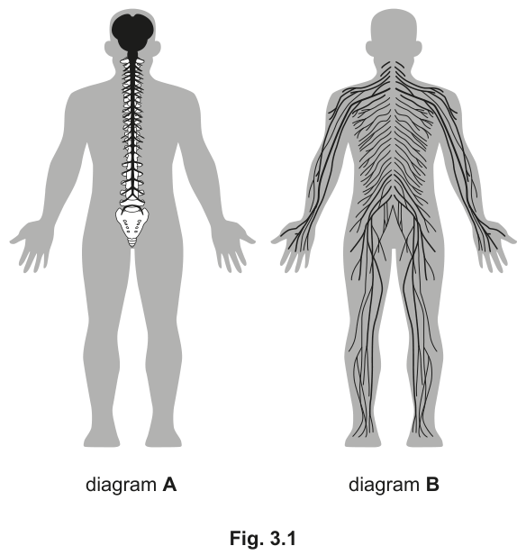
Fig. 3.1
Show full working
- 1
Four marks, two diagrams: give each diagram a name (1 mark) and say what it is made of (1 mark).
A name on its own ("CNS") earns only half of the marks for that diagram.
- 2
Diagram A shows only the brain and the spinal cord (running down inside the backbone). Name: central nervous system (CNS).
The brain and spinal cord together are the CNS.
- 3
What it is made of: the brain and the spinal cord.
This is the second mark for diagram A. Name both parts.
- 4
Diagram B shows a network of nerves reaching every part of the body. Name: peripheral nervous system (PNS).
Nerves spread through the body are the PNS.
- 5
What it is made of: the nerves outside the brain and spinal cord (outside the CNS).
"Outside the CNS" is the key idea for the second mark.
Diagram A: the central nervous system (CNS), made of the brain and spinal cord. Diagram B: the peripheral nervous system (PNS), made of the nerves outside the brain and spinal cord.
"Describe the parts" = name + what it is made of, for each one.
Your turn
- 14 marks
(a) Name the two structures that make up the central nervous system.
(b) What makes up the peripheral nervous system?
(c) State the overall job of the nervous system.Show solution
- 1
(a) The brain and the spinal cord.
Two marks — one for each structure.
- 2
(b) The nerves outside the brain and spinal cord.
"Outside" is the key word; the PNS is everything that is not the CNS.
- 3
(c) It coordinates and regulates body functions.
Use the syllabus words: "coordinates and regulates".
Answer(a) Brain and spinal cord. (b) The nerves outside the brain and spinal cord. (c) It coordinates and regulates body functions.
- 1
- 25090/21 O/N 2024 Q3(a)2 marks
Fig. 3.1 shows the central nervous system and some other parts of a man's body.
Name the two parts of the central nervous system labelled A and B.
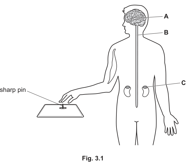
Fig. 3.1
Show solution
- 1
A is inside the skull, at the top of the CNS: brain.
The CNS has only two parts, so find which is which from their position.
- 2
B is the long structure running down the back from the brain: spinal cord.
The spinal cord runs down the back, protected by the backbone.
AnswerA = brain; B = spinal cord.
- 1
- 35090/11 M/J 2024 Q231 mark
Which statement describes the roles of components of the nervous system?
Options
A Effectors respond to environmental changes and send impulses to the central nervous system (CNS).
B Parts of the central nervous system (CNS) can only transmit impulses to a single effector at a time.
C Parts of the peripheral nervous system (PNS) carry impulses to effectors and receptors from the central nervous system (CNS).
D Receptors respond to environmental changes and send impulses to the central nervous system (CNS).Stuck? Show hint
Use the route stimulus → receptor → coordinator → effector → response. Which way do impulses go at each end?
Show solution
- 1
D is correct: receptors detect changes (stimuli) and send impulses to the CNS.
This matches the start of the route: stimulus → receptor → CNS.
- 2
A is wrong: effectors do not detect changes, and they receive impulses from the CNS — they do not send impulses to it.
The option swaps the jobs of receptors and effectors.
- 3
B is wrong: the CNS can send impulses to many effectors at the same time (for example, many muscles at once when you run).
Nothing in the nervous system limits it to one effector.
- 4
C is wrong: impulses from the CNS go to effectors, but not to receptors. Receptors send impulses towards the CNS.
"and receptors" is the error — the direction for receptors is the wrong way round.
AnswerD
- 1
The rest of this note
Can you do all of these?
State that the nervous system (brain, spinal cord and nerves) coordinates and regulates body functions; name the brain and spinal cord as the CNS and the nerves outside them as the PNS
Identify sensory, relay and motor neurones on a diagram by where they start and end; give a similarity and a difference between sensory and motor neurones; state that electrical impulses travel along neurones
Define a reflex action (rapid, automatic) and trace any reflex arc from receptor to effector, including the blink and pupil reflexes; predict the effect of a block at any point
Describe a synapse, its structure and the three events there; explain why impulses go one way; explain how a drug that mimics or blocks the neurotransmitter works
Describe sense organs as groups of receptor cells, and link receptor density to sensitivity
Label the ten structures of the eye and give each function in the syllabus words
Explain the pupil reflex and accommodation step by step; read pupil-diameter and lens-shape graphs; calculate a percentage decrease
Define a hormone (and say why saliva, bile and enzymes are not hormones); match each endocrine gland to its hormones and find it on a diagram
Explain how adrenaline raises blood glucose and heart rate and give situations when it is released
Compare nervous and hormonal control using faster/slower and shorter/longer
Define homeostasis; explain negative feedback and find a set point on a graph
Explain why blood glucose is controlled; describe the roles of the pancreas, liver, insulin and glucagon without mixing up glucagon and glycogen; give the signs and treatment of Type 1 diabetes
Label a skin section; explain insulation, the roles of the hypothalamus and skin receptors, and how sweating, shivering, hair erector muscles, vasodilation and vasoconstriction control body temperature; explain why a mammal uses more oxygen in the cold