Biology 9700/24 — October/November 2025
Cambridge AS Level · AS Level Structured Questions · worked solutions for every part, with the mark scheme
Topics The Mitotic Cell Cycle · Cell Structure · Transport in Mammals · Biological Molecules · Immunity · Infectious Diseases · +4 more
There are similarities and differences between the structure of a typical plant cell and a typical animal cell.
The nucleus of plant cells and animal cells contains chromosomes. In interphase of the cell cycle, individual chromosomes are present but cannot be seen. The chromosome material is known as chromatin.
Changes occur in interphase, which result in a difference between the chromatin in the G1 phase compared with the chromatin in the G2 phase.
State and explain the difference in chromatin in the G1 phase compared with chromatin in the G2 phase.
Answer
- (Semi-conservative) DNA replication has occurred in the S phase between and ;
- As a result, in there is double the amount of DNA / chromatin compared with ; each chromosome now consists of two sister chromatids (two DNA molecules joined at the centromere) rather than a single DNA molecule.
In G2 there is twice the amount of DNA and each chromosome has two sister chromatids (two DNA molecules joined at the centromere) because DNA replication has occurred in the S phase between G1 and G2.
Background Concept
Interphase is the part of the cell cycle between two mitoses. It is divided into three sub-phases:
- G1 – the cell grows and carries out its normal metabolism; each chromosome consists of a single DNA molecule packaged with histones.
- S phase – DNA replication occurs. Replication is semi-conservative: each of the two new DNA molecules keeps one of the original strands and gains a new complementary strand. After S phase, each chromosome is made of two identical DNA molecules.
- G2 – the cell continues to grow and prepares for mitosis; the DNA is still decondensed (so it is visible only as chromatin, not as discrete chromosomes) and the chromosomes each consist of two identical sister chromatids joined at the centromere.
Mitosis itself (prophase → telophase) is when the chromosomes condense enough to be visible, line up, separate and are pulled to opposite poles.
Understanding the Question
The question has already told you that the chromosome material in interphase is called chromatin, and that it is present but not visible. It asks you to compare chromatin in G1 and G2 and to state the difference and explain why it exists. Two marks are available, so the mark scheme expects one mark for the cause (S phase) and one mark for the consequence (doubled DNA / sister chromatids).
Approach
Think of the cell cycle as a timeline: G1 → S → G2 → M. The only event between G1 and G2 that alters the amount of DNA is S phase. Whatever the change is, it must be explained by S phase. Once you identify S phase as the cause, the consequence is mechanical: one DNA molecule per chromosome becomes two.
Step-by-Step Reasoning
- Between G1 and G2 lies the S phase, during which semi-conservative DNA replication takes place. This is the event the examiner wants you to identify. The mark scheme rejects statements that place replication in G1 or G2 itself — replication happens in S.
- The replication doubles the amount of DNA. By G2 each chromosome is therefore made of two DNA molecules rather than one. These two molecules are called sister chromatids and are joined at the centromere.
- You can express the difference in any of the equivalent ways the mark scheme accepts: "double the amount of DNA", "each chromosome has two chromatids instead of one" or "each chromosome with 1 DNA molecule in G1 has 2 DNA molecules in G2".
Key Takeaways
- The amount of DNA doubles during S phase, not during G1 or G2.
- After S phase (i.e. in G2) every chromosome is a pair of sister chromatids joined at a centromere.
- Until mitosis, the DNA remains decondensed as chromatin — being double the amount does not by itself make it visible.
Common Mistakes
- Stating that DNA replication occurs in G1 or G2 — the mark scheme explicitly rejects this.
- Writing simply "more chromatin in G2" without linking the increase to S phase / DNA replication.
- Confusing chromatin (the interphase state) with chromosomes (the mitotic state) and saying chromosomes form in G2.
- Saying the cell has "more chromosomes" in G2 — the number of chromosomes is unchanged; only the amount of DNA per chromosome has doubled.
Things to Be Careful About
- The cell is diploid throughout, so the increase is per chromosome, not a doubling of the chromosome number.
- The marks are split: one for cause, one for consequence. If you only state the consequence ("more DNA") without the cause (S phase / replication), you will only earn one of the two marks.
Describe the features of a nucleus, other than containing chromatin.
You may use the space below the lines for a diagram.
Answer
Any three from:
- Nuclear envelope – a double membrane that surrounds the chromatin; the outer membrane is continuous with the rough endoplasmic reticulum (and may bear ribosomes); it encloses the genetic material and controls what enters and leaves the nucleus.
- Nuclear pores – channels through the nuclear envelope; allow (m)RNA and ribosomal subunits to leave the nucleus, allow proteins to enter, and prevent DNA from leaving.
- Nucleolus – a spherical, densely staining region inside the nucleus; site of rRNA synthesis and assembly of ribosomal subunits.
- Nucleoplasm – the fluid that surrounds the chromatin inside the nucleus.
A nucleus has a double-membrane nuclear envelope (with pores) surrounding nucleoplasm, and contains one or more nucleoli.
Background Concept
The nucleus is the largest organelle in a eukaryotic cell and is the store of the cell's genetic information. To understand its features, think about the jobs it has to do:
- Protect the DNA from cytoplasmic damage.
- Separate transcription (which must happen in the nucleus) from translation (which happens in the cytoplasm).
- Control which molecules pass between nucleus and cytoplasm.
- Manufacture the RNA and ribosomal subunits needed for protein synthesis.
Each structural feature of the nucleus corresponds to one of these jobs.
Understanding the Question
The question asks you to describe the features of a nucleus, other than the chromatin already mentioned. The mark scheme (3 marks) lets you pick any three of: the nuclear envelope, nuclear pores, nucleolus and nucleoplasm. For each feature you should give enough structural or functional detail to be worth a mark. The question also allows (but does not require) a diagram.
Approach
For each named structure, state what it is and what it does. Function alone is often enough to score the detail mark; the mark scheme explicitly says descriptions from a correctly labelled diagram are also accepted, so a sketch is a valid alternative to text if you are not confident writing the words.
Step-by-Step Reasoning
- Nuclear envelope – The nucleus is bounded by two membranes (a double membrane). The envelope encloses the chromatin, holding the genetic material safely inside the cell. The outer membrane is continuous with the rough endoplasmic reticulum and may carry ribosomes on its cytoplasmic face. By being selectively permeable it controls which substances enter and leave the nucleus.
- Nuclear pores – These are small gaps in the nuclear envelope where the two membranes fuse. They form channels through which mRNA, tRNA and ribosomal subunits can pass out of the nucleus, and through which proteins (such as histones and transcription factors) enter. The pores are small enough to prevent DNA from leaving.
- Nucleolus – A spherical, densely staining region inside the nucleus, made of condensed rRNA and protein. It is the site of rRNA synthesis and the assembly of ribosomal subunits before they are exported through the nuclear pores. There is usually one nucleolus, but some cells have more.
- Nucleoplasm – The semi-fluid material that fills the space inside the nuclear envelope and bathes the chromatin. It is the site in which the contents of the nucleus are suspended.
The mark scheme requires any three of these; most candidates name nuclear envelope + nuclear pores + nucleolus and supply a one-line detail for each.
Key Takeaways
- The nuclear envelope is a double membrane that separates nuclear and cytoplasmic contents.
- Nuclear pores regulate transport between the two compartments.
- The nucleolus makes rRNA and assembles ribosomal subunits.
- The nucleoplasm is the internal medium of the nucleus.
Common Mistakes
- Saying the nucleus has a "membrane" (singular) instead of a double membrane — the mark scheme specifically requires the doubled nature.
- Calling the pores "holes" or saying "stuff passes through the membrane" without naming nuclear pores.
- Confusing the nucleolus with a nucleus, or describing it as "where DNA is stored" (it is rRNA, not DNA).
- Listing "contains DNA/chromosomes/chromatin" as a feature of the nucleus — the stem has already told you this and excluded it.
Things to Be Careful About
- A labelled diagram is acceptable in place of a written description (the question allows it). If you draw, label nuclear envelope, nuclear pore and nucleolus clearly with lines pointing to the correct structures.
- Spelling matters at this level — nucleolus, nucleoplasm and nuclear envelope are precise CIE terms.
Starch, cellulose and pectins are polysaccharides found in plant cells but not in animal cells.
Pectins are complex polysaccharides that are found in the cell wall.
Describe the structural features of starch that are different from the structural features of cellulose.
Answer
Any four from:
- Starch is made of -glucose monomers, whereas cellulose is made of -glucose.
- The -glucose monomers in starch are all oriented the same way; in cellulose every other -glucose is rotated through relative to its neighbour.
- Starch contains -1,4 glycosidic bonds; amylopectin (a component of starch) additionally has -1,6 glycosidic bonds at branch points.
- Starch is a mixture of two polymers: amylose (which is helical/coiled) and amylopectin (which is branched); cellulose is a single, linear, unbranched polymer.
- There are no hydrogen bonds holding starch molecules together; cellulose chains are extensively cross-linked by hydrogen bonds (forming microfibrils).
Starch is made of α-glucose (α-1,4 bonds) with monomers oriented the same way and consists of helical amylose plus branched amylopectin, whereas cellulose is β-glucose with 180°-rotated monomers forming linear, H-bonded chains.
Background Concept
Starch and cellulose are both polymers of glucose, but the small chemical difference between α- and β-glucose produces very different polymers with very different roles:
- α-glucose has its –OH on carbon 1 below the ring; successive α-1,4 linkages produce chains that coil. This is the monomer of starch (and glycogen).
- β-glucose has its –OH on carbon 1 above the ring; successive β-1,4 linkages force every other glucose to flip , producing straight chains that pack tightly together. This is the monomer of cellulose.
Starch itself is a mixture: amylose (a long, unbranched, helical chain of α-1,4 glucose) and amylopectin (a long, branched chain of α-1,4 glucose with α-1,6 branch points). Cellulose is a single, linear polymer of β-1,4 glucose, and adjacent chains are held together by many hydrogen bonds to form strong microfibrils.
Understanding the Question
This is a "describe the structural features of starch that are different from the structural features of cellulose" question. It is not asking for a full description of either — only for the differences. The mark scheme rewards up to four differences and gives a mark for any correct, distinct contrast. It also issues some sharp "R" rules:
- Do not earn a mark by stating an incorrect feature of cellulose.
- Do not say "starch can be helical or branched" (it must specify which component is which).
- Do not credit "α-helix" — that is a protein structure.
Approach
Work from the molecule outwards:
- Start with the monomer (α vs β glucose).
- Move to the glycosidic bond (α-1,4 vs β-1,4) and the orientation of adjacent monomers.
- Then the polymer-level structure: starch is a mixture of two polymers, one helical and one branched; cellulose is linear.
- Finally the intermolecular features: starch has no H-bonds between chains; cellulose chains are tied together by H-bonds into microfibrils.
This is a logical order from the smallest to the largest scale, and each step gives a mark-scheme point.
Step-by-Step Reasoning
- Monomer (mp1). Starch is built from -glucose; cellulose is built from -glucose. The two differ only in the position of the –OH on C1. (You are not penalised for also noting the type of bond — α-1,4 for starch, β-1,4 for cellulose — but stating just "glycosidic bond" or "1,4 bond" is shared with cellulose, so it is not a difference on its own.)
- Monomer orientation (mp2). In starch every α-glucose is oriented the same way as the last. In cellulose every other β-glucose is rotated . This is what allows cellulose chains to lie straight and pack closely together.
- Two polymers in starch (mp3) and the α-1,6 bonds (mp4). Starch is a mixture of amylose (a helical, unbranched chain of α-1,4 glucose) and amylopectin (a chain of α-1,4 glucose with α-1,6 branch points). Cellulose is just a single linear polymer — it is not a mixture. The α-1,6 bond is unique to amylopectin (and glycogen) and is the basis of branching.
- Shape (mp5). Amylose is helical/coiled, amylopectin is branched; cellulose is straight, linear and unbranched.
- Hydrogen bonding (mp6). Starch molecules are not held to one another by hydrogen bonds. In cellulose, the –OH groups of adjacent chains form extensive hydrogen bonds, drawing the chains into tight, parallel bundles called microfibrils. This is what makes cellulose a structural material and starch a storage material.
Pick any four of these for a full-mark answer.
Key Takeaways
- The α/β distinction in the glucose monomer is the root cause of the very different structures of starch and cellulose.
- Starch is a storage polysaccharide: helical, branched, easily hydrolysed.
- Cellulose is a structural polysaccharide: straight, unbranched, H-bonded into microfibrils.
- A useful mnemonic: "Starch = Same orientation, Single monomer direction, Storage; Cellulose = Contrasting orientation ( turns), Chains H-bonded, Cell wall."
Common Mistakes
- Stating features that are shared (e.g. "both are made of glucose", "both have 1,4 bonds") as if they were differences — these do not earn a mark.
- Describing cellulose incorrectly to contrast it with starch (e.g. "cellulose is branched"). The mark scheme rejects an incorrect cellulose feature and refuses the mark for that point.
- Writing "α-helix" for amylose — this is a protein motif and is specifically rejected.
- Writing "starch can be helical or branched" without naming amylose and amylopectin.
- Writing "no hydrogen bonds in starch" without contrasting it with cellulose's extensive H-bonding.
Things to Be Careful About
- The 1,4 vs 1,6 distinction is the basis of branching. Make clear that it is the α-1,6 bond that creates branches in amylopectin.
- "Glycosidic bond" on its own is too vague — quote α-1,4 (and α-1,6) for starch and β-1,4 for cellulose.
- Units and chemical spelling matter: α-glucose, β-glucose, glycosidic bond, hydrogen bond.
Cell wall pectins can vary in different plant cell types and in different stages of cell development. Pectin molecules are released from cells as basic structures and then modified within the cell wall by adding side chains.
RG-I is a pectin molecule with a variable structure. The basic structure is a repeated disaccharide made from two different monosaccharides. RG-I has three different side chains that can be added in different positions.
Monoclonal antibodies (mAbs) are used to investigate the structure, location and role of cell wall pectins.
Suggest and explain why scientists need to use a number of different monoclonal antibodies when investigating a pectin such as RG-I.
Answer
- A monoclonal antibody (mAb) has a single, specific antigen-binding site / variable region, so each mAb binds to only one particular antigen / epitope (shape).
- RG-I is a structurally variable molecule: the basic disaccharide backbone can carry different side chains added in different positions, giving it many different shapes and therefore many different epitopes.
- No single mAb can recognise all of these different epitopes, so scientists need a range of different mAbs, each complementary to a particular epitope, to detect, locate and study the different forms and modifications of RG-I (including tracking when side chains are added during development).
Each mAb is specific to one epitope, but RG-I has a variable structure with many different side chains and epitopes, so multiple different mAbs are needed to recognise the different forms of the molecule.
Background Concept
Monoclonal antibodies (mAbs) are antibodies made by a single clone of B-lymphocytes fused with a myeloma cell to form a hybridoma. Every antibody produced by one hybridoma line is identical, with the same antigen-binding site (variable region) and therefore the same specificity — it will bind to only one particular epitope (a small region of an antigen with a complementary shape).
This specificity is what makes mAbs so useful as research tools: an mAb is essentially a molecular probe that recognises one structural feature of one target molecule. If the target molecule has many different structural features, no single mAb can recognise them all — you need a panel of mAbs, each recognising a different epitope.
The pectin RG-I (rhamnogalacturonan-I) is a backbone of a repeated disaccharide (rhamnose–galacturonic acid) onto which different side chains of other sugars can be added after the molecule is exported to the cell wall. The side chains and their positions vary between cell types and developmental stages, so an RG-I molecule is a family of related structures rather than a single fixed molecule.
Understanding the Question
The stem gives you several clues:
- RG-I is variable (different side chains, different positions).
- mAbs are specific to one antigen.
- The question asks you to suggest and explain why several mAbs are needed.
The 3-mark mark scheme expects: a mark for the specificity of mAbs; a mark for the variability of RG-I; and a mark for the consequence (different mAbs for different epitopes / different forms of RG-I, allowing the scientists to identify and locate them and to study how the molecule is modified during development).
Approach
- Why an mAb is so specific — one variable region, one complementary epitope.
- Why RG-I needs many probes — its structure varies, so its epitopes vary.
- What the scientists gain by using several mAbs — they can identify, locate and compare the different forms; they can track when side chains are added during cell development.
Step-by-Step Reasoning
- mAb specificity. Each mAb has a single type of antigen-binding site (variable region) whose shape is complementary to one specific epitope. It will bind to that epitope and not to others. This is the basis of the mAb's use as a probe — a single mAb is a single, highly specific detector.
- RG-I variability. The basic RG-I disaccharide backbone can carry any of three different side chains, attached in different positions and at different times during cell development. As a result, the same molecule exists in many structural forms in different cell types and at different developmental stages. Each form presents a different set of epitopes on its surface.
- Consequence for the experiment. A single mAb, being specific to one epitope, can recognise only one of these many forms. To investigate the whole family of RG-I structures — to detect which forms are present, where in the cell wall they are located, and when during development each modification appears — scientists need a panel of different mAbs, each specific to a different epitope, and use them in combination. An additional bonus is that using several mAbs reveals which structures are similar (cross-reaction) and which are unique, giving information about the precise shape of the pectin.
Key Takeaways
- mAb specificity comes from the variable region of a single antibody clone — one mAb = one epitope.
- A structurally variable molecule exposes a range of different epitopes.
- Studying a variable molecule therefore needs a panel of mAbs, each one a probe for a different epitope.
- This principle is widely used to map cell-surface molecules, identify cell types in tissue sections, and follow biochemical changes during development.
Common Mistakes
- Writing "the active site binds to the antigen" — antibodies do not have an active site; they have an antigen-binding site / variable region. The mark scheme explicitly ignores "active site".
- Saying scientists need several mAbs "because there are several pectins" without explaining the variability of structure within one pectin (RG-I itself is the variable molecule).
- Vague statements such as "to get better results" or "to be more accurate" — these do not address specificity.
- Failing to link side-chain addition to development, and therefore failing to use the development clue in the stem.
Things to Be Careful About
- Use the mark-scheme terms: antigen-binding site or variable region, not active site.
- Note the epitope as the precise thing the mAb recognises — this is the level of detail the mark scheme rewards.
- Make the link to the development context: side chains are added after export to the cell wall, so the molecule a young cell makes is not the same as the molecule in a mature cell — different mAbs let the scientists see this changing pattern.
Cholera, malaria and tuberculosis (TB) are infectious diseases caused by unicellular organisms.
For each of the diseases listed, state whether the disease is caused by a eukaryotic or prokaryotic organism.
cholera ______
malaria ______
TB ______
Answer
cholera — prokaryotic ;
malaria — eukaryotic ;
TB — prokaryotic ;
cholera — prokaryotic; malaria — eukaryotic; TB — prokaryotic
Background Concept
The prokaryote/eukaryote distinction is the first major division in cell biology. Prokaryotes (bacteria) are small, single-celled organisms with no membrane-bound nucleus; their DNA is circular and lies free in the cytoplasm, and they have small (70S) ribosomes. Eukaryotes have a true nucleus and membrane-bound organelles; their cells are usually larger and contain 80S ribosomes. The three pathogens in this question are all unicellular (the question stem reminds us of this) but they are not all prokaryotic.
Understanding the Question
You are given three infectious diseases — cholera, malaria and TB — and asked whether each is caused by a prokaryotic or a eukaryotic organism. The mark is awarded as a single combined credit, so all three must be correct to score.
Approach
Recall the causative agent of each disease, then decide whether that agent is a bacterium (prokaryote) or a non-bacterial single-celled organism (typically a eukaryote such as a protist).
Step-by-Step Reasoning
- Cholera is caused by Vibrio cholerae — a bacterium → prokaryotic.
- Malaria is caused by Plasmodium species (e.g. P. falciparum) — these are protists → eukaryotic.
- TB is caused by Mycobacterium tuberculosis — a bacterium → prokaryotic.
All three statements together earn the single mark.
Key Takeaways
- Bacteria (including Vibrio cholerae and Mycobacterium tuberculosis) are prokaryotes.
- Protists (including Plasmodium) are eukaryotes.
- "Unicellular" is not the same as "prokaryotic" — Plasmodium is unicellular but eukaryotic.
Common Mistakes
- Calling Plasmodium a bacterium because it is small and single-celled.
- Confusing TB with a viral disease (it is bacterial).
- Treating the three answers as three separate marks; in fact they are combined — one error loses the mark.
Things to Be Careful About
- Viruses are neither prokaryotic nor eukaryotic (they are acellular) — but none of the three diseases in this question is viral.
- The mark scheme awards the credit only when all three answers are correct.
State the term given to an organism that causes diseases such as cholera, malaria and TB.
Answer
pathogen
pathogen
Background Concept
A pathogen is an organism (or, more loosely, an agent) that causes disease in its host. The category includes bacteria, viruses, fungi and protists. The term is a general one that covers any disease-causing biological agent, whether cellular or acellular.
Understanding the Question
You are asked for the single biological term that applies to an organism causing diseases such as cholera, malaria and TB. The expected answer is one word.
Approach
Recall the standard biology term that applies to all three example diseases in the question.
Step-by-Step Reasoning
The correct term is pathogen. It applies to all three example diseases because the causative agents of cholera (Vibrio cholerae), malaria (Plasmodium) and TB (Mycobacterium tuberculosis) are all pathogens.
Key Takeaways
- "Pathogen" is the umbrella term for any disease-causing organism.
Common Mistakes
- Writing "germ" (informal; not credited in biology).
- Specifying a category of pathogen (e.g. "bacterium", "virus", "protist") — too narrow, since the term must cover all three example diseases.
Things to Be Careful About
- The term is pathogen, not "pathogen" in any other form.
Malaria is caused by species from the genus Plasmodium.
Ring cells are one stage in the complex life cycle of Plasmodium that are found within red blood cells. Fig. 2.1 is a scanning electron micrograph showing two ring cells, surrounded by the membrane of a red blood cell, which has just lysed (burst).
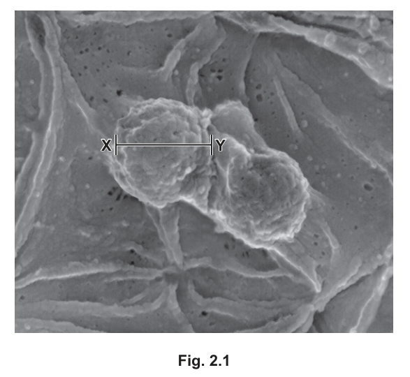
The actual diameter of the ring cell along the length X–Y is .
Calculate the magnification of the image shown in Fig. 2.1.
Give your answer to 3 significant figures.
magnification = ______
Working
length of line X–Y on the printed image = 24 mm
convert to µm (same unit as actual size):
apply the magnification formula:
Answer
magnification =
× 12 000
Background Concept
Magnification is the factor by which a microscope or printed image enlarges the specimen. It is given by the formula
Both quantities must be in the same units before dividing. The two common unit conversions needed in Cambridge questions are and .
Understanding the Question
You are told that the actual diameter of a Plasmodium ring cell along line X–Y is , and you are asked to calculate the magnification of the printed image. The answer must be given to 3 significant figures. To solve this you must first measure the length of X–Y on the printed image (using a ruler), convert to the same unit as the actual size, then apply the magnification formula.
Approach
- Measure the line X–Y on the printed image (in mm).
- Convert that measurement to µm.
- Divide image size (µm) by actual size (2 µm).
- Round to 3 significant figures.
Step-by-Step Reasoning
- Image length of X–Y on the printed micrograph ≈ (the mark scheme accepts – for full credit).
- Convert: .
- Magnification .
- to 3 significant figures is (or, written unambiguously, ).
Key Takeaways
- The magnification formula is image ÷ actual (with both in the same unit).
- Always convert units before dividing.
- Measure the printed image carefully — a small error in measurement gives a large error in magnification.
Common Mistakes
- Dividing the actual size by the image size (formula inverted) — would give .
- Forgetting to convert mm to µm — would give 12 (off by a factor of 1000).
- Reporting the answer with no thought to significant figures, or only to 2 sig figs () when 3 were asked for.
Things to Be Careful About
- The image measurement is the image size; the is the actual size. Putting the wrong value on top of the fraction is the most common error.
- Always include units in the working.
- Round only at the end of the calculation.
Describe how Plasmodium is transmitted from a person with malaria into the blood stream of an uninfected person.
Answer
- a vector / female Anopheles mosquito feeds on / takes a blood meal from an infected person ;
- Plasmodium / the parasite is in the blood of the infected person (and is ingested with the blood meal) ;
- Plasmodium migrates to / develops in the mosquito's salivary glands (AVP) ;
- the mosquito feeds on an uninfected person and injects Plasmodium together with saliva (containing anticoagulant) into the bloodstream ;
A female Anopheles mosquito takes a blood meal from an infected person and ingests Plasmodium from the blood; the parasite migrates to the mosquito's salivary glands; the mosquito then feeds on an uninfected person and injects Plasmodium (with its saliva/anticoagulant) into the bloodstream.
Background Concept
Malaria is caused by protists of the genus Plasmodium (e.g. P. falciparum). The parasite has a complex life cycle that requires two hosts: a human (or other mammal) and a female Anopheles mosquito. Only female mosquitoes feed on blood, because they need the protein in blood to develop their eggs; only certain Anopheles species are efficient vectors. Transmission of the parasite from one human to the next occurs during a mosquito blood meal — the mosquito ingests the parasite from an infected person and later injects it (in its saliva) into an uninfected person.
Understanding the Question
You are asked to describe how Plasmodium is transmitted from a person with malaria into the blood stream of an uninfected person. The answer must cover (a) the vector, (b) the blood meal, (c) the location of the parasite, and (d) the injection step. The mark scheme credits any three of these for 3 marks.
Approach
Trace the transmission cycle step-by-step: mosquito feeds on infected human → parasite is in the blood → parasite develops in the mosquito → mosquito feeds on uninfected human and injects the parasite with its saliva.
Step-by-Step Reasoning
- A female Anopheles mosquito takes a blood meal from a person with malaria.
- Plasmodium is in the blood of the infected person, so it is ingested with the blood meal.
- (AVP) The parasite develops in the mosquito and migrates to its salivary glands.
- When the (same or a different) mosquito feeds on an uninfected person, it injects saliva — containing an anticoagulant (to keep blood flowing) together with Plasmodium — into the new host's bloodstream.
Key Takeaways
- The vector is specifically the female Anopheles mosquito.
- The parasite must be in the blood of the infected host to be picked up by the mosquito.
- Transmission occurs when the mosquito injects saliva containing the parasite into the new host.
- The cycle requires both an infected and an uninfected human plus the mosquito vector.
Common Mistakes
- Naming "mosquito" without specifying that it is the female Anopheles species.
- Saying the mosquito "bites" without mentioning that it injects saliva carrying the parasite.
- Omitting the role of the mosquito's salivary glands.
- Placing Plasmodium in the mosquito's mouth or gut generally, rather than the salivary glands.
Things to Be Careful About
- Plasmodium is in the blood, not just inside the body generally — the mosquito acquires it by taking a blood meal.
- The injection happens during feeding: saliva is delivered into the new host's skin along with the anticoagulant and the parasite.
- If a different mode of transmission (e.g. blood transfusion) is mentioned, the mark scheme only allows mark points 1 and 3 plus a relevant AVP — the standard mosquito route is expected.
In a person with malaria, phagocytes destroy infected red blood cells. The phagocytes respond to the presence of particular molecules in the outer phospholipid bilayer of the cell surface membrane.
The cell surface membrane of a healthy, uninfected red blood cell shows an uneven distribution of types of phospholipid making up the bilayer (membrane asymmetry). For example, most of the phospholipid phosphatidylserine (PS) is located in the inner layer, facing the cytoplasm.
After a red blood cell is infected, a much higher proportion of PS is found in the outer layer of the cell surface membrane, facing blood plasma.
Suggest how the presence of PS in the outer layer can cause a response in a phagocytic cell.
Answer
- (phagocyte) has (specific) receptors (for PS) ;
- PS binds to (phagocyte / cell surface) receptors (to stimulate a response) ;
- (the presence of) PS (in the outer layer) causes chemotaxis / attracts the phagocyte to the red blood cell ;
Phagocytes have specific receptors for PS; PS in the outer leaflet binds to these receptors and triggers a response in the phagocyte (e.g. chemotaxis / movement towards the red blood cell).
Background Concept
Phagocytes (such as macrophages and neutrophils) recognise cells to be engulfed by means of receptors on their cell surface membrane. These receptors bind to specific molecules (ligands) displayed on the target cell. Binding triggers a response in the phagocyte: chemotaxis (directional movement towards the target) and the initiation of phagocytosis. In a healthy red blood cell, phosphatidylserine (PS) is kept almost entirely on the inner leaflet of the bilayer; in aged, damaged, apoptotic or infected red blood cells, some PS flips to the outer leaflet, where it acts as an "eat-me" signal.
Understanding the Question
The question asks how the appearance of PS on the outer leaflet of the red blood cell membrane causes a response in a phagocytic cell. The answer needs to describe the receptor on the phagocyte, the binding event, and the resulting response (chemotaxis or activation of phagocytosis). The mark scheme credits any two of these for 2 marks.
Approach
Connect the appearance of a new outer-leaflet molecule (PS) to receptor-mediated recognition by the phagocyte, then to a downstream response (chemotaxis or activation of engulfment).
Step-by-Step Reasoning
- Phagocytes have specific receptors on their cell surface membrane that recognise PS.
- When PS is exposed on the outer leaflet of the red blood cell, it binds to these receptors.
- This receptor–ligand binding stimulates a response in the phagocyte — e.g. chemotaxis (the phagocyte moves towards the red blood cell) and the initiation of phagocytosis.
Key Takeaways
- PS in the outer leaflet acts as a signal recognised by phagocyte receptors.
- The response is receptor-mediated, not non-specific.
- This is a general mechanism for clearing aged, damaged, apoptotic or infected cells.
Common Mistakes
- Saying the phagocyte "sees" or "recognises" the cell without mentioning receptors — too vague for the mark.
- Calling PS an "antigen" — the mark scheme explicitly rejects this.
- Confusing which cell has the receptor (the phagocyte) and which displays the ligand (the red blood cell).
Things to Be Careful About
- The word "receptor" (or "binding site", which is also accepted) must appear; "antigen" is not credited here.
- The mark scheme accepts a named phagocyte (e.g. macrophage, neutrophil, monocyte, Kupffer cell) as the cell type carrying the receptor.
Suggest why some uninfected red blood cells in a person with malaria can also be destroyed by phagocytic cells.
Answer
(some) PS is naturally present in the outer layer of (some) uninfected red blood cells, so these cells can be recognised / detected by phagocytes ;
Some PS is naturally present in the outer layer of uninfected red blood cells, allowing phagocytes to recognise and destroy them.
Background Concept
Although most PS is normally kept on the inner leaflet, a small amount can flip to the outer leaflet spontaneously as the membrane lipids move about. The membrane is a fluid structure in which individual lipid molecules occasionally switch leaflets without enzyme help, so even some healthy, uninfected red blood cells can display a low level of outer-leaflet PS, or can become damaged in other ways (age, mechanical stress, attached antibody or parasite fragment) that mark them for destruction.
Understanding the Question
The question asks why some uninfected red blood cells in a person with malaria are also destroyed by phagocytes. The simplest and most direct answer is that a low background level of PS on uninfected cells is enough to trigger recognition.
Approach
Consider the possibilities: spontaneous PS exposure, attached parasite fragments, displayed foreign antigens, antibodies bound to the cell, or just age- / damage-related destruction. The mark scheme accepts any of these.
Step-by-Step Reasoning
- Some PS is naturally present in the outer leaflet of even uninfected red blood cells (because phospholipids move randomly between leaflets from time to time).
- This PS is recognised by phagocyte receptors, so a few uninfected red blood cells are also engulfed.
- (Alternative, also accepted by the mark scheme: fragments of parasite / antibody / foreign antigen may become attached to the surface of an uninfected cell, marking it for destruction.)
Key Takeaways
- Random membrane events (occasional flip of PS to the outer layer) can mark even healthy cells for destruction.
- A low level of "eat-me" signal is enough; phagocytes don't need a strong signal to act.
Common Mistakes
- Saying "phagocytes can't tell infected and uninfected apart" — too vague and doesn't explain the mechanism.
- Stating that uninfected cells are weaker and so break more easily — not the focus of the question.
Things to Be Careful About
- The mark scheme offers several acceptable answers; the simplest is the spontaneous appearance of some PS in the outer layer of an uninfected cell.
- The mark is for explaining the destruction — the answer must identify a reason, not just describe the event.
Research has shown that Plasmodium uses cholesterol from the cell surface membrane of the red blood cell it has infected.
Answer
- to make (its own) cell membrane(s) / cell surface membrane ;
- to maintain / regulate stability / fluidity of (its) membranes ;
- Plasmodium cannot / does not have the gene to synthesise cholesterol ;
To form cell membranes; to maintain the stability/fluidity of its membranes; Plasmodium cannot synthesise its own cholesterol.
Background Concept
Cholesterol is a lipid component of eukaryotic cell membranes. It sits among the phospholipid fatty-acid tails and (a) reduces membrane fluidity by restraining phospholipid movement, and (b) increases mechanical stability. Plasmodium is a eukaryote (a protist) and so makes cell membranes that contain cholesterol. It cannot make cholesterol itself and must obtain it from its host.
Understanding the Question
The question asks why Plasmodium needs cholesterol. The mark is in the context of Plasmodium (not the host red blood cell). The expected answer involves either membrane structure, fluidity / stability, or the parasite's inability to make its own.
Approach
Think about cholesterol's role in cell membranes, then apply this to the parasite's own membranes and consider the parasite's metabolism.
Step-by-Step Reasoning
- Plasmodium needs cholesterol to make / maintain its own cell surface membrane (or other internal membranes).
- Cholesterol helps maintain the stability / fluidity of those membranes.
- Plasmodium cannot synthesise cholesterol itself, so it must take it from the host red blood cell.
Key Takeaways
- Cholesterol is essential for the structure and proper fluidity / stability of eukaryotic cell membranes.
- Parasites often steal lipids they cannot make from the host.
Common Mistakes
- Saying cholesterol is needed for energy — it is not used as a fuel.
- Stating the context as the red blood cell rather than Plasmodium — the mark scheme rejects this.
- Saying cholesterol is needed "to enter the red blood cell" — the mark scheme explicitly rejects this framing.
Things to Be Careful About
- The context of the mark is Plasmodium, not the host cell. Statements about what cholesterol does for the red blood cell are not credited here.
- The mark scheme accepts either structural / fluidity arguments or the inability-to-synthesise argument; any one is enough.
The use of cholesterol by Plasmodium causes a decrease in the quantity of cholesterol in the cell surface membrane of the red blood cell.
Outline how a decrease in cholesterol could affect the cell surface membrane of the red blood cell.
Answer
- the membrane becomes more fluid / fluidity increases ;
- (membrane) stability decreases / is reduced ;
- permeability (to polar molecules / ions / water) increases ;
The membrane becomes more fluid; stability is reduced; permeability to polar molecules, ions and water increases.
Background Concept
Cholesterol has two main effects on a phospholipid bilayer:
- It reduces membrane fluidity by interfering with the movement of phospholipid fatty-acid tails.
- It increases membrane stability (mechanical strength) and decreases permeability to small water-soluble molecules and ions.
Removing cholesterol reverses all three effects.
Understanding the Question
The question asks how a decrease in cholesterol would affect the red blood cell's cell surface membrane. The answer should describe the consequences of removing cholesterol.
Approach
Identify cholesterol's normal roles, then predict what changes when there is less of it.
Step-by-Step Reasoning
- Less cholesterol → the membrane becomes more fluid (because there is less restraint on phospholipid movement).
- Less cholesterol → membrane stability decreases (because cholesterol is normally a stabilising component).
- Less cholesterol → membrane permeability to polar molecules, ions and water increases (because cholesterol normally reduces permeability).
Key Takeaways
- Decreased cholesterol makes the membrane more fluid, less stable and more permeable.
- These effects are the opposite of what cholesterol normally does.
Common Mistakes
- Saying the membrane becomes less fluid or more stable (the opposite of the correct answer).
- Mentioning only one effect — the mark scheme credits any two.
- Citing temperature as the cause — references to temperature are ignored.
Things to Be Careful About
- "Less cholesterol" → "more fluid" (not less).
- Don't confuse the effects of adding cholesterol with the effects of removing it.
- If the candidate writes the answer in the wrong direction, the mark scheme allows one "compensation mark" if two of the three terms (fluidity / stability / permeability) are correctly changed.
To achieve membrane asymmetry, which is an essential feature of a healthy cell, the red blood cell needs a supply of ATP and must maintain a very low concentration of calcium ions () within the cytoplasm.
Table 2.1 shows details of three membrane enzymes that are involved in the movement of phospholipids between the inner and outer layers.
Table 2.1
key
PS = phosphatidylserine
PE = phosphatidylethanolamine
PC = phosphatidylcholine
| enzyme | enzyme action |
|---|---|
| flippase | hydrolyses ATP and moves PS and PE from the outer to the inner layer |
| floppase | hydrolyses ATP and moves PC from the inner to the outer layer |
| scramblase | after binding , randomly moves PS, PE and PC between layers |
A healthy red blood cell has most PS and PE located in the inner layer and most PC located in the outer layer.
Phospholipids can occasionally move between layers without the action of an enzyme, so the continued activity of flippase and floppase is needed.
With reference to Table 2.1, explain why the action of the enzymes flippase and floppase involves the hydrolysis of ATP.
Answer
- flippase and floppase carry out an active / energy-requiring process ;
- they move (named) phospholipids against the concentration gradient (from a layer where they are less concentrated to one where they are more concentrated) ;
It is an active, energy-requiring process because the enzymes move phospholipids against their concentration gradient.
Background Concept
Enzymes that hydrolyse ATP are typically powering an energy-requiring step. In this case flippase and floppase move specific phospholipids from one leaflet of the bilayer to the other — but they move them against their concentration gradient (e.g. flippase moves PS and PE from the outer layer, where they are sparse, to the inner layer, where they are concentrated). Movement against a concentration gradient requires energy, supplied by the hydrolysis of ATP.
Understanding the Question
The question asks why flippase and floppase hydrolyse ATP. The expected answer is that they are carrying out an energy-requiring (active) process because the phospholipids are moving against their concentration gradient.
Approach
Identify the work the enzymes do (moving specific phospholipids in a specific direction) and the energy requirement this implies (against the gradient).
Step-by-Step Reasoning
- Flippase and floppase move phospholipids from a layer where they are less concentrated to a layer where they are more concentrated (i.e. against the concentration gradient).
- Movement against a concentration gradient is an active process, which requires energy from ATP hydrolysis.
Key Takeaways
- ATP is required whenever work is done against a gradient.
- Flippase and floppase are powered by ATP, not by the random "flip-flop" of phospholipids (which is too slow to maintain asymmetry on its own).
Common Mistakes
- Using the term "active transport" — the mark scheme ignores this term here, because the phospholipids are not crossing the whole membrane, just moving between leaflets. "Active transport" is described as movement of substances across a membrane.
- Saying the process "needs energy" without identifying what work the energy does.
Things to Be Careful About
- The phospholipids are moving between the two leaflets of one membrane, not across the membrane as a whole.
- The mark is for identifying the reason ATP is needed (against the gradient), not just for the word "active".
Blood plasma has a higher concentration of than the cytoplasm of red blood cells.
Suggest one way in which a red blood cell can have a very low concentration of when blood plasma has a higher concentration of .
Answer
- the red blood cell (actively) transports / pumps out of the cell (against the concentration gradient) ;
The cell actively transports / pumps Ca²⁺ out of the cell (against the concentration gradient).
Background Concept
A concentration gradient tends to drive ions from a region of higher concentration to a region of lower concentration. If the desired direction is the opposite (low inside, high outside), the cell must use active transport — a carrier protein powered by ATP — to pump the ion against the gradient. Calcium ions () are typically kept at very low cytoplasmic concentrations in this way; specialised -ATPases pump out of the cytoplasm (or into the endoplasmic reticulum).
Understanding the Question
The question asks how a red blood cell can have a lower concentration than the blood plasma (which is higher in ). The expected answer involves active transport of out of the cell.
Approach
Apply the principle of active transport against a concentration gradient.
Step-by-Step Reasoning
- Red blood cells actively transport / pump out of the cell, using ATP and a -ATPase carrier protein in the membrane.
- This is movement against the concentration gradient (cytoplasm < plasma).
- The result is a much lower concentration in the cytoplasm than in the plasma.
Key Takeaways
- Maintaining a low cytoplasmic requires active transport.
- A carrier protein ( pump) is essential; this is not a passive property of the membrane.
Common Mistakes
- Saying the membrane is impermeable to — this is a passive property and does not by itself explain a maintained gradient. The mark scheme explicitly rejects this.
- Saying "there are no channel proteins" — not a valid mechanism for keeping out; in fact would still leak in through any available pathway.
- "Active transport" is the right idea, but the explanation must identify that is being moved out of the cell.
Things to Be Careful About
- The mark scheme accepts "pump" or "active transport" but not "membrane impermeable" alone.
- The context is the entry of (which would normally diffuse in) and how the cell deals with it.
The concentration of within the red blood cell increases when the cell is infected with the malarial parasite. This leads to the loss of membrane asymmetry.
With reference to Table 2.1, suggest and explain how membrane asymmetry is lost when the concentration of within the red blood cell increases.
Answer
- (the increase in intracellular ) activates scramblase (so it begins to function) ;
- scramblase moves PC from the outer to the inner layer, and PS / PE from the inner to the outer layer (i.e. the opposite direction to the action of flippase and floppase) ;
- scramblase activity is higher than / overwhelms / is not counteracted by the activity of flippase and floppase ;
- the asymmetric distribution / organisation of phospholipids in the cell surface membrane is therefore lost / disrupted / more random / less organised ;
Increased Ca²⁺ activates scramblase, which moves PC into the inner layer and PS/PE into the outer layer (opposite to flippase and floppase). Scramblase activity is higher than (or is not counteracted by) flippase and floppase, so the asymmetric distribution of phospholipids is lost.
Background Concept
Healthy red blood cells maintain membrane asymmetry using three enzymes (Table 2.1):
- Flippase uses ATP to move PS and PE from the outer to the inner layer.
- Floppase uses ATP to move PC from the inner to the outer layer.
- Scramblase is inactive at low and, when binds, becomes active and moves PS, PE and PC randomly between the two layers.
When rises, scramblase becomes active and its non-specific, non-directional activity opposes the directional work of flippase and floppase. Because scramblase is more active and not regulated by ATP, the asymmetric distribution is lost. This is the molecular basis of the PS "flip" that marks cells for phagocytosis, and a key step in apoptosis.
Understanding the Question
The question asks how loss of membrane asymmetry follows from an increase in intracellular , with reference to Table 2.1. The expected answer involves (a) activation of scramblase by , (b) what scramblase then does (move phospholipids in the wrong direction compared with the asymmetry), (c) the inability of flippase and floppase to counteract it, and (d) the resulting disruption of asymmetry. The mark scheme credits any three of these for 3 marks.
Approach
- Note that activates scramblase.
- Describe what scramblase does once active — moving phospholipids in the opposite direction to the asymmetry.
- Note that flippase and floppase are overwhelmed (or cannot counteract the effect), so asymmetry is lost.
Step-by-Step Reasoning
- The increase in cytoplasmic binds to scramblase, activating it. (In low , scramblase is suppressed.)
- Active scramblase moves phospholipids non-directionally, but in particular: PC from the outer to the inner layer, and PS / PE from the inner to the outer layer. These movements oppose the actions of flippase (PS / PE in) and floppase (PC out).
- Because scramblase activity is higher (or less regulated) than flippase / floppase activity, the two ATP-driven enzymes cannot counteract the effect of scramblase.
- The result is a loss of the normal asymmetric distribution — phospholipids are now in the "wrong" layer, and the membrane organisation is disrupted / randomised.
Key Takeaways
- Scramblase is -dependent and non-specific; flippase and floppase are ATP-dependent and specific.
- A rise in shifts the balance towards scramblase, destroying asymmetry.
- This is the molecular basis of the PS "flip" that marks cells for phagocytosis, and a step in apoptosis.
Common Mistakes
- Saying scramblase "randomly moves" phospholipids without specifying the wrong direction (PC into the inner layer, PS/PE into the outer layer). The mark scheme ignores "randomly moves" alone.
- Confusing the role of scramblase with the role of flippase or floppase.
- Saying "ATP runs out" — possible but not the main mechanism described in Table 2.1; not the credited answer unless explicitly qualified.
- Saying "membrane asymmetry is restored" — the opposite of the question.
Things to Be Careful About
- In low , scramblase is suppressed; the increase in is what activates it.
- The loss of asymmetry is the consequence of scramblase moving phospholipids in the opposite direction to flippase and floppase, not just the fact that it moves them at all.
- "Higher proportion of PC in the inner layer" / "higher proportion of PS / PE in the outer layer than normal" are both acceptable descriptions of the disrupted distribution.
Small interfering RNA (siRNA) is a short length of double-stranded RNA (dsRNA), approximately 21 to 25 nucleotides long. siRNA helps to regulate protein synthesis in cells.
(a) Fig. 3.1 is an outline summary of one way in which a primary transcript can be processed to produce a molecule of siRNA. The transcript does not code for a sequence of amino acids.
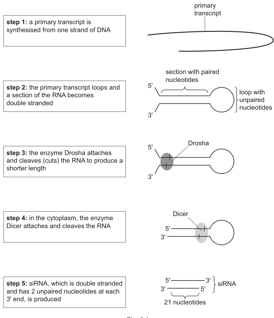
In step 2 in Fig. 3.1, the single-stranded primary transcript loops and forms a section where dsRNA is present.
Explain how it is possible for the double-stranded section of RNA to be held together in step 2 and maintained this way in steps 3, 4 and 5.
Answer
- Complementary base pairing occurs in the double-stranded section: adenine (A) pairs with uracil (U), and guanine (G) pairs with cytosine (C).
- Hydrogen bonds form between these complementary base pairs and hold the two strands together; these hydrogen bonds are maintained through steps 3, 4 and 5.
Complementary base pairing (A–U and G–C) held together by hydrogen bonds.
Background Concept
DNA and RNA are polynucleotides made up of nucleotide monomers. Each nucleotide has a phosphate–sugar backbone and a nitrogenous base. In double-stranded nucleic acids (DNA–DNA, RNA–RNA, or DNA–RNA hybrids) the two strands are held together by hydrogen bonds between complementary base pairs. In RNA the base pairs are adenine–uracil (A–U) and guanine–cytosine (G–C); DNA uses A–T and G–C.
A hairpin forms when a single RNA strand folds back on itself so that a stretch of bases pairs with a complementary stretch further down the same strand. The bases within the stem pair with each other; the bases in the loop itself remain unpaired. This is intramolecular base pairing.
Hydrogen bonds are weak, non-covalent interactions that allow the two strands to be separated when needed (e.g. during replication, transcription, or, as in this question, when an enzyme needs to cleave the RNA). Phosphodiester bonds, in contrast, hold adjacent nucleotides together within a single strand.
Understanding the Question
The question asks how the double-stranded section in step 2 is held together, and why this bonding is still present in steps 3, 4 and 5, where the molecule is being shortened by the enzymes Drosha and Dicer. The key concept is that hydrogen bonds between complementary bases are what holds the two strands together throughout this processing.
Approach
Identify the chemistry: complementary bases (A–U, G–C) form hydrogen bonds. Distinguish this from phosphodiester bonding, which holds adjacent nucleotides within a single strand (the question is about bonding between the two strands, not within them).
Step-by-Step Reasoning
- The two strands of RNA in the double-stranded section are held together because their bases are complementary: A pairs with U, and G pairs with C. Because the strand loops back on itself, this is intramolecular base pairing.
- The hydrogen bonds between these base pairs are the forces holding the two strands together. They are weak, reversible interactions, which is why they can be maintained even while the molecule is being processed.
- The hydrogen bonds persist through steps 3, 4 and 5 because the remaining sections still have complementary sequences that can base pair — even after Drosha removes the loop and Dicer cleaves the molecule, the surviving 21-nt section is still base-paired along its length.
- Phosphodiester bonds (which hold the sugar–phosphate backbone together) are not the answer: the question is about the bonding between the two strands, not within them.
Key Takeaways
- RNA can form intramolecular double-stranded structures (hairpins) through complementary base pairing.
- Hydrogen bonds between complementary bases (A–U and G–C) hold the two strands together.
- Phosphodiester bonds hold adjacent nucleotides within a single strand — they are not what holds the two strands together.
Common Mistakes
- Saying "thymine" instead of "uracil" — this is RNA, not DNA, so it must be uracil.
- Describing the bonds as "strong" or as covalent bonds.
- Confusing the role of phosphodiester bonds (within a strand) with hydrogen bonds (between strands).
- Saying the strands are held together by phosphodiester bonds.
Things to Be Careful About
- The transcript is RNA, so the base pairs are A–U and G–C (not A–T). The mark scheme explicitly says: if thymine is stated, only the H-bond mark can be credited.
- The question asks about the bonding between the two strands, not within a single strand.
After step 3, the shorter dsRNA produced by the action of Drosha is transported to the cytoplasm, where it is cleaved further by Dicer to produce ds siRNA.
Suggest why two different enzymes, Drosha and Dicer, are needed to cut dsRNA into shorter lengths.
Answer
- Drosha and Dicer have different active sites, each shaped to fit a different site/length of dsRNA, so each enzyme cleaves at its own specific location.
- Drosha and Dicer work in different cellular locations: Drosha acts in the nucleus (where the primary transcript is found) and Dicer acts in the cytoplasm (where the Drosha-cleaved dsRNA is transported to).
Different enzymes with different active-site specificities (and operating in different cell locations) are required to cleave the dsRNA at different specific sites.
Background Concept
Enzymes are biological catalysts that are highly specific to their substrates. This specificity arises from the three-dimensional shape of the enzyme's active site, which is complementary to the substrate. The lock-and-key and induced-fit models describe how this specificity works. When the substrate fits into the active site, an enzyme–substrate (ES) complex forms and the reaction proceeds.
Drosha and Dicer are both RNase III family endonucleases that cleave double-stranded RNA. They have different active-site shapes and operate in different cellular compartments: Drosha acts in the nucleus and Dicer acts in the cytoplasm.
Understanding the Question
The question asks why two different enzymes (Drosha and Dicer) are needed to sequentially process the dsRNA into shorter pieces. The answer lies in enzyme specificity and the conditions under which each operates.
Approach
Apply the concept of enzyme specificity: different enzymes have different active sites that recognise and cleave at different specific sites on the dsRNA. Consider also the different conditions/locations in which each enzyme operates.
Step-by-Step Reasoning
- Drosha and Dicer are both enzymes that cleave dsRNA, but they have different active sites with different specificities.
- Drosha's active site is specific for the longer dsRNA section in the nucleus; it recognises a particular site (e.g. a specific sequence or structural feature) and cleaves there, removing the loop.
- Dicer's active site is specific for a shorter section of dsRNA in the cytoplasm; it cleaves the molecule further to produce the final siRNA length (~21–25 nucleotides).
- The active sites of Drosha and Dicer have different shapes, so each is complementary to a different substrate (a different length or location of dsRNA).
- Additionally, Drosha functions in the nucleus (where the primary transcript is processed) while Dicer functions in the cytoplasm. They may also require different conditions (e.g. pH, ions, cofactors) for optimal activity, and Dicer may be too large to cross the nuclear envelope.
Key Takeaways
- Enzymes are specific to their substrates due to the shape of their active sites.
- Two different enzymes can perform sequential processing steps because each has a different active-site specificity.
- Drosha and Dicer are localised differently in the cell (nucleus vs cytoplasm), which is one reason both are needed.
Common Mistakes
- Vague answers like "they are different enzymes" without explaining the specificity of their active sites.
- Not mentioning the different locations (nucleus vs cytoplasm) or different substrate specificities.
- Saying the enzymes cut in different places without explaining WHY (because of active-site specificity).
Things to Be Careful About
- The question asks for a SUGGESTION, so the answer can include additional valid points (AVP).
- The mark scheme credits suggestions such as Dicer being too large to enter the nucleus, or Drosha being exposed to degradation in the cytoplasm.
From 2018, siRNA has been used as a therapeutic drug to treat a number of diseases.
The presence of molecules of siRNA in the cytoplasm can result in the cleavage of messenger RNA (mRNA) molecules coding for a protein involved in the disease. This prevents the synthesis of the protein.
Describe the differences between a molecule of mRNA and a molecule of siRNA, such as the siRNA shown in step 5 in Fig. 3.1.
Answer
- mRNA is single-stranded, whereas the siRNA in step 5 is double-stranded (with 2 unpaired nucleotides at each 3' end).
- mRNA has a much longer sequence of nucleotides (often hundreds to thousands), whereas siRNA is only 21–25 nucleotides long.
- mRNA codes for a sequence of amino acids (a protein), whereas siRNA does not code for amino acids.
mRNA is single-stranded, longer and codes for amino acids; siRNA is double-stranded (with 3' overhangs), short (~21–25 nt) and does not code for amino acids.
Background Concept
mRNA (messenger RNA) is a single-stranded RNA molecule that is transcribed from DNA in the nucleus and carries the genetic code to the ribosomes for translation. mRNA contains the codons that specify the amino-acid sequence of a protein. It is synthesised 5' to 3' and can be hundreds to thousands of nucleotides long, depending on the size of the protein it encodes.
siRNA (small interfering RNA) is a short double-stranded RNA molecule (~21–25 nucleotides long) involved in post-transcriptional gene silencing. It does NOT code for amino acids; instead, it regulates gene expression by targeting specific mRNA molecules for degradation. The mature siRNA has 2 unpaired (overhanging) nucleotides at each 3' end.
Understanding the Question
The question asks for a description of the differences between mRNA and siRNA, referring to the siRNA shown in step 5 of Fig. 3.1. The siRNA in step 5 is double-stranded with 2 unpaired nucleotides at each 3' end.
Approach
Compare the two molecules on the basis of strandedness, length, and coding function.
Step-by-Step Reasoning
- mRNA is single-stranded; siRNA (as shown in step 5) is double-stranded with 2 unpaired nucleotides at each 3' end (the 3' overhangs).
- mRNA has a long sequence of nucleotides (it can be hundreds to thousands of nucleotides long depending on the protein); siRNA is short, only 21–25 nucleotides long.
- mRNA codes for a sequence of amino acids (a protein); siRNA does not code for amino acids — it is a regulatory molecule.
- (Additional) siRNA has both paired and unpaired nucleotides; mRNA has only unpaired nucleotides (no internal base pairing).
- (Additional) siRNA has antiparallel strands (one runs 5'→3', the other 3'→5'); mRNA is a single strand running 5'→3'.
Key Takeaways
- mRNA: single-stranded, codes for amino acids, long (hundreds to thousands of nt).
- siRNA: double-stranded with 3' overhangs, does not code for amino acids, short (~21–25 nt).
- The two molecules have different functions: mRNA is a template for translation, siRNA is a regulatory molecule that targets mRNA for cleavage.
Common Mistakes
- Saying mRNA is double-stranded (it is single-stranded).
- Saying siRNA codes for amino acids (it does not).
- Not specifying the siRNA's structure (double-stranded with 2 unpaired nucleotides at each 3' end).
- Confusing the functions: mRNA is for protein synthesis, siRNA is for gene regulation.
Things to Be Careful About
- The siRNA in step 5 has 2 unpaired nucleotides at each 3' end (the 3' overhangs).
- The question refers specifically to the siRNA shown in step 5 of Fig. 3.1, so refer to its structure.
Fig. 3.2 summarises how mRNA coding for a protein involved in disease can be targeted and cleaved by siRNA.
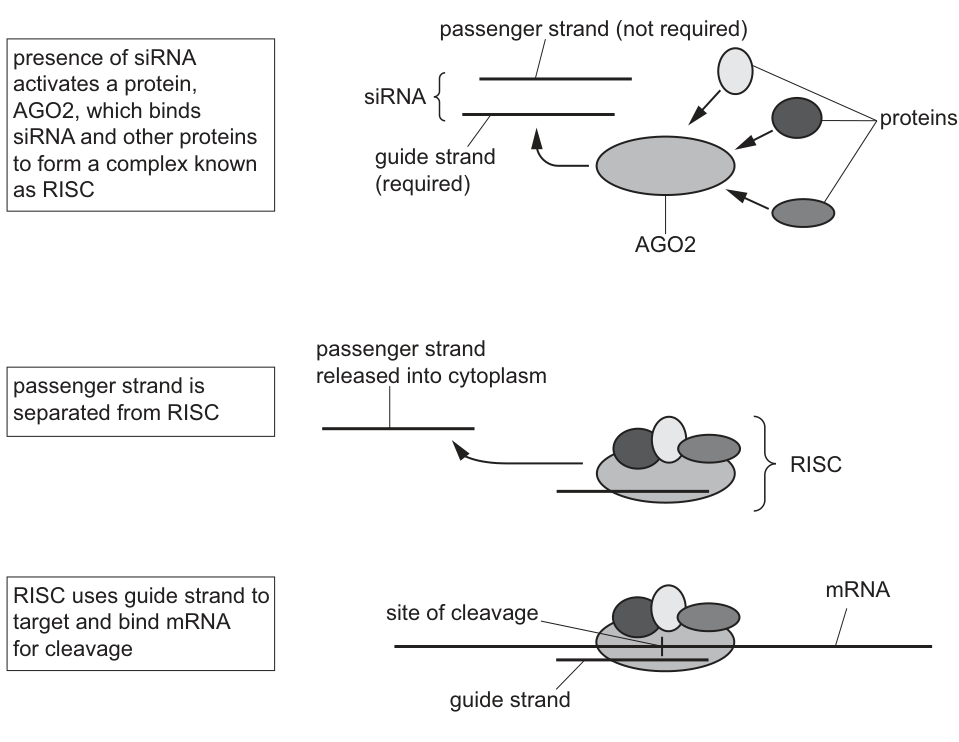
With reference to Fig. 3.2, state why the passenger strand needs to be separated and released from the RISC.
Answer
- The passenger strand must be released so that the guide strand is exposed and can form hydrogen bonds with the complementary bases on the target mRNA. If the passenger strand remained, it would base-pair with the guide strand and block it from binding to the mRNA.
The passenger strand must be released so the guide strand can base-pair with the target mRNA.
Background Concept
The RISC (RNA-Induced Silencing Complex) is a multi-protein complex that uses siRNA to target and cleave specific mRNA molecules. The siRNA has two strands: the guide strand (which is complementary to the target mRNA and remains in RISC) and the passenger strand (which is discarded). The guide strand's bases need to be exposed so they can pair with the target mRNA by complementary base pairing.
Understanding the Question
Fig. 3.2 shows the mechanism of mRNA cleavage by siRNA. The question asks why the passenger strand needs to be separated and released from the RISC.
Approach
The passenger strand must be released so that the guide strand can bind to the target mRNA by complementary base pairing. If the passenger strand remained, it would base-pair with the guide strand and prevent the guide strand from binding to the mRNA.
Step-by-Step Reasoning
- The guide strand in RISC is the strand that needs to base-pair with the target mRNA, because the guide strand has a sequence complementary to the target mRNA.
- While the siRNA is double-stranded, the guide strand's bases are paired with the passenger strand's bases and are not exposed to the cytoplasm.
- The passenger strand must be removed so the guide strand's bases are exposed and can form hydrogen bonds with the complementary bases on the target mRNA.
- If the passenger strand remained, it would base-pair with the guide strand (since they are complementary), preventing the guide strand from base-pairing with the target mRNA.
Key Takeaways
- The guide strand must be exposed (i.e., not paired with the passenger strand) so it can base-pair with the target mRNA.
- The passenger strand is complementary to the guide strand, so it would block the guide strand from binding to the mRNA if it remained.
Common Mistakes
- Vague answers like "it needs to be removed for the complex to work."
- Not mentioning the role of the guide strand in binding to the mRNA.
- Not explaining that the passenger strand base-pairs with the guide strand.
Things to Be Careful About
- The "ora" (or reverse argument) is also credited: "passenger strand prevents guide strand binding" or "guide strand cannot bind to mRNA if passenger strand remains".
The aim of siRNA therapy is to prevent or decrease the synthesis of a protein involved in the disease being treated.
A target mRNA molecule can be cleaved in a different location by a RISC with a different siRNA.
Suggest how cleaving mRNA in different locations will have different effects on protein synthesis and explain how these different effects can result in a lack of functioning protein.
Answer
- If cleavage removes the start codon, the mRNA cannot be translated (or the ribosome cannot bind/initiate), so no polypeptide is produced.
- If cleavage occurs within the coding sequence, a shorter mRNA is produced, which is translated into a shorter polypeptide chain. The shorter polypeptide cannot fold into the correct tertiary structure, so it is non-functional and may be degraded in the cytoplasm.
- If cleavage removes the stop codon, translation continues past the normal end of the protein. The polypeptide may not be released from the ribosome, leading to its degradation, and so no functioning protein is produced.
Different cleavage sites remove different essential regions of the mRNA, leading to no protein, a short/non-functional protein, or a polypeptide that cannot be released from the ribosome — all resulting in no functioning protein.
Background Concept
Translation is the process by which ribosomes read mRNA codons and assemble a polypeptide chain. The mRNA has a start codon (AUG) near the 5' end, which is where translation begins, and a stop codon (UAA, UAG or UGA) at the 3' end, which signals the end of translation and release of the polypeptide from the ribosome.
A polypeptide must be of a certain length and have the correct amino-acid sequence to fold into a functional tertiary structure and perform its function. If any of these are disrupted, the protein will not function.
Understanding the Question
The question asks how cleaving mRNA in different locations can have different effects on protein synthesis, and how these different effects can result in a lack of functioning protein. The question is essentially asking about the consequences of different cleavage sites on the protein product.
Approach
Consider the different ways mRNA can be cleaved: near the start codon, within the coding sequence, or near the stop codon. Each of these will have different effects on the protein product. Connect each effect to "no functioning protein."
Step-by-Step Reasoning
- Loss of start codon (or cleavage near the 5' end): If the mRNA is cleaved such that the start codon (AUG) is removed, the mRNA cannot be properly translated (the ribosome cannot initiate translation), so no polypeptide is produced. In some cases the cleaved mRNA is degraded before it can attach to the ribosome at all.
- Cleavage in the middle of the coding sequence: This produces a shorter mRNA, which is translated into a shorter polypeptide chain. The shorter polypeptide cannot fold into the correct tertiary structure (the 3D shape depends on length and R-group interactions), so it is non-functional.
- Loss of stop codon (or cleavage near the 3' end): If the mRNA is cleaved such that the stop codon is removed, the ribosome continues translation past the normal end of the protein. The polypeptide may not be released from the ribosome, leading to its degradation in the cytoplasm, so no functioning protein is produced.
- Connecting to "no functioning protein":
- No protein is made at all (if cleavage is before/at the start codon).
- A shorter/abnormal protein is made, which cannot fold correctly and so is non-functional.
- A longer/aberrant protein is made, which may be degraded or may fold incorrectly.
- All of these effects result in a lack of functioning protein.
Key Takeaways
- Different locations of cleavage result in different effects on translation:
- Cleavage near the start codon → no protein (no translation initiation).
- Cleavage within the coding sequence → shorter protein (which cannot fold correctly).
- Cleavage near the stop codon → longer/aberrant protein (which may be degraded or not released from the ribosome).
- All of these effects result in no functioning protein.
- siRNA therapy exploits this by targeting disease-related mRNAs for cleavage at specific sites.
Common Mistakes
- Not linking the cleavage effects to "no functioning protein" specifically.
- Saying the protein will be "different" or "wrong" without explaining HOW it's wrong.
- Not mentioning the role of the start and stop codons in translation.
- Saying the polypeptide "changes shape" without specifying it cannot form the correct tertiary structure.
Things to Be Careful About
- The question is worth 3 marks, so aim for 3 distinct points.
- "Cleaved mRNA results in loss of start codon" and "mRNA may not attach to ribosome" are two separate but related points.
- "Cleaved mRNA results in shorter polypeptide chain" and "polypeptide does not fold correctly" are two separate points.
- The question wants connections between the cleavage effects AND the lack of functioning protein.
In mammals, the gas exchange system includes a set of branching airways that carry air to and from the gas exchange surface.
Air from the external atmosphere passes through different types of airway to reach the gas exchange surface.
Name the type of airway of the gas exchange system that branches into airways known as bronchioles.
Answer
Bronchus
Bronchus
Background Concept
The mammalian gas exchange system is a branching network of tubes that conducts air from the external atmosphere to the gas exchange surface (the alveoli). Starting at the top, the pathway is: nasal cavity / mouth → pharynx → larynx → trachea → two primary bronchi (one to each lung) → secondary and tertiary bronchi → bronchioles → alveolar ducts → alveolar sacs → alveoli. The trachea divides into the left and right primary bronchi at a level called the carina, and each bronchus then branches repeatedly into smaller bronchi and finally into bronchioles (airways less than about 1 mm in diameter that lack cartilage in their walls).
Understanding the Question
The question asks which airway branches into bronchioles. The wording is direct and the only correct answer is the airway immediately upstream of the bronchioles in the hierarchy.
Approach
Recall the order of branching and identify the airway directly above the bronchioles.
Step-by-Step Reasoning
The sequence of branching is: trachea → bronchus → bronchioles → alveoli. The bronchus is the airway that gives rise to the bronchioles. The plural 'bronchi' refers to the two main airways entering each lung, and each of these branches into smaller bronchi which then become bronchioles.
Key Takeaways
- The gas exchange airways branch in a strict sequence: trachea → bronchus → bronchioles → alveoli.
- Bronchi are still large enough to require cartilage support in their walls; bronchioles are not.
Common Mistakes
- Writing 'bronchioles' (the answer must be the airway that branches INTO bronchioles, not the bronchioles themselves).
- Writing 'trachea' (the trachea does not directly branch into bronchioles; it branches into bronchi first).
Things to Be Careful About
The command word is 'name', so a single word is sufficient. Spelling matters: 'bronchus' (singular) / 'bronchi' (plural).
Fig. 4.1 is a photomicrograph of a section through a bronchiole.
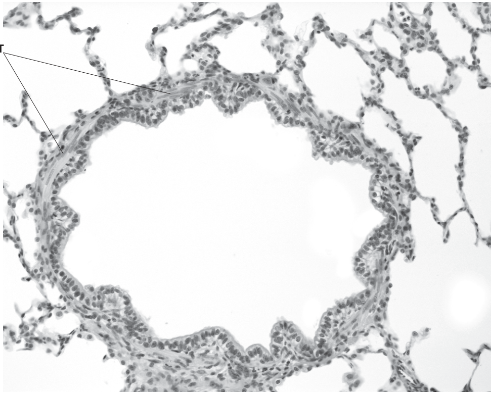
The bronchiole shown in Fig. 4.1 is not part of the gas exchange surface.
Name one structure in the gas exchange system, visible in Fig. 4.1, where gas exchange is carried out.
Answer
Alveolus (alveolar sac / alveolar duct)
Alveolus (alveolar sac / alveolar duct)
Background Concept
The gas exchange surface in mammals is the alveolar epithelium. Alveoli are tiny, thin-walled, sac-like outpocketings at the end of the airway tree. They are surrounded by a dense capillary network so that oxygen and carbon dioxide can diffuse across the respiratory surface. They are the only sites in the gas exchange system across which gas exchange actually occurs; the rest of the system is purely conducting.
Understanding the Question
The question states that the bronchiole shown in Fig. 4.1 is not part of the gas exchange surface, and asks the candidate to name a structure, visible in the same image, where gas exchange does occur. The micrograph shows the central bronchiole with its folded epithelium and outside the bronchiole are numerous thin-walled spaces — these are the alveoli.
Approach
Look at the structures outside the bronchiole in the photomicrograph. Recognise the cluster of thin-walled air spaces as the alveoli (often appearing as a honeycomb of small irregular spaces).
Step-by-Step Reasoning
In Fig. 4.1, the central feature is a bronchiole with a wavy/folded epithelium and an outer wall of smooth muscle. Surrounding it is lung parenchyma made up of many small, thin-walled air spaces. These are the alveoli. The bronchioles conduct air to the alveoli, but it is the alveoli themselves where oxygen and carbon dioxide cross the respiratory surface.
Key Takeaways
- The conducting portion (trachea, bronchi, bronchioles) only carries air; the respiratory portion (alveoli, alveolar sacs, alveolar ducts) is where gas exchange occurs.
- In a histological section, alveoli appear as clusters of small, thin-walled air spaces adjacent to conducting airways.
Common Mistakes
- Writing 'bronchiole' itself (the question explicitly says the bronchiole is NOT part of the gas exchange surface).
- Writing 'lung' (the lung is the whole organ, not a specific structure).
Things to Be Careful About
The mark scheme accepts alveolus, alveolar sac or alveolar duct. Any of these earns the mark.
Answer
Smooth muscle (cell)
Smooth muscle (cell)
Background Concept
The wall of a bronchiole is built from (inside out): a ciliated cuboidal/columnar epithelium, a basement membrane, a layer of smooth muscle, and a thin outer layer of connective tissue with elastic fibres. Because the bronchiole has no cartilage to hold it open, the smooth muscle layer is functionally important: when it contracts (bronchoconstriction) the airway narrows, and when it relaxes (bronchodilation) the airway widens. This is the basis of asthma medication action (e.g. salbutamol causes bronchodilation).
Understanding the Question
The label T in Fig. 4.1 points to the layer outside the folded epithelium of the bronchiole wall. The question asks the candidate to name the type of cell found in this layer.
Approach
Identify the location of T in the image (between the epithelium and the surrounding alveolar tissue) and recall what cell type occupies that position in a bronchiole.
Step-by-Step Reasoning
T points to the muscular wall of the bronchiole. The dominant cell type in this layer is the smooth muscle cell. Smooth muscle is involuntary (not under conscious control) and is innervated by the autonomic nervous system.
Key Takeaways
- The wall of a bronchiole contains smooth muscle (and lacks cartilage).
- Smooth muscle controls airway diameter via bronchoconstriction and bronchodilation.
Common Mistakes
- Writing 'muscle' without the qualifier 'smooth' (cardiac and skeletal muscle are wrong here).
- Writing 'elastic fibre' or 'cartilage' (these are not the dominant cell type in this layer).
Things to Be Careful About
The answer must specify smooth muscle — 'muscle cell' on its own is too vague for this mark.
State the features that help to identify the type of airway shown in Fig. 4.1 as a bronchiole and not the other types of airway present in the gas exchange system.
Answer
Any three from:
- No cartilage present (unlike the trachea/bronchi which have cartilage rings or plates)
- Few or no goblet cells in the epithelium (the trachea/bronchi have many goblet cells)
- A thin layer / patches of smooth muscle in the wall (less than in trachea/bronchi)
- Thinner wall overall
- Convoluted / wavy / folded lumen lining (the lining of the trachea/bronchi is smoother)
- Less elastic tissue / fewer elastic fibres than in the trachea/bronchi
Three distinguishing features of a bronchiole visible in the image: no cartilage; few/no goblet cells; thin patches of smooth muscle; thinner wall; folded lumen; less elastic tissue.
Background Concept
The walls of the airways change in composition as they branch and become smaller:
- Trachea and bronchi: C-shaped (trachea) or irregular plates (bronchi) of hyaline cartilage support the wall. The epithelium is pseudostratified ciliated columnar with many goblet cells. Submucosal mucous and serous glands are present. Smooth muscle is present between the cartilage and the epithelium. Plenty of elastic fibres in the wall.
- Bronchioles: Cartilage is absent. The epithelium is ciliated but becomes simpler (cuboidal) with few or no goblet cells. The wall is dominated by smooth muscle, and elastic fibres are still present but in smaller amounts than in larger airways.
- The smaller the airway, the simpler the wall.
The bronchiole is the portion of the airway tree that lacks cartilage, so it can constrict and dilate significantly under the control of the smooth muscle layer — this is what makes bronchioles the focus of asthma pathophysiology.
Understanding the Question
The question asks the candidate to identify features of the bronchiole visible in Fig. 4.1 that distinguish it from the trachea and the bronchi. Three marks are available, so three distinct features should be given.
Approach
Look at the image systematically. For each wall component (cartilage, epithelium, muscle, elastic tissue, lumen shape) compare what is visible in the bronchiole to what would be present in a trachea or bronchus. Choose the three most clearly visible distinguishing features.
Step-by-Step Reasoning
The image shows:
- No cartilage — the bronchiole wall has no dense pink/blue cartilage plates or rings around it. The trachea/bronchi have prominent cartilage in the wall.
- Folded/wavy lumen lining — the epithelium bulges into the lumen in folds. In the trachea/bronchi the cartilage holds the lumen open and the lining appears relatively smooth and circular.
- Few/no goblet cells — the epithelium is simple, lacking the dense population of pale goblet cells seen in the pseudostratified epithelium of the trachea/bronchi.
- Thin layer of smooth muscle (T) — present but in patches/thin layer, unlike the thicker smooth muscle in larger airways.
- Thinner overall wall compared to trachea/bronchi.
- Less elastic tissue in the wall.
Any three of these are valid. The strongest three, based purely on what the image shows, are: no cartilage; convoluted/folded lumen; and few/no goblet cells.
Key Takeaways
- Bronchioles are recognised histologically by the absence of cartilage, the absence (or scarcity) of goblet cells, a relatively thin wall, and a folded epithelium.
- The smaller the airway, the less supporting tissue (cartilage) and the more dominant the smooth muscle layer.
- The gas exchange system shows a clear structural gradient from trachea to alveoli, and being able to identify where you are in that gradient from a micrograph is a key skill.
Common Mistakes
- Listing features that are true of all airways (e.g. 'has a lumen', 'is lined by epithelium') — these do not distinguish a bronchiole from other airways.
- Writing 'no muscle' — the bronchiole has smooth muscle; it is the cartilage that is absent.
- Writing 'no cilia' — bronchioles are still ciliated; it is the goblet cells and cartilage that are reduced or absent.
Things to Be Careful About
- The mark scheme allows 'ORA' (or reverse argument) for goblet cells: 'few/no goblet cells' OR 'trachea/bronchus have (more) goblet cells' — both earn the mark.
- 'Thin wall' alone is acceptable as a distinguishing feature.
- Avoid vague answers like 'smaller' or 'less developed' — be specific about which component is reduced or absent.
Blood is pumped to the lungs in the pulmonary circulation.
The lungs also receive a supply of blood from the systemic circulation.
Explain why the pulmonary circulation and the systemic circulation need to supply blood to the lungs.
Answer
Any two from:
- The systemic circulation supplies the lung tissue itself with oxygenated blood (and glucose) so that the lung cells can respire / obtain ATP for their metabolic activities.
- The pulmonary circulation brings deoxygenated blood to the alveolar capillaries so that oxygen can be absorbed and carbon dioxide can be excreted at the gas exchange surface.
- The lungs are the site of gas exchange (O₂ uptake and CO₂ excretion) — the pulmonary circulation serves this function.
- Cells (including lung cells) need oxygen and glucose for (aerobic) respiration to release energy / ATP.
Pulmonary circulation delivers deoxygenated blood for gas exchange at the alveoli; systemic circulation delivers oxygenated blood (and glucose) to supply the lung tissue itself with O₂ and nutrients for aerobic respiration.
Background Concept
Mammals have a closed double circulation consisting of:
- The pulmonary circulation: right side of the heart → pulmonary artery → lungs → pulmonary veins → left side of the heart. This circulation carries deoxygenated blood to the lungs and returns oxygenated blood to the heart.
- The systemic circulation: left side of the heart → aorta → body tissues (including the lungs themselves) → vena cava → right side of the heart. This carries oxygenated blood at high pressure to supply the body's tissues.
A common misconception is that only the pulmonary circulation supplies the lungs. In fact, the bronchial circulation (a branch of the systemic circulation) supplies oxygenated blood to the lung tissue itself — the smooth muscle, the connective tissue, the pleura, and the walls of the larger airways. Without this, lung cells would not receive oxygen or glucose for their own aerobic respiration.
Understanding the Question
The question asks the candidate to explain why BOTH the pulmonary circulation AND the systemic circulation are needed to supply blood to the lungs. The two marks require two distinct ideas. The key is to recognise that the two circulations serve different functions even though they both go to the lungs.
Approach
Separate the question into 'what does the pulmonary circulation do for the lungs?' and 'what does the systemic circulation do for the lungs?'. Then bring in the idea that lung cells themselves are living tissue and need their own blood supply.
Step-by-Step Reasoning
- Pulmonary circulation: carries deoxygenated blood from the right ventricle via the pulmonary artery to the alveolar capillaries. Here, oxygen diffuses into the blood and carbon dioxide diffuses out. This is the gas exchange function — it is the reason the lungs exist as an organ.
- Systemic circulation (via the bronchial arteries): supplies oxygenated blood to the lung tissue itself — to the smooth muscle of the airways, the walls of the bronchi, the pleura and the connective tissue. Lung cells are living, metabolically active cells that need oxygen and glucose for aerobic respiration to make ATP. They cannot rely on the deoxygenated blood that arrives via the pulmonary arteries because that blood is being processed for gas exchange; it is not supplying the lung cells' own metabolic needs.
- Bringing the two together: the lungs need both — the pulmonary circulation for gas exchange, and the systemic circulation to feed the lung tissue itself.
Key Takeaways
- The lungs have a dual blood supply: the pulmonary circulation (for gas exchange) and the bronchial circulation (part of the systemic circulation, to supply the lung tissue with O₂ and glucose for respiration).
- It is a common error to think the lungs are only supplied by the pulmonary circulation. The systemic supply to the lungs is essential for the lung cells' own metabolism.
- Cells throughout the body — including lung cells — need a continuous supply of oxygen and glucose for aerobic respiration to release energy as ATP.
Common Mistakes
- Stating only that 'the pulmonary circulation brings deoxygenated blood and the systemic brings oxygenated blood' without linking each to its function (gas exchange vs. supplying lung tissue). The mark scheme wants the reasoning, not just the description.
- Saying the systemic circulation supplies 'the body' (this is too vague) rather than specifically the lung tissue.
- Confusing the two circulations (e.g. saying the systemic circulation is for gas exchange).
Things to Be Careful About
- The mark scheme explicitly REJECTS the bare phrase 'for body cells' (R body cells). The answer must specifically refer to the lung cells/tissue, not the body generally.
- The two ideas must be distinct: 'pulmonary for gas exchange' and 'systemic for supplying the lung tissue' are the two cleanest points.
- The question says 'explain', so a reason must accompany each point — not just a description of the two circulations.
Veins transport blood towards the heart. The structure of a vein is adapted to its function.
The inner layer of a vein is the tunica intima, composed of a single layer of endothelial cells that form a protective barrier.
When the tunica intima of a blood vessel is damaged, endothelial cells can carry out mitosis to allow tissue repair to occur.
The spindle that is formed during mitosis is composed of spindle fibres.
Name the cell structures that are organised to form spindle fibres during mitosis.
Answer
Microtubules
Microtubules
Background Concept
During mitosis, chromosomes must be separated accurately so that each daughter cell receives a complete set. This movement is carried out by the mitotic spindle — a dynamic, bipolar array of fibres that extends from opposite poles of the cell and attaches to chromosomes at their centromeres via kinetochores. The spindle is not a permanent structure; it assembles at the start of mitosis and disassembles once chromosome segregation is complete.
The spindle fibres themselves are built from microtubules — hollow cylindrical polymers made of alternating α-tubulin and β-tubulin protein dimers. Microtubules belong to the cytoskeleton along with microfilaments (made of actin) and intermediate filaments (made of keratin-like proteins). In animal cells, microtubules are nucleated and organised by the centrosomes (each containing a pair of centrioles), which migrate to opposite poles and act as the spindle poles. Plant cells lack centrioles but still form spindles from microtubules nucleated at the cell's polar regions.
Understanding the Question
The stem sets the scene: when the tunica intima of a blood vessel is damaged, the endothelial cells undergo mitosis to repair the tissue. This part then asks specifically about the molecular structures that make up the spindle. The question is a direct factual recall — a one-word (one-term) answer worth one mark.
Approach
Recall that the spindle is a cytoskeletal structure. Identify which cytoskeletal filament type forms it. Be careful to distinguish microtubules (the correct answer) from other cytoskeletal elements.
Step-by-Step Reasoning
- Spindle fibres are not made of chromatin, DNA, or membrane — they are structural filaments of the cytoskeleton.
- There are three classes of cytoskeletal filament: microfilaments (actin), intermediate filaments, and microtubules (tubulin).
- Of these, only microtubules are used to build the spindle apparatus — they polymerise from tubulin dimers and grow/shrink dynamically (a property called dynamic instability) to capture chromosomes, align them at the metaphase plate, and then pull the sister chromatids apart.
- Therefore the cell structures that organise to form spindle fibres are microtubules.
Key Takeaways
- Spindle fibres = microtubules (polymers of α/β-tubulin).
- Microtubules are nucleated by centrosomes/centrioles at the spindle poles in animal cells.
- Distinguish microtubules from microfilaments (actin) — the latter are involved in cytokinesis (the contractile ring), not spindle formation.
Common Mistakes
- Writing "microfilaments" or "actin" — these form the contractile ring during cytokinesis, not the spindle.
- Writing "chromosomes" or "chromatin" — chromosomes are what the spindle moves; they do not compose the spindle.
- Writing "centrioles" or "centrosomes" — these organise the spindle but are not themselves the spindle fibres.
Things to Be Careful About
The mark scheme requires the exact word "microtubules". "Tubulin" alone is not credited because tubulin is the protein subunit, not the assembled structure. "Spindle fibres" is the structure as a whole, not the components.
Answer
Any two from:
- The (daughter) chromosomes are at opposite poles of the cell (in two groups)
- The nuclear envelope reassembles around each set of chromosomes
- The nucleolus reappears within each reforming nucleus
- The (daughter) chromosomes become diffuse / decondense (into chromatin)
- The spindle disassembles
Daughter chromosomes are at opposite poles and the nuclear envelope reassembles around each set, forming two new nuclei (with the spindle disassembling and chromosomes decondensing).
Background Concept
Mitosis is conventionally divided into four stages: prophase, metaphase, anaphase, and telophase. Each stage has a characteristic set of events that moves the cell closer to producing two genetically identical daughter nuclei.
Telophase is the final stage of nuclear division. It essentially reverses the events of prophase: after the sister chromatids have been pulled to opposite poles during anaphase, the cell reforms two separate nuclei — one around each set of chromosomes. The defining features of telophase are:
- The daughter chromosomes (now single chromatids) are at the two poles.
- A new nuclear envelope reassembles around each set of chromosomes, forming two nuclei.
- Nucleoli reappear within the new nuclei (they had disassembled during prophase).
- The chromosomes decondense — they uncoil and become long, thin, diffuse chromatin, no longer visible as discrete chromosomes.
- The spindle microtubules disassemble, freeing the tubulin subunits for re-use.
Telophase ends when two distinct daughter nuclei exist. Cytokinesis (division of the cytoplasm) usually occurs in parallel with or just after telophase, but it is a separate process.
Understanding the Question
The question uses the command word "describe", which means state the key events that occur during telophase. Two marks are available — choose any two creditable points from the mark scheme's list. The stem has set the context: mitosis in endothelial cells of a damaged blood vessel. The same events occur in any mitotic cell.
Approach
Mentally run through the sequence of mitosis: prophase → metaphase → anaphase → telophase. Identify which events characterise telophase specifically (not anaphase, not cytokinesis). Pick two precise, mark-scheme-worthy points.
Step-by-Step Reasoning
- Position of chromosomes — at the end of anaphase, the sister chromatids (now independent daughter chromosomes) have been pulled to opposite poles. In telophase they are therefore at the poles, in two separate groups. Mark-scheme wording: "(daughter) chromosomes at poles".
- Nuclear envelope reassembly — during prophase the nuclear envelope broke down into vesicles. In telophase these vesicles fuse around each set of chromosomes, reforming two nuclear envelopes. The required wording is "reassembles" or "re-forms", not just "forms".
- Nucleolus reappears — the nucleolus disassembled in prophase. In telophase it reappears within each new nucleus, marking the resumption of rRNA synthesis.
- Chromosomes decondense — having reached the poles, the chromosomes uncoil and become long, thin, diffuse chromatin, restoring the interphase appearance. The mark scheme accepts "decondense", "become diffuse", "become long and thin", "uncoil", or "become chromatin". "Chromosomes disappear" is not credited because they are still present, just less condensed.
- Spindle disassembles — the spindle microtubules depolymerise. The mark scheme accepts "disassembles", "breaks down", or any equivalent wording.
Any two of these five points earn the two marks.
Key Takeaways
- Telophase reverses prophase: nuclear envelope reassembles, nucleoli reappear, chromosomes decondense.
- Daughter chromosomes are already at the poles when telophase begins — telophase is about reforming nuclei, not moving chromosomes.
- Telophase is the nuclear division step; cytokinesis is the cytoplasmic division step and is conceptually separate.
Common Mistakes
- Saying "chromosomes move to the poles" — this is anaphase, not telophase.
- Saying "two nuclei form" without saying where or what encloses them — the mark scheme requires the nuclear envelope to reassemble around each set of chromosomes.
- Saying "chromosomes disappear" — this is rejected; they become diffuse chromatin, they do not vanish.
- Saying "the cell divides into two" — this is cytokinesis, not telophase.
- Only describing one event when two are required.
Things to Be Careful About
- "Two nuclei form" alone is not credited if it is unclear whether you mean two new nuclei (which is fine) or whether it is being used to describe the cell splitting (rejected). Pair it with "nuclear envelope reassembles around each set".
- "Nuclear membrane forms" is accepted; "nucleus forms" without mentioning the envelope is borderline — add "envelope" for safety.
- Use "reassembles" or "re-forms" rather than "forms" to make it clear that telophase restores what prophase dismantled.
Explain how the structure of a vein is related to its function.
You do not need to include details of the structure of the tunica intima and its function.
Answer
Any three from:
- Valves — to prevent backflow of blood / to help move blood back towards the heart / to ensure one-way flow of blood.
- Tunica externa (adventitia) — the (thickest) outer layer, contains collagen, to protect the vein and prevent it collapsing from external forces and to maintain its shape.
- Tunica media — contains smooth muscle, which provides mechanical support and can change the diameter of the lumen to help move the low-pressure blood back towards the heart; also contains elastic fibres, which accommodate changes in blood volume / lumen diameter (allowing the vein to stretch and recoil).
- Thin walls and large lumen (relative to wall thickness) — the large lumen holds a larger volume of (low-pressure) blood and minimises resistance to flow from the walls.
(Three creditable points are required; any combination of structure + function from the above earns the marks.)
Three creditable feature–function links, e.g.: valves prevent backflow; tunica externa/adventitia with collagen protects and prevents collapse; tunica media smooth muscle and elastic fibres support the wall, change lumen diameter and accommodate changes in blood volume; thin walls with large lumen hold a larger volume of low-pressure blood and reduce resistance.
Background Concept
Veins are blood vessels that carry blood towards the heart. Because they typically carry blood at low pressure (especially in the venous system below the heart), their wall structure is adapted to withstand compression rather than high pulsatile pressure, and to actively assist the return of blood against gravity.
A typical vein has the same three concentric layers as an artery, but in different proportions:
- Tunica intima — the thin innermost layer, a single layer of endothelial cells on a basement membrane (this question excludes it from discussion).
- Tunica media — the middle layer, containing smooth muscle and elastic fibres. In a vein this layer is much thinner than in an artery because the blood pressure is low.
- Tunica externa / adventitia — the outermost layer, the thickest layer in a vein, dominated by collagen fibres with some elastic fibres.
Two additional features distinguish veins from arteries:
- Valves — folds of the tunica intima projecting into the lumen, particularly important in veins of the limbs, ensuring one-way flow.
- Large lumen relative to total wall thickness.
Understanding the Question
The stem states "Veins transport blood towards the heart. The structure of a vein is adapted to its function." Part (b) asks for an explanation of how the structure of a vein is related to its function. The command word is explain, so the answer must pair a structural feature with its functional consequence — not just list structures. Three marks are available, so the answer must give three credible points. The tunica intima is explicitly excluded by the question.
Approach
Work through the wall from outside to inside (or inside to outside) and for each layer or feature, ask: "Why does the vein need this? What problem does it solve?" The key functional problems of a vein are: (a) low blood pressure so blood tends to pool and flow back, (b) the vessel may be compressed by surrounding tissues, (c) the volume of blood returning varies with posture and activity, (d) blood must be propelled towards the heart against gravity.
Step-by-Step Reasoning
Feature 1 — Valves.
- Structure: inward-projecting folds of the tunica intima, present especially in limb veins.
- Function: when blood tries to flow backwards (or when surrounding muscles compress the vein), the valve cusps close, preventing backflow. This ensures one-way flow back towards the heart and helps overcome the effect of gravity, particularly when standing.
- Note: the mark scheme rejects "semilunar valve" — semilunar valves are in the heart (aortic and pulmonary), not in veins. Use "valve" alone.
Feature 2 — Tunica externa / adventitia.
- Structure: the outermost and (in a vein) thickest layer, dominated by collagen fibres.
- Function: collagen is a tough, inextensible fibrous protein. It protects the vein from mechanical damage and prevents the vein from collapsing under external pressure (e.g. from contracting skeletal muscles in the limbs). It also helps maintain the shape of the vessel.
Feature 3 — Tunica media — smooth muscle.
- Structure: middle layer containing smooth muscle.
- Function: smooth muscle can contract to reduce the lumen diameter (venoconstriction), which squeezes blood towards the heart — important because venous blood is at low pressure. Venoconstriction also helps to maintain blood pressure and to adjust the capacity of the venous system to redistribute blood volume.
Feature 4 — Tunica media — elastic fibres.
- Structure: elastic fibres are also present in the (thinner than in arteries) tunica media.
- Function: elastic fibres allow the vein to stretch and recoil as blood volume changes — accommodating variations in the volume of blood returning (e.g. during exercise). They are not as prominent as in arteries because the pressure changes are much smaller.
Feature 5 — Thin walls and large lumen (relative to wall thickness).
- Structure: the overall wall is thin compared with an artery, and the lumen is wide relative to that wall.
- Function: the wide lumen holds a larger volume of blood — useful because veins act as a capacitance / blood reservoir (most of the blood volume at rest is in the venous system). It also minimises the resistance to flow (lower surface-area-to-volume ratio inside the lumen, less friction), so blood flows easily despite the low pressure.
Any three of the above structure–function pairs earn the three marks. Strongest answers give three distinct features, each correctly matched.
Key Takeaways
- Vein structure is dominated by the tunica externa (collagen), not the tunica media — the opposite of arteries.
- Veins carry low-pressure blood, so they need valves to prevent backflow, a tough outer layer to prevent collapse, and a wide lumen to act as a capacitance reservoir.
- Smooth muscle in the tunica media assists venous return (venoconstriction) and elastic fibres accommodate volume changes.
- Structure–function pairs must always be named together; structure alone earns nothing.
Common Mistakes
- Saying only "the wall is thin" or "the lumen is large" with no functional explanation.
- Naming "semilunar valves" — these are heart valves, not vein valves, and the mark scheme rejects this.
- Saying the wall "stretches and recoils to prevent bursting" — veins do not face bursting pressures; the mark scheme rejects this wording.
- Describing artery features (thick muscular tunica media, no valves) instead of vein features.
- Including the tunica intima — the question explicitly excludes this layer.
- Saying valves "stop backflow" is too vague; the function is to ensure one-way flow towards the heart (against gravity).
Things to Be Careful About
- "Thin wall" must be qualified: thin compared with an artery of similar size, and with a large lumen relative to that wall.
- The tunica externa is the thickest layer in a vein — say so, because candidates often say (wrongly) that it is the thinnest.
- Both structure AND function are required for each mark — pairing "tunica media contains smooth muscle" with "for support / to change lumen diameter" is what earns the mark.
- Stay strictly within vein structure: do not introduce artery or capillary features, even for contrast.
State and explain why the same leaf of a plant can be described as a source or as a sink, depending on the stage of maturity (age) of the leaf.
Answer
- A young, developing leaf is a sink because it is not yet photosynthesising / cannot yet make its own sugars, so it receives / imports assimilates (e.g. sucrose) from other parts of the plant and uses them for growth.
- A mature, fully-expanded leaf is a source because it is photosynthesising, producing assimilates (e.g. sucrose) which it exports via the phloem to other parts of the plant (other sinks).
Young/immature leaf = sink (imports assimilates because it is not yet photosynthesising); mature leaf = source (exports assimilates because it is photosynthesising).
Background Concept
In the phloem, organic assimilates (mainly sucrose, but also amino acids and other products of photosynthesis) are moved from sources — regions that produce a net surplus of these substances — to sinks — regions that use or store them but do not produce enough for their own needs. Sources are typically mature photosynthesising leaves, but also any storage organ that is being remobilised (e.g. a germinating seed's cotyledons). Sinks include roots, developing fruits, growing buds, and — importantly — young, developing leaves. A single organ is not permanently one or the other; its role depends on the balance between what it makes and what it consumes.
Understanding the Question
The command word is state and explain. You need to give one mark-worthy point about the leaf as a sink and one about the leaf as a source (the mark scheme is split exactly 1 + 1 for two marks, and gives credit for any two features under each role). The question is asking you to link the leaf's stage of maturity to its function in phloem transport.
Approach
Think of a leaf's life in two phases:
- While it is still expanding, its chlorophyll content and photosynthetic capacity are too low to support its own growth, and it imports sucrose from elsewhere — sink behaviour.
- Once it is fully expanded, it photosynthesises more than enough for its own needs and exports the surplus — source behaviour.
You must give both sides; an answer that only explains one role will score only one mark at most.
Step-by-Step Reasoning
Mark 1 — sink features (any two accepted):
- The leaf is young / immature / developing / still growing.
- It is not (yet) photosynthesising, so it cannot make its own sugars / organic compounds.
- It therefore receives assimilates / photosynthates / organic compounds (e.g. sucrose) from other parts of the plant.
- It needs these for energy and as raw materials for growth.
Mark 2 — source features (any two accepted):
- The leaf is mature / fully developed / no longer growing.
- It is photosynthesising / synthesising assimilates (e.g. sucrose).
- It translocates / exports / provides these assimilates to other parts of the plant (other sinks such as roots, fruits, buds, young leaves).
A good candidate answer simply states each role with the reason in one or two sentences each.
Key Takeaways
- A source–sink relationship is dynamic, not fixed to one organ — the same leaf changes role as it matures.
- The driver is whether the organ is a net producer or a net consumer of assimilates at that moment.
- Mass flow in the phloem goes from source to sink along a turgor-pressure gradient, and a single plant contains many sources and many sinks at any one time.
Common Mistakes
- Saying the leaf is a source because it has chlorophyll — chlorophyll is present from the start, but net export only begins once photosynthesis exceeds respiration. The mark scheme rewards "mature / photosynthesising / exporting", not the mere presence of chlorophyll.
- Saying the leaf is a sink because it "uses" sugars — vague; the mark scheme requires the link to it importing assimilates from elsewhere, because it cannot yet make enough of its own.
- Giving only one side of the comparison — this limits you to one mark, not two.
Things to Be Careful About
- "Starch" alone is rejected unless accompanied by another correct named assimilate — sucrose is the principal transport sugar in most plants.
- The mark scheme explicitly accepts "named" assimilates (e.g. sucrose, amino acids) in place of the generic term, so any correct named assimilate is fine.
- "Energy" is acceptable as a reason why a sink needs assimilates, but it must be tied to the import, not stated on its own.
Fig. 6.1 lists seven types of plant cell found in leaves.
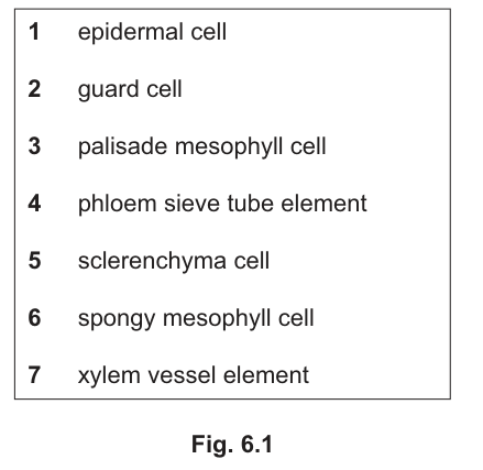
Match the correct type of cell from the list in Fig. 6.1 with each statement, A to E.
Each cell type can be used once, more than once, or not at all.
The first match has been done for you.
A This is a thick-walled cell that provides support. ______ 5
B This cell is one of a pair of cells that form a stoma. ______
C This cell receives water to build up hydrostatic pressure for mass flow. ______
D This cell needs water for photosynthesis and is columnar-shaped. ______
E This cell secretes a waxy substance to help prevent water loss. ______
Answer
A 5 (already given) — sclerenchyma cell
B 2 — guard cell
C 4 — phloem sieve tube element
D 3 — palisade mesophyll cell
E 1 — epidermal cell
B = 2, C = 4, D = 3, E = 1
Background Concept
A typical dicotyledonous leaf is built from a small set of specialised cell types, each with features that suit its function. Recall the seven types in Fig. 6.1:
- Epidermal cell (1): flat, irregularly shaped cells forming the outer protective skin of the leaf; the upper epidermal cells secrete the cuticle (waxy layer) that reduces water loss.
- Guard cell (2): kidney-shaped cells paired around a pore (the stoma); their unevenly thickened walls open and close the stoma to control gas exchange and transpiration.
- Palisade mesophyll cell (3): tall, columnar cells packed with chloroplasts just below the upper epidermis; the main site of photosynthesis and therefore the main user of water for this purpose.
- Phloem sieve tube element (4): a living but enucleate cell joined end-to-end to form sieve tubes; companion cells load sucrose into them, and water follows osmotically, building up the hydrostatic (turgor) pressure that drives mass flow.
- Sclerenchyma cell (5): a dead cell at maturity with a thick, lignified secondary cell wall — provides mechanical support (often around vascular bundles).
- Spongy mesophyll cell (6): irregularly shaped, loosely packed cells with large air spaces between them; allow gas diffusion between the stomata and the palisade layer.
- Xylem vessel element (7): a dead, hollow, lignified tube element; carries water and mineral ions up the plant under tension (transpiration pull).
Understanding the Question
You are given five descriptions (A–E) and a numbered list of seven cell types. A is pre-filled (sclerenchyma = 5). For each remaining description (B–E), pick the cell type whose defining structural/functional feature matches. The list says cells can be used once, more than once, or not at all, so guard against assuming each is used only once.
Approach
Read each statement, identify the key function or feature it describes, then scan the list for the cell whose definition most closely fits. Useful key words to listen for: stoma → guard cell; mass flow / hydrostatic pressure / sucrose loading → sieve tube element; columnar / photosynthesis → palisade mesophyll; waxy / cuticle → epidermal cell; thick wall / support → sclerenchyma.
Step-by-Step Reasoning
A — "thick-walled cell that provides support" → 5 (sclerenchyma cell). Already given. Sclerenchyma has a heavily lignified secondary wall and is dead at maturity; it gives rigidity, often around vascular bundles and at leaf margins.
B — "one of a pair of cells that form a stoma" → 2 (guard cell). Stomata are flanked by a pair of guard cells; their differential wall thickening and turgor changes open and close the pore.
C — "receives water to build up hydrostatic pressure for mass flow" → 4 (phloem sieve tube element). Loading of sucrose into the sieve tube lowers its water potential, water enters by osmosis, and the resulting high hydrostatic (turgor) pressure drives mass flow from source to sink.
D — "needs water for photosynthesis and is columnar-shaped" → 3 (palisade mesophyll cell). Palisade cells are characteristically columnar and densely packed with chloroplasts — the principal photosynthetic cells, with water as the electron donor in the light-dependent reactions.
E — "secretes a waxy substance to help prevent water loss" → 1 (epidermal cell). The epidermal cells of the upper (and lower) surface secrete cutin, forming the waxy cuticle that is the leaf's first defence against uncontrolled evaporation.
Key Takeaways
- Each cell type in a leaf is recognisable by both a structural feature (shape, wall, contents) and a functional consequence (support, transport, photosynthesis, gas exchange, water-proofing).
- The sieve tube's role in generating hydrostatic pressure is the basis of the mass-flow hypothesis for phloem transport.
- Palisade cells are columnar and chloroplast-rich specifically to maximise light absorption near the upper surface.
- Guard cells are the only cells in the epidermis that contain chloroplasts — a useful diagnostic if a question asks how to recognise them.
Common Mistakes
- C: choosing xylem vessel element (7) instead of phloem sieve tube element (4). Xylem transports water under tension (negative pressure); phloem is the one that builds up positive hydrostatic pressure to drive mass flow of assimilates. Mass flow of assimilates = phloem.
- D: choosing spongy mesophyll (6) instead of palisade (3). Spongy cells are rounded/irregular and not the main photosynthetic cells; the columnar, chloroplast-rich cells are the palisade layer.
- E: choosing guard cell (2) instead of epidermal cell (1). Guard cells surround the stoma but do not secrete the cuticle — that is the job of the ordinary pavement epidermal cells.
- Assuming each cell type is used exactly once. The question's stem says they may be used more than once or not at all — useful to check on past-paper questions of this type.
Things to Be Careful About
- The marking scheme for this part is one mark per correct match; there is no partial credit for a near-miss. Do not write two possibilities for the same letter.
- The wording of the stem (e.g. hydrostatic pressure for mass flow) is chosen to be unambiguous — it points directly to phloem, not xylem. Re-read the stem carefully before matching.
- The cuticle is secreted by all epidermal cells, not just those of the upper surface, so "epidermal cell" is the right general answer even though the bulk of cuticle is on the upper side.