Biology 9700/23 — October/November 2025
Cambridge AS Level · AS Level Structured Questions · worked solutions for every part, with the mark scheme
Topics Cell Structure · Nucleic Acids and Protein Synthesis · Enzymes · Cell Membranes and Transport · Transport in Plants · Biological Molecules · +4 more
Many substances can move through cell surface membranes between the cytoplasm of animal cells and the extracellular environment.
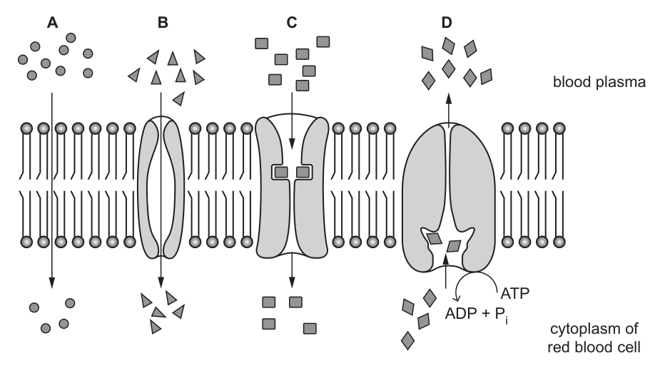
A student made a drawing to summarise the movement of substances across the cell surface membranes of mammalian red blood cells.
Fig. 1.1 shows the drawing made by the student:
- Each arrow indicates the movement of a substance through the membrane.
- The number of each of the 4 shapes represents the relative concentrations of each substance in the cytoplasm and in the blood plasma.
The student carried out research and made a list of some of the substances found in red blood cells as shown in Fig. 1.2.
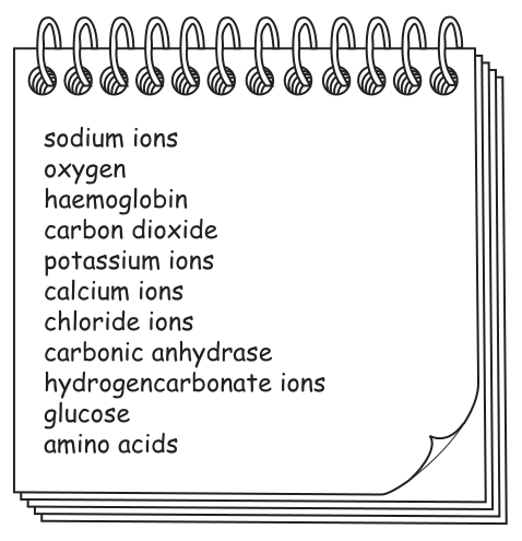
Table 1.1 shows information about the 4 types of movement of substances across the cell surface membranes of red blood cells as shown in Fig. 1.1.
Complete Table 1.1 using the information in Fig. 1.1 and Fig. 1.2.
Table 1.1
| letter from Fig. 1.1 | type of movement | name of part of membrane involved | example of a substance that moves across the membrane (from Fig. 1.2) |
|---|---|---|---|
| A | simple diffusion | phospholipids | |
| B | facilitated diffusion | calcium ions | |
| C | facilitated diffusion | ||
| D |
Answer
Completed Table 1.1:
| letter | type of movement | name of part of membrane involved | example of substance |
|---|---|---|---|
| A | simple diffusion | phospholipids | oxygen (or carbon dioxide) |
| B | facilitated diffusion | channel protein | calcium ions |
| C | facilitated diffusion | carrier protein | glucose |
| D | active transport | carrier (pump) protein | sodium ions |
See table; key entries: A = oxygen/CO2; B = channel protein; C = carrier protein and glucose; D = active transport, carrier protein, sodium ions.
Background Concept
The cell surface membrane is a fluid phospholipid bilayer in which proteins are embedded (the fluid mosaic model). Substances cross the membrane by different mechanisms depending on their size, polarity, charge, and the direction in which they need to move relative to their concentration gradient.
The four mechanisms in this question are:
- Simple diffusion – small, non-polar molecules (e.g. , ) pass directly between the phospholipid tails, down their concentration gradient, without the involvement of membrane proteins. No ATP is required.
- Facilitated diffusion via a channel protein – small charged ions (e.g. , , , ) pass through a water-filled protein pore, down their electrochemical gradient. The protein does not change shape.
- Facilitated diffusion via a carrier protein – larger or polar molecules (e.g. glucose, amino acids) bind to a carrier protein that changes shape to move them across the membrane, down their concentration gradient.
- Active transport – a carrier (pump) protein moves substances against their concentration gradient. This requires energy from the hydrolysis of ATP (ATP → ADP + ). A well-known example is the sodium–potassium pump, but sodium ions alone moving outward against their gradient is also valid.
Understanding the Question
Fig. 1.1 shows four labelled transport processes (A–D) across a red blood cell membrane. Fig. 1.2 lists substances found in (or moving across) red blood cells. The candidate must complete the missing cells in Table 1.1 by drawing on the information in both figures and knowledge of which substances use which transport route.
The table already has: the type of movement for A and B, the membrane component for A, the substance for B (calcium ions). The student must complete the rest.
Approach
Read Fig. 1.1 carefully to identify what each letter is showing:
- A – arrows go straight through the bilayer (no protein involved); molecules are small circles, with more in the plasma than in the cytoplasm → simple diffusion through phospholipids.
- B – arrows pass through a tube-like protein, with a clear central pore, molecules are small and triangular; facilitated diffusion through a channel protein; substance already given as calcium ions.
- C – arrows pass through a protein that encloses the molecule (no open pore); facilitated diffusion through a carrier protein.
- D – arrow points from the cytoplasm to the plasma, against the concentration gradient (more molecules inside than outside), and ATP is shown being hydrolysed to ADP + → active transport through a carrier/pump protein.
Then select, from the Fig. 1.2 list, the substance that fits each mechanism for a red blood cell.
Step-by-Step Reasoning
- Letter A (simple diffusion through phospholipids). The substance must be small and able to dissolve in the phospholipid bilayer. From the list, oxygen and carbon dioxide fit. (Both are accepted.) The mark scheme allows either.
- Letter B (facilitated diffusion). The membrane part is a channel protein – it forms a pore. Calcium ions are already given; they diffuse inward through the channel down their electrochemical gradient. (1 mark for "channel protein".)
- Letter C (facilitated diffusion). The protein shown has no open pore, so it must be a carrier protein. The substance is one that is too large/polar to diffuse through a channel or the bilayer. Glucose is the standard example; amino acids, chloride, potassium, and sodium ions are also acceptable by the mark scheme. (1 mark for "carrier protein" and 1 mark for the substance, usually glucose.)
- Letter D (active transport). Type of movement is active transport (movement against the gradient, using ATP). The membrane part is a carrier (pump) protein. From the list, sodium ions are the textbook example actively pumped out of red blood cells; potassium ions, chloride ions and amino acids are also acceptable. (1 mark for "active transport"; 1 mark combined for the membrane part and substance.)
Key Takeaways
- Simple diffusion moves small, non-polar molecules directly through the phospholipid bilayer; no protein, no ATP.
- Facilitated diffusion uses either a channel protein (small ions) or a carrier protein (larger polar molecules) and still requires a downhill gradient; no ATP.
- Active transport uses a carrier/pump protein, ATP, and moves substances against their concentration gradient.
- In red blood cells, oxygen and carbon dioxide cross by simple diffusion; calcium ions diffuse through a channel; glucose diffuses via a carrier protein; sodium ions are pumped out by active transport.
Common Mistakes
- Putting haemoglobin or carbonic anhydrase as the substance for any row – these are large cytoplasmic proteins and do not cross the cell surface membrane.
- Confusing a channel with a carrier protein – the mark scheme gives these as a single paired point, so getting only one of them right loses a mark.
- Stating "sodium–potassium pump" alone without identifying the actual substance (sodium or potassium ions) – this is rejected by the mark scheme.
- Giving the wrong type of movement for D, e.g. "facilitated diffusion" – the ATP symbol and the upward arrow against the gradient unambiguously indicate active transport.
- Putting glucose in for D, since active transport of glucose occurs in some cells (e.g. intestinal epithelium) – but the red blood cell uses facilitated diffusion for glucose; the only active transport shown should match a substance that genuinely is pumped in/out of red blood cells.
Things to Be Careful About
- The type of movement for D is given by both the direction of the arrow and the presence of ATP – either clue is sufficient to identify it as active transport.
- The mark scheme treats "B = channel protein" and "C = carrier protein" as a single paired point – but the C substance (glucose) is a separate mark.
- For D, "carrier/pump protein" and "sodium ions" together are credited; the mark scheme is happy with either potassium ions, chloride ions, or amino acids as an alternative substance.
- Avoid describing oxygen as moving by "osmosis" or by facilitated diffusion in a red blood cell – in red blood cells oxygen enters by simple diffusion.
Some viruses infect plants through the surfaces of damaged leaves. These plant viruses can travel from one leaf cell to another without having to pass through any cell surface membranes.
Explain how some plant viruses can travel from one cell to another without passing through cell surface membranes.
Answer
- Plant cells are connected to neighbouring cells by plasmodesmata, which are cytoplasmic strands that pass through the cell walls. (1 mark)
- These plasmodesmata allow substances (including viruses) to pass directly from the cytoplasm of one cell to the cytoplasm of the next, without crossing any cell surface membrane. (1 mark)
- This is the symplast pathway; viruses are small enough to pass through plasmodesmata.
Plasmodesmata provide direct cytoplasmic connections between adjacent plant cells, so viruses can move through them via the symplast pathway without crossing any cell surface membrane.
Background Concept
Plant cells are separated from one another by cell walls, but the walls are not continuous barriers between cells. Narrow cytoplasmic channels called plasmodesmata (singular: plasmodesma) pass through the cell walls, linking the cytoplasm of one cell directly to the cytoplasm of the next. In effect, almost all the living cytoplasm of a plant forms a single continuous network – the symplast.
The symplast pathway is one of two routes by which water and dissolved substances can move through plant tissues; the other is the apoplast pathway, which goes through the continuous mesh of cell walls and intercellular spaces without ever crossing a cell surface membrane. Movement of substances through plasmodesmata (symplast) is a third option that is especially relevant to cell-to-cell transport of solutes and signalling molecules, and is exploited by plant viruses.
Understanding the Question
This part tests knowledge of how plant cells are joined and how that arrangement allows materials (specifically, viruses) to move from cell to cell without ever having to cross a phospholipid bilayer. The question stem makes clear that these viruses infect plants through damaged leaves and then spread from cell to cell.
Approach
Identify the structure that allows direct cytoplasm-to-cytoplasm contact between adjacent plant cells, name it, and explain why viruses (which are very small) can fit through it.
Step-by-Step Reasoning
- Plasmodesmata are cytoplasmic connections between plant cells that pass through the cell walls. They provide a continuous channel of cytoplasm linking one cell to the next. (1 mark)
- Viruses are small enough to pass through plasmodesmata. Typical plant viruses such as Tobacco Mosaic Virus are about 15–18 nm in diameter, while the cytoplasmic channel of a plasmodesma is around 50 nm wide, so viral particles can move through. Movement through plasmodesmata via the symplast pathway means the virus moves from the cytoplasm of one cell to the cytoplasm of the next without ever crossing a cell surface membrane. (1 mark for the size argument, or for stating that the route is the symplast pathway.)
- AVP – the mark scheme allows a further mark for a quantitative size comparison or for additional correct detail about the symplast route.
Key Takeaways
- Plasmodesmata are the cytoplasmic channels that connect adjacent plant cells.
- Movement through plasmodesmata = the symplast pathway.
- Plant viruses exploit this pathway because the channels are wide enough (relative to viral size) to allow whole viral particles to pass between cells without ever crossing a phospholipid bilayer.
Common Mistakes
- Saying the viruses cross through the cell wall alone – the cell wall is fully permeable to small molecules, but the key point is that plasmodesmata link the cytoplasm of one cell to the next.
- Confusing the symplast and apoplast pathways – the apoplast is the network of cell walls and intercellular spaces (no membrane crossing required, but not direct cytoplasm-to-cytoplasm). The mark scheme ignores an apoplast reference unless it is correctly distinguished.
- Saying the virus "squeezes between the cells" or "through the cell wall" without mentioning plasmodesmata.
Things to Be Careful About
- The question is specifically about the structural reason viruses do not have to cross a membrane. The answer must mention the cytoplasmic connection (plasmodesmata), not just "they are small".
- The mark scheme ignores apoplast references that are not described correctly. A safe approach is to name the symplast pathway and the plasmodesmata, and to give the size argument as the third point.
Collagen is a fibrous protein found in many tissues in animals.
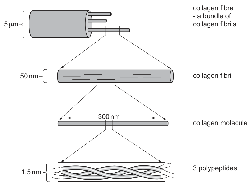
Fig. 2.1 shows the composition of a collagen fibre.
Answer
- Each polypeptide is a (left-handed) helix.
- The three polypeptides are tightly coiled/wound around each other to form a triple helix.
Three polypeptides, each a left-handed helix, are tightly wound together to form a triple helix.
Background Concept
Collagen is the most abundant fibrous protein in animals. Each collagen molecule (tropocollagen) is built from three polypeptide chains, called α-chains. Each α-chain folds into a left-handed helix — note this is NOT the right-handed α-helix found in many globular proteins. When three of these left-handed helices wind around a common central axis, they form a right-handed triple helix. The triple helix is stabilised by hydrogen bonds between the backbones of adjacent polypeptides, but bonding detail is not required for this part.
Understanding the Question
The question asks for a description (worth 2 marks) of how the three polypeptides are arranged in a single collagen molecule. Fig. 2.1 shows the triple helix at the bottom of the hierarchy (1.5 nm diameter), but the candidate must describe it in words, not just point to the figure.
Approach
Identify two distinct descriptive points about the structure: (1) the shape of each individual polypeptide, and (2) how the three fit together. The mark scheme accepts any two from three possible points, so a strong answer mentions both the helix and the triple helix.
Step-by-Step Reasoning
- Helical shape of each polypeptide. Each α-chain adopts a left-handed helical conformation. The mark scheme explicitly rejects "α-helix" — that is a different, right-handed structure found in globular proteins such as haemoglobin and myoglobin.
- Triple helix. The three polypeptides wind tightly around one another, producing a right-handed triple helix, also called tropocollagen.
- Optional third point. The polypeptides can be described as "coiled" or "wound" around each other if the candidate needs an alternative wording.
Any two of these three points earn the 2 marks. A complete answer gives both the helix and the triple helix.
Key Takeaways
- Each collagen polypeptide is a left-handed helix (NOT an α-helix).
- Three such helices wind together to form a right-handed triple helix.
- The handedness distinction matters: the α-helix of globular proteins is right-handed, while collagen's individual helix is left-handed.
Common Mistakes
- Writing "α-helix" — the mark scheme rejects this; collagen is not α-helical.
- Describing the helix without specifying the triple-helix arrangement.
- Including hydrogen-bonding details (these are ignored for part (a)(i) and belong in (a)(ii) or (c)(ii)).
Things to Be Careful About
- "Left-handed" is the correct handedness for each individual polypeptide chain.
- The triple helix as a whole is right-handed, but the candidate is being asked about the 3 polypeptides themselves, so focus on the coiling/triple-helix arrangement.
- The mark scheme says "I bonding" — ignore any mention of hydrogen bonds here.
With reference to Fig. 2.1, explain how the molecules of collagen are arranged and held together in a collagen fibril.
Answer
- Collagen molecules are arranged in parallel.
- The molecules are staggered (their ends are not aligned with each other).
- The molecules are held together by covalent bonds between the R-groups of amino acids on different molecules.
Parallel, staggered molecules held together by covalent cross-links between R-groups on adjacent molecules.
Background Concept
Collagen fibrils are formed by many tropocollagen molecules packed together in a regular pattern. To maximise mechanical strength, the molecules lie parallel to one another and are offset (staggered) so that weak points along one molecule are not aligned with weak points in the next. The fibril is held together by covalent cross-links that form between the R-groups (side chains) of specific amino acid residues, particularly lysine and hydroxylysine. These covalent bonds give collagen its very high tensile strength.
Understanding the Question
This is a "describe/explain" question worth 3 marks. The phrase "with reference to Fig. 2.1" tells the candidate to describe what the figure shows — the staggered, parallel molecules. The answer has two halves: arrangement and bonding.
Approach
Split the answer cleanly: first describe the arrangement (parallel + staggered), then describe the bonding (covalent, between R-groups, on different molecules). The arrangement points can be read straight off the figure, where the 300 nm molecules are drawn in parallel rows with offset ends.
Step-by-Step Reasoning
Arrangement (1 mark each):
- Parallel. The molecules lie alongside each other, all pointing in the same direction. This allows them to share load along their length.
- Staggered. The molecules are offset — the end of one molecule is not at the same point as the end of its neighbour. This staggering prevents all the weak "ends" from lining up and creating a fracture plane.
Bonding (max 2 marks; only 1 of these is needed for the third mark):
3. Covalent bonds/links. The molecules are held together by covalent cross-links. Note: hydrogen bonds are explicitly IGNORED by the mark scheme for this part.
4. Between R-groups. The covalent bonds form between the R-groups (side chains) of amino acids on DIFFERENT molecules. The mark scheme rejects answers that list other bond types alongside covalent bonds.
5. AVP. A valid alternative point is that the covalent bonds form specifically between lysine or hydroxylysine residues.
A complete 3-mark answer is: parallel + staggered + (covalent bonds OR between R-groups OR AVP).
Key Takeaways
- Collagen molecules in a fibril are arranged in parallel and staggered.
- Covalent cross-links form between the R-groups of amino acids on adjacent molecules.
- These cross-links often involve lysine or hydroxylysine residues.
- Hydrogen bonds are ignored for this part of the mark scheme.
Common Mistakes
- Mentioning hydrogen bonds (rejected for this part).
- Listing multiple bond types together, e.g., "covalent and hydrogen bonds" (rejected — covalent only).
- Saying the molecules are "side by side" without specifying parallel/staggered.
- Failing to specify that the bonds are between R-groups on different molecules.
Things to Be Careful About
- The mark scheme says "I hydrogen bonds" — do not credit hydrogen bonds as the holding bond here.
- "Between R-groups of amino acids on different molecules" is the key phrase; the cross-links are inter-molecular, not within one molecule.
- The bonds form between amino acid SIDE CHAINS (R-groups), not the polypeptide backbone.
Collagen fibres and elastic fibres are found in the structures of the gas exchange system of mammals.
Suggest two properties of collagen that contribute to the function of cartilage in the trachea in the gas exchange system.
Answer
- Provides (high tensile) strength to keep the trachea open / prevent it from collapsing.
- Allows flexibility for breathing movements and bending of the trachea.
Provides tensile strength to keep the trachea open; allows flexibility for breathing movements and bending.
Background Concept
The trachea (windpipe) is a tube that carries air between the larynx and the bronchi. It is supported by C-shaped rings of hyaline cartilage, which prevent the trachea from collapsing during the pressure changes of breathing. Hyaline cartilage is a connective tissue whose matrix is rich in type II collagen fibres embedded in a gel of proteoglycans and water. Collagen gives the cartilage its mechanical strength, while the proteoglycan matrix gives it resilience. The trachea must also flex with movements of the neck and change diameter slightly during breathing, so the cartilage must be both strong and flexible.
Understanding the Question
This is a "suggest" question worth 2 marks, asking the candidate to identify two properties of collagen that suit it to its role in tracheal cartilage. The candidate must link the molecular property to the physiological function in the gas exchange system.
Approach
Think about what the trachea needs from its cartilage support: (1) it must stay open against the negative pressure of inhalation and the positive pressure of exhalation — this needs strength; and (2) it must allow the neck to bend and the trachea to change diameter as the lungs expand — this needs flexibility. Both of these are properties of collagen.
Step-by-Step Reasoning
- Strength. Collagen has very high tensile strength because of the covalent cross-links between R-groups of amino acids on adjacent molecules (covered in part (a)(ii)). This strength keeps the trachea open and prevents it from collapsing when air pressure inside changes during breathing.
- Flexibility. Despite its strength, the collagen molecules can slide slightly past one another, giving the cartilage some flexibility. This allows the trachea to bend with movements of the neck and to change in diameter during breathing.
Both points each earn 1 mark.
Key Takeaways
- Collagen's covalent cross-links give it high tensile strength.
- Collagen's structure also allows some flexibility.
- These two properties together suit collagen for its role in tracheal cartilage — strong enough to stay open, flexible enough to allow breathing and bending.
Common Mistakes
- Saying "strong" without specifying "tensile" strength.
- Confusing collagen's role (strength) with elastic fibres' role (recoil).
- Saying the cartilage is "rigid" — it is not; it must be flexible.
- Failing to link the property to the function in the trachea.
Things to Be Careful About
- The mark scheme accepts "strength to keep trachea open" and similar phrasings.
- "Flexibility" must be linked to a specific function (breathing, bending, change in diameter).
- This question requires applying knowledge of collagen to a new context — the candidate should think about what the trachea needs, not just list properties of collagen.
Answer
Elastic fibres recoil to help expel air during exhalation (and allow stretching of the alveoli during inhalation without rupture).
Recoil to help expel air during exhalation; allow stretching without rupture during inhalation.
Background Concept
Elastic fibres are made mainly of the protein elastin (with fibrillin scaffolding). Elastin can be stretched considerably and then recoil passively to its original length. In the lungs, elastic fibres are found in the walls of the alveoli and the bronchioles, where they provide the elastic recoil that is essential for efficient breathing.
Understanding the Question
This is a "state" question worth 1 mark. The candidate needs to give the function of elastic fibres in the alveoli.
Approach
Think about what happens to the alveoli during the breathing cycle: during inhalation, the alveoli expand as the lungs fill with air; during exhalation, the lungs recoil to push air out. Elastic fibres are responsible for the recoil that drives exhalation.
Step-by-Step Reasoning
The mark scheme accepts two equivalent answers:
- Recoil during exhalation. Elastic fibres stretch during inhalation and then recoil during exhalation, helping to expel air from the lungs. This is the more commonly expected answer.
- Stretching without rupture. During inhalation, elastic fibres allow the alveoli to stretch considerably without rupturing, accommodating the increased volume of air.
Either answer earns the 1 mark.
Key Takeaways
- Elastic fibres recoil during exhalation, helping to expel air.
- They also allow the alveoli to stretch during inhalation without rupturing.
- The passive recoil of elastic fibres is what makes exhalation an energy-efficient process.
Common Mistakes
- Saying "to empty the lungs" — the mark scheme rejects this wording as too vague.
- Saying "to help exhalation" without specifying the recoil mechanism.
- Confusing elastic fibres with collagen (which provides strength, not recoil).
Things to Be Careful About
- "Recoil" is the key technical word — make sure to use it.
- Either the stretching function or the recoil function earns the mark.
- The question asks for the function in the ALVEOLI specifically, but the same function applies to elastic fibres throughout the lung tissue.
Table 2.1 shows the DNA triplets in the two strands of DNA in part of a gene that codes for one of the polypeptides in collagen.
Table 2.1
| non-transcribed strand | GGT | CCA | ATG | GGT | CCC | CGA | GGT | CCC | CCA | GGT |
| template strand | CCA | GGT | TAC | CCA | GGG | GCT | CCA | GGG | GGT | CCA |
| amino acid | gly |
Table 2.2 shows the triplets of bases in DNA and the amino acids for which they code.
The table can be used to determine the sequence of the amino acids in a polypeptide.
Table 2.2
| first base | second base: T | second base: C | second base: A | second base: G | third base |
|---|---|---|---|---|---|
| T | TTT phe TTC phe TTA leu TTG leu | TCT ser TCC ser TCA ser TCG ser | TAT tyr TAC tyr TAA stop TAG stop | TGT cys TGC cys TGA stop TGG trp | T C A G |
| C | CTT leu CTC leu CTA leu CTG leu | CCT pro CCC pro CCA pro CCG pro | CAT his CAC his CAA gln CAG gln | CGT arg CGC arg CGA arg CGG arg | T C A G |
| A | ATT ile ATC ile ATA ile ATG met | ACT thr ACC thr ACA thr ACG thr | AAT asn AAC asn AAA lys AAG lys | AGT ser AGC ser AGA arg AGG arg | T C A G |
| G | GTT val GTC val GTA val GTG val | GCT ala GCC ala GCA ala GCG ala | GAT asp GAC asp GAA glu GAG glu | GGT gly GGC gly GGA gly GGG gly | T C A G |
Complete Table 2.1 to show the amino acids coded by the DNA nucleotide sequence in Table 2.1.
Answer
(gly)-pro-met-gly-pro-arg-gly-pro-pro-gly
gly-pro-met-gly-pro-arg-gly-pro-pro-gly
Background Concept
The genetic code is read in triplets called codons. Each codon in mRNA specifies one amino acid (or a stop signal). The non-transcribed (sense/coding) strand of DNA has the same sequence as mRNA except that T in DNA is replaced by U in mRNA. So to read the amino acid sequence from a DNA sequence, the candidate can either use the non-transcribed strand directly (treating T as if it were U), or take the template strand, find its mRNA complement, and read the codons. The simpler method is to use the non-transcribed strand.
The codon table provided (Table 2.2) is organised by the first, second, and third bases of each triplet. It is a DNA codon table, so the codons match the DNA sequence directly (no need to convert to mRNA).
Understanding the Question
The question gives the non-transcribed strand sequence and asks the candidate to fill in the 9 missing amino acids. The first amino acid (gly) is given as a hint. The full sequence is 10 amino acids long.
Approach
Read each triplet of the non-transcribed strand and look it up in the codon table. Convert to the three-letter abbreviation of the amino acid.
Step-by-Step Reasoning
Reading the non-transcribed strand triplets from Table 2.1:
| Triplet | Amino acid |
|---|---|
| GGT | gly (given) |
| CCA | pro |
| ATG | met |
| GGT | gly |
| CCC | pro |
| CGA | arg |
| GGT | gly |
| CCC | pro |
| CCA | pro |
| GGT | gly |
So the complete sequence is: gly-pro-met-gly-pro-arg-gly-pro-pro-gly.
The pattern shows glycine every third amino acid (positions 1, 4, 7, 10), which is characteristic of collagen and is discussed in part (c)(ii).
Key Takeaways
- The non-transcribed strand has the same sequence as mRNA (with T instead of U).
- DNA codon tables can be read directly from the non-transcribed strand.
- GGN codes for glycine, CCN codes for proline, ATG codes for methionine, CGN codes for arginine.
Common Mistakes
- Reading the template strand instead of the non-transcribed strand, which would give the complementary codons.
- Confusing the row and column headings of the codon table.
- Misreading the third base (e.g., GGT vs GGC vs GGA vs GGG — all code for glycine, but other triplets are more sensitive to the third base).
- Using the wrong strand for a specific triplet when the table has overlapping information.
Things to Be Careful About
- The codon table given in the question is a DNA codon table — read it directly with T, not U.
- The third base matters: CCA and CCC both code for proline, but the candidate must use the correct one for each position.
- ATG codes for methionine (met) — this is also the start codon, but in this context it is just an internal methionine.
The sequence of amino acids that you have worked out is representative of the whole of the collagen polypeptide.
Explain how the sequence of amino acids makes the polypeptide suitable as a component of a collagen molecule.
Answer
- Glycine is every third amino acid in the sequence.
- Glycine has the smallest R-group (a single hydrogen atom).
- The small R-group allows the three polypeptides to pack tightly together, and the NH group of glycine can form many hydrogen bonds with adjacent amino acids, stabilising the triple helix.
Every-third-glycine has the smallest R-group, allowing tight packing and many H-bonds between the three polypeptides of the collagen molecule.
Background Concept
The primary structure of a protein is its sequence of amino acids. This sequence determines how the polypeptide folds and how it interacts with other polypeptides. Collagen has a very distinctive primary structure: every third amino acid is glycine, and the other two positions are often occupied by proline or other small amino acids. This repeating pattern is what allows the three polypeptides to wind together into a tight, regular triple helix.
Glycine is unique among the 20 standard amino acids because its R-group is just a single hydrogen atom. This makes it the smallest amino acid by far. In a triple helix, the three polypeptide backbones come together very tightly at every third residue; only a hydrogen-sized R-group can fit into the crowded centre of the helix without disrupting the structure.
Understanding the Question
This is an "explain" question worth 3 marks. The candidate must explain how the amino acid sequence (with glycine every third position) makes the polypeptide suitable as a component of a collagen molecule. The answer requires linking the primary structure to the higher-level structure of collagen.
Approach
Break the answer into three parts: (1) describe the pattern (glycine every third), (2) state the key property of glycine (smallest R-group), and (3) explain the consequence for the collagen molecule (tight packing + hydrogen bonds stabilising the triple helix).
Step-by-Step Reasoning
- Glycine is every third amino acid. Looking at the sequence gly-X-Y-gly-X-Y-gly-X-Y-gly, the pattern is obvious: glycine occupies every third position. This regular pattern is characteristic of collagen.
- Glycine has the smallest R-group. Glycine's side chain is just a hydrogen atom (H), making it by far the smallest of the 20 amino acids. The mark scheme accepts "smallest R-group", "smallest amino acid", or "H as R-group" as equivalents.
- Allows tight folding / close fit. Because glycine is so small, the three polypeptides can be wound tightly together without their R-groups clashing. The mark scheme accepts "tight folding", "fit closely together", or "compactness".
- Hydrogen bonding. The NH group of glycine (in the peptide backbone) can form hydrogen bonds with the carboxyl group (C=O) of an amino acid residue on an adjacent polypeptide. Many such hydrogen bonds form between the three polypeptides, stabilising the triple helix.
- AVP. Alanine and proline also have relatively small R-groups and contribute to the tight packing.
Any three of these points earn the 3 marks. A common full-mark answer covers points 1, 2/3 (combined), and 4/5 (combined).
Key Takeaways
- Glycine every third amino acid is the signature of collagen.
- Glycine's tiny R-group (just H) is essential for the tight triple-helix structure.
- The NH of glycine forms many H-bonds with adjacent polypeptides, stabilising the molecule.
- This is a classic example of how primary structure determines higher-level structure.
Common Mistakes
- Saying glycine is "small" without specifying "smallest".
- Forgetting to mention hydrogen bonds at all.
- Saying glycine "bonds" without specifying hydrogen bonds.
- Stating the pattern without explaining WHY it matters (the "suitability" aspect).
- Confusing glycine with alanine (alanine has a methyl group, not just H).
Things to Be Careful About
- The mark scheme accepts "compactness" as an alternative to "tight folding".
- The hydrogen-bond detail is a separate point from the "smallest R-group" point — the candidate should mention both.
- The question is about why the SEQUENCE is suitable, so the candidate must mention the repeating pattern (every third = glycine) and the property of glycine.
Two mutations, P and Q, can have an effect on the primary structure of the polypeptide.
- Mutation P is a deletion of the first nucleotide pair in the DNA nucleotide sequence shown in Table 2.1.
- Mutation Q is a substitution of G with T as the first base in the DNA nucleotide sequence shown in Table 2.1.
State the effects of the mutations, P and Q, on the primary structure of the polypeptide.
mutation P ______
mutation Q ______
Answer
Mutation P (deletion of first nucleotide pair):
- Changes the sequence/order of amino acids in the primary structure (a frameshift — all subsequent amino acids are different).
Mutation Q (substitution of G with T as the first base):
- The first amino acid changes from glycine to cysteine; the rest of the sequence is unchanged.
P: changes the sequence/order of amino acids (frameshift). Q: first amino acid changes (gly to cys), rest of the sequence is unchanged.
Background Concept
Gene mutations change the DNA base sequence and can alter the primary structure of the protein produced. There are several types:
- Substitution — one nucleotide pair is replaced by another. This usually affects only ONE codon, so at most one amino acid is changed (or a stop codon may be introduced, truncating the protein).
- Deletion — one or more nucleotide pairs are removed. This causes a FRAMESHIFT: all subsequent codons are read in a different reading frame, so all subsequent amino acids are altered.
- Insertion — one or more nucleotide pairs are added. Similar effect to deletion — a frameshift.
The genetic code is read in non-overlapping triplets starting from a fixed point (usually AUG / methionine). A deletion anywhere except at the very end of a complete codon causes the reading frame to shift.
Understanding the Question
This is a "state" question worth 2 marks (1 mark per mutation). The candidate must describe the effect of each mutation on the primary structure of the polypeptide.
Approach
For each mutation, work out the new DNA sequence, read the new codons, and compare the new amino acid sequence to the original (gly-pro-met-gly-pro-arg-gly-pro-pro-gly).
Step-by-Step Reasoning
Mutation P — deletion of the first nucleotide pair:
- Original non-transcribed strand: GGT CCA ATG GGT CCC CGA GGT CCC CCA GGT
- After deletion of the first G: G_T CCA ATG GGT CCC CGA GGT CCC CCA GTT (shifted by one base)
- Reading in triplets from the new start: GTC CAA TGG GTC CCC GAG GTC CCC AGT T…
- All codons after the first one are in a different reading frame.
- The first amino acid changes (from gly to val), and ALL subsequent amino acids are different.
- Mark scheme answer: "changes the sequence / order, of amino acids (in primary structure)".
- Note: the mark scheme says "I shorter" — do NOT credit "the polypeptide is shorter" as the main effect, even though it may be true if a stop codon appears later.
Mutation Q — substitution of G with T as the first base:
- Original non-transcribed strand: GGT CCA ATG GGT CCC CGA GGT CCC CCA GGT
- After substitution: TGT CCA ATG GGT CCC CGA GGT CCC CCA GGT
- First triplet changes: GGT → TGT. From the codon table, GGT codes for gly, and TGT codes for cys.
- Subsequent triplets (CCA, ATG, etc.) are unchanged.
- So: the first amino acid changes from gly to cys; the rest of the sequence is unchanged.
- Mark scheme answer: "cys (instead of gly) is first amino acid, and no change to the rest (of sequence / order, of amino acids)".
Key Takeaways
- A deletion causes a frameshift — the entire downstream amino acid sequence is altered.
- A substitution affects only the codon in which it occurs (and only the amino acid if the new codon codes for a different one).
- The mark scheme for mutation P ignores the answer "shorter" — the focus is on the change in sequence, not the length.
- For mutation Q, both parts of the answer are needed: which amino acid changes AND that the rest is unchanged.
Common Mistakes
- Saying the polypeptide is "shorter" for mutation P (rejected by the mark scheme — "I shorter").
- Saying "no change" for mutation Q (incorrect — one amino acid does change).
- Failing to specify that mutation Q only affects the first amino acid.
- Confusing deletion with substitution — they have very different effects.
- Saying the protein is "longer" for mutation P (not the main effect and not always true).
Things to Be Careful About
- The mark scheme requires both halves of the mutation Q answer: which amino acid is changed AND that the rest is unchanged.
- For mutation P, the mark scheme explicitly ignores "shorter" — the candidate should focus on the change in sequence.
- The candidate should distinguish between primary structure (the answer the question asks for) and higher levels of structure.
- The question is about the polypeptide, so the candidate should not discuss the wider effect on the whole protein or on the organism.
Answer
- The active site of the enzyme is not exactly complementary to the substrate before binding (only partially complementary).
- As the substrate enters the active site, the shape of the active site changes (a conformational change), moulding around the substrate.
- The active site now has a shape complementary to the substrate, so an enzyme-substrate complex forms.
- After the products leave, the active site / enzyme returns to its original shape / conformation.
The active site and substrate are initially only partially complementary; when the substrate binds, the active site changes shape (a conformational change) and moulds around the substrate so that an enzyme-substrate complex forms; the active site then returns to its original shape after the products are released.
Background Concept
Enzymes are biological catalysts — globular proteins that speed up metabolic reactions without being used up. They work because of the specific three-dimensional shape of a region called the active site, where the substrate (the molecule being acted on) binds. The amino acid side chains in the active site form temporary bonds with the substrate, holding it in the correct orientation for the reaction to occur, and so the enzyme lowers the activation energy of the reaction — the energy barrier that has to be overcome for the reaction to proceed.
There are two classical models for how an enzyme recognises and binds its substrate:
- Lock-and-key hypothesis — the active site has a fixed shape that is already exactly complementary to the substrate before binding (like a key fitting a lock). This is now considered too rigid, because it cannot easily explain how some enzymes act on several related substrates or how allosteric regulation works.
- Induced-fit hypothesis — the active site is not exactly the right shape at first; it changes shape when the substrate binds, moulding itself around the substrate to achieve a better fit. This is the accepted modern model.
Understanding the Question
The command word is describe — give an ordered, factual account of the induced-fit model: the initial state, what changes when the substrate binds, and what happens after the products are released. The question is worth 3 marks, so 3 clear points are needed. Crucially, the mark scheme requires that the answer refer to shape or conformation at least once, otherwise the marks are capped at 2.
Approach
Tell the story of one substrate molecule entering the active site: (1) before binding, the fit is imperfect; (2) during binding, the active site physically changes shape; (3) this new shape gives a better fit and forms the enzyme–substrate complex; (4) afterwards the products leave and the active site returns to its original shape. Remember to use the words "shape" or "conformational change" — they are the keywords the examiner is looking for.
Step-by-Step Reasoning
- Point 1 — initial imperfect fit. Before the substrate arrives, the active site is not yet the right shape for the substrate. The mark scheme requires the idea that the active site and substrate are "only partially / not complementary".
- Point 2 — conformational change on binding. As the substrate enters, the enzyme changes shape around it. This is the central tenet of induced fit: the active site moulds itself around the substrate. The word "shape" or "conformational change" is essential here.
- Point 3 — good fit achieved / ES complex forms. The result of the conformational change is that the active site is now complementary to the substrate, so an enzyme–substrate (ES) complex forms. This can also be phrased as the active site achieving a "good fit" with the substrate.
- Point 4 — return to original shape. Once catalysis is complete, the products leave the active site and the enzyme returns to its original shape, ready to bind another substrate molecule. This reversible step shows that the enzyme is a true catalyst and is unchanged at the end of the reaction.
Key Takeaways
- The induced-fit hypothesis explains how the same enzyme can act on several similar substrates and how the active site is dynamic, not rigid.
- The conformational change is the key event: it creates the precise geometry needed to lower the activation energy.
- After catalysis, the enzyme is unchanged and can be reused.
- "Shape" and "conformation" are the exact words the mark scheme looks for — use them.
Common Mistakes
- Describing only the lock-and-key model. If the answer ignores the conformational change and treats the active site as a fixed shape, the mark scheme caps the marks at 2 (from MP4 onwards) because the answer is describing lock-and-key, not induced fit.
- Saying the substrate changes shape. The mark scheme explicitly rejects this — only the enzyme / active site changes shape; the substrate is the molecule being acted on.
- Omitting the words "shape" or "conformation". This caps the answer at 2 marks. Always include the keyword.
- Forgetting the recovery step. Failing to mention that the active site returns to its original shape loses a marking point and gives an incomplete picture of catalysis.
Things to Be Careful About
- "Active site" must be the subject of the shape change, not the substrate.
- The enzyme–substrate complex is a transient state — make clear that the products are released and the enzyme is unchanged afterwards.
- "Lowers the activation energy" is a separate mark-worthy idea; if you have space, mention it.
Many marine organisms can become attached to hard surfaces such as rocks or the surfaces of ships. These organisms are known as fouling organisms.
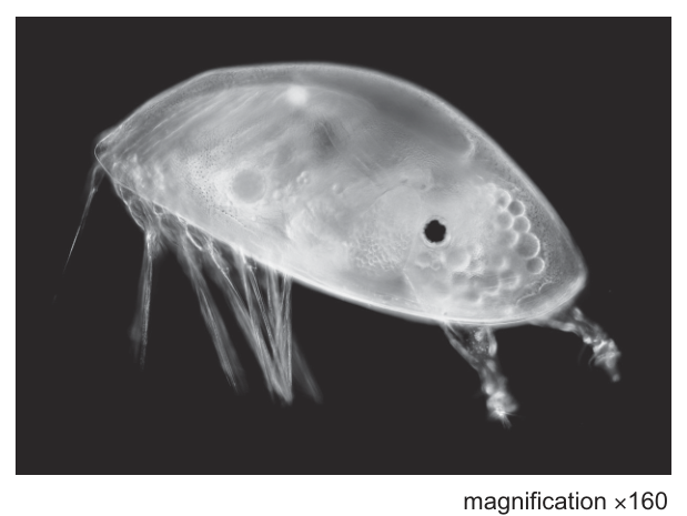
The larva of the acorn barnacle, Amphibalanus amphitrite, is an example of a fouling organism. One of these barnacle larvae is shown in Fig. 3.1.
The larvae of A. amphitrite use a protein to attach themselves to the surfaces of ships.
It is expensive to remove fouling organisms from ships. Scientists have developed substances to prevent the attachment of larvae. However, some of these substances are toxic and have been responsible for a decrease in marine biodiversity.
Scientists investigated the effect of using an immobilised protease, subtilisin A, to prevent the attachment of the larvae of A. amphitrite to surfaces.
The scientists used 4 different concentrations of subtilisin A which had been immobilised onto the surface of a polymer film. As a control they used denatured subtilisin A immobilised onto the surface of the same polymer. Glass slides were also used as a control.
The larvae were released into 6 tanks of artificial sea water:
- 4 tanks, each with a polymer surface and a different concentration of immobilised subtilisin A
- 1 tank with a polymer surface and denatured immobilised subtilisin A
- 1 tank with glass slides instead of a polymer surface.
The number of larvae that attached to the different surfaces in the tanks was counted after 24 hours and again after 48 hours.
The number of larvae attached in each tank was expressed as the percentage of the total number of larvae released in each tank. The results are shown in Fig. 3.2.
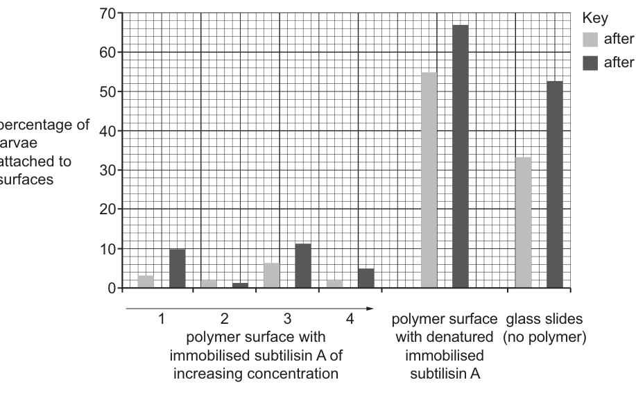
With reference to the data in Fig. 3.2, discuss whether subtilisin A is effective in preventing the attachment of the larvae.
Answer
- Subtilisin A is effective because the percentage of larvae attaching to polymer surfaces with immobilised subtilisin A (tanks 1–4) is much lower (all below ≈ 15 % at both 24 h and 48 h) than in the controls: the polymer with denatured subtilisin A (≈ 55 % at 24 h, ≈ 68 % at 48 h) and the glass slides (≈ 34 % at 24 h, ≈ 53 % at 48 h).
- However, it is not 100 % effective — some larvae still attach in all four experimental tanks.
- It generally becomes less effective at 48 h (e.g. tank 1 rises from ≈ 3 % to ≈ 10 %; tank 3 from ≈ 7 % to ≈ 12 %); only tank 2 becomes more effective over this time, falling from ≈ 2 % to ≈ 1 %.
- There is no clear trend of greater effectiveness with increasing subtilisin A concentration (e.g. tank 2 is the most effective at 48 h but is not the highest concentration).
- 48 hours is a short period of time, so long-term effectiveness is unknown, and no repeats were carried out, so the data may not be valid.
Subtilisin A reduces attachment to < 15 % compared with ≈ 34–68 % in the controls, but it is not 100 % effective, generally becomes less effective at 48 h (only tank 2 improves), shows no clear dose–response, and the data are limited by a short timescale and lack of repeats.
Background Concept
Fouling organisms (such as barnacle larvae) attach to ship hulls using adhesive proteins. Proteases are enzymes that hydrolyse (break) peptide bonds in proteins; if a protease can hydrolyse the adhesive protein, the larvae cannot attach. Immobilising the enzyme — fixing it to a polymer surface — keeps the enzyme localised, easy to recover, and reusable, and allows the breakdown products (small peptides and amino acids) to be washed away. This question evaluates whether this strategy actually works in practice, by reading percentage-attachment data from a bar chart.
Understanding the Question
The stem describes an experiment with six tanks: four with polymer surfaces coated in different concentrations of immobilised subtilisin A (the experimental treatments), one with denatured subtilisin A (control 1 — same polymer and immobilisation, but the enzyme is non-functional, so any difference is due to the enzyme's activity, not the polymer), and one with bare glass slides (control 2 — no polymer at all, the natural surface). The dependent variable is the percentage of larvae that attached after 24 h and 48 h, shown in Fig. 3.2.
The command word is discuss — you must weigh up both sides, presenting evidence FOR effectiveness AND evidence AGAINST it (or limitations), then reach a reasoned conclusion. The question is worth 4 marks, so you need 4 distinct, well-supported points. Note: the mark scheme says to ignore any comparison between the two controls — focus on experimental vs. control comparisons.
Approach
Read off the bar heights for each treatment at both time points, then:
- Compare the experimental bars (tanks 1–4) with the control bars — is there a clear difference? Quantify it.
- Look at how the experimental bars change with time — is effectiveness maintained or lost over 48 h?
- Look at how the experimental bars change with subtilisin A concentration — is there a dose–response?
- Identify limitations of the data (short timescale, no repeats, possible enzyme instability).
Step-by-Step Reasoning
- Evidence of effectiveness (point 1). At 48 h, attachment in the experimental tanks (tanks 1–4) is between ≈ 1 % and ≈ 12 %, while the denatured-enzyme control is ≈ 68 % and the glass control is ≈ 53 %. The difference is several-fold and consistent across all four experimental tanks. Subtilisin A is therefore reducing attachment compared with both controls.
- Evidence of incomplete effectiveness (point 2). Even in the best experimental tank (tank 2 at 48 h) ≈ 1 % of larvae still attach, and in the worst experimental tank (tank 3 at 48 h) ≈ 12 % attach. The treatment is not 100 % effective.
- Effect of time (points 3 and 4). In three of the four experimental tanks (1, 3, 4) the percentage attached is higher at 48 h than at 24 h, suggesting the treatment becomes less effective with time. Only tank 2 shows a decrease from 24 h to 48 h. The mark scheme explicitly looks for this observation.
- No concentration effect (point 5). There is no clear pattern: tank 2 (the second-lowest concentration) is the most effective at 48 h, while tank 3 (the third concentration) is the least effective. This makes it hard to choose an "optimal" concentration and is a weakness of the data.
- Limitations (points 6, 7). 48 hours is a short period for a ship's hull — fouling builds up over weeks or months, so we cannot tell whether the treatment would still work long-term. There are also no repeats, so the variability between tanks is unknown and the result may not be statistically reliable. The mark scheme also accepts the idea that the immobilised enzyme may not be perfectly stable — it could detach or denature over time, which fits with the time-related decrease in effectiveness.
Key Takeaways
- To discuss effectiveness you must use the data — quote specific numbers or trends, not vague generalities.
- A "good" experiment has controls, repeats and a sensible timescale; the absence of any of these is itself a valid point in a discussion.
- A dose–response (clear trend with concentration) is usually expected from an enzyme experiment; its absence here is a meaningful finding.
- "Discuss" demands balance: present both the supportive evidence and the limitations before reaching an overall conclusion.
Common Mistakes
- Only stating that it is effective. A one-sided answer misses half the marks. You must also discuss the limitations.
- Comparing the two controls against each other. The mark scheme explicitly ignores this — focus on the experimental vs. control comparison.
- Vague quantifiers. "Lower" or "less" are weaker than quoting ≈ 10 % vs ≈ 68 %.
- Saying it is "not effective". The data clearly show a substantial reduction, so the treatment is partially effective — discuss to what extent and under what conditions.
- Ignoring the data and talking about the biology of subtilisin A. The question says "with reference to the data" — your points must be tied to Fig. 3.2.
Things to Be Careful About
- Read the bar chart carefully: tank 2 at 48 h is the only tank where attachment decreases — most candidates miss this.
- Note that both controls are high and all four experimental bars are low — this contrast matters more than the precise numbers.
- "Short period of time" and "no repeats" are two separate mark-worthy points, not one.
- The comparison "tank 2 has lower attachment than tank 3" is a valid within-experiment comparison and earns a separate point.
The scientists extended their investigation by applying the polymer with immobilised subtilisin A to the outside of the bottom of small ships.
Two factors that need to be taken into consideration in this type of investigation are the temperature and pH of the sea water.
Outline two other factors that need to be taken into consideration when investigating the suitability of immobilised subtilisin A as an anti-fouling agent for ships.
Answer
- How long the immobilised subtilisin A remains active (stable) on the surface when exposed to sea water — i.e. whether the enzyme detaches, denatures, or loses activity over time.
- The effect of subtilisin A (or its products) on other (non-target) marine organisms — whether it is toxic to other species and therefore reduces marine biodiversity.
Stability / how long the immobilised enzyme remains active, and effect on other (non-target) marine organisms / marine biodiversity.
Background Concept
A useful experiment tests whether the treatment works under the actual conditions of use. The stem of part (b) already mentions that temperature and pH of the sea water must be controlled — these are the conditions that most strongly affect enzyme activity. But for a real anti-fouling coating on a ship's hull, several other practical and ecological factors matter:
- The coating must stay on the ship and remain active for a useful time — if the enzyme detaches or denatures in sea water the coating is useless.
- The coating must not harm other marine organisms — the stem notes that the toxic anti-fouling substances previously used caused a decrease in marine biodiversity. A new treatment that solved fouling but wiped out other species would be unacceptable.
The question is about extending the laboratory finding (effective in tanks for 48 h) to a real ship — what else needs to be considered to make that jump safely?
Understanding the Question
The command word is outline — give a brief, clear description of each factor without lengthy explanation. You need exactly two factors (worth 2 marks). The question says "other factors" — so you must avoid the ones already mentioned (temperature, pH). The mark scheme also ignores cost, so do not write about cost. The factors should be relevant to either the practical application (will it work on a ship?) or the wider impact (will it harm other organisms?).
Approach
Look at the mark-scheme list and pick the two factors that are most important and most clearly distinct from the ones already mentioned:
- Length of time the enzyme remains active / stable on the surface (MP 7) — practical, real-world durability.
- Effect on other marine organisms / biodiversity (MP 8) — ecological impact, directly addresses the biodiversity concern raised in the stem.
These two are independent of each other and between them cover the "will it work?" and "should it be used?" questions.
Step-by-Step Reasoning
- Factor 1: stability of the immobilised enzyme. A ship's hull is in sea water for months at a time. The data only cover 48 hours, and the trend in tanks 1, 3 and 4 already shows attachment rising by 48 h. The scientists need to find out whether the enzyme remains active for longer periods, and whether the immobilisation (attachment to the polymer) is stable in real sea water (e.g. does the polymer degrade? does the enzyme detach? does salt or marine growth inactivate it?). Without this, the treatment may be only briefly effective.
- Factor 2: effect on other marine organisms. The stem specifically warns that previous toxic anti-fouling agents reduced marine biodiversity. Subtilisin A is a protease — its products are small peptides and amino acids, which may be harmless, but the scientists cannot assume this. They should test whether subtilisin A (or its breakdown products) is toxic to other species, whether it harms organisms that graze on the polymer coating, and whether it is effective (and safe) against other fouling species besides A. amphitrite.
Other valid factors (e.g. type of polymer, way of fixing polymer to ship, salinity of sea water, presence of enzyme inhibitors in sea water, density/concentration of enzyme) could also be chosen, but the two above best match the mark-scheme points MP 7 and MP 8 and most directly address the context.
Key Takeaways
- When applying a laboratory result to a real-world context, consider both practical durability and wider ecological / safety impact.
- "Outline" needs only brief, point-form answers — do not write a paragraph for each.
- Always look for the keywords the examiner will credit: stability / activity over time, effect on other organisms, etc.
Common Mistakes
- Repeating temperature or pH. The question says "two OTHER factors" — the stem has already covered these.
- Writing about cost. The mark scheme explicitly ignores cost — do not waste a mark on it.
- Being too vague. "How well it works" is too vague to score; "how long the enzyme remains active" is specific and mark-worthy.
- Writing about factors already controlled in the lab experiment (e.g. number of larvae). The question is about extending the work to real ships, so the factors must be relevant to that step.
Things to Be Careful About
- Choose factors that are independent of temperature and pH (the stem excludes these).
- The mark scheme lists many acceptable factors; you only need two, but they must be specific and biology-relevant.
- Real-world ecological concerns (biodiversity, toxicity to non-target species) are particularly important in this question because the stem explicitly raises them.
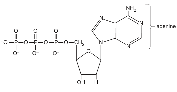
Fig. 4.1 shows a phosphorylated nucleotide which is one of the monomers that is used to synthesise DNA during replication.
Answer
Monomers are repeating units (sub-units / molecules) that join together to form a larger molecule called a polymer. In DNA, the monomers are the four nucleotides (containing the bases A, T, G and C).
Repeating units/sub-units/molecules (nucleotides) that join to form a polymer (polynucleotide / DNA strand).
Background Concept
A monomer is a small, repeating molecular unit that links with other identical or similar units to form a larger molecule called a polymer. In nucleic acids, the monomer is the nucleotide; many nucleotides linked by phosphodiester bonds form a polynucleotide strand (a single DNA or RNA chain). Each DNA nucleotide contains a deoxyribose sugar, a phosphate group, and one of four nitrogenous bases (A, T, G or C).
Understanding the Question
The stem of part (a) introduces Fig. 4.1, a phosphorylated nucleotide (dATP) that is a monomer used to build DNA during replication. Part (a)(i) is a straightforward definitional recall task, asking what is meant by the term 'monomers of DNA'.
Approach
The mark scheme credits any of: repeating / units / sub-units / molecules / nucleotides / used to make a polymer / polynucleotide / strand of DNA. A complete answer combines the idea of a repeating unit with the idea that it builds up a larger molecule.
Step-by-Step Reasoning
- A monomer must be a repeating sub-unit (the 'mono-' prefix indicates a single unit that occurs many times).
- The context is DNA, so the relevant monomers are the four DNA nucleotides (dAMP, dTMP, dGMP, dCMP).
- These monomers link together by phosphodiester bonds between the 3'-OH of one sugar and the 5'-phosphate of the next, producing a polynucleotide (a single DNA strand).
- Two such strands (antiparallel, complementary) then form the double helix.
Key Takeaways
- 'Monomer' means a small repeating sub-unit.
- The monomers of DNA are nucleotides (each containing deoxyribose, a phosphate and a nitrogenous base).
- Linking monomers together gives a polymer / polynucleotide (a single DNA strand).
Common Mistakes
- Saying only 'a molecule that joins together' — too vague; must include the idea of a repeating unit or a polymer product.
- Naming the components of a nucleotide (sugar, phosphate, base) instead of defining the term 'monomer'.
- Confusing monomer with 'macromolecule' or with 'nucleic acid' itself.
Things to Be Careful About
- Use the term 'repeating unit / sub-unit' to earn the mark; 'molecule' alone is acceptable on this mark scheme but 'repeating' is the key concept.
- Mention the polymer product (DNA strand / polynucleotide / polymer) to be fully credited.
State how ATP differs in structure from the phosphorylated nucleotide shown in Fig. 4.1.
Answer
In ATP the pentose sugar is ribose (with an –OH group on C2), whereas the nucleotide in Fig. 4.1 has deoxyribose (with an –H on C2).
(Pentose/sugar is) ribose (not deoxyribose) OR C2 of the pentose has –OH not –H.
Background Concept
ATP (adenosine 5'-triphosphate) and dATP (deoxyadenosine 5'-triphosphate) have almost identical structures: both have the nitrogenous base adenine attached to a pentose sugar, with three phosphate groups linked to the 5' carbon of the sugar. The single structural difference is the pentose sugar: ATP contains ribose, while dATP (the monomer in DNA replication, shown in Fig. 4.1) contains deoxyribose. Deoxyribose lacks the hydroxyl (–OH) group on the 2' carbon, having only a hydrogen (–H) there instead. This 2' difference gives the two sugars their names: ribose (with –OH) and deoxyribose ('de-oxy' = without oxygen on C2).
Understanding the Question
The stem of part (a) shows dATP, a phosphorylated deoxyribonucleotide. Part (a)(ii) asks the candidate to identify a single structural difference between ATP and the molecule shown. The mark scheme allows either naming the sugar (ribose vs deoxyribose) or specifying the change at C2.
Approach
Compare the two molecules' sugar components. The phosphate groups and adenine base are identical, so the only difference lies in the pentose sugar. State which sugar is in ATP and contrast it with the deoxyribose shown in Fig. 4.1.
Step-by-Step Reasoning
- Both ATP and the nucleotide in Fig. 4.1 contain: adenine, a pentose sugar and a chain of three phosphate groups attached to the 5' carbon of the sugar.
- The pentose in Fig. 4.1 (dATP) is deoxyribose — note the –OH on C3 and the –H on C2 shown on the diagram.
- The pentose in ATP is ribose, which has –OH on both C2 and C3.
- Therefore ATP and dATP differ only in the sugar: ATP has ribose while dATP has deoxyribose (or, equivalently, the 2' carbon carries an –OH in ATP but an –H in dATP).
Key Takeaways
- ATP and dATP are otherwise identical; the sugar is the only structural difference.
- Ribose has –OH at C2 and C3; deoxyribose has –OH only at C3 and –H at C2.
- This single –OH change explains why ATP is the energy currency of the cell while dNTPs are the building blocks of DNA.
Common Mistakes
- Stating that ATP has a different base (e.g. 'ATP has adenine but DNA nucleotides have different bases') — incorrect; both molecules shown have adenine here.
- Saying ATP has 'one' or 'two' phosphate groups — both molecules shown have three; the number of phosphates is not the difference.
- Confusing the position: some candidates state the difference at C3 instead of C2.
Things to Be Careful About
- Only one structural difference is asked for; do not list multiple.
- Either name the sugar (ribose / deoxyribose) or specify the C2 change; both are accepted on the mark scheme.
Answer
Respiration
Respiration
Background Concept
ATP (adenosine triphosphate) is the universal energy currency of cells. It is generated continuously in all living cells — prokaryotic and eukaryotic, plant and animal — by the process of respiration, in which organic substrates (most commonly glucose) are oxidised to release energy. This energy is used to phosphorylate ADP + Pi to ATP. Respiration may be aerobic (in the presence of oxygen, producing most ATP via oxidative phosphorylation in mitochondria) or anaerobic (in the cytoplasm, producing far less ATP via glycolysis and fermentation).
Understanding the Question
The stem of part (a) has set up the idea of a phosphorylated nucleotide used in DNA replication. Part (a)(iii) now steps back and asks which process in all cells generates ATP. The phrase 'in all cells' is a key clue: photosynthesis is not universal (it only occurs in chloroplast-containing cells), so it cannot be the answer.
Approach
Recall the single process common to every living cell that produces ATP. The two main candidates are respiration (universal) and photosynthesis (only in photosynthetic cells). The qualifier 'in all cells' rules out photosynthesis.
Step-by-Step Reasoning
- ATP can be generated by substrate-level phosphorylation (in glycolysis and the Krebs cycle) and by oxidative phosphorylation (electron transport chain on the inner mitochondrial membrane in eukaryotes or on the plasma membrane in prokaryotes).
- All of these mechanisms together constitute respiration.
- Respiration occurs in every living cell, so the process universal to all cells is respiration.
- Photosynthesis is restricted to chloroplast-containing cells (in plants, algae and cyanobacteria) and so does not occur in all cells.
Key Takeaways
- Respiration is the universal process by which cells regenerate ATP from ADP + Pi.
- It includes glycolysis (cytoplasm), the link reaction, the Krebs cycle (mitochondrial matrix) and oxidative phosphorylation (inner mitochondrial membrane).
- The wording 'in all cells' deliberately excludes photosynthesis, which is restricted to photosynthetic cells.
Common Mistakes
- Writing 'photosynthesis' — wrong, because this only occurs in chloroplast-containing cells.
- Writing 'the Krebs cycle' or 'oxidative phosphorylation' — these are stages of respiration, not the overall process name. The mark scheme wants the umbrella term 'respiration'.
- Writing 'metabolism' — too vague; respiration is the specific answer.
Things to Be Careful About
- The single mark is for one word: 'respiration'.
- 'Respiration' as a biological term (cellular respiration) should not be confused with 'breathing' (ventilation in animals).
Fig. 4.2 shows a short length of a DNA molecule. One of the strands of the DNA molecule is labelled Y.
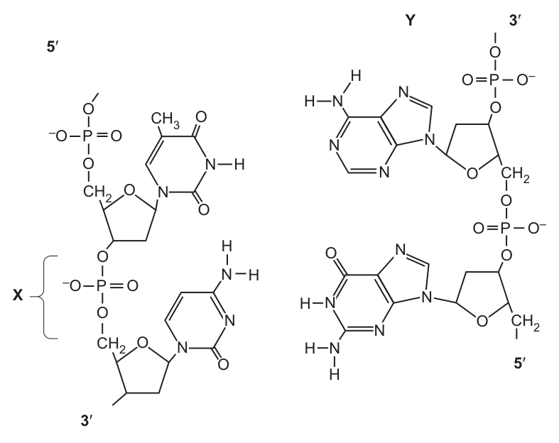
Complete Fig. 4.2 by drawing dotted lines to represent all the hydrogen bonds between the two strands of the DNA molecule.
Answer
Add two dotted lines between adenine (A) on strand Y and thymine (T) on the opposite strand, and three dotted lines between cytosine (C) and guanine (G), to represent the hydrogen bonds of complementary base pairing.
2 dotted lines between A and T, 3 dotted lines between C and G (placed at the correct H-bond donor/acceptor positions on the bases).
Background Concept
In the DNA double helix, the two antiparallel strands are held together by hydrogen bonds between complementary base pairs. The base-pairing rules are: adenine (A) on one strand always pairs with thymine (T) on the other via two hydrogen bonds, and cytosine (C) always pairs with guanine (G) via three hydrogen bonds. A purine (A or G, with two rings) always pairs with a pyrimidine (T or C, with one ring), giving the helix a uniform width. The hydrogen bonds form between specific donor (–NH or –NH2) and acceptor (C=O or ring N) atoms on the inside edges of each base — and it is the position of these donor/acceptor groups that determines the geometry of the base pair.
Understanding the Question
Fig. 4.2 shows a short DNA segment with two base pairs: the upper pair is T (left strand) and A (on strand Y, right); the lower pair is C (left) and G (right). Part (b)(i) asks the candidate to add dotted lines on the figure to represent the hydrogen bonds between the bases. The mark scheme rewards 1 mark for two correct H-bonds on A–T and 1 mark for three correct H-bonds on C–G. If neither is positioned correctly but the right number of lines is drawn (2 between A–T and 3 between C–G), 1 mark is still awarded as a fallback.
Approach
Identify the two base pairs (A–T at the top, C–G at the bottom). Remember the H-bond counts (2 and 3 respectively). For each base pair, place the dotted lines between the donor and acceptor groups on the inside edges of the two bases — i.e. between the –NH/–NH2 (donor) and C=O / N (acceptor) groups facing each other across the helix axis, not on the outer edges.
Step-by-Step Reasoning
- In Fig. 4.2 the top base on the left is thymine (T) and the top base on strand Y is adenine (A). This is the A–T pair.
- A and T form two hydrogen bonds. Draw two dotted lines:
- one between the C2 carbonyl oxygen of thymine (O on the upper left of T) and the –NH2 of adenine (the top –NH on A);
- one between the N3 of thymine (the –N–H on T) and the N1 of adenine (the bottom ring N of A).
- In Fig. 4.2 the lower base on the left is cytosine (C) and the lower base on the right is guanine (G). This is the C–G pair.
- C and G form three hydrogen bonds. Draw three dotted lines:
- between the C2 carbonyl oxygen of cytosine and the –NH2 of guanine (top –NH2 of G);
- between the N3 of cytosine and the N1 of guanine (the inner ring N of G);
- between the –NH2 of cytosine and the C6 carbonyl oxygen of guanine (bottom O of G).
- The lines should join the correct donor/acceptor atoms on the inside edges of the bases (the side facing the helix axis), not the outside.
Key Takeaways
- A pairs with T via 2 hydrogen bonds; C pairs with G via 3 hydrogen bonds.
- Hydrogen bonds form between specific donor and acceptor groups on the inside edges of the bases.
- The number and placement of the H-bonds is what makes A–T and C–G distinguishable in a diagram and is what the examiner checks.
Common Mistakes
- Drawing 3 H-bonds between A and T and 2 between C and G (numbers reversed).
- Drawing the correct number of lines but in the wrong place (e.g. on the outside of the bases or off the base entirely) — partial credit, 1 mark maximum under the fallback rule.
- Connecting the H-bonds to incorrect atoms (e.g. the C–H of thymine, which cannot H-bond), so the lines do not start or end on a real donor/acceptor.
- Drawing solid lines or dashes that are not dotted; the question specifies 'dotted lines' (the conventional way to depict non-covalent bonds).
Things to Be Careful About
- Use dotted lines, not solid or dashed lines.
- Place each line between a real H-bond donor (–NH, –NH2) and a real H-bond acceptor (C=O, ring N).
- 1 mark is available for the correct number of lines (2 for A–T, 3 for C–G) even if the positioning is imperfect; the second mark requires correct positioning at chemically valid donor/acceptor atoms.
Answer
Phosphodiester (bond)
Phosphodiester
Background Concept
The sugar–phosphate backbone of a DNA strand is held together by phosphodiester bonds. Each bond forms between the 3'-OH of one deoxyribose sugar and the 5'-phosphate of the next nucleotide, with the loss of a water molecule. The bond is called a phosphodiester because it contains two ester linkages (one to the 3' carbon of the upper sugar and one to the 5' carbon of the next sugar) joined through a central phosphate group. In Fig. 4.2 the brace labelled X spans the region between two adjacent sugars on the left strand, encompassing the central phosphate group; this entire structure is a single phosphodiester bond.
Understanding the Question
Part (b)(ii) is a one-word recall question. The candidate is given Fig. 4.2 with bond X explicitly labelled and asked to name it. The mark scheme accepts only 'phosphodiester'.
Approach
Recognise that bond X lies between the 3' carbon of one sugar and the 5' carbon of the next sugar via a phosphate — the definition of a phosphodiester bond.
Step-by-Step Reasoning
- The brace X in Fig. 4.2 covers one phosphate group and the two C–O bonds linking it to the 3' and 5' carbons of two adjacent sugars.
- A bond formed between two sugars via a single phosphate group, producing two ester linkages, is called a phosphodiester bond.
- The term 'phosphodiester' is the single correct answer; 'ester bond' alone is too vague and would not be credited.
Key Takeaways
- Phosphodiester bonds link adjacent nucleotides in a DNA (or RNA) strand.
- They form between the 3'-OH of one sugar and the 5'-phosphate of the next.
- They give the backbone its directionality (5' to 3'), important in replication, transcription and translation.
Common Mistakes
- Writing 'ester bond' or 'glycosidic bond' — these are different bonds (glycosidic bonds link the base to the sugar; ester bonds are individual chemical groups, not the full backbone linkage).
- Writing 'hydrogen bond' — this holds the two strands together, not the backbone.
- Writing 'peptide bond' — peptides are in proteins, not nucleic acids.
Things to Be Careful About
- Spelling: 'phosphodiester' is one word, no hyphen; 'phospho-diester' with a hyphen is usually not accepted.
- The bond is sometimes called a 'phosphodiester linkage' — both forms are accepted.
Inhibitors are substances that prevent biological processes in a variety of different ways.
Table 5.1 shows some antibiotics, their modes of action and the diseases which they are used to treat.
Table 5.1
| antibiotic | mode of action | disease |
|---|---|---|
| erythromycin | binds to ribosomes to inhibit translation | cholera |
| penicillin | enzyme inhibitor | tetanus |
| rifampicin | inhibits the function of RNA polymerase in transcription | tuberculosis |
Suggest why erythromycin can inhibit translation in the bacterium that causes cholera and not inhibit translation in humans who are infected with this pathogen.
Answer
- Bacteria have 70S ribosomes (smaller) while human cells have 80S ribosomes (larger).
- Erythromycin can only bind to 70S / bacterial ribosomes, because the binding site is not present on 80S / human ribosomes.
Erythromycin only binds to 70S ribosomes found in bacteria; human cells have 80S ribosomes that lack the antibiotic's binding site.
Background Concept
Ribosomes are the cellular machinery that carries out translation, reading mRNA codons and assembling the corresponding polypeptide chain. A key difference between prokaryotes and eukaryotes is the size (and composition) of their ribosomes:
- Prokaryotic ribosomes (e.g. in Vibrio cholerae) are 70S, made of a 50S large subunit and a 30S small subunit.
- Eukaryotic ribosomes (e.g. in human cells) are 80S, made of a 60S large subunit and a 40S small subunit.
The "S" stands for Svedberg units, which describe sedimentation rate in a centrifuge (and therefore size/density, not mass). The two ribosome types also differ in their rRNA molecules and the proteins that make them up. Antibiotics exploit these differences: drugs that target bacterial ribosomes are described as selectively toxic because they disrupt pathogen function without damaging the host's own protein synthesis.
Understanding the Question
The question uses Table 5.1, which states that erythromycin "binds to ribosomes to inhibit translation" and is used to treat cholera (caused by the bacterium Vibrio cholerae). The candidate must explain why this antibiotic stops translation in the bacterial pathogen but leaves translation in the infected human host unaffected.
The command word is "suggest why" — this is essentially an explain question that requires the candidate to give the structural reason for the selectivity. The marking scheme offers up to five possible points; any two earn full marks.
Approach
The reasoning flows from a single underlying principle: selective toxicity depends on structural differences between host and pathogen. The candidate should:
- State the ribosome-size difference (70S vs 80S).
- Explain that erythromycin's binding site is only present on the bacterial 70S ribosome, so it cannot attach to a human 80S ribosome.
An additional mark-scheme point worth noting is that human mitochondria do contain 70S ribosomes (an evolutionary remnant of their prokaryotic origin), but erythromycin cannot easily enter mitochondria, so even those ribosomes are largely protected.
Step-by-Step Reasoning
- Identify the relevant structure. Erythromycin targets ribosomes, so the question is asking about a structural difference between bacterial and human ribosomes.
- State the difference. Bacteria (prokaryotes) have 70S ribosomes; humans (eukaryotes) have 80S ribosomes. This is the foundational marking point.
- Link the difference to the drug's action. Erythromycin only binds to a specific site that is present on the 70S ribosome. Human 80S ribosomes lack that binding site, so the antibiotic has nothing to attach to and translation continues normally in human cells.
- (Optional reinforcement.) The two ribosome types also differ in the rRNA molecules and ribosomal proteins they contain — this is the molecular reason the binding site exists on one but not the other.
- (Optional reinforcement.) Although human mitochondria have 70S ribosomes, erythromycin cannot cross the mitochondrial membranes to reach them in significant amounts, so human protein synthesis is largely unaffected.
A complete two-mark answer therefore pairs the size difference (70S vs 80S) with the binding-site difference ("only binds to 70S" / "no binding site on 80S").
Key Takeaways
- Antibiotics that target protein synthesis (e.g. erythromycin, streptomycin, chloramphenicol, tetracyclines) are selectively toxic because bacterial (70S) and human (80S) ribosomes are structurally different.
- The principle of selective toxicity is fundamental to antimicrobial chemotherapy: the drug must damage the pathogen more than the host.
- The 70S/80S distinction is one of the most heavily tested pieces of comparative cell biology at AS level.
Common Mistakes
- Stating only that "bacteria and humans have different ribosomes" without giving the actual sizes (70S vs 80S) — vague statements do not earn the mark.
- Saying "erythromycin cannot enter human cells" — this is too vague; the antibiotic simply does not bind the host's ribosomes, and even where 70S ribosomes exist (mitochondria) the drug has limited access.
- Confusing the Svedberg values (e.g. writing "bacteria have 80S and humans have 70S").
- Treating "selective toxicity" as a magic property rather than a consequence of structural differences.
Things to Be Careful About
- The command word is "suggest why", not "describe" — a one-line answer giving the ribosome difference plus the binding-site difference is sufficient for two marks.
- Svedberg units are not additive: a 50S + 30S does not equal an 80S because Svedberg measures sedimentation rate, not mass. A 70S ribosome is genuinely smaller than an 80S ribosome.
- The marking scheme explicitly notes that references to "complementary/specific" binding are accepted as alternatives to the binding-site wording.
Answer
Penicillin acts on bacterial cell walls (peptidoglycan / murein), and human cells have no cell walls / peptidoglycan, so there is no target for the drug.
Human cells have no cell walls / peptidoglycan for penicillin to act on.
Background Concept
Penicillin is a β-lactam antibiotic. Its mode of action depends on the chemistry of the bacterial cell wall.
- Bacterial cell walls are made of peptidoglycan (also called murein) — a mesh of polysaccharide chains cross-linked by short peptide bridges.
- Penicillin binds irreversibly to and inhibits transpeptidase (also called penicillin-binding protein, PBP), the enzyme that forms the peptide cross-bridges between peptidoglycan strands. Without these cross-links the wall weakens and the bacterium takes up water, swells, and lyses.
- Human cells have no cell wall at all — they are bounded only by a plasma membrane. There is no peptidoglycan and no transpeptidase, so penicillin has nothing to attack.
This is another example of selective toxicity: the drug only disrupts a structure that exists in the pathogen.
Understanding the Question
Table 5.1 lists penicillin as an "enzyme inhibitor" used to treat tetanus (caused by the bacterium Clostridium tetani). The question asks the candidate to state why this drug does not affect the human host's own cells. The answer must identify the absence of a suitable target.
The command word is "state" — this requires a single, direct factual statement. One mark is awarded.
Approach
The candidate should:
- Identify the target of penicillin (bacterial cell wall / peptidoglycan).
- State that this target is absent in human cells.
A clean, one-sentence answer is enough.
Step-by-Step Reasoning
- From Table 5.1, penicillin's mode of action is "enzyme inhibitor". Combined with knowledge of penicillin's mechanism, the relevant enzyme is transpeptidase, which makes peptide cross-bridges in peptidoglycan.
- Penicillin therefore acts on the bacterial cell wall.
- Human cells do not have a cell wall (only a cell-surface membrane) and contain no peptidoglycan.
- Conclusion: there is no target for penicillin in human cells, so the drug does not act on them.
A complete one-mark answer must explicitly mention cell walls / peptidoglycan and the fact that human cells lack them. Vague answers such as "penicillin only affects bacteria" do not earn the mark because they do not give the structural reason.
Key Takeaways
- Penicillin disrupts bacterial cell wall synthesis by inhibiting transpeptidase.
- Human cells have no cell wall — and therefore no peptidoglycan and no transpeptidase — so penicillin has no target.
- This is the textbook example of selective toxicity in the AS syllabus.
Common Mistakes
- Writing only "penicillin only affects bacteria" — too vague; the mark scheme requires a structural reason (cell wall / peptidoglycan).
- Saying "human cells have a different cell wall" — incorrect; human cells have no cell wall at all.
- Confusing penicillin's target with that of antiviral drugs (e.g. reverse transcriptase inhibitors) or antibiotics that target ribosomes (e.g. erythromycin).
- Mentioning penicillinase — the mark scheme explicitly ignores (rejects) any reference to penicillinase here.
Things to Be Careful About
- "Murein" is an accepted alternative to "peptidoglycan" at A-level.
- The question is worth only one mark, so a short, direct answer is appropriate.
- Avoid giving a long description of how penicillin works in bacteria — the question asks specifically why it does not act on human cells.
Explain one way in which rifampicin may inhibit the action of RNA polymerase in transcription.
Answer
Rifampicin binds to the active site of RNA polymerase, blocking the substrate (nucleotides) from binding, so no phosphodiester bonds can be formed and mRNA cannot be made.
Rifampicin blocks the active site of RNA polymerase, preventing nucleotides from binding and so preventing mRNA synthesis.
Background Concept
Transcription is the synthesis of an RNA molecule from a DNA template, catalysed by the enzyme RNA polymerase. In bacteria, a single RNA polymerase (with a σ-factor that recognises promoters) makes all types of RNA — mRNA, tRNA and rRNA. The enzyme works by:
- Binding to a promoter sequence on the DNA.
- Unwinding the DNA double helix to expose the template strand.
- Aligning complementary RNA nucleotides (ATP, GTP, CTP, UTP) with the template.
- Joining these nucleotides together with phosphodiester bonds to build the new RNA strand.
Enzyme inhibitors slow or stop an enzyme's activity. The two main types at AS level are:
- Competitive inhibitors — bind to the active site, blocking substrate access. Their effect can be overcome by increasing substrate concentration.
- Non-competitive inhibitors — bind to an allosteric site elsewhere on the enzyme, changing the shape of the active site so the substrate can no longer bind effectively. Increasing substrate concentration does not overcome the inhibition.
Understanding the Question
Table 5.1 states that rifampicin "inhibits the function of RNA polymerase in transcription" and is used to treat tuberculosis (caused by Mycobacterium tuberculosis). The question asks the candidate to explain one way rifampicin may inhibit RNA polymerase.
The command word is "explain" but with a single-mark weighting — the candidate only needs to give one credible mechanism. The mark scheme offers five alternatives, of which the most straightforward is competitive inhibition at the active site.
Approach
The candidate should choose one of the inhibition mechanisms that is consistent with the question and state it clearly:
- The most commonly taught mechanism: rifampicin binds to the active site of RNA polymerase, blocking substrate (nucleotide) binding.
- Alternative: rifampicin binds to an allosteric site, altering the active-site shape.
- Alternative: rifampicin attaches to the DNA template, physically blocking RNA polymerase.
- Alternative: rifampicin is incorporated into the growing RNA chain, but no further nucleotide can be added (chain termination).
Any one of these is sufficient. The most "biology-textbook" answer is the first.
Step-by-Step Reasoning
- Identify what RNA polymerase does. It catalyses the formation of phosphodiester bonds between RNA nucleotides during transcription.
- Identify the structure of the enzyme relevant to a drug. The active site is the region where the substrates (ribonucleotides) bind and the reaction occurs.
- Apply the principle of competitive inhibition. If rifampicin has a shape complementary to the active site, it can occupy that site, preventing nucleotides from binding.
- State the consequence. With the active site blocked, no phosphodiester bonds can be formed and mRNA cannot be synthesised — transcription is inhibited.
This is exactly the mechanism by which rifampicin acts in reality: it binds to the β-subunit of bacterial RNA polymerase and physically blocks the elongation of the RNA chain.
Key Takeaways
- Antibiotics can inhibit any step of a pathogen's central dogma — transcription (rifampicin), translation (erythromycin) or cell-wall synthesis (penicillin).
- Many antibiotics act as enzyme inhibitors, either competitively (at the active site) or non-competitively (at an allosteric site).
- Drug mechanisms often mirror normal enzyme inhibition concepts taught in the Enzymes topic.
Common Mistakes
- Writing only that "rifampicin stops transcription" — this is a restatement of the question, not an explanation. The candidate must describe how it stops transcription.
- Confusing rifampicin's target with that of erythromycin (ribosomes) or penicillin (cell-wall enzymes).
- Failing to mention either the active site (for a competitive model) or a specific consequence (e.g. no phosphodiester bonds formed).
- Using vague language such as "it interferes with the enzyme" — without specifying the binding location or effect, this does not score.
Things to Be Careful About
- The question is only worth one mark, so the candidate should give one clear, specific mechanism — not a list of all possibilities.
- "Fits into the active site" is an acceptable alternative to "binds to the active site" at A-level.
- The mark scheme explicitly accepts a non-competitive-inhibition explanation (binding to an allosteric site) as an alternative, so either model of inhibition is fine.
A student studied the structure of a mammalian heart.
The student took a photograph of the left side of a dissected heart as shown in Fig. 6.1.
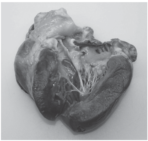
Identify two features of the left side of the heart visible in Fig. 6.1 and explain how each feature is adapted to the function of the heart.
feature ______
explanation ______
feature ______
explanation ______
Answer
Feature 1: Thick (muscular) wall of the left ventricle
Explanation: Generates the high force / high pressure required to pump blood through the systemic circulation (over a long distance and against the resistance of the arterioles).
Feature 2: (Left) atrioventricular / bicuspid / mitral valve
Explanation: Prevents backflow of blood / maintains one-way flow of blood from the left ventricle back into the left atrium during ventricular systole.
Acceptable alternative Feature 2: Chordae tendineae (tendinous cords)
Acceptable alternative Explanation: Hold the atrioventricular valve in position during ventricular systole / prevent the valve from turning inside out when the ventricle contracts.
See working — two features with explanations as above.
Background Concept
The left side of the heart receives oxygenated blood from the lungs via the pulmonary veins and pumps it into the systemic circulation through the aorta. The left ventricle must therefore generate enough force to overcome the high resistance of the systemic vascular bed and push blood through the entire body, while the left atrium only needs to deliver blood a short distance into the ventricle. This functional asymmetry is reflected in the anatomy visible on dissection: a much thicker ventricular wall, the bicuspid (mitral) valve between atrium and ventricle, fibrous chordae tendineae tethering the valve cusps to papillary muscles, and (at the outflow) the aortic semi-lunar valve.
Understanding the Question
The student has photographed the internal left side of a dissected mammalian heart (Fig. 6.1) and must pick out two features actually visible in that photograph and explain how each is adapted to the heart's function. The question explicitly restricts the answer to the left side — features of the right side (tricuspid valve, pulmonary valve, thinner right ventricular wall) are not creditworthy.
Approach
Read each labelling line on the photograph as a list of identifiable structures (ventricular wall thickness, atrial wall, valve(s), chordae tendineae, papillary muscles, aorta). For each, decide whether the marking scheme rewards a feature paired with a functional explanation. The most reliable pairs are:
- Thick LV wall ↔ high-pressure systemic pumping
- Bicuspid valve ↔ prevents backflow into the atrium
- Chordae tendineae ↔ hold valve during systole
Avoid common traps: 'withstands high pressure' is explicitly rejected; simply naming 'valves' without specifying which one is risky because the tricuspid (right) is rejected.
Step-by-Step Reasoning
Feature 1 — thick muscular wall of the left ventricle. Cardiac muscle is striated and rich in mitochondria, but the wall thickness itself is the key adaptation. A thicker wall can generate a greater contractile force, raising intraventricular pressure to roughly 120 mmHg in systole, sufficient to overcome peripheral resistance and perfuse all systemic tissues. The right ventricle wall is roughly one-third this thickness because it only has to pump blood the short distance to the lungs at lower pressure.
Feature 2 — the bicuspid (mitral / left atrioventricular) valve. Two fibrous cusps close at the start of ventricular systole. Because pressure in the ventricle rises sharply above atrial pressure, the valve would otherwise invert into the atrium; its closure ensures blood exits only via the aorta. The mark scheme credits "prevents backflow" or "maintains one-way flow" but specifically rejects the tricuspid or pulmonary valve here.
Alternative Feature 2 — chordae tendineae. These thin tendinous cords attach the AV valve cusps to papillary muscles. As the ventricle contracts, the papillary muscles also contract, pulling the cords taut and stopping the valve cusps from being pushed back into the atrium (a 'blowback'). The mark scheme allows 'ligaments' to be ignored and 'high tensile strength unqualified' to be ignored, so the function must be stated.
Key Takeaways
- Wall thickness in the heart mirrors the pressure work each chamber must do (LV >> RV; atria thin).
- Valves are essential for one-way flow; the chordae tendineae prevent valve inversion under high ventricular pressure.
- On the left side of the heart, only the bicuspid valve and the aortic semi-lunar valve are creditworthy; never credit the tricuspid or pulmonary valve.
Common Mistakes
- Naming the tricuspid or pulmonary valve — these belong to the right side and are rejected.
- Writing 'withstands high pressure' instead of 'generates high force/pressure' — explicitly rejected by the mark scheme.
- Stating that the wall is thick 'because it contains cardiac muscle' — this is descriptive, not an adaptation; the function is the generation of high force to overcome systemic resistance.
- Treating 'ligaments' as a synonym for chordae tendineae — the mark scheme ignores this.
Things to Be Careful About
- Restrict features to those on the left side; the photograph's right-side structures must not be named.
- Each feature must be paired with an explanation of adaptation, not merely a restatement of the structure.
- A correct feature with a wrong/creditless explanation still scores only the feature mark (1), not 2.
Cardiac muscle is described as myogenic. This means the electrical activity controlling the rhythm of a regular heartbeat begins within the muscle tissue of the heart.
Describe how electrical activity within the heart controls each heartbeat.
Answer
- The sinoatrial node (SAN) in the wall of the right atrium initiates the heartbeat by releasing waves of excitation / electrical impulses (action potentials).
- These impulses spread across the walls of both atria, causing atrial systole (atria contract).
- Impulses are prevented from passing directly to the ventricles by a layer of non-conducting tissue / fibrous ring between the atria and ventricles.
- The impulse reaches the atrioventricular node (AVN), which delays the impulse by ~0.1 s to allow the atria to finish emptying.
- The AVN passes the impulse to the Purkyne tissue (via the bundle of His) which conducts it down the septum.
- The Purkyne tissue carries the impulse to the base / apex of the ventricles, so the ventricles contract from the base upwards, pumping blood out through the aorta and pulmonary artery.
See working — six points covering the SAN–atria–AVN–Purkyne sequence and the resulting contraction pattern.
Background Concept
Cardiac muscle is myogenic — it can contract without external nervous stimulation because the rhythm is generated within the heart itself. The pacemaker is the sinoatrial node (SAN), a small region of specialised muscle cells in the wall of the right atrium. From the SAN, the impulse spreads through the atrial walls to the atrioventricular node (AVN), then down the bundle of His and the Purkyne fibres in the interventricular septum, and finally to the ventricular walls. The arrangement ensures the atria contract before the ventricles, and that ventricular contraction begins at the apex and travels upwards towards the aorta and pulmonary artery.
Understanding the Question
The stem defines 'myogenic' and asks for a description — not just a list — of how the electrical activity within the heart controls each heartbeat. The description must connect electrical events (impulses, conduction, delay) to mechanical events (atrial systole, ventricular systole) in the correct order, and the mark scheme offers six discrete marking points from which any four are credited.
Approach
Trace the impulse along its anatomical route, naming each structure in turn and stating what happens there. Three steps matter particularly: (a) impulse generation at the SAN, (b) the delay at the AVN, and (c) the route from AVN to ventricular muscle via the bundle of His / Purkyne fibres.
Step-by-Step Reasoning
- SAN fires. The SAN spontaneously depolarises and initiates a wave of excitation that spreads across both atrial walls. The mark scheme credits either 'waves of excitation', 'waves of depolarisation' or 'electrical impulses / action potentials'.
- Atria contract. The electrical event produces atrial systole — both atria contract simultaneously, pushing blood through the open AV valves into the ventricles.
- Non-conducting tissue / fibrous ring between the atria and ventricles stops the impulse passing directly. Without this insulating tissue the impulse would short-circuit straight to the ventricles, preventing the AVN delay.
- AVN delays the impulse (~0.1 s). This delay ensures atrial contraction is complete (and the ventricles are full) before ventricular systole begins.
- AVN → bundle of His → Purkyne tissue. The Purkyne fibres conduct the impulse rapidly down the interventricular septum.
- Ventricles contract from the base / apex upward. Because the Purkyne fibres spread the impulse from the apex upwards, the ventricular muscle contracts from the bottom up, squeezing blood out through the semi-lunar valves into the aorta and pulmonary artery.
Key Takeaways
- Cardiac muscle is myogenic: the SAN is the pacemaker.
- The order is SAN → atria → AVN (with delay) → Purkyne tissue → ventricles.
- The non-conducting tissue and the AVN delay are essential for coordinated atrial-then-ventricular contraction.
- Ventricular contraction starts at the apex and travels upwards to maximise ejection.
Common Mistakes
- Writing 'AVN' without explaining its role in delaying the impulse — the delay (~0.1 s) is the mark.
- Omitting the non-conducting / fibrous ring between atria and ventricles.
- Saying the ventricles 'contract from the top down' — the correct direction is base / apex upwards.
- Confusing the bundle of His with the Purkyne fibres (bundle of His is the short connecting trunk; Purkyne fibres spread through the ventricular walls).
Things to Be Careful About
- The mark scheme penalises once if AVN is used and then use error-carried-forward — but you should still write AVN in full at least once so the examiner can award the credit.
- 'Stimulates the atria to contract' must clearly attach the impulse to the muscle, not just 'causes atrial systole' with no electrical cause.
- If you list six points, ensure they are sequential — the SAN must precede the AVN, the AVN must precede the Purkyne tissue.
Tissue fluid is formed from blood plasma as it flows through capillaries.
Fig. 6.2 is a diagram of a capillary and some adjacent tissue cells in a capillary network.
Answer
Any two of:
- Provides an aqueous / watery (external) environment for cells, maintaining the correct water potential so that cells do not dehydrate / lose excessive water.
- Supplies oxygen to cells (oxygen diffuses from the blood into the tissue fluid and then into the cells).
- Removes carbon dioxide from cells (carbon dioxide diffuses from the cells into the tissue fluid and then into the blood).
- Supplies nutrients such as glucose / amino acids / ions to cells.
- Removes metabolic / toxic waste such as urea from cells.
- Allows movement / passage of (named) cell-signalling molecules / hormones / ligands between cells.
- Acts as a medium for the movement of macrophages / neutrophils / phagocytes for defence.
The two strongest choices for the exam are oxygen supply and carbon dioxide removal (these cover the gas-exchange role) OR the aqueous-environment role and a named-nutrient supply.
See working — any two of the listed functions.
Background Concept
Tissue fluid is the interstitial fluid that bathes every cell in the body. It is formed by filtration of plasma out of capillaries and is returned to the circulation mainly via the lymphatic system. Because every cell of the body is surrounded by tissue fluid, this fluid must (i) provide a chemically and osmotically suitable environment and (ii) act as the exchange medium between blood and cells for everything the cells need (oxygen, glucose, amino acids, ions, hormones) and everything they need to lose (carbon dioxide, urea, heat).
Understanding the Question
This is a 'state' / 'describe' sub-question worth 2 marks, asking for two functions of tissue fluid. The mark scheme lists many valid functions; the examiner will accept any two clearly-stated and biologically distinct roles.
Approach
Categorise the mark-scheme points into three groups and pick one from each group if you want maximum coverage:
- Environment — provides the aqueous medium.
- Exchange of substances — supply of oxygen / nutrients; removal of carbon dioxide / wastes.
- Defence / communication — movement of phagocytes; carriage of signalling molecules / hormones.
Step-by-Step Reasoning
- Aqueous environment. Cells would dehydrate and shrink in a dry tissue space; tissue fluid keeps their external water potential similar to that of cytoplasm and prevents excessive osmotic water loss.
- Oxygen supply. Blood delivers oxygen bound to haemoglobin in red cells; oxygen diffuses from blood plasma through tissue fluid into cells for aerobic respiration.
- Carbon dioxide removal. CO₂ produced by respiration diffuses out of cells into tissue fluid and from there into the plasma for transport to the lungs.
- Nutrient supply. Glucose, amino acids and ions move with the filtrate and diffuse into cells; tissue fluid is the medium across which this happens.
- Waste removal. Urea and other metabolic wastes diffuse out of cells into tissue fluid and are carried away by the blood (or lymph).
- Phagocyte movement. Tissue macrophages and neutrophils migrate through tissue fluid to sites of infection / damage.
- Signalling molecules. Local hormones, cytokines and other ligands diffuse through tissue fluid to act on neighbouring cells (paracrine signalling).
Key Takeaways
- Tissue fluid is the exchange medium between blood and cells.
- Its functions fall into three groups: environment, substance exchange, defence / signalling.
- In the exam, the cleanest pair is usually O₂ supply + CO₂ removal (the gas-exchange role).
Common Mistakes
- Writing 'transports substances' on its own — the mark scheme rejects a vague 'transport' claim; you must name at least one substance moved in each direction.
- Confusing tissue fluid with blood plasma or lymph; the three are related but distinct (lymph forms from excess tissue fluid and is returned to the blood).
- Listing 'white blood cells' without naming macrophages / neutrophils — the mark scheme ignores generic 'white blood cell' here.
Things to Be Careful About
- Each of the two functions must be clearly distinct (gas exchange ≠ nutrient supply unless you have phrased one as 'oxygen' and the other as 'glucose').
- If you want to mention 'oxygen', name it; 'respiratory gases' is acceptable but only as an alternative to naming both O₂ and CO₂.
Answer
- At the arteriole end of the capillary the hydrostatic pressure of the blood (plasma) is high (greater than the hydrostatic pressure of the tissue fluid outside).
- This pressure difference forces fluid out of the capillary — ultrafiltration: plasma (water) and small solutes are pushed out of the blood.
- Small molecules such as glucose, amino acids and ions leave the blood, while (large) plasma proteins remain inside the capillary (because they are too large to pass through the capillary wall). Red blood cells and platelets also remain in the blood.
- The fluid passes through pores / gaps / fenestrations in the capillary wall (between / through the endothelial cells) — these are visible in Fig. 6.2 as the small openings in the wall of the capillary.
See working — hydrostatic pressure, ultrafiltration of plasma with small solutes, retention of plasma proteins, passage through capillary pores.
Background Concept
Tissue fluid is filtered out of blood at the arteriole end of every capillary because the blood here is under considerable hydrostatic pressure (the heart has just pumped it through the arterioles). The capillary wall is only one endothelial cell thick and contains small gaps (pores / fenestrations) between adjacent endothelial cells. Anything small enough to pass through these pores — water, ions, glucose, amino acids, urea, oxygen, carbon dioxide — is pushed out by the pressure. Anything too large — plasma proteins (especially albumin), and of course the blood cells themselves — stays inside the capillary. As blood continues along the capillary, hydrostatic pressure falls and the osmotic effect of the retained plasma proteins starts to draw water back in at the venule end.
Understanding the Question
The question says 'with reference to Fig. 6.2'. Fig. 6.2 shows a longitudinal section through a capillary, with red blood cells inside and tissue cells outside, and arrows showing fluid movement. So your answer must explicitly use pressure to drive fluid out, must mention the pores in the wall, and must distinguish between small solutes (which leave) and plasma proteins / cells (which stay). The mark scheme lists 4–5 marking points from which any 3 are credited.
Approach
Work through the steps of filtration in order, in language the diagram supports:
- Pressure at the arteriole end.
- Filtration of fluid through the wall.
- What leaves (small solutes) and what stays (plasma proteins, cells).
- The route through the wall — the pores / fenestrations shown in the figure.
Step-by-Step Reasoning
- High hydrostatic pressure at the arteriole end. Blood enters the capillary network under pressure (residual from ventricular systole and the resistance of the arterioles). This pressure is the driving force for filtration.
- Ultrafiltration. The high pressure forces plasma (water) and dissolved small molecules out through the capillary wall. This is described as 'ultrafiltration' because the filter (the capillary wall) holds back large particles.
- Selectivity of the filter. Glucose, amino acids, ions, urea, oxygen and carbon dioxide are small enough to pass through the pores. Plasma proteins (especially albumin) are too large, and cells (RBCs, WBCs, platelets) are far too large — they remain in the blood.
- Route — the pores. Fig. 6.2 shows the capillary wall as a thin layer with small gaps between endothelial cells. Fluid, ions and small molecules pass through these gaps (or through fenestrations in the endothelial cells) into the tissue space, where it bathes the adjacent tissue cells.
Key Takeaways
- Tissue fluid is formed by ultrafiltration at the arteriole end of the capillary.
- The driving force is hydrostatic pressure of the blood, which exceeds the hydrostatic pressure of the tissue fluid.
- Plasma proteins and blood cells remain in the blood; small solutes (glucose, amino acids, ions, O₂, CO₂, urea) and water pass through pores in the capillary wall.
- The route is through pores / gaps / fenestrations between endothelial cells.
Common Mistakes
- Saying 'diffusion' alone — diffusion does occur for individual solutes, but the bulk movement of fluid out is driven by pressure (ultrafiltration), not diffusion.
- Stating that red blood cells leave the capillary — they do not; this is explicitly rejected.
- Saying plasma proteins leave — they remain in the blood; this is the basis of the oncotic pressure that draws water back in at the venule end.
- Omitting any reference to pressure — without this the 'filtration' claim has no driving force.
Things to Be Careful About
- The question says 'with reference to Fig. 6.2' — you must mention features of the figure (the wall, the pores, the cells in the capillary) so the examiner can see you used the diagram.
- A common answer that earns only 1 or 2 marks is 'blood is under pressure, fluid is pushed out' — you must additionally say what passes through (small solutes, not proteins/cells) and through what (the pores / gaps in the wall).
Measles is a common disease caused by a virus. Vaccination to prevent the disease has been very successful.
A child receives a vaccine for measles.
Explain why only some of the T-lymphocytes in the child respond to the measles vaccine.
Answer
- T-lymphocytes carry specific T-cell receptors that are complementary to only one (or a very few) particular antigen(s). Each T-lymphocyte in the body carries a different receptor.
- The antigen(s) in the measles vaccine bind only to those T-lymphocytes whose receptors are complementary to that antigen.
- As a result, only those specific T-lymphocytes are activated / stimulated / selected (clonal selection); the rest are not activated and so do not respond.
See working — specificity of T-cell receptors and clonal selection/activation.
Background Concept
Each lymphocyte expresses receptors of a single specificity — generated randomly during the lymphocyte's development. The body therefore contains a vast repertoire of lymphocytes, each capable of recognising one specific antigen. When a pathogen (or a vaccine) enters the body, the antigenic epitopes it carries bind only to those lymphocytes whose receptors happen to be complementary — a process called clonal selection. The selected lymphocyte is then activated and proliferates.
In this question, the measles vaccine contains antigens (typically surface proteins of the measles virus). Of the millions of T-lymphocytes circulating in the child, only a tiny minority will have a T-cell receptor complementary to a measles antigen. These are the ones that respond.
Understanding the Question
The stem says the child receives a measles vaccine and asks why only some T-lymphocytes respond. This is essentially asking the candidate to apply clonal selection to T-lymphocytes specifically. The mark scheme explicitly accepts 'epitope' as an alternative wording for 'antigen', and explicitly ignores the word 'pathogen'.
Approach
Construct a short chain of three statements, even though only two marks are available — covering specificity, binding, and activation/selection.
Step-by-Step Reasoning
- Specificity of T-cell receptors. Each T-lymphocyte has receptors specific to one antigen (or a few closely related antigens). The receptors vary between lymphocytes.
- Antigen–receptor binding. Antigen from the vaccine binds to the T-lymphocytes whose receptors are complementary in shape. This is the recognition step.
- Clonal selection / activation. Only the T-lymphocytes to which the antigen binds are activated (or 'selected'); the others are not stimulated and therefore do not respond.
Key Takeaways
- Lymphocytes are antigen-specific by virtue of their receptors.
- Antigen binding activates only the matching lymphocyte — the basis of clonal selection.
- This principle applies equally to T-lymphocytes and B-lymphocytes.
Common Mistakes
- Writing 'some T-lymphocytes do not respond because they are not exposed to the antigen' — the mark scheme ignores this kind of statement if it is the only one made; the selective response is the point.
- Writing 'the vaccine only contains a few antigens, so only some T-lymphocytes are exposed' — this is technically an alternative point (the mark scheme accepts it as AVP), but on its own it does not demonstrate understanding of clonal selection.
- Confusing the role of T-lymphocytes with B-lymphocytes (which produce antibodies) or with macrophages (which present antigen).
Things to Be Careful About
- Do not write 'respond' on its own; the mark scheme specifically ignores 'respond' as the only verb for activation — pair it with 'activated', 'stimulated' or 'selected'.
- The terms 'epitope' and 'antigen' are accepted alternatives here — both are correct.
Describe the events that occur in an immune response to a vaccine that result in lymphocytes that provide long-term immunity to measles.
Answer
- Antigen presentation. Macrophages engulf and process antigen from the vaccine and present it on their surface (antigen presentation).
- T-helper cell activation / clonal selection. A T-helper lymphocyte whose receptor is complementary to the presented antigen binds to the macrophage and is activated (clonal selection).
- Clonal expansion. The activated T-helper cell divides repeatedly by mitosis to produce a clone of identical T-helper cells.
- B-lymphocyte activation. B-lymphocytes with complementary receptors bind the antigen and, with the help of cytokines released by the activated T-helper cells, are also activated and undergo clonal expansion.
- Cytokine release. T-helper cells release cytokines that stimulate both B- and T-lymphocytes to multiply and differentiate.
- Memory cell formation. Some of the activated B- and T-lymphocytes differentiate into memory (B and T) lymphocytes.
- Long-term immunity. These memory cells remain in the circulation (and lymphoid tissue) for a long time / are long-lived, providing long-term immunity. On subsequent exposure to the measles antigen, these memory cells can rapidly recognise and mount a secondary response, producing antibody faster and in greater quantity than in the primary response.
See working — antigen presentation → clonal selection → clonal expansion → cytokines → memory cell formation → long-term immunity.
Background Concept
A vaccine presents the immune system with harmless antigenic material that mimics the surface of a pathogen. The body's adaptive response unfolds in two linked phases:
- The primary response is slower, generates antibodies from B-lymphocytes (with T-helper-cell help), and produces memory cells that persist after the antigen is cleared.
- The secondary response is faster and larger because memory cells recognise the antigen immediately, allowing rapid clonal expansion and antibody production before the pathogen can cause disease.
Long-term immunity therefore depends on the memory cells that survive the primary response. A vaccine that fails to generate memory cells fails to protect against later infection.
Understanding the Question
The stem specifies 'events that occur in an immune response to a vaccine that result in lymphocytes that provide long-term immunity to measles'. This wording deliberately steers the candidate towards (a) a primary response and (b) memory-cell formation. The mark scheme caps the answer at 4 marks if memory cells / immunological memory are not explicitly mentioned — so the link between the response and long-term immunity is essential.
Approach
Lay the events out in chronological order:
- Antigen-presenting cell (macrophage) takes up antigen.
- Antigen presentation to a T-helper cell with a complementary receptor.
- T-helper cell activation and clonal expansion.
- Cytokine release by T-helper cells.
- B-lymphocyte activation (with T-helper-cell help) and clonal expansion.
- Differentiation of some lymphocytes into memory (B and T) cells.
- Persistence of memory cells in the circulation / lymphoid tissue.
Step-by-Step Reasoning
- Antigen presentation. Macrophages phagocytose antigen from the vaccine, process it internally and present fragments on their surface bound to MHC molecules. This is the antigen-presentation step.
- T-helper cell activation / clonal selection. A T-helper lymphocyte with a receptor complementary to the presented antigen binds and is activated.
- Clonal expansion. The activated T-helper cell divides repeatedly by mitosis, producing a clone of identical T-helper cells — clonal expansion.
- Cytokine release. Activated T-helper cells secrete cytokines that stimulate other lymphocytes (B-cells and other T-cells) to proliferate and differentiate.
- B-cell activation. B-lymphocytes with complementary surface antibodies bind the antigen and, with the help of cytokines from T-helper cells, are activated and undergo clonal expansion, then differentiate into plasma cells (which secrete antibody) and memory B-cells.
- T-cell differentiation. Some of the proliferating T-cells differentiate into memory T-cells (in addition to effector T-cells).
- Long-term immunity. The memory B- and T-cells are long-lived and persist in the circulation and lymphoid organs, ready to mount a rapid secondary response if the measles antigen is encountered again.
Key Takeaways
- Long-term immunity depends on the memory cells generated during the primary response.
- The primary response requires antigen presentation, T-helper-cell activation, clonal expansion, cytokine help and B-cell activation.
- Memory cells are long-lived; on re-exposure they trigger a faster, larger secondary response.
- Cytokines from T-helper cells are the bridge between T-cell and B-cell responses.
Common Mistakes
- Writing only 'antibodies are produced' without mentioning memory cells — capped at 4 marks.
- Forgetting that B-cells need T-helper-cell help to be fully activated (T-dependent antigen).
- Describing the secondary response instead of the primary response — the question asks about events occurring in response to a vaccine (the primary response), not on later natural infection.
- Saying 'more memory cells' rather than 'memory cells are present / have been formed' — the mark scheme ignores 'more' here; what matters is that memory cells exist after the response.
Things to Be Careful About
- Mention memory cells explicitly — the answer is capped at 4 marks without them.
- Make sure the steps are in the right sequence (antigen presentation → T-helper activation → clonal expansion → cytokines → memory cell formation).
- 'Long-lived' or 'remain in circulation for a long time' both credit the persistence of memory cells.
- The mark scheme rewards B- and T-lymphocyte memory cells; you do not need to specify which subtypes, but doing so (memory B, memory T) adds clarity.