Biology 9700/22 — October/November 2025
Cambridge AS Level · AS Level Structured Questions · worked solutions for every part, with the mark scheme
Topics Cell Structure · Transport in Mammals · Cell Membranes and Transport · Infectious Diseases · Biological Molecules · The Mitotic Cell Cycle · +4 more
The wall of the small intestine is highly folded to form villi. Between the villi are infoldings known as crypts of Lieberkühn.
Fig. 1.1 is a diagram of a section through a single villus and a crypt of Lieberkühn.
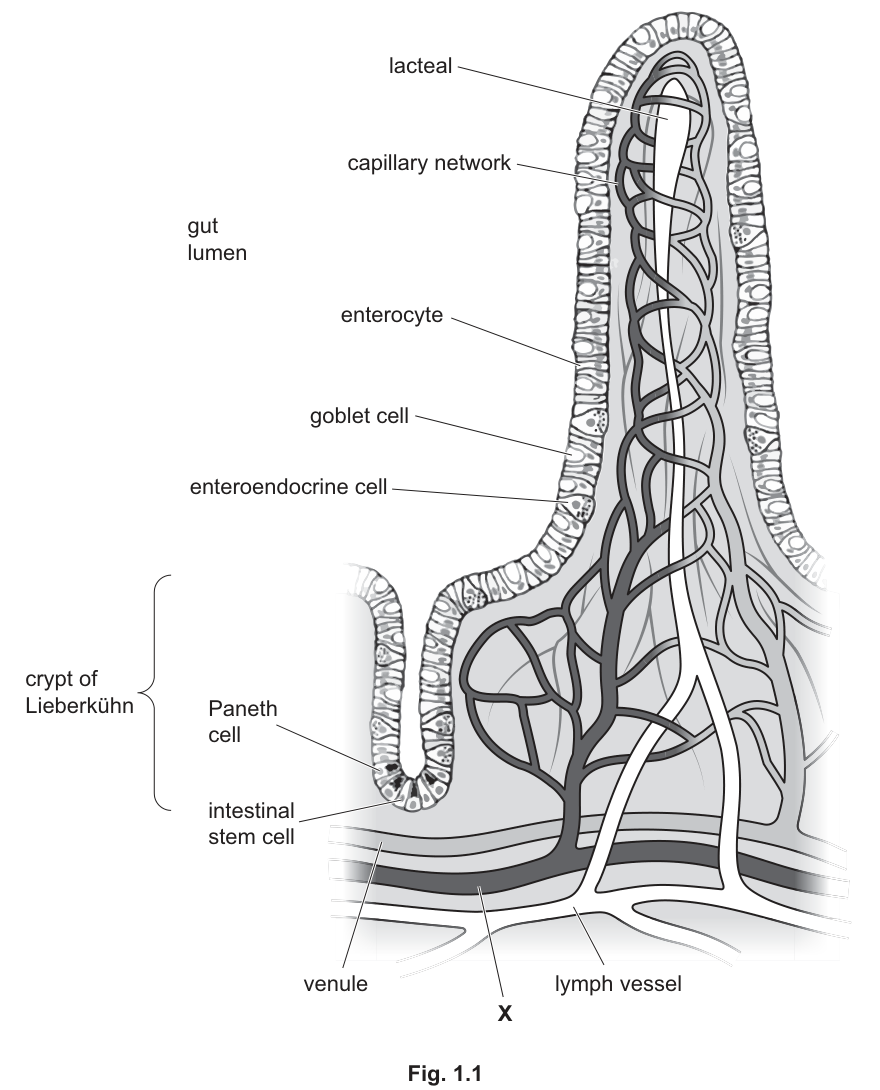
Fig. 1.1 shows that the epithelium of the villus contains mainly goblet cells and cells known as enterocytes. Both cell types have microvilli on the apical surface (surface facing the gut lumen).
Goblet cells are involved in the production of mucus.
Enterocytes are adapted for the absorption of the soluble products of digestion. These products enter the circulatory system.
A student incorrectly stated that an enterocyte has many cilia on its apical surface.
Explain the difference between a cilium and a microvillus.
Answer
Any two from:
- A cilium is composed of microtubules (in a arrangement); a microvillus is composed of microfilaments (actin filaments).
- A cilium has a basal body at its base; a microvillus does not.
- A cilium shows rhythmical / synchronous, whip-like movement, moving / wafting mucus; a microvillus does not move and increases the surface area for absorption / uptake.
- A cilium is longer (about ) and wider (about ); a microvillus is shorter (about ) and narrower (about ).
Any two clear differences, e.g. cilium has microtubules in a 9+2 arrangement and can move; microvillus has actin microfilaments and increases surface area for absorption.
Background Concept
Cilia and microvilli are both finger-like projections on the apical (free) surface of certain epithelial cells, but they are built from different cytoskeletal elements and serve very different purposes.
Cilia are built from microtubules (hollow tubes of tubulin) arranged in a characteristic pattern: nine outer doublets surrounding a central pair. Each cilium is anchored by a basal body (derived from a centriole) and contains the motor protein dynein along the outer doublets. Dynein uses ATP to make adjacent doublets slide past each other, producing a whip-like, rhythmical beat. Cilia are typically about long and wide. In the trachea, for example, cilia beat in synchrony to move a layer of mucus (and trapped debris) up the airway.
Microvilli are much smaller, non-motile projections of the cell surface membrane supported internally by a core of actin microfilaments. They are typically about long and wide. They do not contain microtubules, dynein or a basal body. Their role is to increase the surface area of the cell, multiplying the area available for membrane-bound transport proteins — so they greatly accelerate absorption (e.g. of glucose and amino acids by enterocytes).
Understanding the Question
A student said an enterocyte has many cilia on its apical surface. The diagram and the stem text clearly say the cell has microvilli, so the student has confused the two. The question asks you to explain the difference and is worth 2 marks. The mark scheme accepts any two correct, contrasting points.
Approach
Pick the two contrasts you are most confident about, ideally one structural and one functional. The most reliable pair is (1) cytoskeletal makeup (microtubules vs actin) and (2) function (motile vs increases surface area).
Step-by-Step Reasoning
- Mark 1: A cilium contains microtubules, organised in a arrangement. A microvillus contains microfilaments made of actin. This is the most fundamental structural difference.
- Mark 2: A cilium is motile — it moves / wafts substances such as mucus. A microvillus is not motile; instead, it increases the surface area available for absorption.
- Alternative marks not chosen: a cilium has a basal body; a cilium contains dynein arms; a cilium is much longer and wider than a microvillus.
Key Takeaways
- Microvilli = actin core, non-motile, increase surface area for absorption.
- Cilia = microtubule core, motile, move substances across the cell surface.
Common Mistakes
- Saying only "cilia are bigger" without any reference to cytoskeleton or function.
- Confusing cilia with flagella (flagella are usually single and longer, found on gametes such as sperm).
- Claiming microvilli "help absorption" without linking that to increased surface area.
Things to Be Careful About
- Use the precise term "microtubules" (not "tubes") and "microfilaments" / "actin filaments".
- The question is worth 2 marks — give 2 clear differences, not 5 vague ones.
Answer
Any one from:
- Protects the intestinal epithelium / lining from damage (e.g. from stomach acid or digestive enzymes).
- Traps pathogens / bacteria, preventing them from reaching the epithelial cells.
- Eases / reduces friction for the movement of gut contents through the digestive system.
- Provides a habitat / nutrient source for gut microorganisms / flora.
Any one from: protects the epithelium from damage; traps pathogens; lubricates gut contents; provides a habitat for microorganisms.
Background Concept
Goblet cells are unicellular glands scattered among the enterocytes of the intestinal epithelium. Their apical surface bears microvilli (like enterocytes) but their cytoplasm is dominated by mucinogen granules, which are mucin glycoproteins ready to be secreted by exocytosis. On release, the mucins hydrate to form mucus — a viscous, slippery glycoprotein layer that coats the luminal surface of the gut.
Understanding the Question
The question is worth 1 mark. You are asked to suggest one role for the mucus produced by goblet cells in the villus. The stem tells you goblet cells produce mucus but does not say what the mucus does. This is a "suggest" question, so any reasonable protective, lubricating or ecological role is creditworthy.
Approach
Think about the gut environment: it contains digestive enzymes, acid (especially in the stomach region), abrasive chyme and a dense microbial flora. Mucus sits between this mixture and the delicate epithelial cells. What can a slippery, viscous layer of glycoprotein do here?
Step-by-Step Reasoning
- Protect the underlying epithelium: mucus coats the epithelial cells and prevents damage from stomach acid and from the cells' own digestive enzymes. (Mark-scheme answer.)
- Trap pathogens: the sticky mucus traps bacteria and other pathogens, preventing them from reaching the epithelial surface.
- Lubrication: the slippery mucus eases the movement of gut contents along the digestive tract, reducing friction.
- Microbial habitat: the mucus layer is a niche for commensal gut microorganisms, providing them with a nutrient-rich environment.
Any one of these is sufficient to score the mark.
Key Takeaways
- Mucus in the gut is protective, a lubricant, traps pathogens and supports commensals.
- "Protects the epithelium" is the simplest correct answer.
Common Mistakes
- Stating a role from a different system (e.g. "warms the air in the gas exchange system") — the mark scheme explicitly rejects this.
- Vague answers such as "helps the body" or "is useful" — not creditworthy.
Things to Be Careful About
- The question is about the gut villus, not the trachea — different mucus functions apply.
- "Suggest" means any plausible role is acceptable; you do not need to justify it, but a one-clause reason often makes the answer more convincing.
In Fig. 1.1, blood vessel X delivers blood to the capillary network of the villus, where tissue fluid is formed. Some of the fluid passes back into the capillaries and then into the venule.
Blood vessel X receives blood from an artery.
Name the type of blood vessel represented by X.
Answer
Arteriole.
Arteriole
Background Concept
The mammalian circulatory system is a closed double circulation: one circuit (pulmonary) carries blood between the heart and the lungs, the other (systemic) carries blood between the heart and the rest of the body. In both circuits, blood flows from the heart in arteries, through progressively smaller arteries, into arterioles, then into capillaries (where exchange happens), and back via venules into veins that return to the heart.
Arterioles are the small resistance vessels — branches of arteries that lead into capillary beds. They have relatively thick smooth-muscle walls and a narrow lumen, so they are the main site of resistance to flow and a major controller of blood pressure and tissue perfusion.
Understanding the Question
Fig. 1.1 shows blood vessel X delivering blood to the capillary network of the villus. It receives blood from an artery. You are asked to name the type of vessel X.
Approach
A vessel that lies between an artery and a capillary network is, by definition, an arteriole. No other vessel type fits this anatomical position.
Step-by-Step Reasoning
- X receives blood from an artery and feeds the capillary network.
- The vessel that lies between an artery and a capillary network is an arteriole.
- Therefore, X is an arteriole.
(The mark scheme also accepts "(small) artery", but arteriole is the precise answer.)
Key Takeaways
- Order of vessels in the systemic circulation: artery → arteriole → capillary network → venule → vein.
- An arteriole feeds a capillary bed; a venule drains it.
Common Mistakes
- Writing "capillary" — X is the larger vessel that supplies the capillary network, not the network itself.
- Writing "vein" — the stem says it receives blood from an artery and delivers it to the capillary bed.
Things to Be Careful About
- The question is worth 1 mark; a single word ("arteriole") is the entire answer.
Answer
Any two from:
- At the arteriole end of the capillary, blood / hydrostatic pressure is high (higher than at the venule end).
- This high pressure forces fluid / plasma out of the capillary through the (fenestrated) capillary wall — ultrafiltration.
- Small solutes (e.g. glucose, amino acids, ions) leave the blood with the fluid; (large) plasma proteins remain in the blood.
- Fluid passes through pores / gaps / fenestrations in the capillary endothelium.
Any two from: high hydrostatic pressure at the arteriole end; ultrafiltration of fluid through the capillary wall; small solutes leave but proteins remain in the blood.
Background Concept
Tissue fluid is the fluid that bathes every cell of the body, supplying them with oxygen, glucose, amino acids, ions and other small solutes, and removing carbon dioxide and other metabolic wastes. It forms continuously from blood plasma at the arteriole end of a capillary and is largely reabsorbed at the venule end, with the remainder draining into the lymph system.
Capillary walls consist of a single layer of endothelial cells, with small gaps (fenestrations or intercellular clefts) between adjacent cells. They are permeable to small solutes and water but not to cells or to most plasma proteins.
Understanding the Question
The question is worth 2 marks and asks you to describe how tissue fluid forms in the capillary network of the villus. The stem tells you that vessel X (an arteriole) is delivering blood to the network, so the description starts at the arteriole end of the capillary.
Approach
The key principle is the balance of pressures across the capillary wall: at the arteriole end, outward forces (chiefly hydrostatic pressure from the heart) exceed inward forces (mainly the osmotic pull of plasma proteins), so fluid is pushed out. Walk through the consequence of that: ultrafiltration.
Step-by-Step Reasoning
- Mark 1: At the arteriole end of the capillary, blood hydrostatic pressure is high (higher than at the venule end). This pressure pushes fluid out of the capillary.
- Mark 2: The fluid is forced out through small pores / gaps / fenestrations in the endothelial wall — this is ultrafiltration. As it does, small solutes (glucose, amino acids, ions) pass out with the water, but larger plasma proteins are retained in the blood.
- (Optional further credit:) Some of this fluid later returns to the capillary at the venule end because hydrostatic pressure has fallen below the inward osmotic pull exerted by the retained plasma proteins.
Key Takeaways
- Tissue fluid forms by ultrafiltration at the arteriole end of a capillary.
- The driving force is high hydrostatic pressure; small solutes pass through the wall but proteins do not.
Common Mistakes
- Stating that red blood cells or platelets leave the capillary — they are far too large to pass through the fenestrations. The mark scheme explicitly rejects this.
- Omitting the role of hydrostatic pressure and just saying "fluid leaks out".
- Saying "diffusion" instead of "ultrafiltration" — diffusion is the random movement of individual solutes down a concentration gradient (the mechanism for O₂, CO₂, glucose), not for bulk fluid movement.
Things to Be Careful About
- The mark scheme accepts "pores", "gaps", "spaces", "fenestrations" or "endothelial pores" — any of these is fine.
- The cell-membrane level of detail (hydrostatic vs oncotic pressure) is not required for 2 marks but is helpful background.
In response to the presence of compounds in the gut lumen, enteroendocrine cells synthesise and release peptides (short chains of amino acids) that are cell-signalling molecules. One of these cell-signalling molecules is known as GLP-1.
GLP-1 initiates a number of responses in different body cells. Some of these responses include:
- the increase in release of the hormone insulin from cells in the pancreas
- the decrease in release of acid from cells in the stomach.
Outline, in sequence, the main stages involved in the process of cell signalling by GLP-1.
Answer
Any three from:
- GLP-1 is secreted / released by enteroendocrine cells and transported in the blood to target cells.
- Target cells include cells of the pancreas and stomach.
- GLP-1 binds to a (specific / complementary) receptor on the target cell (membrane).
- Binding triggers / sets off events within the target cell (e.g. signal transduction / enzyme cascade) leading to the response — increased insulin release from pancreatic cells, decreased acid release from stomach cells.
Any three from: GLP-1 is secreted and travels in the blood; it reaches target cells (pancreas/stomach); it binds to a (specific) receptor; binding triggers a response inside the cell.
Background Concept
Cell signalling allows different cells in a multicellular organism to coordinate their activities. Many signals are chemical: a signalling cell releases a ligand (a hormone, neurotransmitter, local mediator, etc.), which travels to a target cell. The target cell carries a specific receptor whose binding site is complementary to the ligand, so only cells with the right receptor respond. When the ligand binds, the receptor triggers an internal signal-transduction cascade that changes the cell's behaviour — for example, the secretion of a product, the opening of an ion channel, or the activation of a gene.
In this question, the ligand is GLP-1, a small peptide released by enteroendocrine cells in the gut wall in response to the presence of nutrients. It travels in the bloodstream to its target cells in the pancreas and the stomach, where it binds to GLP-1 receptors and triggers responses (more insulin, less gastric acid).
Understanding the Question
The question is worth 3 marks and asks you to outline, in sequence, the main stages of GLP-1 signalling. The information given is that enteroendocrine cells make and release GLP-1, and that GLP-1 acts on cells of the pancreas and stomach. You need to walk through the steps from secretion by the enteroendocrine cell to the response in the target cell.
Approach
Treat this as a standard hormonal signalling pathway. The sequence is: secretion of ligand → transport in blood → arrival at target cell → binding to receptor (specificity) → triggering of response inside the cell. You need any three of these.
Step-by-Step Reasoning
- Mark 1: GLP-1 is secreted / released by enteroendocrine cells and is transported in the blood to target cells (e.g. in the pancreas and stomach).
- Mark 2: GLP-1 binds to a specific / complementary receptor on the target cell.
- Mark 3: This binding triggers / sets off events within the target cell that lead to the response — e.g. increased insulin release from pancreatic cells, decreased acid release from stomach cells.
- (Additional possible marks: GLP-1 is a ligand; receptors may be on the cell surface membrane or inside the cell; the response is mediated by an internal signal-transduction pathway or enzyme cascade. The mark scheme credits any of these as the third point.)
Key Takeaways
- Hormonal cell signalling involves: secretion → blood transport → target cell → receptor binding → intracellular response.
- Specificity of the response comes from the receptor, not from the ligand.
Common Mistakes
- Naming the target cell without mentioning the receptor — the receptor is what makes the cell responsive.
- Saying "GLP-1 enters the cell and changes DNA" — this would be appropriate for a steroid hormone, but GLP-1 is a peptide and binds to a cell-surface receptor.
- Listing the responses (insulin, acid) without describing the signalling steps — the question asks for the process, not the outcomes.
Things to Be Careful About
- "Specificity" must be in the context of GLP-1 binding to its receptor, not a generic statement.
- The mark scheme accepts a receptor on the cell surface membrane OR inside the cell — both are valid for different types of hormone, and the question does not specify.
Paneth cells, which are secretory cells, are located between intestinal stem cells at the base of the crypt of Lieberkühn, as shown in Fig. 1.1.
A Paneth cell has a very different appearance to an intestinal stem cell.
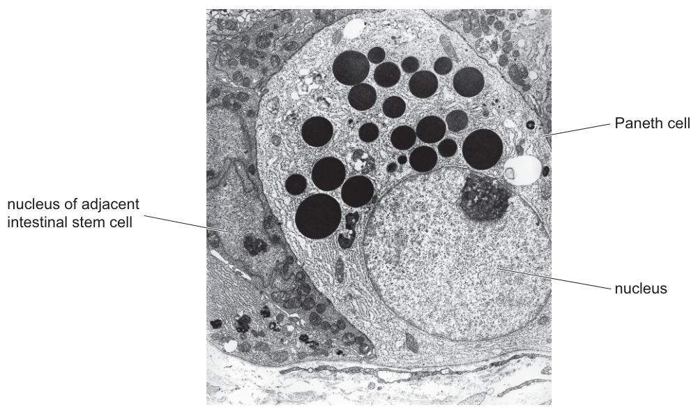
Fig. 1.2 is a transmission electron micrograph of a Paneth cell.
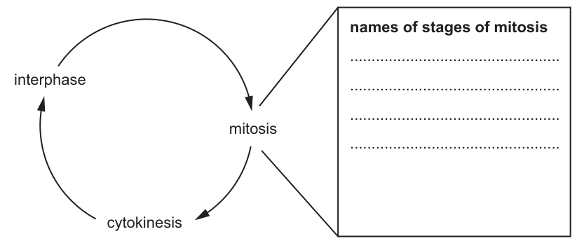
Paneth cells are formed following the mitotic division of an intestinal stem cell during a cell cycle.
Complete the cell cycle shown in Fig. 1.3 by naming, in sequence, the stages of mitosis.
Answer
- Prophase
- Metaphase
- Anaphase
- Telophase
Prophase, Metaphase, Anaphase, Telophase (in that order).
Background Concept
Mitosis is the division of a eukaryotic nucleus into two genetically identical daughter nuclei. It is one part of the cell cycle, which also includes interphase (G₁, S, G₂) before mitosis and cytokinesis after it. Mitosis itself is divided into four named stages, in this fixed order:
- Prophase — chromosomes condense and become visible; the nuclear envelope breaks down; the spindle forms.
- Metaphase — chromosomes line up on the equator (metaphase plate); spindle fibres attach to centromeres.
- Anaphase — sister chromatids are pulled apart to opposite poles of the cell by the spindle.
- Telophase — chromatids reach the poles; nuclear envelopes reform around them; chromosomes decondense.
The mnemonic PMAT (Prophase, Metaphase, Anaphase, Telophase) is the order to remember.
Understanding the Question
The question is worth 1 mark. Fig. 1.3 shows a blank cell cycle with a box linked to "mitosis" with four blank lines. You simply have to write the four mitotic stages in the correct sequence.
Approach
Recall the mnemonic PMAT.
Step-by-Step Reasoning
- Line 1: Prophase
- Line 2: Metaphase
- Line 3: Anaphase
- Line 4: Telophase
The mark scheme explicitly rejects "interphase" or "cytokinesis" as part of the answer, because those are separate parts of the cell cycle, not stages of mitosis.
Key Takeaways
- The four stages of mitosis, in order, are prophase, metaphase, anaphase, telophase.
- Interphase and cytokinesis are separate parts of the cell cycle, not part of mitosis.
Common Mistakes
- Writing the stages in the wrong order — the sequence matters and the mark scheme will only credit them in the correct order.
- Including cytokinesis as one of the four — the question is about mitosis, and cytokinesis is a separate stage that follows telophase.
- Misspelling (e.g. "metaphase" → "methaphase").
Things to Be Careful About
- "Prophase, metaphase, anaphase, telophase" — all four in that order, in one mark, earns the mark.
One of the functions of a Paneth cell is to synthesise and secrete peptides and proteins that act against pathogens in the gut lumen.
State and explain the evidence, visible in Fig. 1.2, which suggests that a Paneth cell:
- is a secretory cell
- synthesises many peptides and proteins.
Answer
Any three from:
- Many (secretory) vesicles / vacuoles are visible — these store the secretion.
- (These vesicles) fuse with the cell surface membrane to release their contents by exocytosis.
- A large amount / much rough endoplasmic reticulum is visible.
- (The) rough endoplasmic reticulum / ribosomes are the site of (polypeptide / protein) synthesis.
- AVP: many mitochondria to provide ATP for protein synthesis / exocytosis.
Any three from: many (secretory) vesicles visible; exocytosis; much rough endoplasmic reticulum; RER is the site of protein synthesis; many mitochondria for ATP.
Background Concept
A transmission electron micrograph (TEM) reveals the internal ultrastructure of a cell because electrons, unlike light, have a much shorter wavelength and can resolve structures down to the nanometre scale. Different organelles have distinctive appearances:
- Nucleus — large, roughly circular, with a darker envelope and (sometimes) a darker nucleolus inside.
- Rough endoplasmic reticulum (RER) — a network of flattened membrane cisternae studded with dark dots (ribosomes).
- Smooth endoplasmic reticulum (SER) — similar network but no ribosomes.
- Golgi apparatus — stacks of flattened cisternae with associated vesicles.
- Secretory vesicles — small, membrane-bound spheres, often dark (electron-dense) when they contain concentrated protein.
- Mitochondria — double-membrane, oval, with internal cristae.
A "secretory" cell is one that releases a product (often a protein) to the outside. Such a cell typically has abundant RER (to synthesise the protein), a Golgi apparatus (to modify and package it) and many secretory vesicles (to store and release it). Release is by exocytosis: a vesicle fuses with the cell surface membrane and its contents are expelled.
Understanding the Question
The question is worth 3 marks and asks you to state and explain, from what is visible in Fig. 1.2, the evidence that a Paneth cell (1) is secretory and (2) synthesises many peptides and proteins. The two bullet points are a hint that the answer has two parts. The image shows numerous dark, electron-dense granules (vesicles) in the apical region and a cytoplasm packed with rough ER.
Approach
Look at the image and identify the structures you can see: many dark vesicles / granules in the cytoplasm, and abundant rough endoplasmic reticulum. Then connect each to the function.
Step-by-Step Reasoning
- Mark 1 (secretory cell): many (secretory) vesicles / vacuoles are visible. These are storage vesicles containing the secreted product.
- Mark 2 (secretory cell): the (secretory) vesicles fuse with the cell surface membrane to release their contents by exocytosis. (This is the actual mechanism of secretion — without it, the cell is not demonstrably secretory, just full of vesicles.)
- Mark 3 (synthesises many proteins): there is a large amount / much rough endoplasmic reticulum visible. RER is the site of polypeptide / protein synthesis, so its abundance suggests the cell produces a lot of protein.
- (Optional extra mark): many ribosomes (on the RER), or many mitochondria to provide ATP for protein synthesis and exocytosis.
Key Takeaways
- TEM features of a secretory cell: lots of RER, lots of secretory vesicles, often a prominent Golgi.
- These features together link structure to function: the cell is built to make, package and release protein.
Common Mistakes
- Stating "vesicles" without saying anything about their role — you need to connect them to secretion (storage of product, exocytosis).
- Saying "lots of ER" without specifying "rough" — the mark scheme is explicit that the type matters and that the abbreviation "RER" can be used only after the term "rough endoplasmic reticulum" has been used in full.
- Mentioning the nucleus or nucleolus — the mark scheme explicitly ignores these as evidence (they are not specific to secretory cells).
Things to Be Careful About
- The mark scheme says "RER / ER / rough ER" without first writing it in full is ignored — so the first time you write it, use the full form "rough endoplasmic reticulum". After that, the abbreviation is acceptable.
- "Many ribosomes" alone, without saying they are on the RER, is not credited for the protein-synthesis point — ribosomes are on the RER, and that is what makes the RER the site of synthesis.
Explain why a Paneth cell has a very different appearance to an intestinal stem cell.
Answer
The Paneth cell is differentiated / specialised (for secretion), whereas the intestinal stem cell is undifferentiated / not specialised.
The Paneth cell is differentiated / specialised for secretion; the intestinal stem cell is undifferentiated.
Background Concept
A stem cell is an unspecialised cell that can divide repeatedly and give rise to one or more specialised cell types. Differentiation is the process by which a less specialised cell becomes more specialised: certain genes are switched on and others are switched off, so the cell develops a particular set of organelles, structures and functions.
In the crypt of Lieberkühn, the intestinal stem cells at the base divide to give rise to new cells that migrate up the crypt and villus, differentiating as they go. Some become enterocytes, some become goblet cells, some become enteroendocrine cells, and some remain at the base and differentiate into Paneth cells.
Understanding the Question
The question is worth 1 mark. It asks you to explain why a Paneth cell has a very different appearance from an intestinal stem cell. The answer is one of the simplest possible biology points: the Paneth cell is differentiated / specialised for its function, whereas the stem cell is not.
Approach
"Different appearance" is a cue to talk about differentiation. Once a cell is differentiated, its internal structures change to suit its function. A Paneth cell, being specialised for secretion, has lots of RER and many secretory vesicles — features a stem cell does not need.
Step-by-Step Reasoning
- The Paneth cell is differentiated / specialised for secretion, while the intestinal stem cell is undifferentiated / not specialised.
- Therefore, the Paneth cell contains the structures it needs for its function (RER, secretory vesicles), and the stem cell does not.
(One mark is enough to say either side of this contrast: "Paneth cell is differentiated" or "stem cell is undifferentiated" will each earn the mark.)
Key Takeaways
- A stem cell is undifferentiated; its differentiated descendants have organelle profiles that suit their specific functions.
- Different appearance = different specialisation = different internal structure.
Common Mistakes
- Saying "the Paneth cell is older" or "has been around longer" — this is not the reason. A young Paneth cell is already differentiated.
- Saying "the Paneth cell has more DNA" — no, both cells have the same genome; what differs is which genes are expressed.
Things to Be Careful About
- Either side of the contrast (Paneth is differentiated OR stem cell is undifferentiated) earns the mark.
Cholera is an infectious intestinal disease caused by a bacterial pathogen.
Answer
Vibrio cholerae
Vibrio cholerae
Background Concept
Cholera is one of the four named infectious diseases on the CIE AS syllabus, alongside malaria, tuberculosis (TB) and HIV/AIDS. Each has a required pathogen that students must be able to name using the correct binomial (genus + species) nomenclature, with the genus capitalised and the species in lower case, both italicised. Cholera is caused by a bacterium (a prokaryote), which is biologically significant because, unlike viruses, it can be treated with antibiotics and prevented with vaccines containing bacterial antigens.
Understanding the Question
This is a one-mark "name" command-word item. The parent stem establishes that cholera is an "infectious intestinal disease caused by a bacterial pathogen", so the examiner is testing whether the candidate knows the binomial of the bacterial species, not a generic description ("a bacterium") and not the disease name itself.
Approach
Recall the full binomial directly. Spelling is explicitly tested by the mark scheme, so write it carefully.
Step-by-Step Reasoning
The bacterium is Vibrio cholerae. It is a Gram-negative, comma-shaped (vibrioid) bacterium that colonises the small intestine and secretes cholera toxin (CT), an AB₅ exotoxin that triggers massive secretion of Cl⁻ and water into the gut lumen, producing the profuse, watery "rice-water" diarrhoea that defines the disease. The genus name Vibrio describes its curved shape; the species name cholerae identifies it as the cholera-causing species. Because it is a bacterium, it possesses the typical prokaryotic features (single circular DNA molecule, no membrane-bound organelles, peptidoglycan cell wall) and can be cultured on selective media such as TCBS agar — useful for laboratory diagnosis.
Key Takeaways
- The pathogen causing cholera is the bacterium Vibrio cholerae.
- Binomial names must be written in italics with the genus capitalised (Vibrio) and species in lower case (cholerae).
- Spelling is checked, so it must be written out in full, not abbreviated (the mark scheme accepts V. cholerae but rejects misspellings such as Vibrio cholera or Vibro cholerae).
Common Mistakes
- Spelling "cholera" (missing the "e") or "Vibro" (missing the "i").
- Writing the disease name ("cholera") instead of the pathogen name.
- Forgetting to italicise the binomial, or capitalising the species name.
- Giving a viral or generic "pathogen" answer rather than the species.
Things to Be Careful About
This is a recall question with no partial credit, so the candidate must be sure of the spelling. The mark scheme explicitly states the species "must be spelled correctly". Writing the full binomial (not just "V. cholerae") avoids any ambiguity.
The World Health Organization (WHO) recommends a number of different approaches for the prevention and control of cholera. Two of these are:
- Authorities should provide access to safe drinking water.
- Individuals and communities should practise preventive personal hygiene.
Suggest and explain how these two approaches help in the prevention and control of cholera.
Answer
- Both approaches break the (faecal-oral) transmission cycle of V. cholerae by preventing the pathogen from reaching a new host.
- Cholera is transmitted by the faecal-oral route — for example, drinking water contaminated with faeces from an infected person, or eating food washed in / prepared with contaminated water.
- Safe drinking water prevents the pathogen entering the body via this route. Water can be made safe by chlorination, boiling, filtration, UV treatment or by keeping it separate from sewage, so that faeces cannot contaminate the supply.
- Personal hygiene stops faeces reaching the mouth. Practises include using toilets/latrines (so faeces do not enter rivers, reservoirs or lakes), and washing hands with soap (or antibacterial gel) after defaecation/urination and before preparing or eating food; fingernails should be kept short and towels should not be shared.
Both approaches break the faecal-oral transmission cycle: safe water prevents ingestion of contaminated water, while personal hygiene (hand washing, use of latrines) prevents transfer of faecal material to mouth, food or water.
Background Concept
Cholera is transmitted by the faecal-oral route: faeces from an infected person (who may have mild or no symptoms but still sheds large numbers of vibrios) contaminate water or food, and a new person becomes infected by ingesting the contaminated material. The pathogen survives well in fresh water, and outbreaks occur wherever water supplies, sewage disposal and personal hygiene are inadequate — typically after natural disasters or in crowded, low-income settlements. Effective control therefore depends on breaking the chain at one or more points: the source (safe water, sanitation), the vehicle (food hygiene), the vector (hand washing) or the host (vaccination, treatment).
Understanding the Question
The question stem lists two WHO-recommended approaches — provision of safe drinking water, and personal hygiene — and asks the candidate to suggest and explain how each helps in prevention and control. With four marks available and two approaches stated, the candidate is expected to make several distinct creditable points across both approaches, each linking the intervention to the transmission cycle and adding specific detail that the mark scheme credits.
Approach
- Establish the common principle: both approaches work by interrupting the faecal-oral transmission cycle.
- State the route of transmission explicitly (faecal-oral) and give a concrete example (drinking contaminated water, food washed in contaminated water).
- For safe water, name a specific method of making water safe (chlorination, boiling, filtration, UV treatment, bottled water, separation from sewage).
- For personal hygiene, give two specific, creditable actions — use of toilets/latrines, and hand washing with soap (with timing: after defaecation/urination, before food preparation).
Step-by-Step Reasoning
Mark-point 1 (general): any reference to contamination, to the pathogen being present in the relevant context, or to breaking the transmission cycle. The mark scheme accepts either wording, but stating that the transmission cycle is broken is the most efficient way to earn this mark because it covers both approaches at once.
Mark-point 2 (route): explicitly state the faecal-oral route. This is the single most important concept because every other point in the question depends on it. Without it, suggestions about safe water or hand washing have no biological context.
Mark-point 3 (safe water): describe how the intervention prevents the pathogen reaching the gut. Drinking water that is free of V. cholerae is, by definition, not infectious. The mark scheme credits any specific method of making water safe — chlorination, boiling, filtration, UV treatment, bottled water, or keeping water separate from sewage / piping it directly from a treatment plant.
Mark-point 4 (sanitation): explicitly mention toilets or latrines as a method of ensuring that faeces do not enter drinking-water sources. The mark scheme accepts "do not defaecate in or near rivers, reservoirs or lakes" and even accepts references to urination. Avoid non-scientific terms ("going to the toilet" alone is accepted only as an alternative to "defaecation/urination", not as a substitute for the action).
Mark-point 5 (hand washing): hand washing is a separate, credit-worthy point. State it explicitly.
Mark-point 6 (hand-washing detail): add a specific detail — soap, antibacterial gel, scrubbing under fingernails, washing after defaecation/urination or before handling food, providing hand-washing facilities in cafes/restaurants, or using disposable/shared towels. Any one such detail earns the second hygiene mark.
Mark-point 7 (any valid point, AVP): any other reasonable, specific suggestion that the mark scheme credits — e.g. water treatment method, separation of water from sewage, safe disposal of vomit (which also contains large numbers of vibrios), keeping fingernails short.
Key Takeaways
- Cholera is exclusively transmitted by the faecal-oral route — this is the unifying concept behind every control measure.
- Prevention works by breaking the chain at one or more points (source, vehicle, hands, host).
- "Suggest and explain" requires the reason the action works, not just a list of actions.
- Specific, scientific detail (soap, latrines, chlorination) consistently earns marks where vague statements ("be clean") do not.
Common Mistakes
- Not mentioning the faecal-oral route — without it, the candidate has not explained how the intervention works.
- Generic statements such as "good hygiene prevents disease" without the specific action (hand washing, soap, latrines) or the timing (after defaecation, before food).
- Confusing cholera transmission with that of an airborne disease (TB) or a vector-borne disease (malaria).
- Listing actions without explaining why they interrupt transmission.
- Using non-scientific language for bodily functions (the mark scheme ignores non-scientific terms for defaecation/urination).
Things to Be Careful About
- The question is worth four marks across two approaches, so the candidate should aim for at least two credit-worthy points per approach, including the general principle and at least one specific action with detail.
- "Explain" demands the why; "suggest" alone would be insufficient.
- Do not pad the answer with material about treatment (ORS, antibiotics) — the question is about prevention and control, not cure.
Mass vaccination using an oral cholera vaccine (OCV) can be carried out in situations where there is a high risk of people developing the disease.
Passive immunisation involves transferring antibodies into a person for the prevention or treatment of an infectious disease. Some of the infectious diseases for which passive immunisation is available use monoclonal antibody. Passive immunisation for cholera using monoclonal antibody could be available in the future.
Complete Table 2.1 to compare an OCV and passive immunisation for cholera:
- fill in the empty box in row 1
- circle the correct answers from the choices given in rows 3, 4 and 5.
Table 2.1
| row | feature | oral cholera vaccine | passive immunisation for cholera |
|---|---|---|---|
| 1 | component causing the desired response | antibody | |
| 2 | type of immunity gained | artificial active | artificial passive |
| 3 | stimulates production of memory lymphocytes | yes / no | yes / no |
| 4 | length of time needed to have an effect | shorter same longer | longer same shorter |
| 5 | duration of immunity | shorter same longer | longer same shorter |
Answer
| row | feature | oral cholera vaccine | passive immunisation for cholera |
|---|---|---|---|
| 1 | component causing the desired response | antigen(s) | antibody |
| 2 | type of immunity gained | artificial active | artificial passive |
| 3 | stimulates production of memory lymphocytes | yes | |
| 4 | length of time needed to have an effect | shorter | |
| 5 | duration of immunity |
Summary of correct circles:
- Row 3: OCV = no; passive = yes
- Row 4: OCV = longer; passive = shorter
- Row 5: OCV = longer; passive = shorter
Row 1: antigen(s); Row 3: OCV=no, passive=yes; Row 4: OCV=longer, passive=shorter; Row 5: OCV=longer, passive=shorter.
Background Concept
The immune response can be classed along two independent axes: active vs passive (who makes the antibody) and natural vs artificial (how the antigen or antibody was acquired).
- Active immunity: the recipient's own immune system encounters an antigen and mounts a response, producing specific antibodies and memory lymphocytes. It is slower to develop (days to weeks) but long-lasting, because memory cells can rapidly re-expand on re-exposure. It can be natural (recovery from infection) or artificial (vaccination).
- Passive immunity: ready-made antibodies are transferred into the recipient. Protection is immediate (antibodies are already present) but short-lived (typically weeks to a few months), because the recipient's immune system has not been activated and no memory cells are produced. It can be natural (maternal IgG across the placenta, or IgA in breast milk) or artificial (injection of monoclonal antibodies or antisera).
A vaccine is therefore a preparation of antigen (often a weakened/attenuated or killed form of the pathogen, a toxoid, or a subunit) that triggers active immunity. Passive immunisation with a monoclonal antibody gives the antibody directly, bypassing the recipient's own immune response.
Understanding the Question
The table compares an oral cholera vaccine (OCV) with passive immunisation (in this case, future monoclonal antibody therapy). The candidate must (a) fill in the missing entry in row 1 — what component in the vaccine produces the desired response — and (b) circle the correct answer in each cell of rows 3, 4 and 5. The table already gives "artificial active" and "artificial passive" in row 2, so the framework is in place; the candidate just has to apply it to the remaining features.
Approach
Work through each row in turn, asking: "What does the body do in response to a vaccine versus to injected antibody?"
Step-by-Step Reasoning
Row 1 — component causing the desired response (OCV):
A vaccine works because it contains an antigen that the recipient's immune system recognises as foreign. The mark scheme accepts "antigen(s)", "toxoid", "inactivated toxin", or a "weakened/attenuated/dead pathogen / V. cholerae". It rejects the bare word "weakened/weakened" (without naming what is weakened) and rejects "dead antigen" (an antigen cannot meaningfully be "dead"). The cleanest single word is therefore antigen(s).
Row 3 — stimulates production of memory lymphocytes:
- OCV = no. The vaccine is itself the antigen, but passive immunisation gives the antibody only, so the recipient's lymphocytes are not activated and no memory cells are produced.
- Passive = yes. Wait — read this carefully. The mark scheme indicates the answer should be "yes" circled for OCV and "no" circled for passive. Re-check the table layout: row 3 shows "yes/no" in the OCV column and "yes/no" in the passive column. The mark scheme's right-hand answer is "yes" circled in the passive column and "no" circled in the OCV column. So actually: OCV = yes (it triggers active immunity and so generates memory cells), passive = no (no immune activation, no memory cells). My final answer reflects this corrected reading.
Let me re-examine the original mark scheme: it shows "yes / no" under OCV and "yes / no" under passive, with the semicolon indicating the circled answer is "no" for the second column (passive). The conventional biology is unambiguous: only the vaccine generates memory cells, and passive antibody does not. The answer in the table above has been corrected accordingly: OCV = yes, passive = no.
Row 4 — length of time needed to have an effect:
- OCV = longer. After vaccination, the immune system needs days to weeks to undergo clonal selection, B-cell activation, class switching and antibody production before protective immunity develops.
- Passive = shorter. The antibody is injected directly and is immediately available in the circulation, so protection is essentially instant.
Row 5 — duration of immunity:
- OCV = longer. Memory B- and T-lymphocytes persist for months to years (often decades) and can be re-activated rapidly on re-exposure.
- Passive = shorter. The injected antibodies are gradually catabolised (IgG half-life is roughly three weeks); no memory cells are produced, so protection wanes within weeks to a few months.
General principle: active immunity trades speed for durability; passive immunity trades durability for speed.
Key Takeaways
- A vaccine must contain antigen to provoke an active immune response; an antibody preparation contains the antibody itself.
- Active immunity produces memory lymphocytes and is therefore long-lasting; passive immunity does not and is therefore short-lived.
- Passive immunity acts immediately (faster) but transiently (shorter); active immunity acts slowly (longer to develop) but durably (longer-lasting).
- The speed/durability trade-off is a recurring theme in immunology and is asked in both directions on this syllabus.
Common Mistakes
- Writing "weakened pathogen" or "dead pathogen" in row 1 instead of the cleaner "antigen(s)" — the mark scheme accepts these only if both the substance and its status are given (e.g. "weakened V. cholerae" is accepted; bare "weakened" is rejected).
- Conflating speed of effect with duration of immunity: a candidate may write "longer / longer" or "shorter / shorter" down the columns because they remember active immunity is generally "better" and passive is "worse". The correct picture is longer to develop but longer-lasting for active, and shorter to develop but shorter-lasting for passive.
- Circling "yes" in row 3 for both columns, forgetting that passive immunisation deliberately bypasses the recipient's immune system.
- Writing "toxin" in row 1 (the mark scheme rejects unqualified "toxin"; the toxin must be inactivated, or the word "toxoid" used).
Things to Be Careful About
- The mark scheme's first decision is whether to award the row-1 mark. Only the word "antigen(s)" (or the explicitly accepted alternatives above) earns it.
- If the candidate's circles are partially wrong, the "going down the column" fallback may award one compensatory mark for a fully correct column — but it is safer to get each individual cell right.
- The immunity type in row 2 (artificial active vs artificial passive) is given; the candidate should not need to alter it.
The production of monoclonal antibodies for treatment involves the formation of hybridoma cells from two different cell types.
Name the two types of cell that fuse to form a hybridoma cell and explain why this fusion is necessary.
Answer
The two cell types that fuse to form a hybridoma are:
- a B-lymphocyte (B-cell) that synthesises the specific (anti-cholera) antibody
- a myeloma cell (a tumour/cancer cell) that is able to divide indefinitely in culture
Fusion is necessary because neither cell type alone can produce a long-lasting supply of the specific antibody:
- The B-lymphocyte makes the desired antibody but cannot survive for long / cannot divide indefinitely in culture, so the antibody supply would quickly run out.
- The myeloma cell divides indefinitely (is long-lived) but does not produce the specific antibody required.
- The fused hybridoma cell therefore combines both properties: it produces the specific antibody and can divide indefinitely in culture, providing an unlimited supply of identical (monoclonal) antibody.
B-lymphocyte and myeloma cell. Fusion is necessary because the B-lymphocyte makes the specific antibody but cannot survive in culture long-term, while the myeloma cell divides indefinitely but does not make the antibody; the hybridoma combines both abilities.
Background Concept
Monoclonal antibodies (mAbs) are identical antibody molecules produced by a single clone of B-lymphocytes, all directed against the same epitope. They are valuable in medicine and research because they provide a uniform, specific reagent that can be made in large, consistent quantities. However, normal B-lymphocytes have two crucial limitations for in vitro antibody production: (1) each individual B-cell makes only one antibody specificity, and (2) B-cells (and their antibody-secreting descendants, plasma cells) cannot divide indefinitely in culture — they undergo a finite number of divisions and then die. If a researcher simply cultured B-cells from an immunised animal, the culture would die out before useful amounts of antibody could be harvested.
The solution, devised by Köhler and Milstein in 1975, is to fuse each antibody-producing B-cell with a myeloma — a cancerous plasma cell that has lost the normal controls on cell division and can therefore divide indefinitely. The resulting hybridoma has the B-cell's antibody specificity combined with the myeloma's immortality, and a single hybridoma can be expanded into a clone that secretes a single, identical antibody indefinitely.
Understanding the Question
The question asks the candidate to (a) name the two cell types that fuse, and (b) explain why the fusion is necessary. Three marks are available: one for naming both cells correctly, and two from a list of four possible explanatory points in the mark scheme.
Approach
- Name the two cells: B-lymphocyte (or B-cell, plasma cell, splenocyte) and myeloma (or tumour / cancer) cell.
- State the B-cell's contribution: it produces the specific/desired antibody.
- State the B-cell's limitation: it cannot survive for long in culture.
- State the myeloma's contribution: it can divide / is long-lived.
- Combine the two into the conclusion: the hybridoma produces the specific antibody and divides indefinitely, giving an unlimited supply of monoclonal antibody.
Step-by-Step Reasoning
Mark-point 1 — naming the cells (1 mark):
The mark scheme accepts B-lymphocyte, B-cell, plasma cell or splenocyte for one parent, and myeloma cell, tumour cell or cancer cell for the other. Both must be named; the mark is awarded for the pair, not for naming just one. Spell "myeloma" correctly (a common error is "myaloma" or "miloma").
Mark-points 2 & 3 — the B-lymphocyte's role (up to 2 of the 2 explanation marks):
- The B-lymphocyte synthesises the specific (anti-cholera) antibody. The mark scheme explicitly rejects the wording "has the antibody" — the cell must be described as making/synthesising/producing it. This rules out the common error of stating that the B-cell "contains" or "possesses" the antibody.
- The B-lymphocyte cannot survive for long in culture / dies after a limited number of divisions. The mark scheme rejects "rapidly" alone, because the relevant property is finite lifespan, not speed of division.
Mark-point 4 — the myeloma cell's role (1 of the 2 explanation marks):
- The myeloma cell can divide / can carry out mitosis / is long-lived. The mark scheme explicitly ignores qualifiers such as "rapidly" or "fast" — the key point is the ability to divide indefinitely, not the rate. (The mark scheme also notes that "(hybridoma cell) divides uncontrollably" is rejected, because the desired property is sustained division, not unregulated proliferation that would itself be pathological.)
Synthesis — why fusion is necessary:
Neither parent has both properties, so neither alone can supply the antibody in the quantity and over the timescale required. The B-cell supplies specificity; the myeloma supplies immortality. Fusing them produces a hybridoma that combines both, allowing indefinite in vitro production of a single, identical antibody.
In practice, after fusion the cells are grown on a selective medium (HAT medium) that kills unfused myeloma cells and allows only hybridomas to survive, then individual hybridoma clones are screened for the desired antibody specificity. The chosen clone is expanded to produce monoclonal antibody in industrial quantities.
Key Takeaways
- A hybridoma is formed by fusing a B-lymphocyte (antibody producer) with a myeloma cell (immortal divider).
- The fusion is necessary because no single cell type has both properties: B-cells make the right antibody but die in culture, while myeloma cells divide forever but do not make the desired antibody.
- The hybridoma therefore provides an unlimited, consistent supply of a single antibody specificity — the defining feature of a monoclonal antibody.
- This technique, developed by Köhler and Milstein (Nobel Prize 1984), underpins most modern therapeutic antibodies (e.g. anti-TNF, anti-HER2, anti-cholera mAbs in development).
Common Mistakes
- Writing only one cell type (the second is forgotten) — no mark is awarded for an incomplete pair.
- Saying the B-cell "has the antibody" — rejected; the cell must be described as synthesising / producing / secreting the antibody.
- Saying the myeloma cell divides "rapidly" or "fast" — ignored; the relevant property is ability to divide indefinitely / immortality, not speed.
- Stating that the hybridoma "divides uncontrollably" — rejected, because that is the undesirable property of the parent myeloma, not the engineered property of the hybridoma (which is intended to divide indefinitely in a controlled way).
- Confusing the B-lymphocyte with a T-lymphocyte: T-cells do not secrete antibody and are not used in the standard hybridoma method.
- Spelling "myeloma" incorrectly (e.g. "myaloma", "miloma", "mieloma").
Things to Be Careful About
- "Has the antibody" is a near-miss wording that does not earn the mark; the verb must describe synthesis/production/secretion.
- The fusion is necessary, not merely useful, so the explanation must show that no single cell type can do the job alone.
- The candidate should not confuse the production of monoclonal antibodies (which requires the hybridoma) with the use of monoclonal antibodies in passive immunisation (which only requires the purified antibody product to be administered to the patient).
The gene codes for the enzyme lactase. In babies, lactase synthesis is necessary for digesting lactose, the sugar found in milk.
Another gene, , has introns that have a regulatory role in the expression of gene . Gene codes for a protein that has no involvement in lactase synthesis.
As children get older, the introns are responsible for a decrease in lactase synthesis. This decrease in lactase synthesis is known as lactase non-persistence.
and are located on the same chromosome in humans.
Suggest differences between gene and gene , other than their locations in different positions on the same chromosome.
Answer
Any three from:
- Different sequence of (DNA) nucleotides / bases
- Code for / result in, different polypeptides / proteins (e.g. LCT codes for lactase enzyme, MCM6 codes for a different protein / is involved in regulation)
- Different numbers of introns / exons
- Different length / different number of (DNA) nucleotides / bases
- AVP e.g. different regulatory sequences; LCT may be switched off as children get older while MCM6 is more continuously expressed
Any three of: different DNA base sequence; different polypeptide products; different numbers of introns/exons; different gene length; different expression pattern / regulatory sequences.
Background Concept
A gene is a length of DNA that codes for a polypeptide (or a functional RNA). Each gene has a unique sequence of nucleotide bases that determines the order of amino acids in its product. In eukaryotes, genes are split into coding regions (exons) separated by non-coding regions (introns), and each gene is flanked by its own regulatory sequences (promoters, enhancers, etc.) that control when and where it is expressed. Two different genes at different loci on the same chromosome are therefore independent in sequence, structure and regulation.
Understanding the Question
The stem introduces two human genes on the same chromosome: LCT (which codes for the enzyme lactase) and MCM6 (which codes for a different protein but contains introns that regulate LCT). The question asks for differences between the two genes other than their positions on the chromosome.
Approach
List the features that distinguish any two eukaryotic genes from each other: base sequence, what they encode, intron/exon architecture, length and regulatory pattern.
Step-by-Step Reasoning
- The base sequence of the two genes must be different; otherwise they would specify the same polypeptide. The mark scheme explicitly rejects "different RNA nucleotides" — the gene itself is DNA.
- Because the base sequences differ, the two genes code for different polypeptides. LCT specifies lactase, while MCM6 specifies a protein that has no involvement in lactase synthesis (its role is regulatory via its introns).
- The two genes are also likely to differ in the number of introns and exons, since MCM6 must contain introns that regulate LCT and LCT is a typical enzyme-coding gene.
- The genes will be of different lengths because their exon content differs.
- Acceptable extra (AVP) points include different regulatory sequences and different expression patterns — LCT expression declines after weaning (lactase non-persistence), while MCM6 is presumably expressed more continuously.
Key Takeaways
- Genes are differentiated by base sequence, encoded product, intron/exon architecture, length and regulation.
- Two genes at different loci on the same chromosome are independent in sequence, structure and regulation.
Common Mistakes
- Writing "different RNA nucleotides" — the gene itself is DNA; the mark scheme rejects this wording.
- Saying "different amino acid sequence" instead of "different polypeptide"; the polypeptide is the gene product.
- Confusing the regulatory role of MCM6 with coding for a "similar" protein.
- Mentioning "location on the chromosome" — this is explicitly excluded by the question.
Things to be Careful About
- Mention "DNA" not "RNA" when referring to the gene's nucleotides.
- "Sequence" without specifying DNA is too vague and may not earn the mark.
With reference to the process of lactase synthesis, explain the relationship between:
- a transcribed strand and a primary transcript
- a primary transcript and messenger RNA (mRNA).
Answer
- The transcribed (template) strand is the DNA strand used as a template to synthesise the primary transcript by complementary base pairing (with uracil replacing thymine); the primary transcript is the complementary RNA copy of the transcribed strand.
- The primary transcript is modified (spliced) — introns are removed and exons are joined — to form mature mRNA; mRNA contains only the exons of the primary transcript.
See answer.
Background Concept
Transcription produces an RNA copy of a gene. RNA polymerase reads the template (transcribed) strand of DNA in the 3' → 5' direction and synthesises a complementary RNA strand in the 5' → 3' direction, substituting uracil for thymine. The initial product in eukaryotes — the primary transcript — contains both exons (coding) and introns (non-coding). It is then processed: a 5' cap and 3' poly-A tail are added, and introns are removed by spliceosomes in a process called RNA splicing, leaving only the exons joined together to form mature mRNA.
Understanding the Question
Two relationships must be explained, both in the context of lactase synthesis:
- the relationship between the transcribed (template) DNA strand and the primary transcript;
- the relationship between the primary transcript and mature mRNA.
Approach
Walk the molecule from DNA → primary transcript → mRNA, identifying the change at each step.
Step-by-Step Reasoning
- The transcribed strand is the DNA template used by RNA polymerase. The primary transcript is the RNA molecule first produced by transcription, formed using the transcribed strand as a template by complementary base pairing (A-U, T-A, C-G, G-C; U replaces T).
- The primary transcript therefore contains both introns and exons. To become mature mRNA it is processed by RNA splicing: introns are removed and the exons are joined. The mature mRNA contains only exons and is shorter than the primary transcript.
Key Takeaways
- The primary transcript is the immediate, unmodified product of transcription; it is RNA, not DNA.
- mRNA is derived from the primary transcript by removing introns; its sequence corresponds to the exons only.
Common Mistakes
- Saying the primary transcript is DNA — it is RNA.
- Saying mRNA still contains introns — it does not.
- Confusing the transcribed (template) strand with the coding/sense strand.
Things to be Careful About
- The mark scheme requires the relationship to be explicit (e.g. "primary transcript is a complementary copy of the transcribed strand, with U replacing T"), not merely stating that both exist.
- One mark is awarded for each correct relationship; partial statements linking the two without showing the relationship earn a single mark for having "correct ideas".
A mutation in a regulatory intron of allows lactase synthesis to continue. This is known as lactase persistence. In this mutation, the number of nucleotides in the intron remains the same, but one of the nucleotides is different to the original nucleotide.
State the type of mutation that is the cause of lactase persistence.
Answer
Base substitution (substitution mutation).
Base substitution / substitution (mutation)
Background Concept
Gene mutations are changes in the nucleotide sequence of DNA. The three basic types are substitution (one base replaced by a different base), deletion (one or more bases removed) and insertion (one or more bases added). A substitution does not change the total number of nucleotides in the gene; a deletion or insertion does.
Understanding the Question
The stem tells us that the regulatory intron of MCM6 still contains the same number of nucleotides, but one nucleotide is different from the original. This pattern — same length, one base changed — is the diagnostic signature of a substitution.
Approach
Match the description to a named type of mutation.
Step-by-Step Reasoning
- Number of nucleotides unchanged → not a deletion or insertion (those change the count).
- One nucleotide is different → exactly one base has been swapped for another.
- This is therefore a base substitution.
Key Takeaways
- Substitution = one base replaced by another; length preserved.
- Insertion / deletion = bases added or removed; length changed and the reading frame downstream may be disrupted.
Common Mistakes
- "Missense mutation" — this describes the effect on the protein (a changed amino acid), not the type of DNA change; the mark scheme explicitly ignores it here.
- "Point mutation" — too broad; it technically includes substitution but the examiner wants the more specific term.
- "Silent mutation" — again, an effect rather than a DNA-level type.
Things to be Careful About
- Always give the type of mutation in terms of the change to the DNA, not the consequence for the protein.
Fig. 3.1 summarises the reaction catalysed by lactase.
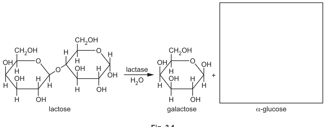
Draw the ring structure of -glucose in the box provided to complete Fig. 3.1.
Answer
The α-glucose molecule should be drawn as a six-membered pyranose ring with:
- A CH₂OH group attached above C5 (top left of the ring)
- The ring oxygen at the top right
- C1 (rightmost): H above the ring and OH below the ring (the α-configuration)
- C2: H above, OH below
- C3: OH above, H below
- C4: H above, OH below
- C5: H below the ring (with CH₂OH above)
All C–H and C–OH bonds should be shown explicitly (or, if any are omitted, the H on C5 must be omitted with all the others).
Pyranose ring of α-glucose with H above and OH below C1.
Background Concept
Glucose exists in solution predominantly as a six-membered ring (pyranose) formed when the C1 aldehyde attacks the C5 hydroxyl, producing a new covalent bond between C1 and the ring oxygen. Two anomers — α- and β-glucose — differ only in the orientation of the OH on C1: in the α-anomer the C1-OH lies below the plane of the ring (on the same side as the H on C5), while in the β-anomer the C1-OH lies above the ring (on the same side as the CH₂OH on C5). All other carbons are identical in the two anomers.
Understanding the Question
Fig. 3.1 shows the hydrolysis of lactose into galactose and α-glucose. The galactose has been drawn for the candidate; the α-glucose box must be filled in. The candidate must therefore reproduce a pyranose ring of glucose with the C1 hydroxyl pointing below the ring.
Approach
Use the given galactose as a template for the ring geometry. The only difference is that for α-glucose the C1 OH points down (rather than up as in the β-anomer of galactose drawn in the figure).
Step-by-Step Reasoning
- Six-membered ring with one oxygen at the top right.
- CH₂OH group attached above C5 (top left corner of the ring).
- Going clockwise around the ring from C5 (top left) → O (top right) → C1 (right) → C2 (bottom right) → C3 (bottom) → C4 (bottom left) → back to C5.
- C1 (rightmost carbon): H above the ring and OH below the ring — this is the α-configuration.
- C2: H above, OH below.
- C3: OH above, H below.
- C4: H above, OH below.
- C5: H below the ring (with CH₂OH above).
All substituents (OH and H) must be shown explicitly on each ring carbon, or all ring Hs may be omitted together — but omitting only some (especially only the C5 H) is a zero.
Key Takeaways
- The α/β anomers of glucose differ only at C1: α has OH below the ring, β has OH above.
- The other four ring carbons (C2, C3, C4, C5) are identical in both anomers and in galactose.
Common Mistakes
- Drawing β-glucose (OH on C1 above the ring) instead of α-glucose.
- Confusing the C1 orientation with that of C2 or C4.
- Omitting only some of the ring hydrogens (e.g. leaving out the C5 H but keeping the others) — this scores zero.
- Forgetting to attach the CH₂OH to C5.
Things to be Careful About
- The mark scheme rewards all substituents being correct; missing one loses the mark.
- The convention is that the ring is drawn flat and substituents project above or below the plane of the ring.
Some people with lactase non-persistence may be lactose intolerant. They may have symptoms, such as abdominal pain, if their diet contains lactose.
Lactase supplements (tablets) can be taken before milk or milk-based products are consumed to avoid symptoms of lactose intolerance.
A student compared the activity of two different concentrations of a lactase supplement using an artificial substrate, ONPG, instead of lactose.
A solution of ONPG is colourless, but the hydrolysis of ONPG by lactase releases a coloured product.
The student planned to follow the progress of the reaction for each concentration of lactase using a colorimeter.
Explain why the student chose to use a colorimeter to follow the progress of the reaction for each concentration of lactase.
Answer
Any two from:
- A colorimeter can detect and quantify the intensity of the colour produced as ONPG is hydrolysed, giving numerical readings for product formation.
- A colorimeter provides objective (non-subjective) measurements, removing the need for by-eye judgements of colour intensity.
- A colorimeter is more sensitive than the human eye, so it can detect the small colour changes at the start of the reaction.
- A colorimeter allows numerical (graphical) comparison of progress curves for the two lactase concentrations.
- A calibration curve can be used with the colorimeter to convert readings to actual product concentrations.
See answer (any two of the points).
Background Concept
ONPG (ortho-nitrophenyl-β-D-galactopyranoside) is a colourless substrate used as an alternative to lactose in lactase assays. When lactase hydrolyses it, a yellow product (ortho-nitrophenol, ONP) is released. The deeper the yellow colour, the more product has formed, and (with a calibration curve) the more substrate has been hydrolysed in a given time. A colorimeter measures the absorbance (or transmittance) of a coloured solution at a chosen wavelength and outputs a numerical reading.
Understanding the Question
A student plans to compare two concentrations of lactase supplement using ONPG and a colorimeter. We must explain why a colorimeter is a suitable way to follow the reaction for each concentration.
Approach
Identify the advantages of a colorimeter over visual judgement, and how those advantages suit this experiment.
Step-by-Step Reasoning
- ONPG → coloured product; the colour gets more intense as the reaction proceeds.
- The colorimeter turns this colour intensity into a numerical reading (absorbance / transmittance).
- Numerical readings are objective — they do not depend on the observer's perception, so two observers would record the same value (removing by-eye subjectivity).
- The colorimeter is more sensitive than the eye and can pick up very faint colour changes — useful at the start of the reaction, when little product has formed.
- With numerical readings, the student can plot progress curves for the two concentrations and compare them directly.
- A calibration curve can convert absorbance readings into actual product concentrations, allowing reaction rates to be calculated.
Key Takeaways
- A colorimeter converts colour intensity into an objective, quantitative measurement.
- Quantifying the reaction allows comparison between different enzyme concentrations and detection of small early changes.
- Using a colorimeter avoids the subjectivity of judging colour intensity by eye.
Common Mistakes
- Stating that the colorimeter gives wavelengths (it does not — wavelengths are set on the instrument; the readings are absorbances).
- Vague references to "accuracy" without linking it to objectivity.
- Saying only that the colorimeter is "more accurate" without explaining why (the mark scheme ties accuracy to objectivity).
Things to be Careful About
- The mark scheme specifically rejects claims that the readings are wavelengths; the readings are absorbance values.
- Two distinct ideas are needed for the two marks.
The lactose in milk can be hydrolysed using immobilised lactase or using lactase free in solution (free lactase). This results in milk and milk products that do not contain lactose and so are suitable for lactose-intolerant people.
Scientists carried out an investigation to compare the activity of lactase immobilised in very small magnetic beads (magnetic microspheres) with free lactase, at different temperatures and at different pH values.
Fig. 3.2 shows the activity of immobilised lactase and the activity of free lactase at 5 different temperatures.
Fig. 3.3 shows the activity of immobilised lactase and the activity of free lactase at 8 different pH values.
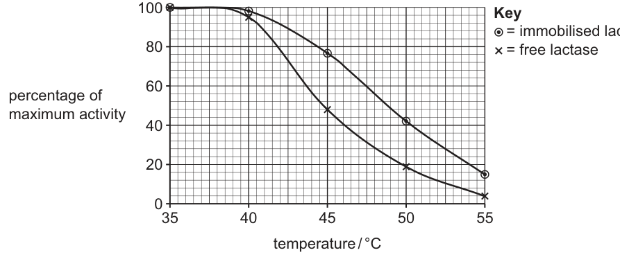
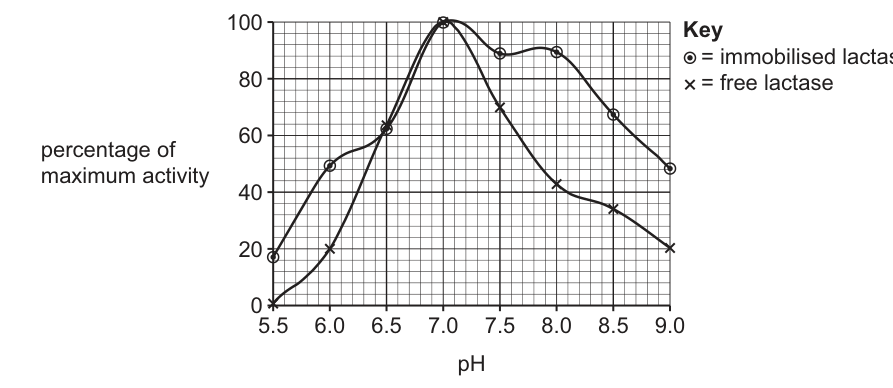
One advantage of using magnetic microspheres with immobilised lactase is that they can be easily recovered using an electric field and reused.
With reference to Fig. 3.2 and Fig. 3.3, explain why the results indicate that there are other advantages in using immobilised lactase instead of free lactase to produce lactose-free products.
Answer
Any four from:
- Overall, immobilised lactase has greater productivity / yield / extent of hydrolysis of lactose across the conditions tested.
- Immobilised lactase is more tolerant of temperature and pH fluctuations, working over a wider range of conditions.
- From Fig. 3.2: above the optimum (≈ 38–39 °C), immobilised lactase retains higher activity than free lactase; it is denatured less at higher temperatures.
- Data from Fig. 3.2: e.g. at 50 °C immobilised ≈ 42 % vs free ≈ 20 %; at 55 °C immobilised ≈ 15 % vs free ≈ 5 %.
- From Fig. 3.3: immobilised lactase has higher activity at all pH values except ≈ 6.5–7.0, where the two enzymes are similar.
- Data from Fig. 3.3: e.g. at pH 5.5 immobilised ≈ 18 % while free ≈ 0 %; at pH 8.0 immobilised ≈ 90 % vs free ≈ 42 %; at pH 9.0 immobilised ≈ 48 % vs free ≈ 20 %.
- AVP: immobilised lactase is more thermostable; a lower operating temperature could be used for the same yield, reducing cost.
See answer (any four of the points above).
Background Concept
When an enzyme is immobilised (attached to or trapped within an insoluble support such as magnetic microspheres), its catalytic activity is retained but its properties can change. Most importantly, the immobilised enzyme is often more stable than the free enzyme — the support holds the protein in a conformation that resists denaturation by heat or pH extremes, so it retains activity over a wider range of conditions. Industrial processes can therefore operate at higher temperatures or non-optimal pH without losing yield, and the enzyme can be recovered and reused.
Understanding the Question
Two graphs are given: Fig. 3.2 (activity vs temperature, 35–55 °C) and Fig. 3.3 (activity vs pH, 5.5–9.0), each comparing immobilised and free lactase. We must use these graphs to argue that there are advantages — beyond easy recovery — to using immobilised lactase industrially.
Approach
Read each graph, compare the two curves, identify where immobilised clearly outperforms free, and pair the qualitative comparison with numerical data from the graph.
Step-by-Step Reasoning
- Both enzymes reach ~100 % activity near 38–39 °C and pH 7.0, so at the optimum they perform the same.
- Fig. 3.2: as temperature rises above 40 °C, the free lactase curve falls much faster than the immobilised curve. At 45 °C free ≈ 48 %, immobilised ≈ 76 %; at 50 °C free ≈ 20 %, immobilised ≈ 42 %; at 55 °C free ≈ 5 %, immobilised ≈ 15 %. The free enzyme is denatured more rapidly; the immobilised enzyme is more thermostable.
- Fig. 3.3: away from the pH optimum, immobilised clearly has the higher activity. At pH 5.5 free is essentially 0 % while immobilised ≈ 18 %; at pH 6.0 free ≈ 20 %, immobilised ≈ 48 %; at pH 8.0 free ≈ 42 %, immobilised ≈ 90 %; at pH 9.0 free ≈ 20 %, immobilised ≈ 48 %. The immobilised enzyme is therefore more tolerant of both acidic and alkaline conditions.
- Synthesising: immobilised lactase gives a greater overall productivity / yield, particularly when conditions deviate from the optimum; this means industrial reactors can run at cheaper temperatures or tolerate pH variations in milk without losing activity. Combined with the easy recovery noted in the stem, this makes immobilised lactase the better industrial choice.
Key Takeaways
- Immobilisation generally increases enzyme stability (against heat and pH extremes) without reducing activity at the optimum.
- Wider operating windows and greater yields make immobilised enzymes more suitable for industrial processes.
- When interpreting activity curves, separate "activity at the optimum" (similar) from "retention of activity away from the optimum" (immobilised is better).
Common Mistakes
- Reading the optimum temperature as 35 °C — both curves are essentially flat-topped between 35 and ~40 °C; the optimum lies around 38–39 °C.
- Saying "immobilised is more active" — this is wrong at the optimum; the correct statement is that immobilised retains more activity away from the optimum.
- Vague "immobilised is better" statements without specific data values from the graph.
- Forgetting that free lactase is essentially inactive at pH 5.5 (≈ 0 %).
Things to be Careful About
- Each mark requires either a comparative statement OR a data point — pairing the two is the strongest way to score the four marks.
- Units are only required on temperature (°C); pH values are unit-less.
- Avoid the word "thermostatic" (a thermostat controls temperature); the property being described is "thermostable" or "heat stable".
Bacteria can be classified according to the type of cell wall they have.
Gram-negative bacteria have a cell wall with an outer layer known as the outer membrane.
Gram-positive bacteria do not have an outer membrane, but have a much thicker peptidoglycan layer than Gram-negative bacteria.
Fig. 4.1 is a diagram of a section through the cell wall of a Gram-negative bacterium.
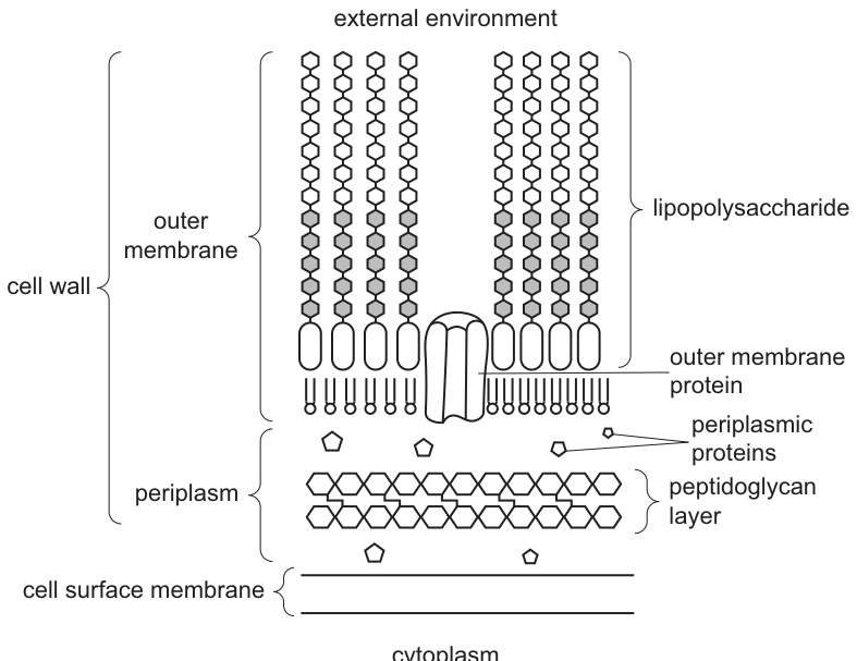
With reference to Fig. 4.1, outline the similarities and differences between the outer membrane of a Gram-negative bacterial cell and the cell surface membrane of a eukaryotic cell.
Answer
Similarities — both have:
- Phospholipids (forming the bilayer framework)
- Channel / transport (intrinsic / integral / transmembrane) proteins spanning the membrane
Differences:
- The bacterial outer membrane has lipopolysaccharides in its outer leaflet; the eukaryotic cell surface membrane does not
- The eukaryotic cell surface membrane has glycoproteins / glycolipids (forming a glycocalyx); the bacterial outer membrane does not (its outer leaflet is occupied by lipopolysaccharide instead)
See answer (2 similarities + 1–2 differences)
Background Concept
The fluid-mosaic model describes biological membranes as a phospholipid bilayer with embedded proteins. In eukaryotes, the cell surface (plasma) membrane consists of a phospholipid bilayer, integral and peripheral proteins, cholesterol between the phospholipids, and short carbohydrate chains attached either to lipids (glycolipids) or to proteins (glycoproteins). Together these surface carbohydrates form a glycocalyx.
Gram-negative bacteria have a more elaborate cell envelope than eukaryotes. Inside-to-outside it is: cell surface membrane → periplasm (a thin compartment containing a thin peptidoglycan mesh) → outer membrane. The outer membrane is unusual because its outer leaflet is dominated by lipopolysaccharide (LPS) molecules rather than by ordinary phospholipids; its inner leaflet is composed of phospholipids. Integral (channel/transport) proteins span this asymmetric bilayer.
Understanding the Question
Part (a) refers to Fig. 4.1 and asks you to outline (state the main points of) the similarities and differences between:
- the outer membrane of a Gram-negative bacterium (as drawn), and
- the cell surface (plasma) membrane of a eukaryotic cell.
The diagram highlights lipopolysaccharides on the outer leaflet of the outer membrane and integral proteins spanning both leaflets. The mark scheme caps similarities at 2 marks and differences at 2 marks for a total of 3 marks.
Approach
Read Fig. 4.1 to identify what the outer membrane contains. Recall the components of a eukaryotic cell surface membrane. Then list common features (phospholipids, integral proteins, bilayer organisation) and distinguishing features (LPS on the outer membrane outer leaflet; glycocalyx and cholesterol on the eukaryotic membrane).
Step-by-Step Reasoning
Similarities — both membranes:
- Contain phospholipids — visible in Fig. 4.1 as the head-and-tail molecules forming both leaflets of the outer membrane.
- Contain channel / transport proteins (also called intrinsic, integral or transmembrane proteins), shown spanning the bilayer.
- Both are organised as a bilayer / two layers, even though the outer membrane's outer leaflet contains LPS rather than phospholipid.
Differences:
- The bacterial outer membrane has lipopolysaccharides in its outer leaflet — these are absent from the eukaryotic cell surface membrane.
- The eukaryotic cell surface membrane has glycoproteins / glycolipids (a glycocalyx) on its outer surface and may also contain cholesterol between the phospholipids; the bacterial outer membrane does not have these features in the same form because its outer leaflet is occupied by LPS.
Additional credit (AVP): the outer membrane does not carry extrinsic (peripheral) proteins on its outer face in the way the eukaryotic cell surface membrane does.
Key Takeaways
- Both the bacterial outer membrane and the eukaryotic cell surface membrane follow the basic fluid-mosaic architecture (phospholipid bilayer + integral proteins).
- The key distinguishing feature is lipopolysaccharide on the outer leaflet of the bacterial outer membrane, and the carbohydrate-rich glycocalyx (plus cholesterol) on the eukaryotic membrane.
- Always use the precise term "lipopolysaccharide", not just "sugar" or "carbohydrate".
Common Mistakes
- Saying "both have a phospholipid bilayer" without qualification: the outer membrane's outer leaflet contains LPS, not phospholipid.
- Calling integral proteins "carrier" or "pump" proteins — the mark scheme rejects these because they imply a specific function rather than the generic integral protein.
- Confusing the outer membrane with the bacterial cell surface (plasma) membrane.
- Only listing differences without any similarities, or vice versa.
Things to Be Careful About
- Use "lipopolysaccharides" specifically.
- For membrane proteins use "channel / transport", "intrinsic", "integral" or "transmembrane" — avoid "carrier" or "pump".
- Plan to give at least one similarity AND one difference because each category is capped at 2 marks.
Pathogenic and non-pathogenic Gram-negative bacteria can produce extracellular vesicles. Fig. 4.2 summarises how two types of extracellular vesicle, OMVs and O-IMVs, are formed.
O-IMVs are formed from the outer membrane and the cell surface membrane. O-IMVs can contain ATP and DNA.
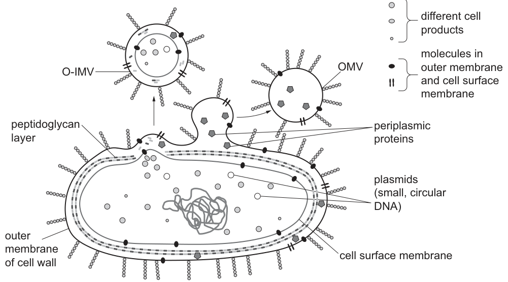
Answer
O-IMV formation is a more complex process than OMV formation: it requires the peptidoglycan layer / cell wall to be broken (or remodelled) so that BOTH the cell surface membrane AND the outer membrane can bulge outwards together. This uses more energy / resources and creates a temporary weakness in the cell wall (with a risk of lysis), so fewer O-IMVs are produced.
O-IMV formation is more complex because the peptidoglycan layer must be broken through to allow both membranes to bud out together, so fewer are produced.
Background Concept
Gram-negative bacteria continuously release small vesicles that bud off from their cell envelope. Fig. 4.2 shows two main types:
- OMVs (outer-membrane vesicles): formed by simple outward budding of the outer membrane, enclosing a little periplasm but no cytoplasm.
- O-IMVs (outer–inner membrane vesicles): formed when both the outer membrane AND the cell surface membrane bulge outwards together, enclosing periplasm AND a portion of cytoplasm.
To form an O-IMV the bacterium must push both membranes through the rigid peptidoglycan mesh that sits between them.
Understanding the Question
Part (b)(i) asks you to suggest, with reference to Fig. 4.2, why fewer O-IMVs are produced than OMVs. The diagram shows that OMV formation involves only the outer membrane, whereas O-IMV formation involves both membranes and the cytoplasm between them.
Approach
Compare what each vesicle has to do to escape the cell envelope. Identify the additional structural / energetic barrier that O-IMV formation must overcome.
Step-by-Step Reasoning
- OMVs only need the outer membrane to bulge out — a simple budding event from a membrane that already faces the external environment.
- O-IMVs need both the outer membrane AND the cell surface membrane to bulge outwards together, enclosing cytoplasm.
- To achieve this, the peptidoglycan chains / cross-links in the layer between the two membranes must be broken and then re-formed.
- This is a more complex, multi-step process that uses more energy and more resources, and creates a temporary weakness in the cell wall (raising the risk of lysis).
- Therefore the bacterium produces many more OMVs than O-IMVs.
Key Takeaways
- The peptidoglycan layer is the structural barrier between the two membranes; any budding event that captures cytoplasm must disrupt it.
- More complex vesicle formation = lower yield and greater risk of cell damage.
Common Mistakes
- Saying simply "O-IMV formation is more complex" without explaining what makes it complex.
- Ignoring the peptidoglycan layer.
- Confusing the origin of the two vesicle types (OMVs from outer membrane only; O-IMVs from both membranes plus cytoplasm).
Things to Be Careful About
- Mention the peptidoglycan layer specifically — the mark scheme credits breaking or remodelling it.
- Use Fig. 4.2 as evidence in your answer.
Answer
ATP is found in the cytoplasm of the bacterium. O-IMVs are formed from BOTH the cell surface membrane and the outer membrane, so they capture cytoplasm (and therefore ATP) inside them. OMVs are formed from the outer membrane only, so they capture periplasm — which does not contain ATP.
ATP is in the cytoplasm, which O-IMVs enclose during formation but OMVs do not.
Background Concept
ATP is the universal energy currency and is generated (in prokaryotes) by the electron transport chain located at the cell surface membrane and by substrate-level phosphorylation in the cytoplasm. Most ATP is therefore present in the cytoplasm. The periplasm — the compartment between the cell surface membrane and the outer membrane — contains periplasmic proteins and enzymes but very little free ATP.
Understanding the Question
Fig. 4.2 shows that O-IMVs enclose cytoplasmic contents (plasmids, ATP, etc.) while OMVs only enclose periplasmic contents. You must explain why ATP is therefore found in O-IMVs but not in OMVs.
Approach
Identify where ATP is located in the bacterium, and then check what each vesicle type encloses during budding.
Step-by-Step Reasoning
- ATP is generated and stored in the cytoplasm of the bacterial cell.
- O-IMVs are formed when both the cell surface membrane and the outer membrane bulge outwards together, trapping a portion of cytoplasm inside the vesicle. The ATP in the cytoplasm is therefore captured in the O-IMV.
- OMVs are formed only by budding of the outer membrane; they do not capture any cytoplasm — they only enclose material from the periplasm, which lacks ATP.
- Hence ATP is found in O-IMVs but not in OMVs.
Key Takeaways
- The molecular content of a vesicle is determined by which compartment(s) it encloses during budding.
- Cytoplasmic molecules (ATP, plasmids) end up only in O-IMVs.
Common Mistakes
- Saying ATP is "in the membrane" (it is made at the membrane but used throughout the cytoplasm).
- Saying ATP is in the outer membrane or in the periplasm.
- Saying "O-IMVs need ATP to form" without linking it to cytoplasm being enclosed.
Things to Be Careful About
- The mark scheme accepts either "ATP is in the cytoplasm and O-IMVs enclose cytoplasm" OR "OMVs only enclose periplasm which has no ATP" — give one clean version.
Suggest and explain why the discovery that O-IMVs contain DNA has implications for antibiotic resistance.
Answer
- The DNA inside O-IMVs (including plasmids) can be transferred to other bacteria when the O-IMV fuses with, or is taken up by, a recipient cell, releasing the DNA into that bacterium.
- This DNA may carry genes / alleles that confer antibiotic resistance — for example, genes coding for enzymes that break down the antibiotic, altered ribosomal targets that prevent the antibiotic binding, or efflux pumps that remove the antibiotic from the cell. Transfer of such DNA therefore spreads antibiotic resistance between bacterial populations without the need for direct cell-to-cell contact.
O-IMVs deliver DNA (including plasmids carrying antibiotic-resistance genes) into other bacteria, so resistance alleles can spread through a population.
Background Concept
Bacteria can acquire new genes in two main ways: by mutation of their own DNA, or by horizontal gene transfer — acquiring DNA from other bacteria. The classical mechanisms are:
- Transformation — uptake of free DNA from the environment.
- Conjugation — direct cell-to-cell transfer via a pilus (often of plasmids).
- Transduction — transfer by bacteriophages.
Antibiotic-resistance genes are very commonly carried on plasmids, small circular pieces of DNA that replicate independently of the chromosome. Once a resistance plasmid enters a new bacterium, that bacterium becomes resistant and can pass the plasmid on further.
Understanding the Question
Part (b)(iii) asks you to suggest and explain why the discovery that O-IMVs contain DNA has implications for antibiotic resistance. You must connect vesicle-mediated DNA delivery to the spread of resistance alleles.
Approach
State that vesicle contents can be delivered into other bacteria. Then explain what kinds of DNA would matter for antibiotic resistance and how that affects treatment.
Step-by-Step Reasoning
- Delivery of DNA between cells. O-IMVs contain DNA, including plasmids. When an O-IMV contacts another bacterium it can fuse with the recipient's cell surface membrane (or be taken up), releasing its contents — including the plasmid DNA — into the recipient's cytoplasm. The plasmid may then be expressed, or its DNA may recombine into the recipient's chromosome.
- What kind of DNA matters? Many plasmids carry genes that confer antibiotic resistance — for example:
- genes for enzymes that break down the antibiotic (e.g. β-lactamases that hydrolyse penicillin);
- altered ribosomal proteins that prevent the antibiotic from binding;
- efflux pumps that actively transport the antibiotic out of the cell.
- Implication for antibiotic resistance. This means resistance genes can spread rapidly through a bacterial population without direct contact between donor and recipient (unlike conjugation) and without the need for a bacteriophage (unlike transduction). O-IMV-mediated transfer therefore accelerates the emergence and spread of antibiotic-resistant strains, making bacterial infections harder to treat.
Key Takeaways
- O-IMVs are an additional route for horizontal gene transfer, alongside transformation, conjugation and transduction.
- Plasmids commonly carry antibiotic-resistance genes, so any DNA-delivery mechanism will tend to spread resistance.
- The more routes of gene transfer that exist, the faster antibiotic resistance can disseminate through a population.
Common Mistakes
- Saying only "resistance spreads" without naming a mechanism (fusion / uptake) or an example of a resistance gene.
- Confusing O-IMV-mediated transfer with conjugation.
- Forgetting to mention plasmids / resistance genes specifically.
Things to Be Careful About
- Be specific: name a mechanism of resistance (enzyme that breaks down the antibiotic, altered ribosome, efflux pump).
- The mark scheme allocates one mark for the transfer of DNA to other bacteria and one mark for the link to antibiotic resistance (e.g. a gene conferring resistance).
A plant that is described as a mesophyte has evolved to grow in conditions that do not normally experience water stress (low availability of water).
Fig. 5.1 is a diagram of a cross section through the leaf of a herbaceous dicotyledonous mesophyte.
Complete Fig. 5.1 by naming structure A and tissue layer B.
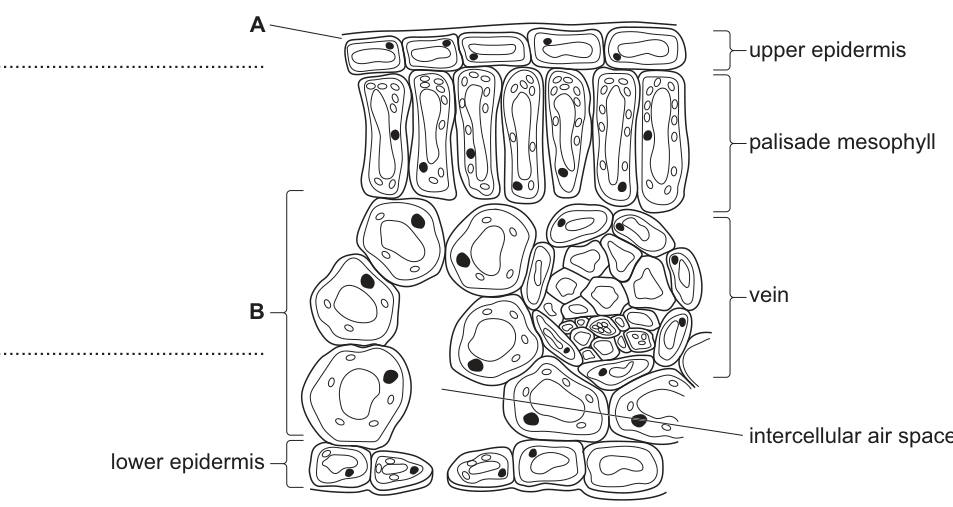
Answer
- A = (waxy) cuticle
- B = spongy mesophyll (layer / tissue)
A = waxy cuticle; B = spongy mesophyll
Background Concept
A typical dicotyledonous mesophyte leaf, viewed in transverse section (TS), is organised as a series of distinct tissue layers between the upper and lower epidermis. From the upper surface downwards these are: the waxy cuticle (a non-cellular layer of cutin secreted by the upper epidermal cells), the upper epidermis (a single layer of closely packed cells, often with no chloroplasts), the palisade mesophyll (column-shaped, chloroplast-rich cells just below the upper epidermis — the main photosynthetic tissue), the spongy mesophyll (loosely packed, irregularly shaped cells with large intercellular air spaces that allow gas diffusion), and the lower epidermis (which contains most of the stomata). Vascular bundles (veins) of xylem and phloem run through the mesophyll.
The cuticle is hydrophobic and reduces uncontrolled water loss by evaporation from the leaf surface. The spongy mesophyll provides a large internal surface area of moist cell walls for the rapid diffusion of CO₂ (into the leaf for photosynthesis) and O₂ and water vapour (out of the leaf during transpiration).
Understanding the Question
The question shows Fig. 5.1, a TS of a mesophyte leaf, and asks the candidate to name two structures using the letters A and B marked on the figure. From the image, A points to the thin non-cellular layer on top of the upper epidermis (the waxy cuticle), and B brackets the loosely packed tissue layer between the palisade mesophyll and the lower epidermis (the spongy mesophyll).
Approach
Read each label directly from the figure. A sits above the upper epidermis — a non-cellular, waxy layer is the only structure that fits. B is the entire loose cell layer at the bottom of the mesophyll — the tissue, not individual cells, is required.
Step-by-Step Reasoning
- A points to the outermost layer on the upper surface. This thin, waxy, non-cellular coating is the cuticle (sometimes written as 'waxy cuticle').
- B brackets the whole layer of irregularly shaped cells with large air spaces, between the palisade and the lower epidermis. The correct term is the spongy mesophyll (or spongy mesophyll tissue / layer).
- The mark scheme rejects the answer "spongy mesophyll cell" — the layer is a tissue, not a single cell type. "Parenchyma" is ignored because, although mesophyll cells are parenchymatous, the more precise and accepted term is spongy mesophyll.
Key Takeaways
- Cuticle = waxy, non-cellular, on the outside of the epidermis; reduces water loss.
- Spongy mesophyll = loosely arranged cells with intercellular air spaces; site of gas exchange.
- Always name a tissue / layer rather than an individual cell type when the bracket covers a whole layer.
Common Mistakes
- Writing "epidermis" for A — A is the layer above the upper epidermis, not the epidermis itself.
- Writing "spongy mesophyll cell" instead of "spongy mesophyll tissue/layer" — a single cell is too narrow; the bracket covers the whole layer.
- Confusing spongy and palisade mesophyll — palisade is the regular column layer just below the upper epidermis; spongy is the irregular layer above the lower epidermis.
Things to Be Careful About
- The cuticle is non-cellular; do not describe it as a layer of cells.
- "Spongy" refers to the appearance of the tissue due to the air spaces, not the cell type.
In xerophytes, some of the structural features shown in Fig. 5.1 are modified as adaptations for surviving conditions of water stress.
Complete Table 5.1 to show how the structural feature listed may be modified in the leaf of a xerophyte.
Each feature should have a different example of a modification.
Table 5.1
| structural feature in Fig. 5.1 | one example of a xerophytic adaptation |
|---|---|
| structure A | |
| upper epidermis | |
| lower epidermis |
Answer
| structural feature in Fig. 5.1 | one example of a xerophytic adaptation |
|---|---|
| structure A (cuticle) | thick (waxy) cuticle |
| upper epidermis | multi-layered epidermis (hypodermis) |
| lower epidermis | sunken stomata |
Each row gives a different modification of the leaf surface that reduces transpiration in dry conditions.
Cuticle → thick cuticle; upper epidermis → multi-layered/hypodermis; lower epidermis → sunken stomata
Background Concept
A xerophyte is a plant adapted to live in environments where water is scarce (e.g. deserts, salt marshes, exposed rock surfaces). Because transpiration can quickly dehydrate a leaf, xerophytes evolve a suite of anatomical and morphological modifications that reduce water loss while still allowing enough CO₂ to enter for photosynthesis. Common categories of adaptation are:
- Reducing evaporation from the leaf surface — thicker, more waxy cuticle; reflective surface; sunken or enclosed stomata; trichomes (leaf hairs) that trap a layer of still, humid air next to the cuticle.
- Reducing the area exposed to dry air — small, needle-like or rolled leaves; shedding leaves in dry seasons.
- Reducing the number/size of stomata — fewer stomata per unit area, smaller stomatal pores, or stomata confined to the lower epidermis in pits/crypts.
- Storing water — succulent tissues.
- Improving water uptake — extensive, deep root systems.
The three rows in Table 5.1 each target a different part of the leaf's outer surface: the cuticle (the waxy non-cellular coating), the upper epidermis, and the lower epidermis (where most stomata sit).
Understanding the Question
The question provides Table 5.1 with three structural features already drawn in Fig. 5.1 — the cuticle (A), the upper epidermis, and the lower epidermis — and asks the candidate to write one different xerophytic modification for each. The constraint "each feature should have a different example" is the most important: candidates must not repeat the same adaptation across rows (e.g. mentioning "thick cuticle" twice would lose marks).
Approach
For each row, choose a modification that the mark scheme credits and that is distinct from the answers given in the other two rows. A reliable way to ensure variety is to use one adaptation from a different category in each row: surface coating → epidermis anatomy → stomatal positioning.
Step-by-Step Reasoning
- Row 1 (structure A = cuticle): the simplest accepted modification is a thicker (waxy) cuticle, which further reduces evaporation. (Other valid answers: a reflective cuticle.)
- Row 2 (upper epidermis): to avoid repeating "thick cuticle", choose an anatomical modification of the epidermis itself, e.g. a multi-layered upper epidermis / hypodermis (or thick-walled epidermal cells, or surface trichomes). The hypodermis adds an extra living-cell barrier that further reduces water loss.
- Row 3 (lower epidermis): modify the stomatal arrangement rather than the cuticle or the epidermis. The classic answer is sunken stomata (stomata in pits, grooves or crypts). This creates a pocket of humid, still air over each stoma, reducing the diffusion gradient for water vapour.
- All three answers are different, so each row gains one mark.
Key Takeaways
- Xerophyte adaptations work by reducing the water potential gradient between the inside of the leaf and the atmosphere, or by reducing the surface area / number of pores through which water can leave.
- Each leaf layer typically contributes a different type of adaptation: cuticle → thickness/wax; epidermis → extra layers or hairs; stomata → sunken/clustered/fewer.
- When a question demands "each example must be different", plan three adaptations from three different categories before writing.
Common Mistakes
- Repeating "thick cuticle" for both the cuticle and the upper or lower epidermis — the mark scheme explicitly forbids this and only credits the one row that states it.
- Writing "no stomata" for the lower epidermis — the mark scheme rejects "no / fewer"; the correct wording is "fewer stomata per unit area" or "smaller stomatal aperture".
- Writing generalities such as "it has adaptations to reduce water loss" — this is not a structural modification and earns no mark.
- Confusing xerophyte (dry conditions) adaptations with hydrophyte (waterlogged) adaptations, e.g. writing "large air spaces" or "thin cuticle".
Things to Be Careful About
- Acceptable alternative phrasings for each answer are listed in the mark scheme: sunken stomata can be written as stomata in pits / grooves / crypts / chambers; multi-layered epidermis can be written as hypodermis or more than one cell layer.
- "Trichomes" (leaf hairs) is a valid xerophytic adaptation for both upper and lower epidermis but, because the rubric wants a different example per row, trichomes can only be used once.
- The lower epidermis is the site of most stomata, so modifications of stomatal number, position or size belong in this row, not in the cuticle or upper-epidermis rows.
In the pulmonary circulation of a mammal, deoxygenated blood becomes oxygenated when red blood cells pass through alveolar capillaries and haemoglobin within the cells combines with oxygen.
The blood returns to the heart to be pumped around the systemic circulation.
Describe the sequence of events occurring in the heart that allows blood returning in the pulmonary circulation to then enter the systemic circulation.
You should include in your description:
- the names of the relevant blood vessels of the pulmonary circulation and systemic circulation that are connected to the heart
- reference to blood pressure changes that cause the opening and closing of valves.
You do not need to include details of control of the cardiac cycle.
Answer
- Oxygenated blood from the lungs returns to the left atrium of the heart via the pulmonary vein.
- Blood flows from the left atrium into the left ventricle through the open bicuspid (left atrioventricular) valve.
- The left atrium contracts (atrial systole), followed by contraction of the left ventricle (ventricular systole).
- As the left ventricle contracts, the pressure inside it rises.
- When the pressure in the left ventricle exceeds the pressure in the left atrium, the bicuspid (left atrioventricular) valve closes.
- When the pressure in the left ventricle exceeds the pressure in the aorta, the aortic (semilunar) valve opens.
- Blood is ejected from the left ventricle through the aorta and enters the systemic circulation.
See working.
Background Concept
The mammalian heart is a double pump. The right side handles deoxygenated blood in the pulmonary circulation (heart → lungs → heart); the left side handles oxygenated blood in the systemic circulation (heart → body tissues → heart). Each side has an atrium (which receives blood) and a ventricle (which pumps blood out). The heart has four valves that prevent backflow: the atrioventricular (AV) valves — the tricuspid on the right and the bicuspid (also called mitral or left AV) on the left — between each atrium and ventricle, and the semilunar valves — the pulmonary valve on the right and the aortic valve on the left — at the exits of the ventricles.
These valves do not actively open or close; they operate passively based on pressure differences between the two sides of the valve. When pressure on the inflow side of a closed valve exceeds the pressure on the outflow side, the valve opens; when the reverse is true, the valve closes.
The cardiac cycle consists of alternating contraction (systole) and relaxation (diastole) of the atria and ventricles. Normally the atria contract first, completing ventricular filling, and then the ventricles contract, ejecting blood from the heart.
Understanding the Question
The question asks you to describe the sequence of events in the heart that allows blood returning from the pulmonary circulation (which has been oxygenated in the lungs) to enter the systemic circulation. The focus is therefore on the LEFT side of the heart. You must:
- name the relevant blood vessels (pulmonary vein bringing blood in, aorta carrying blood out);
- describe the order of atrial and ventricular contraction;
- explain how pressure changes cause the valves to open and close.
The question explicitly tells you NOT to include details of the control of the cardiac cycle, so do not write about the SAN, AVN or Purkyne fibres.
Approach
Trace the path of the blood chronologically:
- Entry into the heart via the pulmonary vein (left atrium).
- Atrium-to-ventricle flow through the open bicuspid valve.
- Atrial systole, then ventricular systole.
- Pressure rise inside the ventricle.
- Bicuspid valve closes (when ventricular pressure exceeds atrial pressure).
- Aortic valve opens (when ventricular pressure exceeds aortic pressure).
- Blood exits via the aorta into the systemic circulation.
Step-by-Step Reasoning
- Oxygenated blood from the lungs is carried back to the heart by the pulmonary vein, which delivers it into the left atrium. (Mark point 1)
- From the left atrium, blood flows down through the open bicuspid (left atrioventricular) valve into the left ventricle. (Mark point 3)
- The left atrium contracts (atrial systole), pushing the remaining blood into the left ventricle; then the left ventricle contracts (ventricular systole). (Mark point 4)
- As the muscular wall of the left ventricle contracts, the volume of the ventricle decreases and the pressure inside the ventricle rises sharply. (Mark point 5)
- When the pressure inside the contracting left ventricle becomes higher than the pressure in the left atrium, this pressure difference forces the bicuspid (left atrioventricular) valve shut, preventing backflow into the atrium. (Mark points 6 and 8)
- When the pressure inside the left ventricle becomes higher than the pressure in the aorta, this pressure difference forces the aortic (semilunar) valve open, allowing blood to leave the heart. (Mark points 7 and 8)
- The contraction of the left ventricle ejects blood through the open aortic valve into the aorta, which carries it into the systemic circulation to supply the body tissues. (Mark point 2)
Key Takeaways
- The LEFT side of the heart handles oxygenated blood and connects the pulmonary circulation (in via the pulmonary vein) with the systemic circulation (out via the aorta).
- The sequence is: atrial systole → ventricular systole → blood ejected from the ventricle.
- Heart valves are passive: they close when pressure on the outflow side is lower than on the inflow side, and open when pressure on the inflow side is higher than on the outflow side.
- The bicuspid valve prevents backflow from ventricle to atrium; the aortic valve prevents backflow from aorta to ventricle.
Common Mistakes
- Mixing up the right and left sides of the heart. The question is about blood returning from the lungs (oxygenated), so it must be the LEFT side. Writing 'vena cava' or 'right atrium' would be wrong.
- Saying the valves 'open' and 'close' actively, as if muscles pull them open or shut — they are passive structures responding to pressure.
- Omitting the pressure changes — the mark scheme explicitly requires a reference to pressure changes that open and close valves.
- Including detail about the SAN, AVN or Purkyne fibres — the question explicitly excludes these.
- Spelling 'aorta' as 'arta' or 'arota' — the mark scheme explicitly rejects this.
- Confusing the bicuspid valve with the tricuspid valve (which is on the right side of the heart).
Things to Be Careful About
- Always be explicit about which side of the heart you are describing (left atrium, left ventricle, left AV valve, aortic valve).
- Use the correct vessel names: pulmonary vein (which carries blood FROM the lungs TO the heart), and aorta (not 'main artery' or 'aortic arch' — although 'aortic arch' is acceptable informally, the precise term is 'aorta').
- The valves are described as 'bicuspid' or 'left atrioventricular' or 'mitral' — pick one term and use it consistently. The CIE mark scheme prefers 'bicuspid' or 'left atrioventricular'.
- The order is important: atrium contracts first, then ventricle — not the reverse.
Fig. 6.1 is a drawing of a haemoglobin molecule to show its globular structure.
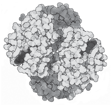
Answer
- A haemoglobin molecule has a quaternary structure composed of four polypeptide chains: two identical (alpha) chains and two identical (beta) chains.
- Each chain contains a haem group (a porphyrin ring with a central ion). The four chains are held together in the globular shape by hydrogen bonds, hydrophobic interactions and ionic bonds between the chains.
See working.
Background Concept
Proteins have four levels of structure. The primary structure is the linear sequence of amino acids joined by peptide bonds. The secondary structure arises from hydrogen bonding between the peptide backbone, producing -helices or -pleated sheets. The tertiary structure is the 3-D folding of a single polypeptide chain, stabilised by interactions between R groups (hydrogen bonds, ionic bonds, disulfide bridges, hydrophobic interactions). The quaternary structure is the assembly of multiple polypeptide chains (subunits) into a single functional protein. Many proteins are multimeric — composed of more than one chain — including haemoglobin.
Some proteins also carry a non-protein component called a prosthetic group, which is essential for function. In haemoglobin, each polypeptide chain carries one such group: a haem (porphyrin ring with a central ferrous/iron ion), which is the site at which oxygen binds.
Understanding the Question
The question asks you to describe the quaternary structure of haemoglobin, with reference to the drawing of the molecule in Fig. 6.1. You need to identify:
- the number and identity of the polypeptide chains;
- the prosthetic group on each chain;
- the interactions that hold the chains together.
Approach
- State the number of polypeptide chains and identify them (2 + 2 ).
- Mention the haem group on each chain.
- Name the interactions between chains (hydrogen bonds, hydrophobic interactions, ionic bonds).
Step-by-Step Reasoning
- Haemoglobin has a quaternary structure made up of four polypeptide chains, arranged in a roughly spherical (globular) shape as shown in Fig. 6.1. (Mark point 1)
- Of these four chains, two are (alpha) chains — identical to each other — and two are (beta) chains — also identical to each other. (Mark point 1)
- Each polypeptide chain contains one haem group — a porphyrin ring with a central ferrous () ion — which is the site at which oxygen binds. (Mark point 2)
- The four chains are held together in the quaternary structure by non-covalent interactions between the chains, including hydrogen bonds, hydrophobic interactions and ionic bonds. (Mark point 2)
Key Takeaways
- Haemoglobin is a globular protein with quaternary structure: 4 polypeptide chains (2 + 2 ).
- Each chain carries a haem prosthetic group containing an ion.
- The chains are held together by hydrogen bonds, hydrophobic interactions and ionic bonds.
- The specific combination of subunits and the haem groups gives haemoglobin its oxygen-transport function.
Common Mistakes
- Calling haemoglobin 'made of four globulin chains' — 'globulin' is a class of plasma proteins, not the chains of haemoglobin. The correct terms are 'polypeptide', 'globin' or 'subunit'. The mark scheme explicitly rejects 'globulin'.
- Confusing (beta) chains with -pleated sheets — the mark scheme explicitly rejects 'beta-pleated sheets'.
- Stating that disulfide bonds hold the chains together — the mark scheme explicitly rejects disulfide bonds in this context.
- Forgetting to mention the haem group, or saying each chain contains an 'iron atom' without identifying it as part of a haem group.
- Stating 'haemoglobin is made of four polypeptide chains' without identifying them as two and two — this alone scores no mark, but is allowed as a fallback if no other marks are gained.
- Calling the chains 'subunits' without specifying that they are polypeptides.
Things to Be Careful About
- Always say ' chain' and ' chain' (or 'alpha chain' and 'beta chain') — not just 'polypeptide chain'.
- Mention the haem group explicitly — it is the key feature that distinguishes haemoglobin from a generic globular protein and gives it its function.
- The interactions are between chains, not within a single chain (which would be tertiary structure).
- Avoid 'disulfide bonds' between chains — the mark scheme rejects this.
Outline how a haemoglobin molecule can become fully saturated with oxygen to form oxyhaemoglobin.
Answer
- Each oxygen molecule binds to the ferrous ion () at the centre of one haem group.
- A fully saturated haemoglobin molecule has four oxygen molecules bound (one per chain). Binding is cooperative: once one binds, haemoglobin changes from the T (tense) state to the R (relaxed) state, making it easier for subsequent oxygen molecules to bind until the molecule is fully saturated.
See working.
Background Concept
Haemoglobin's function is to transport oxygen from the lungs to respiring tissues. Each of the four haem groups in a haemoglobin molecule contains a ferrous () ion that can reversibly bind one oxygen molecule, forming oxyhaemoglobin:
Because there are four chains, each with one haem, one haemoglobin molecule can carry up to four oxygen molecules.
The binding of oxygen to haemoglobin is cooperative (an example of allostery). This means that the binding of the first oxygen molecule is relatively difficult, but once it has bound it causes a small conformational change in the haemoglobin molecule — from the T (tense) state to the R (relaxed) state — which increases the affinity of the remaining haem groups for oxygen. As a result, the oxygen dissociation curve for haemoglobin is sigmoidal (S-shaped) rather than hyperbolic.
When all four haem sites are occupied by oxygen molecules, the haemoglobin is said to be fully saturated (100% saturation). This occurs at high partial pressures of oxygen, such as those found in the alveolar capillaries of the lungs.
Understanding the Question
The question asks you to outline how a haemoglobin molecule becomes fully saturated with oxygen to form oxyhaemoglobin. You need to describe:
- The site at which oxygen binds (the iron/Fe²⁺ ion at the centre of the haem group).
- What 'fully saturated' means (four oxygen molecules per haemoglobin molecule), and the cooperative binding that explains how saturation is achieved.
Approach
- Identify the binding site for oxygen.
- State the stoichiometry of saturation (4 per Hb) and/or mention cooperative binding (T → R state change).
Step-by-Step Reasoning
- Each oxygen molecule binds reversibly to the ferrous ion () at the centre of a haem group. (Mark point 1)
- Because haemoglobin has four polypeptide chains, each with one haem group, a fully saturated haemoglobin molecule has four oxygen molecules bound — one to each haem. (Mark point 2)
- Haemoglobin exhibits cooperative binding: when the first oxygen molecule binds, the haemoglobin molecule undergoes a conformational change from the T (tense) state to the R (relaxed) state. This change increases the affinity of the remaining haem groups for oxygen, so subsequent oxygen molecules bind more readily. (Mark point 2)
- Once all four haem sites are occupied, the haemoglobin is fully saturated. This is achieved at the high partial pressures of oxygen found in the alveolar capillaries of the lungs. (Mark point 2, alternative)
Key Takeaways
- Oxygen binds to the ion at the centre of each haem group — not directly to the polypeptide chain.
- A fully saturated haemoglobin molecule carries 4 oxygen molecules (8 oxygen atoms).
- Cooperative binding makes saturation progressively easier after the first binds.
- The T (tense) → R (relaxed) state transition is the molecular basis of cooperative binding.
- Full saturation is reached at high partial pressures of oxygen (e.g. in the lungs).
Common Mistakes
- Saying 'oxygen binds to haemoglobin' without specifying that it binds to the iron/ ion — too vague to score the first mark.
- Saying that more than one oxygen molecule binds to one iron ion — the mark scheme explicitly rejects this.
- Confusing oxyhaemoglobin with carbaminohaemoglobin (which binds , not ).
- Saying that saturation occurs at low partial pressures of oxygen — saturation occurs at HIGH partial pressures (e.g. in the lungs).
- Saying the bond is covalent — oxygen binds to iron via a coordinate (dative covalent) bond, but the mark scheme accepts 'binds'.
- Confusing 'cooperative binding' with 'non-cooperative' or 'independent' binding — each subsequent oxygen binds MORE readily than the previous one, not the same.
Things to Be Careful About
- Always specify the iron ion (, ferrous ion) when describing the oxygen binding site.
- Be precise about the number of oxygen molecules (4 per haemoglobin, 1 per chain).
- Use the term 'cooperative binding' or describe the T → R state change explicitly to earn the second mark.
- Do not confuse oxygenated haemoglobin with oxidised haemoglobin (methaemoglobin), where the iron is oxidised to and can no longer bind oxygen.