Biology 9700/21 — October/November 2025
Cambridge AS Level · AS Level Structured Questions · worked solutions for every part, with the mark scheme
Topics Biological Molecules · Cell Membranes and Transport · Transport in Mammals · Enzymes · Infectious Diseases · Immunity · +5 more
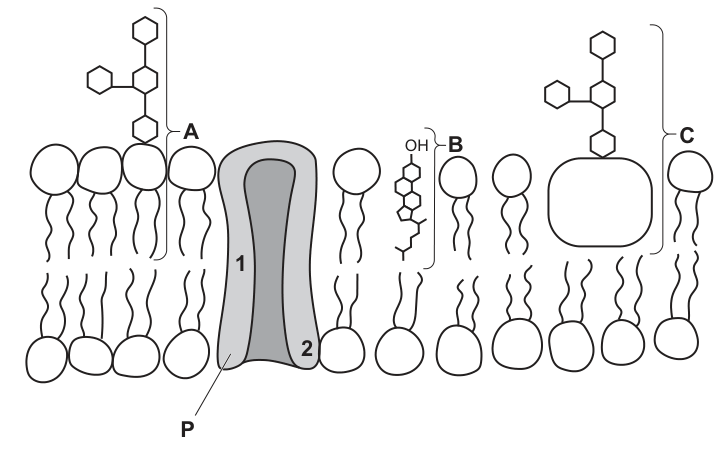
Fig. 1.1 shows a diagram of the fluid mosaic model of the structure of the cell surface membrane.
Answer
- A: glycolipid
- B: cholesterol
- C: glycoprotein
A: glycolipid; B: cholesterol; C: glycoprotein
Background Concept
The fluid mosaic model describes the cell surface membrane as a phospholipid bilayer in which various proteins, cholesterol molecules and short carbohydrate chains are embedded or attached. Three labelled features on Fig. 1.1 each correspond to a recognisable membrane component:
- Glycolipid — a short, often branched, carbohydrate chain covalently attached to the polar head of a phospholipid. It sits on the extracellular face only and contributes to cell recognition and signalling.
- Glycoprotein — a short, branched carbohydrate chain attached to a membrane protein. Like glycolipids it projects from the extracellular surface and is important in cell recognition, adhesion and as part of receptors.
- Cholesterol — a small lipid molecule with a hydroxyl (-OH) group at one end and a rigid steroid ring system. It slots between the phospholipid tails in the hydrophobic core and modulates membrane fluidity.
Understanding the Question
The stem presents a labelled diagram of the fluid mosaic model. The candidate is asked to name the three labelled structures A, B and C. A and C are both carbohydrate-containing features that project from the extracellular surface; B is the molecule embedded among those tails.
Approach
Look at what each carbohydrate chain is attached to, and at the position of the third molecule:
- A is a carbohydrate attached to a phospholipid head → glycolipid.
- B is a small molecule with an -OH group sitting between the fatty acid tails → cholesterol.
- C is a carbohydrate attached to a protein → glycoprotein.
Step-by-Step Reasoning
- A: The branched carbohydrate chain on the left of the diagram is bonded to the round polar head of a phospholipid, not to a protein. A carbohydrate linked to a phospholipid is, by definition, a glycolipid.
- B: The molecule bearing the -OH group is wedged between the fatty acid tails in the hydrophobic core. Its size, shape and -OH group identify it as cholesterol.
- C: The branched carbohydrate chain on the right is bonded to the box-shaped integral protein (P). A carbohydrate attached to a protein is a glycoprotein.
Key Takeaways
- Glycoprotein vs glycolipid depends purely on what the carbohydrate is bonded to: a protein → glycoprotein; a phospholipid → glycolipid.
- Cholesterol is identified by its -OH group and its position among the fatty acid tails.
Common Mistakes
- Swapping A and C by looking only at the position of the carbohydrate chain rather than what it is bonded to.
- Calling cholesterol a "phospholipid" or "protein" — it is its own class of membrane lipid.
Things to Be Careful About
- Both glycoproteins and glycolipids are restricted to the extracellular face of the membrane.
- Cholesterol's hydroxyl group gives it a slightly polar end; the rest of the molecule is hydrophobic.
Draw an arrow on Fig. 1.1 to show the movement of a molecule across the membrane into the cell by facilitated diffusion.
Answer
Draw a vertical arrow passing through the central channel of protein P, with the arrowhead pointing from the extracellular side (above the membrane) downward into the cell (intracellular side).
Arrow through the channel of protein P, pointing down into the cell
Background Concept
Facilitated diffusion is the passive movement of ions or polar molecules across a membrane through a transport protein, moving down their concentration gradient (high → low) without using ATP. Because the interior of the phospholipid bilayer is hydrophobic, ions and polar molecules cannot diffuse directly between the phospholipid tails — they must pass through a channel protein (forming an aqueous pore) or be carried by a carrier protein.
In Fig. 1.1, protein P is drawn as a transmembrane protein with a clear central channel, identifying it as a channel protein suitable for facilitated diffusion.
Understanding the Question
The stem shows the fluid-mosaic diagram and asks the candidate to annotate it with an arrow showing facilitated diffusion into the cell. The question stem fixes the route (facilitated diffusion) and the direction (into the cell); the candidate must place the arrow correctly on the figure.
Approach
Two marks are available:
- The arrow must pass through the protein (not between phospholipids).
- The arrow must point in the correct direction (downward, into the cell).
Step-by-Step Reasoning
- Identify protein P as the only integral protein drawn with an open central channel suitable for facilitated diffusion.
- Draw the arrow passing through this channel, between the regions labelled 1 and 2 inside the protein.
- The extracellular face of the membrane is shown at the top of the diagram, so "into the cell" means the arrow points downward, ending below the membrane.
- The arrow must NOT be drawn through the phospholipid bilayer — that would represent simple diffusion, not facilitated diffusion.
Key Takeaways
- Facilitated diffusion is shown on diagrams by an arrow through a transport protein (not through the bilayer).
- Direction always follows the concentration gradient; here "into the cell" → downward.
Common Mistakes
- Drawing the arrow through the phospholipid bilayer between the tails — this depicts simple diffusion, not facilitated diffusion, and scores 0/2.
- Drawing the arrow in the wrong direction (out of the cell).
- Drawing the arrow near the protein rather than clearly through its channel.
Things to Be Careful About
- The arrowhead must be unambiguous; examiners need to see clearly that the arrow ends inside the cell.
- The arrow must pass through protein P's central channel — not down the side of the protein between P and the bilayer.
Explain how the fluidity of the membrane would change if the proportion of unsaturated fatty acids in the phospholipid bilayer increased.
Answer
- Fluidity of the membrane increases.
- Unsaturated fatty acids contain C=C double bonds.
- The double bonds cause the fatty acid chains to kink / bend.
- The kinks prevent unsaturated fatty acids from lying as close together as saturated fatty acids (or increase the distance between phospholipids).
- This weakens the hydrophobic interactions between the fatty acid chains (and between fatty acid chains and cholesterol).
Membrane fluidity increases because unsaturated fatty acids have kinked chains that prevent tight packing and weaken hydrophobic interactions between tails
Background Concept
The hydrophobic core of the cell surface membrane is built from the fatty acid tails of phospholipids. These tails are held together laterally by hydrophobic interactions (van der Waals forces between adjacent non-polar chains).
- Saturated fatty acids have only single C–C bonds, so their chains are straight and pack tightly together, producing a more viscous, less fluid membrane.
- Unsaturated fatty acids contain one or more C=C double bonds. Each double bond introduces a rigid kink of about 30° into the chain, so the tails cannot lie alongside one another as neatly.
Tighter packing → stronger hydrophobic interactions → lower fluidity; looser packing → weaker hydrophobic interactions → higher fluidity.
Understanding the Question
The stem fixes the scenario: the proportion of unsaturated fatty acids in the bilayer is increased. The candidate must explain how this affects membrane fluidity, going from the molecular structure through to the macroscopic property.
Approach
Build a chain of cause-and-effect: C=C double bond → kink → reduced packing → weakened hydrophobic interactions → increased fluidity. Any three of the linked statements earn the three marks; ideally present all of them in order.
Step-by-Step Reasoning
- Fluidity increases. State the overall outcome first.
- Unsaturated fatty acids contain C=C double bonds. This is the structural starting point.
- The double bonds cause the chains to kink / bend. Without this kink, the rest of the reasoning fails.
- Kinks prevent the unsaturated chains from packing as closely as saturated chains (equivalently: they increase the distance between phospholipids). This is the immediate geometric consequence.
- The looser packing weakens the hydrophobic interactions between adjacent fatty acid chains (and between chains and cholesterol). This is the intermolecular consequence that produces the change in fluidity.
Key Takeaways
- Membrane fluidity is determined largely by how tightly the fatty acid tails can pack together.
- Double bonds in unsaturated fatty acids introduce kinks that reduce packing and weaken hydrophobic interactions, raising fluidity.
- Cholesterol buffers these changes by filling gaps between tails at moderate temperatures and restricting movement at high temperatures.
Common Mistakes
- Saying "fluidity decreases" — the opposite of the truth.
- Claiming unsaturated fatty acids "contain fewer hydrogens" or "are saturated with water" — confusing terminology.
- Stopping at "double bonds cause kinks" without linking kinks to packing and then to fluidity.
Things to Be Careful About
- The reverse argument (ORA) is credited by the mark scheme: "unsaturated fatty acids cannot lie as close together as saturated fatty acids."
- Hydrophobic interactions are between fatty acid tails themselves, not just with cholesterol; the mark scheme accepts either.
The R-groups of amino acids give them different properties.
Suggest how the properties of the R-groups of the amino acids at position 1 of P may differ from the R-groups of the amino acids at position 2.
Answer
- Position 1 (in contact with the hydrophobic fatty acid tails) has amino acids with hydrophobic / non-polar R-groups.
- Position 2 (lining the aqueous channel through which polar molecules / ions pass) has amino acids with hydrophilic / polar / ionic R-groups.
Position 1: hydrophobic/non-polar R-groups; Position 2: hydrophilic/polar/ionic R-groups
Background Concept
Every amino acid has a central carbon bonded to an amino group (-NH2), a carboxyl group (-COOH), a hydrogen atom and a variable side chain — the R-group. The chemistry of the R-group dictates how an amino acid behaves in different chemical environments:
- Hydrophobic / non-polar R-groups (e.g. in valine, leucine, isoleucine, phenylalanine) avoid water and prefer non-polar surroundings.
- Hydrophilic / polar / ionic R-groups (e.g. in serine, lysine, aspartate, glutamate) interact readily with water or with charged species.
In a folded membrane protein, R-groups orient themselves so that hydrophobic R-groups face the hydrophobic fatty-acid tails, while hydrophilic R-groups face either the aqueous cytoplasm, the extracellular fluid or an aqueous channel through the protein.
Understanding the Question
Protein P in Fig. 1.1 has two distinct environments inside it:
- Position 1: the part of the protein in contact with the hydrophobic fatty acid tails.
- Position 2: the lining of the central aqueous channel through which polar/charged molecules pass.
The candidate must suggest how the R-groups at these two positions differ.
Approach
Apply the principle that R-groups orient according to the polarity of their surroundings (like interacts with like):
- Hydrophobic surroundings → hydrophobic R-groups.
- Aqueous / polar surroundings → hydrophilic R-groups.
Step-by-Step Reasoning
- Position 1 is embedded in the non-polar fatty acid core. To be thermodynamically stable in this environment, the R-groups there must be hydrophobic (non-polar) — they will not interact favourably with water, but they will pack against the lipid tails.
- Position 2 lines an aqueous channel through which polar molecules or ions move. To interact with water and/or with the passing molecules, the R-groups there must be hydrophilic / polar / ionic.
Only one mark is offered, so either side of the comparison is acceptable as long as the polarity is correctly assigned to a specific position.
Key Takeaways
- R-group orientation in a membrane protein is dictated by the polarity of the surrounding environment.
- Hydrophobic exterior of an integral protein ↔ hydrophobic R-groups; aqueous pore or aqueous face ↔ hydrophilic R-groups.
Common Mistakes
- Stating the amino group (-NH2) or carboxyl group (-COOH) instead of the R-group — these are present in every amino acid and do not vary.
- Reversing the polarity: claiming position 1 has polar R-groups or position 2 has non-polar R-groups.
- Vague answers such as "the R-groups are different" without saying how.
Things to Be Careful About
- The question is about the R-groups, not the backbone amine/carboxyl groups.
- The mark scheme allows either side of the comparison — naming only position 1 or only position 2 is enough, provided it is correctly polar and correctly linked to its environment.
Haemoglobin is a globular protein containing haem groups.
Explain how the presence of haem groups allows the haemoglobin molecule to transport oxygen.
Answer
- Each haem group contains an iron () ion.
- Each haem group binds to one oxygen molecule (), allowing haemoglobin to carry oxygen.
Each haem group contains an iron (Fe²⁺) ion that binds reversibly to one O₂ molecule.
Background Concept
Haemoglobin is a globular protein made up of four polypeptide chains (two α and two β), each of which carries a non-protein prosthetic group called a haem group. The haem group is a flat porphyrin ring with a single iron ion at its centre. Because this iron is in the reduced (ferrous) state, , it can form a reversible coordinate bond with an oxygen molecule, allowing haemoglobin to load oxygen in the lungs and unload it where it is needed.
Understanding the Question
This is a two-mark "explain" question asking the candidate to identify the structural feature of a haem group that enables oxygen transport, and how that feature performs the job. The mark scheme rewards two specific points — the iron ion and the binding of oxygen.
Approach
State the two complementary facts clearly:
- The chemical identity of the binding site — an iron(II) ion.
- What that ion does — binds to (forms a bond with) one oxygen molecule.
No mention of the globin chains, the four-subunit structure or cooperativity is needed for these two marks.
Step-by-Step Reasoning
- Point 1: A haem group contains an iron ion in the +2 oxidation state (ferrous, ). The mark scheme accepts "iron", "iron ion" or "ferrous ion".
- Point 2: This ion forms a reversible bond with one oxygen molecule, so each of the four haem groups in a haemoglobin molecule can carry one . A maximum of four oxygen molecules can therefore be carried per haemoglobin.
- The two points together answer the question: the iron ion provides the binding site, and the binding of to that site is what allows haemoglobin to transport oxygen.
Key Takeaways
- The haem prosthetic group is the functional unit that binds oxygen; the globin chains provide the protein scaffold and the cooperative behaviour.
- Iron must be in the reduced state for oxygen to bind; oxidation to produces methaemoglobin, which cannot carry oxygen.
- Binding is reversible (no need to state for this mark, but it is essential for transport).
Common Mistakes
- Writing "iron" without specifying it is the ion (or the form) — the mark scheme still accepts this, but it is sloppy and should be avoided in extended answers.
- Stating that the haem contains oxygen, or that the globin binds oxygen — the binding site is the iron ion inside the haem, not the protein.
- Describing only the protein structure of haemoglobin without naming the iron/oxygen binding — this misses both marking points.
Things to Be Careful About
- The mark scheme insists the iron ion is named; "iron atom" alone is acceptable but less precise.
- "Binds to an oxygen molecule" is the required wording; "carries" or "attracts" oxygen is too vague to credit by itself.
- The state matters: do not imply the iron has any other oxidation state.
In certain situations, such as during intense exercise, a person may breathe very quickly and deeply. This is known as hyperventilation.
This hyperventilation causes more carbon dioxide to be exhaled. As a result, there is a lower concentration of carbon dioxide in the blood that passes through the capillary network in respiring tissues. This increases the affinity of haemoglobin for oxygen so that there is a decrease in the release of oxygen from red blood cells.
Explain why a decrease in the concentration of carbon dioxide in the blood passing through respiring tissues leads to a decrease in the release of oxygen from red blood cells.
Answer
- Less diffuses into the red blood cells.
- Less reacts with water, so less carbonic acid is formed (catalysed by carbonic anhydrase).
- Less carbonic acid dissociates, so fewer ions and hydrogencarbonate ions are produced.
- Fewer ions bind to haemoglobin, so less haemoglobinic acid is formed.
- With fewer ions bound to haemoglobin, haemoglobin has a higher affinity for , so less oxygen is released from red blood cells into the respiring tissues.
(Any three of the points above, taken in the order that gives a logical sequence.)
Lower CO₂ → less H⁺ formed via carbonic acid → less H⁺ binds to haemoglobin → haemoglobin holds onto O₂ more tightly → less O₂ released.
Background Concept
Haemoglobin's affinity for oxygen is not fixed — it depends on the local chemical environment, especially the concentration of carbon dioxide and therefore of hydrogen ions. This is the Bohr effect (or Bohr shift): increasing (and ) shifts the oxygen dissociation curve to the right, lowering haemoglobin's affinity for and promoting release. Conversely, lower shifts the curve to the left, raising the affinity and reducing release.
Inside the red blood cell, is rapidly hydrated to carbonic acid by the enzyme carbonic anhydrase:
The produced then binds to haemoglobin to form haemoglobinic acid (HHb), stabilising the deoxygenated (T) state. This is why more at a respiring tissue promotes more release — and why less (after hyperventilation) does the opposite.
Understanding the Question
The stem tells us that hyperventilation lowers the concentration in the blood arriving at respiring tissues, and that this raises haemoglobin's affinity for , so less is released. The question asks the candidate to explain why the lower produces this effect — that is, to lay out the chemical chain from concentration through to oxygen release. Three marks are available for any three of the linked points in the chain.
Approach
Walk the chain in the right order. Start outside the red blood cell and work inwards:
- in plasma → inside red blood cell.
- + → (carbonic acid).
- → + .
- + haemoglobin → HHb (haemoglobinic acid).
- Less HHb means haemoglobin holds more tightly, so less is released.
Any three steps in this chain, written clearly, will earn the three marks.
Step-by-Step Reasoning
- Lower in plasma → less diffuses into the red blood cell. The concentration gradient between plasma and red blood cell is smaller, so the diffusion rate falls. (1 mark)
- Less reacts with water. Carbonic anhydrase inside the red blood cell normally catalyses this reaction rapidly. With less substrate, less (carbonic acid) is produced. (1 mark)
- Less carbonic acid dissociates, so fewer and ions are formed. (1 mark)
- Fewer ions bind to haemoglobin, so less haemoglobinic acid is formed. (1 mark, but only one of "less produced" or "less HHb formed" is needed as alternatives depending on which steps are credited.)
- Haemoglobin retains a higher affinity for , so less is released from red blood cells into the respiring tissues. (1 mark, the conclusion that the question is ultimately asking for)
A high-scoring answer will pick three of these in a logical sequence — usually the first, second and last, or the second, fourth and last.
Key Takeaways
- The Bohr effect is a feedback mechanism that normally matches oxygen delivery to metabolic demand.
- Hyperventilation disrupts this: by lowering , it actually reduces delivery to tissues, which is why hyperventilating before swimming underwater (to "load up on oxygen") is physiologically misleading.
- Carbonic anhydrase and the chloride shift together allow rapid conversion of in the red blood cell; the is mopped up by haemoglobin, keeping blood pH relatively stable.
Common Mistakes
- Skipping the chemistry and just writing " affects haemoglobin's affinity for " — this restates the stem and earns no marks.
- Stating that " binds to haemoglobin" without referring to and carbonic acid. Some does form carbaminohaemoglobin, but the mark scheme credits this only as an alternative to the -mediated pathway; the dominant effect is via .
- Confusing cause and effect — saying "less released causes less to be removed" is backwards.
- Using "amount" instead of "concentration" — the stem refers to concentration, and the mark scheme requires this precision.
Things to Be Careful About
- The question is about the blood passing through respiring tissues, so the relevant comparison is between hyperventilated blood and normal blood at the tissues, not in the lungs.
- Three marks, so three clear, distinct points are needed. Don't write five vague ones and hope one of them matches.
- The mark scheme's "AVP" allows credit for the role of carbaminohaemoglobin or for a description of the conformational change when binds; either is acceptable if the main points have been covered.
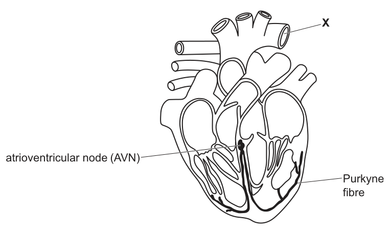
Fig. 2.1 is a diagram of a section through the heart.
Describe and explain how the tunica media (middle layer of the wall) of blood vessel X adapts the blood vessel for its function.
Answer
- Blood vessel X is the aorta; it carries oxygenated blood at high pressure from the left ventricle to the body tissues (systemic circulation).
- The tunica media of X contains a high proportion of elastic fibres and a relatively low proportion of smooth muscle.
- The elastic fibres allow the wall of X to stretch and expand to accommodate the surges (pulses) of blood entering the aorta during ventricular systole, preventing the vessel from bursting.
- Recoil of the elastic fibres between surges pushes the blood onward, maintaining a high pressure and continuous flow of blood to the tissues.
The aorta's tunica media is rich in elastic fibres; these stretch to absorb the pulse of blood from ventricular systole and recoil to maintain high pressure and continuous flow.
Background Concept
Arteries, especially those close to the heart such as the aorta, have walls built to withstand and modulate high, pulsatile pressure. Their tunica media (the middle layer of the wall, sandwiched between the tunica intima and tunica adventitia) is the structural layer that gives an artery its characteristic properties.
In elastic (conducting) arteries like the aorta, the tunica media is dominated by elastic fibres (mostly elastin), with relatively little smooth muscle. In muscular (distributing) arteries further from the heart, the balance shifts towards smooth muscle, allowing vasoconstriction and vasodilation to control distribution of blood to specific organs.
The two key mechanical properties of elastic fibres are:
- Stretch under pressure, allowing the vessel to expand.
- Recoil when the pressure falls, returning the wall to its original size and pushing the blood forward.
Together, these convert the pulsatile output of the heart into a more continuous flow downstream — a property sometimes called the Windkessel effect.
Understanding the Question
The diagram (Fig. 2.1) labels X at the top of the heart, where the great arteries arise. X is therefore the aorta, the largest elastic artery. The candidate must:
- State the function of the aorta in terms of pressure and destination.
- Identify the structural feature of its tunica media (the relative composition).
- Explain how that feature adapts the vessel for carrying high-pressure, pulsatile blood.
Three marks: one for the high-pressure/function statement, and two from the structural/functional pair (composition and one piece of stretch/recoil explanation, with the third mark going to the additional stretch or recoil detail).
Approach
Structure the answer in three short, linked sentences:
- Sentence 1: What X is and what it does (high pressure, to body tissues).
- Sentence 2: The composition of its tunica media (lots of elastic fibres, little smooth muscle).
- Sentence 3: What the elastic fibres do — stretch on the pulse, recoil between pulses — to keep flow going.
Step-by-Step Reasoning
- Function: X is the aorta, which receives blood directly from the left ventricle and distributes it at high pressure to the systemic circulation. (1 mark)
- Structure: The tunica media of the aorta has a high proportion of elastic fibres and a relatively low proportion of smooth muscle compared with a muscular artery. (1 mark, paired with the next)
- Stretch: When the left ventricle contracts (ventricular systole), a surge of blood is forced into the aorta. The elastic fibres in the tunica media allow the wall of the aorta to stretch and expand, accommodating this surge without rupturing. (1 mark)
- Recoil: When the ventricle relaxes, the elastic fibres recoil to their original length. This recoil squeezes the blood onward, maintaining a high pressure and producing a more continuous flow downstream. (1 mark available, but only two are needed for three marks total when paired with the function point)
The mark scheme accepts either (a) the composition + (b) one stretch/recoil explanation as two of the three, or (a) the composition + (b) two stretch/recoil details as alternatives. Candidates should aim to include both stretch and recoil to be safe.
Key Takeaways
- Elastic arteries (e.g. aorta, pulmonary artery) are close to the heart and dominated by elastin; muscular arteries (further from the heart) are dominated by smooth muscle.
- The elastic properties of the aorta smooth the pulsatile output of the heart into a more continuous flow, and they also help to maintain diastolic blood pressure.
- This is a classic "structure–function" question: knowing the composition of a tissue is only useful if you can say what that composition does.
Common Mistakes
- Treating the question as if it were about all arteries, not the aorta specifically — the proportions of elastic fibre vs smooth muscle are very different between elastic and muscular arteries.
- Confusing the layers: the tunica intima is the innermost layer (with the endothelium), the tunica media is in the middle, and the tunica adventitia is the outer connective-tissue layer. Candidates sometimes credit the wrong layer.
- Stating only "elastic fibres allow the artery to stretch" without explaining why stretching is useful (i.e. to accommodate the pulse of blood and prevent rupture) and without mentioning recoil.
- Stating that elastic fibres contract — they are passive; they recoil because of their elastic nature.
Things to Be Careful About
- "High proportion of elastic fibres and low proportion of smooth muscle" is the structural difference; the mark scheme credits this combined statement.
- The function statement should mention high pressure and the destination (body tissues / systemic circulation). Just saying "carries blood" is too vague.
- The mark scheme offers a fallback credit (1 mark) for "stretch and recoil with one detail" if no other marks are scored, so even a minimal answer is worth a mark.
Describe the role of the atrioventricular node (AVN) and the Purkyne tissue in the cardiac cycle.
Answer
- Both the AVN and Purkyne tissue are involved in coordinating the contraction (systole) of the heart muscle.
- AVN: It is the only pathway by which electrical impulses can pass from the atria to the ventricles.
- The AVN introduces a delay of about , so the ventricles contract after the atria have contracted.
- This delay allows the atria to empty completely and the ventricles to fill with blood before ventricular systole begins.
- Purkyne tissue: It carries the electrical impulses down the septum to the base (apex) of the ventricles and through the ventricular walls.
- This causes the ventricular muscle to contract simultaneously from the base upwards, squeezing blood upwards into the aorta and pulmonary artery.
The AVN delays the impulse so atria contract first and fill the ventricles; the Purkyne tissue then spreads the impulse through the ventricular walls so they contract together from the base upwards.
Background Concept
The heart is myogenic — it generates its own electrical rhythm without needing nervous stimulation. The rhythm originates in the sinoatrial node (SAN) in the wall of the right atrium, which acts as the heart's natural pacemaker. From the SAN, a wave of electrical excitation spreads across the atrial walls, causing both atria to contract almost simultaneously. The impulse then reaches the atrioventricular node (AVN), situated in the wall of the right atrium near the septum.
The AVN is the only electrical connection between the atria and the ventricles; the rest of the septum is non-conducting fibrous tissue. The AVN introduces a brief delay (about ) before passing the impulse on, via the bundle of His and then the Purkyne fibres, which spread the excitation rapidly through the muscular walls of the ventricles, from the apex (base of the ventricle) upwards.
The result is a coordinated cycle: atria contract together, ventricles fill, then ventricles contract together from the bottom up, ejecting blood efficiently into the aorta and pulmonary artery.
Understanding the Question
Part (c)(ii) asks the candidate to describe the roles of the AVN and the Purkyne tissue in the cardiac cycle. The marks are split across the two structures, and the candidate must address both to score full marks (the mark scheme's "must attempt AVN and Purkyne tissue to gain max"). Four marks are available, taken from any four of the listed credit points.
Approach
Write two short paragraphs, one for each structure:
- AVN paragraph: its position as the only conduction pathway from atria to ventricles, and the consequence of the delay it introduces (sequential atrial then ventricular contraction, and ventricular filling).
- Purkyne tissue paragraph: how it carries the impulse into the ventricular walls, and the consequence (synchronised contraction from the apex upward, efficient ejection).
A sentence linking the two ("both are involved in coordinating contraction of the heart muscle") is also creditworthy.
Step-by-Step Reasoning
- Linking statement (1 mark if used as the first point): Both AVN and Purkyne tissue coordinate the contraction (systole) of the heart / heart muscle / ventricles.
- AVN — conduction pathway (1 mark): The AVN is the only route by which electrical impulses can travel from the atria to the ventricles. (The fibrous septum between them is non-conducting.)
- AVN — delay (1 mark): The AVN introduces a delay of about before passing the impulse to the ventricles. The mark scheme accepts "a delay" or a numerical value of .
- AVN — consequence of delay (1 mark): Because of this delay, the ventricles contract after the atria. This allows the atria to empty fully and the ventricles to fill with blood before ventricular systole begins. (Either the timing statement or the filling statement will earn a mark.)
- Purkyne tissue — conduction (1 mark): The Purkyne tissue carries / transmits the impulse down the septum to the base of the ventricles and through the walls of the ventricles.
- Purkyne tissue — consequence (1 mark): This causes the ventricular muscle to contract together / simultaneously / at the same time / from the base upwards, so blood is squeezed efficiently out of the ventricles into the aorta and pulmonary artery.
Four marks are needed; candidates can pick any four of the six points above (the first three are the AVN points and the last two are the Purkyne points, so a balanced answer addresses both structures).
Key Takeaways
- The heart's conduction system coordinates the timing and sequence of atrial and ventricular contraction.
- The AVN's role is principally about timing — delaying the impulse so that the atria contract first and the ventricles fill.
- The Purkyne fibres' role is principally about distribution — spreading the impulse rapidly through the ventricular muscle so that it contracts in a coordinated, efficient way.
- This is myogenic control: no nerve impulse is required for the basic heartbeat, although nerves and hormones (e.g. adrenaline) modulate the rate.
Common Mistakes
- Calling the AVN a "nerve" or the impulse a "nerve impulse" — the conduction system is electrical (waves of depolarisation in cardiac muscle), not nervous. The mark scheme rejects "nerve impulses".
- Saying the AVN "generates" impulses — it does not; the SAN does. The AVN only delays and relays.
- Saying the impulse travels "from the AVN to the Purkyne tissue" without explaining what each structure does.
- Describing the Purkyne fibres as "supplying blood" or "carrying oxygen" — they are specialised cardiac muscle cells that conduct electrical impulses.
- Stating that the ventricles contract "from the top down" — they actually contract from the base (apex) upwards, which is why blood is squeezed efficiently towards the arteries at the top of the heart.
- Forgetting to mention that the AVN is the only pathway from atria to ventricles — this is a key part of its role.
Things to Be Careful About
- The mark scheme uses "impulses" / "wave of excitation" and accepts either; the term "nerve impulse" is explicitly rejected once.
- The numerical delay is "about " — the word "about" or the unit alone is acceptable; the mark scheme accepts "a delay" or "".
- The mark scheme requires that the answer addresses both AVN and Purkyne tissue for full marks; a one-sided answer cannot score more than 2 of the 4 marks.
- The question asks for a description of roles, not an explanation of how the conduction system generates the rhythm — the SAN is the pacemaker and is not required here.
The lock-and-key hypothesis and the induced-fit hypothesis are used to describe the interaction of enzymes and their substrates.
Describe one similarity and one difference between the lock-and-key hypothesis and the induced-fit hypothesis.
similarity ______
difference ______
Answer
Similarity: In both hypotheses the substrate binds to the active site of the enzyme, forming an enzyme–substrate complex.
Difference: In the induced-fit hypothesis the active site is not initially fully complementary to the substrate and changes shape as the substrate binds, whereas in the lock-and-key hypothesis the active site is already exactly complementary to the substrate.
Similarity: substrate binds to the active site; Difference: active site is not fully complementary and changes shape on binding (induced-fit) versus already fully complementary (lock-and-key).
Background Concept
Enzymes are biological catalysts whose action depends on a region called the active site. Two complementary models describe how a substrate interacts with this active site:
- Lock-and-key hypothesis: the active site has a fixed, rigid shape that is already exactly complementary to the substrate, like a key fitting a lock. No shape change occurs on binding.
- Induced-fit hypothesis: the active site is not an exact match at first; binding of the substrate causes a small, reversible change in the conformation of the active site so that it moulds around the substrate. The change is small and does not destroy the enzyme.
Both hypotheses accept that enzymes are highly specific and that an enzyme–substrate complex forms before product is released.
Understanding the Question
This is a 2-mark comparison question. The command word is describe, which only requires a statement of fact — no explanation of why. One mark is for a valid similarity and one mark for a valid difference, so the answer should give a single, sharp point under each heading rather than a list of everything the student knows.
Approach
Start with the most obvious shared feature: the idea of substrate binding at the active site. Then pick the single most distinctive feature of the induced-fit model — the shape change of the active site on binding — and contrast it directly with the rigid, pre-formed active site of the lock-and-key model.
Step-by-Step Reasoning
Similarity point. Both hypotheses propose that:
- the substrate binds to the active site of the enzyme, forming an enzyme–substrate complex;
- this is converted to product which leaves the active site, freeing the enzyme for reuse.
Any of these earns the similarity mark.
Difference point. The defining feature of induced-fit that is absent from lock-and-key is the conformational change of the active site. In lock-and-key the active site is already shaped to fit the substrate perfectly; in induced-fit the active site reshapes itself around the substrate as it binds. This is the contrast the examiner is looking for.
Key Takeaways
- Both models feature an enzyme–substrate complex formed at the active site.
- The distinguishing feature of induced-fit is a reversible shape change of the active site; lock-and-key has no such change.
- A good comparison pairs an identical process in both with one specific contrasting detail.
Common Mistakes
- Writing only a similarity or only a difference — half marks lost.
- Saying the enzyme is "denatured" by the shape change in induced-fit — wrong: the change is small and reversible.
- Confusing which model has the rigid active site and which has the flexible one.
- Giving a vague answer such as "they are similar because they both involve enzymes" — no biological content earns no mark.
Things to Be Careful About
The mark scheme offers several alternative similarities (substrate binds to active site; ES complex forms; product leaves active site; enzyme is unaltered for re-use; activation energy is lowered; high specificity). Only one is needed, so choose the clearest. For the difference, the mark scheme credits any statement that the active site is not fully complementary and/or changes shape on substrate binding.
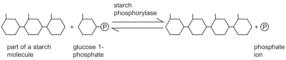
Phosphorylase enzymes can catalyse the synthesis of starch and the breakdown of starch in some plant tissues. A reaction catalysed by starch phosphorylase is shown in Fig. 3.1.
A student carried out an experiment to study the synthesis of starch by phosphorylase found in potato tissue.
The student was provided with a solution, E, extracted from potato tissue. The extract was filtered to remove all the starch grains.
Extract E contained biological molecules from the potato tissue including phosphorylase but no starch.
Iodine solution was used to confirm that starch was not present in extract E.
State the colour observed when iodine solution was added to a sample of extract E.
Answer
Orange (or brown / amber / yellow-brown / yellow-orange).
Orange / brown / amber
Background Concept
The iodine test for starch relies on the iodine ions (triiodide, ) slipping inside the helical coil of amylose and forming a coloured charge-transfer complex. When amylose is present the complex is an intense blue-black; in its absence the iodine solution simply retains the colour of the reagent itself.
Understanding the Question
The question gives away the answer in its stem: the student filtered the extract to remove starch grains and the question states that the extract contained no starch. The colour seen on adding iodine therefore cannot be blue-black. The candidate must name the colour that iodine shows when starch is absent.
Approach
This is a recall point. State one of the accepted colour terms for iodine on its own (without starch).
Step-by-Step Reasoning
Iodine solution (potassium iodide–iodine, ) without starch is orange / brown / amber. Once any of these is written, the mark is awarded. The mark scheme also accepts the intermediate descriptive terms "yellow-brown" and "yellow-orange".
Key Takeaways
- Iodine + starch → blue-black (positive test).
- Iodine + no starch → orange / brown / amber (negative test).
Common Mistakes
- Writing "blue-black" because the candidate remembers the test without reading the stem — the question says there is no starch, so a positive colour is impossible.
- Writing "colourless" or "clear" — the reagent is coloured, and what is observed is the colour of the reagent itself.
Things to Be Careful About
The colour is the colour of the iodine reagent, not the colour of the sample. Avoid describing it as "no colour change" — there has been no reaction because starch is absent, but the solution is still coloured.
The student added a small drop of a dilute starch solution and a solution of glucose 1-phosphate to a test-tube containing extract E.
Samples of the reaction mixture in the test-tube were removed every minute and a drop of iodine solution was added to each.
The student used a colorimeter to measure the absorbance of the solution in each sample.
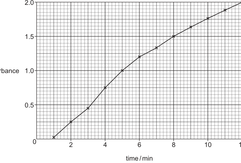
The results are shown in Fig. 3.2.
Explain the results shown in Fig. 3.2.
Answer
- Phosphorylase (in extract E) catalyses the addition of glucose 1-phosphate to the starch chain, so more starch is synthesised as time progresses.
- Iodine forms a blue-black complex in the presence of starch; the more starch present, the darker / more intense the blue-black colour.
- A darker-coloured solution absorbs more light, so the colorimeter records a higher absorbance reading at later times.
(Any three of the points above gain the available marks.)
Phosphorylase synthesises more starch over time; more starch → darker blue-black colour with iodine → higher absorbance.
Background Concept
Starch phosphorylase catalyses the reversible reaction shown in Fig. 3.1:
By adding glucose 1-phosphate (the substrate) and a starter of starch to extract E (which contains the enzyme but no starch of its own), the forward reaction is favoured and starch is built up.
Iodine does not directly measure starch; it forms a deep blue-black charge-transfer complex when it threads into the helical structure of amylose. The intensity of this colour depends on how much starch is present.
A colorimeter passes light of a chosen wavelength through the sample and measures how much is transmitted. Absorbance is , so the darker (more strongly absorbing) the solution, the higher the absorbance reading.
Understanding the Question
The graph (Fig. 3.2) shows absorbance rising from about at min to about at min, with a gently decreasing gradient. The student took a sample every minute and added iodine to it. The question is explain the results — so the candidate must describe the chain of events that links the chemistry in the tube to the reading on the colorimeter.
Approach
Trace the chain backwards from the curve to the chemistry:
absorbance rising → colour getting darker → more iodine-starch complex → more starch in the tube → phosphorylase has been synthesising starch over time.
Each arrow in the chain earns (or contributes to) a mark.
Step-by-Step Reasoning
- Extract E contains phosphorylase. With glucose 1-phosphate and a starch primer present, phosphorylase catalyses the addition of glucose-1-phosphate units onto the growing starch chain, so the amount of starch in the tube increases over time.
- Each minute, a sample is taken and mixed with iodine solution. Iodine forms a blue-black complex when starch is present.
- The more starch there is in a sample, the darker (more intense) the blue-black colour becomes.
- A more strongly coloured solution transmits less light through the colorimeter cuvette, so the meter records a higher absorbance.
The curve therefore rises steadily. The shallow decrease in gradient at later times is consistent with the reaction slowing (substrate becoming limiting, or starch-iodine absorbance approaching saturation), but the question does not specifically reward this detail.
Key Takeaways
- Enzyme catalysis increases the concentration of a product over time.
- Iodine quantifies starch through colour intensity.
- A colorimeter converts colour intensity into a numerical absorbance reading.
Common Mistakes
- Saying "more starch absorbs more light" without mentioning the iodine-starch complex — iodine itself is the chromophore only when bound inside the amylose helix.
- Forgetting the role of the enzyme and attributing starch synthesis to "the starch added to the tube" — the small initial drop of dilute starch cannot account for the absorbance rising to .
- Implying that phosphorylase breaks down starch here — the forward reaction of Fig. 3.1 builds starch.
Things to Be Careful About
The mark scheme requires the candidate to make the link between enzyme catalysis and absorbance explicitly. It is not enough to say "the absorbance increased because more starch was made" — the role of iodine (which gives the colour) and of the colorimeter (which converts colour to absorbance) must be in the chain.
After 12 minutes, the student added a solution containing phosphate ions to the reaction mixture. The student continued taking samples every minute, adding iodine solution to each sample.
The absorbance of the solution in each of these samples was measured. The results showed that the absorbance decreased over time.
Suggest why the absorbance decreased.
Answer
Phosphorylase catalyses the reversible reaction shown in Fig. 3.1. Adding phosphate ions shifts the equilibrium in the reverse direction, so the enzyme now breaks down starch (releasing glucose 1-phosphate). Less starch means a less intense blue-black colour with iodine, hence a lower absorbance.
Adding phosphate ions drives the reverse reaction (starch breakdown), reducing starch concentration and therefore absorbance.
Background Concept
Fig. 3.1 makes the reversibility of the phosphorylase reaction explicit by drawing the arrows pointing both ways:
The same enzyme, starch phosphorylase, catalyses both directions. In a closed system at equilibrium the concentrations of starch and glucose 1-phosphate remain steady. If the concentration of any species is perturbed — for example by adding more phosphate ions () — the system responds to oppose the change (Le Chatelier's principle). Adding product drives the reaction backwards, so starch is broken down again into glucose 1-phosphate and the original starch chain.
Understanding the Question
At 12 min, the reaction has accumulated a large amount of starch and run down much of the glucose 1-phosphate. The student then adds a solution containing phosphate ions. The absorbance subsequently decreases. The question asks for the reason.
Approach
Connect the addition of phosphate to the direction of the reaction, then link the new starch concentration to the colorimeter reading.
Step-by-Step Reasoning
- Phosphorylase catalyses a reversible reaction (Fig. 3.1).
- Adding phosphate ions raises the concentration of one of the products on the right-hand side.
- By Le Chatelier's principle the position of equilibrium shifts to the left: starch is broken down, releasing glucose 1-phosphate and shortening the starch chain.
- With less starch in the mixture, iodine produces a less intense blue-black colour.
- A paler solution transmits more light, so the absorbance recorded by the colorimeter falls.
Any one of the creditable points is enough for the mark; together they form a complete causal chain.
Key Takeaways
- A reversible enzyme catalyses both forward and reverse directions.
- Adding product to a reversible reaction at equilibrium drives the reaction in the reverse direction.
- A falling starch concentration is observable as a falling colorimeter absorbance.
Common Mistakes
- Saying the enzyme is "denatured" or "inactivated" by the phosphate — there is no evidence of this in the question and it is not the mechanism.
- Confusing the role of phosphate: it is a product, not a substrate, of starch synthesis.
- Failing to mention the reversible nature of the phosphorylase reaction shown in Fig. 3.1.
Things to Be Careful About
The mark scheme allows the credit to be given simply for "reaction in Fig. 3.1 goes in the opposite direction" or for "breakdown of starch catalysed by the enzyme". A single such statement is enough for the mark, but a more complete answer (including the consequence for absorbance) is safer and demonstrates understanding.
Muscle cells contain glycogen phosphorylase.
Fig. 3.3 shows the effect of caffeine on the activity of glycogen phosphorylase at different concentrations of substrate.
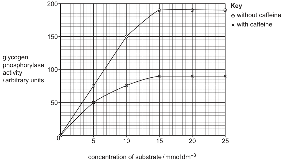
A student concluded that caffeine acts as a non-competitive inhibitor of glycogen phosphorylase.
Explain how the results in Fig. 3.3 support this conclusion.
Answer
- Caffeine reduces glycogen phosphorylase activity at every substrate concentration tested.
- The maximum activity () reached with caffeine (~90 a.u.) is much lower than the without caffeine (~190 a.u.) — adding more substrate cannot restore the original rate.
- Both curves reach their plateau at the same substrate concentration (), so the (substrate concentration at half ) is unchanged by caffeine.
- A lower with unchanged that cannot be overcome by extra substrate is the signature of a non-competitive inhibitor.
Caffeine lowers Vmax but Km is unchanged; the inhibition cannot be overcome by adding more substrate — features characteristic of non-competitive inhibition.
Background Concept
Enzyme inhibitors fall into two main classes:
-
Competitive inhibitors bind to the active site and compete with the substrate. Because substrate and inhibitor are rivals, the effect of a competitive inhibitor can be overcome by increasing substrate concentration. The maximum rate () is therefore unchanged (it can still be reached at high substrate concentration), but more substrate is needed to reach any given fraction of , so (the substrate concentration at half ) increases.
-
Non-competitive inhibitors bind to a site other than the active site (an allosteric site) and change the shape of the active site so that even bound substrate cannot be converted to product efficiently. Because the inhibitor does not compete with substrate for the active site, adding more substrate does not overcome the inhibition: falls. The substrate concentration at which the (now lower) is approached is essentially unchanged, so is unaffected.
The diagnostic graphical fingerprints are therefore:
| Feature | Competitive | Non-competitive |
|---|---|---|
| unchanged | decreased | |
| increased | unchanged | |
| Overcome by substrate? | yes | no |
Understanding the Question
Fig. 3.3 plots glycogen phosphorylase activity (y-axis, arbitrary units, –) against substrate concentration (x-axis, –). Two curves are shown: one without caffeine (the upper curve, plateaus at ) and one with caffeine (the lower curve, plateaus at ). Both plateaus occur at roughly the same substrate concentration (). The candidate must use these graphical features to justify the student's conclusion that caffeine acts as a non-competitive inhibitor.
Approach
Pick the two diagnostic features listed in the table above and verify each one against the graph:
- Compare the heights of the two plateaus → .
- Compare the substrate concentrations at which the plateaus are reached → .
- Comment on whether adding more substrate overcomes the inhibition.
Step-by-Step Reasoning
- has decreased. The plateau (maximum activity) without caffeine is about arbitrary units, but with caffeine it is only about — less than half the original maximum.
- is unchanged. Both curves reach half of their respective values at the same substrate concentration. The substrate concentration at which the curves level off () is the same in the presence and absence of caffeine.
- The inhibition cannot be overcome by adding more substrate. Even at substrate (well above the plateau point), the with-caffeine curve still sits at a.u., far below the no-caffeine plateau of a.u.
The combination of a lower with an unchanged and an inhibition that is not relieved by extra substrate is the textbook signature of non-competitive inhibition. Caffeine therefore does not compete with substrate for the active site — it must be acting elsewhere on the enzyme molecule, altering the shape of the active site so that bound substrate is processed less efficiently.
Key Takeaways
- Non-competitive inhibition lowers without changing and cannot be overcome by adding substrate.
- The key graphical evidence in this question is the lower plateau at the same substrate concentration as the control curve.
- Always compare BOTH and when classifying an inhibitor from a graph — a single feature is rarely sufficient.
Common Mistakes
- Stating only that the activity is "lower with caffeine" — this alone is consistent with any type of inhibition; the candidate must explain why it points specifically to non-competitive.
- Confusing the two classes: claiming that caffeine is competitive because adding substrate increases activity in both curves — substrate still increases activity in non-competitive inhibition up to the (lower) plateau.
- Saying " is the same" or " is increased" — both are inconsistent with the graph.
- Failing to use numbers from the graph to support the answer; the mark scheme explicitly rewards "use of data from the graph".
Things to Be Careful About
The mark scheme credits any combination of the following: (i) reduction in activity at all concentrations, (ii) plateau at a (much) lower activity, (iii) not reached, (iv) same with and without caffeine, (v) use of data from the graph. A complete answer should mention at least the change in , the unchanged , and the inability of extra substrate to overcome the effect, with at least one quoted number from the graph.
Fig. 4.1 is a photomicrograph of a copepod. These animals are found living in sea water and in fresh water environments.
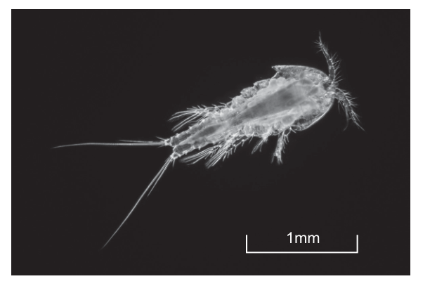
The outer surface of a copepod is covered in a layer of the polysaccharide chitin.
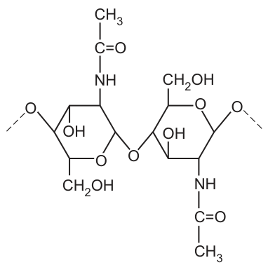
Fig. 4.2 shows part of a chitin molecule.
Answer
Circle the oxygen atom of the C1–O–C4 linkage between the two N-acetylglucosamine rings — i.e. the single –O– that joins C1 of one monomer to C4 of the next.
The central –O– atom linking C1 of one N-acetylglucosamine to C4 of the next (the β-1,4-glycosidic bond).
Background Concept
Chitin is a structural polysaccharide that forms the exoskeletons of arthropods (including copepods) and the cell walls of fungi. It is a polymer of N-acetylglucosamine (NAG) — a glucose derivative in which the –OH on C2 is replaced by an –NH–C(=O)–CH3 (N-acetyl) group. Monomers are joined by β-1,4-glycosidic bonds formed by condensation: the –OH on C1 of one NAG and the –OH on C4 of the next lose water to form a C–O–C bridge. The β designation means the –O– on the anomeric carbon (C1) is on the same face as the –CH2OH on C5 (the upper face in a Haworth projection); 1,4 specifies the linkage is between C1 and C4.
Understanding the Question
Fig. 4.2 shows two N-acetylglucosamine monomers in Haworth projection joined through a glycosidic bond. The candidate must circle that bond — the C–O–C oxygen bridging the two rings, NOT the ring oxygen, NOT the –OH groups, and NOT the oxygen of the N-acetyl side chain.
Approach
Locate the oxygen that sits between the two rings (outside the ring structure), connecting C1 of the right-hand monomer to C4 of the left-hand monomer. That single –O– is the glycosidic bond. Circle it.
Step-by-Step Reasoning
- Each pyranose ring contains one ring oxygen (inside the hexagon, between C1 and C5). This ring oxygen is part of the monomer's own ring and is not the glycosidic bond.
- The –OH groups on the ring carbons (C1, C2, C3, C4) are hydroxyls, not the glycosidic bond.
- The C=O oxygen of the N-acetyl side chain is part of that side chain and is not the glycosidic bond.
- The single oxygen drawn BETWEEN the two rings — connecting C1 of one to C4 of the next — is the glycosidic bond, produced by condensation. Circle this oxygen.
Key Takeaways
- A glycosidic bond is the C–O–C linkage between two sugar (or sugar-derivative) monomers, formed by condensation and broken by hydrolysis.
- In chitin the linkage is specifically β-1,4: β-orientation at C1 joining to C4 of the next monomer.
- Distinguish the glycosidic-bond oxygen from the ring oxygen and from the hydroxyl oxygens.
Common Mistakes
- Circling the ring oxygen (the O inside the hexagonal ring).
- Circling an –OH group on a ring carbon instead of the bridging –O–.
- Circling the C=O oxygen of the N-acetyl group.
Things to Be Careful About
There are several O atoms in the structure. Only the one forming a bridge between the two rings — connecting C1 of one NAG to C4 of the next — is the glycosidic bond.
In aquatic environments, Vibrio cholerae can live on the surface of copepods. V. cholerae secretes enzymes to hydrolyse chitin to its N-acetylglucosamine monomers. These can be broken down to provide carbon, nitrogen and a source of energy.
Draw the monomer that is formed when chitin is hydrolysed by V. cholerae.
Answer
Draw a single N-acetylglucosamine monomer (a six-membered pyranose ring) showing a free –OH on C1 and a free –OH on C4 — the two carbons that were joined in the glycosidic bond. Include the –CH2OH on C5 and the –NH–C(=O)–CH3 (N-acetyl) group on C2.
A single N-acetylglucosamine monomer with free –OH groups on both C1 and C4 (the two carbons that were joined in the glycosidic bond).
Background Concept
A condensation reaction joins two monomers with the loss of water, forming a glycosidic bond. Hydrolysis is the reverse: a water molecule is added across the glycosidic bond, a hydrogen goes to one carbon and a hydroxyl goes to the other. After hydrolysis both carbons that were originally joined now carry a free –OH group.
N-acetylglucosamine (NAG) is the monomer of chitin. It is a pyranose (six-membered ring) with:
- C1: anomeric carbon (carries –OH in the free monomer)
- C2: carries an –OH AND an –NH–C(=O)–CH3 (N-acetyl) group
- C3: carries an –OH
- C4: carries an –OH (in the polymer this –OH was used to form the β-1,4-glycosidic bond)
- C5: connected to a –CH2OH group
- Ring oxygen between C1 and C5.
Understanding the Question
V. cholerae secretes chitinase enzymes that hydrolyse the β-1,4-glycosidic bond of chitin. Hydrolysis of each glycosidic bond produces two free NAG monomers, each with a restored –OH at the carbon that was previously joined. The question asks the candidate to draw ONE of these monomers, making the C1 and C4 hydroxyl groups visible (these are the diagnostic features showing hydrolysis has occurred).
Approach
Draw one pyranose ring in Haworth projection, label or mark the C1 and C4 positions clearly, and show an –OH group on each. The other features (–CH2OH on C5, –NHCOCH3 on C2, –OH on C3) should be present to make the structure unambiguously an NAG monomer rather than glucose.
Step-by-Step Reasoning
- Sketch a six-membered ring with the ring oxygen at the back-right.
- At C5 (back-left, attached to the ring oxygen) draw a –CH2OH group pointing up.
- At C2, draw the –OH (down) and the N-acetyl group –NH–C(=O)–CH3 (up).
- At C3 draw an –OH (down).
- At C4 draw an –OH (up) — this –OH has been restored by hydrolysis of the glycosidic bond.
- At C1 draw an –OH (down for α or up for β is acceptable) — this –OH has also been restored by hydrolysis of the glycosidic bond.
- Mark or label C1 and C4 so it is clear which carbons carry the –OH groups from hydrolysis.
The candidate earns one mark for showing the –OH on C1 and one mark for showing the –OH on C4. The remaining features (–CH2OH, –NHCOCH3, ring oxygen) are needed to make the drawing unambiguously an N-acetylglucosamine monomer, but the marks focus on C1 and C4.
Key Takeaways
- Hydrolysis of a glycosidic bond regenerates –OH groups on the two carbons that were joined.
- N-acetylglucosamine differs from glucose only by the N-acetyl group on C2.
- A drawing must show the key diagnostic features (here, the –OH groups on C1 and C4) to earn the marks.
Common Mistakes
- Drawing glucose instead of N-acetylglucosamine (forgetting the –NH–C(=O)–CH3 on C2).
- Showing the –OH only on C1 or only on C4 (only one of the two marks earned).
- Forgetting the –OH on C4, or putting it on the wrong carbon (e.g. C3).
- Drawing two monomers still joined by a glycosidic bond (no hydrolysis has occurred).
Things to Be Careful About
- The –OH on C1 should be drawn explicitly; the marker cannot assume the ring oxygen is the C1–OH.
- The newly added –OH groups are exactly on the carbons that were joined in the glycosidic bond (C1 and C4 in chitin).
- The –NHCOCH3 group is on C2 (not C1, C3 or C4); this is what distinguishes NAG from glucose.
V. cholerae is a pathogen that causes cholera.
Scientists studied the transmission of V. cholerae in groups of people living in an area where there is a high number of cases of cholera.
Some families living in this area filtered their water through several layers of folded fabric from old clothing. The folded fabric traps particles and organisms larger than .
The scientists recorded the number of cases of cholera in families that filtered their water through folded fabric and compared this to the number of cases of cholera recorded in families that did not filter their water through the folded fabric.
The results are shown in Fig. 4.3.
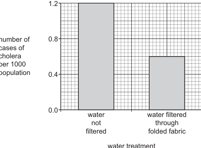
Suggest possible explanations for the results shown in Fig. 4.3.
Answer
- Filtering water through folded fabric reduces cases of cholera because it physically removes copepods (and the V. cholerae attached to their chitinous surface) from drinking water.
- The fabric only traps particles and organisms larger than 20 µm; some V. cholerae live free in the water and pass straight through, and some copepods are smaller than 20 µm and also pass through, so filtering does not eliminate the pathogen.
- Cholera can also be transmitted by routes other than drinking water, for example food contaminated with faeces (crops fertilised with sewage, food washed in contaminated water, food handled by an infected person), so filtering water alone cannot prevent all infections.
- Families who chose to filter their water may also have had better personal hygiene overall (e.g. washing hands with soap after defecation), which independently reduces transmission and contributes to the lower case rate in the filtered group.
Filtering removes copepods (carrying V. cholerae) from water, reducing transmission, but cholera persists because some bacteria/copepods pass through the fabric, the disease can also be spread by food, and people who filter may have other protective hygiene behaviours.
Background Concept
Vibrio cholerae is the bacterium that causes cholera, a severe diarrhoeal disease transmitted by the faecal–oral route. The bacterium is shed in the faeces of infected people and can contaminate water sources. In aquatic environments, V. cholerae is often found attached to the chitinous exoskeletons of copepods (zooplankton crustaceans). This association is biologically important: the copepod provides a surface for attachment and the bacterium can even obtain nutrients by hydrolysing chitin.
Cholera transmission is typically through drinking water contaminated with faeces (or with copepods carrying V. cholerae), but it can also occur via food contaminated with faecal material or directly via the hands of an infected person. Prevention therefore targets multiple routes: water purification, food hygiene, hand washing and sanitation.
Understanding the Question
Fig. 4.3 shows that families filtering water through folded fabric had ~0.6 cases of cholera per 1000, compared with ~1.1 cases per 1000 in families not filtering. The question asks for possible explanations of these results — i.e. reasons both for the reduction AND for the persistence of cases.
Approach
Work through the logic of the experiment step by step:
- What does the filter do? (Traps particles >20 µm.)
- What is trapped? (Copepods with attached bacteria.)
- Why is cholera reduced but not eliminated? (Some pathogens pass through, and there are other transmission routes.)
- Are there other factors that could account for the difference? (Behavioural/hygiene differences between the two groups.)
Step-by-Step Reasoning
- Filtering helps because it removes copepods. Copepods in the water carry V. cholerae on their chitinous surface. By removing copepods, the filter removes a major reservoir of the bacterium. With fewer bacteria in the water, fewer people are infected. This explains why filtered-water families have a lower case rate.
- The filter does not remove all bacteria. The fabric only traps particles and organisms larger than 20 µm. V. cholerae is a curved rod roughly 1–2 µm long — much smaller than 20 µm — and many bacteria live free in the water (not attached to copepods). These free-living bacteria pass straight through. Some copepods are also smaller than 20 µm (copepod species and life stages vary in size), and these also pass through. This explains why filtered-water families still have some cases.
- Other transmission routes exist. Cholera is not transmitted only by water. Contaminated food is a major route — vegetables fertilised with human faeces or irrigated with contaminated water, food handled by an infected person without proper hand washing, or seafood taken from contaminated water. Filtering water does not address these routes, so some cases still occur.
- Behavioural confounding. Families who chose to filter their water may differ systematically from those who did not. They may be more health-conscious, more educated about cholera, and more likely to practise other protective behaviours (hand washing with soap, safe food preparation, using latrines). These behaviours independently reduce transmission. Part of the reduction in cases may therefore be due to confounding factors rather than the filter itself.
- Other water treatments. Some families in either group may also have used other water treatments (boiling, chlorination, settling), which would affect the case rate independently of filtering.
Key Takeaways
- V. cholerae lives attached to copepods in water; removing copepods reduces transmission.
- The 20 µm pore size of folded fabric is too large to remove free-living bacteria.
- Cholera has multiple transmission routes; controlling water alone cannot eliminate it.
- Observational comparisons of behaviours can be confounded by other differences between groups.
Common Mistakes
- Vague answers such as "the water is cleaner" without specifying that copepods (and the bacteria attached to them) are removed.
- Failing to acknowledge that filtering does not eliminate cholera — the question requires explanation of the partial effect.
- Saying only "the bacteria are smaller than 20 µm so the filter does not work" (true, but only one of several reasons the filter is imperfect).
- Forgetting the possibility of confounding by other hygiene behaviours.
Things to Be Careful About
- The 20 µm threshold is the size limit for what the fabric traps. Bacteria (~1–2 µm) and many small copepods pass through.
- "Possible explanations" allows several reasons — the candidate should give as many as the marks allow (up to four from the mark scheme's list).
- Avoid attributing the entire reduction to the filter alone — confounders and alternative routes are part of the explanation.
The World Health Organization (WHO) recommends the use of an oral cholera vaccine (OCV) to protect people living in an area where a cholera outbreak has occurred.
People who receive an OCV and make changes in their behaviour are less likely to have a serious case of cholera.
Describe one change in behaviour that a person can make, other than purifying water, to help prevent a serious case of cholera.
Answer
Wash hands thoroughly with soap after defecation (and before preparing or eating food).
Wash hands thoroughly with soap after defecation (and before preparing or eating food).
Background Concept
Cholera is a faecal–oral disease: the bacterium is shed in the faeces of infected people and reaches a new host through the mouth. The main transmission routes are (1) drinking water contaminated with faeces, (2) eating food contaminated with faeces (e.g. unwashed vegetables, food handled by an infected person), and (3) contaminated hands touching the mouth. Personal-hygiene measures that interrupt these routes are a key part of cholera control, alongside water purification and vaccination.
The oral cholera vaccine (OCV) provides specific immune protection against V. cholerae but is most effective when combined with behavioural changes that reduce the inoculum (number of bacteria) a person is exposed to.
Understanding the Question
The question asks for ONE behaviour, other than purifying water, that helps prevent a serious case of cholera. The behaviour must be specific and scientifically credible.
Approach
Think about the faecal–oral route and identify a behavioural step that interrupts it before the bacteria reach the mouth in numbers large enough to cause serious disease.
Step-by-Step Reasoning
- Faeces are the source of the bacterium. If faecal material is removed from the hands before the hands touch food or the mouth, transmission is broken.
- Hand washing with soap after defecation (and after handling faeces) is one of the most effective single behaviours for breaking the faecal–oral route. Soap helps dislodge bacteria and the running water washes them away.
- Hand washing before preparing or eating food is also important — it prevents transfer of bacteria from the hands to food that will then be eaten.
- Other valid behaviours (also credited by the mark scheme) include: washing fruit and vegetables in clean water before eating; avoiding food grown using faeces as fertiliser; covering food to prevent flies from contaminating it; keeping fingernails short.
Key Takeaways
- Cholera is a faecal–oral disease; the faecal–oral route can be broken at several points.
- Hand washing is a simple, low-cost, evidence-based intervention.
- The OCV and behavioural changes are complementary: the vaccine reduces the severity of any infection that occurs, while behavioural changes reduce the chance of infection in the first place.
Common Mistakes
- Saying "boil water" or "purify water" — explicitly excluded by the question.
- Vague answers such as "be hygienic" or "keep clean" without specifying what to do.
- Saying "use a vaccine" or "take medicine" — the question asks for a behaviour, not a medical intervention.
Things to Be Careful About
- The mark scheme requires the behaviour to be specific and qualified. "Wash hands" alone may not earn the mark; "wash hands with soap after defecation" or "wash hands before preparing food" is more precise.
The antibiotic tetracycline is used to treat cholera. However, some bacteria that cause cholera have evolved resistance to this antibiotic.
Scientists have reported that resistant bacteria have an extra protein in their cell surface membrane. This protein has been found to use ATP.
Suggest how the presence of this protein in the cell surface membrane gives V. cholerae resistance to tetracycline.
Answer
- The protein is a carrier in the cell surface membrane that uses ATP to actively transport tetracycline out of the bacterial cell (an efflux pump).
- Tetracycline is therefore removed from the cell faster than it can accumulate, so its intracellular concentration never reaches a level high enough to inhibit protein synthesis on the ribosomes; the bacterium survives.
The protein uses ATP to actively transport (efflux) tetracycline out of the cell, preventing the antibiotic from accumulating to a concentration that would inhibit bacterial protein synthesis.
Background Concept
Antibiotics work by entering bacterial cells and interfering with essential processes — for example, tetracycline binds to bacterial ribosomes and blocks protein synthesis. Resistance can arise through several mechanisms:
- Modification of the antibiotic target (e.g. a mutation in the ribosomal binding site so tetracycline cannot bind).
- Enzymatic breakdown of the antibiotic (e.g. β-lactamases breaking down penicillin).
- Reduced entry of the antibiotic into the cell (e.g. loss or modification of a porin).
- Active efflux of the antibiotic out of the cell using an ATP-dependent pump.
The clue in the question is that the new protein in the resistant bacteria is in the cell surface membrane AND uses ATP. ATP use by a membrane transport protein is the signature of active transport (facilitated diffusion does not require ATP; primary active transport uses ATP directly).
Understanding the Question
The question states that resistant bacteria have an extra membrane protein that uses ATP, and asks how this gives resistance to tetracycline. The candidate must connect (a) ATP use to active transport and (b) active transport of tetracycline to resistance.
Approach
Recognise that a membrane protein using ATP is an active-transport pump. The functional consequence for the bacterium is that the pump moves something across the membrane against its concentration gradient. In antibiotic resistance, the most common such pump is an efflux pump that removes the antibiotic from the cytoplasm.
Step-by-Step Reasoning
- Tetracycline normally enters the bacterial cell by diffusion and binds to the 30S ribosomal subunit, where it blocks the attachment of aminoacyl-tRNA and so inhibits protein synthesis. Without new proteins, the bacterium cannot grow and dies.
- Resistant bacteria have acquired an extra membrane protein. The protein uses ATP — i.e. it is an active-transport pump.
- The most likely substrate of this pump is tetracycline itself (or tetracycline bound to another molecule). The pump actively transports tetracycline out of the cell.
- Because the pump uses ATP, it can move tetracycline out even when the intracellular concentration of tetracycline is high (against the concentration gradient, if necessary).
- As a result, the intracellular concentration of tetracycline stays below the level needed to inhibit the ribosomes. The bacterium continues to make proteins and survives in the presence of the antibiotic.
- A bacterium carrying the gene for this pump has a strong selective advantage whenever tetracycline is present, so the resistance allele spreads through the population.
Key Takeaways
- ATP-driven membrane proteins perform primary active transport.
- A common mechanism of antibiotic resistance is active efflux: pumping the antibiotic out of the cell.
- The selective pressure (tetracycline in the environment) drives the spread of the resistance gene.
Common Mistakes
- Saying the protein "breaks down tetracycline" — this would be an enzymatic mechanism, not an active-transport one; the ATP clue points to a pump, not an enzyme.
- Saying the protein "prevents tetracycline entering" — this is reduced entry, which does not require ATP.
- Failing to mention that ATP use means active transport (the question explicitly tells the candidate the protein uses ATP — this must be used).
- Saying the protein "uses tetracycline as an energy source" — tetracycline is not metabolised for energy; the protein uses ATP to move tetracycline.
Things to Be Careful About
- The question gives the ATP clue deliberately — the answer must use it.
- "Efflux pump" or "active transport of tetracycline out of the cell" are both credit-worthy phrasings.
- The mark is for the mechanism (active transport / efflux of tetracycline) and its consequence (tetracycline cannot reach the ribosome in inhibitory concentration).
For some vaccines, there may not be an effective secondary immune response when a person is infected by the specific pathogen. The antibodies that are produced do not act on the pathogen. This is known as immune evasion.
Evolution of resistance of bacteria to antibiotics occurs more frequently than immune evasion.
Suggest why bacteria evolve resistance to antibiotics more frequently than vaccines that lose their effectiveness in protecting against bacterial pathogens.
Answer
- Antibiotics are used to treat an existing infection, when a very large population of bacteria is already present in the body. The antibiotic is a strong selection pressure that kills sensitive cells; any mutant that is resistant — often because of a single point mutation at a single target site (e.g. the ribosome) — will survive and reproduce, so resistance evolves rapidly.
- Vaccines are given before exposure, so when the pathogen enters the body the secondary immune response is rapid and effective, killing the pathogen before a large population can establish. In addition, vaccines often contain several different antigens, so the pathogen would need to accumulate several mutations simultaneously to evade all the antibodies produced — a much rarer event than a single resistance mutation.
Antibiotic resistance arises faster because selection acts on a large bacterial population during treatment and often needs only one mutation, whereas immune evasion would need multiple mutations and the pathogen is rapidly eliminated by vaccine-induced immunity before mutants can establish.
Background Concept
Evolution by natural selection requires four conditions:
- Genetic variation (arising from random mutation).
- Heritability of the variation.
- Differential survival and reproduction (selection pressure).
- Time for the favoured variants to spread through the population.
The rate of evolution under a given selection pressure depends on:
- The mutation rate (how often new variants arise).
- The population size (more individuals = more mutants per generation).
- The strength of the selection pressure (how strongly the environment favours the variant).
- The number of mutations required for the new phenotype (fewer = faster).
- The generation time of the organism.
Understanding the Question
The question notes that bacteria evolve resistance to antibiotics more frequently than pathogens evolve to evade vaccines (immune evasion). The candidate must explain why this difference exists, using principles of evolution.
Approach
Compare the two scenarios in terms of the variables listed above: which one has the larger population under selection, which has stronger selection, which requires more mutations, and which is given more time to act.
Step-by-Step Reasoning
-
Population size and timing of selection
- Antibiotics are used AFTER infection, when a very large population of bacteria is already replicating in the patient. There are billions of bacteria, and among them a small number will already carry random mutations — including a mutation conferring resistance. The antibiotic kills all the sensitive cells, leaving the resistant mutants to multiply. Strong selection, large population → rapid evolution of resistance.
- Vaccines are given BEFORE infection, so the immune system is already primed. When the pathogen enters, the secondary immune response is fast and strong, killing the pathogen before it can establish a large population. There is little time or opportunity for the pathogen to mutate, and even if it does, it is eliminated before the mutant can spread.
-
Number of mutations required
- Antibiotic resistance often requires a single point mutation that alters one site in the bacterial cell (e.g. the ribosomal target of tetracycline, the cell-wall precursor binding site of penicillin, an efflux-pump regulator). One mutation is statistically likely to occur in a large population.
- Immune evasion requires the pathogen to escape the many different antibodies produced against the several antigens in a vaccine. A single mutation will not achieve this — the pathogen would need multiple simultaneous mutations, which is extremely unlikely in a small, short-lived population.
-
Multiplicity of targets
- An antibiotic typically targets a single molecule or process in the bacterium (e.g. protein synthesis, cell-wall synthesis, DNA replication). One change in that one target can confer resistance.
- The immune response targets many different epitopes (antigenic sites) on the pathogen. A pathogen would need to change all of them to fully evade the response — far more changes are required.
-
Antibiotic misuse
- Antibiotics are sometimes misused (e.g. given for viral infections, courses not completed, used as growth promoters in agriculture). Misuse increases the frequency and strength of the selection pressure and exposes large mixed populations of bacteria to the antibiotic, accelerating the evolution of resistance.
Key Takeaways
- Evolution is faster when (a) the population is larger, (b) the selection pressure is stronger, (c) the new phenotype needs fewer mutations, and (d) the generation time is shorter.
- Antibiotic resistance ticks all four boxes: large bacterial populations, strong selection by the drug, often a single-point mutation, and rapid bacterial division.
- Vaccine evasion ticks few of the boxes: small pathogen populations, strong immune clearance, multiple antigens requiring multiple mutations.
- The comparison is between two evolutionary processes, not between the strength of the drug and the strength of the vaccine.
Common Mistakes
- "Bacteria reproduce quickly" — true, but not the main point; the population size and selection pressure are the key factors.
- "Antibiotics are stronger than vaccines" — this is not biologically meaningful; the comparison is about evolution, not about the strength of the medical intervention.
- Forgetting the role of population size in producing the original mutants.
- Ignoring the multi-antigen nature of vaccines, which makes evasion a multi-mutation event.
Things to Be Careful About
- The question is about EVOLUTION of resistance vs EVOLUTION of immune evasion, not about the effectiveness of antibiotics vs vaccines.
- "Multiple antigens" is a key concept: vaccines present many targets simultaneously, so the pathogen must change many things to evade them.
- Use the language of evolution: mutation, selection pressure, population size, differential survival.
Fig. 5.1 is a photomicrograph of a longitudinal section through part of the stem of a plant.
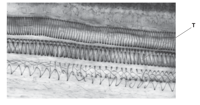
Answer
Water (and dissolved mineral ions).
Water (and mineral ions).
Background Concept
Xylem is the plant tissue responsible for the upward transport of water (and the dissolved mineral ions absorbed from the soil) from the roots to the leaves and other aerial parts. Xylem vessels are dead, hollow, elongated cells with thickened, lignified walls. Because their cytoplasm has been lost, they form continuous tubes through which the transpiration stream can be pulled upwards by the cohesion–tension mechanism.
The micrograph in Fig. 5.1 shows a longitudinal section through a stem with structure T labelled on a xylem vessel. The dark spiral pattern running around the vessel wall is the characteristic secondary thickening of an immature (still-extending) xylem element, where lignin has been deposited in a helical pattern.
Understanding the Question
The question simply asks for one substance that is moved inside the structure labelled T (a xylem vessel). The mark scheme accepts any one correct transported substance.
Approach
Reach for the standard list of xylem contents. The two main categories are water and dissolved mineral ions (e.g. nitrate, magnesium, potassium). Plant hormones such as cytokinin and gibberellin are also carried dissolved in the xylem sap, but the examiner will accept the most direct answers.
Step-by-Step Reasoning
- T is a xylem vessel, identified by the spiral (helical) secondary thickening of lignin in its wall.
- The main bulk flow in xylem is the transpiration stream: water plus the mineral ions absorbed from the soil solution.
- Any one of the accepted answers — water, mineral ions, or a named ion such as nitrate — will earn the mark.
Key Takeaways
- Xylem transports water and mineral ions; the upward flow is the transpiration stream driven by evaporation from the leaves and cohesion of water molecules.
- The spiral wall thickening seen in young xylem is the lignin deposition pattern that will eventually form a complete secondary wall.
Common Mistakes
- Naming 'glucose' or 'sugar' — these are translocated in the phloem, not the xylem.
- Naming 'oxygen' or 'carbon dioxide' — these are gases exchanged by diffusion, not bulk-transported in the xylem.
- Confusing the vessel (xylem) with a tracheid, sieve tube or companion cell.
Things to Be Careful About
- The vessel labelled T has a spiral thickening pattern typical of a young protoxylem element; the same labelling rules apply to it as to a mature xylem vessel.
State the name of the substance that forms the spiral thickening around the structure labelled T in Fig. 5.1.
Answer
Lignin.
Lignin
Background Concept
The wall of a xylem vessel is thickened by the deposition of lignin, a hard, waterproof, phenolic polymer. Lignin is laid down after the cell has extended, and in young xylem it is deposited in patterns such as rings, spirals, reticulate or scalariform thickenings. As the vessel matures, the entire secondary wall may become lignified, leaving only small pits where the wall is unthickened.
Lignin makes the wall:
- waterproof (so the vessel does not leak),
- mechanically strong (so it resists the negative pressure generated by the transpiration stream), and
- resistant to microbial and enzymatic attack.
Understanding the Question
The student is asked to identify the chemical that forms the visible dark spiral in the wall of vessel T. The mark scheme accepts only 'lignin' (or a recognisable variant such as 'lignified').
Approach
Identify T as xylem by its position and by the spiral pattern in the wall. Recall that the wall-thickening substance in xylem is lignin.
Step-by-Step Reasoning
- The helical bands running around the wall of T are the secondary thickenings of a protoxylem vessel.
- These thickenings are made of lignin.
- One word is sufficient: lignin.
Key Takeaways
- The dark, patterned wall thickenings of xylem are made of lignin.
- Lignin gives the vessel its mechanical strength and waterproofing, both essential for withstanding the tension of the transpiration stream.
Common Mistakes
- Writing 'cellulose' — cellulose is the main constituent of the primary cell wall in all plants; the thickening here is specifically lignin.
- Writing 'suberin' or 'cutin' — these waterproof the Casparian strip and leaf cuticle, not xylem walls.
- Writing 'sclerenchyma' — that is a cell type, not a wall substance.
Things to Be Careful About
- Spelling: 'lignin' (one 'g'), not 'lignen' or 'ligin'.
A scientist studied the effect of leaf temperature on the rate of transpiration from the leaves of the wheat plant, Triticum aestivum.
The scientist repeated the investigation using the cotton plant, Gossypium hirsutum.
The results of the investigation are shown in Table 5.1.
Table 5.1
| leaf temperature / | rate of transpiration of T. aestivum / | rate of transpiration of G. hirsutum / |
|---|---|---|
| 25 | 7 | 9 |
| 30 | 12 | 10 |
| 35 | 15 | 11 |
| 40 | 22 | 12 |
| 45 | 31 | 14 |
Suggest explanations for the relationship between leaf temperature and the rate of transpiration of T. aestivum as shown in Table 5.1.
Answer
- As leaf temperature increases, the rate of transpiration of T. aestivum increases.
- (Higher temperature provides more) heat energy to evaporate water from the surfaces of mesophyll cells / to break hydrogen bonds between water molecules.
- Water molecules have more kinetic energy at higher temperature, so they diffuse out of the leaf (through the stomata) faster down the steeper water potential gradient.
- AVP e.g. stomata open wider / more stomata are open at higher temperatures, so more water vapour can diffuse out.
Rate of transpiration increases with leaf temperature because more heat energy is available to evaporate water, water molecules have greater kinetic energy so diffuse out faster, and stomata open more at higher temperatures.
Background Concept
Transpiration is the loss of water vapour from the leaves (and other aerial parts) of a plant. The driving force is the evaporation of water from the wet cell walls of the spongy mesophyll into the air spaces inside the leaf, followed by diffusion of the water vapour out of the leaf through open stomata, down a water potential (concentration) gradient.
Three factors limit the rate of this process:
- The supply of energy for evaporation (latent heat of vaporisation must be supplied to break hydrogen bonds in liquid water).
- The rate of diffusion, which is proportional to kinetic energy and to the steepness of the gradient.
- The number and aperture of the open stomata.
Temperature affects all three.
Understanding the Question
Table 5.1 shows the rate of transpiration of T. aestivum (wheat) at five temperatures between 25 °C and 45 °C. The values rise from 7 to 31 . The student must explain this positive relationship, producing three linked creditable points.
The mark scheme is explicit: any three of (i) overall trend, (ii) more heat energy, (iii) higher evaporation / more energy to break hydrogen bonds, (iv) higher kinetic energy, (v) higher rate of diffusion / steeper gradient, (vi) more open stomata at higher temperatures.
Approach
State the trend first, then give physical reasons for it. Think of the journey of a water molecule: it must (1) be evaporated from the wet cell wall (requires energy), (2) diffuse out of the leaf (kinetic energy and a gradient), and (3) get through the stomata (which respond to temperature). Address each step briefly.
Step-by-Step Reasoning
- Trend: as leaf temperature rises from 25 °C to 45 °C, the rate of transpiration in T. aestivum rises from 7 to 31 .
- Evaporation: higher temperature means more heat energy is available to supply the latent heat of vaporisation. The water molecules in the wet mesophyll walls therefore evaporate into the leaf's internal air spaces at a higher rate.
- Diffusion: the water vapour molecules in the warmer leaf have greater kinetic energy, so they move faster, and the difference in water vapour concentration between the inside of the leaf and the outside air (the gradient) becomes steeper. Both effects increase the rate of diffusion out through the stomata.
- Stomata: at higher temperatures, stomatal guard cells tend to open more (in many species up to an upper limit), allowing more water vapour to escape.
Any three of these earn the three marks.
Key Takeaways
- Transpiration rate increases with temperature because temperature affects every step of the water-loss pathway: evaporation, diffusion and stomatal opening.
- Note that the relationship is approximately exponential rather than linear — the doubling of rate between 30 °C and 45 °C reflects the rapid rise in vapour pressure deficit with temperature.
- The wheat curve is steeper than the cotton curve, indicating wheat is a mesophyte while cotton has xeromorphic features.
Common Mistakes
- Stating only that 'more water evaporates' without explaining why (i.e. without invoking kinetic energy or latent heat).
- Saying 'transpiration is caused by temperature' — temperature is a factor, not a cause; the cause is the gradient driving diffusion.
- Confusing transpiration with evaporation from the soil (evaporation) or with the lifting of water (the transpiration stream).
- Saying 'water is lost faster because the plant needs cooling' — this teleological wording is not credited; the physical explanation must be given.
Things to Be Careful About
- The data in Table 5.1 refer specifically to wheat (T. aestivum); the question asks for an explanation for wheat, not for cotton.
- Three distinct points are required. Repeating the same idea in different words will only earn one mark.
Suggest one difference between the structure of the leaves of T. aestivum and the leaves of G. hirsutum that could explain the results shown in Table 5.1.
Answer
Cotton leaves have a thicker waxy cuticle than wheat leaves, which reduces evaporation of water from the leaf surface.
(Equivalent answers: cotton leaves have fewer / sunken stomata / stomata only on the lower epidermis / trichomes / curled leaves, all of which reduce transpiration.)
A xeromorphic feature of cotton leaves (e.g. thicker waxy cuticle, fewer stomata, sunken stomata or trichomes) that reduces water loss.
Background Concept
Cotton (Gossypium hirsutum) is a plant that evolved in seasonally dry habitats and shows several xeromorphic (drought-adapted) features in its leaves:
- a thick, waxy cuticle on the epidermis,
- stomata that are sunken in crypts or restricted to the lower epidermis,
- trichomes (leaf hairs) that trap a layer of still, humid air next to the leaf surface,
- leaves that curl or roll when water-stressed, again trapping humid air.
Wheat (Triticum aestivum) is a mesophyte: its leaves are flat, broad and thin, with a relatively thin cuticle and a high density of stomata on both surfaces, so it transpires freely.
Understanding the Question
The data show that the transpiration rate of cotton rises only slowly with temperature (9 → 14 ), whereas wheat's rises steeply (7 → 31 ). The student must identify one structural feature of cotton that could account for its consistently lower and less temperature-sensitive rate. Only one such feature is needed for the single mark.
Approach
Think of the leaf as an evaporating surface. Anything that reduces (a) the area of wet cell wall exposed to internal air spaces, (b) the cut-off of vapour reaching the outside air, or (c) the leaf's temperature will reduce transpiration. The accepted answers all fit one of these categories.
Step-by-Step Reasoning
- The mark scheme accepts any of: fewer stomata per unit area; thicker (waxy) cuticle; trichomes / hairs; curled / rolled / folded leaves; sunken stomata (in grooves, crypts, chambers or pits); multilayered epidermis / hypodermis; smaller stomatal aperture; stomata confined to the lower epidermis.
- Choose the one that is most easily described and most easily justified.
- Example answer: cotton leaves have a thicker waxy cuticle than wheat leaves, which reduces evaporation of water from the epidermal surface and lowers the rate of water loss to the atmosphere.
Key Takeaways
- Cotton is a xerophyte; wheat is a mesophyte. The data fit this classification.
- Each xeromorphic feature reduces transpiration by a different mechanism: cuticle (cuts epidermal loss), sunken stomata (lengthen the diffusion path and trap humid air), trichomes (reflect light and trap humid air), curling (lowers exposed area and traps humid air).
Common Mistakes
- Vague answers such as 'cotton is drought-resistant' — this is the conclusion, not the structural feature. The mark scheme requires a specific structure.
- Naming a feature that is not really xeromorphic, e.g. 'wider leaves' or 'larger stomata' — these would increase, not decrease, transpiration.
- Saying 'cotton has a waxy cuticle but wheat does not' — wheat also has a cuticle, just a thinner one. State the comparison.
Things to Be Careful About
- One mark, one feature. Do not waste time listing five; one well-chosen, well-justified feature scores the mark.
- 'Stomata' alone (without saying fewer / sunken / on lower epidermis) is too vague to credit.
Nematodes are microscopic animals that infect a wide range of economically important plant crops including cotton plants. Nematodes feed on the roots of plants, limiting their growth.
When root cells become infected, the nematodes disrupt the plant mitotic cell cycle. This causes the formation of a special type of feeding cell (cell G) in the plant, from which the nematodes absorb nutrients. Cell G is formed as a result of multiple cell cycles without any cytokinesis.
Suggest how cell G differs from cells produced during a mitotic cell cycle that has not been disrupted by a nematode.
Answer
Cell G is a single, large cell containing more than one nucleus (it is multinucleate), because the nuclei divided by mitosis but cytokinesis did not occur.
Cell G is larger and contains more than one nucleus (multinucleate).
Background Concept
A normal mitotic cell cycle consists of:
- Interphase (G1, S, G2) — DNA replicates during S phase.
- Mitosis — the nucleus divides so that the chromosomes are shared equally between two daughter nuclei.
- Cytokinesis — the cytoplasm divides, partitioning the organelles and cytosol into two separate daughter cells.
The result of one complete cycle is two genetically identical daughter cells, each with one nucleus and a normal (2n) chromosome complement.
The question states that nematodes 'disrupt the plant mitotic cell cycle' so that cell G is 'formed as a result of multiple cell cycles without any cytokinesis'. This means the nuclear division (mitosis) keeps happening, but the cytoplasm never splits.
Understanding the Question
The student must identify ONE observable difference between cell G (formed by mitosis without cytokinesis) and a normal product of the mitotic cell cycle.
The mark scheme accepts: more than one nucleus in each cell; more chromosomes; larger cells; more organelles.
Approach
Track what is preserved and what is lost when cytokinesis is omitted:
- The nucleus is divided normally → each 'round' adds one more nucleus to the same cytoplasm.
- The cytoplasm is not divided → all the organelles and cytosol from every 'round' accumulate in one cell.
- The chromosomes are still being segregated correctly inside each daughter nucleus.
Step-by-Step Reasoning
- A normal mitotic cycle ends with two separate daughter cells, each with one nucleus.
- A nematode-disrupted cycle ends with a single cell that contains the nuclei of all the 'daughters' that should have been produced, plus all the cytoplasm and organelles of those daughters combined.
- The clearest, most easily stated difference is therefore: cell G is multinucleate — it contains more than one nucleus, where a normal cell contains exactly one.
- The cell is also larger, contains more organelles, and (in terms of chromosome number per nucleus) still has the same ploidy as a normal cell of that plant.
Key Takeaways
- 'Cell cycle' refers to the whole sequence including cytokinesis. If cytokinesis is omitted, the cell grows and the nucleus divides repeatedly without partitioning the cytoplasm.
- A multinucleate single cell is a hallmark of cells in which mitosis has occurred but cytokinesis has not — for example, the coenocytic hyphae of some fungi, the early endosperm of flowering plants, and the cells described here.
Common Mistakes
- Saying 'cell G is polyploid / has more chromosomes' — the chromosomes are correctly segregated at each mitosis, so ploidy is unchanged; the cell has more nuclei and organelles, not more chromosomes per nucleus.
- Saying 'cell G has more DNA' — the total DNA per cell is greater (because there are more nuclei), but the DNA per nucleus is the same. The cleaner answer is 'more nuclei'.
- Confusing 'cell G' with the endoreplication result in part (ii).
Things to Be Careful About
- Distinguish this part (no cytokinesis → multinucleate) clearly from part (ii) (no mitosis → polyploid).
Nematodes can also stimulate a process called endoreplication. This process causes a plant cell to go through multiple S phases during one cell cycle without entering mitosis or undergoing cytokinesis.
State how the nucleus of a cell that has been through endoreplication may differ from the nucleus of a cell in the same plant that has not been affected by the nematode.
Answer
The nucleus of a cell that has been through endoreplication contains more than two copies of each chromosome (it is polyploid) / more DNA / more chromatin than the nucleus of an uninfected cell, because DNA has replicated several times but the nucleus has not divided.
The nucleus is polyploid (contains more than two copies of each chromosome) / contains more DNA.
Background Concept
During a normal cell cycle:
- S phase (part of interphase) replicates the DNA so that each chromosome consists of two sister chromatids.
- Mitosis segregates those chromatids into two daughter nuclei, so each daughter nucleus has the same chromosome number as the parent.
- Cytokinesis then partitions the cytoplasm.
The key concept here is that DNA replication and nuclear division are separate, independently regulated events. In endoreplication, the cell repeatedly enters S phase (so the DNA doubles each time) but never enters mitosis, so the chromosomes are never segregated.
Understanding the Question
The question states that endoreplication causes a plant cell to go through multiple S phases in a single cell cycle without entering mitosis or undergoing cytokinesis. The student must identify ONE way in which the resulting nucleus differs from a normal nucleus in the same plant.
The mark scheme accepts: more than two copies of each chromosome; more DNA / chromatin; polyploidy; (proportionately) larger nuclei.
Approach
Track the DNA. Each S phase doubles the DNA. After S phases with no mitosis, the cell has copies of each chromosome in a single nucleus. A normal diploid cell has 2 copies; an endoreplicated cell has 4, 8, 16… copies. The nucleus is polyploid.
Step-by-Step Reasoning
- In an uninfected cell, one S phase per cell cycle gives 2 copies of each chromosome in the nucleus — the diploid (2n) state.
- In an endoreplicated cell, multiple S phases give 4, 8, 16, … copies per nucleus — polyploidy.
- Each additional S phase doubles the DNA content of the nucleus; the nucleus therefore contains more DNA, more chromatin, and (proportionately) is larger.
- A clean single-sentence answer is: the nucleus contains more than two copies of each chromosome (it is polyploid).
Key Takeaways
- Endoreplication increases the DNA content of a single nucleus without producing more nuclei — distinct from multinucleation, which is the result of part (i).
- Endopolyploidy is a normal feature of many differentiated plant cells, including the suspensor, the endosperm haustoria, and the antipodal cells of the embryo sac. The giant feeding cells induced by nematodes (cell G and its endoreplicated neighbours) are an extreme version of a normal developmental phenomenon.
Common Mistakes
- Saying 'more nuclei' — this is the answer to part (i), not part (ii). Endoreplication does not increase the number of nuclei; it increases the DNA content of the one existing nucleus.
- Saying 'the nucleus divides' — it does not; mitosis is explicitly omitted.
- Conflating polyploidy (extra chromosome copies inside one nucleus) with aneuploidy (an abnormal chromosome number).
Things to Be Careful About
- 'Polyploid' or 'more than two copies of each chromosome' is the precise term; 'more chromosomes' on its own is ambiguous and is not credited on its own.
- The question asks about the NUCLEUS, not the whole cell. Keep the answer nuclear.
Alveolar macrophages are phagocytes found in the human gas exchange system. They produce hydrolytic enzymes, such as lysozyme, to digest pathogens entering the alveolus.
State the term used to describe the sequence of nucleotides in the DNA of the alveolar macrophage that codes for a protein, such as lysozyme.
Answer
gene
gene
Background Concept
A gene is the basic unit of inheritance. In molecular terms, a gene is a sequence of DNA nucleotides that contains the instructions to make a functional product — usually a protein (a polypeptide), although some genes code for functional RNA molecules such as tRNA or rRNA. The order of the bases along the gene determines the order of amino acids in the polypeptide it encodes. The complete set of genes in an organism is its genome, and different cell types express different subsets of these genes, which is why an alveolar macrophage can make lysozyme while other cells, such as red blood cells, cannot.
Understanding the Question
This is a "state" command worth 1 mark. The question provides a strong hint: "the sequence of nucleotides in the DNA ... that codes for a protein, such as lysozyme." This phrasing is essentially the textbook definition of a gene, so the candidate only needs to recall the technical term.
Approach
Recognise that the wording "sequence of nucleotides in DNA that codes for a protein" is the definition of a gene. There is no other single-word term in CIE biology that fits this description.
Step-by-Step Reasoning
- A gene is defined as a length of DNA that codes for a protein (or for a functional RNA).
- The lysozyme produced by the alveolar macrophage is a protein, so the DNA sequence that codes for it is, by definition, a gene.
- The mark scheme requires only the single word "gene" for one mark.
Key Takeaways
- A gene = a sequence of DNA that codes for a protein (or for a functional RNA).
- Each gene carries the information for one polypeptide chain (with some qualifications for genes that are alternatively spliced).
- Cell-type-specific gene expression is what allows different cells to produce different proteins, even though every nucleated cell in the body contains the same DNA.
Common Mistakes
- Writing "allele" — an allele is a variant form of a gene, not the general term for a protein-coding DNA sequence.
- Writing "chromosome" — a chromosome is a long DNA molecule that contains many genes plus non-coding DNA.
- Writing "DNA" — DNA is the chemical; a gene is a specific functional section of that chemical.
- Writing "genome" — the genome is the complete set of genes, not a single protein-coding unit.
Things to Be Careful About
- The question is phrased to give away the answer. A candidate who knows the definition will recognise the term immediately.
- Do not over-elaborate: "state" questions are one-mark, one-word questions, and adding extra words risks contradicting the mark scheme.
Synthesis of lysozyme occurs in two stages. The first stage occurs in the nucleus using one strand of DNA to synthesise mRNA.
State the name of the strand of DNA that is used to synthesise mRNA.
Answer
template (transcribed) strand
template (transcribed) strand
Background Concept
DNA is a double helix of two antiparallel strands held together by complementary base pairing (A with T, G with C). To make mRNA, the enzyme RNA polymerase must use only one of these two strands as a template. It reads this template strand in the 3' to 5' direction and, by complementary base pairing (with U replacing T), synthesises a new mRNA strand in the 5' to 3' direction. The strand used as the template is called the template strand, the antisense strand, or the transcribed strand. The other strand of DNA — the one not used as the template — is called the coding strand or sense strand, and its sequence is identical to the mRNA (except that T in DNA corresponds to U in mRNA).
Understanding the Question
This is a "state" question worth 1 mark. The stem tells the candidate that "one strand of DNA" is used to synthesise mRNA. The candidate only needs to give the name of that strand. The mark scheme accepts either "template" or "transcribed" strand.
Approach
Recall the two names given in CIE biology for the strand of DNA that is read by RNA polymerase during transcription. Choose the more familiar of "template strand" and "transcribed strand".
Step-by-Step Reasoning
- Transcription uses only one DNA strand to make mRNA; this is the template strand (also called the antisense or transcribed strand).
- The mRNA sequence is complementary to the template strand (and identical — except U for T — to the coding/sense strand).
- For one mark, the candidate needs the term "template" (or "transcribed") and the word "strand".
Key Takeaways
- The template (antisense / transcribed) strand of DNA is the one actually used by RNA polymerase to synthesise mRNA.
- The other strand, the coding (sense) strand, has the same base sequence as the mRNA (with T → U).
- Different genes can be transcribed from either strand of the same DNA molecule, but each gene has a fixed directionality of transcription.
Common Mistakes
- Writing "coding strand" or "sense strand" — this is the OTHER strand, the one with the same sequence as the mRNA.
- Writing "leading strand" or "lagging strand" — these terms describe DNA replication, not transcription.
- Writing "both strands" — only one strand is used as the template for any given gene.
- Calling the mRNA itself the "template" — the question asks for the name of the DNA strand, not the mRNA.
Things to Be Careful About
- "Transcribed strand" is also accepted by the mark scheme, but the more standard and unambiguous CIE term is "template strand".
- Do not confuse the language of transcription (template, coding) with the language of replication (leading, lagging) — they are different processes with different terminology.
Answer
ribosome
ribosome
Background Concept
Translation is the second stage of protein synthesis. The mature mRNA molecule, exported from the nucleus to the cytoplasm, is decoded by ribosomes — small, dense organelles made of ribosomal RNA (rRNA) and protein. A ribosome has a large and a small subunit, which clamp around the mRNA. The ribosome exposes three sites (A, P and E) where tRNA molecules carrying specific amino acids bind. The ribosome moves along the mRNA codon by codon, catalysing peptide bond formation between the amino acids, until it reaches a stop codon and the completed polypeptide is released. Free ribosomes in the cytoplasm make proteins for use inside the cell, while ribosomes bound to the rough endoplasmic reticulum (RER) make proteins for secretion, lysosomes or membranes. Lysozyme, which is secreted by the macrophage, is therefore synthesised on RER-bound ribosomes — but the organelle that actually performs the translation is still the ribosome.
Understanding the Question
This is a "name" question worth 1 mark. The candidate only needs to identify the organelle on which translation occurs. The mark scheme requires "ribosome" and explicitly ignores "70S" or "80S" — so candidates should give just the name, not the size class.
Approach
Recall the locations of the two stages of protein synthesis: transcription in the nucleus, translation on ribosomes in the cytoplasm (or attached to the RER).
Step-by-Step Reasoning
- The mRNA produced inside the nucleus is exported to the cytoplasm.
- In the cytoplasm, the mRNA is decoded by ribosomes, which assemble the amino acid chain (polypeptide) one codon at a time.
- For 1 mark, the candidate must write "ribosome". Giving "RER" or "cytoplasm" or "70S ribosome" is not credited by the mark scheme.
Key Takeaways
- Transcription = nucleus; Translation = ribosome (in the cytoplasm, on the RER, or inside mitochondria/chloroplasts).
- The ribosome is the only organelle that physically carries out translation.
- Whether the protein is for intracellular use (free ribosomes) or for secretion/membranes (RER-bound ribosomes) does not change the answer to "where is translation?" — it is still the ribosome.
Common Mistakes
- Writing "rough endoplasmic reticulum" or "RER" — the ribosome is the actual translation machinery; the RER is the membrane the ribosome may be bound to.
- Writing "cytoplasm" — translation happens in the cytoplasm, but at ribosomes, not in solution.
- Writing "70S" or "80S ribosome" — the mark scheme explicitly ignores size specifications, and giving them is at best superfluous.
- Writing "Golgi apparatus" — the Golgi modifies and packages proteins after translation; it does not synthesise them.
Things to Be Careful About
- "Name" questions need only a single correct term. Adding qualifiers (70S/80S, free/RER-bound) does not earn extra marks and risks contradicting the mark scheme.
- The answer "ribosomes" (plural) is just as acceptable as "ribosome" (singular).
Lysozyme destroys bacterial cells by hydrolysing bonds in peptidoglycan.
Explain how the hydrolysis of bonds in peptidoglycan leads to the destruction of bacterial cells.
Answer
- Peptidoglycan is the main structural component of the bacterial cell wall, so hydrolysing the bonds in peptidoglycan weakens the cell wall.
- The cell wall can no longer resist the turgor pressure of the cytoplasm, so the cell cannot maintain its shape/turgor and water enters by osmosis.
- The cell swells and bursts — it undergoes osmotic lysis.
Hydrolysing peptidoglycan weakens the bacterial cell wall, so the cell cannot maintain turgor and undergoes osmotic lysis.
Background Concept
Bacterial cells are surrounded by a rigid cell wall made of a polymer called peptidoglycan (also known as murein). Peptidoglycan is built from alternating N-acetylglucosamine (NAG) and N-acetylmuramic acid (NAM) sugar units, joined by beta-1,4-glycosidic bonds, and the chains are cross-linked by short peptide bridges to form a single, strong mesh around the cell. This mesh is what gives the bacterium its shape and — crucially — what stops it from bursting. The cytoplasm of a bacterium contains many dissolved solutes, so it has a much lower (more negative) water potential than the dilute fluid that bathes the alveoli. Water therefore tends to enter the bacterium by osmosis all the time, generating a substantial inward (turgor) pressure on the cell wall. In a healthy cell, the peptidoglycan mesh resists this pressure. Lysozyme is an enzyme that hydrolyses the beta-1,4-glycosidic bonds in peptidoglycan, breaking the polymer into shorter pieces and weakening the wall.
Understanding the Question
This is an "explain" question worth 2 marks. The candidate must give a connected chain of reasoning, not just three isolated keywords. The mark scheme rewards three creditable points (weakened cell wall; cell cannot maintain turgor; osmotic lysis), and any 2 of the 3 will score 2 marks — but the cleanest answers link them in the order: hydrolysis → weakened wall → osmotic failure → lysis.
Approach
- Identify what peptidoglycan does in the cell wall (resists turgor pressure).
- State the consequence of breaking it (the wall is weakened).
- Trace the biophysical consequence (water enters by osmosis, turgor cannot be resisted, the cell bursts — osmotic lysis).
Step-by-Step Reasoning
- Peptidoglycan is the structural mesh of the bacterial cell wall. Hydrolysing bonds in it breaks the mesh, so the cell wall is weakened.
- Because the cytoplasm has a more negative water potential than the surrounding alveolar fluid, water continually enters the bacterium by osmosis. A weakened wall cannot resist this turgor pressure, so the cell cannot maintain turgor.
- The cell continues to take up water, swells, and the cytoplasmic membrane ruptures — the bacterium undergoes osmotic lysis.
- 2 marks are available; the mark scheme accepts any 2 of these 3 linked points, so a well-organised answer that gives all 3 in a chain scores full marks.
Key Takeaways
- Peptidoglycan gives the bacterial cell wall its mechanical strength.
- Lysozyme is a hydrolytic enzyme that breaks the beta-1,4-glycosidic bonds in peptidoglycan, weakening the wall.
- A weakened wall cannot resist the osmotic inflow of water into the bacterium, so the cell swells and undergoes osmotic lysis.
- This is why lysozyme is an effective innate antimicrobial agent in tears, saliva, nasal secretions and alveolar fluid.
Common Mistakes
- Stating that lysozyme "kills the bacterium" without explaining how — too vague; "explain" requires the mechanism.
- Saying the bacterium "shrinks" or "loses water" — the opposite happens; water enters the cell because the cytoplasm is more concentrated than the surrounding dilute fluid.
- Saying lysozyme "digests the bacterium" — it digests only the cell wall, not the whole cell; once the wall is broken, the cell lyses.
- Saying the cell wall "disappears" or "is destroyed" — the wall is weakened, not eliminated, and the chain of reasoning to turgor and lysis is what earns the marks.
- Stating "the cell bursts" without using or explaining osmotic inflow — the bursting is osmotic, driven by the water potential difference between the cell and its surroundings.
Things to Be Careful About
- The mark scheme specifically uses the word "turgor" — although bacteria are single-celled, they generate a turgor pressure analogous to that in plant cells, and the term is the precise one expected.
- "Osmotic lysis" is the precise biological term; "bursting" is acceptable in plain English but using the technical term is better.
- Because there are 2 marks, the answer does not have to be long — but it does have to be connected. Two isolated keywords, e.g. "weakens wall" and "bursts", without linking them, would only score 1 of the 2 marks.
- The mark scheme uses the word "cell wall" as the required object of "weakens" — saying "weakens the bacterium" or "weakens the cell" is less precise.
Alveolar macrophages are found in contact with squamous epithelial cells in the walls of alveoli.
Answer
- The cells lining the alveoli are flattened (squamous) epithelial cells.
- This gives a short diffusion distance between the alveolar air and the blood in the adjacent capillaries, so O2 and CO2 can diffuse rapidly across the alveolar wall.
The cells lining the alveoli are flattened (squamous), giving a short diffusion distance for gas exchange.
Background Concept
Gas exchange between the alveolar air and the blood in the surrounding pulmonary capillaries occurs entirely by passive diffusion. Fick's law of diffusion states that the rate of diffusion is directly proportional to the surface area of the exchange surface and the concentration difference across it, and inversely proportional to the thickness of the barrier. To maximise the rate of gas exchange, gas exchange surfaces are therefore typically very thin, have a very large surface area, are moist, and are ventilated and perfused. In the alveoli, the thinness of the barrier is achieved by the structure of the cells that line the alveoli.
Understanding the Question
This is an "explain" question worth 2 marks. It is specifically about the CELLS lining the alveoli (the squamous epithelium, also called Type I pneumocytes), not the alveoli as a whole. The candidate must give the structural feature of the cells (they are flattened) and link it to its functional consequence (it gives a short diffusion distance for gas exchange). The mark scheme rewards exactly these two points.
Approach
- Recall the structure of the alveolar wall: a single layer of very thin, flattened (squamous) epithelial cells.
- Link the thinness of the cells to the diffusion distance the gases must cross.
- State, in each case, both the structure and the function.
Step-by-Step Reasoning
- The cells that line the alveoli are squamous epithelial cells — they are very thin, almost like pancakes.
- Because they are thin, the total thickness of the alveolar wall (cells + fused basement membrane + capillary endothelium) is only about 0.5–1 µm in places.
- This very short diffusion distance allows O2 and CO2 to cross the alveolar wall rapidly by diffusion, which is essential for gas exchange.
- For 2 marks, the candidate should give 1 mark for stating that the cells are flattened/thin/squamous, and 1 mark for linking this to a short diffusion distance for gas exchange.
Key Takeaways
- The cells lining the alveoli are squamous (flattened) epithelial cells, also called Type I pneumocytes.
- Their thinness minimises the diffusion distance between the alveolar air and the blood in the surrounding capillaries.
- This is one of the Fick's law adaptations of the gas exchange system: a thin barrier maximises the rate of diffusion.
Common Mistakes
- Saying "large surface area" — true of the alveoli as a whole (because there are many small alveoli), but it is not an adaptation of the CELLS themselves.
- Saying "good blood supply" or "many capillaries" — also true of the gas exchange system, but is a feature of the surrounding circulation, not of the alveolar lining cells.
- Saying "moist surface" — true of alveoli, but is a property of the surfactant/fluid layer, not of the cells.
- Saying "maintains a concentration gradient" — true of the gas exchange system, but the gradient is maintained by ventilation and circulation, not by the cells themselves.
- Describing the cells only as "thin" without explaining why this helps — "explain" requires the link to diffusion distance.
Things to Be Careful About
- The question is specifically about the CELLS, so answers about other features of the gas exchange system (surface area, ventilation, blood supply) do not earn marks here.
- "Flattened" or "squamous" both describe the cells; "thin" is also acceptable. The functional link is to "short diffusion distance" or "short distance for diffusion".
- The mark scheme requires the link to gas exchange (or to diffusion of O2/CO2), not just the structural fact on its own.
Answer
- On inhalation, the elastic fibres in the alveolar wall stretch, allowing the alveoli to expand without bursting/rupturing.
- On exhalation, the elastic fibres recoil, helping to push air out of the alveoli (and out of the lungs).
Elastic fibres stretch on inhalation to prevent the alveoli from bursting, and recoil on exhalation to help expel air from the lungs.
Background Concept
The lungs are elastic organs that change volume many times each minute as air moves in and out. The connective tissue of the alveolar walls and the smaller bronchioles contains elastic fibres — bundles of the protein elastin that are arranged in a wavy pattern. Elastic fibres can be stretched by an applied force and then return passively to their original length when the force is removed. This stretch-and-recoil property is what allows the lungs to expand during inhalation and to deflate during exhalation without being damaged, and without requiring muscular work to push the air out.
Understanding the Question
This is a "describe" question worth 2 marks. The candidate must state the role of the elastic fibres during BOTH phases of breathing. The mark scheme rewards: (1) stretch on inhalation to prevent rupture of the alveoli, and (2) recoil on exhalation to help expel air. If a candidate says only "they stretch and recoil" without linking to either phase, the mark scheme allows 1 mark as a fallback, but better answers give the full two-phase description.
Approach
- Identify the two mechanical properties of elastic fibres: they stretch and they recoil.
- Link each property to a specific phase of the breathing cycle and to a specific consequence.
Step-by-Step Reasoning
- On inhalation, the diaphragm and external intercostal muscles contract, increasing the volume of the thorax. The lungs are pulled open, and the elastic fibres in the alveolar wall stretch. This stretch allows the alveoli to expand without bursting or rupturing.
- On exhalation, the inspiratory muscles relax, the thorax recoils, and the elastic fibres in the alveolar wall recoil too. This recoil helps to push air out of the alveoli and out of the lungs.
- For 2 marks, both points must be made; ideally, the candidate should link each property (stretch, recoil) to a phase of breathing (inhalation, exhalation) and a consequence (preventing rupture, expelling air).
Key Takeaways
- Elastic fibres give the alveolar walls their mechanical resilience.
- They stretch on inhalation, preventing the alveoli from bursting as they expand.
- They recoil on exhalation, helping to expel air from the lungs.
- This is part of the reason that normal expiration at rest is a passive process — the elastic recoil of the lungs and chest wall does the work of pushing air out.
Common Mistakes
- Saying elastic fibres "help in gas exchange" — they do not; they are a mechanical, not a respiratory, feature.
- Saying they "make the lungs stronger" — vague; the precise words are "stretch" and "recoil".
- Confusing elastic fibres with smooth muscle — smooth muscle is in the walls of the bronchioles (where it controls airway diameter) and is not what allows the alveoli to expand and recoil.
- Stating only "stretch and recoil" without linking to either phase of breathing — the mark scheme allows 1 mark for this fallback, but the full 2 marks require the phase and the consequence for each direction.
- Saying elastic fibres "contract" — they do not contract; only muscle contracts. Elastic fibres passively return to their original length when the stretching force is removed.
Things to Be Careful About
- "Describe" requires both directions of the property and a link to the breathing cycle. A single sentence like "they stretch and recoil" is worth only 1 mark under the fallback rule.
- The mark scheme uses the words "burst" and "rupture" for what the stretching prevents; "overstretching" is also accepted. The mark scheme uses "expel air" for what the recoil helps with.
- Do not confuse elastic fibres (passive, stretch and recoil) with smooth muscle (active, contracts).