Biology 9700/14 — October/November 2025
Cambridge AS Level · Multiple Choice (AS Level) · answer key with instant marking and worked solutions
Topics Biological Molecules · Transport in Plants · Cell Structure · Nucleic Acids and Protein Synthesis · Transport in Mammals · Enzymes · +5 more
Tap an option under each question to check it — your score builds as you go.
Which structures are visible in an appropriately stained plant cell using a high power () light microscope?
Options
| centrioles | mitochondria | starch grains | |
|---|---|---|---|
| A | ✓ | ✗ | ✓ |
| B | ✓ | ✗ | ✗ |
| C | ✗ | ✓ | ✗ |
| D | ✗ | ✓ | ✓ |
key
✓ = visible
✗ = not visible
Working
- Centrioles: absent from higher plant cells, and at ~0.2 µm in diameter they are below the resolution of a light microscope. Not visible. ✗
- Mitochondria: ~0.5–1.0 µm in diameter. When stained (e.g. with Janus Green), they are resolvable at ×400. Visible. ✓
- Starch grains: large (several µm) amyloplasts in plant storage tissues; clearly visible at ×400, especially with iodine stain. Visible. ✓
This matches option D.
Answer
D
D
Background Concept
A standard light microscope, even at the maximum practical magnification of about ×400, has a resolving power of roughly 0.2 µm (200 nm). To be seen, a structure must be (i) large enough to be resolved, (ii) present in the cell type being examined, and (iii) made visible by an appropriate stain that creates contrast between the structure and the surrounding cytoplasm.
The three structures in question:
- Centrioles are barrel-shaped organelles (~0.2 µm diameter × ~0.4 µm long) that organise the mitotic spindle in animal cells. They are not present in higher plant cells (plant cells use other microtubule-organising centres), and even where present their size is at the very limit of light-microscope resolution, so they are not seen in a routine preparation.
- Mitochondria are typically 0.5–1.0 µm wide and 1–10 µm long. They lie above the resolution limit of a light microscope, but because they are largely colourless they require a vital stain such as Janus Green (which is reduced by the mitochondrial electron transport chain) to make them visible against the cytoplasm.
- Starch grains are large amyloplasts (often 5–50 µm across) found abundantly in plant storage tissues such as potato tuber or bean cotyledon. They are highly refractile and become intensely blue-black with iodine (iodine in potassium iodide) solution, making them very conspicuous at ×400.
Understanding the Question
The question lists three structures (centrioles, mitochondria, starch grains) and asks which combination would actually be seen in a plant cell viewed at ×400 with appropriate staining. The candidate must evaluate each structure on two grounds: is it present in a plant cell, and is it large enough / sufficiently stained to be resolved?
Approach
For each structure, ask two questions:
- Is it present in a plant cell?
- Can it be resolved at ×400 with an appropriate stain?
Then select the option whose pattern of ticks and crosses matches the conclusions.
Step-by-Step Reasoning
- Centrioles: Plant cells do not contain centrioles. Even if they did, their size (~0.2 µm) is at the resolution limit of a light microscope. Not visible. ✗
- Mitochondria: Plant cells contain many mitochondria, and with the Janus Green stain they appear as small blue-green rods/dots at ×400. Visible. ✓
- Starch grains: Plant storage cells are packed with starch grains, easily seen unstained as oval refractile bodies and, with iodine, stained intensely blue-black. Visible. ✓
The pattern ✗, ✓, ✓ corresponds to option D.
Key Takeaways
- Light microscope resolution (~0.2 µm) is the limiting factor, not magnification alone.
- Plant cells lack centrioles; this immediately rules out any option in which centrioles are ticked.
- Mitochondria and starch grains are large enough (especially starch grains) and stainable (mitochondria with Janus Green) to be visible at ×400 in a plant cell.
Common Mistakes
- Ticking centrioles because they are a "common organelle" — they are absent from plant cells.
- Leaving starch grains unticked because iodine is not always added in routine preparations — at ×400 starch grains are large and refractile even without stain, and they are an obvious feature of plant storage cells.
- Confusing resolution with magnification and assuming that "higher power" alone makes smaller structures visible.
Things to Be Careful About
- A ×400 light microscope can resolve structures down to about 0.2 µm; ribosomes (~0.025 µm), endoplasmic reticulum membranes, and centrioles are below this limit.
- "Appropriately stained" is the key phrase in the stem — it permits the use of stains like Janus Green (mitochondria) and iodine (starch grains), so the question is not asking what is visible unstained.
- Plant vs animal cell contents differ: centrioles are an animal-cell feature, while starch grains and large central vacuoles are plant-cell features.
The images show an eyepiece graticule and a stage micrometer scale viewed through a microscope.
The smallest divisions of the stage micrometer scale are .
The images also show a photomicrograph of a transverse section through a plant root using the same eyepiece graticule and the same magnification.
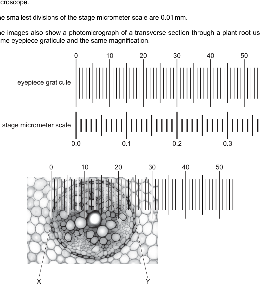
What is the actual width of the vascular tissue in the plant root between X and Y?
Options
A
B
C
D
Working
- Calibrate the graticule from the stage micrometer alignment: eyepiece graticule divisions .
- One graticule division:
- Read X and Y on the photomicrograph: and , giving a span of about graticule divisions.
- Actual width:
Answer
C
C
Background Concept
Two scales are used together on a light microscope:
- The eyepiece graticule is a small disc bearing a scale (here –) that sits inside the eyepiece. It is therefore magnified along with the specimen, so each division of the graticule does NOT represent a fixed real distance — its real value depends on the objective lens in use.
- The stage micrometer is a slide with an accurately ruled scale (smallest divisions here). Unlike the graticule, the stage micrometer has a known real size, so it is used to calibrate the graticule at each magnification.
The calibration procedure is to superimpose the two scales and note how many graticule divisions line up with a known number of stage-micrometer divisions. The result — micrometres per graticule division — is the conversion factor used to turn any later graticule reading into a real length.
Understanding the Question
Fig. 2.1 shows three panels:
- The eyepiece graticule alone (scale –).
- The graticule aligned with the stage micrometer, where the stage reading lines up with graticule mark .
- A photomicrograph of a transverse section through a plant root, with the same eyepiece graticule overlaid and the vascular tissue marked between X (left side) and Y (right side).
The question asks for the actual (real) width of the vascular tissue between X and Y, expressed in micrometres. The correct option is the one that matches the value calculated from the calibration.
Approach
- Use the alignment in the middle panel to find how many micrometres one graticule division represents.
- Read the graticule positions of X and Y on the bottom panel and subtract.
- Multiply the graticule-distance by the micrometres-per-division conversion factor.
- Choose the option that matches the result, remembering .
Step-by-Step Reasoning
Step 1 — Calibrate. From the middle panel:
So each graticule division represents:
Step 2 — Read X and Y. On the photomicrograph, X lies just to the right of the graticule mark and Y lies just before the mark — roughly at graticule values and . The span is therefore about graticule divisions (any reasonable read between and is acceptable, because all give answers in the same order of magnitude).
Step 3 — Calculate the actual width.
Step 4 — Select the closest option. The four options are , , and . The result matches option C.
Why the distractors fail:
- and are both too small — they correspond to a student who read the graticule correctly but forgot to convert mm to μm (treating each division as instead of ).
- is roughly the value if the X–Y span were taken to be the entire visible field from to , or if the student mistakenly used per division (a over-estimate of the conversion factor).
Key Takeaways
- An eyepiece graticule must be calibrated at every magnification with a stage micrometer because its divisions have no fixed real length.
- Calibration gives micrometres per graticule division, the conversion factor used in every later measurement on that graticule.
- Unit conversion () is the most common source of error in these questions; always state the unit on every numerical result.
- When answering an MCQ, compute carefully, then check that the chosen option is plausibly of the right order of magnitude before selecting it.
Common Mistakes
- Forgetting the conversion between mm and μm, giving an answer that is too small by a factor of 1000 (the and options).
- Using the wrong alignment values, e.g. reading the graticule end-marks as – aligned with – on the stage micrometer (which is not what the figure shows).
- Reading X or Y off the wrong scale — confusing the graticule numbers with the stage-micrometer numbers (which only run to ).
- Ignoring the calibration step and treating the graticule as if it were already a micrometre scale.
Things to Be Careful About
- Always state the calibration explicitly: write the equivalence you read from the figure and the resulting μm-per-division value before you use it.
- Keep the conversion factor and the graticule distance in their correct units; the common error is to mix them.
- Read X and Y to the nearest small graticule division, but do not invent precision beyond the figure — any answer in the range – is consistent with the picture, and only one of the offered options (C) lies in that range.
- This is a single-mark MCQ, so the working above is for understanding; on the paper, a brief calibration statement and the final letter is sufficient.
It is thought that some organelles in eukaryotic cells evolved from free-living prokaryotes.
Which organelles have features that suggest they evolved from free-living prokaryotic ancestors?
Options
A chloroplasts and mitochondria
B chloroplasts and Golgi bodies
C mitochondria and smooth endoplasmic reticulum
D Golgi bodies and smooth endoplasmic reticulum
Working
The endosymbiotic theory proposes that mitochondria and chloroplasts originated from free-living prokaryotes that were engulfed by an ancestral eukaryotic cell. Key evidence supporting this:
- Both organelles have a double membrane (consistent with engulfment by phagocytosis).
- Both contain their own circular DNA, free of histones, like bacterial DNA.
- Both contain 70S ribosomes, the same size as bacterial ribosomes (not the 80S ribosomes of the eukaryotic cytoplasm).
- Both reproduce by binary fission, like bacteria.
Golgi bodies and smooth endoplasmic reticulum do not show these prokaryotic features; they are part of the endomembrane system derived from invaginations of the plasma membrane.
Answer
A
A
Background Concept
The endosymbiotic theory (proposed by Lynn Margulis, 1967) states that two eukaryotic organelles — mitochondria and chloroplasts — originated from free-living prokaryotic cells that were engulfed by a larger ancestral host cell. Instead of being digested, they established a mutually beneficial (symbiotic) relationship: the host provided protection and nutrients, while the engulfed prokaryote supplied either ATP (in the case of the aerobic bacterium that became the mitochondrion) or photosynthetic products (in the case of the cyanobacterium that became the chloroplast). Over evolutionary time, most genes were transferred to the host nucleus, but the organelles retained some of their original prokaryotic characteristics.
The evidence for this theory is based on features that mitochondria and chloroplasts share with prokaryotes but that other eukaryotic organelles do not share:
- A double membrane — the outer membrane is derived from the host's plasma membrane (from engulfment), while the inner membrane is derived from the prokaryote's original plasma membrane.
- Circular DNA that is not associated with histones, very similar to bacterial chromosomal DNA.
- 70S ribosomes (smaller than the 80S ribosomes found in the eukaryotic cytoplasm and on the rough endoplasmic reticulum), identical in size to bacterial ribosomes.
- Binary fission as their mode of division, rather than mitosis.
By contrast, the Golgi apparatus and the smooth endoplasmic reticulum are part of the eukaryotic endomembrane system. They are thought to have arisen from infoldings of the host cell's own plasma membrane, not from engulfed prokaryotes. They have no DNA of their own, no ribosomes, and a single bounding membrane.
Understanding the Question
This is a multiple-choice question (AS Paper 1 style) asking the candidate to identify which pair of eukaryotic organelles shows features consistent with having evolved from free-living prokaryotic ancestors. The correct option is the pair that BOTH have endosymbiotic features. Only one option can be correct.
Approach
Recall the endosymbiotic theory and list the organelles that show the four key prokaryotic features (double membrane, own circular DNA, 70S ribosomes, binary fission). The only two organelles that fit this description are mitochondria and chloroplasts. Therefore, the answer must include both of them.
Step-by-Step Reasoning
- Option A — chloroplasts and mitochondria: ✓ Both organelles possess a double membrane, circular DNA, 70S ribosomes, and reproduce by binary fission. They are the textbook examples of endosymbiotic organelles.
- Option B — chloroplasts and Golgi bodies: ✗ Chloroplasts are endosymbiotic, but Golgi bodies are not — they are stacks of flattened membrane-bound cisternae with no DNA and no ribosomes, derived from the endomembrane system.
- Option C — mitochondria and smooth endoplasmic reticulum: ✗ Mitochondria are endosymbiotic, but smooth ER is a network of membranes continuous with the rough ER, with no DNA or ribosomes.
- Option D — Golgi bodies and smooth endoplasmic reticulum: ✗ Neither has endosymbiotic features.
Only Option A pairs two organelles that both have prokaryotic features.
Key Takeaways
- The endosymbiotic theory explains the origin of mitochondria (from an aerobic prokaryote) and chloroplasts (from a photosynthetic cyanobacterium).
- The four diagnostic prokaryotic features of these organelles are: double membrane, own circular DNA, 70S ribosomes, and binary fission.
- Other membrane-bound organelles (Golgi, ER, lysosomes, vacuoles) are part of the endomembrane system and do not have these features.
Common Mistakes
- Confusing the rough ER with an endosymbiotic organelle because it has ribosomes attached — but its ribosomes are 80S (eukaryotic) cytoplasmic ribosomes, not 70S, and the rough ER has no own DNA.
- Choosing an option that mixes one correct organelle with one incorrect one (B or C). The question requires BOTH organelles in the pair to show prokaryotic ancestry.
- Forgetting that chloroplasts, like mitochondria, have a double membrane — students often only recall this for mitochondria.
Things to Be Careful About
- "70S" and "80S" refer to sedimentation coefficients during centrifugation, not strictly to size, but they reliably distinguish prokaryotic from eukaryotic ribosomes.
- The endosymbiotic theory is supported by the SAME features in BOTH mitochondria and chloroplasts — when asked for an endosymbiotic organelle, the answer is always one of these two, never Golgi, ER, lysosomes, or the nucleus.
A student completed a table showing the structures that are found in four different types of cells.
| cell structure | cell Q | cell R | cell S | cell T |
|---|---|---|---|---|
| cell surface membrane | ✓ | ✓ | ✓ | ✓ |
| cellulose cell wall | ✓ | ✓ | ✗ | ✓ |
| chloroplast | ✓ | ✗ | ✗ | ✗ |
| mitochondrion | ✓ | ✓ | ✗ | ✓ |
| nucleus | ✓ | ✓ | ✗ | ✗ |
key
✓ = present
✗ = not present
How many of these types of cells can be found in plants?
Options
A 1
B 2
C 3
D 4
Working
Classify each cell using the features shown:
- Cell Q – cell surface membrane, cellulose cell wall, chloroplast, mitochondrion and nucleus all present → typical plant cell (e.g. palisade mesophyll cell).
- Cell R – cellulose cell wall and nucleus present, but no chloroplast → still a plant cell (a non-photosynthetic cell such as a root or storage cell).
- Cell S – only a cell surface membrane; no cell wall, no nucleus, no organelles → prokaryote (bacterium); not a plant cell.
- Cell T – cellulose cell wall and mitochondrion present, but no nucleus and no chloroplast → a sieve tube element (a phloem cell that has lost its nucleus at maturity); found in plants.
Plant cells = Q, R and T → 3 cells.
Answer
C
C
Background Concept
Plant cells are eukaryotic, so they always have a nucleus (with the single exception of sieve tube elements, which lose their nucleus during differentiation), a cell surface membrane, mitochondria and (outside the meristems) a cellulose cell wall. The cellulose cell wall is the single most reliable diagnostic feature of a plant cell.
However, not every plant cell contains every textbook organelle. A cell's features reflect its function:
- Photosynthetic cells (palisade and spongy mesophyll) contain chloroplasts as well as the other usual plant-cell structures.
- Non-photosynthetic plant cells (root hair cells, xylem vessel elements, storage parenchyma, epidermal cells) have a cellulose cell wall and nucleus but no chloroplasts.
- Sieve tube elements of the phloem are highly specialised: at maturity they lose their nucleus, ribosomes and much of the cytoplasm, leaving only a cell surface membrane, a cellulose cell wall and a few mitochondria (to supply ATP for active loading of sucrose). Their metabolic function is performed by the adjoining companion cell, which does retain a nucleus.
Prokaryotic cells (bacteria) have a cell surface membrane, but no nucleus, no membrane-bound organelles and their cell wall (where present) is made of peptidoglycan, not cellulose.
Understanding the Question
The question gives a 5 × 4 table of structures (cell surface membrane, cellulose cell wall, chloroplast, mitochondrion, nucleus) for four unknown cells Q, R, S and T. The command word is "How many…" — a simple count once each cell is correctly identified. The trap is to assume that any cell with a cellulose cell wall must be a "textbook" plant cell, and that any cell without a nucleus cannot be a plant cell.
Approach
- Use the presence/absence of a cellulose cell wall as the first filter for "plant or not plant".
- For cells with a cellulose cell wall, check the nucleus to decide whether it is a typical plant cell (Q, R) or a specialised phloem sieve tube element (T).
- For cells without a cellulose cell wall and without a nucleus, identify as prokaryote (S).
- Count those that belong in a plant.
Step-by-Step Reasoning
Cell Q has every structure listed: cell surface membrane, cellulose cell wall, chloroplast, mitochondrion and nucleus. This is the "textbook" plant cell — a photosynthetic cell such as a palisade or spongy mesophyll cell. ✓ plant
Cell R has a cellulose cell wall, cell surface membrane, mitochondrion and nucleus, but no chloroplast. A plant cell that does not photosynthesise — e.g. a root cortical cell, a root hair cell or a storage parenchyma cell. ✓ plant
Cell S has only a cell surface membrane; no cell wall, no chloroplast, no mitochondrion, no nucleus. The absence of a nucleus (and of all membrane-bound organelles) identifies it as a prokaryote — a bacterium. Bacteria are not plants, so this is ✗ not a plant cell.
Cell T has a cellulose cell wall, a cell surface membrane and a mitochondrion, but no chloroplast and no nucleus. A eukaryotic plant cell must have a nucleus — but the mature sieve tube element of the phloem is the well-known exception: its nucleus and most organelles degenerate during development so that the lumen is open for efficient mass flow of assimilates. A few mitochondria remain to power active loading at the source. So cell T is a sieve tube element, which is found in plants. ✓ plant
Total plant cells = Q + R + T = 3.
Key Takeaways
- A cellulose cell wall is the defining structural feature of plant cells, but plants contain many cell types and not all of them are photosynthetic.
- Sieve tube elements are specialised phloem cells that lose their nucleus at maturity while retaining a cellulose cell wall and a small number of mitochondria — a classic exam "exception".
- Prokaryotes can be ruled out by the absence of both a nucleus and membrane-bound organelles (here, cell S).
- When asked "how many plant cells?", scan for cellulose cell walls first, then check the nucleus to catch the sieve tube element trap.
Common Mistakes
- Stopping at two plant cells (Q and R) and missing that cell T is a sieve tube element. The mark scheme here deliberately includes a cell that looks like a "bacterium" because of its missing nucleus, but the cellulose cell wall and mitochondrion reveal it as a plant cell.
- Counting cell S as a plant cell because it has a cell surface membrane. Animal cells and bacterial cells both have cell surface membranes, so this single feature is not enough — the cellulose cell wall is what defines a plant cell.
- Treating "no nucleus" as automatically meaning "not a plant cell". Sieve tube elements and xylem vessel elements (which are dead) are the two main plant-cell exceptions.
Things to Be Careful About
- The question asks which cells can be found in plants, not which are "typical" plant cells. Specialised cells such as sieve tube elements still count.
- Read the table carefully: a ✗ in the chloroplast row does not rule out a plant cell, and a ✗ in the nucleus row does not rule out a plant cell on its own.
- Mark scheme-style wording matters: a cellulose cell wall is the diagnostic feature, not just "a cell wall" — bacterial cell walls are made of peptidoglycan.
Some features of cells are listed.
1 cytoplasm
2 cell surface membrane
3 ribosomes
Which features are found in animal cells and also in prokaryotic cells?
Options
A 1, 2 and 3
B 1 and 2 only
C 1 and 3 only
D 2 and 3 only
Working
Both animal (eukaryotic) cells and prokaryotic cells possess:
- Cytoplasm — present in all cells.
- Cell surface membrane — present in all cells.
- Ribosomes — present in both; prokaryotic ribosomes are 70S, while eukaryotic ribosomes are 80S, but ribosomes as a structure are universal.
All three features are shared, so the answer is A.
Answer
A
A
Background Concept
All cells — whether prokaryotic (e.g. bacteria) or eukaryotic (e.g. animal, plant, fungal) — share a small set of universal features. These are the minimum requirements for a living cell:
- A cell surface membrane (plasma membrane), a phospholipid bilayer that encloses the cell and controls what enters and leaves.
- Cytoplasm (or cytosol), a watery matrix in which metabolic reactions occur and where ribosomes are suspended.
- Ribosomes, the site of protein synthesis, made of rRNA and protein.
- DNA as the genetic material.
What distinguishes prokaryotes from eukaryotes is the presence of membrane-bound organelles (nucleus, mitochondria, ER, Golgi, etc.) in eukaryotes — prokaryotes lack these but still have the basic features above.
Understanding the Question
The question lists three cell features — cytoplasm (1), cell surface membrane (2), and ribosomes (3) — and asks which are found in both animal cells and prokaryotic cells. The four options vary in which combination of these three features is selected.
The command word is implicit "which" — the candidate must simply identify the correct set.
Approach
Check each numbered feature against both cell types:
- Animal cell: has cytoplasm ✓, has cell surface membrane ✓, has ribosomes ✓.
- Prokaryotic cell: has cytoplasm ✓, has cell surface membrane ✓, has ribosomes ✓ (smaller 70S type, but still ribosomes).
If a feature is present in BOTH cell types, include it. All three qualify, giving the full set 1, 2 and 3.
Step-by-Step Reasoning
- Cytoplasm — A fluid/gel matrix filling the cell interior. Present in every cell. Prokaryotes have cytoplasm (containing the nucleoid region and ribosomes), and animal cells have cytoplasm (containing organelles). ✓ for both.
- Cell surface membrane — A phospholipid bilayer forming the outer boundary of all cells. Prokaryotes have one (and additionally may have a cell wall outside it), and animal cells have one (with no cell wall). ✓ for both.
- Ribosomes — Site of protein synthesis. Eukaryotic ribosomes are 80S; prokaryotic ribosomes are 70S. The size differs but the structure is universal. ✓ for both.
Distractors worth noting:
- Options B, C and D each omit one of the three shared features, suggesting a candidate who is uncertain whether prokaryotes have a cell surface membrane (they do — every cell has one), cytoplasm (they do), or ribosomes (they do, 70S).
- A common error is to think prokaryotes have no cell surface membrane because the word "membrane-bound organelles" is associated with eukaryotes. The cell surface membrane is the outer cell boundary in prokaryotes, not an internal organelle, so it is always present.
The correct answer is the set containing all three: A.
Key Takeaways
- The minimum universal features of all cells are: cell surface membrane, cytoplasm, ribosomes, and DNA.
- Prokaryotes differ from eukaryotes in lacking a nucleus and other membrane-bound organelles, but they still have a plasma membrane, cytoplasm, and ribosomes.
- Ribosome size differs (70S in prokaryotes, 80S in eukaryotes) but ribosomes as a structure are universal.
Common Mistakes
- Excluding the cell surface membrane (2) because prokaryotes are sometimes said to "lack membrane-bound structures" — the cell surface membrane is the boundary of the cell itself, not an internal organelle, so it is present.
- Excluding ribosomes (3) because they differ in size between prokaryotes and eukaryotes — the question asks about the feature (ribosomes), not the specific type.
- Confusing the cell wall (present in prokaryotes, absent in animal cells) with the cell surface membrane (present in both).
Things to Be Careful About
- "Membrane-bound" in biology often refers to internal organelles, not the cell surface membrane. The cell surface membrane is universal.
- The cell wall is NOT a feature in this question; even if it were, animal cells lack a cell wall, so it would not be shared.
- Prokaryotic ribosomes are smaller (70S) and are the target of many antibiotics (e.g. streptomycin binds the 30S subunit), but the basic structure of rRNA + protein is conserved across all life.
Trehalose is a non-reducing disaccharide.
Separate samples of a mixture of fructose and trehalose were tested with Benedict’s solution before and after acid hydrolysis.
What will be the results of these Benedict’s tests?
Options
| before acid hydrolysis | after acid hydrolysis | |
|---|---|---|
| A | ✓ | ✓ |
| B | ✓ | ✗ |
| C | ✗ | ✓ |
| D | ✗ | ✗ |
key
✓ = positive result with Benedict’s test
✗ = negative result with Benedict’s test
Working
- Fructose is a monosaccharide and a reducing sugar → gives a positive Benedict's test on its own.
- Trehalose is a non-reducing disaccharide → does not give a positive Benedict's test.
- Therefore, the mixture of fructose + trehalose gives a positive result before acid hydrolysis (because fructose is present). ✓
- Acid hydrolysis breaks the glycosidic bond in trehalose, releasing two glucose molecules, both of which are reducing sugars. Fructose is unaffected.
- After acid hydrolysis, the solution contains fructose + glucose + glucose — all reducing sugars → positive Benedict's test. ✓
Answer
A
A
Background Concept
Benedict's test detects reducing sugars. A reducing sugar is one that has a free aldehyde (–CHO) or ketone (–C=O) group that can be oxidised, donating electrons to Cu²⁺ in the blue Benedict's reagent and reducing it to a brick-red precipitate of copper(I) oxide (Cu₂O).
- All monosaccharides are reducing sugars. This includes glucose, fructose, galactose, ribose and maltose's component sugars.
- Some disaccharides are reducing (maltose, lactose) because one of their monosaccharide units still has a free anomeric carbon (the carbon that was involved in the glycosidic bond on the other unit is free, so it can open to expose the reactive group).
- Non-reducing disaccharides (sucrose, trehalose) have BOTH anomeric carbons locked in the glycosidic bond, so there is no free –CHO or –C=O group available to reduce Cu²⁺. They give a negative Benedict's test directly.
Acid hydrolysis of a disaccharide boils the sugar with dilute hydrochloric acid. The acid catalyses the breaking of the glycosidic bond, releasing the component monosaccharides. For trehalose (glucose–α,α-1,1-glucose), hydrolysis yields two glucose molecules.
Understanding the Question
The mixture contains two sugars:
- Fructose — a reducing monosaccharide.
- Trehalose — a non-reducing disaccharide of two glucose units.
We are asked what Benedict's test gives:
- on the original mixture, and
- on the mixture after it has been boiled with acid (acid hydrolysis).
The two results are then matched to the four options A–D, where ✓ = positive and ✗ = negative.
The command word is implicit but the question is essentially "predict" — you have to apply the rules above to the specific case of fructose + trehalose.
Approach
For each condition (before and after hydrolysis), ask:
- Which sugars are present?
- Is each one reducing or non-reducing?
- Overall, is the mixture reducing?
Then write ✓ or ✗ for each condition and match to the table.
Step-by-Step Reasoning
Before acid hydrolysis:
- The mixture contains fructose and trehalose.
- Fructose is a reducing sugar → contributes a positive result.
- Trehalose is non-reducing → contributes nothing.
- The mixture as a whole contains a reducing sugar, so Benedict's test is positive (✓).
After acid hydrolysis:
- Acid hydrolysis cleaves the glycosidic bond in trehalose, releasing two glucose molecules. Glucose is a reducing sugar.
- Fructose is unaffected by the acid (it is already a monosaccharide, so there is no bond to break).
- The hydrolysed mixture therefore contains glucose + glucose + fructose — all reducing sugars.
- Benedict's test is again positive (✓).
So the pattern is ✓ before, ✓ after → this matches option A.
Why the other options are wrong:
- B (✓, ✗): Wrong because acid hydrolysis of a non-reducing disaccharide releases reducing monosaccharides, so the result cannot become negative.
- C (✗, ✓): Wrong before hydrolysis because fructose in the mixture should already give a positive result.
- D (✗, ✗): Wrong on both counts. The mixture contains the reducing sugar fructose, so it must be positive before hydrolysis; and after hydrolysis there are even more reducing sugars present, so it cannot be negative.
A common trap is to assume that "non-reducing disaccharide" means the whole mixture must be non-reducing, forgetting that the other component (fructose) is itself a strong reducing sugar.
Key Takeaways
- All monosaccharides (including fructose) are reducing sugars and give a positive Benedict's test.
- A non-reducing disaccharide gives a negative Benedict's test directly, but a positive test after acid hydrolysis — this is the standard two-step procedure used to identify a non-reducing sugar.
- Acid hydrolysis breaks the glycosidic bond in a disaccharide, releasing the constituent monosaccharides.
- When asked about a mixture, consider each component separately and combine the outcomes.
Common Mistakes
- Assuming the mixture must be non-reducing because trehalose is. Fructose is a reducing sugar and is unaffected by the presence of trehalose — the mixture is reducing from the start.
- Thinking acid hydrolysis destroys sugars or makes them non-reducing. It does the opposite: it liberates reducing monosaccharides from the disaccharide.
- Confusing sucrose with trehalose — both are non-reducing, but the same logic applies to sucrose (glucose + fructose), and hydrolysis of sucrose would give glucose + fructose, both reducing.
- Forgetting that fructose is a ketone, not an aldehyde, and therefore doubting it is reducing. Fructose is a reducing sugar because, in solution, it can isomerise to give a form with a free aldehyde group (or directly tautomerise through the enediol intermediate) that can be oxidised by Cu²⁺.
Things to Be Careful About
- Read the key in the table carefully: ✓ = positive, ✗ = negative.
- "Before acid hydrolysis" and "after acid hydrolysis" are two separate tests on related but distinct samples — there is no single sample that is tested twice.
- Acid hydrolysis requires boiling with dilute HCl, then neutralising with NaHCO₃ (or similar) before doing Benedict's test, because Benedict's reagent is alkaline. The mark scheme takes this as understood; you do not need to write the neutralisation step for this MCQ, but remember it for structured questions.
- The question says trehalose is "a non-reducing disaccharide" — take this as given; you do not need to justify why it is non-reducing, only to use that fact.
Which molecules have a structural formula that contains C=O bonds?
1 glucose
2 glycerol
3 protein
Options
A 1, 2 and 3
B 1 and 2 only
C 1 and 3 only
D 2 and 3 only
Working
- Glucose is an aldehyde sugar (with an open-chain form containing a terminal –CHO group); the C=O of the aldehyde is present in its structural formula.
- Glycerol has the structure — all three carbons carry –OH groups, so there is no C=O bond.
- Proteins contain peptide bonds (–CO–NH–) linking amino acid residues; the C=O of the peptide bond is present in their structural formula.
Answer
C
C
Background Concept
A carbon–oxygen double bond, written C=O, is the defining feature of the carbonyl group. Carbonyls appear in several biologically important functional groups:
- Aldehyde (–CHO), found in the open-chain form of glucose and other aldo-sugars.
- Ketone (>C=O), found in fructose and in intermediates such as pyruvate.
- Carboxyl (–COOH), found in fatty acids and in the side chain (R group) of amino acids.
- Peptide (amide) bond (–CO–NH–), the link between amino acids in a polypeptide chain, which contains a C=O.
Molecules whose carbons are bonded only to –H, –OH, or to other carbons by single bonds (e.g. glycerol, the saturated hydrocarbon backbone of fatty acids, ribose in its cyclic form) do not contain C=O bonds.
Understanding the Question
The command is implicit: identify which of the three named molecules (glucose, glycerol, protein) have a structural formula that includes at least one C=O bond. You must decide on a YES/NO basis for each of the three and then match the resulting pattern to the options A–D.
Approach
For each molecule, write or visualise its structural formula and check whether any carbon is double-bonded to oxygen:
- Glucose — look for the aldehyde group at C1 of the open-chain form.
- Glycerol — list the three carbons and the groups attached to each.
- Protein — recall that amino acids are joined by peptide bonds, each of which has a C=O.
Step-by-Step Reasoning
- Glucose (an aldohexose, ) has the open-chain structure . The carbon at position 1 is part of an aldehyde group –CHO, which is a C=O double bond. ✓ C=O present.
- Glycerol () has the structure . Every carbon is bonded to either –H or –OH only; there is no C=O. ✗ C=O absent.
- Protein — amino acids are linked by peptide bonds (–CO–NH–). Each peptide bond contains a C=O between the carbonyl carbon and the oxygen. ✓ C=O present (as part of the peptide bond backbone).
Therefore the molecules that contain C=O are 1 and 3 only, which matches option C.
Key Takeaways
- An aldehyde (–CHO), ketone (>C=O), carboxyl (–COOH) and peptide bond (–CO–NH–) all contain C=O.
- Glycerol is a triol (three –OH groups on three carbons); it has no C=O and is a common distractor in this type of question.
- The C=O of the peptide bond is the carbonyl that makes proteins give a positive result in tests (e.g. the biuret test detects peptide bonds, not the C=O alone, but the C=O is structurally present).
Common Mistakes
- Choosing A (1, 2 and 3) by assuming glycerol "must" contain a C=O because it is part of a lipid — it does not; the C=O appears in the fatty acid portion of a triglyceride, not in the glycerol backbone.
- Choosing B (1 and 2 only) by forgetting that the peptide bond contains a C=O.
- Confusing hydroxyl (–OH) with carbonyl (C=O) — both contain oxygen but only the latter has a double bond to it.
Things to Be Careful About
- The cyclic (Haworth) form of glucose does not visibly show a C=O, but the structural formula of glucose encompasses the open-chain form, which does. The mark scheme credits the open-chain representation.
- Read the wording of MCQ options carefully: "1 and 3 only" is option C, not option B (which is "1 and 2 only").
Which statement about monosaccharides and disaccharides is correct?
Options
A Sucrose is a non-reducing disaccharide produced from two identical monosaccharides joined together by a condensation reaction.
B The hydrolysis of the reducing disaccharide maltose produces a reducing monosaccharide, and a non-reducing monosaccharide.
C Maltose is a non-reducing disaccharide that contains two identical reducing monosaccharides connected by a glycosidic bond.
D The addition of a water molecule to the glycosidic bond in a sucrose disaccharide produces two different reducing monosaccharides.
Working
A — Sucrose is a non-reducing disaccharide, but it is formed from two different monosaccharides (α-glucose + fructose), joined by an α-1,4 glycosidic bond. The word "identical" makes A wrong.
B — Maltose is a reducing disaccharide, and its hydrolysis gives two molecules of glucose, both of which are reducing. So B is wrong on two counts (maltose is reducing, and both products are reducing).
C — Maltose does contain two identical glucose units joined by an α-1,4 glycosidic bond, but maltose is a reducing disaccharide, not non-reducing. So C is wrong.
D — Hydrolysis adds a water molecule across the glycosidic bond of sucrose, breaking it. The products are glucose (an aldose, reducing) and fructose (a ketose, reducing) — two different reducing monosaccharides. ✓
Answer
D
D
Background Concept
Monosaccharides are single sugar units (e.g. glucose, fructose, galactose). A sugar is classified as reducing if it can donate electrons (be oxidised) — in practice, this means its anomeric carbon (C1 in aldoses, C2 in ketoses) is free, i.e. not locked in a glycosidic bond. Common reducing monosaccharides are glucose, fructose, galactose and mannose.
Disaccharides are formed when two monosaccharides join by a condensation reaction, releasing one water molecule and forming a glycosidic bond. The reverse reaction — adding water to break the bond — is hydrolysis.
A disaccharide is reducing if at least one of its two component monosaccharides still has a free anomeric carbon (i.e. its glycosidic bond does not lock up both anomeric carbons). It is non-reducing only if the glycosidic bond joins the two anomeric carbons together, leaving no free aldehyde or ketone group.
Key examples to know:
- Maltose = glucose + glucose (α-1,4 bond) → reducing
- Lactose = galactose + glucose (β-1,4 bond) → reducing
- Sucrose = glucose + fructose (α-1,β-2 bond, joining both anomeric carbons) → non-reducing
Understanding the Question
This is a single-best-answer MCQ testing whether you can (i) identify whether a named disaccharide is reducing or non-reducing, (ii) recall what monosaccharides it is built from, and (iii) recognise the products of its hydrolysis. Each option embeds one correct fact alongside one incorrect fact, so you have to check every clause.
Approach
Take each option in turn, isolate the claims, and test each against the facts above. The correct answer must be fully correct in every clause; any single wrong detail disqualifies it.
Step-by-Step Reasoning
Option A. "Sucrose is a non-reducing disaccharide produced from two identical monosaccharides…" The first part is right (sucrose is non-reducing), but the second is wrong: sucrose is made from two different monosaccharides, glucose and fructose. A is incorrect.
Option B. "Hydrolysis of the reducing disaccharide maltose produces a reducing monosaccharide, and a non-reducing monosaccharide." Maltose is indeed reducing, but hydrolysis of maltose yields two glucose molecules — both of which are reducing. There is no non-reducing monosaccharide product. B is incorrect.
Option C. "Maltose is a non-reducing disaccharide that contains two identical reducing monosaccharides…" The composition is right (two glucose units joined α-1,4), but maltose is a reducing disaccharide, not non-reducing. C is incorrect.
Option D. "The addition of a water molecule to the glycosidic bond in a sucrose disaccharide produces two different reducing monosaccharides." Hydrolysis of sucrose breaks the α-1,β-2 glycosidic bond and gives glucose + fructose — two different monosaccharides, and both are reducing (glucose is an aldose with a free C1; fructose is a ketose with a free C2 that, under alkaline conditions, can tautomerise to give a free aldehyde). Every clause in D is correct. D is correct.
Key Takeaways
- Sucrose is the only common non-reducing disaccharide you meet at AS Level; maltose and lactose are both reducing.
- "Non-reducing" specifically means the glycosidic bond ties up the anomeric carbons of both sugar units, leaving no free carbonyl to be oxidised.
- Hydrolysis products of a disaccharide are always the same two monosaccharides from which it was made by condensation.
- Fructose is reducing even though it is a ketose — a common source of confusion.
Common Mistakes
- Saying maltose is non-reducing (it is reducing — only one of its two anomeric carbons is involved in the glycosidic bond).
- Thinking fructose is non-reducing because it is a ketose (it is reducing because, in alkaline solution, it tautomerises to give a free aldehyde).
- Confusing "different" with "identical" in the composition of sucrose (it is glucose + fructose, two different sugars).
- Assuming that hydrolysis of any disaccharide gives one reducing and one non-reducing sugar (true only for sucrose's effect on Benedict's reagent because both products are reducing — this is why sucrose itself is non-reducing but its hydrolysate is reducing).
Things to Be Careful About
- The mark scheme only credits the option that is entirely correct. A partly-correct statement scores nothing on an MCQ.
- Use the precise terms "α-1,4 glycosidic bond" (maltose), "β-1,4" (lactose) and "α-1,β-2" (sucrose) in extended-response questions.
- In a Benedict's test, sucrose gives a negative result directly, but a positive result after acid hydrolysis — a classic Paper 2 / Paper 3 discriminator.
Which row is correct for glycogen?
Options
| glycogen is hydrolysed rapidly due to its branched structure | glycogen is insoluble so the water potential of cells is unaffected | glycogen is a compact molecule because it contains many -glucose molecules | |
|---|---|---|---|
| A | ✓ | ✓ | ✓ |
| B | ✓ | ✓ | ✗ |
| C | ✗ | ✗ | ✓ |
| D | ✗ | ✓ | ✗ |
key
✓ = correct for glycogen
✗ = incorrect for glycogen
Working
Statement 1 — glycogen is hydrolysed rapidly due to its branched structure: TRUE. Glycogen has many α-1,6-glycosidic branch points, giving numerous free ends. Hydrolytic enzymes can therefore act on many ends simultaneously, releasing glucose quickly.
Statement 2 — glycogen is insoluble so the water potential of cells is unaffected: TRUE. Glycogen is a large, highly branched polymer that is insoluble in water. Because it does not dissolve in the cytosol, it does not lower the water potential of the cell, so it can be stored in large amounts without osmotic consequences.
Statement 3 — glycogen is a compact molecule because it contains many β-glucose molecules: FALSE. Glycogen is a polymer of α-glucose, not β-glucose (β-glucose polymers form cellulose, which is unbranched). Glycogen is compact because of its branching, not because of β-glucose.
Statements 1 and 2 are correct; statement 3 is incorrect.
Answer
B
B
Background Concept
Glycogen is the main storage polysaccharide in animals, found especially in liver and muscle cells. It is built from a single type of monomer — α-glucose — joined by two kinds of glycosidic bond:
- α-1,4-glycosidic bonds form the straight chains.
- α-1,6-glycosidic bonds form the branch points (roughly every 8–12 glucose units along the chain).
The resulting molecule is a large, compact, highly branched polymer. The branching is the key to almost all of glycogen's functional properties and is what distinguishes it from starch (amylopectin is branched but less so; amylose is unbranched) and from cellulose (an unbranched β-glucose polymer).
Three biological consequences of this structure are relevant to this question:
- Rapid mobilisation. Each branch end is a free C1 carbon that glycogen phosphorylase and debranching enzyme can act on. With many branches there are many ends available, so glucose units can be released simultaneously.
- Insolubility. The polymer is so large and so extensively internally hydrogen-bonded that it does not dissolve in the cytoplasm.
- No osmotic effect. Because it is insoluble, it behaves as an inert solid in the cell — it does not contribute to the solute potential (and so does not change the water potential) of the cytosol, even when very large amounts are stored.
A second related storage property: it is compact, but the reason is the branching, not the type of glucose.
Understanding the Question
The candidate must judge three statements about glycogen and select the row in which the ✓ and ✗ markings match reality. Two are true and one is false. The trick is the false statement: it names β-glucose as the monomer, which is wrong. β-glucose monomers form cellulose (in plants) and chitin (in fungi/arthropods); glycogen is built from α-glucose.
The command is essentially evaluate — for each column decide whether the statement is correct for glycogen, then match the pattern of ticks to the answer choices.
Approach
- For each statement, ask: does the claimed property follow from glycogen's known structure?
- Test statement 3 carefully because it is the easiest mark to drop (α/β confusion).
- Match the resulting pattern (T, T, F) to the answer grid.
Step-by-Step Reasoning
-
Statement 1 — branching and rapid hydrolysis (TRUE). Hydrolysis of glycogen begins at the free C4 ends of each chain. Because glycogen branches about every 8–12 glucose units, it has many more free ends per molecule than an unbranched polymer of the same molecular mass. Many enzyme molecules can therefore act in parallel, releasing α-glucose (or, via glycogen phosphorylase, glucose 1-phosphate) quickly. This is biologically important: muscle cells must liberate glucose rapidly during exercise, and liver cells must release glucose to the blood between meals. The statement is correct.
-
Statement 2 — insolubility and water potential (TRUE). Glycogen is a huge polymer (up to ~120 000 glucose units in liver glycogen) and is held in a tight, branched conformation with extensive internal hydrogen bonding. It is therefore insoluble in water. Insoluble macromolecules do not affect the solute (and therefore water) potential of the cell, so they can accumulate to high concentrations without causing the cell to take in water by osmosis. This is exactly why animals store carbohydrate as a polymer (glycogen) rather than as free glucose. The statement is correct.
-
Statement 3 — compact because of β-glucose (FALSE). Glycogen is built from α-glucose, not β-glucose. The orientation of the –OH group on C1 is the only structural difference between the two anomers, but the consequences are large:
- α-glucose forms helical, branched polymers (starch, glycogen) that are easily broken down by animals.
- β-glucose forms straight, unbranched chains (cellulose) that pack into rigid microfibrils — ideal for plant cell walls but indigestible to most animals.
So glycogen is not a β-glucose polymer. It is compact because of branching, not because of the type of glucose. The statement is incorrect.
-
Matching the pattern (T, T, F) to the grid:
- A: T, T, T — wrong (statement 3 is not true)
- B: T, T, F — correct
- C: F, F, T — wrong (statements 1 and 2 are true)
- D: F, T, F — wrong (statement 1 is true)
The correct answer is B.
Key Takeaways
- Glycogen is a branched polymer of α-glucose, with α-1,4 linkages in the chains and α-1,6 linkages at branch points.
- Branching → many free ends → rapid hydrolysis by glycogen phosphorylase and debranching enzyme.
- Large size + extensive internal H-bonding → insolubility → no effect on the cell's water potential.
- Glycogen is not made of β-glucose; β-glucose polymers are structural (cellulose).
Common Mistakes
- Confusing α- and β-glucose polymers. Glycogen = α; cellulose = β. Any option claiming glycogen contains β-glucose is wrong on this point.
- Thinking glycogen is soluble. It is not. Free glucose, in contrast, is very soluble and would dramatically lower the cell's water potential if stored in large quantities — this is exactly why animals store it as an insoluble polymer.
- Thinking unbranched polymers hydrolyse faster than branched ones. The opposite is true: more branches give more free ends for enzymes to attack, so hydrolysis is faster, not slower.
Things to Be Careful About
- The "compact" property in statement 3 is genuinely true of glycogen, but the reason given (β-glucose) is wrong. The correct reason is the branched structure.
- "Hydrolysed rapidly" is a relative term — it is fast compared with an unbranched polymer of similar mass, and the rate is biologically meaningful during, e.g., vigorous exercise.
- Use the precise term: glycogen is built from α-glucose joined by glycosidic bonds (α-1,4 and α-1,6). Vague answers such as "it's made of sugar" will not earn structural marks in extended questions.
The diagram shows a section of a polysaccharide.
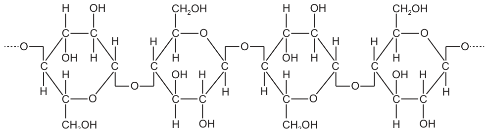
Which statement about this polysaccharide is correct?
Options
A It coils into a tight helix that provides a compact shape for energy storage in cells.
B It has high tensile strength when arranged into fibrils in plant cell walls.
C It folds into a globular shape that is soluble in water and easily transported.
D It combines with protein molecules to act as cell markers on the cell surface membrane.
Working
The diagram shows a straight-chain polysaccharide of β-glucose units linked by β-1,4-glycosidic bonds, with every alternate glucose rotated through . This is cellulose.
- A describes a compact helical storage molecule (starch/glycogen) — built from α-glucose with α-1,4 (and α-1,6) bonds. Wrong.
- B describes cellulose, whose long, straight, hydrogen-bonded chains form microfibrils of high tensile strength in plant cell walls. Correct.
- C describes a soluble, folded globular protein — not a polysaccharide. Wrong.
- D describes glycoproteins/glycolipids on the cell-surface membrane — not cellulose. Wrong.
Answer
B
B
Background Concept
Polysaccharides are long-chain polymers of monosaccharide units (here, glucose) joined by glycosidic bonds. The type of glycosidic bond and the orientation of successive glucose units dictate the three-dimensional shape of the polymer, and the shape in turn dictates the function.
- α-glucose polymerised through α-1,4 (and α-1,6) bonds gives starch (amylose + amylopectin) in plants and glycogen in animals. The α-linkage makes the chain curve, allowing it to coil into a compact helix ideal for energy storage.
- β-glucose polymerised through β-1,4 bonds gives cellulose. Because every alternate β-glucose is rotated by relative to its neighbour, the long chain is straight, not coiled. Hundreds of parallel chains hydrogen-bond to one another into microfibrils, which are extremely strong in tension. Cellulose microfibrils form the load-bearing framework of plant cell walls.
Glycoproteins and glycolipids are quite different: they are short, branched carbohydrate chains (often oligosaccharides) attached to membrane proteins or lipids, serving as cell-surface markers for cell recognition and signalling — not as structural fibrils.
Understanding the Question
The question shows a labelled diagram of four glucose units joined head-to-tail. The orientation of the –OH on carbon 1 alternates above and below the plane of the ring, indicating β-1,4-glycosidic bonds and the characteristic flip of every other glucose. This is diagnostic of cellulose.
The command word is "Which statement … is correct?" — this is a multiple-choice item asking for the option that correctly describes a property of this polysaccharide. The marks reward (1) identifying cellulose and (2) matching it to the right functional statement.
Approach
- Identify the polymer from the bond geometry (β-1,4, alternating flip = cellulose).
- Read each option and decide which function fits cellulose.
- Eliminate the other three by recognising which other molecule they actually describe.
Step-by-Step Reasoning
- Option A: "coils into a tight helix … compact shape for energy storage" — this is a textbook description of amylose (part of starch) or glycogen, both built from α-glucose. Cellulose cannot coil because its β-1,4 bonds and alternating glucose orientation keep the chain straight. Wrong.
- Option B: "high tensile strength when arranged into fibrils in plant cell walls" — straight cellulose chains hydrogen-bond laterally to form microfibrils; these fibrils are the principal load-bearing component of plant cell walls. Correct.
- Option C: "folds into a globular shape that is soluble in water and easily transported" — this describes a globular protein (e.g. haemoglobin, enzymes), not a polysaccharide. Polysaccharides like cellulose are insoluble. Wrong.
- Option D: "combines with protein molecules to act as cell markers on the cell surface membrane" — this describes glycoproteins (and glycolipids). Cellulose is a structural cell-wall polysaccharide and is not found as a membrane marker. Wrong.
Key Takeaways
- Bond geometry determines polysaccharide function: α-1,4 → storage (coil); β-1,4 → structural (straight fibril).
- The flip of alternate β-glucose units is the visual cue that identifies cellulose.
- Functions of membrane carbohydrates (recognition/signalling) are carried out by glycoproteins and glycolipids, not cellulose.
- Globular solubility describes proteins, not polysaccharides.
Common Mistakes
- Confusing starch/glycogen (α, helical, storage) with cellulose (β, straight, structural) — a very common error driven by treating all glucose polymers as "the same".
- Choosing D because "carbohydrate + protein = glycoprotein" sounds plausible — but cellulose is not a membrane carbohydrate; membrane markers are short, branched oligosaccharides.
- Choosing C because "soluble and transported" sounds biologically reasonable — but the statement describes a protein, not the polysaccharide shown.
Things to Be Careful About
- Read the bond carefully: an –OH above the ring on C1 (β configuration) versus below the ring (α configuration) is the deciding visual clue.
- "Fibrils" + "tensile strength" + "plant cell walls" is a tight, cellulose-specific trio — if all three appear together, the answer is cellulose.
- Do not be misled by "compact" or "helical" — those belong with α-linked storage polysaccharides.
Thermostable enzymes can maintain their tertiary structure at high temperatures.
Scientists studying the primary structure of a thermostable enzyme found that the enzyme contained:
● more charged amino acids
● fewer polar amino acids that are not charged
● more hydrophobic amino acids
than enzymes that are not thermostable.
Which row shows a correct comparison between this thermostable enzyme and an enzyme that is not thermostable?
Options
| number of amino acids forming interactions at the centre of a thermostable enzyme compared to an enzyme that is not thermostable | number of ionic bonds in a thermostable enzyme compared to the number in an enzyme that is not thermostable | |
|---|---|---|
| A | less than | more than |
| B | less than | less than |
| C | more than | less than |
| D | more than | more than |
Working
- Hydrophobic amino acids have non-polar R groups that cluster in the interior of the folded protein, forming hydrophobic interactions. The thermostable enzyme has more hydrophobic amino acids, so it has more amino acids forming interactions at the centre.
- Charged amino acids have R groups that can form ionic bonds (salt bridges) with oppositely charged R groups. The thermostable enzyme has more charged amino acids, so it has more ionic bonds.
Answer
D
D
Background Concept
Proteins have four levels of structure. The primary structure is the linear sequence of amino acids joined by peptide bonds. The tertiary structure is the 3D folding of the polypeptide, held in place by interactions between the R groups (side chains) of the amino acids. These R-group interactions include:
- Hydrophobic interactions between non-polar R groups, which cluster together in the interior of the protein, away from the aqueous cytoplasm.
- Ionic bonds (salt bridges) between positively and negatively charged R groups.
- Hydrogen bonds between polar R groups (or between polar R groups and water on the surface).
- Disulfide bonds between two cysteine residues.
An enzyme's tertiary structure is what gives it its specific 3D shape, including the active site. Thermostable enzymes (e.g. from thermophilic bacteria) maintain this tertiary structure — and therefore their function — at high temperatures that would denature most other enzymes. The extra stability comes from additional or stronger R-group interactions holding the folded shape in place.
Understanding the Question
The scientists found that the thermostable enzyme has a different amino acid composition than a non-thermostable one:
- More charged amino acids
- Fewer polar (uncharged) amino acids
- More hydrophobic amino acids
The question asks us to predict the consequences of these three compositional differences for:
- The number of amino acids forming interactions at the centre of the protein
- The number of ionic bonds in the protein
We must pick the row that correctly predicts BOTH of these consequences in the thermostable enzyme relative to the non-thermostable one.
Approach
The strategy is to map each amino acid property to:
- Where in the folded protein those amino acids tend to be located.
- What type of bond those amino acids tend to form.
Then compare the thermostable and non-thermostable enzymes on each of the two criteria, and select the matching row.
Step-by-Step Reasoning
1. Hydrophobic amino acids at the centre.
Hydrophobic R groups are non-polar and are repelled by water. During protein folding, they are buried in the hydrophobic core of the protein, where they associate with one another via hydrophobic interactions. The thermostable enzyme has more hydrophobic amino acids, so more of its amino acids are packed into the core forming hydrophobic interactions. Therefore the number of amino acids forming interactions at the centre is more than in a non-thermostable enzyme.
2. Charged amino acids form ionic bonds.
Ionic bonds form between R groups carrying opposite full charges (e.g. on lysine and on aspartate). The thermostable enzyme has more charged amino acids, providing more R groups that can pair up to form ionic bonds. Therefore the number of ionic bonds is more than in a non-thermostable enzyme.
(As a bonus check, "fewer polar uncharged amino acids" simply means fewer residues available to form hydrogen bonds on the surface — this is consistent with the more compact, more strongly bonded interior of a thermostable enzyme.)
3. Matching to the options.
| Interactions at centre | Ionic bonds | |
|---|---|---|
| A | less than | more than |
| B | less than | less than |
| C | more than | less than |
| D | more than | more than |
Both predictions are "more than", which is row D.
Key Takeaways
- The primary structure (amino acid sequence) determines the tertiary structure (3D fold) because different R groups form different bonds and occupy different positions in the folded protein.
- Hydrophobic R groups → buried in the core, forming hydrophobic interactions.
- Charged R groups (e.g. lysine, aspartate, glutamate, arginine) → can form ionic bonds (salt bridges).
- Polar uncharged R groups (e.g. serine, threonine, asparagine) → typically on the surface, forming hydrogen bonds with water.
- Thermostable enzymes tend to have additional/stronger R-group interactions (more ionic bonds, denser hydrophobic core) that resist the disruptive effect of high temperatures on tertiary structure.
Common Mistakes
- Confusing "polar" with "charged". Polar uncharged amino acids (e.g. serine) form hydrogen bonds, not ionic bonds. The question explicitly separates these two groups; "more charged" is the relevant fact for ionic bonds.
- Assuming hydrophobic amino acids stay on the surface. They cluster in the interior, not the exterior; this is the opposite of what a student who has confused "hydrophilic" and "hydrophobic" might write.
- Choosing C. This would only be correct if charged amino acids were repelled by one another, but opposite charges attract and form ionic bonds; like charges do not, but the more-charged statement still favours more ionic bonds overall.
- Choosing A. A common trap: the student assumes the more compact, less charged surface means fewer interactions overall, but the question specifies the centre (where hydrophobic interactions dominate) and ionic bonds (where charged R groups dominate) — both are increased.
Things to Be Careful About
- The phrase "interactions at the centre" refers specifically to the hydrophobic core, not to all tertiary-structure bonds. Do not count surface hydrogen bonds here.
- "Number of amino acids forming interactions at the centre" is asking about the count of residues participating in those core interactions, which scales with the number of hydrophobic amino acids present.
- Distinguish between the three facts given: more charged, fewer polar uncharged, more hydrophobic. Each maps to a specific structural consequence, and only two are directly tested in the answer options.
Collagen is a strong, elastic molecule that is often found in the connective tissues of animals. Glycine is an amino acid found at regular positions in the primary structure of a collagen polypeptide.
Which row shows features of collagen that contribute to its strength and elasticity?
Options
| glycine is found on the outside of the helical polypeptides to allow them to lie closer together | disulfide bonds form between glycine amino acids to join the polypeptides together | |
|---|---|---|
| A | ✓ | ✓ |
| B | ✓ | ✗ |
| C | ✗ | ✓ |
| D | ✗ | ✗ |
key
✓ = contributes to the strength and elasticity of collagen
✗ = does not contribute to the strength and elasticity of collagen
Working
Statement 1: "Glycine is found on the outside of the helical polypeptides to allow them to lie closer together."
- Glycine has the smallest side chain of any amino acid (a single hydrogen atom, –H).
- This tiny R-group allows three collagen polypeptide chains to wind tightly around each other in a triple helix, with glycine residues packed into the interior (centre) of the helix, not the outside.
- Therefore this statement is FALSE — ✗ (does not contribute to strength/elasticity).
Statement 2: "Disulfide bonds form between glycine amino acids to join the polypeptides together."
- Glycine contains no sulfur in its side chain, so it cannot form disulfide bonds.
- Collagen's strength comes from hydrogen bonding between chains and covalent cross-links between lysine/hydroxylysine residues — not disulfide bonds (these are characteristic of keratin).
- Therefore this statement is FALSE — ✗.
Both statements are ✗.
Answer
D
D
Background Concept
Collagen is the most abundant protein in mammals and the classic example of a fibrous protein. Each collagen molecule is a tropocollagen unit made of three polypeptide (α) chains wound around each other in a right-handed triple helix. This quaternary/tertiary arrangement gives collagen its characteristic combination of high tensile strength (resistance to pulling forces) and limited elasticity (it can stretch slightly but resists breaking).
Two structural features are central to this question:
-
The role of glycine. Every third amino acid in the primary structure of a collagen α-chain is glycine, giving the repeating sequence –X–Y–Gly– (where X is often proline and Y is often hydroxyproline). Glycine's R-group is simply a hydrogen atom, making it the smallest amino acid. When three chains pack into a triple helix, the available space at the centre/inside of the helix is very narrow, and only a hydrogen side chain fits there without distorting the structure. Glycine residues therefore occupy the interior positions of the helix, allowing the chains to lie very close together and maximising inter-chain hydrogen bonding, which gives collagen its strength.
-
Bonds that stabilise collagen. The chains are held together by hydrogen bonds between the backbones of adjacent α-chains. Additional covalent cross-links form between the side chains of lysine and hydroxylysine residues on neighbouring molecules (catalysed by the enzyme lysyl oxidase); these cross-links stiffen collagen fibres and are responsible for the increasing toughness of collagen with age.
Disulfide bonds (S–S) are NOT a feature of collagen. They are characteristic of other structural proteins, notably keratin (hair, nails, skin) and insulin, where the –CH₂–SH side chains of two cysteine residues oxidise to form a covalent bridge. Glycine cannot participate in disulfide bonding because it has no sulfur atom in its side chain.
Understanding the Question
This is a multiple-choice question presenting two statements about collagen. The candidate must decide, for each statement, whether it correctly describes a structural feature that contributes to collagen's strength and elasticity, marking it ✓ (yes) or ✗ (no), and then select the row with the correct combination.
Both statements in the question are false descriptions of collagen — they sound plausible at first glance, which is the trap. Recognising each as wrong is the whole point of the question.
Approach
Evaluate each statement independently against what you know about collagen's structure, then read the answer grid to find the row that matches both judgements.
Step-by-Step Reasoning
Evaluating Statement 1 — glycine's position:
- "Glycine is found on the outside of the helical polypeptides…" — INCORRECT.
- Glycine residues project inward into the centre of the triple helix. They do not sit on the outside.
- The reason glycine is interior is exactly because its tiny R-group (–H) allows the three bulky, proline-rich chains to pack tightly together. Larger side chains would sterically clash at the helix axis.
- So this statement does not describe a feature of collagen. ✗
Evaluating Statement 2 — disulfide bonds:
- "Disulfide bonds form between glycine amino acids to join the polypeptides together." — INCORRECT on two counts.
- Glycine has no –SH group, so it cannot form disulfide bonds.
- Collagen is not stabilised by disulfide bonds at all; it is stabilised by hydrogen bonds within the triple helix and covalent lysine/hydroxylysine cross-links between tropocollagen molecules.
- Disulfide bonds are characteristic of keratin, not collagen.
- So this statement does not describe a feature of collagen. ✗
Reading the table:
| Statement 1 | Statement 2 | Row |
|---|---|---|
| ✓ | ✓ | A |
| ✓ | ✗ | B |
| ✗ | ✓ | C |
| ✗ | ✗ | D |
Since both statements are ✗, the correct row is D.
Key Takeaways
- Collagen is a fibrous protein whose α-chains form a right-handed triple helix.
- Glycine is found every third residue and packs into the interior of the triple helix because it is the smallest amino acid — this is what allows the chains to lie close together.
- Collagen's stability comes from inter-chain hydrogen bonds and lysine/hydroxylysine covalent cross-links, not from disulfide bonds.
- Disulfide bonds are a feature of keratin and a few other proteins, but not collagen.
Common Mistakes
- Assuming glycine is on the outside of the helix — it is on the inside. Many students remember "glycine is small so it allows close packing" but forget where this smallness is structurally important.
- Confusing collagen with keratin. Both are fibrous structural proteins, but keratin (hair/nails) is rich in cysteine and stabilised by disulfide bonds, whereas collagen (tendons/bone/skin) is rich in glycine, proline and hydroxyproline and is not.
- Assuming all amino acids can form disulfide bonds. Only cysteine (and occasionally cystine, its dimer) carries the –CH₂–SH side chain required.
Things to Be Careful About
- The phrase "lie closer together" in Statement 1 is a deliberate red herring — close packing is a feature of collagen, but it is achieved by glycine being on the inside, not the outside.
- "Elastic" in the question refers to collagen's limited extensibility under tension; it should not be confused with the high elasticity of elastin, a different fibrous protein.
- When the table uses "✓ = contributes" and "✗ = does not contribute," read the statements literally — a wrong description of a real feature still scores ✗ because the description is incorrect.
Which statements describe the action of an extracellular enzyme?
1 synthesis of a polynucleotide in the nucleus during DNA replication
2 digestion of macromolecules in the lumen of the small intestine
3 synthesis of ATP molecules in the mitochondria
Options
A 1 and 2
B 1 and 3
C 2 and 3
D 2 only
Answer
Extracellular enzymes catalyse reactions outside the cell. Statement 1 (DNA replication in the nucleus) and statement 3 (ATP synthesis in mitochondria) are intracellular. Statement 2 (digestion in the lumen of the small intestine) is extracellular.
Answer
D
D
Background Concept
Enzymes are biological catalysts that speed up metabolic reactions. They are classified by where they act:
- Intracellular enzymes are synthesised and function inside the cell (e.g. DNA polymerase in the nucleus, respiratory enzymes on mitochondrial membranes).
- Extracellular enzymes are secreted by the cell and act outside it, typically in a body cavity, duct or the gut lumen (e.g. amylase, trypsin, lipase in the small intestine; cellulase in fungal hyphae).
A useful rule of thumb: if the substrate is too large to enter the cell, or the product is needed externally, the enzyme is released to act on it in the surrounding environment.
Understanding the Question
The question asks which statements describe an extracellular enzyme. The options combine three statements, and we must judge each one on whether the named process happens inside a cell (intracellular) or outside cells (extracellular).
Approach
For each statement, identify the location of the reaction and decide whether the enzyme responsible acts inside or outside the cell.
Step-by-Step Reasoning
- Statement 1 – synthesis of a polynucleotide in the nucleus during DNA replication. DNA replication occurs inside the nucleus, catalysed by DNA polymerases. The enzymes are working inside the cell → intracellular. ✗
- Statement 2 – digestion of macromolecules in the lumen of the small intestine. The lumen of the small intestine is the cavity outside the cells of the intestinal wall. Enzymes such as trypsin, lipase and maltase are secreted by the pancreas or by cells lining the gut and act on substrates (proteins, lipids, carbohydrates) in this lumen. → extracellular. ✓
- Statement 3 – synthesis of ATP molecules in the mitochondria. ATP synthesis on the inner mitochondrial membrane is catalysed by ATP synthase and the electron transport chain enzymes, all inside the mitochondrion and therefore inside the cell → intracellular. ✗
Only statement 2 describes an extracellular enzyme, so the answer is D.
Key Takeaways
- Extracellular = outside the cell (e.g. digestive enzymes in the gut lumen, lysozyme in tears).
- Intracellular = inside the cell (e.g. DNA polymerase, respiratory enzymes, ribosomes).
- A common exam trap: the gut wall contains cells, but the lumen is extracellular.
Common Mistakes
- Choosing A or C because "digestion" sounds like a general enzyme activity; forgetting that location is the key criterion.
- Treating ATP synthesis as "energy work outside the cell" — in fact ATP is made internally and exported.
Things to Be Careful About
- "Lumen" means the inside space of a tubular organ — it is technically outside the body in a topological sense, and extracellular enzymes are released into it.
- For an MCQ, the answer only needs the correct letter; the brief reasoning above is for understanding, not for the answer line.
Which statement about the induced-fit hypothesis of enzyme action is correct?
Options
A An enzyme that uses the induced-fit mechanism will increase the activation energy of a reaction.
B The binding of a substrate causes conformational changes in an enzyme.
C The active site of an enzyme changes to become the same shape as the substrate.
D An enzyme that uses an induced-fit mechanism has decreased specificity to the substrate.
Working
The induced-fit hypothesis proposes that the active site is not a perfectly pre-formed complement to the substrate. When the substrate binds, the enzyme undergoes a small conformational change that moulds the active site around the substrate, bringing catalytic groups into the correct position. This lowers (not raises) the activation energy, the active site becomes complementary to the substrate only after binding (it is not identical to the substrate), and specificity is retained (often enhanced) rather than decreased.
Answer
B
B
Background Concept
Enzymes are biological catalysts — globular proteins that speed up reactions by lowering the activation energy. Two classical models describe how an enzyme recognises and binds its substrate:
- Lock-and-key hypothesis: the active site has a rigid, pre-formed shape that is already an exact complement to the substrate, like a key fitting a lock. No shape change in the enzyme is required.
- Induced-fit hypothesis (proposed by Daniel Koshland, 1958): the active site is not a perfect fit initially. Binding of the substrate induces a conformational change in the enzyme, moulding the active site around the substrate. This brings catalytic amino acid side-chains into the correct geometry to strain bonds in the substrate, stabilise the transition state, and lower the activation energy. Once products leave, the enzyme returns to its original conformation.
A key consequence of induced fit is that the active site becomes complementary to the transition state, not to the substrate itself, which is why enzymes are so catalytically powerful. Specificity is retained (and often refined) by the precise re-arrangement of residues; it is not lost.
Understanding the Question
This is a multiple-choice question asking which single statement correctly describes the induced-fit mechanism. Each option tests a different misconception or a genuine feature of the model:
- A claims enzymes raise the activation energy.
- B states that substrate binding causes a conformational change in the enzyme.
- C states the active site becomes the same shape as the substrate.
- D claims induced-fit reduces specificity.
The command word is implicit: identify the correct statement.
Approach
Recall the central tenet of the induced-fit hypothesis — the substrate induces a shape change in the enzyme after binding. Then test each option against what the model actually says and against what you know about enzyme catalysis in general.
Step-by-Step Reasoning
- Option A is wrong. Catalysts, including enzymes, decrease the activation energy of a reaction; they never increase it. This is true whether the enzyme operates by lock-and-key or induced-fit.
- Option B is correct. Induced fit explicitly proposes that when the substrate enters the active site, it triggers a small conformational change in the enzyme. This re-shapes the active site so that catalytic residues make optimal contact with the substrate and the transition state. This is the defining feature of the model.
- Option C is wrong. The active site becomes complementary to the substrate (and especially to the transition state); it does not become the same shape as the substrate. "Same shape" would imply the substrate and active site are identical, which is meaningless — the active site is a pocket/binding site, not a copy of the substrate. It also contradicts induced fit because the change in shape is in the enzyme, not the substrate.
- Option D is wrong. Induced fit does not reduce specificity. Each enzyme still binds a specific substrate (or small group of related substrates), and the induced conformational change actually refines the fit and catalytic geometry. If anything, induced fit contributes to the high catalytic efficiency and selectivity of enzymes.
Therefore B is the only correct statement.
Key Takeaways
- The induced-fit hypothesis states that substrate binding induces a conformational change in the enzyme.
- The active site becomes complementary to the substrate/transition state, not identical to it.
- Enzymes always lower the activation energy, regardless of the model used to describe binding.
- Induced fit preserves (and refines) specificity — it is not a "looser" version of lock-and-key.
Common Mistakes
- Confusing "complementary to" with "same shape as" the substrate — the active site fits the substrate like a glove, it does not become a copy of it.
- Thinking that any shape change in an enzyme means loss of specificity — induced fit is a precise, functional re-arrangement, not random wobble.
- Forgetting the basic rule that catalysts lower, never raise, activation energy.
Things to Be Careful About
- "Complementary shape" is the wording the mark scheme expects; avoid "same shape", "identical", or "similar shape", which are imprecise.
- Activation energy and specificity are general features of enzyme catalysis; do not attribute their loss to a particular binding model.
- Read every option carefully — the question asks for the correct statement, not the "most correct" one.
Graph J shows the effect of pH on the rate of an enzyme reaction. Graph K shows the effect of substrate concentration on the rate of an enzyme reaction.
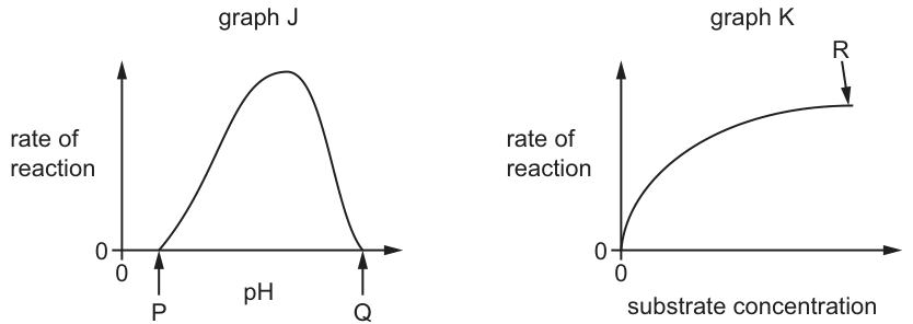
A student wrote four statements about the graphs.
1 In graph J, the enzyme is denatured at point Q.
2 In graph J, the enzyme is inactive at point P.
3 In graph K, the rate of reaction is directly proportional to the substrate concentration.
4 In graph K, is reached at point R.
Which statements are correct?
Options
A 1, 2 and 3
B 1, 2 and 4
C 1, 3 and 4
D 2, 3 and 4
Working
Statement 1 – At point Q (very high pH), the rate is 0 because the extreme pH disrupts the hydrogen and ionic bonds holding the tertiary structure, permanently changing the shape of the active site. The enzyme is denatured. ✓
Statement 2 – At point P (very low pH), the rate of reaction is 0; the enzyme is not catalysing any reaction, so it is correctly described as inactive. ✓
Statement 3 – Direct proportionality would require a straight line through the origin. Graph K rises steeply at first and then curves to a plateau, so the rate is not directly proportional to substrate concentration. ✗
Statement 4 – At point R the curve has levelled off on the plateau; further increases in substrate concentration do not increase the rate because all active sites are saturated. This is . ✓
Correct statements: 1, 2 and 4.
Answer
B
B
Background Concept
Effect of pH on enzyme activity (Graph J). Enzymes are globular proteins whose tertiary structure is held together by hydrogen bonds, ionic bonds, hydrophobic interactions and disulfide bridges. The shape of the active site depends on this tertiary structure. Each enzyme has an optimum pH at which the ionisation states of amino acid side-chains in the active site are ideal for binding substrate. As pH moves away from the optimum, the ionisation of these side-chains changes, substrate binding becomes less effective, and the rate falls. At extreme pH values on both sides of the optimum, enough bonds are disrupted to permanently alter the shape of the active site — the enzyme is denatured and can no longer function even if returned to optimum pH.
Effect of substrate concentration on enzyme activity (Graph K). At low substrate concentrations, active sites are largely empty and the rate is limited by how often enzyme–substrate (ES) complexes form; the rate therefore rises steeply as substrate increases. As substrate concentration rises further, more active sites become occupied, until eventually all of them are saturated at any one moment. From that point onwards, the rate cannot increase further — adding more substrate makes no difference because there are no free active sites. The maximum rate achieved is called and it depends on the enzyme concentration, not the substrate concentration.
The shape of Graph K is the classic Michaelis–Menten curve. Direct proportionality (a straight line through the origin) only holds at very low substrate concentrations, where the rate is essentially linear with [S]. It does not hold across the whole curve.
Understanding the Question
Two graphs are given. Graph J is a bell-shaped curve of rate vs pH with point P on the low-pH side (rate = 0) and point Q on the high-pH side (rate = 0). Graph K is the Michaelis–Menten curve of rate vs substrate concentration, with point R on the plateau region. The student is asked to evaluate four statements, two about each graph, and select the option listing all the correct ones. The command word is implicit: identify which statements are biologically correct.
Approach
Evaluate each statement one at a time against what the graph shows and what the biology says:
- Does Q represent a denatured enzyme? — Yes: extreme pH permanently distorts the active site.
- Does P represent an inactive enzyme? — Yes in the sense that the rate is 0, so the enzyme is not catalysing; this is a true description of the enzyme's functional state at that pH.
- Is the rate directly proportional to substrate concentration? — No: only the very first, near-linear portion of the curve is proportional; the whole curve is not.
- Is reached at R? — Yes: R lies on the plateau where rate is no longer increasing with substrate.
Step-by-Step Reasoning
Statement 1 — "In graph J, the enzyme is denatured at point Q."
At Q, pH is far above the optimum and the rate has fallen to 0. Under these conditions the high concentration of OH⁻ (or H⁺ in a strongly basic environment, depending on the buffer) alters the ionisation of R-groups, breaking hydrogen and ionic bonds that maintain tertiary structure. The active site loses its specific shape and the substrate can no longer bind. The change is permanent — the enzyme is denatured. True.
Statement 2 — "In graph J, the enzyme is inactive at point P."
At P, pH is far below the optimum and the rate is 0. The word "inactive" simply means the enzyme is not catalysing any reaction, which is exactly what the graph shows (rate = 0). Whether the underlying cause is denaturation or simply a non-binding active site, the functional observation is that the enzyme is not active. The statement is correct as a description of the enzyme's activity. True.
Statement 3 — "In graph K, the rate of reaction is directly proportional to the substrate concentration."
Direct proportionality means for some constant , i.e. a straight line through the origin. Graph K is curved — steeply rising at low [S] and plateauing at high [S]. Only the very beginning of the curve approximates a straight line; the curve as a whole is not proportional. False.
Statement 4 — "In graph K, is reached at point R."
Point R is marked on the flat, plateau portion of the curve. By definition is the maximum rate at which the enzyme can work, achieved once all active sites are saturated with substrate. R is exactly on this plateau. True.
Statements 1, 2 and 4 are correct → answer B.
Key Takeaways
- An enzyme's pH curve is bell-shaped; rate falls to 0 at extreme pH on both sides of the optimum because the active-site shape is lost (denaturation).
- The substrate-concentration curve is a hyperbola (Michaelis–Menten), with a steep rise at low [S] and a plateau at high [S].
- is the plateau rate, reached when all active sites are saturated; it depends on enzyme concentration, not on substrate concentration.
- "Directly proportional" means a straight line through the origin — it is not the same as "rate increases with substrate".
Common Mistakes
- Saying the rate is "directly proportional" to substrate concentration across the whole curve. Only the initial, near-linear portion is approximately proportional; the full curve is hyperbolic.
- Saying the enzyme is denatured at P but not at Q (or vice versa). Both extreme pH values denature the enzyme — denaturation is not a one-sided event.
- Saying depends on substrate concentration. depends on enzyme concentration; increasing [S] only reveals it.
- Confusing with . is the substrate concentration at which the rate is half of — it is a point on the rising part of the curve, not on the plateau.
Things to Be Careful About
- The exact wording of each statement matters. Statement 2 uses the word "inactive" (not "denatured") — "inactive" is a true functional description because the rate is 0, even though the underlying cause at extreme pH is denaturation.
- Read each point's position on the graph carefully: P is on the low-pH axis, Q is on the high-pH axis, R is on the substrate-concentration plateau.
- Use the precise term "" (or "maximum rate") rather than vague phrases such as "top of the graph" or "fastest point".
The diagram shows a section through a cell surface membrane.
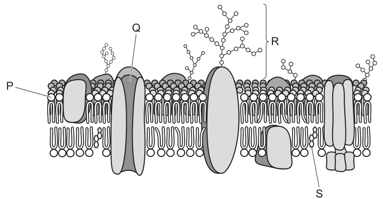
What is a correct role for a labelled molecule?
Options
A P is involved in controlling membrane stability.
B Q is involved in active transport.
C R is involved in cell signalling.
D S is involved in diffusion of ions.
Working
- P is a cholesterol molecule — its role is to regulate membrane fluidity, not stability.
- Q is a channel protein — channel proteins are involved in facilitated diffusion, not active transport (which uses carrier proteins/pumps).
- R is a glycoprotein — glycoproteins act as receptors and are involved in cell signalling and cell recognition. ✓
- S is a peripheral protein on the inner surface — it is not a channel for ion diffusion.
Answer
C
C
Background Concept
The fluid mosaic model describes the cell surface membrane as a phospholipid bilayer in which proteins move laterally. Embedded in or associated with the bilayer are several types of molecules, each with characteristic roles:
- Phospholipids form the bilayer; their hydrophilic heads face the aqueous environments on either side of the membrane, while the hydrophobic tails face inwards, creating a barrier to water-soluble molecules.
- Cholesterol molecules sit among the phospholipid tails. They regulate membrane fluidity — at high temperatures they reduce fluidity by restricting phospholipid movement, and at low temperatures they prevent the membrane from becoming too rigid by disrupting the regular packing of phospholipid tails. They also reduce permeability to small water-soluble molecules such as ions.
- Channel proteins span the bilayer and form a water-filled pore that allows specific ions or small polar molecules to move across the membrane by facilitated diffusion (down their concentration/electrochemical gradient; no ATP required).
- Carrier proteins bind specific molecules and change shape to move them across. These are the proteins used in active transport (which requires ATP) as well as in some forms of facilitated diffusion.
- Glycoproteins have a carbohydrate chain attached to a protein on the outer surface of the membrane. The carbohydrate chains of glycoproteins (and glycolipids) function as receptor sites for signalling molecules (e.g. hormones, neurotransmitters) and as cell recognition markers, enabling the immune system to distinguish self from non-self.
- Peripheral proteins are not embedded in the bilayer; they sit on the surface and are often involved in cell signalling, anchoring the cytoskeleton, or acting as enzymes — but they do not form channels for ion diffusion.
Understanding the Question
The question shows a fluid-mosaic-style diagram of a cell surface membrane and labels four components: P (cholesterol), Q (a channel protein), R (a glycoprotein, identified by the carbohydrate chain on the outer surface), and S (a peripheral protein on the inner surface). The candidate must identify which of the four options correctly describes the role of a labelled molecule.
Approach
Match each label to its structure, then check whether the stated role in each option is biologically correct. Eliminate the options in which the role is wrong for the identified structure.
Step-by-Step Reasoning
- Option A — "P is involved in controlling membrane stability": P is cholesterol. Cholesterol's accepted role is to regulate membrane fluidity and reduce permeability to water-soluble molecules, not to control "stability". Stability implies holding the membrane together structurally, which is the role of the phospholipid bilayer and the cytoskeleton, not cholesterol. This option is therefore incorrect because of imprecise terminology.
- Option B — "Q is involved in active transport": Q is a channel protein. Channel proteins provide a hydrophilic pore that allows ions/polar molecules to move down their electrochemical gradient — this is facilitated diffusion, a passive process. Active transport requires carrier proteins (pumps) and ATP. This option is incorrect.
- Option C — "R is involved in cell signalling": R is a glycoprotein — a protein with a carbohydrate chain projecting into the extracellular space. Glycoproteins act as receptors that bind specific signalling molecules (hormones, neurotransmitters, growth factors), triggering responses inside the cell. They are also involved in cell–cell recognition. This option is correct.
- Option D — "S is involved in diffusion of ions": S is a peripheral protein attached to the inner face of the membrane. Peripheral proteins are not channel proteins and do not form pores for ions. Diffusion of ions is performed by channel proteins (or by carrier proteins in some cases). This option is incorrect.
The only option pairing a correct structure with its correct role is C.
Key Takeaways
- Cholesterol regulates fluidity (not stability) of the membrane.
- Channel proteins → facilitated diffusion of ions/small polar molecules (passive).
- Carrier proteins → active transport (and some facilitated diffusion).
- Glycoproteins/glycolipids → cell signalling and cell recognition (receptor sites and markers).
- Peripheral proteins → structural, enzymatic, or signalling roles, but not ion channels.
Common Mistakes
- Confusing channel proteins with carrier proteins, leading to the wrong belief that channel proteins carry out active transport.
- Stating that cholesterol "stabilises" the membrane instead of "regulates fluidity" — the CIE mark scheme often rejects "stability" for cholesterol.
- Confusing glycoproteins (protein + carbohydrate) with glycolipids (lipid + carbohydrate); both have similar cell-signalling/recognition roles.
- Assuming any protein in the membrane is automatically a transport protein — peripheral proteins are often structural or enzymatic.
Things to Be Careful About
- Read the outer vs inner orientation of the membrane in the diagram: the carbohydrate chain of a glycoprotein is always on the outer (extracellular) surface.
- Use the exact CIE terminology: "regulate fluidity" for cholesterol, "facilitated diffusion" for channel proteins, and "active transport" specifically for carrier proteins using ATP.
- Be precise about which structure the option refers to — a tempting-sounding option (such as A, which sounds plausible) may be rejected because of one wrong word.
When red blood cells are put into distilled water they burst (haemolysis).
Which statements explain this haemolysis?
1 The water potential of the surrounding liquid is lower than the water potential of the contents of the red blood cell.
2 The cell surface membranes of red blood cells are not supported by cell walls.
3 More water moves into the red blood cells by osmosis than leaves the cells.
4 Water enters the red blood cells by osmosis but does not leave the cells.
Options
A 1 and 3
B 1 and 4
C 2 and 3
D 2 and 4
Working
Distilled water has a water potential of (the highest/least negative). The cytoplasm of a red blood cell contains dissolved solutes (haemoglobin, ions) giving it a negative water potential. So the surrounding liquid has a HIGHER water potential than the cell contents, not lower.
Statement 1: Incorrect — the surrounding liquid has a higher water potential than the cell contents.
Statement 2: Correct — red blood cells are animal cells and have no cell wall, so the cell surface membrane is unsupported and bursts when too much water enters.
Statement 3: Correct — because the water potential outside is higher than inside, the net movement of water is into the cells by osmosis.
Statement 4: Incorrect — osmosis is a dynamic process; water moves in both directions, but net movement is into the cell.
Answer
C
C
Background Concept
Water potential () is the tendency of a solution to lose water; pure water has the highest water potential at , and any solution containing solutes has a lower (more negative) water potential. Water moves down a water potential gradient by osmosis — a special case of diffusion involving water molecules moving across a partially permeable membrane.
Animal cells and plant cells respond differently to osmosis:
- An animal cell in distilled water (higher external ) gains water, swells, and may burst because the cell surface membrane is the only structural boundary.
- A plant cell in the same situation becomes turgid, but the rigid cell wall prevents bursting.
Red blood cells are biconcave animal cells packed with haemoglobin (a protein) and various ions in their cytoplasm, giving the cytoplasm a negative water potential.
Understanding the Question
The question asks which statements correctly explain haemolysis — the bursting of red blood cells in distilled water. We must evaluate each statement for biological accuracy.
- Distilled water:
- Red blood cell cytoplasm: (negative, due to dissolved solutes)
- Water moves from higher (outside) to lower (inside), down the gradient.
Approach
Go through each statement and decide whether it is biologically true:
- Check the direction of the water potential gradient.
- Check whether red blood cells have a supporting cell wall.
- Check the direction of net water movement.
- Check whether osmosis is a one-way process.
Step-by-Step Reasoning
Statement 1: "The water potential of the surrounding liquid is lower than the water potential of the contents of the red blood cell."
- WRONG. Distilled water () has a HIGHER water potential than red blood cell cytoplasm (which is negative). The gradient is the opposite way round.
Statement 2: "The cell surface membranes of red blood cells are not supported by cell walls."
- CORRECT. Red blood cells are animal cells — they lack a cell wall. Without this rigid outer support, the membrane alone cannot resist the increasing internal pressure as water enters, so the cell bursts. (This is why plant cells in the same situation simply become turgid.)
Statement 3: "More water moves into the red blood cells by osmosis than leaves the cells."
- CORRECT. Because the external water potential is higher, the net (overall) movement of water is into the cell. The cell gains water and swells.
Statement 4: "Water enters the red blood cells by osmosis but does not leave the cells."
- WRONG. Osmosis is a passive, reversible process — water molecules cross the membrane in both directions all the time. In distilled water, water still leaves, but the rate of entry is greater than the rate of exit, giving net inflow.
Statements 2 and 3 are correct → answer is C.
Key Takeaways
- Distilled water has the highest water potential (); any solution has a lower (more negative) water potential.
- Osmosis is a two-way movement of water; the net movement is down the water potential gradient.
- Animal cells lack cell walls, so they burst (haemolyse) in hypotonic solutions, whereas plant cells become turgid.
- When evaluating "explain" statements in MCQs, check the direction of gradients and the direction of movement carefully.
Common Mistakes
- Saying water potential of distilled water is "low" — it is the highest possible water potential at .
- Thinking osmosis is one-way — water moves in BOTH directions continuously; only the net flow is in one direction.
- Confusing animal and plant cells — many candidates think red blood cells have a cell wall; they do not.
- Confusing concentration with water potential — a dilute solution can still have a negative water potential.
Things to Be Careful About
- "Lower" water potential means more negative; pure water has the highest water potential.
- Osmosis is described by NET movement; do not state water only flows one way.
- The CIE mark scheme requires the direction of the gradient to be correct — state whether the outside is higher or lower in than the inside.
The diagrams show the shape and size of two types of cell.
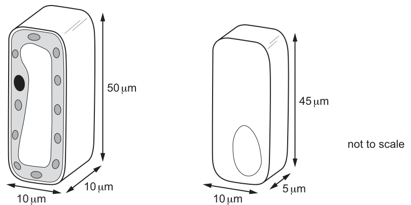
Which statement about the palisade cell and epithelial cell shown in the diagrams is correct?
Options
A An increase in surface area reduces the distance for gases to reach the centre of the cell.
B The surface area of the palisade mesophyll cell is greater than the columnar epithelial cell.
C The surface area to volume ratio is greater in the columnar epithelial cell than the palisade mesophyll cell.
D The volume of the palisade mesophyll cell is greater than that of the columnar epithelial cell.
Working
Palisade mesophyll cell ():
Columnar epithelial cell ():
The columnar epithelial cell has the greater SA:V ratio ().
Answer
C
C
Background Concept
Cells exchange materials (gases, nutrients, waste) with their surroundings across the plasma membrane. The rate at which a cell can absorb what it needs and remove what it doesn't depends on the surface area to volume ratio (SA:V). As a cell grows larger, its volume (which scales with the cube of a linear dimension) increases faster than its surface area (which scales with the square). The result is that a bigger cell has proportionally less membrane per unit of cytoplasm — diffusion alone cannot serve its metabolic demands, and the cell either stays small, flattens, develops internal membranes, or subdivides.
For a rectangular prism of sides :
The first formula adds the three distinct face areas and doubles them because each face has an opposite.
Understanding the Question
Fig. 18.1 shows two rectangular-prism cells, with dimensions and one of the two derived quantities (SA or V) supplied. We must use the dimensions to test four statements about their surface area, volume, and SA:V ratio.
- Palisade mesophyll cell: , SA stated as .
- Columnar epithelial cell: , V stated as .
The command word is implicit ("Which statement … is correct?") — only the option that survives numerical checking is the answer.
Approach
- Calculate the missing quantity for each cell (volume of the palisade; surface area of the epithelial cell) to confirm the given figures.
- Compute the SA:V ratio for each cell.
- Test each option against the calculated values.
Step-by-Step Reasoning
Palisade mesophyll cell:
The calculated SA matches the figure supplied, confirming the geometry.
Columnar epithelial cell:
The calculated V matches the figure supplied.
Option A — "An increase in surface area reduces the distance for gases to reach the centre of the cell." Increasing surface area does not by itself shorten the diffusion path to the cell's centre. The relevant factor is the linear size of the cell, not its surface area. Incorrect.
Option B — Surface area difference: , not . Incorrect.
Option C — for the epithelial cell () > for the palisade cell (). Correct.
Option D — Volume difference: , not . Incorrect.
Key Takeaways
- The SA:V ratio of a cell governs the efficiency of exchange between the cell and its surroundings.
- Volume scales with the cube and surface area with the square of linear dimension — so larger cells have lower SA:V ratios unless their shape changes (flattening, folding, microvilli).
- Always compute SA from and V from for a rectangular prism — do not confuse them.
- The question's stated values (SA = for the palisade cell; V = for the epithelial cell) are a built-in check of your arithmetic, not a substitute for it.
Common Mistakes
- Using only one or two dimensions when computing SA or V — leads to errors like treating the cell as a flat rectangle or a single rod.
- Assuming the cell with the larger raw surface area also has the larger SA:V ratio. Here the smaller epithelial cell has the larger ratio because its volume shrinks more than its surface area.
- Reading as for the SA difference — the distractor is designed to trap hasty arithmetic.
- Reading as for the volume difference — same trap.
- Believing that an increase in surface area automatically shortens the diffusion distance to the cell's centre — a common misconception.
Things to Be Careful About
- Always carry units ( for SA, for V) and check the option's numbers against the calculated difference.
- Quote SA:V ratios to two significant figures ( vs ) when comparing.
- The diagram is marked "not to scale" — rely on the dimensions given, not the visual appearance.
- Be precise with the rectangular-prism formula: there are six faces in three opposite pairs, hence the factor of .
The DNA content of a cell changes as the mitotic cell cycle proceeds through a series of stages.
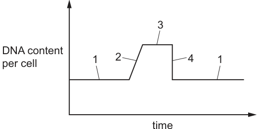
Which row correctly identifies when DNA replicates and when the nuclear envelope breaks down?
Options
| DNA replicates | nuclear envelope breaks down | |
|---|---|---|
| A | 1 | 3 |
| B | 1 | 4 |
| C | 2 | 3 |
| D | 2 | 4 |
Working
- DNA replication occurs during S phase of interphase, when the DNA content of the cell doubles. On the graph this corresponds to the rising portion — stage 2.
- The nuclear envelope breaks down during prophase of mitosis. By this point DNA replication is already complete, so the cell is at the doubled DNA level (the plateau before cytokinesis). This corresponds to stage 3.
- Stage 1 = G1 (baseline DNA), stage 2 = S phase (DNA replicating), stage 3 = G2 + mitosis (doubled DNA, including prophase), stage 4 = cytokinesis/telophase (DNA content halved back to baseline).
Answer
C
C
Background Concept
The mitotic cell cycle consists of interphase (G1, S, G2) followed by mitosis (prophase, metaphase, anaphase, telophase) and cytokinesis. Two events are key to interpreting a DNA-content graph:
- DNA replication happens during S phase of interphase. Each chromosome is duplicated to form two sister chromatids, so the total DNA per cell doubles (e.g. from 2C to 4C). No visible change in chromosome number occurs — the cell still has the same chromosomes, just with double the DNA.
- Nuclear envelope breakdown happens during prophase of mitosis. Prophase is the first mitotic stage, occurring after DNA replication is complete. The nuclear envelope disassembles so the spindle fibres can attach to the chromosomes via their centromeres.
A DNA content graph therefore shows:
- a flat baseline (G1)
- a rising slope (S phase, DNA replicating)
- a high plateau (G2 + mitosis, DNA doubled; nuclear envelope breaks down during this plateau)
- a sharp drop back to baseline (cytokinesis, when the two daughter cells separate)
Understanding the Question
The graph in Fig. 19.1 plots DNA content per cell against time with five labelled regions. The question asks which row correctly identifies:
- The region in which DNA replicates (the rising portion where DNA doubles)
- The region in which the nuclear envelope breaks down (a mitotic event that occurs while DNA is still at the doubled level)
We must identify each event based on the DNA content shown at that stage.
Approach
Map each numbered region to the corresponding stage of the cell cycle, then locate the two named events within that sequence:
- Region 1 (flat, low): G1
- Region 2 (rising): S phase → DNA replication occurs here
- Region 3 (flat, high): G2 + mitosis (prophase, metaphase, anaphase) → nuclear envelope breaks down in prophase, which lies on this plateau
- Region 4 (falling): cytokinesis/telophase — DNA content is halved as the cell divides
The answer therefore matches region 2 with DNA replication and region 3 with nuclear envelope breakdown.
Step-by-Step Reasoning
- Region 1 (baseline): DNA content is at its lowest, stable value. This represents G1 phase, before any replication has occurred.
- Region 2 (rising slope): DNA content is increasing — the cell is making new DNA. This can only be S phase, when each chromosome is being replicated to form sister chromatids. DNA replication therefore happens in region 2.
- Region 3 (high plateau): DNA content is at its maximum (doubled). This plateau covers G2 plus mitosis (prophase, metaphase, anaphase). The nuclear envelope breaks down during prophase, the very first stage of mitosis, which sits on this plateau. So the nuclear envelope breaks down in region 3.
- Region 4 (sharp drop): DNA content halves suddenly. This represents cytokinesis / telophase, when the cell physically divides into two daughter cells, each returning to the baseline DNA content.
- Return to region 1: The new daughter cells are back in G1 of the next cell cycle.
Matching the events to the regions: DNA replication = 2; nuclear envelope breakdown = 3 → Option C.
Key Takeaways
- On a DNA content graph, S phase = the rising slope; mitosis = the high plateau; cytokinesis = the sharp drop.
- DNA must replicate before mitosis begins, so the nuclear envelope cannot break down until the cell is already on the plateau — i.e. a mitotic event can never occur during the rising portion.
- Recognising the shape of this graph is a high-yield skill — it appears frequently on CIE 9700 papers.
Common Mistakes
- Confusing the rising portion with mitosis — it is actually S phase (interphase); mitosis occurs on the high plateau, not on the slope.
- Placing nuclear envelope breakdown during the falling portion (region 4): by this stage cytokinesis is underway and the cell is splitting in two; the nuclear envelope had already broken down earlier, in prophase.
- Treating the plateau as a single undifferentiated block — G2 and mitosis share the same DNA content, so the plateau covers both, but only mitosis includes the nuclear-envelope breakdown.
Things to Be Careful About
- The nuclear envelope breaks down at the start of mitosis (prophase), not at the end. It reforms during telophase. On the graph, both events occur within the plateau region, so do not try to separate them into different numbered regions.
- DNA content per cell halves during cytokinesis because the doubled DNA is partitioned into two daughter cells, each receiving a single chromatid from every chromosome.
- A common distractor (options A and D) places DNA replication in region 1, which is incorrect because region 1 is flat — no new DNA is being made there.
The diagram shows three cycles of cell division from an original cell of cell type P in bone marrow. Some of the cells that form are cell type Q.
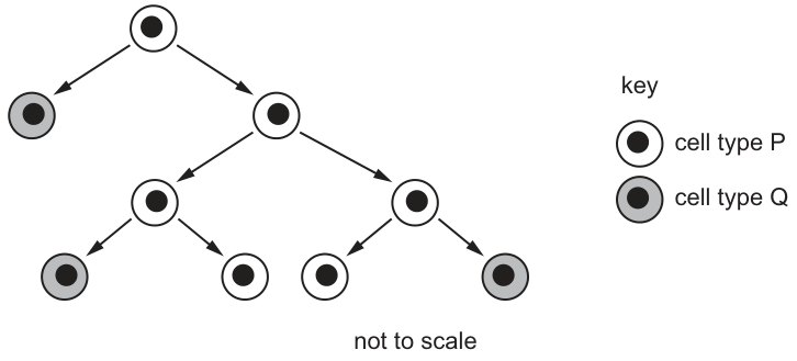
Which row correctly identifies cell type P and cell type Q?
Options
| cell type P | cell type Q | |
|---|---|---|
| A | cancer cell | differentiated cell |
| B | cancer cell | stem cell |
| C | differentiated cell | stem cell |
| D | stem cell | differentiated cell |
Working
The diagram shows asymmetric division: each time a type P cell divides, one daughter remains type P (self-renewal) while the other becomes type Q. This is the defining behaviour of a stem cell. In bone marrow, the stem cells are haematopoietic stem cells, which divide to produce differentiated blood cells (e.g. red blood cells, neutrophils, lymphocytes).
Therefore cell type P = stem cell, and cell type Q = differentiated cell.
Answer
D
D
Background Concept
A stem cell is an unspecialised cell that has two defining properties:
- Self-renewal — it can divide to produce more identical stem cells, so the stem cell pool is maintained.
- Potency — it can divide and differentiate into one or more specialised (differentiated) cell types.
In bone marrow, the resident stem cells are haematopoietic (multipotent) stem cells. They give rise to all the cellular components of blood: erythrocytes (red blood cells), several lineages of leucocytes (white blood cells — neutrophils, lymphocytes, monocytes, eosinophils, basophils), and platelets (from megakaryocytes).
A key feature of stem cell division is that it is often asymmetric: each mitosis produces one daughter cell that remains a stem cell and one daughter cell that is committed to differentiation. The committed cell undergoes further mitotic divisions, with each generation becoming progressively more specialised until a terminally differentiated blood cell is produced.
Understanding the Question
Fig. 20.1 is a cell lineage (family tree) diagram. Read it from top to bottom:
- The top circle (cell type P) divides into two daughter cells.
- One of those daughters is again a type P circle; the other has become a type Q (shaded) circle.
- The pattern repeats through three cycles, so type P cells persist at the top of every level while type Q cells accumulate below.
The question asks us to identify the biological identity of P and Q, with reference to bone marrow.
Approach
- Look for a cell type whose division can (a) produce more of itself AND (b) give rise to a different, more specialised cell type. This combination is the hallmark of a stem cell.
- Eliminate the other options by checking what the bone marrow contains and whether the diagram matches that biology.
Step-by-Step Reasoning
- Option A — cancer cell / differentiated cell: A cancer cell's defining property is uncontrolled proliferation that does not typically yield differentiated functional cells in an orderly lineage. It would not reliably give rise to a recognisable differentiated bone-marrow cell. Reject.
- Option B — cancer cell / stem cell: Same problem as A for P. Also, Q being a stem cell contradicts the diagram, which shows Q as the terminal, shaded end-product that no longer divides. Reject.
- Option C — differentiated cell / stem cell: A terminally differentiated cell (e.g. a mature red blood cell or neutrophil) cannot divide, so P could not be a source of further cycles. Reject.
- Option D — stem cell / differentiated cell: The diagram's behaviour — P self-renewing while generating a different (Q) cell at each cycle — is exactly the asymmetric division of a haematopoietic stem cell producing differentiated blood cells. This matches bone marrow biology. ✓
Key Takeaways
- Self-renewal + differentiation in the same division = stem cell.
- Bone marrow contains haematopoietic stem cells that continuously replenish the blood cell lineages.
- A cell lineage diagram is read like a family tree: identical symbols that persist represent the self-renewing population; new symbols at the tips represent differentiated products.
- Terminally differentiated cells usually cannot divide, so any cell shown producing daughters in a lineage tree cannot itself be terminally differentiated.
Common Mistakes
- Calling cell type P a cancer cell because the cell "keeps dividing." Cancer cells divide uncontrollably but the orderly production of a distinct, specialised daughter alongside a self-renewing daughter is the stem cell pattern, not a tumour.
- Confusing stem cell with embryonic cell or assuming all stem cells are pluripotent. The bone marrow stem cell is multipotent (restricted to blood lineages) but is still a stem cell.
- Assuming cell type Q is a stem cell because "it came from a dividing cell." Q is the shaded end-product that does not continue the lineage — it is the differentiated output.
Things to Be Careful About
- The diagram's "three cycles of cell division" wording refers to three successive mitotic events, not three daughter cells. Counting the shaded cells (five in the figure) is not the test; the pattern of self-renewal is.
- "Bone marrow" is the context clue: it points directly to haematopoietic stem cells and their differentiated blood-cell products, ruling out generic interpretations.
- For MCQs, use each option to eliminate rather than searching for the "right-sounding" one — the diagram's structure eliminates three of the four options decisively.
A student made a drawing of two chromosomes at stage K of a mitotic cell cycle.
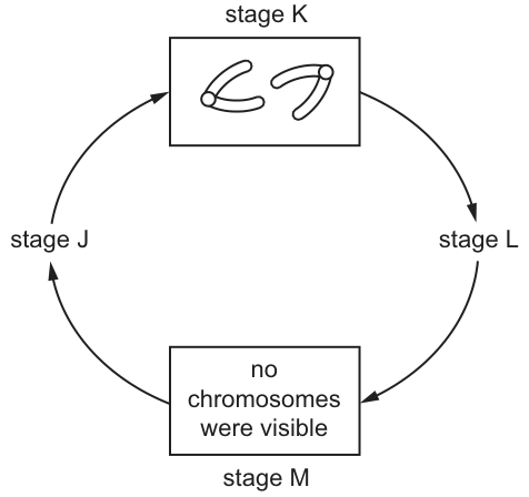
Which row shows the possible events that occur in stages J, L and M?
Options
| J | L | M | |
|---|---|---|---|
| A | spindle attachment | cytokinesis | DNA replication |
| B | chromosome condensation | spindle attachment | cytokinesis |
| C | DNA replication | chromosome condensation | spindle attachment |
| D | cytokinesis | DNA replication | chromosome condensation |
Working
Stage K shows two chromosomes, each with two sister chromatids, so K is metaphase (or late prophase) — chromosomes are condensed and aligned with spindle attached.
The cycle runs J → K → L → M → J, so working backwards and forwards from K:
- M shows no chromosomes visible → M is interphase → DNA replication occurs here.
- J is the stage that follows interphase and precedes the visible-chromosome stage → J is prophase, where chromosomes attach to the spindle.
- L follows K (metaphase) → L is anaphase/telophase, leading to cytokinesis.
Answer
A
A
Background Concept
The cell cycle consists of interphase (G₁, S, G₂) followed by mitosis (prophase, metaphase, anaphase, telophase) and finally cytokinesis. The key distinguishing feature of each stage is the state of the genetic material:
- Interphase: DNA exists as diffuse chromatin — no discrete chromosomes are visible under the light microscope. DNA replication occurs during the S phase.
- Prophase: Chromatin condenses into visible chromosomes, each composed of two sister chromatids joined at the centromere. The spindle begins to form and attaches to chromosomes at their centromeres via kinetochores.
- Metaphase: Chromosomes (still as sister chromatids) line up at the equator, fully attached to spindle fibres.
- Anaphase: Sister chromatids separate and are pulled to opposite poles.
- Telophase/Cytokinesis: Nuclear envelopes re-form and the cytoplasm divides, producing two daughter cells.
Understanding the Question
The diagram shows a circular cell cycle with four labelled stages — J, K, L and M — connected by arrows in the order J → K → L → M → J.
- Stage K contains a drawing of two chromosomes, each clearly consisting of two sister chromatids joined at a centromere. This identifies K as a stage where chromosomes are condensed and visible — consistent with prophase or metaphase.
- Stage M is explicitly labelled "no chromosomes were visible." This identifies M as interphase, when DNA is in the form of chromatin.
The question asks which row correctly assigns the events "spindle attachment," "chromosome condensation," "cytokinesis" and "DNA replication" to stages J, L and M.
Approach
Identify what each stage of the cell cycle must contain, given the clues in the figure, then match those events to the four options. Work forwards and backwards from the two anchor stages K and M.
Step-by-Step Reasoning
Anchoring with M: Stage M is labelled "no chromosomes were visible." This unambiguously places M in interphase. The defining molecular event of interphase (specifically the S phase) is DNA replication. So M = DNA replication.
This immediately eliminates:
- Option B, which places cytokinesis at M (cytokinesis is the final stage of mitosis, not interphase).
- Option D, which places chromosome condensation at M (condensation happens in prophase, when chromosomes are clearly visible, not when "no chromosomes were visible").
Anchoring with K: Stage K shows two chromosomes, each as two sister chromatids. K is therefore a stage within mitosis where chromosomes are condensed and visible — prophase or metaphase.
Determining J: The stage before K, in the direction of the cycle, is J. The cell has just exited interphase (M) and is entering mitosis. The first major event of mitosis is spindle attachment (prophase/prometaphase) as the spindle forms and attaches to chromosomes via their kinetochores. So J = spindle attachment.
This eliminates Option C, which places DNA replication at J (DNA replication occurs in interphase, not in the stage immediately after interphase).
Determining L: The stage after K, in the direction of the cycle, is L. If K is metaphase (chromosomes aligned with sister chromatids), then L must be the next stage — anaphase and telophase, culminating in cytokinesis, when the cytoplasm divides and two daughter cells form. So L = cytokinesis.
This matches Option A exactly:
- J = spindle attachment
- L = cytokinesis
- M = DNA replication
Checking Option A in full:
- M (interphase, no chromosomes visible) → DNA replication ✓
- J (just after interphase) → spindle attachment (prophase) ✓
- K (chromosomes with sister chromatids) → metaphase ✓
- L (after metaphase) → cytokinesis (telophase) ✓
The full sequence is therefore: M (interphase, DNA replication) → J (prophase, spindle attachment) → K (metaphase, chromosomes aligned) → L (anaphase/telophase, cytokinesis) → back to M.
Key Takeaways
- The cell cycle is a continuous loop; the diagram in the question shows only four snapshots, so each stage must be deduced from both the visual clue and its position in the cycle.
- "No chromosomes visible" is the classic signature of interphase — DNA is dispersed as chromatin, and this is when DNA replication occurs.
- A drawing of chromosomes as two sister chromatids indicates prophase or metaphase; the spindle is attached to chromosomes at their centromeres.
- Cytokinesis is the final event of the mitotic phase, separating the cytoplasm into two daughter cells.
Common Mistakes
- Confusing the direction of the cell cycle. The arrow goes M → J, so M (interphase) precedes J, not the other way round.
- Assuming that "spindle attachment" only happens in metaphase — in reality, spindle microtubules attach to chromosomes during prophase/prometaphase, even though alignment at the equator is only complete by metaphase.
- Placing DNA replication at a stage where chromosomes are visible — DNA replication strictly occurs in interphase (S phase), when no discrete chromosomes can be seen.
- Confusing cytokinesis (cytoplasmic division) with karyokinesis (nuclear division) — cytokinesis is the very last step.
Things to Be Careful About
- Read the figure carefully: the label "no chromosomes were visible" in stage M is the key clue that fixes M as interphase.
- The drawing in stage K shows two chromosomes, each with two chromatids — this is the textbook appearance of a chromosome after S phase and during G₂/prophase/metaphase, before sister chromatids separate.
- When asked to match events to stages, always anchor first to the stage with the most distinctive visual feature (here, M) before reasoning about neighbouring stages.
During DNA replication, the two original strands of a DNA molecule separate.
Which diagram shows the direction in which DNA polymerase moves along each of the original strands during DNA replication?
Options
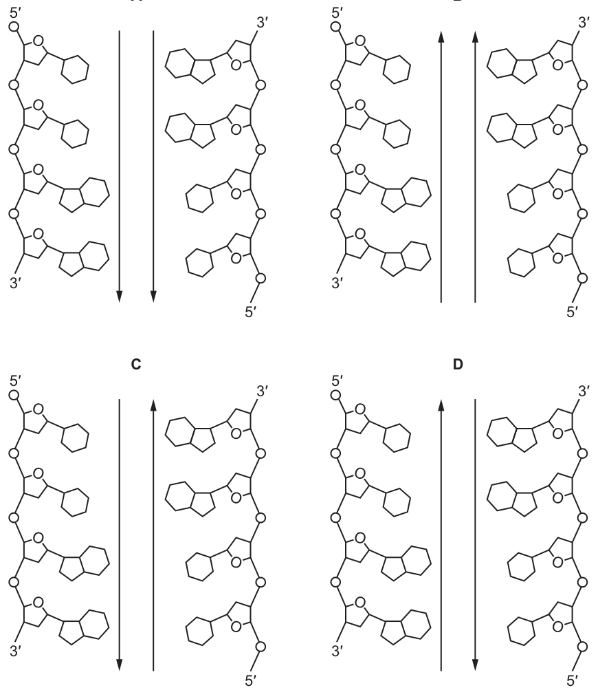
Working
The two DNA template strands are antiparallel:
- Left strand: 5' (top) → 3' (bottom)
- Right strand: 3' (top) → 5' (bottom)
DNA polymerase can only add nucleotides to the 3' end of the new strand, so it moves along the template strand in the 3' → 5' direction.
- On the left template strand, the 3' end is at the bottom and the 5' end is at the top, so DNA polymerase moves upwards.
- On the right template strand, the 3' end is at the top and the 5' end is at the bottom, so DNA polymerase moves downwards.
This matches diagram D (left arrow up, right arrow down).
Answer
D
D
Background Concept
DNA has a double helix made of two polynucleotide strands that run in opposite directions — this is called an antiparallel arrangement. Each strand has a sugar-phosphate backbone with a 5' end (where the 5' carbon of the deoxyribose is exposed) and a 3' end (where the 3' carbon is exposed). In a DNA molecule, the 5' end of one strand aligns with the 3' end of the other.
During semi-conservative replication, the two strands separate and each acts as a template for a new complementary strand. The enzyme DNA polymerase catalyses the addition of free DNA nucleotides to the growing new strand. A critical constraint is that DNA polymerase can only add nucleotides to the 3' end of the new strand. This means:
- The new strand is always synthesised in the 5' → 3' direction.
- Consequently, DNA polymerase must read (move along) the template strand in the 3' → 5' direction.
Because the two template strands are antiparallel, DNA polymerase must move in opposite directions on the two strands. The strand being synthesised continuously towards the replication fork is the leading strand, while the other is synthesised in short Okazaki fragments away from the fork — the lagging strand.
Understanding the Question
The question shows four diagrams (A–D), each depicting two antiparallel DNA template strands with their 5' and 3' ends labelled. Arrows on each strand indicate the direction in which DNA polymerase moves along that template strand. The task is to identify which diagram correctly shows the direction of polymerase movement on BOTH template strands.
In every diagram:
- The left strand has its 5' end at the top and its 3' end at the bottom.
- The right strand has its 3' end at the top and its 5' end at the bottom.
The only thing that varies between options is the direction of the two arrows.
Approach
For each strand, identify where the 3' end and 5' end lie, then determine which direction along the strand corresponds to 3' → 5' (the direction DNA polymerase must travel). Apply this to the left strand and the right strand separately, then match to the correct option.
Step-by-Step Reasoning
-
Left template strand: The 5' end is at the top, and the 3' end is at the bottom. Therefore, moving along this strand in the 3' → 5' direction means going from the bottom to the top — i.e., upwards. So the left arrow should point up.
-
Right template strand: The 3' end is at the top, and the 5' end is at the bottom. Therefore, moving along this strand in the 3' → 5' direction means going from the top to the bottom — i.e., downwards. So the right arrow should point down.
-
Evaluating the options:
- A: both arrows down — wrong (left polymerase should be moving up)
- B: both arrows up — wrong (right polymerase should be moving down)
- C: left arrow down, right arrow up — wrong (both directions are reversed)
- D: left arrow up, right arrow down — correct
-
The answer is D.
Key Takeaways
- DNA strands are antiparallel: one runs 5'→3', the other runs 3'→5'.
- DNA polymerase can only add nucleotides to the 3' end of the growing strand, so it moves along the template in the 3'→5' direction.
- Because the two template strands run in opposite directions, DNA polymerase moves in opposite directions on the two strands — this is the structural reason for the existence of leading and lagging strands.
Common Mistakes
- Forgetting the directional constraint: thinking DNA polymerase can move in either direction, leading to answers A or B (both arrows the same way).
- Confusing template and new strand directions: a new strand is built 5'→3', but the question asks about movement along the template, which is 3'→5'.
- Not reading the 5'/3' labels carefully: swapping which end of each strand carries which label leads to choosing C (the mirror image of the correct answer).
Things to Be Careful About
- The question asks about the direction of polymerase movement along each original (template) strand, not the new strand.
- Always read the 5' and 3' labels on the diagram carefully — the left strand in every option is 5'-top/3'-bottom, and the right strand is 3'-top/5'-bottom.
- A common visual trap is option C, which has the arrows reversed; double-check by going back to the 3'→5' rule for each strand individually.
Which statements about anticodons are correct?
1 The base sequence of an anticodon will always be the same as that of the corresponding triplet on the transcribed DNA strand.
2 There are 20 naturally occurring amino acids, so therefore there are only 20 anticodons as each anticodon is specific for only one amino acid.
3 The formation of hydrogen bonds between the bases of the anticodon with those of the complementary mRNA codon must occur before the new peptide bond can form.
Options
A 1, 2 and 3
B 1 and 2 only
C 1 and 3 only
D 3 only
Working
Statement 1 is false. The "transcribed DNA strand" is the template (antisense) strand. The mRNA codon is complementary to this template strand, and the tRNA anticodon is in turn complementary to the mRNA codon. So the anticodon has the same base sequence as the template strand — except that DNA contains T while tRNA contains U. The two sequences are therefore not identical.
Statement 2 is false. The genetic code is degenerate: more than one codon (and hence more than one anticodon) can specify the same amino acid. With four bases read in triplets there are possible codons, so there are many more than 20 anticodons.
Statement 3 is true. Hydrogen bonding between the anticodon and the mRNA codon positions the correct tRNA (carrying the correct amino acid) in the A site of the ribosome. Only after this correct positioning can a new peptide bond be catalysed by peptidyl transferase between the new amino acid and the growing polypeptide chain.
Only statement 3 is correct.
Answer
D
D
Background Concept
Translation is the second stage of protein synthesis and takes place on ribosomes in the cytoplasm. It converts the sequence of codons on an mRNA molecule into a chain of amino acids. Three types of RNA are involved:
- mRNA carries the genetic message in the form of triplets called codons.
- tRNA has an anticodon loop at one end; the three bases of the anticodon pair with the complementary mRNA codon by hydrogen bonding. Each tRNA also carries, at its 3′ end, the specific amino acid corresponding to its anticodon.
- rRNA, together with proteins, makes up the ribosome, which has a P site holding the tRNA attached to the growing polypeptide and an A site into which the next tRNA enters.
The template (antisense) DNA strand is the strand actually used as the template during transcription. The mRNA is synthesised complementary to this template strand, so the mRNA has the same base sequence as the other (coding/sense) DNA strand, except that uracil (U) replaces thymine (T). The anticodon on the tRNA is complementary to the mRNA codon, so it has the same base sequence as the template DNA strand, again with U replacing T.
The genetic code is degenerate: because there are possible codons but only 20 common amino acids, most amino acids are specified by more than one codon (and therefore by more than one anticodon). A few codons do not code for any amino acid — they are stop signals.
A new peptide bond is formed by the enzyme peptidyl transferase (a ribozyme) in the large ribosomal subunit, joining the amino acid on the tRNA in the A site to the polypeptide chain held by the tRNA in the P site. This only happens once the incoming tRNA has been correctly positioned by codon–anticodon base pairing.
Understanding the Question
This is a multiple-choice question asking which of three statements about anticodons are correct. The candidate must judge each statement independently and select the option that lists every correct statement and no incorrect ones.
- Statement 1 tests whether the candidate knows how the anticodon sequence relates to the original DNA sequence.
- Statement 2 tests understanding of the degeneracy of the genetic code and the number of possible anticodons.
- Statement 3 tests the sequence of events during translation at the ribosome.
The correct answer must include only those statements that are true.
Approach
Test each statement in turn against the mark scheme by recalling:
- The relationship between the template DNA strand, mRNA codon, and tRNA anticodon, including the T/U difference.
- The degeneracy of the genetic code and the number of possible codons/anticodons.
- The order of events in translation: codon–anticodon hydrogen bonding (to check the correct tRNA is in place) must precede peptide bond formation.
Step-by-Step Reasoning
Statement 1 — "The base sequence of an anticodon will always be the same as that of the corresponding triplet on the transcribed DNA strand."
- The transcribed (template/antisense) DNA strand is complementary to the mRNA codon, except that T is used instead of U.
- The tRNA anticodon is complementary to the mRNA codon, but uses U instead of T.
- Therefore the anticodon matches the template strand except for the T → U swap. The sequences are not literally identical, so the word "always be the same" makes this statement false.
- (Note: this statement would also be false if "transcribed DNA strand" is taken to mean the coding/sense strand — the anticodon would then be complementary to it, not identical.)
Statement 2 — "There are 20 naturally occurring amino acids, so therefore there are only 20 anticodons as each anticodon is specific for only one amino acid."
- The genetic code is degenerate: most amino acids are coded for by more than one codon, and therefore by more than one anticodon. For example, leucine, serine and arginine each have six codons.
- With 4 bases in triplets, there are possible codons (and 64 corresponding anticodons), of which 61 code for amino acids and 3 are stop codons.
- So there are many more than 20 anticodons. Statement 2 is false.
Statement 3 — "The formation of hydrogen bonds between the bases of the anticodon with those of the complementary mRNA codon must occur before the new peptide bond can form."
- A tRNA carrying its amino acid enters the A site of the ribosome. The anticodon pairs with the mRNA codon by hydrogen bonding between complementary bases (A–U and G–C).
- This pairing is what positions the correct amino acid opposite the codon. Only after this correct positioning does peptidyl transferase catalyse peptide bond formation between the new amino acid and the growing polypeptide chain on the tRNA in the P site.
- Therefore codon–anticodon hydrogen bonding must occur before the peptide bond is formed. Statement 3 is true.
Only statement 3 is correct, so the answer is D.
Key Takeaways
- The tRNA anticodon has the same base sequence as the DNA template strand, but with U replacing T. It is complementary to the mRNA codon.
- The genetic code is degenerate: codons, more than 20 anticodons, code for about 20 amino acids.
- During translation, hydrogen bonding between the anticodon and the mRNA codon correctly positions the incoming tRNA in the A site. Only then can peptidyl transferase catalyse formation of the new peptide bond.
Common Mistakes
- Confusing the template and coding strands of DNA — many students think the "transcribed strand" is the strand with the same sequence as the mRNA. It is in fact the template (antisense) strand.
- Forgetting that DNA uses T and RNA uses U — this subtle difference is what makes statement 1 false.
- Assuming one anticodon per amino acid — this ignores the degeneracy of the genetic code.
- Thinking peptide bond formation happens before the tRNA is in place — without codon–anticodon pairing, the correct amino acid is not delivered to the ribosome, and the wrong peptide bond would be at risk of forming.
Things to Be Careful About
- The phrase "transcribed DNA strand" can be ambiguous. In A-level biology, treat it as the template (antisense) strand — the strand used as a template by RNA polymerase.
- When judging statements in a "which are correct" question, evaluate each statement independently; do not assume that a partially correct statement earns a mark.
- Distinguish carefully between "complementary" (base pairs with) and "the same as" (identical). Statement 1 uses "the same as", which is the more demanding wording and which makes the statement false.
Part of a DNA molecule is transcribed to form an RNA molecule that is then modified.
Which row describes the RNA molecule before and after modification?
Options
| before modification | after modification | |
|---|---|---|
| A | contains exons and introns | contains exons only |
| B | contains exons and introns | contains introns only |
| C | contains exons only | contains introns only |
| D | contains introns only | contains exons only |
Working
Transcription of a gene produces a primary transcript (pre-mRNA) that is a complementary copy of the template DNA strand. This pre-mRNA contains both exons (coding sequences) and introns (non-coding sequences) in the same order as they occur in the gene.
Modification (RNA processing / splicing) occurs in the nucleus before the mRNA leaves for translation. During splicing, the introns are excised and the exons are joined together. The mature mRNA therefore contains exons only.
Answer
A
A
Background Concept
A eukaryotic gene is not a continuous stretch of coding sequence. It is split into two kinds of segments:
- Exons — the expressed sequences; these carry the information that will be translated into amino acids (and also untranslated regions flanking the coding sequence).
- Introns — the intervening sequences; these are present in the gene and in the initial RNA transcript but do not code for amino acids.
When RNA polymerase II transcribes a gene, it produces a primary RNA transcript (pre-mRNA) that is a faithful complementary copy of the entire gene — exons and introns alike.
Before this pre-mRNA can be translated, it must be modified in the nucleus. The key modification is splicing, carried out by a ribonucleoprotein complex called the spliceosome. The spliceosome recognises specific sequences at the exon–intron boundaries (splice sites), excises each intron as a lariat structure, and ligates the exons together. The result is a shorter, mature mRNA that contains only exons and is exported through nuclear pores to the ribosomes in the cytoplasm.
A useful memory aid: exons are EXpressed, introns are IN between (and are chucked out).
Understanding the Question
This is an MCQ testing the candidate's recall of the order in which exons and introns appear in a newly synthesised RNA molecule versus the same molecule after it has been processed.
- The "before modification" column corresponds to the primary transcript / pre-mRNA immediately after transcription.
- The "after modification" column corresponds to the mature mRNA after splicing has occurred in the nucleus.
The command word is implicit (the candidate must identify the correct row), so the answer is the option whose entries match the biology described above.
Approach
State the two biological facts in the correct order:
- Pre-mRNA, straight after transcription, contains the full gene sequence — both exons and introns.
- Splicing removes the introns; mature mRNA therefore contains only exons.
Then select the option that matches these two statements.
Step-by-Step Reasoning
- Step 1 — Recall the primary transcript. Transcription copies the entire gene, so the pre-mRNA contains both coding and non-coding regions: exons + introns.
- Step 2 — Apply splicing. Modification in the nucleus removes the introns, so the mature mRNA contains exons only.
- Step 3 — Match to the table. The row that says "contains exons and introns → contains exons only" is row A.
- Step 4 — Eliminate the others.
- B is wrong: introns are not the only thing left after splicing — they are removed, not retained.
- C is wrong: the primary transcript contains introns as well as exons, so it does not start with "exons only".
- D is wrong on both counts: the initial transcript is not "introns only" (it has exons too), and the mature mRNA is not "exons only" starting from a pure-intron molecule.
Key Takeaways
- The primary RNA transcript (pre-mRNA) contains exons and introns.
- Modification (splicing) removes the introns, leaving a mature mRNA that contains exons only.
- Splicing occurs in the nucleus before the mRNA is translated.
Common Mistakes
- Confusing the direction of the change: thinking the cell "adds" introns during modification. Introns are not added — they are removed.
- Believing that the primary transcript already contains only exons because textbooks emphasise exons as the coding parts. In reality the cell first transcribes everything, then edits.
- Mixing up the terms: remembering that "introns are removed" but then forgetting they were ever present, leading to a choice that does not state "exons and introns" before modification.
Things to Be Careful About
- Splicing is only one of several modifications to pre-mRNA. A 5′ cap and a poly-A tail are also added, but neither changes the exon/intron content, so the answer to this question is unaffected.
- The question concerns a typical eukaryotic gene; prokaryotic mRNA (which is rarely split by introns and is not spliced in the same way) is not the context being tested here.
- The mark scheme is unambiguous: there is no ecf or alternative wording — only option A correctly describes the state of the RNA before and after modification.
When a gene mutation occurs, which of the following may be altered, resulting in the production of a non-functional protein?
1 amino acid sequence
2 DNA nucleotide sequence
3 mRNA nucleotide sequence
Options
A 1, 2 and 3
B 1 and 2 only
C 2 and 3 only
D 2 only
Working
A gene mutation is a change in the base sequence of DNA.
- Statement 2 (DNA nucleotide sequence): directly altered — this is where the mutation occurs.
- Statement 3 (mRNA nucleotide sequence): altered — mRNA is transcribed from the mutated DNA template, so a changed base is copied into the mRNA.
- Statement 1 (amino acid sequence): altered — a change in the mRNA codon can change the amino acid incorporated during translation, producing a non-functional protein.
All three are altered, so 1, 2 and 3 are correct.
Answer
A
A
Background Concept
A gene mutation is a change in the sequence of nucleotide bases (A, T, C, G) along a DNA molecule. The most common types taught at AS Level are:
- Substitution — one base is replaced by a different base.
- Deletion — one or more bases are lost.
- Insertion — one or more extra bases are added.
The central dogma describes the flow of genetic information: DNA is transcribed into mRNA, which is then translated into a protein at the ribosome. Because each mRNA codon (a triplet of bases) codes for a specific amino acid, any change in the base sequence can change the amino acid sequence of the protein. If the new amino acid disrupts the protein's three-dimensional shape — for example by altering a critical bond in the active site of an enzyme — the protein becomes non-functional.
Understanding the Question
The question asks which of three molecular sequences MAY be altered by a gene mutation such that a non-functional protein is produced. The three candidates are:
- The amino acid sequence of the protein
- The DNA nucleotide sequence
- The mRNA nucleotide sequence
The command word is essentially "identify which apply" — we are selecting the correct combination.
Approach
Apply the central dogma step by step:
- Where does the mutation occur? → In the DNA.
- What is the immediate consequence for the mRNA? → It will carry the same change (because it is transcribed from the DNA template).
- What is the downstream consequence for the protein? → The amino acid sequence may be different, and the protein may no longer function.
Therefore all three statements can be true for a single gene mutation.
Step-by-Step Reasoning
- Statement 2 (DNA nucleotide sequence): True. By definition, a gene mutation is a change in the DNA base sequence, so the DNA is always altered.
- Statement 3 (mRNA nucleotide sequence): True. During transcription, the DNA template strand is used to synthesise a complementary mRNA molecule. A mutated base in the DNA template will therefore be transcribed into a complementary (mutated) base in the mRNA.
- Statement 1 (amino acid sequence): True. During translation, ribosomes read mRNA codons three bases at a time and use tRNA to deliver the corresponding amino acid. A change in the mRNA codon may result in a different amino acid being inserted into the polypeptide chain. A substitution that does not change the encoded amino acid (because of the degeneracy of the code) would not alter the protein, but the question only requires that the sequence MAY be altered — and substitutions that do change the amino acid, plus any deletion or insertion (which causes a frameshift), do alter it.
Because all three can be altered, the answer is A (1, 2 and 3).
Key Takeaways
- A gene mutation always alters the DNA base sequence.
- That change is propagated to the mRNA by transcription.
- It may then alter the amino acid sequence of the protein at translation.
- Even a single amino acid change can produce a non-functional protein (for example, sickle-cell anaemia results from a single glutamate → valine substitution in haemoglobin).
Common Mistakes
- Choosing B (1 and 2 only) — forgetting that the mutation is also carried into the mRNA, since mRNA is a direct copy of the DNA template.
- Choosing C (2 and 3 only) — forgetting that the changed mRNA codon can change the amino acid incorporated during translation.
- Choosing D (2 only) — the most common error; it overlooks the downstream effects on mRNA and protein.
- Confusing gene mutation with chromosome mutation. A gene mutation specifically alters the base sequence of a single gene, not the number or structure of whole chromosomes.
Things to Be Careful About
- A substitution may or may not change the amino acid (because of the degenerate genetic code), but the question only requires that the amino acid sequence MAY be altered.
- A deletion or insertion (other than in multiples of three bases) causes a frameshift, which alters every amino acid downstream and almost always produces a non-functional protein.
- The word "may" is important: the question does not claim that every mutation alters all three, only that all three are possible consequences.
The diagrams show the arrangements of phloem and xylem tissues in some plant organs.
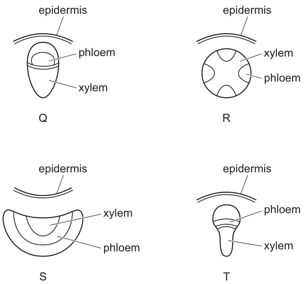
Where will arrangements Q, R, S and T be found in plants?
Options
| Q | R | S | T | |
|---|---|---|---|---|
| A | leaf | root | stem | root |
| B | root | stem | root | leaf |
| C | stem | stem | leaf | root |
| D | stem | root | leaf | stem |
Working
- Q — a discrete vascular bundle with phloem on the outside and xylem on the inside, surrounded by a single epidermis line: this is the collateral bundle of a stem.
- R — a central star-shaped xylem with phloem patches sitting between the arms of the xylem star: this radial arrangement is characteristic of a root.
- S — a broad, flattened section with xylem on the inner (upper) side and phloem on the outer (lower) side: this is the vascular bundle of a leaf.
- T — a collateral bundle (phloem outside, xylem inside) drawn in a different shape: also from a stem.
Matching: Q = stem, R = root, S = leaf, T = stem.
Answer
D
D
Background Concept
Vascular plants transport water, mineral ions and assimilates through two continuous tissue systems:
- Xylem — conducts water and dissolved mineral ions from the roots up to the shoots and leaves. In an angiosperm, the conducting elements are xylem vessels (and tracheids), supported by xylem fibres and xylem parenchyma.
- Phloem — translocates organic solutes (mainly sucrose) from sources to sinks. It consists of sieve tube elements and companion cells, plus phloem fibres and parenchyma.
The way these two tissues are arranged within a vascular bundle is diagnostic of the organ:
- Collateral bundle — phloem and xylem lie side by side on the same radius, with phloem on the outside (towards the epidermis) and xylem on the inside (towards the centre of the stem). This is the typical arrangement in dicot stems and in leaf vascular bundles (in a leaf, the xylem is on the upper side and the phloem on the lower side).
- Radial arrangement — the xylem forms a central star or cross, with the phloem strands sitting between the projecting arms of the xylem, alternating with it on different radii. This is the diagnostic arrangement of a dicot root.
- Scattered / closed bundles — in monocot stems the bundles are scattered through the ground tissue, but each individual bundle is still collateral.
Understanding the Question
The question presents four cross-sections (Q, R, S, T) of plant tissue, each labelled with the positions of phloem, xylem and epidermis. The task is to recognise which plant organ (stem, root or leaf) each section is taken from, then pick the row of the option table that matches.
The command word here is implicit: the candidate must identify the organ from the arrangement of vascular tissue.
Approach
For each section, ask two questions:
- Are the phloem and xylem arranged side by side on the same radius (collateral) or on alternating radii (radial)?
- Is the bundle embedded in a ring of ground tissue (stem), arranged as a star with alternating phloem strands (root), or set into a flat photosynthetic lamina (leaf)?
- A collateral bundle with phloem outer and xylem inner = stem (or leaf, depending on shape).
- A radial xylem star with phloem between the arms = root.
- A flat, lens-shaped section with xylem on the upper surface and phloem on the lower = leaf.
Step-by-Step Reasoning
-
Q shows a discrete oval bundle with phloem on the outside (near the epidermis) and xylem on the inside. This is a classic collateral vascular bundle of a dicot stem, where the bundles form a ring near the periphery of the stem. ⇒ stem.
-
R shows a central, star-shaped (or cross-shaped) xylem with discrete phloem patches located between the projecting arms of the xylem, on different radii. The xylem occupies the very centre of the section, which never happens in a stem — the central pith of a stem is parenchyma, not xylem. This radial arrangement is the unmistakable signature of a root (specifically a young dicot root in which the protoxylem points outwards and the metaxylem fills the centre). ⇒ root.
-
S shows a long, broad, flattened section rather than a round stem. The xylem sits on the upper (inner) side of the section and the phloem on the lower (outer) side. The flattened shape and the position of the xylem towards what would be the upper epidermis of a leaf identify this as a leaf vascular bundle (a minor vein seen in cross-section through the lamina). ⇒ leaf.
-
T shows another collateral bundle — phloem outer, xylem inner — drawn in a slightly different, more elongated shape. Because the arrangement is collateral and the bundle is discrete (not radial and not a flat lamina), it is also from a stem. ⇒ stem.
Reading off the matches:
- Q = stem, R = root, S = leaf, T = stem.
This row matches option D.
Key Takeaways
- Stems have collateral vascular bundles, with phloem on the outside and xylem on the inside; in a TS the bundles are arranged in a ring, each a discrete unit.
- Roots have a radial arrangement: xylem in a central star/cross with phloem strands between the arms, on different radii from the xylem.
- Leaves have collateral bundles set in a flat lamina; in a cross-section the xylem lies on the upper (adaxial) side and the phloem on the lower (abaxial) side.
- Recognising whether phloem and xylem are on the same radius (collateral) or different radii (radial) is the single most useful first step when identifying an unfamiliar vascular cross-section.
Common Mistakes
- Calling Q a leaf because phloem is on the outside. A leaf bundle is also collateral, but it sits in a flat photosynthetic lamina; Q is a discrete bundle embedded in ground tissue, i.e. a stem.
- Calling R a stem because the section is round. A stem's centre is pith, not xylem; if xylem occupies the very centre and the phloem is on different radii, it is a root.
- Calling S a stem because the xylem is on top. The flattened, elongated shape and the position of the bundle within a lamina are what mark S as a leaf.
- Confusing phloem and xylem: phloem is on the outside of a stem bundle, but in a root the phloem sits between the arms of the xylem star, not on the outside of the section.
Things to Be Careful About
- The label lines in the diagram point to the tissue, not to the organ — read the arrangement, not just the labels.
- "Phloem outside, xylem inside" is correct for both a dicot stem bundle and a leaf vein; the difference is the shape of the whole section (round stem vs flat lamina) and the relative position of the bundle within it.
- Monocot stems also have collateral bundles, but they are scattered rather than arranged in a ring; the question's options do not require distinguishing monocot from dicot stems, only stem from root from leaf.
- Keep the orientation of "inside/outside" consistent: in any cross-section, the epidermis marks the outside, and the centre of a stem marks the inside.
Which feature of xylem vessel elements allows them to have reduced resistance to water movement?
Options
A Lignin forms an incomplete secondary wall.
B New vessels transport extra water as a plant grows.
C There are no cross walls between vessel elements.
D Vessel elements join to form narrow tubes.
Working
Xylem vessel elements are dead, hollow cells stacked end to end. During their development, the end walls break down completely, so each vessel is a continuous, unobstructed tube. This means water moving up the xylem encounters no cross walls to slow it down, giving reduced resistance to flow.
- A — Lignin forms a complete (not incomplete) secondary wall and provides support; it does not reduce flow resistance.
- B — Describes a consequence of growth, not a structural feature of the vessel element itself.
- D — Vessels are wide, not narrow; a wide lumen lowers resistance.
Answer
C
C
Background Concept
Xylem is the plant tissue responsible for transporting water and dissolved mineral ions from the roots to the leaves and other aerial parts. It is composed of xylem vessel elements and tracheids (both dead at maturity), together with living xylem parenchyma and supporting fibres.
A xylem vessel element starts life as a living cell, but as it matures:
- The protoplast (cytoplasm) dies, leaving an empty lumen.
- A lignified secondary cell wall is laid down, giving mechanical strength and preventing the tube from collapsing under the negative pressure (tension) generated by transpiration pull.
- The end walls break down completely, so adjacent vessel elements join into one long, continuous tube called a xylem vessel.
Water moves up these vessels in a continuous column driven by the transpiration stream (cohesion–tension theory). Anything that interrupts this column — air embolisms, narrow lumens, or cross walls — increases resistance and reduces flow rate.
Understanding the Question
The command word is "which feature … allows them to have reduced resistance to water movement?" This is a structure–function question. We must pick the option that describes a structural feature of the vessel element itself, and explain (or recognise) how that feature directly lowers resistance to water flow.
Approach
Recall the structural features of a mature xylem vessel element: hollow lumen, lignified wall, no end walls, wide diameter. Then check each option to see which one is both a real feature of the vessel element AND the one that lowers flow resistance.
Step-by-Step Reasoning
- Option A — "Lignin forms an incomplete secondary wall." The wording is wrong on two counts. Lignin in xylem forms a continuous, complete secondary wall (often in annular, spiral, scalariform, reticulate or pitted patterns, but always an unbroken layer around the cell). Lignin's role is to provide support and waterproofing, not to reduce flow resistance. Reject.
- Option B — "New vessels transport extra water as a plant grows." This is not a structural feature of an individual vessel element; it is a statement about the plant as a whole producing more vessels as it grows. Reject.
- Option C — "There are no cross walls between vessel elements." Correct. Each vessel element is open at both ends (perforation plates replace the original end walls). The result is a long, continuous tube with no internal partitions to interrupt the water column. Resistance to flow through a tube is proportional to the length of the tube and inversely proportional to the fourth power of the radius (Poiseuille's law), so removing cross walls is a major reduction in resistance. Accept.
- Option D — "Vessel elements join to form narrow tubes." Vessels are in fact wide tubes; a wide lumen (large radius) further reduces resistance. The statement that they are narrow is biologically incorrect. Reject.
Key Takeaways
- Xylem vessel elements are dead, hollow, lignified cells whose end walls have broken down.
- The absence of cross walls gives a continuous, wide tube → low resistance to water flow.
- Lignin provides support (preventing collapse under tension), not reduced flow resistance.
- Structure–function: structure of the vessel (no end walls, wide lumen, lignified) directly matches its function (rapid, unobstructed water transport).
Common Mistakes
- Choosing A because students remember "lignin = xylem" but misremember lignin as a thin or incomplete layer; lignin actually forms a complete secondary wall.
- Choosing D because "narrow" sounds like it should be efficient, but in fluid flow through a tube a wider lumen gives less resistance (Poiseuille's law: resistance ∝ 1/r⁴).
- Confusing xylem vessel elements with sieve tube elements of the phloem. Phloem sieve tubes do retain perforated end walls (sieve plates) and remain alive, so they are not a continuous open tube.
Things to Be Careful About
- "Reduced resistance" is a specific physics idea: it means water can flow more easily. The structural cause is the absence of cross walls (and the wide lumen).
- The question is about the vessel element's structure, not about the whole plant's water relations — that rules out option B.
- Resist the temptation to pick the option that simply mentions a true xylem feature (lignin, narrowness) when the wording is biologically wrong.
Which statement describes movement through a plant in the apoplast pathway?
Options
A Water moves through the cell walls.
B Water moves through the cytoplasm.
C Water moves through the plasmodesmata.
D Water moves through the vacuoles.
Working
The apoplast pathway is the route taken by water through the non-living continuum of cell walls and intercellular spaces, without crossing any plasma membrane. Option A states this directly.
Answer
A
A
Background Concept
Water can move across the root cortex from the epidermis to the xylem via three interconnected routes:
- Apoplast pathway — water moves through the cell walls and the air spaces between cells. This is a continuous, non-living network; water here does not cross any plasma membrane and so is not stopped by the membrane barrier until it reaches the endodermis, where the Casparian strip blocks further apoplastic flow and forces water into the symplast.
- Symplast pathway — water moves through the cytoplasm of cells, passing from cell to cell via plasmodesmata (the cytoplasmic channels that connect adjacent cells). Because the cytoplasm is living, the plasma membrane bounds this pathway at every cell.
- Vacuolar pathway — water moves from vacuole to vacuole across the tonoplast and cytoplasm; this is sometimes regarded as a subdivision of the symplast route.
Understanding the Question
This is a multiple-choice question testing direct recall of what the apoplast pathway is. The candidate must match the description to the correct option.
Approach
Recall that "apoplast" literally refers to the non-living structural framework of the plant (cell walls and intercellular spaces) and so water in the apoplast pathway is by definition moving through cell walls.
Step-by-Step Reasoning
- Option A — "Water moves through the cell walls." This is the textbook definition of the apoplast pathway. ✓
- Option B — "Water moves through the cytoplasm." This describes the symplast pathway. ✗
- Option C — "Water moves through the plasmodesmata." Plasmodesmata are the intercellular cytoplasmic channels of the symplast pathway. ✗
- Option D — "Water moves through the vacuoles." This is the vacuolar pathway. ✗
Therefore A is correct.
Key Takeaways
- Apoplast = cell walls and intercellular spaces (non-living).
- Symplast = cytoplasm linked by plasmodesmata (living, bounded by plasma membranes).
- The Casparian strip at the endodermis blocks the apoplast route, forcing water into the symplast before it can enter the xylem.
Common Mistakes
- Confusing apoplast with symplast — the key distinction is whether water crosses the plasma membrane (symplast) or stays within the cell wall network (apoplast).
- Choosing plasmodesmata for the apoplast — plasmodesmata are features of the symplast, not the apoplast.
Things to Be Careful About
The word "apoplast" itself comes from Greek roots meaning "away from the formed/ living material" (apo- = away from, plastos = formed), which is a useful mnemonic: the apoplast is the non-living, structured framework through which water can flow freely without crossing any membrane.
What is the correct name for the attraction due to the hydrogen bonding between water molecules during mass flow in plants?
Options
A transpiration
B cohesion
C tension
D adhesion
Working
The cohesion-tension theory describes the upward movement of water in the xylem. It distinguishes between several forces:
- Transpiration — the evaporation of water from the mesophyll cell walls and its loss as water vapour through the stomata.
- Cohesion — the attractive force between like molecules; in the xylem, this is the hydrogen bonding between adjacent water molecules that holds the water column together.
- Tension — the negative hydrostatic pressure (pull) generated in the xylem as water is lost by transpiration at the leaves; this pulls the water column upward.
- Adhesion — the attraction between unlike molecules; here, between water molecules and the cellulose walls of the xylem vessels, which helps to counteract gravity.
The question specifically asks for the force due to hydrogen bonding between water molecules, which is the definition of cohesion.
Answer
B
B
Background Concept
The cohesion-tension theory explains how water is moved from the roots to the leaves of a plant through the xylem. The driving force is the evaporation of water from the leaves (transpiration), which creates a negative pressure (tension) in the xylem. This tension pulls a continuous column of water upward, and that column remains intact because of two intermolecular forces:
- Cohesion — the attraction between water molecules themselves, caused by hydrogen bonding between the partially positive hydrogen of one water molecule and the partially negative oxygen of another.
- Adhesion — the attraction between water molecules and the walls of the xylem vessels (cellulose), which also helps the column resist the pull of gravity.
These two forces, together with the tension generated by transpiration, allow water to be pulled up tall trees without the column breaking.
Understanding the Question
This is a single-mark multiple choice question that tests precise vocabulary. The stem points specifically to the attraction due to the hydrogen bonding between water molecules during mass flow in plants. The candidate must match this description to one of the four options, all of which are key terms in the cohesion-tension theory.
Approach
Translate each option into its precise definition and compare it to the wording in the stem:
- Transpiration refers to the loss of water vapour, not an intermolecular attraction.
- Cohesion refers to attraction between like molecules (water–water) by hydrogen bonding — this matches the stem exactly.
- Tension refers to the negative pressure (pull) in the xylem, not a hydrogen-bonding force.
- Adhesion refers to attraction between unlike molecules (water–xylem wall), not water–water.
Step-by-Step Reasoning
- The stem describes "attraction due to hydrogen bonding between water molecules" — the key phrase is "between water molecules", i.e. water–water interactions.
- Cohesion is, by definition, the force of attraction between molecules of the same substance. In the xylem, cohesion is provided by hydrogen bonds between water molecules.
- Adhesion is the attraction between water and the xylem wall, so it does not match "between water molecules".
- Transpiration is the loss of water vapour, a process not an intermolecular force. Tension is the pulling force generated by that loss; it is a pressure, not a hydrogen-bonding attraction.
- The only option that matches the definition in the stem is B, cohesion.
Key Takeaways
- Cohesion = water-to-water attraction via hydrogen bonding; it maintains the continuity of the water column in the xylem.
- Adhesion = water-to-xylem wall attraction; it counteracts gravity and supports capillarity.
- Tension = the negative pressure (pull) created by transpiration; it provides the driving force for upward movement.
- Transpiration = the evaporation of water from leaves; it is the ultimate source of the pull.
Common Mistakes
- Choosing A, transpiration — confusing the driving process with the intermolecular force.
- Choosing C, tension — confusing the negative pressure that pulls the column with the hydrogen-bonding force that holds it together.
- Choosing D, adhesion — forgetting that adhesion is between water and the xylem wall, not between water molecules themselves.
Things to Be Careful About
- The wording "between water molecules" is the discriminator: water–water = cohesion; water–wall = adhesion.
- The cohesion-tension theory uses both cohesion AND tension; remember that cohesion keeps the column intact, while tension actually pulls it up.
- A small number of questions may use "mass flow" loosely; in this question the phrase refers specifically to the upward mass flow of water in the xylem (transpiration stream), not the mass flow of assimilates in the phloem.
Which row shows the cause of mass flow in the phloem and the direction of movement of phloem sap by mass flow?
Options
| cause of mass flow in the phloem | direction of movement of phloem sap by mass flow | |
|---|---|---|
| A | hydrostatic pressure gradient | sink to source |
| B | hydrostatic pressure gradient | source to sink |
| C | water potential gradient | sink to source |
| D | water potential gradient | source to sink |
Mass flow in the phloem is driven by a hydrostatic pressure gradient, established when sugars are actively loaded at the source (raising the solute concentration, lowering the water potential so water enters by osmosis and raises turgor pressure) and unloaded at the sink (lowering turgor). The resulting pressure difference pushes phloem sap from source to sink through the sieve tubes.
Answer
B
B
Background Concept
In the phloem, sugars (mainly sucrose) and other assimilates are transported from where they are made or stored (the source) to where they are used or stored (the sink). The mechanism responsible is the mass flow hypothesis (proposed by Münch and supported by subsequent evidence).
Key points about phloem transport:
- Sieve tube elements form continuous tubes with perforated end walls (sieve plates), allowing bulk flow of sap.
- Companion cells carry out metabolic activities, including actively loading sucrose into the sieve tubes at the source.
- A hydrostatic pressure gradient is generated between source and sink regions, and this is the direct cause of bulk flow.
The loading process at the source:
- Companion cells actively transport H⁺ out using a proton pump, using ATP.
- The resulting H⁺ gradient drives sucrose co-transport into the companion cell (and then into the sieve tube) via a symporter.
- This raises the solute concentration inside the sieve tube, lowering the water potential ().
- Water enters the sieve tube by osmosis from the xylem, raising the hydrostatic (turgor) pressure at the source end.
At the sink:
- Sucrose is actively or passively unloaded into sink cells.
- The water potential inside the sieve tube rises (becomes less negative), so water leaves by osmosis, lowering the hydrostatic pressure at the sink end.
The pressure difference between source and sink is the hydrostatic pressure gradient that drives the bulk flow of phloem sap.
Understanding the Question
This is a multiple-choice question asking two things:
- What is the cause of mass flow in the phloem?
- What is the direction of phloem sap movement?
A common confusion: candidates sometimes say "water potential gradient" (which sounds similar to "hydrostatic pressure gradient"). The mark scheme is precise — the cause is a hydrostatic pressure gradient, not a water potential gradient per se. (Although water potential differences drive the entry/exit of water, the actual flow through the sieve tube is driven by the pressure difference.)
Approach
Eliminate the wrong options:
- A and C both say "sink to source" — this is wrong direction. Phloem moves sugars from where they are produced (source, usually photosynthesising leaves) to where they are used/stored (sink, e.g. roots, fruits, growing tips).
- C and D say "water potential gradient" — this is incorrect. The driving force inside the sieve tube is the hydrostatic (turgor) pressure gradient, not a water potential gradient. (Water potential is relevant to the entry and exit of water, but the bulk flow of sap within the tube is driven by pressure.)
- B correctly pairs hydrostatic pressure gradient (the cause) with source to sink (the direction).
Step-by-Step Reasoning
- At the source (e.g., a photosynthesising leaf), sucrose is actively loaded into the sieve tube via a proton pump and sucrose-H⁺ co-transporter.
- The increased solute concentration lowers the water potential inside the sieve tube, so water enters from the xylem by osmosis, raising the hydrostatic pressure.
- At the sink, sucrose is unloaded, raising the water potential in the sieve tube, so water leaves and the hydrostatic pressure falls.
- The difference in hydrostatic pressure between the high-pressure source end and the low-pressure sink end produces a hydrostatic pressure gradient.
- This pressure gradient pushes the phloem sap (a solution of sugars, amino acids, and other substances) in bulk along the sieve tubes from the source to the sink.
The flow is passive in terms of the bulk movement itself — the energy expended is at the loading step (proton pumps), which sets up the pressure difference.
Key Takeaways
- The cause of mass flow in phloem is a hydrostatic pressure gradient.
- The direction of movement is from source to sink.
- Sources are typically mature photosynthesising leaves; sinks include roots, fruits, seeds and growing tissues.
- Active loading of sucrose at the source (using proton pumps and co-transporters, hence ATP) is what sets up the pressure difference.
- Note: although water potential gradients are involved in the entry/exit of water at source/sink, the actual flow through the sieve tube is driven by the pressure gradient, not the water potential gradient itself.
Common Mistakes
- Confusing hydrostatic pressure gradient with water potential gradient — these are different. The water potential gradient causes water to enter or leave the sieve tube; the resulting hydrostatic pressure gradient is what drives bulk flow.
- Saying the direction is sink to source — this is wrong; phloem carries assimilates from where they are made to where they are used.
- Confusing phloem (transports sugars, amino acids) with xylem (transports water and mineral ions from roots to leaves).
Things to Be Careful About
- The phrase "water potential gradient" is a distractor — it is the hydrostatic pressure gradient that drives mass flow in the phloem.
- The direction "source to sink" applies to phloem sap; the water that enters and exits the sieve tube travels from xylem into the sieve tube at the source and back into xylem at the sink.
- Note: the source and sink relationship is not fixed — a growing root tip is a sink, but once mature and storing starch, it may become a source during spring mobilisation.
- Make sure you can describe the role of the proton pump and co-transporter in the active loading of sucrose.
The graph shows the thickness of the walls of the main blood vessels to and from a mammalian heart.
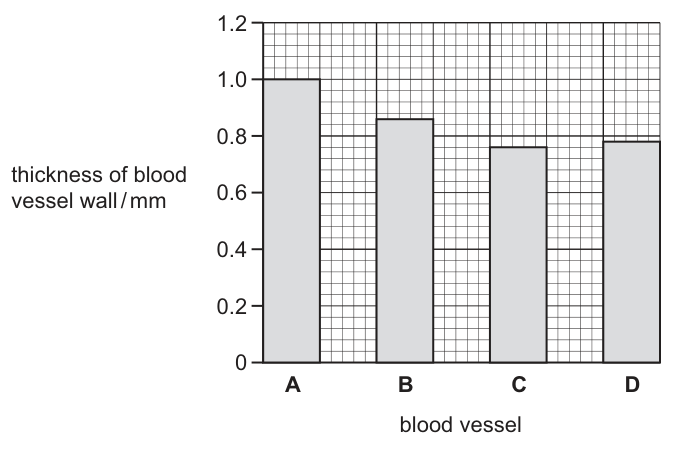
Which blood vessel is the pulmonary artery?
Options
A A
B B
C C
D D
Working
The pulmonary artery is an artery, so its wall is thicker than the walls of veins (the vena cava and pulmonary vein) because arteries must withstand the higher pressure generated by ventricular contraction.
However, the pulmonary artery carries blood only to the lungs, so it experiences a lower pressure than the aorta (which serves the entire systemic circulation). Its wall is therefore thinner than the aorta but thicker than any vein.
Ranking the wall thicknesses:
- A (1.0 mm) — thickest → aorta
- B (~0.85 mm) — second thickest → pulmonary artery
- D (~0.8 mm) → vena cava
- C (0.75 mm) — thinnest → pulmonary vein
Answer
B
B
Background Concept
The mammalian circulatory system is a closed double circulation: blood passes through the heart twice for each complete circuit. The right side of the heart handles the pulmonary circuit (heart → lungs → heart), while the left side handles the systemic circuit (heart → body tissues → heart). This produces four major vessels around the heart:
- Aorta — carries oxygenated blood from the left ventricle to the body.
- Vena cava — returns deoxygenated blood from the body to the right atrium.
- Pulmonary artery — carries deoxygenated blood from the right ventricle to the lungs.
- Pulmonary vein — returns oxygenated blood from the lungs to the left atrium.
Blood vessel wall thickness is closely matched to the blood pressure the vessel must withstand. Arteries have thick, muscular, elastic walls to cope with the surge of high-pressure blood pumped out during ventricular systole, whereas veins have thinner walls because they operate at much lower pressure. The aorta experiences the highest pressure of all vessels (it is closest to the powerful left ventricle and feeds the high-resistance systemic circulation), and so it has the thickest wall. The pulmonary artery also has to withstand arterial pressure, but the pulmonary circuit is a much shorter, lower-resistance loop than the systemic circuit, so its operating pressure — and therefore its wall thickness — is less than the aorta but more than any vein.
Understanding the Question
The figure is a bar chart of the wall thicknesses (in mm) of the four main vessels leading to and from the heart. The question is essentially asking the candidate to identify which vessel must be the pulmonary artery, given only its wall thickness. The command word is "which", so the candidate must pick a single letter and justify the ranking.
The key biological fact to apply is the relative pressure ranking: aorta > pulmonary artery > veins (vena cava ≈ pulmonary vein). Translating that into wall-thickness ranking gives aorta > pulmonary artery > vena cava / pulmonary vein, which then maps directly onto the four bars.
Approach
- Recall that wall thickness ∝ blood pressure in the vessel.
- Rank the four vessels by the pressure they normally experience.
- Match that ranking to the four bar heights in the figure.
Step-by-Step Reasoning
Step 1 — rank pressures.
- Aorta: highest pressure (left ventricle pumps into the entire systemic circulation, which has high peripheral resistance).
- Pulmonary artery: lower than aorta, but still arterial (right ventricle pumps into the lungs).
- Vena cava and pulmonary vein: low pressure (no ventricular pump directly behind them; they rely on skeletal-muscle pump, respiratory pump, and valves).
Step 2 — translate to wall thickness.
Wall thickness follows pressure. So the expected order is:
- Aorta (thickest) > pulmonary artery > vena cava ≈ pulmonary vein (thinnest).
Step 3 — read the graph.
From the bar chart:
- A = 1.0 mm (thickest)
- B ≈ 0.85 mm (second thickest)
- D ≈ 0.8 mm
- C = 0.75 mm (thinnest)
Step 4 — match.
- A → aorta
- B → pulmonary artery ✓
- D → vena cava
- C → pulmonary vein
Key Takeaways
- Arteries have thicker walls than veins because they carry blood at higher pressure.
- Of all vessels, the aorta has the thickest wall, because it receives blood directly from the left ventricle and feeds the high-resistance systemic circulation.
- The pulmonary artery has the second-thickest wall: it is still an artery (so thicker than any vein), but it operates at lower pressure than the aorta because the pulmonary circuit has lower resistance.
- A useful memory aid: "arteries = thick, veins = thin; of the arteries, aorta is the thickest because it faces the highest pressure."
Common Mistakes
- Choosing A (the aorta). Students often pick the thickest bar thinking "artery = thickest wall". The question asks specifically for the pulmonary artery, not any artery; the aorta is the other thick-walled artery.
- Choosing C or D (a vein). Confusing artery with vein is a fundamental error — all arteries (including the pulmonary artery) have thicker walls than any vein.
- Forgetting that the pulmonary artery carries deoxygenated blood. This is a common trap: students assume all arteries carry oxygenated blood. The pulmonary artery is an exception — it carries deoxygenated blood from the right ventricle to the lungs.
Things to Be Careful About
- The pulmonary artery and pulmonary vein are named by direction of flow relative to the heart/lungs, not by oxygen content. Don't let the word "artery" trick you into thinking it must carry oxygenated blood.
- The question gives wall thickness only — no information about whether the vessel carries oxygenated or deoxygenated blood is needed to solve it. The ranking argument alone is sufficient.
- The vena cava and pulmonary vein have similar (thin) walls; either could plausibly be C or D here. Don't be distracted by the very small difference between them — what matters is the clear gap between arteries (A and B) and veins (C and D).
The photomicrographs show blood smears from two different people. Person J does not have a disorder that affects their blood. Person K does have a disorder that affects their blood.
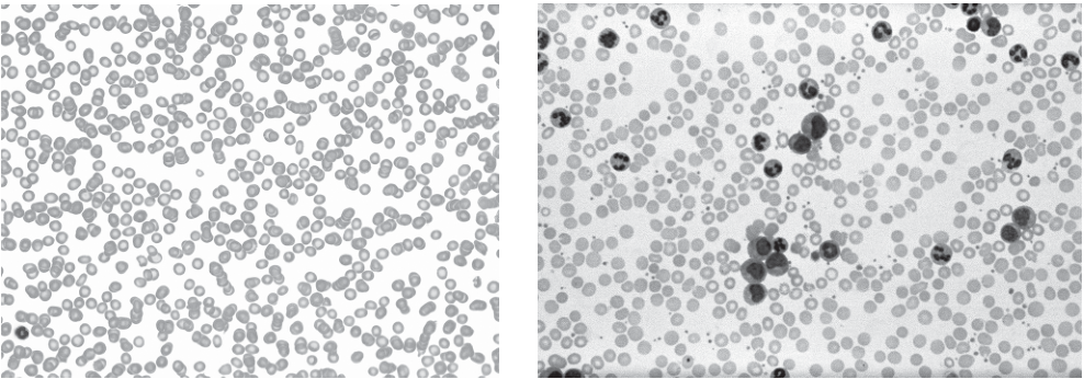
The table shows some disorders that increase or decrease the number of neutrophils, red blood cells or lymphocytes.
| disorders related to blood count changes | ||
|---|---|---|
| increase | decrease | |
| neutrophil | chronic inflammation | autoimmune neutropenia |
| red blood cell | heart failure | anaemia |
| lymphocyte | chronic leukaemia | sepsis |
Which disorder does person K have?
Options
A autoimmune neutropenia
B heart failure
C sepsis
D chronic leukaemia
Working
Person K's blood smear shows many more white blood cells than Person J's. The white blood cells visible in person K's smear are large with a round nucleus and little cytoplasm — characteristic of lymphocytes (not neutrophils, which have a multi-lobed nucleus, nor red blood cells, which are small and anucleate).
The table shows that the only disorder causing an increase in lymphocytes is chronic leukaemia.
Answer
D
D
Background Concept
A normal blood smear contains three main cellular components visible by light microscopy: large numbers of small, pale, anucleate red blood cells (erythrocytes), and a much smaller population of larger white blood cells (leucocytes), together with platelets. White blood cells are subdivided into:
- Neutrophils — multilobed nucleus, fine granular cytoplasm; the most abundant white cell in normal blood.
- Lymphocytes — single large round nucleus occupying most of the cell, with only a thin rim of cytoplasm.
- Monocytes — large kidney-shaped nucleus.
A clearly elevated white cell count in a blood film therefore needs to be examined to determine which leucocyte type has increased, because different disorders raise different populations.
Understanding the Question
The question gives a normal control (Person J) and an affected individual (Person K), and asks the student to identify Person K's disorder from a short list using the table provided. The key task is therefore:
- Recognise which type of white blood cell has increased in Person K's smear.
- Read across the table to find the disorder that increases that cell type.
The image caption states that Person K's smear shows a significantly higher number of large, dark-stained white blood cells (lymphocytes), which is the decisive observation.
Approach
Identify the cell type showing the change (lymphocytes, not neutrophils and not red blood cells), then read the table for the disorder that increases lymphocytes. Only one option in the table increases lymphocytes: chronic leukaemia.
Step-by-Step Reasoning
- Compare the two smears: the population of small pale erythrocytes looks similar in both, so red cell count is not markedly changed → rule out heart failure and anaemia (which affect red cell numbers).
- The increased cells in Person K are large, darkly stained and have a single round nucleus with a thin rim of cytoplasm — these are the defining features of lymphocytes, not neutrophils (which would have a lobed nucleus and granular cytoplasm).
- Because the change is an increase in lymphocytes, the relevant cell–direction pair in the table is "lymphocyte — increase — chronic leukaemia".
- The other lymphocyte entry (sepsis) is paired with a decrease, not an increase, so it does not match the observation.
- The neutrophil-related entries (chronic inflammation / autoimmune neutropenia) are ruled out because the increased cell type is not the neutrophil.
- Therefore the disorder is chronic leukaemia, option D.
Key Takeaways
- Neutrophils have multi-lobed nuclei; lymphocytes have a single large round nucleus with little cytoplasm — this is the single most useful distinguishing feature in a blood smear.
- When a question provides a table of cell-count changes linked to disorders, the procedure is: (1) identify the cell type whose count has changed, (2) note the direction of the change, (3) read across to the matching disorder.
- "Leukaemia" literally means a proliferation of white cells, and chronic leukaemia characteristically produces a large excess of lymphocytes visible on a blood film.
Common Mistakes
- Confusing neutrophils and lymphocytes on a smear — neutrophils have a clearly multi-lobed nucleus; lymphocytes have one large round nucleus. Students who misidentify the cell type will look in the wrong row of the table and pick an incorrect option.
- Forgetting to check the direction of change. "Sepsis" is in the table under lymphocytes, but it is paired with a decrease. The smear shows an increase, so sepsis does not fit.
- Picking an answer based only on the cell type being white blood cells without confirming which type.
Things to Be Careful About
- Always compare the affected smear against the normal control in the same question; it is much easier to see the relative change than to judge a count from one image alone.
- Match both the cell type AND the direction (increase vs decrease) to the table — single-axis matching loses marks.
- Use precise cell-type names (neutrophil, lymphocyte, red blood cell) rather than vague terms such as "white blood cell" or "blood cell" when describing the observation.
Which statement correctly describes the Bohr shift?
Options
A the decrease in affinity of haemoglobin for oxygen that occurs when pH is lowered
B the increase in affinity of haemoglobin for oxygen that occurs when oxygen concentration rises
C the increase in affinity of haemoglobin for oxygen that occurs when pH is at an optimum
D the release of oxygen from oxyhaemoglobin in the absence of carbonic acid
Working
The Bohr shift describes how a decrease in pH (an increase in H+ concentration) reduces haemoglobin's affinity for oxygen. This causes the oxygen dissociation curve to shift to the right, so more oxygen is released at respiring tissues where CO2 is high and pH is low.
Answer
A
A
Background Concept
Haemoglobin is a globular protein with four subunits, each containing a haem group that can reversibly bind one O2 molecule. The binding of O2 to one subunit increases the affinity of the remaining subunits for O2 — this is cooperative binding and gives the oxygen dissociation curve its characteristic sigmoid (S-shaped) form. The position of this curve along the x-axis is determined by the affinity of haemoglobin for oxygen, and several physiological factors can shift it left (higher affinity) or right (lower affinity).
The Bohr shift specifically describes the effect of pH (and therefore CO2) on this affinity. In actively respiring tissues:
- CO2 enters red blood cells and is converted to carbonic acid (H2CO3) by the enzyme carbonic anhydrase.
- Carbonic acid dissociates into HCO3- and H+.
- The resulting increase in H+ concentration lowers the pH.
- H+ ions bind to haemoglobin, stabilising its deoxygenated (T/taut) form and reducing its affinity for O2.
The result is a rightward shift of the dissociation curve: at any given partial pressure of oxygen, less O2 is bound and more is released to the surrounding respiring tissues. In the lungs, where CO2 is low and pH is higher, haemoglobin reverts to its higher-affinity (R/relaxed) form and readily loads O2.
Understanding the Question
The command word "describes" requires the candidate to identify the statement that correctly captures what the Bohr shift is. The four options test three common misconceptions:
- confusing pH with oxygen concentration as the trigger,
- confusing decreased affinity with increased affinity, and
- omitting the role of pH altogether (option D).
The correct definition must include both elements: a change in pH AND a corresponding change in haemoglobin's affinity for O2.
Approach
Recall the precise definition: the Bohr shift is the decrease in affinity of haemoglobin for oxygen that occurs when pH is lowered (i.e. when [H+] rises). Match this to the options.
Step-by-Step Reasoning
- Option A — "the decrease in affinity of haemoglobin for oxygen that occurs when pH is lowered". This states both the direction of the affinity change (decrease) and the trigger (lower pH). It is the textbook definition of the Bohr shift. ✓
- Option B — names oxygen concentration as the trigger. A rise in O2 concentration does not define the Bohr shift; the Bohr shift is specifically a pH/CO2 effect. ✗
- Option C — refers to an "optimum pH", which is a concept from enzyme kinetics, not gas transport. The Bohr shift is not about an optimum but about the effect of decreasing pH. ✗
- Option D — describes oxygen release "in the absence of carbonic acid". Carbonic acid is precisely what forms in respiring tissues and drives the Bohr shift, so this is the opposite of the correct situation. ✗
Key Takeaways
- The Bohr shift = a rightward shift of the oxygen dissociation curve caused by lowering pH (rising [H+]/CO2).
- Physiological purpose: ensures respiring tissues, which are more acidic, receive more O2.
- The converse effect in the lungs (higher pH, lower CO2) increases haemoglobin's affinity so it can load O2 efficiently.
Common Mistakes
- Reversing the direction of the affinity change — confusing the Bohr shift with the (non-existent) "left shift" described by options B and C.
- Wrong trigger — naming O2 concentration rather than pH/CO2.
- Confusing carbonic acid — the Bohr shift requires the presence of carbonic acid (and its H+), not its absence.
- Confusing with enzyme pH optima — haemoglobin is not an enzyme; it does not have an "optimum pH" in that sense.
Things to Be Careful About
- Use the term affinity precisely: the Bohr shift is a decrease in affinity, which is the same as a rightward shift of the dissociation curve.
- Remember the directionality: lower pH → lower affinity → more O2 released to tissues.
- The Bohr shift is often discussed alongside the chloride shift, but these are two different phenomena — do not mix them up (the chloride shift refers to HCO3- exchange between red cells and plasma).
Which row correctly describes the features for two of the heart chambers?
Options
| left atrium | left ventricle | |
|---|---|---|
| A | large force of contraction to allow blood to travel a long distance | large force of contraction to allow blood to travel a long distance |
| B | small force of contraction as blood only needs to travel a short distance | large force of contraction to allow blood to travel a long distance |
| C | small force of contraction as blood only needs to travel a short distance | small force of contraction as blood only needs to travel a short distance |
| D | large force of contraction to allow blood to travel a long distance | small force of contraction as blood only needs to travel a short distance |
Working
The left atrium receives oxygenated blood from the pulmonary veins and only needs to push it the short distance into the left ventricle, so it requires a small force of contraction. The left ventricle must pump oxygenated blood through the aorta to the entire body (systemic circulation), a long distance, so it requires a large force of contraction. This matches option B.
Answer
B
B
Background Concept
The mammalian heart is a double pump with a left side and a right side, each side consisting of an atrium and a ventricle. The thickness of the muscular wall of each chamber is matched to the force of contraction it must generate, which in turn depends on how far the blood must travel after leaving that chamber.
- The right atrium receives deoxygenated blood from the body via the venae cavae and passes it to the right ventricle (short distance).
- The right ventricle pumps deoxygenated blood to the lungs via the pulmonary artery (short distance, low-pressure pulmonary circuit).
- The left atrium receives oxygenated blood from the lungs via the pulmonary veins and passes it to the left ventricle (short distance).
- The left ventricle pumps oxygenated blood through the aorta to the entire systemic circulation — a long, high-resistance pathway.
Because the left ventricle works against a much greater distance and resistance, its wall is the thickest of the four chambers and it generates the largest force of contraction. The atria have thin walls and produce only a small force, sufficient to top up the ventricles.
Understanding the Question
The question presents a table with four answer options, each giving a description of the left atrium and the left ventricle. The candidate must select the row in which both descriptions are correct.
Approach
For each option, check two things against the structure-to-function principle:
- Is the stated force of contraction for the left atrium correct (i.e. small, since blood only travels to the left ventricle)?
- Is the stated force of contraction for the left ventricle correct (i.e. large, since blood travels the long systemic route)?
Step-by-Step Reasoning
- Option A states both chambers need a large force. This is wrong for the left atrium, which only has to push blood a short distance into the left ventricle.
- Option B states the left atrium needs a small force (blood travels only a short distance to the left ventricle) and the left ventricle needs a large force (blood travels the long systemic route). Both statements are correct.
- Option C states both chambers need a small force. This is wrong for the left ventricle, which must generate a large force to drive blood around the whole body.
- Option D reverses the correct pairing, giving the left atrium a large force and the left ventricle a small force — both incorrect.
Therefore, the correct answer is B.
Key Takeaways
- The thickness of the muscular wall of a heart chamber is proportional to the force of contraction it must generate.
- Atria (both left and right) have thin walls because they only push blood into the adjacent ventricle.
- The left ventricle has the thickest wall because it must pump blood through the high-resistance systemic circulation to the entire body.
Common Mistakes
- Confusing the left and right sides of the heart. Both atria are low-pressure chambers with thin walls; the difference is the type of blood (oxygenated vs deoxygenated), not the wall thickness or contraction force.
- Assuming all ventricles work the same way. The right ventricle has a thinner wall than the left because the pulmonary circuit is shorter and at lower pressure than the systemic circuit.
- Choosing an option because the description of one chamber sounds right, without checking the second column.
Things to Be Careful About
- Read every column in the table — the question requires both descriptions to be correct, so a partially correct option scores nothing.
- Use the precise term "force of contraction" rather than vague alternatives such as "strength" or "power" when justifying the answer.
The photomicrograph shows a transverse section of part of the human gas exchange system.
Which row shows the correct name and function of one of the labelled structures in the photomicrograph?
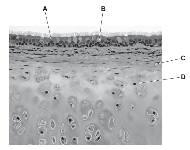
Options
| name | function | |
|---|---|---|
| A | ciliated epithelial cell | traps particles |
| B | goblet cell | secretes mucus |
| C | cartilage | prevents collapse of trachea |
| D | smooth muscle | contracts to expand airways |
Working
The photomicrograph is a T.S. of the trachea. The four labels indicate:
- A — ciliated epithelial cells (pseudostratified columnar epithelium with cilia at the luminal surface)
- B — a goblet cell (paler cell among the epithelium)
- C — connective tissue (submucosa) immediately beneath the epithelium
- D — hyaline cartilage (chondrocytes sitting in lacunae within the matrix)
Checking each option (name must match the labelled structure AND the function must be correct):
- A — ciliated epithelial cell → "traps particles". Name fits, but the function is wrong. Cilia BEAT to move mucus; it is the mucus (produced by goblet cells) that traps particles.
- B — goblet cell → "secretes mucus". Name fits label B and the function is correct. ✓
- C — cartilage → "prevents collapse of trachea". The function is correct, but the name does not match label C — label C points to the submucosa, not cartilage.
- D — smooth muscle → "contracts to expand airways". The name does not match label D (label D is cartilage) and the function is also wrong — smooth muscle contracts to CONSTRICT the airway, not expand it.
Answer
B
B
Background Concept
The trachea is the largest conducting airway of the human gas exchange system. Its wall is built up from several layers, each with a clearly identifiable structure and role:
- Pseudostratified ciliated columnar epithelium lines the lumen. Every cell reaches the basement membrane, but not all reach the surface, giving a "false" appearance of layering. Cilia project from the apical surface.
- Goblet cells are mucus-secreting cells scattered among the ciliated cells. They release sticky mucus that traps inhaled dust, microbes and other particles, forming part of the mucociliary escalator.
- Cilia beat in a coordinated, wave-like fashion that drives the layer of mucus (with its trapped particles) upward toward the pharynx, where it is swallowed.
- Submucosa is a layer of loose connective tissue just deep to the epithelium. It contains blood vessels, lymphatics and nerves that supply the mucosa.
- Hyaline cartilage forms incomplete C-shaped rings around the trachea. Because the trachea is a soft tube exposed to large pressure changes during breathing, the rigid cartilage keeps the airway patent (prevents collapse) when intrathoracic pressure falls on inhalation.
- Smooth (trachealis) muscle bridges the open ends of each C-ring posteriorly. When it contracts, it narrows the tracheal lumen (e.g. during the fight-or-flight response to reduce dead space); when it relaxes, the lumen widens.
In a micrograph, hyaline cartilage is unmistakable: it stains a pale, glassy blue/pink and contains scattered chondrocytes sitting in lacunae (the small dark spaces within the matrix).
Understanding the Question
A photomicrograph of a transverse section of the trachea is shown with four labels (A–D). The question asks us to pick the row in which the name correctly identifies a labelled structure AND the function stated for it is biologically accurate. Both columns must be right for the row to score.
Approach
For each option, perform a two-part check:
- Does the name in the left column correspond to the structure pointed to by the matching label in the micrograph?
- Is the function in the right column a correct description of that structure?
Only the row that passes BOTH tests is the answer.
Step-by-Step Reasoning
-
Row A — ciliated epithelial cell; traps particles.
Name matches label A (ciliated epithelium). However, cilia do not "trap" particles — they MOVE the mucus. Trapping is the job of the mucus itself (produced by goblet cells). The function is wrong → reject. -
Row B — goblet cell; secretes mucus.
Name matches label B (the pale, oval cell among the epithelium) AND the function is the textbook role of goblet cells. Both columns correct → this is the answer. -
Row C — cartilage; prevents collapse of trachea.
The function is true of tracheal cartilage, but label C in the micrograph points to the submucosa (the connective tissue layer immediately deep to the epithelium), not to the cartilage. The cartilage is at D. So the name-label pairing is wrong → reject. -
Row D — smooth muscle; contracts to expand airways.
Two errors. First, label D is cartilage, not smooth muscle, so the name-label pairing is wrong. Second, smooth muscle in the trachea contracts to constrict (narrow) the airway, not to expand it. Both columns wrong → reject.
Key Takeaways
- The tracheal wall is layered: ciliated epithelium + goblet cells → submucosa → cartilage (with smooth muscle bridging the C-ring).
- Mucus traps particles; cilia move the mucus — these are two distinct roles often confused.
- In a micrograph, cartilage is recognised by chondrocytes in lacunae within a homogeneous matrix; submucosa is loose connective tissue, much more cellular and fibrous in appearance.
- Smooth muscle in the airways is constrictor muscle, not dilator.
Common Mistakes
- Saying cilia "trap" particles. Cilia MOVE; mucus TRAPS.
- Confusing the cartilage (D) with the submucosa (C) when reading a micrograph. Cartilage has very few cells (chondrocytes in lacunae) in a glassy matrix; the submucosa looks like ordinary connective tissue with many small fibroblast-type nuclei and fibres.
- Thinking that smooth muscle expands the airway. It contracts the trachealis muscle and NARROWS the lumen.
- Picking a row because the function sounds right, without checking that the name matches the label.
Things to Be Careful About
- This is a double-match question: the name must fit the label AND the function must be correct. A row that gets only one of the two right still scores zero.
- The cartilage of the trachea is C-shaped, not a complete ring; the gap is bridged posteriorly by smooth muscle, which is why airway diameter can still be regulated.
- The mark scheme accepts one correct row only; do not try to argue for two. Once you have confirmed that B has both the correct name and the correct function, stop.
Infectious diseases may be caused by bacteria, protoctists and viruses. Different pathogens may be transmitted by airborne droplets, from faeces to mouth (faecal-oral), by sexual intercourse and by vectors.
Four examples of infectious diseases are listed.
1 cholera
2 HIV/AIDS
3 malaria
4 tuberculosis
Which table classifies the four diseases correctly to show the type of pathogen that causes the disease and the mode of transmission?
Options
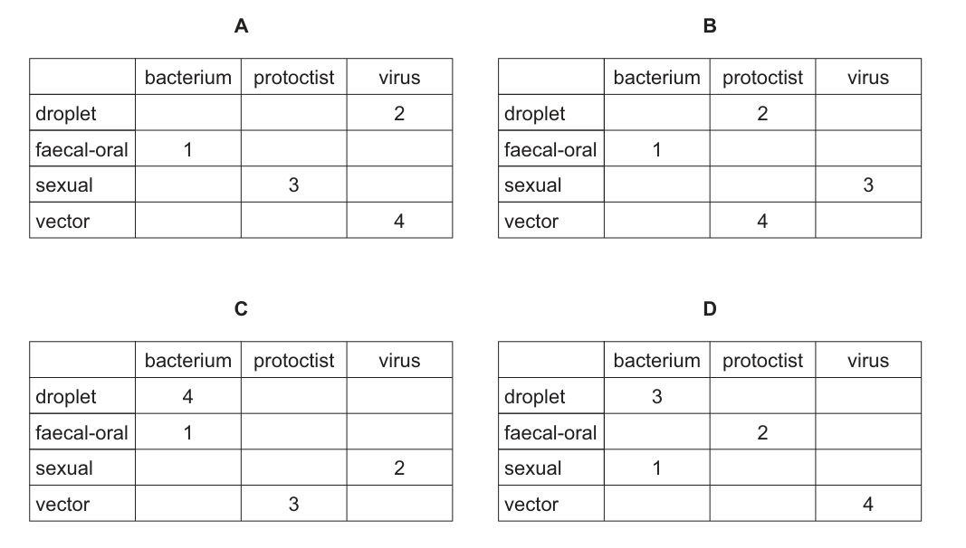
Working
Classify each disease by pathogen type and mode of transmission:
- 1 — Cholera: caused by Vibrio cholerae (a bacterium); transmitted by the faecal–oral route (contaminated water/food).
- 2 — HIV/AIDS: caused by HIV (a virus); transmitted by sexual intercourse (and other body-fluid routes).
- 3 — Malaria: caused by Plasmodium (a protoctist); transmitted by a vector (the female Anopheles mosquito).
- 4 — Tuberculosis: caused by Mycobacterium tuberculosis (a bacterium); transmitted by airborne droplets (coughing/sneezing).
The required table must therefore show:
- droplet → 4 (bacterium)
- faecal–oral → 1 (bacterium)
- sexual → 2 (virus)
- vector → 3 (protoctist)
Only Table C places all four diseases in the correct pathogen–transmission cells.
Answer
C
C
Background Concept
Infectious diseases are caused by pathogens — microorganisms (or, in the case of viruses, non-cellular particles) that can invade a host and cause harm. CIE classifies the pathogens of these four diseases into three categories:
- Bacteria — single-celled prokaryotes (e.g. Vibrio cholerae, Mycobacterium tuberculosis).
- Protoctists — single-celled eukaryotes with a true nucleus; the disease-causing ones are often referred to as protozoa (e.g. Plasmodium, the malaria parasite).
- Viruses — non-cellular particles consisting of a nucleic-acid core inside a protein capsid (e.g. HIV, the human immunodeficiency virus).
The mode of transmission is the route by which the pathogen passes from an infected individual (or reservoir) to a new host:
- Droplet (airborne) — pathogens carried in droplets produced by coughing, sneezing or talking, which are then inhaled.
- Faecal–oral — pathogens shed in faeces contaminate water, food or hands and enter a new host via the mouth.
- Sexual — pathogens passed in body fluids (semen, vaginal secretions, blood) during sexual intercourse.
- Vector — pathogens transferred by a living organism (typically an arthropod such as a mosquito) that bites an infected host and then transmits the pathogen when it feeds on a new one.
Understanding the Question
The question lists four diseases (1 cholera, 2 HIV/AIDS, 3 malaria, 4 tuberculosis) and four tables (A–D). Each table is a 4 × 3 grid crossing transmission mode (droplet, faecal–oral, sexual, vector) with pathogen type (bacterium, protoctist, virus), with each disease number placed in one cell. The task is to identify the table that places every disease in the correct cell.
Approach
First, decide the pathogen type and primary mode of transmission for each of the four diseases independently. Then check which table matches all four pairings at once. A single mismatch eliminates that table.
Step-by-Step Reasoning
1 — Cholera. Cholera is caused by Vibrio cholerae, a curved Gram-negative bacterium. It is spread when faecal matter from an infected person contaminates drinking water or food (poor sanitation), so the route is faecal–oral. Any table that puts 1 in droplet, sexual or vector is wrong on this row, and any table that labels it a protoctist or virus is also wrong.
2 — HIV/AIDS. AIDS is caused by the human immunodeficiency virus (HIV), a retrovirus. Its principal route of transmission listed here is sexual intercourse (via exchange of body fluids). Tables that put 2 in droplet, faecal–oral or vector, or that label it a bacterium or protoctist, are wrong on this row.
3 — Malaria. Malaria is caused by Plasmodium species (typically P. falciparum or P. vivax), which are protoctists. The parasite is transmitted by the bite of an infected female Anopheles mosquito — i.e. a vector. So 3 must sit in the vector row in the protoctist column.
4 — Tuberculosis (TB). TB is caused by Mycobacterium tuberculosis, a slow-growing bacterium. It is spread through the air when an infected person coughs or sneezes — the droplet (airborne) route. So 4 must sit in the droplet row in the bacterium column.
Checking each option against this classification:
- A places 2 (HIV/AIDS) in the droplet row and 3 (malaria) in the sexual row — both wrong.
- B places 2 (HIV/AIDS) in the droplet row and 4 (TB) in the vector row — both wrong.
- C places 4 (TB, bacterium) in droplet, 1 (cholera, bacterium) in faecal–oral, 2 (HIV/AIDS, virus) in sexual, and 3 (malaria, protoctist) in vector — every disease is in the correct cell.
- D places 3 (malaria) in droplet and 1 (cholera) in sexual — both wrong.
Only option C is fully correct.
Key Takeaways
- Cholera → bacterium (Vibrio cholerae) → faecal–oral.
- HIV/AIDS → virus (HIV) → sexual (and other body-fluid routes).
- Malaria → protoctist (Plasmodium) → vector (female Anopheles mosquito).
- Tuberculosis → bacterium (Mycobacterium tuberculosis) → droplet (airborne).
- When faced with a classification table, fixing the correct cell for one or two diseases you are most confident about (here, TB as droplet and malaria as vector) is the fastest way to eliminate the wrong options.
Common Mistakes
- Confusing cholera with malaria because both are associated with tropical regions — they have completely different pathogens and routes (bacterium / faecal–oral vs protoctist / vector).
- Assuming HIV can be transmitted by droplets like flu or TB — it cannot; it requires the exchange of body fluids (sexual contact, contaminated needles, mother-to-child).
- Putting TB into the vector category because of confusion with malaria, or assuming TB is viral because it affects the lungs.
- Calling Mycobacterium a "virus" or Plasmodium a "bacterium" — these are precisely the category errors the question is designed to expose.
Things to Be Careful About
- Read every cell of every table before choosing — a single misplacement rules an option out, but two tables can each contain three correct entries and differ in only one.
- The CIE syllabus classifies Plasmodium as a protoctist, not a protozoan (protozoa are a subgroup of protoctists). Use the syllabus term.
- The binomial names Vibrio cholerae, Mycobacterium tuberculosis and Plasmodium should be written in italics with a capital genus and lowercase species if quoted in an extended answer.
- Transmission by vector here is restricted to a living organism that carries the pathogen; the question is not referring to fomites or vehicles such as contaminated needles.
Scientists investigated the sensitivity and resistance of a bacterium called Clostridium difficile to four antibiotics. Colonies of bacteria were grown on agar plates for 24 hours, after which a different antibiotic was added to each plate. The number of bacterial colonies that died, stopped growing and continued growing were then counted. A bacterial colony is a group of bacterial cells that have multiplied from one bacterium.
Which antibiotic would be the best to treat a person infected with Clostridium difficile?
Options
| antibiotic | number of colonies that died | number of colonies that stopped growing | number of colonies that continued growing | |
|---|---|---|---|---|
| A | ampicillin | 2 | 5 | 13 |
| B | clindamycin | 4 | 8 | 6 |
| C | metronidazole | 13 | 6 | 1 |
| D | cefoxitin | 9 | 5 | 4 |
Working
An effective antibiotic should kill or stop the growth of as many bacterial colonies as possible, with the fewest colonies still growing. Compare the "continued growing" column (and combine "died" + "stopped growing") for each antibiotic:
- A (ampicillin): 13 still growing — ineffective
- B (clindamycin): 6 still growing — limited effect
- C (metronidazole): 1 still growing — most effective
- D (cefoxitin): 4 still growing — intermediate
Metronidazole killed 13 colonies and stopped growth in 6, leaving only 1 colony still growing, making it the most effective treatment.
Answer
C
C
Background Concept
Antibiotics are chemicals that kill bacteria (bactericidal) or prevent them from multiplying (bacteriostatic). Different antibiotics target different bacterial processes — for example, penicillins such as ampicillin inhibit cell wall synthesis, while metronidazole damages bacterial DNA.
A key idea in infectious disease treatment is antibiotic sensitivity testing: exposing a pathogen to different antibiotics to find the one that is most effective against it. This is particularly important when:
- the bacterium is a known cause of serious infection (e.g. Clostridium difficile, which causes antibiotic-associated colitis),
- antibiotic resistance is suspected, and
- empirical treatment needs to be guided toward the most reliable option.
A useful antibiotic is one that either kills the bacteria or stops them growing, leaving as few as possible still able to multiply.
Understanding the Question
The question gives a data table summarising the response of C. difficile colonies to four different antibiotics. Three outcomes are recorded for each:
- Died — the antibiotic killed the colony (bactericidal effect).
- Stopped growing — the colony was inhibited but not killed (bacteriostatic effect).
- Continued growing — the colony was unaffected, indicating resistance to that antibiotic.
The command is to choose the best antibiotic to treat an infected person. The "best" one is the antibiotic that leaves the fewest colonies still able to grow — i.e. the one that C. difficile is most sensitive to.
Approach
- Read off the "continued growing" column for each antibiotic — lower is better.
- For the leading candidate, also check that the combined killed + stopped-growing number is high, confirming the effect is real and not an artefact of small numbers.
- Select the option with the smallest number still growing.
Step-by-Step Reasoning
Reading the "continued growing" column:
- A (ampicillin): 13 colonies continued growing — most colonies survived, so ampicillin is the least effective.
- B (clindamycin): 6 colonies continued growing — moderate effectiveness.
- C (metronidazole): 1 colony continued growing — by far the most effective.
- D (cefoxitin): 4 colonies continued growing — intermediate.
Checking option C in detail: 13 colonies died and a further 6 stopped growing, leaving only 1 colony (out of 20) still able to grow. This represents 19 out of 20 colonies affected — the highest combined bactericidal + bacteriostatic effect of all four antibiotics. This matches the real-world clinical picture: metronidazole is the standard first-line treatment for mild-to-moderate C. difficile infection.
Key Takeaways
- The best antibiotic for an infection is the one to which the pathogen is most sensitive, indicated experimentally by the lowest number of colonies still able to grow.
- "Died" + "stopped growing" together give the total colonies affected, while "continued growing" reveals resistance.
- This kind of in-vitro sensitivity testing underpins the choice of antibiotic therapy in clinical practice.
Common Mistakes
- Picking the antibiotic that killed the most colonies without considering how many continued to grow — for example, ampicillin killed only 2 but its main problem is the 13 colonies still growing.
- Choosing clindamycin (B) on the basis of "stopped growing" alone (8) and ignoring that 6 colonies continued to grow, including resistant survivors that can reinfect the patient.
- Confusing the meaning of "stopped growing" with "cured": a bacteriostatic effect only works while the drug is present, so survivors may resume growth.
Things to Be Careful About
- "Sensitivity" and "resistance" are technical terms: a sensitive bacterium is inhibited/killed by the antibiotic at therapeutic doses; a resistant one continues to grow.
- Always look at the resistance column (continued growing) first, because a single surviving colony can repopulate and cause relapse.
- A low total in "died" combined with a high total in "continued growing" (as with ampicillin) signals widespread resistance, not just weak killing.
A vaccine is used to create artificial active immunity. After a person has been given a vaccine, it takes a period of time before they develop long-term immunity to the disease.
Which statement about this period of time explains the delay in developing long-term immunity?
Options
A No memory cells have been produced from B-lymphocytes.
B No plasma cells have been produced from B-lymphocytes.
C The primary immune response has not produced enough antibodies.
D The secondary immune response has not produced enough antibodies.
Working
Long-term immunity depends on memory cells (B-memory and T-memory cells), which persist after the primary response and enable a rapid, large secondary response on re-exposure to the antigen.
- A – Correct. Long-term immunity arises only once memory cells have been produced from activated B-lymphocytes; until this happens, protection cannot be long-lasting.
- B – Wrong. Plasma cells secrete antibodies, but antibodies decline after the primary response and do not, by themselves, provide long-term immunity.
- C – Wrong. The quantity of antibody in the primary response affects short-term protection, not long-term (memory-based) immunity.
- D – Wrong. The secondary response occurs only after a subsequent exposure to the antigen, not during the delay following first vaccination.
Answer
A
A
Background Concept
Vaccination triggers artificial active immunity by introducing a harmless form of an antigen (e.g. attenuated pathogen, inactivated toxin, or surface protein) so that the recipient's own immune system mounts a response. The first exposure of the body to a particular antigen produces a primary immune response:
- Antigen-presenting cells (e.g. macrophages) take up and present the antigen.
- Helper T-cells activate B-lymphocytes.
- Activated B-lymphocytes proliferate and differentiate into two key cell types:
- Plasma cells – short-lived cells that secrete large quantities of antibody specific to the antigen.
- Memory cells (B-memory and T-memory cells) – long-lived cells that persist for months, years, or decades after the antigen has been cleared.
Memory cells are the cellular basis of long-term immunity. On re-exposure to the same antigen, they are rapidly reactivated, producing a secondary immune response that is faster, stronger, and dominated by IgG — usually before symptoms appear.
Understanding the Question
The question asks why there is a delay between receiving a vaccine and having long-term immunity. The key phrase is "long-term immunity", which immediately points to memory cells rather than antibodies. The answer must explain the cellular event that has not yet happened during the lag period after vaccination.
Approach
- Identify what provides long-term immunity → memory cells (not antibodies).
- Look for the option that describes the absence of memory cells, which is precisely why long-term protection has not yet been established.
- Eliminate the options that refer to plasma cells, primary antibodies, or the secondary response — none of these define long-term immunity.
Step-by-Step Reasoning
-
Option A – "No memory cells have been produced from B-lymphocytes." ✅ This correctly identifies that long-term immunity requires memory cells, and these have not yet been generated during the early phase after vaccination. As soon as memory cells have been produced and clonally expanded, the individual has long-term immunity.
-
Option B – "No plasma cells have been produced from B-lymphocytes." ❌ Plasma cells secrete antibodies, which give short-term protection during the primary response. They are not the basis of long-term immunity, so their absence does not explain the delay in long-term immunity.
-
Option C – "The primary immune response has not produced enough antibodies." ❌ Antibody titre in the primary response determines immediate, short-term neutralisation of the antigen. It does not generate long-term immunity — even a strong primary antibody response eventually wanes.
-
Option D – "The secondary immune response has not produced enough antibodies." ❌ The secondary response only occurs after a second exposure to the antigen. After a single vaccination, no secondary response has yet taken place, so this option describes the wrong time-point in the immune timeline.
Key Takeaways
- Long-term immunity = memory cells, not antibodies.
- The primary response produces both plasma cells (short-term antibody defence) and memory cells (long-term protection).
- The delay after vaccination corresponds to the time needed for clonal selection, proliferation, and differentiation of B-lymphocytes into memory cells.
- A subsequent infection triggers a rapid secondary response from these pre-existing memory cells.
Common Mistakes
- Confusing antibody titre with immunity — high antibody levels after a first exposure do not guarantee lasting protection; antibodies wane, but memory cells persist.
- Choosing an option that mentions plasma cells or primary antibodies — these explain short-term, immediate defence rather than long-term immunity.
- Choosing an option that mentions the secondary response — the secondary response is the consequence of memory cells existing, not the cause of long-term immunity being established.
Things to Be Careful About
- Read the question carefully: it asks about the reason for the delay in developing long-term immunity, not about how antibodies clear the initial infection.
- Remember that both B-lymphocytes and T-lymphocytes produce memory cells, but the question specifically asks about the production step that has not yet happened — formation of memory cells from B-lymphocytes.
An antiserum to a snake toxin can be obtained by injecting the toxin into a horse. The antiserum is made from blood plasma taken from the horse a few weeks later. The antiserum is injected into a person who has been bitten by the same species of snake.
Which type of immunity occurs as a result of using this antiserum?
Options
A artificial active
B artificial passive
C natural active
D natural passive
Working
The person receives ready-made antibodies (in the antiserum) rather than producing their own, so the immunity is passive. Because the antibodies are introduced by deliberate medical intervention (injection) rather than acquired through everyday exposure, the immunity is artificial.
Answer
B
B
Background Concept
Immunity can be classified along two independent axes:
- Active vs passive — active immunity arises when the recipient's own immune system is stimulated to mount a response (producing antibodies and memory cells); passive immunity arises when the recipient is given ready-made antibodies produced by another organism.
- Natural vs artificial — natural immunity is acquired through everyday exposure (e.g. infection, or maternal antibodies crossing the placenta or in breast milk); artificial immunity is acquired through deliberate medical intervention (e.g. vaccination, injection of an antiserum).
Combining the two axes gives four categories: natural active, natural passive, artificial active, and artificial passive.
Understanding the Question
The stem describes the production and use of an antiserum. A horse is injected with snake toxin, mounts its own immune response and produces antibodies against it. Plasma containing these antibodies (the antiserum) is harvested weeks later and injected into a snake-bite victim. We are asked which of the four immunity categories this represents in the human recipient.
Approach
Ask two questions about the human recipient:
- Did the person's own immune system produce the antibodies, or were they supplied ready-made? → ready-made = passive.
- How did the antibodies enter the person — through normal exposure or by deliberate medical intervention? → by injection = artificial.
Step-by-Step Reasoning
- The horse, not the person, produced the antibodies in response to the toxin. The person is therefore not actively making antibodies — this rules out A and C (both active).
- The antibodies reach the person by injection of a prepared antiserum, a deliberate medical procedure, not by ordinary exposure — this rules out natural categories (C and D).
- The combination of ready-made antibodies given by injection is the textbook definition of artificial passive immunity (B). The protection is immediate (because antibodies are already present) but short-lived, because the recipient's immune system has not been stimulated to produce memory cells.
Key Takeaways
- Antiserum therapy (e.g. against snake venom, tetanus, rabies) is the classic example of artificial passive immunity.
- A useful mnemonic: passive = borrowed immunity; active = self-made immunity.
- The "natural/artificial" axis refers to how the immunity is acquired, not what is in the injection.
Common Mistakes
- Choosing A (artificial active) because an injection is involved — forgetting that "active" requires the recipient to make their own antibodies.
- Choosing C (natural active) on the grounds that the person was "exposed" to the toxin via the bite — the bite did not give the person the antiserum, and the person did not make their own antibodies.
- Choosing D (natural passive) because the antibodies are not made by the recipient — forgetting that the route of delivery (injection) makes it artificial, not natural.
Things to Be Careful About
- Classify from the perspective of the recipient (the bitten person), not the donor horse.
- "Passive" describes the source of the antibodies; "artificial" describes the route of administration. The two judgements are independent.
Which statement describes the feature of monoclonal antibodies that allows them to accurately treat a specific disease?
Options
A They bind to human hormones to produce a colour change.
B A fluorescent dye can be attached to them.
C They have active sites that are complementary to antigens.
D They can carry toxic chemicals.
Working
Monoclonal antibodies are produced from a single clone of hybridoma cells and so are identical and specific to one antigen epitope. This specificity is exploited in treatment by linking (conjugating) them to toxic chemicals — for example, cytotoxic drugs, radioactive isotopes, or other therapeutic agents. The antibody then delivers the toxic payload directly to the diseased cells (e.g. cancer cells) that display the target antigen, sparing healthy cells.
- A — describes a detection/colour-change use, not treatment.
- B — fluorescent labelling is a diagnostic/research tool, not a treatment feature.
- C — antibodies have variable-region binding sites (not 'active sites', which is enzyme terminology) that are complementary to antigens; this is the basis of their specificity but the wording is biologically incorrect.
- D — describes the therapeutic delivery of toxic chemicals specifically to target cells; this is the feature that enables accurate treatment.
Answer
D
D
Background Concept
Monoclonal antibodies (mAbs) are antibodies produced by a single clone of B-lymphocyte–myeloma hybrid cells (hybridomas). Because they originate from one parent cell, every antibody molecule is identical and binds to the same epitope on the same antigen. This single-epitope specificity is the foundation of all their applications — in diagnosis, in research, and in therapy.
In therapy, the specificity of a monoclonal antibody is exploited by using it as a targeting device. A toxic payload — a cytotoxic drug, a radioactive isotope, or a potent cell-killing molecule — is chemically attached (conjugated) to the antibody. The antibody then circulates in the body and binds only to cells carrying the matching antigen (commonly a protein found in large numbers on the surface of cancer cells). Once bound, the toxic component is delivered precisely to the diseased cell, maximising damage to the target while minimising harm to healthy tissue. This approach is the basis of antibody–drug conjugates such as trastuzumab emtansine and of radioimmunotherapy.
Understanding the Question
The question asks for the feature of monoclonal antibodies that allows them to accurately treat a specific disease. 'Accurately treat' is the key phrase — it points to the therapeutic use, in which the antibody's specificity is converted into a precision drug-delivery system. The question is testing whether the candidate can identify the correct therapeutic feature and avoid confusions with other mAb uses (diagnosis, labelling) or incorrect terminology (e.g. confusing antibodies with enzymes).
Approach
Eliminate the options systematically:
- Decide whether each option describes a use of monoclonal antibodies.
- Where the option does refer to a real feature of mAbs, check whether the feature relates to treatment (not diagnosis) and whether the biological terminology is correct.
- Select the option that uniquely fits the description of an accurate treatment feature.
Step-by-Step Reasoning
- Option A — Antibodies can indeed be linked to enzymes to produce colour changes (e.g. ELISA, home pregnancy tests), but this is a diagnostic application, not a treatment. Incorrect for the question.
- Option B — Fluorescent dyes attached to antibodies are used in research and imaging to locate specific molecules or cells. This is a detection/visualisation tool, not a treatment mechanism. Incorrect.
- Option C — The biological wording is wrong. Antibodies do not have 'active sites' (this is enzyme terminology arising from the lock-and-key/induced-fit model of enzyme–substrate interaction). Antibodies have variable regions in their Fab portions, which form antigen-binding sites complementary in shape to a specific epitope. Even if the idea (complementarity to antigens) is true, the precise terminology required by Cambridge is 'binding site', not 'active site'. As phrased, this option is rejected on a terminology mark.
- Option D — Correct. The defining therapeutic feature of monoclonal antibodies is that they can be conjugated to toxic chemicals (drugs, radioisotopes, toxins) and deliver these selectively to cells bearing the target antigen. This is what allows accurate treatment of a specific disease (typically cancers such as HER2-positive breast cancer or B-cell lymphomas).
Key Takeaways
- Monoclonal antibodies are identical antibodies specific to a single epitope.
- Their therapeutic advantage is their specificity, which is exploited by conjugating them to toxic drugs, radioisotopes, or toxins so the poison is delivered only to target cells.
- Antibodies have binding sites (variable regions), not 'active sites' — that term belongs to enzymes.
Common Mistakes
- Choosing C because it 'sounds right'. The concept of antibody–antigen complementarity is correct, but the term 'active site' makes the statement biologically inaccurate; Cambridge markers reject this wording.
- Confusing diagnostic uses of mAbs (colour change, fluorescent labelling) with therapeutic uses — diagnosis finds the disease; treatment cures it.
- Forgetting that the accuracy of mAb therapy comes from targeted delivery of a toxic agent, not from the antibody itself killing the cell directly (most therapeutic mAbs act by recruiting immune effectors or blocking signalling, but the unique feature allowing accurate targeting is the ability to carry toxins/drugs specifically to the diseased cell).
Things to Be Careful About
- Distinguish active site (enzyme) from binding site (antibody).
- Distinguish diagnosis (locating/identifying disease) from treatment (curing/controlling disease).
- The question specifies 'feature that allows them to accurately treat a specific disease' — the word 'specifically' points to the targeting capability conferred by attaching toxic chemicals.
Your score so far
Answer a question to start scoring
Your marks add up here as you work through the paper.