Biology 9700/12 — October/November 2025
Cambridge AS Level · Multiple Choice (AS Level) · answer key with instant marking and worked solutions
Topics Biological Molecules · Cell Structure · Enzymes · Nucleic Acids and Protein Synthesis · Cell Membranes and Transport · Transport in Plants · +5 more
Tap an option under each question to check it — your score builds as you go.
A student observed some cells using a microscope with an eyepiece graticule.
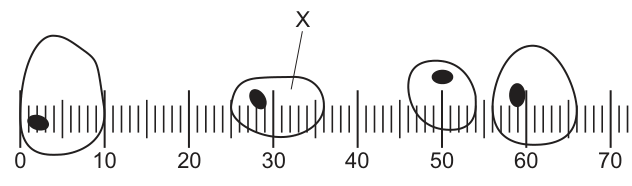
They then replaced the slide containing the cells with a stage micrometer with a millimetre scale, and lined up the eyepiece graticule as shown.
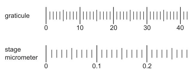
What is the maximum diameter of the cell labelled X?
Options
A
B
C
D
Working
Step 1 — Calibrate the eyepiece graticule using Fig. 1.2:
Step 2 — Read off the cell in Fig. 1.1:
Cell X has a maximum diameter of about eyepiece graticule units.
Step 3 — Calculate the actual diameter:
Answer
C
C
Background Concept
A light microscope has two scales for measuring size:
- The eyepiece graticule is a small glass disc with a scale etched on it that sits inside the eyepiece lens. You see it superimposed on the specimen. Because it is part of the eyepiece, the size of one graticule division is not fixed — it changes whenever you change objective lens, because the total magnification changes.
- The stage micrometer is a slide with an accurately known scale (usually divisions, totalling or ). It is a real, certified scale and does not change with magnification.
To find the real size of a specimen you must therefore calibrate the graticule against the stage micrometer at the magnification you are using. Only then can you read graticule units off a real specimen and convert them into a length.
Useful unit relationships:
Understanding the Question
The question gives two figures:
- Fig. 1.1 — the cells the student is looking at, with cell X drawn across the graticule scale. The scale runs from to graticule units.
- Fig. 1.2 — the calibration: the graticule has been aligned with the stage micrometer so that graticule units line up with on the stage micrometer.
The command word is "What is the maximum diameter…?" — this is a calculation, not a recall question. We must (1) work out how many µm one graticule division represents, then (2) multiply by the number of graticule divisions the cell spans at its widest point.
The distractors in the options are there to catch three classic errors: forgetting to convert mm → µm (giving ), reading the cell as roughly units instead of units (giving ), and missing the factor of in the unit conversion (giving ).
Approach
Two-step method:
- Calibration step — use Fig. 1.2 to convert graticule units into µm. The conversion factor comes from dividing the known length on the stage micrometer by the number of graticule units it occupies.
- Measurement step — read the cell's maximum width in graticule units from Fig. 1.1, then multiply by the conversion factor from step 1.
Always do the unit conversion explicitly so that you do not mix mm and µm.
Step-by-Step Reasoning
Step 1 — Calibration (Fig. 1.2).
The graticule's mark lines up with the stage micrometer's mark. The graticule's mark lines up with the stage micrometer's mark. So
Convert mm to µm (multiply by ):
So one graticule division represents
Step 2 — Measure cell X (Fig. 1.1).
Cell X's longest axis runs from roughly graticule mark to about mark –, i.e. a maximum width of about graticule units.
Step 3 — Convert to µm.
This is the option marked C.
Key Takeaways
- An eyepiece graticule must be calibrated at the magnification in use; one division's value changes with the objective.
- The calibration is done by aligning the graticule against a stage micrometer and reading the µm-per-division conversion factor directly.
- Always state units and convert mm → µm () when reading the stage micrometer.
- A red blood cell is about – across; many plant mesophyll / palisade cells are –. A value of is biologically sensible for a cell seen with a objective — a useful sanity check.
Common Mistakes
- Forgetting the unit conversion. Reading and leaving the answer in mm, or multiplying the wrong number of zeroes. This usually produces a wildly wrong answer (e.g. or ).
- Reading the cell as ~13 units instead of ~14.6 — gives (option B). Take the widest part of the cell, not a narrower axis.
- Counting small sub-divisions on the graticule as full units. Each large numbered mark is one unit; the small ticks are or of a unit depending on the graticule.
- Mixing up the two scales in Fig. 1.2: the top scale is the graticule (variable), the bottom scale is the stage micrometer (fixed in mm).
Things to Be Careful About
- The calibration ratio only holds at the magnification used in Fig. 1.2. If the objective is changed, the calibration must be redone.
- Quote the answer with the correct unit. The option that has the right number but the wrong unit (e.g. instead of ) is a deliberately placed distractor.
- "Maximum diameter" means the longest straight-line distance across the cell, not an average or the short axis.
Which structures will be present in a cell that causes cholera?
1 circular DNA
2 cytoplasmic DNA
3 70S ribosomes
Options
A 1, 2 and 3
B 1 and 2 only
C 1 and 3 only
D 2 and 3 only
Working
Cholera is caused by Vibrio cholerae, a prokaryotic bacterium. Therefore, the cell has the features of a prokaryote:
- 1. Circular DNA ✓ — present; the bacterial chromosome is a single circular DNA molecule located in the nucleoid.
- 2. Cytoplasmic DNA ✓ — present; the circular chromosome lies free in the cytoplasm because there is no nuclear envelope.
- 3. 70S ribosomes ✓ — present; prokaryotes have 70S ribosomes (smaller than the 80S ribosomes of eukaryotes).
All three features are characteristic of a prokaryotic cell.
Answer
A
A
Background Concept
Cholera is caused by the bacterium Vibrio cholerae. Bacteria are prokaryotes — cells that lack a membrane-bound nucleus and other membrane-bound organelles. Several structural features distinguish prokaryotes from eukaryotes:
- The genetic material is a single circular DNA molecule that lies free in the cytoplasm in a region called the nucleoid (not enclosed by a nuclear envelope).
- The only ribosomes are 70S (composed of a 50S large subunit and a 30S small subunit), which are smaller than the 80S ribosomes found in the cytoplasm of eukaryotic cells.
- There are no mitochondria, chloroplasts, endoplasmic reticulum, or other membrane-bound organelles.
- Some prokaryotes also carry small accessory circles of DNA called plasmids, but these are not part of the standard feature list required here.
Because the chromosomal DNA is in the cytoplasm, the term cytoplasmic DNA can be used to describe it — this is not restricted to mitochondrial or chloroplast DNA in eukaryotes; any DNA that is not enclosed in a nucleus qualifies.
Understanding the Question
The question presents three candidate features and asks which are present in the cholera-causing cell. Each feature must be evaluated against the structure of Vibrio cholerae.
Approach
- Identify the type of cell (prokaryote — bacterium).
- Check each numbered feature against the prokaryotic checklist.
- Select the option that includes every correct feature and excludes every incorrect one.
Step-by-Step Reasoning
- Vibrio cholerae is a bacterium → prokaryote.
- Feature 1 — circular DNA: Prokaryotes have one circular DNA molecule as their chromosome. ✓ Present.
- Feature 2 — cytoplasmic DNA: The circular chromosome is located in the cytoplasm (no nuclear envelope separates it from the rest of the cell). So yes, the cell contains DNA in its cytoplasm. ✓ Present.
- Feature 3 — 70S ribosomes: Prokaryotes synthesise proteins on 70S ribosomes (not the 80S ribosomes of eukaryotic cytoplasm). ✓ Present.
- All three features apply → the correct option lists 1, 2 and 3.
Key Takeaways
- Vibrio cholerae is a prokaryote; recall the defining prokaryotic features when a bacterial pathogen is named.
- "Circular DNA" and "cytoplasmic DNA" are not mutually exclusive in a prokaryote — the single chromosome is both circular and in the cytoplasm.
- 70S vs 80S ribosomes is a quick, reliable prokaryote/eukaryote marker.
Common Mistakes
- Selecting B or C because students wrongly assume "cytoplasmic DNA" only refers to mitochondrial/chloroplast DNA in eukaryotes. In a prokaryote, the chromosome itself is in the cytoplasm.
- Selecting D by misremembering that bacteria have 80S ribosomes — only the eukaryotic cytoplasm has 80S ribosomes; mitochondria and chloroplasts also have 70S.
- Selecting B by thinking prokaryotes lack circular DNA — this is the reverse of the truth.
Things to Be Careful About
- The phrasing "cytoplasmic DNA" is a generic description of any DNA located in the cytoplasm, not a specific term for organelle DNA.
- S values for ribosomes are not additive in the way some students expect; a 70S ribosome is a complete, functional prokaryotic ribosome, not half of an 80S one.
Which organelles have a partially permeable membrane?
1 Golgi apparatus
2 lysosome
3 mitochondrion
4 ribosome
Options
A 1, 2 and 3
B 1, 2 and 4
C 1, 3 and 4
D 2, 3 and 4
Working
- The Golgi apparatus is surrounded by a single membrane that is partially permeable.
- A lysosome is bounded by a single membrane that is partially permeable.
- A mitochondrion has a double membrane (outer and inner), and these membranes are partially permeable.
- A ribosome is not surrounded by a membrane at all — it is a complex of rRNA and protein, so it cannot be described as having a partially permeable membrane.
Therefore the organelles with a partially permeable membrane are 1, 2 and 3.
Answer
A
A
Background Concept
Eukaryotic cells contain many organelles, but only some of them are membrane-bound. A membrane-bound organelle is surrounded by one or more phospholipid bilayers, which exhibit the property of partial permeability — they allow some molecules to pass through freely (or via channels/carriers) while restricting others.
The key organelle structures in this question are:
- Golgi apparatus: a stack of flattened, single-membrane-bound cisternae involved in modifying, sorting and packaging proteins and lipids.
- Lysosome: a single-membrane-bound vesicle containing hydrolytic (digestive) enzymes, used to break down waste materials and engulfed pathogens.
- Mitochondrion: a double-membrane-bound organelle (outer membrane and folded inner membrane forming cristae), the site of aerobic respiration and ATP production.
- Ribosome: a small spherical structure made of ribosomal RNA (rRNA) and protein. It has no membrane at all — it is a ribonucleoprotein complex, not a membrane-bound organelle.
Understanding the Question
The question is asking the candidate to identify which of the four listed structures are enclosed by a membrane that is partially permeable. This is a two-step test: first, does the organelle have a membrane, and second (for those that do), is that membrane partially permeable?
Approach
To answer, check each organelle in turn:
- Does it have a membrane (yes/no)?
- If yes, are all biological membranes partially permeable? (Yes — partial permeability is a defining property of any phospholipid bilayer.)
So the practical test reduces to: which of the four are membrane-bound organelles?
Step-by-Step Reasoning
- 1 — Golgi apparatus: bounded by a single phospholipid bilayer (membrane). ✓ Partially permeable.
- 2 — Lysosome: bounded by a single phospholipid bilayer (membrane). ✓ Partially permeable.
- 3 — Mitochondrion: bounded by a double phospholipid bilayer (outer + inner membrane). ✓ Partially permeable.
- 4 — Ribosome: no membrane. ✗ Cannot be described as having a partially permeable membrane.
The only organelle that fails the test is the ribosome, so 1, 2 and 3 are correct. This matches option A.
Key Takeaways
- Ribosomes are not membrane-bound organelles — they are naked ribonucleoprotein particles found free in the cytoplasm or bound to rough endoplasmic reticulum.
- All membrane-bound organelles (single- or double-membraned) have membranes that are partially permeable.
- "Partially permeable" applies to every biological membrane; it is the term used to describe the selective barrier that allows some substances through but not others.
Common Mistakes
- Choosing C (1, 3 and 4) or D (2, 3 and 4) by mistakenly including the ribosome as a membrane-bound organelle.
- Confusing the ribosome with the rough endoplasmic reticulum, which is membrane-bound (the RER has ribosomes studded on its cytoplasmic surface, but it is the RER that has the membrane, not the ribosome itself).
Things to Be Careful About
- A ribosome consists of a large and a small subunit made of rRNA and protein — it has no phospholipid bilayer around it, so it cannot be described as "partially permeable".
- Both single-membraned (Golgi, lysosome) and double-membraned (mitochondrion) organelles count as having a partially permeable membrane.
Mouse cells were grown in a dish containing a growth medium. Radioactively labelled amino acids were added to the growth medium. The table shows the time taken for the radioactively labelled amino acids to appear in three different organelles, R, S and T.
| organelle | time taken for radioactivity to appear/min |
|---|---|
| R | 2 |
| S | 9 |
| T | 35 |
Which row correctly identifies the organelles?
Options
| R | S | T | |
|---|---|---|---|
| A | Golgi body | lysosome | rough endoplasmic reticulum |
| B | Golgi body | rough endoplasmic reticulum | lysosome |
| C | rough endoplasmic reticulum | Golgi body | lysosome |
| D | rough endoplasmic reticulum | lysosome | Golgi body |
Working
Amino acids are assembled into proteins on the ribosomes of the rough endoplasmic reticulum (RER), so radioactivity appears there first (R, 2 min).
Proteins are then transported in vesicles to the Golgi body for modification, so radioactivity appears there second (S, 9 min).
Lysosomes are formed when vesicles containing hydrolytic enzymes bud off from the trans face of the Golgi, so radioactivity appears there last (T, 35 min).
Order: R = RER, S = Golgi body, T = lysosome.
Answer
C
C
Background Concept
Eukaryotic cells contain a system of membrane-bound organelles that work together to synthesise, modify and dispatch proteins. The main steps and organelles in this secretory pathway are:
- Rough endoplasmic reticulum (RER) – studded with ribosomes. Proteins destined for secretion, insertion into membranes, or delivery to lysosomes are synthesised here. As the nascent polypeptide emerges into the lumen of the RER, it is folded and may be glycosylated.
- Vesicles – small membrane-bound sacs bud off the RER and carry the protein to the next organelle.
- Golgi body (Golgi apparatus) – a stack of flattened cisternae. Here proteins are further modified (e.g. carbohydrate chains trimmed or extended, sulphation) and sorted according to their final destination.
- Secretory vesicles / lysosomes – vesicles bud from the trans face of the Golgi. Some fuse with the plasma membrane to release their contents (exocytosis); others become lysosomes, which contain hydrolytic enzymes that digest worn-out organelles, engulfed bacteria and debris.
A useful summary: RER → vesicles → Golgi → vesicles → lysosome (or plasma membrane).
Understanding the Question
This is a pulse–chase style experiment. Mouse cells in culture are given growth medium containing amino acids whose atoms have been replaced with radioactive isotopes (e.g. or ). The cells incorporate these labelled amino acids into newly made proteins, and at successive times the cells can be fixed, sectioned and the location of the radioactivity pinpointed to a particular organelle by autoradiography.
The table tells us:
- R is labelled at 2 min (very early)
- S is labelled at 9 min (intermediate)
- T is labelled at 35 min (much later)
We must match R, S, T in time order to the three organelles listed in the options.
Approach
The key principle is that a protein travelling through the secretory pathway passes through the organelles in a fixed order, so the organelle reached first will be the one closest to the site of protein synthesis (the RER), and the one reached last will be the furthest downstream (the lysosome in this list). All we need to do is write out the order:
and then check which option assigns the organelles to R, S, T in exactly that order.
Step-by-Step Reasoning
-
First (R, 2 min) – rough endoplasmic reticulum. Ribosomes on the RER translate mRNA and the new polypeptide is threaded directly into the RER lumen / membrane. Because the amino acids are added to the medium and reach the cytosol almost immediately, the first organelle in which radioactivity becomes detectable is the one doing the synthesising – the RER.
-
Second (S, 9 min) – Golgi body. After folding and initial processing in the RER, proteins are packaged into transport vesicles that bud from the RER and fuse with the cis face of the Golgi. Inside the Golgi the proteins are further modified, and this delay of a few minutes is exactly what gives the 9 min figure for S.
-
Third (T, 35 min) – lysosome. Lysosomal enzymes are themselves made on the RER, processed through the Golgi, and then tagged with mannose-6-phosphate in the cis-Golgi. This tag directs them into vesicles that bud from the trans-Golgi and mature into lysosomes. The longer lag (35 min) reflects the time needed to synthesise, traffic and concentrate enough labelled enzyme in newly forming lysosomes before the signal is detectable.
-
Match to the options.
- A: Golgi, lysosome, RER – wrong on all three
- B: Golgi, RER, lysosome – wrong on R and S
- C: RER, Golgi, lysosome – matches our order
- D: RER, lysosome, Golgi – wrong on S and T
Therefore the correct row is C.
Key Takeaways
- The secretory pathway has a fixed order: RER → Golgi → (lysosome / secretion / membrane).
- In a pulse–chase experiment, the order in which radioactivity appears in different organelles reveals their position in the pathway.
- Lysosomal enzymes take the longest to appear because they must be made, processed, tagged with mannose-6-phosphate, sorted and concentrated in new lysosomes before becoming detectable.
- The same logic lets you predict the order for any combination of RER, Golgi, lysosome, secretory vesicle and plasma membrane.
Common Mistakes
- Putting the Golgi body first because it is "associated with packaging". Packaging is the final step, not the first; synthesis happens on the RER.
- Putting the lysosome second, assuming it is "near" the Golgi. Although lysosomes are derived from the Golgi, the enzymes have still had to be synthesised on the RER first, so the RER must be labelled before the Golgi and before the lysosome.
- Confusing smooth and rough ER. Only the rough ER has ribosomes and is the site of synthesis of secretory / lysosomal proteins. The smooth ER handles lipid synthesis and detoxification.
- Treating the times as absolute rather than relative. The 2, 9 and 35 min values matter only as an order; what counts is the sequence R, then S, then T.
Things to Be Careful About
- The question specifies mouse cells in culture – a eukaryotic system, so the secretory pathway described above applies (it would not apply in prokaryotes, which have no RER, Golgi or lysosomes).
- "Radioactively labelled amino acids" tells you the cells are making proteins; this immediately points to ribosome-bearing organelles, ruling out mitochondria or the nucleus as the first stop.
- Read the columns carefully. R, S and T are organelles, and the rows give a complete triple; the answer must assign all three correctly, not just one or two.
- Lysosomes are formed from the Golgi, not from the RER – this is why lysosomes are reached after the Golgi, not before.
- A common distractor pattern in this type of question swaps the Golgi and RER. Always anchor the answer to the principle: synthesis first, processing second, packaging/destination last.
The electron micrograph shows a section through a cell.
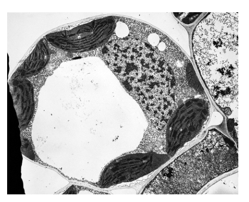
What would be present in this type of cell?
Options
| mitochondria | peptidoglycan cell wall | histone proteins | |
|---|---|---|---|
| A | ✓ | ✓ | ✗ |
| B | ✓ | ✗ | ✓ |
| C | ✗ | ✓ | ✗ |
| D | ✗ | ✗ | ✓ |
key
✓ = present
✗ = not present
Working
The micrograph shows chloroplasts (with grana), a large central vacuole, mitochondria, and a cell wall — this is a eukaryotic plant cell.
- Mitochondria: present (eukaryotic organelle, visible in the micrograph).
- Peptidoglycan cell wall: absent (peptidoglycan is found in bacterial cell walls, not in plant or animal cells).
- Histone proteins: present (eukaryotic DNA is wound around histones to form nucleosomes).
Only row B matches: mitochondria ✓, peptidoglycan cell wall ✗, histone proteins ✓.
Answer
B
B
Background Concept
The three structures tested belong to different cellular locations and cell types, and the key is to know which goes where:
- Mitochondria are membrane-bound organelles that carry out aerobic respiration. They occur in virtually all eukaryotic cells (animals, plants, fungi, and most protists), but not in prokaryotes such as bacteria.
- Peptidoglycan (also called murein) is the structural polysaccharide–peptide polymer that forms bacterial cell walls. It is the target of antibiotics such as penicillin. Plant cell walls are made of cellulose, fungal walls of chitin, and animal cells have no cell wall at all — so peptidoglycan is exclusive to bacteria.
- Histone proteins are small, positively charged proteins that eukaryotic DNA wraps around (about 1.65 turns around a histone octamer) to form nucleosomes, which then fold further into chromatin. Bacteria have DNA-associated proteins sometimes loosely called "histone-like," but they are not true histones in the CIE sense, so for exam purposes, histone proteins are a marker of eukaryotic cells.
Understanding the Question
The micrograph shows a section through a cell with unmistakable features: dark, oval chloroplasts with visible internal grana (stacks of thylakoids), a large clear central vacuole occupying most of the cell, a rigid outer cell wall, and smaller mitochondria in the cytoplasm. This combination — chloroplasts + large central vacuole + cellulose cell wall — identifies the cell as a eukaryotic plant cell.
The question asks which of the three listed features would be present in this type of cell, and the answer must be selected from a table of four possible combinations.
Approach
- Confirm the cell type from the micrograph.
- For each of the three features, decide present/absent.
- Match to the option that has exactly that pattern.
Step-by-Step Reasoning
- Mitochondria → present. The micrograph already shows them, and every eukaryotic cell (including plant cells) contains mitochondria for ATP production by aerobic respiration.
- Peptidoglycan cell wall → absent. Peptidoglycan is specific to the bacterial cell wall. Plant cell walls are made of cellulose, so this feature is rejected regardless of cell type.
- Histone proteins → present. Eukaryotic DNA is packaged with histones into nucleosomes. Because the cell is eukaryotic, histones are present.
The pattern present ✓, peptidoglycan ✗, histone ✓ corresponds to row B.
Key Takeaways
- Peptidoglycan is a prokaryote-only marker — seeing it in an answer usually signals that the distractor is describing a bacterium.
- Histones, the nuclear envelope, membrane-bound organelles (mitochondria, ER, Golgi, chloroplasts in plants) are all eukaryote-only features.
- Identifying a cell from an electron micrograph depends on spotting the diagnostic combination: a plant cell = chloroplasts + large central vacuole + cellulose cell wall; an animal cell = centrioles, no wall, no chloroplasts; a bacterium = no nucleus, no membrane-bound organelles, a peptidoglycan wall.
Common Mistakes
- Choosing A because the cell clearly has a cell wall — confusing cellulose (plant) with peptidoglycan (bacterial).
- Choosing C because of the same cell-wall confusion.
- Choosing D because the cell has a wall and "feels" non-animal — forgetting that plant cells are eukaryotic and so do have mitochondria and histones.
- Assuming any cell shown in an EM must be animal — chloroplasts and a large central vacuole are diagnostic of plant cells.
Things to Be Careful About
- "Cell wall" alone is not enough: state the type (cellulose in plants, peptidoglycan in bacteria, chitin in fungi).
- CIE treat histone proteins as a eukaryotic feature; bacterial DNA-binding proteins are not credited as histones.
- The presence of chloroplasts is the single clearest visual cue that this is a plant (or algal) cell rather than an animal or bacterial cell.
A student carried out four tests for biological molecules. The observations are shown in the table.
| test | observations |
|---|---|
| iodine | orange |
| biuret | purple |
| Benedict’s | orange |
| emulsion | clear |
The diagrams show molecules or parts of molecules.
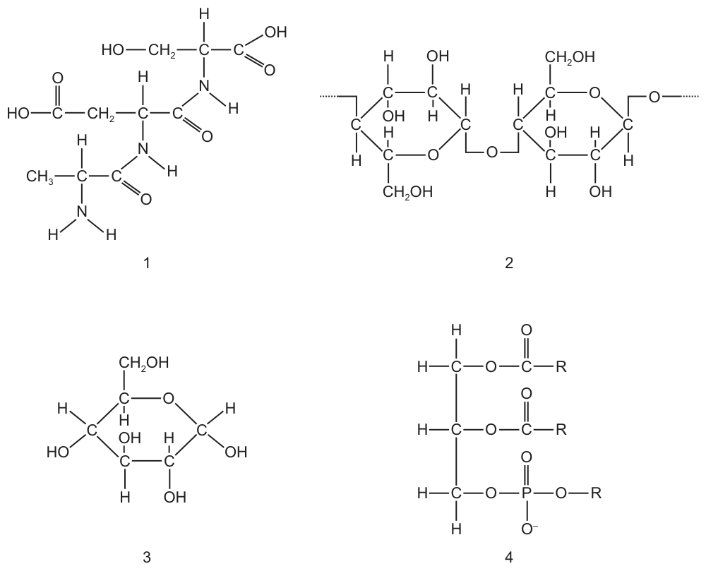
Which pair is present in the solution?
Options
A 1 and 2
B 1 and 3
C 2 and 3
D 3 and 4
Working
Interpreting the four test results:
- Iodine = orange (negative) → starch is absent.
- Biuret = purple (positive) → protein is present.
- Benedict's = orange (positive) → a reducing sugar is present.
- Emulsion = clear (negative) → lipid is absent.
Matching to the four structures:
- 1 = tripeptide (amino acids joined by peptide bonds) → protein → present.
- 2 = segment of a polysaccharide (note the dotted bonds indicating it is part of a longer chain, e.g. starch/glycogen) → would be detected by iodine, not Benedict's, and iodine was negative → absent.
- 3 = α-glucose, a monosaccharide and reducing sugar → present.
- 4 = phospholipid (a lipid) → emulsion was clear → absent.
The two structures present in the solution are 1 and 3.
Answer
B
B
Background Concept
The four standard qualitative tests for biological molecules each rely on a specific colour change caused by a particular functional group or bond type:
- Iodine test for starch — iodine (iodine–potassium iodide solution) is yellow-brown/orange. Inside the helical coils of amylose it forms a starch–iodine complex that is blue-black. Any other colour (yellow, orange, brown) means starch is absent or present in too low a concentration to detect.
- Biuret test for peptide bonds (proteins) — copper(II) ions in alkaline solution complex with the lone pairs on the nitrogen atoms of peptide bonds, producing a violet/purple colour. A blue result (copper(II) hydroxide) means no (or too little) protein.
- Benedict's (or Fehling's) test for reducing sugars — a reducing sugar has a free anomeric carbon whose ring can open to expose an aldehyde group; this reduces the blue copper(II) ions in Benedict's reagent to red copper(I) oxide. The colour sequence, from least to most reducing sugar, is blue → green → yellow → orange → brick-red. "Orange" therefore means a moderate amount of reducing sugar is present.
- Emulsion test for lipids — lipids are immiscible with water; when shaken with ethanol and then water they form a milky-white cloudy emulsion. A clear solution means no (or insufficient) lipid.
The key biological distinction that matters here is the difference between a monosaccharide, a disaccharide and a polysaccharide. Only the structures that have a free anomeric carbon (a hemiacetal that can open to an aldehyde) are reducing sugars. A free α- or β-glucose molecule has this free anomeric carbon and is therefore a reducing sugar. The disaccharide maltose also has a free anomeric carbon on one of its two glucose units, so maltose is reducing. However, in a long polysaccharide such as starch or glycogen, every glucose unit is joined to its neighbours by glycosidic bonds, so there is no free anomeric carbon — the polysaccharide is not a reducing sugar. A polysaccharide is detected by the iodine test, not Benedict's.
Understanding the Question
The student has performed all four standard tests on a single solution and recorded the colours. We must combine those four colour results with the four molecular structures (1–4) to decide which pair of molecules is genuinely present in the solution. The trick is that the colour results must be read with two distinctions in mind: (a) what the colour tells us is present (purple biuret and orange Benedict's) and (b) what the colour tells us is absent (orange iodine and clear emulsion). The structure labels are "molecules or parts of molecules", and one of them (structure 2) is shown as a segment of a longer chain — recognising this is what makes the question discriminating.
Approach
First, write a one-line conclusion for each of the four test results. Then match each labelled structure to the molecule type it represents and ask, for each, whether the test for that molecule is positive or negative. Only structures whose molecule type is consistent with a positive test can be in the solution.
The matching is:
- Tripeptide with three peptide bonds → protein → biuret.
- Two glucose units with dotted bonds on both ends → part of a polysaccharide chain (e.g. starch) → iodine, not Benedict's.
- Single α-glucose ring → monosaccharide / reducing sugar → Benedict's.
- Glycerol with two fatty acid chains and a phosphate group → phospholipid / lipid → emulsion.
Step-by-Step Reasoning
Iodine = orange.
Iodine is only blue-black with starch. Orange means starch is absent. Therefore any structure that is a polysaccharide (structure 2, the disaccharide/polysaccharide segment) is not present. (Note: a free disaccharide such as isolated maltose would not give a positive iodine test either, so the negative iodine alone does not exclude a disaccharide — but structure 2 is drawn as a chain segment, indicating starch, which is what really rules it out.)
Biuret = purple.
This is the classical positive protein colour, produced by Cu²⁺–peptide bond complexation. The solution therefore contains protein. Structure 1 is a tripeptide (three amino acids joined by two peptide bonds) — it is a small protein/peptide and is consistent with the positive biuret. So structure 1 is present.
Benedict's = orange.
Benedict's starts blue; heating with a reducing sugar reduces the Cu²⁺ to Cu₂O. The observed orange precipitate (rather than green or brick-red) tells us a reducing sugar is present in moderate amount. Structure 3 is α-glucose, a monosaccharide and a reducing sugar — it is consistent with a positive Benedict's. So structure 3 is present. (Structure 2 would not give a positive Benedict's because, as drawn, it is part of a polysaccharide chain with no free anomeric carbon.)
Emulsion = clear.
A cloudy white emulsion is the positive lipid result; a clear solution means no lipid. Structure 4 is a phospholipid (a type of lipid) — it is therefore absent from the solution.
Conclusion. Only structures 1 (tripeptide, protein) and 3 (α-glucose, reducing sugar) match the four observed colours. The pair present is 1 and 3, which is option B.
Key Takeaways
- Iodine is specific for polysaccharide (starch), not for any sugar; blue-black = starch, orange/brown = no starch.
- Biuret detects peptide bonds; any molecule containing them (proteins and peptides) gives the violet colour.
- Benedict's detects reducing sugars (free anomeric carbon). Free monosaccharides and maltose are reducing; a polysaccharide is not.
- The emulsion test detects lipids, including phospholipids.
- A drawing with dotted bonds at both ends of a sugar unit is shorthand for "this is part of a longer chain" — i.e. a polysaccharide segment — and the test you should associate with it is the iodine test, not Benedict's.
- Always match each test to the molecule the test actually detects; don't be distracted by structural features (e.g. a phospholipid looks very different from a triglyceride, but the emulsion test treats them both as lipid).
Common Mistakes
- Picking A (1 and 2) because the student recognises that both a protein and a "sugar" seem consistent with the two positive tests. The error is missing that structure 2 is a polysaccharide segment, not a free disaccharide — it would be picked up by iodine, which was negative.
- Picking C (2 and 3) because both are carbohydrates. The error is forgetting that biuret was positive (purple) for protein, so a protein (structure 1) must be present and one of the sugars must therefore be wrong.
- Picking D (3 and 4) because glucose is a sugar and a phospholipid is a biological molecule. The error is ignoring the clear emulsion result, which excludes lipid, and forgetting the positive biuret, which requires a protein.
- Misreading "orange Benedict's" as negative. Orange is positive — it is the colour produced by a moderate amount of reducing sugar. Only the original blue colour of unused Benedict's reagent indicates absence.
Things to Be Careful About
- The order of the colour sequence in Benedict's (blue → green → yellow → orange → brick-red) is what makes "orange" a positive, semi-quantitative result. Do not write "Benedict's was negative" simply because the colour is not brick-red.
- "Orange" in the iodine test means negative for starch (iodine itself is yellow-brown/orange; the blue-black colour is the positive). Context — which test and which reagent — is what makes the colour meaningful.
- When a structure is drawn with dotted/wavy bonds at the ends, treat it as part of a polymer, not a discrete small molecule. The test that detects it is the polymer-specific test (iodine for starch/glycogen), not the monomer test (Benedict's).
- A phospholipid still counts as a lipid for the emulsion test — do not assume the test only detects triglycerides.
- The question asks which pair is present; you must therefore eliminate every option containing a molecule whose test was negative (no starch, no lipid) or omitting the molecule whose test was positive (protein).
The diagram shows some relationships between features of carbohydrates.
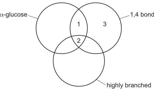
Which row correctly matches the carbohydrate with some of its features?
Options
| 1 | 2 | 3 | |
|---|---|---|---|
| A | amylopectin | glycogen | amylose |
| B | amylose | amylopectin | cellulose |
| C | cellulose | glycogen | sucrose |
| D | glycogen | amylose | amylopectin |
Working
The Venn diagram partitions carbohydrates by three features:
- α-glucose (left circle)
- 1,4 bond (right circle)
- highly branched (bottom circle)
So the three labelled regions require:
- Region 1 (α-glucose ∩ 1,4 bond only, not highly branched) → a polymer of α-glucose with only 1,4 glycosidic bonds, unbranched.
- Region 2 (all three) → a polymer of α-glucose that is branched.
- Region 3 (1,4 bond ∩ highly branched, not α-glucose) → a polymer with 1,4 bonds that is not built from α-glucose.
Matching each carbohydrate:
- Amylose — α-glucose monomer, only 1,4 bonds, unbranched helix → fits Region 1.
- Amylopectin — α-glucose monomer, 1,4 bonds in the main chain, branched (via 1,6 bonds at branch points) → fits Region 2.
- Cellulose — β-glucose monomer (so not α-glucose), 1,4 bonds only, and clearly distinguished from the α-glucose polymers → fits Region 3.
Glycogen is also an α-glucose polymer and is even more highly branched than amylopectin, so it could be argued for Region 2, but only B places amylose in Region 1 and amylopectin in Region 2 correctly.
Answer
B
B
Background Concept
The biologically important polysaccharides of glucose differ in three structural ways:
- The monomer: α-glucose or β-glucose. The two isomers differ in the orientation of the –OH group on C1. Polymerising α-glucose gives a storage polysaccharide; polymerising β-glucose gives a structural one.
- The glycosidic bond: 1,4 bonds link glucose units in a straight chain; 1,6 bonds create branch points.
- The degree of branching: unbranched (helical or straight), moderately branched, or highly branched.
| Polysaccharide | Monomer | Bonds | Branching |
|---|---|---|---|
| Amylose | α-glucose | 1,4 only | Unbranched (helix) |
| Amylopectin | α-glucose | 1,4 (main chain) + 1,6 (branches) | Moderately branched |
| Glycogen | α-glucose | 1,4 (main chain) + 1,6 (branches) | Highly branched |
| Cellulose | β-glucose | 1,4 only | Unbranched (straight chains) |
These features are exactly what the Venn diagram in Fig. 7.1 partitions.
Understanding the Question
Fig. 7.1 is a three-set Venn diagram with circles labelled α-glucose, 1,4 bond and highly branched. Three regions are numbered and each is identified with a different carbohydrate in the answer table. The task is to choose the row that correctly places a carbohydrate in each region.
The three regions correspond to:
- Region 1: features α-glucose and 1,4 bond, but is not highly branched.
- Region 2: features all three (α-glucose, 1,4 bond, and highly branched).
- Region 3: features 1,4 bond and highly branched, but is not made of α-glucose.
Approach
Recall, for each named carbohydrate, which combination of the three features it has, then locate it in the Venn diagram. Work by elimination:
- A carbohydrate in Region 1 must be built from α-glucose, joined by 1,4 bonds, and be unbranched.
- A carbohydrate in Region 2 must be built from α-glucose, joined by 1,4 bonds, and be branched.
- A carbohydrate in Region 3 must NOT be built from α-glucose, but must have 1,4 bonds and be branched (the "highly branched" cue is the main way to distinguish Region 3 from Region 1: the carbohydrate is not α-glucose, so the only other linear 1,4 polymer, cellulose, is the natural candidate).
Step-by-Step Reasoning
Region 1 — amylose (row B): Amylose is the unbranched component of starch. Its chains are formed entirely from α-glucose joined by 1,4 glycosidic bonds, which coil into a helix. It has α-glucose ✓, has 1,4 bonds ✓, is not highly branched ✓. So amylose sits inside the α-glucose and 1,4-bond circles but outside the highly-branched circle — exactly Region 1.
Region 2 — amylopectin (row B): Amylopectin is the branched component of starch. Like amylose it is built from α-glucose with 1,4 bonds along the main chain, but it also has 1,6 bonds that create branch points. Therefore it has α-glucose ✓, has 1,4 bonds ✓, and is branched ✓ — all three circles — so it sits in the central Region 2.
Region 3 — cellulose (row B): Cellulose is built from β-glucose (not α-glucose), and the units are joined by 1,4 bonds. The cellulose chains are straight, unbranched and pack together via hydrogen bonds to give plant cell walls their tensile strength. In the Venn diagram cellulose belongs inside the 1,4-bond circle but outside the α-glucose circle — i.e. in Region 3. The branched aspect of this region is best read as the distinguishing "branching" cue that separates Region 3 (β-glucose, 1,4 only) from Region 1 (α-glucose, 1,4 only): Region 3 in this question is the "β-glucose, 1,4-bond" slot, which is unambiguously cellulose.
Eliminating the other rows:
- Row A puts amylopectin in Region 1 — but amylopectin is branched, so it cannot sit outside the highly-branched circle.
- Row C puts cellulose in Region 1 — but cellulose is built from β-glucose, not α-glucose, so it cannot be inside the α-glucose circle.
- Row D puts glycogen in Region 1 (glycogen is highly branched, so it cannot be in the unbranched region) and amylose in Region 2 (amylose is unbranched, so it cannot be in the central region where all three circles overlap).
Only row B places each carbohydrate in a region consistent with its known structure.
Key Takeaways
- Amylose = α-glucose, 1,4 bonds only, unbranched (helix).
- Amylopectin = α-glucose, 1,4 (main chain) + 1,6 (branches), branched.
- Glycogen = α-glucose, 1,4 (main chain) + 1,6 (branches), more branched than amylopectin — animal storage form.
- Cellulose = β-glucose, 1,4 bonds only, unbranched, structural.
- A 1,4-bond polymer that is not built from α-glucose must be built from β-glucose, which is cellulose.
Common Mistakes
- Confusing amylose and amylopectin. Amylose is the unbranched helix; amylopectin is the branched one. They are both components of starch, but only amylopectin is branched.
- Treating glycogen and amylopectin as identical. Both are α-glucose with 1,4 and 1,6 bonds, but glycogen is more highly branched (branched roughly every 8–12 glucose units, versus every 24–30 in amylopectin).
- Forgetting that cellulose is built from β-glucose, not α-glucose. The β-1,4 linkage gives very different properties — straight chains that hydrogen-bond together, rather than a storage helix.
- Reading the Venn diagram regions carelessly. A carbohydrate in a region must satisfy all the features of that region; the "not in this circle" part of a region is just as important as the "in these circles" part.
Things to Be Careful About
- Use precise terminology: "α-glucose", "β-glucose", "1,4 glycosidic bond", "1,6 glycosidic bond". Vague terms like "glucose polymer" do not earn credit.
- Region 3 in this Venn diagram is identified by not being α-glucose while having 1,4 bonds — that is the cellulose slot. The "highly branched" label on the lower circle is the cue that pulls the candidate's attention away from the simple "1,4-bond-only" feature and towards the structural contrast between amylose (Region 1) and cellulose (Region 3).
- The diagram does not ask you to identify every feature of each carbohydrate; it asks you to identify which region of the Venn diagram each one belongs to. Work from each carbohydrate's known structure, not from a vague impression.
The diagram shows a triglyceride molecule.
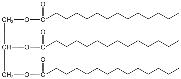
Which statement is correct for the structure and function of this triglyceride?
Options
A The glycerol head forms hydrogen bonds with water in cytoplasm to produce oil droplets.
B The hydrophobic fatty acids allow only small molecules to pass through the cell surface membrane.
C It packs closely with identical triglyceride molecules to form energy reserves in lysosomes.
D It has long hydrocarbon chains that release high energy output per unit mass when oxidised.
Working
A triglyceride consists of a glycerol molecule bonded (via ester bonds) to three long fatty acid (hydrocarbon) chains. During aerobic respiration, these long hydrocarbon chains are oxidised, releasing a large amount of energy per unit mass — approximately twice that of carbohydrates. This makes triglycerides highly efficient energy-storage molecules.
- A is wrong: triglycerides are hydrophobic (insoluble in water) and do not form oil droplets via hydrogen bonding with water.
- B is wrong: the cell surface membrane is a phospholipid bilayer, not a triglyceride layer, and small non-polar molecules pass through the phospholipid region, not because of triglyceride fatty acids.
- C is wrong: triglyceride energy reserves are stored in lipid droplets in the cytoplasm (adipose tissue), not in lysosomes.
- D is correct: the long hydrocarbon (fatty acid) chains yield a high energy output per unit mass on oxidation.
Answer
D
D
Background Concept
A triglyceride is formed when three fatty acid molecules are joined to one glycerol molecule by ester bonds (a condensation reaction releasing three water molecules). Each fatty acid is a long hydrocarbon chain (a chain of –CH₂– groups terminating in a –CH₃) with a carboxyl group (–COOH) at one end. Because the hydrocarbon chains are non-polar, the whole triglyceride molecule is hydrophobic and insoluble in water.
Triglycerides are the body's main long-term energy store. When oxidised during aerobic respiration, the C–H and C–C bonds in the hydrocarbon chains are broken, releasing a large amount of energy. Lipids yield roughly twice as much energy per gram as carbohydrates (about 38 kJ g⁻¹ versus 17 kJ g⁻¹ for carbohydrates), which is why they are so effective for energy storage.
Understanding the Question
The question presents a structural diagram of a triglyceride (glycerol backbone with three attached fatty acid chains) and asks which statement correctly links its structure to its function. The command word "correct" means only one option is fully accurate; the others contain at least one structural or functional error.
Approach
For each option, check two things: (1) is the structural claim about a triglyceride accurate, and (2) is the functional/biological consequence correct? Eliminate any option that confuses triglycerides with phospholipids, misplaces their cellular location, or misrepresents their chemistry.
Step-by-Step Reasoning
-
Option A claims the glycerol part of the triglyceride forms hydrogen bonds with water to produce oil droplets. This is incorrect because the glycerol is attached via ester bonds to three large hydrophobic fatty acid chains, which dominate the molecule's behaviour. The overall molecule is hydrophobic and forms oil droplets because it cannot hydrogen-bond with water, not because it does.
-
Option B attributes the membrane's selective permeability to the "hydrophobic fatty acids" of a triglyceride. This confuses triglycerides with phospholipids. The cell surface membrane is built from a phospholipid bilayer, where the two fatty acid tails create a hydrophobic core. Small, non-polar molecules (e.g. O₂, CO₂) diffuse through this region. Triglycerides are not structural components of the membrane in this way.
-
Option C places triglyceride energy reserves in lysosomes. This is wrong. Triglycerides are stored in cytoplasmic lipid droplets (in adipocytes of adipose tissue, or in the cytoplasm of many other cell types). Lysosomes contain hydrolytic enzymes for breaking down macromolecules, not energy-storage lipids.
-
Option D correctly identifies that triglyceride molecules contain long hydrocarbon chains which, when oxidised in respiration, release a high energy output per unit mass. This is the defining structure–function relationship of triglycerides as energy stores.
Key Takeaways
- Triglycerides = glycerol + 3 fatty acids joined by ester bonds.
- Long hydrocarbon (fatty acid) chains → high energy yield on oxidation → excellent energy storage.
- Triglycerides are hydrophobic; they do not form hydrogen bonds with water.
- Membranes are made of phospholipids, not triglycerides.
- Triglyceride stores are in cytoplasmic lipid droplets, not lysosomes.
Common Mistakes
- Confusing triglycerides with phospholipids (only phospholipids form bilayers in membranes; triglycerides do not).
- Thinking the glycerol "head" of a triglyceride is hydrophilic enough to hydrogen-bond with water — the three large hydrophobic chains dominate the molecule's properties.
- Misplacing lipid storage in lysosomes rather than cytoplasmic lipid droplets / adipose tissue.
- Believing "small molecules pass through membrane because of fatty acids" — it is specifically the hydrophobic fatty acid tails of phospholipids that allow small non-polar molecules to cross.
Things to Be Careful About
- The word "triglyceride" specifically means a storage lipid with three fatty acids; phospholipids have only two fatty acids and a phosphate group, giving them an amphipathic (partly hydrophilic) character.
- Energy yield per unit mass is a key concept: lipids ≈ 2× the energy of carbohydrates per gram, which is why they are preferred for long-term energy storage in animals.
Enzyme P has been found in a tropical grass.
● The enzyme catalyses the hydrolysis of the fungal polysaccharide, chitin, into amino sugars.
● It also inhibits the activity of an enzyme in locust guts which catalyses the digestion of amylose.
Which row describes the actions of enzyme P?
Options
| reaction catalysed | reaction inhibited | |
|---|---|---|
| A | hydrolysis of glycosidic bonds | condensation of glycosidic bonds |
| B | hydrolysis of glycosidic bonds | hydrolysis of glycosidic bonds |
| C | hydrolysis of peptide bonds | condensation of glycosidic bonds |
| D | hydrolysis of peptide bonds | hydrolysis of glycosidic bonds |
Working
- Chitin is a polysaccharide (polymer of N‑acetylglucosamine monomers). Its monomers are joined by glycosidic bonds, so hydrolysing chitin into amino sugars means hydrolysis of glycosidic bonds.
- Amylose is also a polysaccharide (polymer of α‑glucose joined by 1,4 glycosidic bonds). Digestion of amylose in the locust gut is therefore also hydrolysis of glycosidic bonds.
- Hence enzyme P catalyses the hydrolysis of glycosidic bonds AND inhibits a reaction that is itself the hydrolysis of glycosidic bonds.
Answer
B
B
Background Concept
A monosaccharide is a single sugar unit (e.g. glucose, fructose, glucosamine). When two or more monosaccharides join together they form a disaccharide, oligosaccharide or polysaccharide, and the bond that holds them together is always a glycosidic bond — an oxygen bridge between two carbon atoms (typically C1 of one sugar and C4 of the next). Hydrolysis of a polysaccharide breaks these glycosidic bonds and releases the free monosaccharide units; condensation joins monosaccharides by forming new glycosidic bonds.
This is true even when the monomers themselves contain other functional groups. Chitin, for example, is a polymer of N‑acetylglucosamine — a glucose molecule that has been modified by replacing the hydroxyl (–OH) on C2 with an N‑acetyl amino group (–NHCOCH₃). The amino group is part of the monomer, not part of the linkage between monomers, so the bonds joining the units are still glycosidic.
The same logic applies to amylose: it is a polymer of α‑glucose with 1,4 glycosidic bonds (and occasional 1,6 branch points in amylopectin). "Digesting" a polysaccharide in a gut means hydrolysing the glycosidic bonds to release glucose, which can then be absorbed.
Understanding the Question
The question describes a single enzyme, P, found in a tropical grass. P does two things:
- It catalyses the hydrolysis of chitin (a fungal polysaccharide) into amino sugars.
- It inhibits a locust gut enzyme that digests amylose (a plant polysaccharide of glucose).
You must decide, for each of the two reactions involved, whether glycosidic or peptide bonds are being broken or formed, and then pick the matching row of the table.
The command word is implicit: which option correctly describes the two actions of enzyme P.
Approach
For each reaction, ask three questions in order:
- What is the substrate? Chitin and amylose are both polysaccharides.
- What type of bond joins the monomers? Polysaccharides have glycosidic bonds, not peptide bonds. (Peptide bonds join amino acids in proteins.)
- What is happening to those bonds? Hydrolysis (breaking with water) or condensation (forming with loss of water)?
Apply this to both the catalysed reaction and the inhibited reaction, then match against the four rows.
Step-by-Step Reasoning
Reaction catalysed by enzyme P
- Substrate: chitin = polymer of N‑acetylglucosamine (a polysaccharide).
- Bond between monomers: glycosidic bond.
- Process described: "hydrolysis of chitin into amino sugars" → adding water to split the polymer → hydrolysis of glycosidic bonds.
- This rules out options C and D, both of which claim the catalysed reaction involves peptide bonds.
Reaction inhibited by enzyme P
- Substrate: amylose = polymer of α‑glucose (a polysaccharide of glucose).
- Bond between monomers: glycosidic bond (specifically α‑1,4).
- Process described: "digestion of amylose" in the locust gut → the locust's amylase is breaking the polysaccharide into glucose → hydrolysis of glycosidic bonds.
- Option A claims the inhibited reaction is "condensation of glycosidic bonds" — but condensation would build a polysaccharide, not digest one. Wrong.
- Option B claims both reactions involve hydrolysis of glycosidic bonds. Correct.
Therefore the row that fits is B.
Key Takeaways
- Polysaccharides, whatever their monomers look like, are joined by glycosidic bonds.
- The word "amino" in "amino sugar" refers to an amino group on the sugar, not to amino‑acid/peptide chemistry. Do not let the word "amino" push you into the peptide‑bond option.
- "Hydrolysis" always means bond‑breaking with water; "condensation" always means bond‑forming with the loss of water. Digestion of a polymer in a gut is always hydrolysis.
- A useful general rule: hydrolysis of a polymer ↔ hydrolysis of the bond type characteristic of that polymer (glycosidic for sugars, peptide for proteins, phosphodiester for nucleic acids, ester for lipids).
Common Mistakes
- Choosing C or D because the chitin product is an "amino sugar", assuming the bonds being broken are peptide bonds. The amino group is part of each monomer; the bonds between monomers are still glycosidic.
- Choosing A because "hydrolysis" of a polysaccharide sounds as if it must be opposed by "condensation". In fact, the inhibited reaction is also a hydrolysis (amylose digestion), not a condensation.
- Confusing the structure of amylose (a glucose polymer) with that of cellulose or starch, or of a protein — any of which would send the answer off in the wrong direction.
Things to Be Careful About
- Read the question precisely: enzyme P catalyses one reaction and inhibits another. Both columns of the table must be filled in, and the right‑hand column refers to the inhibited reaction, not to enzyme P's own action.
- Distinguish "amino sugar" (e.g. glucosamine, N‑acetylglucosamine) from "amino acid" (the building block of proteins). The two are chemically very different despite the shared word.
- Be sure the word "hydrolysis" in the question is the same "hydrolysis" the answer is asking about — both columns should describe the type of bond being broken, not a vague direction of the reaction.
Which bonds between amino acids hold the shape of the secondary structure and also the tertiary structure of proteins?
Options
A peptide bonds
B disulfide bonds
C hydrogen bonds
D ionic bonds
Answer
C
C
Background Concept
Proteins have four levels of structure, each stabilised by different bonds:
- Primary structure: the linear sequence of amino acids linked by peptide bonds (covalent) formed by condensation between the carboxyl group of one amino acid and the amino group of the next.
- Secondary structure: regular folding of the polypeptide backbone into an α-helix or β-pleated sheet, stabilised by hydrogen bonds between the C=O of one peptide bond and the N–H of another.
- Tertiary structure: the overall 3D folding of the polypeptide, stabilised by interactions between R groups — these include hydrogen bonds, ionic bonds (between oppositely charged R groups), disulfide bridges (covalent, between cysteine –SH groups), and hydrophobic interactions (between non-polar R groups).
- Quaternary structure: the association of multiple polypeptide chains, held by the same R-group interactions as tertiary structure plus sometimes disulfide bonds.
Understanding the Question
The question asks which single type of bond is responsible for BOTH the secondary structure AND the tertiary structure of a protein. The four options (peptide, disulfide, hydrogen, ionic) correspond to the major bond types found in proteins.
Approach
Identify the bond(s) involved in each level of structure and find the overlap:
- Peptide bonds → only primary structure
- Disulfide bonds → only tertiary (and quaternary) structure
- Ionic bonds → only tertiary (and quaternary) structure
- Hydrogen bonds → both secondary (between backbone peptide groups) AND tertiary (between R groups)
The only bond type appearing at both levels is hydrogen bonding.
Step-by-Step Reasoning
- Peptide bonds (A) hold amino acids together in the primary structure — they form the backbone itself, not the folding of it. They are not directly responsible for secondary or tertiary structure.
- Disulfide bonds (B) are strong covalent bonds between two cysteine residues. They are found in tertiary (and quaternary) structure but not in secondary structure.
- Hydrogen bonds (C) form in the secondary structure between the partial positive charge on the N–H hydrogen and the partial negative charge on the C=O oxygen of the peptide backbone, producing the regular patterns of α-helices and β-pleated sheets. The same type of bond also forms in tertiary structure between, for example, an –OH on one R group and a C=O on another, helping to maintain the overall 3D shape. This is the correct answer.
- Ionic bonds (D) form between positively and negatively charged R groups (e.g. –NH₃⁺ and –COO⁻) and contribute to tertiary (and quaternary) structure, but they do not hold the secondary structure.
Key Takeaways
- Hydrogen bonding is the only interaction common to both secondary and tertiary protein structure.
- The distinction between the two levels: in secondary structure H-bonds are between backbone atoms, while in tertiary structure H-bonds (and other interactions) are between R groups.
- Knowing the bond type for each structural level is a high-yield recall point.
Common Mistakes
- Choosing A (peptide bonds) because they are the most familiar bond in proteins — but peptide bonds define the primary structure, not the folding.
- Choosing B (disulfide) or D (ionic) because they are involved in tertiary structure — but these do not stabilise secondary structure.
- Forgetting that hydrogen bonds in proteins occur in two different contexts (backbone vs R groups).
Things to Be Careful About
- The question is a multiple-choice with one correct answer. Watch for "and also" — both levels must be satisfied.
- Although tertiary structure involves several bond types, the question only asks for the one shared with secondary structure, so do not list multiple answers.
Resilin is a protein found in the wings of insects. It is an elastic, fibrous protein and scientists have suggested that it may have an important role in allowing the wings of an insect to fold and unfold without damage.
Peptidyl transferase is an enzyme which catalyses the condensation of amino acids during translation to form resilin.
Which statements are correct?
1 Peptidyl transferase and resilin have structural roles in the insect.
2 Peptidyl transferase is a globular protein.
3 Peptidyl transferase is soluble in water.
Options
A 1 and 2
B 1 and 3
C 1 only
D 2 and 3
Working
Statement 1: Peptidyl transferase and resilin have structural roles in the insect.
- Resilin does have a structural role (gives elasticity in wings).
- Peptidyl transferase is an enzyme, so its role is catalytic (condensation of amino acids), not structural.
- Statement 1 is incorrect.
Statement 2: Peptidyl transferase is a globular protein.
- Most enzymes are globular proteins (compact, roughly spherical, with specific 3-D active sites).
- Statement 2 is correct.
Statement 3: Peptidyl transferase is soluble in water.
- Globular proteins (including most enzymes) are water-soluble because hydrophilic R-groups are on their outer surface.
- Statement 3 is correct.
Answer
D
D
Background Concept
Proteins are classified into two broad structural categories based on their shape and solubility:
- Fibrous proteins have long, parallel polypeptide chains forming fibres or sheets. They are usually insoluble in water and have a structural role (e.g. collagen in tendons, keratin in hair, elastin/ resilin in elastic tissues). Their regular secondary structure (often α-helix or β-pleated sheet) is held together by many cross-links.
- Globular proteins have polypeptide chains folded into a compact, roughly spherical shape. Hydrophilic (water-loving) R-groups sit on the outside and hydrophobic R-groups are tucked inside, so they are generally soluble in water. Globular proteins usually have a functional role — for example as enzymes, hormones, antibodies or transport proteins — and they possess a specific 3-D active site / binding site.
Because most enzymes are globular proteins, they are typically water-soluble and perform a catalytic role (speeding up metabolic reactions) rather than a structural one. Peptidyl transferase is the enzyme that catalyses peptide bond formation between amino acids during translation. It is the enzymatic activity of the large ribosomal subunit (it is actually a ribozyme — catalysed by rRNA — but for the purpose of this question it behaves as a protein enzyme made of ribosomal proteins).
Understanding the Question
The question asks you to evaluate three statements about resilin and peptidyl transferase, then pick the combination that is correct. The stem gives you crucial information:
- Resilin = elastic, fibrous protein with a structural role in wings.
- Peptidyl transferase = an enzyme catalysing condensation of amino acids during translation.
So the test is whether you can:
- Distinguish structural (fibrous) from catalytic (globular enzyme) roles.
- Recall the general properties of globular proteins (and therefore of enzymes).
The command word "Which statements are correct?" directs you to evaluate each independently, then pick the option that contains only the true ones.
Approach
Test each statement in turn using the definitions above:
- For each protein, identify whether it is fibrous or globular from the description given.
- Assign structural or functional (catalytic) role accordingly.
- Apply the general solubility rule for globular proteins.
Step-by-Step Reasoning
Statement 1 — "Peptidyl transferase and resilin have structural roles in the insect."
- Resilin is explicitly described as a fibrous protein that allows wings to fold and unfold → it is structural. ✓
- Peptidyl transferase is described as an enzyme that catalyses a condensation reaction → its role is catalytic, not structural. ✗
- One part is wrong, so the whole statement is incorrect.
Statement 2 — "Peptidyl transferase is a globular protein."
- All enzymes in this context are treated as globular proteins (compact, spherical, with a specific active site that binds substrate).
- This is the standard classification for enzymes and is correct.
Statement 3 — "Peptidyl transferase is soluble in water."
- A consequence of being a globular protein is water-solubility: hydrophilic R-groups face outwards, allowing hydrogen bonding with water.
- The condensation reaction it catalyses takes place in the aqueous cytosol of the ribosome, so it must be soluble. Correct.
Statements 2 and 3 are correct → option D.
Key Takeaways
- Fibrous proteins (e.g. resilin, collagen, keratin) → structural, insoluble.
- Globular proteins (e.g. most enzymes, haemoglobin, antibodies) → functional, soluble.
- Enzymes are globular and water-soluble so they can act in solution in the cytoplasm or other body fluids.
- Distinguish structural from catalytic roles carefully — a protein that makes a structural protein (peptidyl transferase making resilin) is not itself structural.
Common Mistakes
- Assuming that because peptidyl transferase makes resilin (a structural protein), peptidyl transferase must also be structural. The function of a protein is determined by what it does, not by the role of the product it makes.
- Forgetting that enzymes are typically soluble globular proteins; treating "protein" as a single undifferentiated category.
- Confusing catalytic with structural roles in general — e.g. saying haemoglobin is structural because it carries oxygen (it is functional/transport, not structural).
Things to Be Careful About
- Read each statement in full and check every part: a statement is only correct if all of it is true (a single false clause makes the whole statement false).
- Use the information given in the stem: the description "elastic, fibrous protein" and "enzyme which catalyses…" are deliberate clues to the classification.
- The exam mark scheme here is just the letter D, so don't second-guess by trying to find a "trick" — straightforward application of the rules above is enough.
Collagen is a protein that helps to strengthen and increase the elasticity and hydration of the skin. Aging can lead to dry skin and the formation of wrinkles, due to the body producing less collagen.
Which row is correct for the structure of collagen in the skin?
Options
| every fourth amino acid is glycine | fibrous protein | |
|---|---|---|
| A | ✗ | ✗ |
| B | ✓ | ✓ |
| C | ✗ | ✓ |
| D | ✓ | ✗ |
key
✓ = correct
✗ = not correct
Working
Collagen is a fibrous protein (long, parallel polypeptide chains forming strong, water-insoluble fibres that give skin its tensile strength and elasticity). This makes the "fibrous protein" column ✓.
In the primary structure of collagen, glycine occurs at every third position in each α-polypeptide chain (the repeating tripeptide motif is –Gly–X–Y–), because only the small glycine side chain can fit into the tight interior of the triple helix. Every fourth amino acid being glycine is therefore ✗.
Only the second statement is correct, so the matching row is C.
Answer
C
C
Background Concept
Collagen is the most abundant protein in mammals and is the major fibrous component of skin, tendons, bone, cartilage, ligaments, blood vessels and the cornea. It gives tissues their tensile strength and, together with elastin and proteoglycans, contributes to skin elasticity and hydration.
Fibrous proteins (like collagen, keratin and elastin) are characterised by long, regular polypeptide chains wound around one another to form rope- or sheet-like structures. They are generally insoluble in water and play structural or mechanical roles rather than acting as enzymes, hormones or transporters (which is the role of globular proteins such as haemoglobin and enzymes).
The primary structure of collagen is a repeating tripeptide:
where X is frequently proline and Y is frequently hydroxyproline. Glycine must appear at every third position because every third residue sits at the very crowded central axis of the triple helix, where the three polypeptide chains come together. Only glycine, with its single hydrogen side chain (–H), is small enough to fit in this tight space; any other side chain would prevent the three chains from packing together.
Three α-polypeptide chains (each with about 1000 amino acids) then wind around each other to form a right-handed triple helix (tropocollagen). These are assembled into fibrils and then fibres, which is what gives collagen its enormous tensile strength.
Understanding the Question
This is a multiple-choice question (Paper 1 style) that asks you to evaluate two statements about collagen and choose the row in the table where the ✓/✗ pattern is correct.
The two statements to test are:
- Every fourth amino acid in collagen is glycine.
- Collagen is a fibrous protein.
The table gives four possible combinations of ✓/✗ for these two claims.
The command word is implicit: select the row that correctly describes the structure of collagen.
Approach
- Decide whether each statement is true or false based on your knowledge of collagen structure.
- Match that pattern to the row in the table.
Step-by-Step Reasoning
Statement 1 — "Every fourth amino acid is glycine":
This is false. The repeating unit in collagen is –Gly–X–Y–, so glycine occurs at every third position, not every fourth. This precise spacing is essential: it places a glycine residue at every position that ends up in the crowded core of the triple helix.
So column 1 = ✗.
Statement 2 — "Fibrous protein":
This is true. Collagen is the textbook example of a fibrous protein — long, rope-like, insoluble, structural.
So column 2 = ✓.
Matching the pattern to the table:
| Row | Col 1 (glycine) | Col 2 (fibrous) |
|---|---|---|
| A | ✗ | ✗ |
| B | ✓ | ✓ |
| C | ✗ | ✓ |
| D | ✓ | ✗ |
The pattern ✗ / ✓ matches row C.
Key Takeaways
- Collagen is a fibrous protein with structural/mechanical roles.
- Its primary structure is a repeating tripeptide –Gly–X–Y–; glycine occurs at every third position (not fourth), because only the small glycine side chain fits into the core of the triple helix.
- X is often proline, Y is often hydroxyproline; three such chains form a right-handed triple helix.
Common Mistakes
- Writing or believing "every fourth amino acid is glycine" — the repeat is every third residue.
- Confusing the test for protein structure that requires "every fourth residue" with a different protein (e.g. some coiled-coil motifs or the heptad repeat of α-keratin, which is a different heptad-based pattern).
- Confusing fibrous and globular proteins — collagen, keratin, elastin, fibroin (silk) are fibrous; haemoglobin, insulin, amylase, antibodies are globular.
Things to Be Careful About
- Be precise about "third" vs "fourth" — this is a favourite detail in MCQs.
- The glycine regularity is a feature of collagen's primary structure; the triple helix is its secondary/tertiary structure. Don't mix the levels.
- A ✓ in this question means the statement is correct, a ✗ means not correct — make sure you read the key before answering.
What is the effect of an enzyme in an enzyme-catalysed reaction?
Options
A decreases the activation energy and decreases the energy yield
B decreases the activation energy and has no effect on the energy yield
C increases the activation energy and increases the energy yield
D decreases the activation energy and increases the energy yield
Working
Enzymes are biological catalysts. They speed up a reaction by lowering the activation energy (the minimum energy reactant molecules must have to react) but they do not change the overall energy change (ΔH or energy yield) of the reaction — the reactants and products are the same, so the difference in their energy levels is unchanged.
- A → wrong: energy yield is not decreased by an enzyme.
- B → correct: activation energy is decreased; energy yield is unaffected.
- C → wrong: enzymes lower (not raise) activation energy, and they do not increase the energy yield.
- D → wrong: the energy yield is unchanged, not increased.
Answer
B
B
Background Concept
An enzyme is a biological catalyst — a globular protein (or, in a few cases, an RNA molecule, i.e. a ribozyme) that increases the rate of a chemical reaction without being consumed. Like all catalysts, it works by lowering the activation energy (): the minimum kinetic energy that reactant molecules must possess, at a given temperature, for a productive collision that leads to products. By stabilising the transition state (often through the induced-fit distortion of substrates at the active site), the enzyme allows a much larger fraction of molecular collisions to be successful at any given temperature, so the reaction proceeds faster.
Critically, a catalyst only changes the pathway (and the height of the activation energy barrier); it does not change the starting point (reactants) or the end point (products). Therefore the overall energy change of the reaction — the difference in energy between reactants and products, sometimes called the energy yield or ΔH — is exactly the same with or without the enzyme.
Understanding the Question
The question asks for the effect an enzyme has on a reaction. The options combine two independent statements:
- The effect on the activation energy.
- The effect on the energy yield (the overall energy change of the reaction).
We need to pick the option that correctly describes both effects.
Approach
- Recall that enzymes lower activation energy. This rules out any option saying activation energy is increased (so C is wrong).
- Recall that enzymes do not change the energy yield of a reaction — the substrates and products are the same, and the enzyme only changes the route between them. This rules out options claiming the energy yield is decreased (A) or increased (D).
- The only option left is B.
Step-by-Step Reasoning
- An energy-profile diagram for an enzyme-catalysed reaction shows the same starting energy (reactants) and ending energy (products) as for the uncatalysed reaction. The only difference is that the "hump" in the middle (activation energy) is lower when the enzyme is present.
- Because the start and end energies are unchanged, the energy released (in an exergonic reaction) or absorbed (in an endergonic reaction) is unchanged. The enzyme does not make the reaction "more exergonic" or "less endergonic".
- A → incorrect because it says energy yield decreases. The energy yield is the difference between product and reactant energy, and the enzyme does not change this.
- B → correct: activation energy is decreased, energy yield is unaffected.
- C → incorrect on both counts: activation energy is not increased, and energy yield is not increased.
- D → incorrect because it says energy yield increases; the energy yield is unchanged by any catalyst.
Key Takeaways
- A catalyst (including an enzyme) lowers activation energy.
- A catalyst does not change the overall energy change (energy yield, ΔH) of a reaction.
- The same is true of inorganic catalysts — this is a general property of catalysis, not something special to enzymes.
Common Mistakes
- Confusing the effect on activation energy with the effect on the overall energy change. Many students wrongly think that if a reaction is "faster", it must "release more energy"; in fact, the energy released is fixed by the chemistry of the bonds broken and formed.
- Choosing D because it feels "more biology-ish" — but biology is precise here: the energy yield is unaffected, not increased.
Things to Be Careful About
- "Energy yield" refers to the overall energy change between reactants and products, not the rate of energy release or the activation energy barrier.
- Enzymes are not used up in the reaction and do not alter the equilibrium position (they speed up both the forward and reverse reactions equally) — so the equilibrium constant, and therefore the position of equilibrium, is unchanged.
How is the Michaelis–Menten constant () used?
Options
A to determine the number of collisions between the enzyme and substrate
B to compare the affinity of enzymes for their substrates
C to find the maximum velocity of an enzyme ()
D to find the optimum rate of reaction
Working
The Michaelis–Menten constant () is defined as the substrate concentration at which the reaction rate is half of . A low means the enzyme reaches half its maximum rate at a low substrate concentration, i.e. it has a high affinity for its substrate; a high means a low affinity. is therefore used to compare how tightly different enzymes bind their substrates.
- A — collision frequency relates to kinetic energy and substrate concentration, not .
- B — correct: is an inverse measure of enzyme–substrate affinity.
- C — is read off the plateau of the rate–substrate curve, not from .
- D — optimum rate is determined by temperature/pH, not by .
Answer
B
B
Background Concept
Enzyme kinetics describes how the rate of an enzyme-catalysed reaction depends on substrate concentration. As [S] increases, the rate rises steeply at first and then levels off as the active sites become saturated, approaching a maximum velocity . The Michaelis–Menten model summarises this with the equation:
The Michaelis–Menten constant is the substrate concentration at which . Because it is the [S] needed to half-saturate the enzyme, it reflects how good the enzyme is at grabbing substrate: a small means only a little substrate is needed, i.e. high affinity; a large means lots of substrate is needed, i.e. low affinity. is therefore an inverse, qualitative measure of enzyme–substrate affinity.
Understanding the Question
This is a single-best-answer multiple-choice question. The command word is "How is … used?" — the candidate must select the option that correctly states the purpose of in enzyme kinetics. The distractors each describe a different kinetic concept (collision frequency, , optimum rate), so the question tests whether is distinguished from those related ideas.
Approach
Recall the definition of and what it does and does not tell you. Match each option to a kinetic concept and check whether it is what is used for.
- A — collision frequency is a factor of kinetic theory (temperature, concentration) and is not what measures.
- B — is the standard index used to compare the affinity of an enzyme for its substrate between different enzymes (or different substrates for the same enzyme).
- C — is the asymptote of the Michaelis–Menten curve, found by extrapolating to saturating [S]; it is not obtained from .
- D — "optimum rate" refers to the rate under optimum temperature/pH, not a kinetic constant.
Step-by-Step Reasoning
- On a rate vs [S] curve, is the plateau; the substrate concentration at is, by definition, .
- Because reaching half-saturation at low [S] implies strong binding, is inversely related to affinity: low = high affinity, high = low affinity.
- This makes the standard tool for comparing how well different enzymes (or an enzyme with different substrates) bind their substrate — option B.
- Option A is wrong because collision frequency depends on temperature, [S] and molecular motion, not on .
- Option C is wrong because is read directly from the graph plateau, not derived from (although the two appear together in the Michaelis–Menten equation).
- Option D is wrong because the optimum rate is set by temperature and pH, not by the kinetic constant .
Key Takeaways
- = [S] that gives .
- Low = high enzyme–substrate affinity; high = low affinity.
- is a comparative index of affinity, not a measure of , rate, or collision frequency.
Common Mistakes
- Confusing with — they are read off different parts of the same curve.
- Thinking a high means a "fast" enzyme; it actually means lower affinity, not lower rate at saturation.
- Saying "tells you the rate" — it tells you the [S] needed to reach half the maximum rate.
Things to Be Careful About
- has units of substrate concentration (e.g. ), not of rate.
- is independent of enzyme concentration; does depend on it.
- "Optimum rate" questions are about temperature/pH, not about .
Some students investigated the effect of pH on the rate of starch digestion by amylase at .
● They added of a amylase solution and of pH 4 buffer to a test-tube.
● They added of a starch solution, containing of starch, to the test-tube.
● They measured the time taken for the starch to be digested (to disappear).
The students then repeated these steps with pH 5, pH 6, pH 7 and pH 8 buffers.
The table shows the results.
| pH | time taken for starch to be digested/minutes |
|---|---|
| 4 | 4.0 |
| 5 | 1.5 |
| 6 | 1.0 |
| 7 | 2.5 |
| 8 | 6.0 |
Which graph is correct for these data?
Options
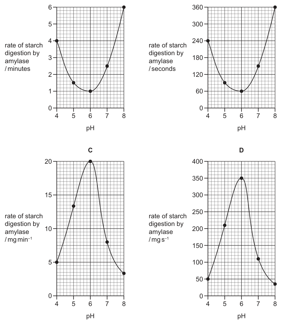
Working
The data are times for a fixed amount of starch (0.02 g = 20 mg) to be digested. Rate = amount of starch ÷ time, so the fastest reaction (shortest time, 1.0 min at pH 6) gives the highest rate, and the slowest (6.0 min at pH 8) gives the lowest. The pH–rate curve for an enzyme is bell-shaped with an optimum at pH 6.
Rate values (mg min⁻¹ = 20 / time):
- pH 4: 20/4.0 = 5
- pH 5: 20/1.5 ≈ 13
- pH 6: 20/1.0 = 20 (maximum)
- pH 7: 20/2.5 = 8
- pH 8: 20/6.0 ≈ 3
Only Graph C has a rate axis (mg min⁻¹) and a bell-shaped curve peaking at pH 6 with these values.
Answer
C
C
Background Concept
Enzymes are biological catalysts that speed up specific reactions. Their activity is highly sensitive to pH because the ionisation state of amino acid side chains in the active site depends on H⁺ concentration. At the optimum pH, the active site has the correct shape and charge to bind substrate most effectively, so the rate of reaction is at its maximum. Away from the optimum, the enzyme's tertiary structure is disturbed (in extreme cases it denatures), and the rate falls. Plotting rate against pH therefore gives a characteristic bell-shaped curve, with the peak at the optimum pH. For amylase, the optimum is around pH 6–7, near neutral.
Understanding the Question
The students measured the time taken for the same quantity of starch (0.02 g) to disappear at five different pH values. The question asks which graph correctly displays these data. The crucial step is recognising that:
- The graph must show rate, not time, because enzyme activity is conventionally expressed as a rate (amount of substrate converted per unit time).
- Rate is inversely proportional to time, so the pH giving the shortest time will give the highest rate.
- The y-axis must carry a rate unit — amount ÷ time (e.g. mg min⁻¹) — appropriate to the data.
From the table: pH 6 has the shortest time (1.0 min), so the curve should peak at pH 6, with lower rates at pH 4, 5, 7, and 8.
Approach
- Convert the time data to rates using rate = amount of starch / time. The amount is 0.02 g = 20 mg, and times are in minutes, so the natural rate unit is mg min⁻¹.
- Compute each rate:
- pH 4 → 20/4.0 = 5 mg min⁻¹
- pH 5 → 20/1.5 ≈ 13 mg min⁻¹
- pH 6 → 20/1.0 = 20 mg min⁻¹ (maximum)
- pH 7 → 20/2.5 = 8 mg min⁻¹
- pH 8 → 20/6.0 ≈ 3 mg min⁻¹
- Look for the graph whose y-axis is a rate in mg min⁻¹, whose curve is bell-shaped, and whose peak sits at pH 6 with values matching these numbers.
Step-by-Step Reasoning
- Graph A has the y-axis labelled "rate … / minutes". "Minutes" alone is a unit of time, not a rate. It also shows a U-shape (minimum at pH 6), which would be appropriate if the time were being plotted, but the y-axis is wrongly labelled as a rate. A low point at pH 6 would mean the slowest reaction, which contradicts the data (pH 6 has the shortest time, i.e. the fastest reaction).
- Graph B has the y-axis labelled "rate … / seconds". Again, seconds is a unit of time. It also has a U-shape, with the minimum at pH 6. This is wrong on two counts: the unit is not a rate, and the shape is inverted.
- Graph C has the y-axis labelled "rate of starch digestion by amylase / mg min⁻¹" — a true rate unit. The curve is bell-shaped, peaking at pH 6, with the value 20 at the peak. The other values read off the graph match the calculations: ~5 at pH 4, ~13 at pH 5, ~8 at pH 7, ~3 at pH 8. This is correct.
- Graph D has the y-axis labelled "rate … / mg s⁻¹". The shape is correct (bell-shaped, peak at pH 6) but the unit is mg s⁻¹, not mg min⁻¹. The original times were measured in minutes, so the rate should be expressed per minute, not per second. The numerical values (~50–350) would correspond to 20 mg / (time in seconds) — i.e. a unit conversion error.
Only Graph C combines the correct shape (bell), the correct peak position (pH 6), and the correct rate unit (mg min⁻¹) consistent with the data given in minutes.
Key Takeaways
- Enzyme rate is the inverse of the time taken for a fixed amount of substrate to be consumed; the optimum pH gives the shortest time and therefore the highest rate.
- pH–rate curves for enzymes are bell-shaped; the peak is the optimum pH.
- The y-axis of a rate graph must carry a true rate unit (amount per unit time, e.g. mg min⁻¹). A unit that is just a time (min or s) is not a rate, even if the question asks about "rate".
- When the data are times in minutes, the rate unit should also be per minute; converting to per second requires an additional step and changes all the numbers by a factor of 60.
Common Mistakes
- Plotting time instead of rate — a U-shaped curve with the minimum at pH 6 fits if the time is plotted, but the question asks for the rate, so the curve must peak at pH 6.
- Choosing mg s⁻¹ when the data are in minutes — students often pick the bell-shaped curve without checking the unit, but mg s⁻¹ is inconsistent with the data and gives numbers 60× too large.
- Confusing the optimum with the worst condition — at the optimum pH the enzyme works fastest, so the time is shortest and the rate is highest.
- Ignoring the amount of starch — because the same amount (0.02 g = 20 mg) was used in every tube, rate is simply 20 ÷ time, which is what makes the y-axis values in Graph C exact integers/near-integers.
Things to Be Careful About
- Always read the y-axis label, including the unit, before judging the shape.
- "Rate" alone is not a complete description; the unit must contain a "per time" element (e.g. mg min⁻¹, g s⁻¹, cm³ min⁻¹).
- A bell-shaped curve is the expected response of an enzyme to pH; a U-shape indicates either the time is being plotted or the labels are swapped.
- Numerical cross-check: at pH 6, 20 mg / 1.0 min = 20 mg min⁻¹, which matches the peak value of Graph C exactly — a useful way to confirm the correct answer quickly.
Which statements are correct for a non-competitive inhibitor of enzyme action?
1 Increasing the concentration of the enzyme’s substrate will reduce its effect.
2 It reduces the activation energy required for a reaction to take place.
3 It reduces the maximum rate of reaction.
Options
A 1 and 3
B 1 only
C 2 and 3
D 3 only
Working
A non-competitive inhibitor binds to a site on the enzyme other than the active site (an allosteric site). This changes the shape of the active site so the substrate can no longer bind, meaning the inhibition is not overcome by adding more substrate.
Evaluating each statement:
- Incorrect — because the inhibitor binds away from the active site, increasing substrate concentration does not out-compete it and so does not reduce its effect. (This is the defining contrast with a competitive inhibitor.)
- Incorrect — inhibitors do not lower activation energy; enzymes do. A non-competitive inhibitor slows the reaction (i.e. raises the effective activation energy by deactivating some enzyme molecules).
- Correct — because some enzyme molecules are effectively inactivated, the maximum rate () is reduced and cannot be restored by adding more substrate.
Only statement 3 is correct.
Answer
D
D
Background Concept
Enzyme inhibitors are molecules that reduce the rate of an enzyme-catalysed reaction. The two main types behave very differently:
- Competitive inhibitors have a shape similar to the substrate and bind to the active site, directly blocking substrate binding. Their effect can be overcome by increasing substrate concentration, because at sufficiently high [S] the substrate out-competes the inhibitor. Competitive inhibitors increase the apparent but leave unchanged.
- Non-competitive inhibitors bind to a site other than the active site (an allosteric site). Binding causes a conformational change that distorts the active site so the substrate can no longer bind or be converted efficiently. Because the inhibitor does not compete for the active site, raising [S] cannot displace it. Non-competitive inhibitors lower and may also change , but the key feature is that is reduced and cannot be restored by more substrate.
Enzymes work by lowering the activation energy () of a reaction. Inhibitors work against the enzyme, so they do not lower ; they effectively make the catalysed reaction harder or prevent it altogether on the inhibited molecules.
Understanding the Question
The question presents three statements and asks which are correct for a non-competitive inhibitor. The answer is a single letter from A–D. The statements test three properties:
- Effect of increasing substrate concentration on the inhibitor's action.
- Effect on activation energy.
- Effect on the maximum rate of reaction ().
The command word is implicit ("which statements are correct") — we need to mark each as true or false and pick the option that matches.
Approach
Apply the defining features of non-competitive inhibition to each statement in turn:
- Statement 1 hinges on whether [S] can overcome the inhibition. For non-competitive inhibitors, it cannot (this is the critical contrast with competitive inhibitors).
- Statement 2 hinges on the role of enzymes (to lower ). Inhibitors do the opposite.
- Statement 3 hinges on what happens to . For non-competitive inhibitors, falls because some enzyme molecules are permanently (for the duration of inhibitor binding) inactivated.
Step-by-Step Reasoning
Statement 1 — "Increasing the concentration of the enzyme's substrate will reduce its effect."
This is the hallmark of a competitive inhibitor, not a non-competitive one. A non-competitive inhibitor does not occupy the active site, so adding more substrate does not displace it and does not restore the original rate. Statement 1 is incorrect for a non-competitive inhibitor.
Statement 2 — "It reduces the activation energy required for a reaction to take place."
Activation energy is lowered by the enzyme's own catalytic action; an inhibitor by definition slows the reaction down, so it does not lower . Statement 2 is incorrect.
Statement 3 — "It reduces the maximum rate of reaction."
A non-competitive inhibitor reduces the effective concentration of functional enzyme. The remaining active enzyme can still be saturated by substrate, but the maximum possible rate is lower because there are fewer working active sites overall. On a rate–vs–[S] graph, the curve plateaus at a lower and adding more substrate never recovers the original maximum. Statement 3 is correct.
Only statement 3 is correct, so the answer is D.
Key Takeaways
- A non-competitive inhibitor binds at an allosteric site, not the active site, so it cannot be out-competed by substrate.
- A non-competitive inhibitor lowers ; this is irreversible (in the kinetic sense) by raising [S].
- Inhibitors do not lower activation energy — they slow the reaction; enzymes are what lower .
- The most-tested distinction is that competitive inhibition is overcome by more substrate; non-competitive inhibition is not.
Common Mistakes
- Confusing non-competitive with competitive inhibition and selecting statements that would only be true for a competitive inhibitor (e.g. saying 1 is correct).
- Thinking all inhibitors lower activation energy — only the enzyme lowers ; an inhibitor reduces the rate.
- Believing a non-competitive inhibitor has no effect on — the defining kinetic effect is a reduced .
Things to Be Careful About
- The wording "will reduce its effect" in statement 1 is a giveaway: it is the property of a competitive inhibitor, not a non-competitive one.
- "Activation energy" is a common trap wording — remember that enzymes lower it; inhibitors never do.
- On a rate–[S] graph: competitive inhibition gives the same at higher [S]; non-competitive inhibition gives a lower that cannot be reached no matter how high [S] is.
What can increase the fluidity of the cell surface membrane at low temperatures?
1 double bonds between carbon atoms in the fatty acid chains
2 cholesterol
3 fatty acids having shorter chains
Options
A 1, 2 and 3
B 1 and 3 only
C 1 only
D 2 and 3 only
Answer
At low temperatures, all three changes increase membrane fluidity:
- Statement 1 — double bonds between carbon atoms in fatty acid chains (correct): cis-double bonds introduce kinks that prevent the fatty acid tails from packing closely together, so the membrane remains more fluid.
- Statement 2 — cholesterol (correct): at low temperatures cholesterol disrupts the orderly packing of phospholipid tails, preventing the bilayer from becoming too rigid and so increasing fluidity.
- Statement 3 — shorter fatty acid chains (correct): shorter tails have weaker van der Waals interactions between adjacent phospholipids, reducing packing and increasing fluidity.
All three statements are correct, so the answer is A.
Answer
A
A
Background Concept
The cell surface membrane is a fluid phospholipid bilayer in which phospholipid molecules can move laterally and (less freely) between the two layers. Membrane fluidity is governed by the interactions between the hydrophobic fatty acid tails of the phospholipids:
- Saturated fatty acid tails are straight and pack tightly together through van der Waals interactions, making the membrane less fluid and more rigid (especially at lower temperatures).
- Unsaturated fatty acid tails contain cis-double bonds that introduce a ~30° kink, preventing close packing and so increasing fluidity.
- Cholesterol sits between the phospholipids with its small polar hydroxyl group near the phosphate heads and its rigid steroid ring structure alongside the fatty acid tails. Its effect on fluidity is temperature-dependent:
- At high temperatures, the rigid steroid rings restrain the movement of the tails and decrease fluidity.
- At low temperatures, cholesterol disrupts the orderly, close packing of the phospholipid tails, preventing the membrane from solidifying and so increasing fluidity.
- Shorter fatty acid chains have less surface area in contact with adjacent chains, so the van der Waals forces between them are weaker, again increasing fluidity.
Understanding the Question
This is an MCQ that asks which of three statements correctly identify factors that can increase the fluidity of the cell surface membrane specifically at low temperatures. The candidate must judge each statement independently and then choose the option that lists all the true ones. The key trap is the role of cholesterol — many students only recall that cholesterol "restricts fluidity" and so wrongly reject statement 2.
Approach
Evaluate each statement against the biology of membrane structure:
- Do double bonds help at low temperature? — Yes, kinks disrupt packing.
- Does cholesterol help at low temperature? — Yes, but only at low T; it does the opposite at high T.
- Do shorter chains help? — Yes, weaker interactions mean less rigid packing.
All three are correct, so the answer is A (1, 2 and 3).
Step-by-Step Reasoning
- Statement 1 (double bonds): At low temperature, the kinetic energy of the molecules is reduced and the bilayer tends to solidify. A cis-double bond forces the fatty acid tail to bend, so neighbouring tails cannot lie side-by-side and crystallise. The membrane therefore stays more fluid. Statement 1 is correct.
- Statement 2 (cholesterol): At low temperature the phospholipid tails try to pack into a near-solid array. The bulky, rigid steroid ring of cholesterol gets in the way of this orderly packing, holding the tails slightly apart and keeping the bilayer fluid. (At high temperature the same rings instead damp down excessive movement.) The question specifically asks about low temperature, so statement 2 is correct.
- Statement 3 (shorter chains): Van der Waals forces between fatty acid tails are short-range and depend on the area of contact. Shorter chains have less contact area, weaker interactions and a lower melting point, so the membrane is more fluid at a given low temperature. Statement 3 is correct.
Because all three statements increase fluidity at low temperatures, the only option containing all three is A.
Key Takeaways
- Membrane fluidity at low temperature is increased by anything that prevents the fatty acid tails from packing tightly: cis-double bonds, shorter chains, and cholesterol.
- Cholesterol has a dual, temperature-dependent effect: it raises fluidity when it is cold and lowers fluidity when it is warm, helping the membrane maintain a fairly constant fluidity (a homeoviscous adaptation).
- The common misconception is to remember only the "restricting" role of cholesterol and therefore reject it in any fluidity question, regardless of temperature.
Common Mistakes
- Rejecting statement 2 because cholesterol is taught as "restricting phospholipid movement". That is only true at high temperatures; at low temperatures cholesterol has the opposite effect.
- Confusing saturated and unsaturated fatty acids: saturated tails have no double bonds and pack tightly, decreasing fluidity; unsaturated tails (with cis-double bonds) increase fluidity.
- Mixing up the effects of longer vs. shorter chains: longer chains increase van der Waals interactions and so decrease fluidity; shorter chains do the opposite.
Things to Be Careful About
- Read the temperature specified in the stem. The cholesterol effect flips sign between low and high temperature.
- "Double bonds" in this context means cis-double bonds in natural membrane lipids; trans-double bonds behave like saturated tails and do not introduce kinks.
- The role of cholesterol is sometimes described as a "fluidity buffer" — it does not simply increase or decrease fluidity, it stabilises it against temperature change.
Which statements about the proteins and glycoproteins in cell surface membranes are correct?
1 They can allow cells to bond together to form tissues.
2 They can recognise messenger molecules like hormones.
3 They can be antigens and allow cell-to-cell recognition.
Options
A 1, 2 and 3
B 1 and 2 only
C 1 and 3 only
D 2 and 3 only
Working
Statement 1: Glycoproteins act as cell adhesion molecules (CAMs), allowing adjacent cells to bind together and form tissues.
Statement 2: Receptor proteins in the membrane bind specific messenger molecules (ligands) such as hormones, triggering a response inside the cell.
Statement 3: The carbohydrate chains of glycoproteins function as antigens/cell-surface markers that allow cells to recognise one another (e.g. ABO blood group antigens).
All three statements are correct.
Answer
A
A
Background Concept
The cell surface membrane is described by the fluid mosaic model. Embedded within or attached to the phospholipid bilayer are various proteins and glycoproteins (proteins with short sugar chains covalently attached). These molecules are not just structural components — they perform a wide range of dynamic roles, including transport, cell–cell adhesion, cell signalling, and cell recognition.
Three roles are relevant to this question:
- Cell adhesion – Glycoproteins on adjacent cells can bind to each other (and to the extracellular matrix), holding cells together to form tissues. Loss of these adhesion molecules is one reason cancer cells can break away from a tumour.
- Reception of signal molecules – Many membrane proteins act as receptors. A specific messenger molecule (a ligand) such as a hormone binds to its receptor on the cell surface, causing a conformational change that triggers an intracellular response.
- Cell recognition / antigens – The carbohydrate chains of glycoproteins project from the outer surface of the membrane and act as cell markers or antigens. The immune system uses these markers to distinguish self from non-self, and they underlie ABO blood groups and tissue rejection in transplants.
Understanding the Question
The question lists three statements, each describing a different function of membrane proteins/glycoproteins. The candidate must judge whether each statement is correct, then choose the option that includes all and only the correct statements.
Approach
Check each statement independently against the known functions of membrane proteins/glycoproteins, then match the set of correct statements to the answer options.
Step-by-Step Reasoning
- Statement 1 – cell–cell adhesion to form tissues. This is correct. Glycoproteins such as cadherins and integrins on adjacent cell surfaces bind to each other (homophilic binding) and to the extracellular matrix, joining cells into tissues. ✓
- Statement 2 – recognition of messenger molecules like hormones. This is correct. Hormone receptors (e.g. the insulin receptor) are integral membrane proteins. The hormone binds to the extracellular domain, and this binding triggers an intracellular signal. ✓
- Statement 3 – acting as antigens for cell-to-cell recognition. This is correct. Glycoproteins carry carbohydrate chains on the outer surface that differ between cell types and between individuals. These function as antigens (cell markers), enabling immune recognition, ABO blood grouping, and tissue matching. ✓
All three statements are correct, so the answer is the option containing 1, 2 and 3.
Key Takeaways
- Membrane proteins and glycoproteins have multiple roles beyond transport, including adhesion, signalling, and recognition.
- Glycoproteins (proteins + carbohydrate) are particularly important as cell markers because the sugar chains project outward from the membrane.
- A single membrane can carry many different proteins performing these functions simultaneously — the membrane is functionally "mosaic".
Common Mistakes
- Confusing receptor proteins (which bind signal molecules) with channel/carrier proteins (which transport substances). Statement 2 is about receptors, not transporters.
- Thinking that only the protein part of a glycoprotein matters; in fact the carbohydrate chains are essential to antigen function (Statement 3).
- Assuming Statement 3 is wrong because it mentions "antigens" — antigens are not only on pathogens; self-antigens on body cells are how the immune system identifies self.
Things to Be Careful About
- The wording "messenger molecules like hormones" includes other ligands (neurotransmitters, growth factors), so the statement is broader than just hormones — this still makes it correct.
- The question does not separate "proteins" from "glycoproteins" — both are referred to throughout, reflecting the fact that the two are functionally and structurally interlinked in the membrane.
In an experiment, four different-sized cubes of colourless agar were placed into a blue dye. The time taken for each cube to turn blue was recorded.
Which row shows the surface area and volume of the cube that took the longest time to turn completely blue?
Options
| surface area / | volume / | |
|---|---|---|
| A | 6.0 | 1 |
| B | 9.5 | 2 |
| C | 12.5 | 3 |
| D | 38.0 | 16 |
Working
The time taken for the dye to reach the centre of the cube depends on the surface area to volume (SA:V) ratio. The smaller the SA:V ratio, the slower diffusion is relative to the volume that must be coloured, so the longer the time taken.
- A: SA:V = 6.0 / 1 = 6.0
- B: SA:V = 9.5 / 2 = 4.75
- C: SA:V = 12.5 / 3 ≈ 4.17
- D: SA:V = 38.0 / 16 ≈ 2.38
The cube in row D has the smallest SA:V ratio, so the dye diffuses in most slowly relative to the volume and it takes the longest to turn completely blue.
Answer
D
D
Background Concept
Diffusion is the passive, net movement of molecules from a region of higher concentration to a region of lower concentration. The rate at which a substance diffuses into a solid block (such as agar) depends on:
- the surface area available for molecules to enter, and
- the distance those molecules must travel to reach the centre.
The surface area to volume (SA:V) ratio captures both of these ideas in a single number. As an object gets larger, its volume grows faster than its surface area (volume scales with the cube of the linear dimension while surface area scales with the square). This means bigger objects have smaller SA:V ratios and so diffusion into them is relatively slower.
Understanding the Question
The question describes an experiment using cubes of colourless agar immersed in blue dye. The dye diffuses in from the surface; the cube takes the longest to turn completely blue when diffusion through the cube is slowest relative to the size of the cube. We are asked which row gives the surface area and volume of the cube that took the longest.
The four rows are different-sized cubes. The values are consistent with cubes of side length , , and respectively (since for a cube, and ).
The command word is "which row shows…", so we identify the row rather than writing a description.
Approach
Find the SA:V ratio for each cube and select the one with the smallest ratio — the largest cube.
Step-by-Step Reasoning
Compute SA:V for each option:
- A:
- B:
- C:
- D:
Cube D has by far the smallest SA:V ratio. It has the most volume to fill but proportionally the least surface through which dye can enter, and the centre is farthest from any face. The dye therefore takes longest to reach the centre and the cube takes longest to turn completely blue.
Key Takeaways
- Larger cubes have smaller SA:V ratios.
- A low SA:V ratio means diffusion is slow relative to volume — a key reason cells and exchange surfaces are small/thin.
Common Mistakes
- Choosing the cube with the largest volume alone (16 cm³) without considering surface area. Both quantities are needed.
- Choosing the cube with the largest surface area (38.0 cm²), thinking more surface means faster — but its volume is much greater so diffusion is still slowest overall.
- Dividing volume by surface area instead of surface area by volume.
Things to Be Careful About
- Always check that the SA:V ratio, not absolute surface area, governs the relative rate of diffusion.
- Confirm the values are for cubes (the question says "cubes"), so SA:V follows the pattern where is the side length.
The stalk of a dandelion flower is a hollow tube called a scape. Pieces of the scape are cut as shown by the dotted lines and placed in sucrose solutions of different water potentials.
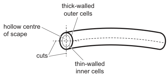
Which diagram shows the piece that is placed in the sucrose solution with the highest water potential?
Options
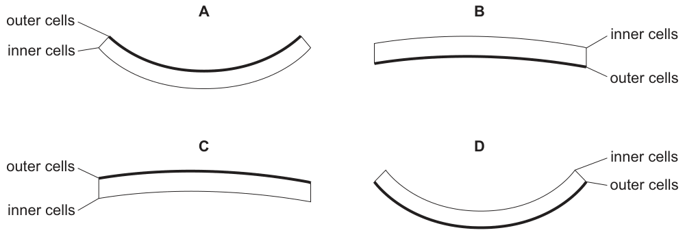
Working
- In the scape, the outer cells have thick walls and the inner cells have thin walls; the outer surface is naturally convex.
- The sucrose solution with the highest water potential is the most dilute, so it has the least negative and is closest to pure water.
- In this solution, both cell types take in water by osmosis and become fully turgid.
- The thin-walled inner cells expand more than the thick-walled outer cells (the rigid thick walls restrict expansion of the outer cells).
- The inner surface therefore becomes longer than the outer surface, so the strip curls more strongly than before — and the outer cells remain on the convex side.
- This matches diagram A: strongly curved with outer cells on the convex side.
Answer
A
A
Background Concept
Water potential () describes the tendency of water to move by osmosis. Pure water has kPa; solutions have more negative values, and the higher the solute concentration, the more negative the water potential.
When a plant cell is placed in a solution:
- → water leaves the cell → the protoplast shrinks and pulls away from the wall (plasmolysis).
- → water enters the cell → the protoplast presses harder on the wall (turgid).
- → no net water movement (incipient plasmolysis / flaccid).
Plant cell walls are elastic. Thin-walled cells can swell considerably when they take up water, while thick-walled cells resist expansion because their rigid cellulose/lignin framework limits how far the protoplast can push the wall outward.
Understanding the Question
The dandelion scape is a hollow tube whose wall has two distinct layers:
- Thick-walled outer cells (originally on the convex/outer side of the natural curve)
- Thin-walled inner cells (originally on the concave/inner side of the natural curve)
A short longitudinal strip is cut from this tube and immersed in one of several sucrose solutions. The question asks which shape (A–D) the strip will have when it has been placed in the solution of highest water potential (i.e. the most dilute sucrose solution).
The command word is which diagram shows…, so the task is to predict the final shape, not to explain all four options. However, reasoning through the contrast with the other options makes the logic clear.
Approach
- Decide which cells will gain or lose the most water in the most dilute (highest-) solution.
- Compare the amount each cell type can change in size, given the wall thickness constraint.
- Translate any difference in surface length into a change in curvature of the whole strip.
- Identify the original orientation (outer cells on the convex side) so the final diagram is correctly labelled.
Step-by-Step Reasoning
Step 1 — Direction of water movement.
A sucrose solution of highest water potential is the most dilute, so is least negative. Plant cell sap typically has a of around kPa or so; any sucrose solution weaker than that is effectively "hypotonic" to the cell. Water therefore enters the cells by osmosis through the partially permeable cell-surface membrane.
Step 2 — Both sides become turgid, but by different amounts.
With water entering on both faces, the cells on each side swell. The crucial asymmetry is wall rigidity:
- Outer cells: thick walls → limited expansion; the protoplast pushes the wall outward, but the wall resists.
- Inner cells: thin walls → larger expansion; the protoplast can push the thin wall outward much more readily.
Step 3 — Differential length change drives curvature.
Because the inner surface elongates more than the outer surface, the strip must bend so that the longer side (inner cells) lies on the outside of the curve. The natural convex side (outer cells) becomes even more strongly convex, and the strip curls more tightly than at the start.
Step 4 — Match to the options.
The final state should be strongly curved, with the original outer (thick-walled) cells still on the convex side. That is exactly diagram A.
Why the other options are wrong:
- B (slightly curved, inner cells convex): would correspond to a low water potential where the inner cells shrink more than the outer cells, reversing the curvature only slightly.
- C (almost straight, outer cells convex): corresponds to a solution roughly isotonic to the cell sap, so neither side changes much and the strip stays near its original mild curvature.
- D (strongly curved, inner cells convex): corresponds to a very low water potential (very concentrated sucrose) — the thin-walled inner cells lose more water and shrink, pulling the strip into a tight curve the opposite way around.
Key Takeaways
- Highest water potential = most dilute solution = greatest water uptake by cells = greatest turgor.
- Differential expansion (or shrinkage) between tissues is governed by wall thickness/rigidity, not by the type of cell alone.
- In any curved plant organ, the tissue layer that expands more determines the convex side of the new shape.
- This principle underlies many classic curvature experiments (e.g. dandelion scape, Nitella internode, germinating coleoptiles), and it is also how plants move organs such as leaves and pulvini.
Common Mistakes
- Assuming both sides expand equally so the strip stays the same shape — ignoring wall thickness.
- Forgetting which surface is the original convex surface and so mis-labelling "outer" and "inner" on the diagrams.
- Confusing water potential with solute concentration: highest water potential = least negative = most dilute solution (NOT most concentrated).
- Selecting D because "it is the most curved": the curvature in D has the wrong side convex and corresponds to a low water potential, not a high one.
Things to Be Careful About
- The scape in the question is already curved; the strip is not starting flat. You are asked for the change relative to the original cut piece.
- "Highest water potential" must be interpreted as the least negative value, i.e. closest to kPa, which is the most dilute sucrose solution.
- Keep the orientation of the labels straight: in Fig. 20.1 the outer cells are on the convex (upper) side; the inner cells are on the concave (lower) side. The strip in the answer must keep that orientation.
Telomeres prevent the loss of genes during DNA replication.
Which row correctly shows cell types that contain telomeres?
Options
| typical animal cell | typical plant cell | typical bacterial cell | |
|---|---|---|---|
| A | ✓ | ✓ | ✓ |
| B | ✓ | ✓ | ✗ |
| C | ✗ | ✗ | ✓ |
| D | ✓ | ✗ | ✗ |
key
✓ = present
✗ = not present
Working
Telomeres are repetitive DNA sequences found at the ends of linear eukaryotic chromosomes. They protect coding regions from being shortened during DNA replication (the end-replication problem).
- Typical animal cell: eukaryotic → linear chromosomes → telomeres present ✓
- Typical plant cell: eukaryotic → linear chromosomes → telomeres present ✓
- Typical bacterial cell: prokaryotic → circular DNA (no ends) → telomeres absent ✗
Answer
B
B
Background Concept
Telomeres are short, repetitive, non-coding nucleotide sequences (in humans, the repeat is TTAGGG) located at the two ends of every linear eukaryotic chromosome. They serve as protective caps, together with associated proteins (the shelterin complex), that stop the chromosome ends from being recognised as DNA damage and prevent them from fusing with neighbouring chromosomes or being degraded.
Their existence is a direct consequence of the end-replication problem. DNA polymerase can only synthesise DNA in the 5' → 3' direction and requires an RNA primer to start. On the lagging strand, when the final RNA primer at the chromosome end is removed, there is no upstream 3' end for DNA polymerase to extend from, so the new strand is shorter than the template. Without telomeres, essential coding sequences at the chromosome ends would be progressively lost with every round of replication.
A separate enzyme, telomerase, lengthens telomeres in cells that need to divide many times (germ cells, stem cells, some white blood cells) by adding the repeat sequence using its own RNA template.
Understanding the Question
The question lists three cell types — a typical animal cell, a typical plant cell, and a typical bacterial cell — and asks which of them contain telomeres. The mark key shows ✓ = present and ✗ = not present, so we must decide for each cell type whether it has telomeres.
The key biological distinction is:
- Eukaryotes (animals, plants, fungi, protists) have their DNA packaged into linear chromosomes inside a nucleus → telomeres are present.
- Most prokaryotes (bacteria) have a single circular chromosome of DNA with no free ends → telomeres are not needed and are not present.
Approach
The reasoning strategy is:
- Identify whether each cell type is eukaryotic or prokaryotic.
- Recall that telomeres are a feature of linear eukaryotic chromosomes.
- Tick the box for every eukaryotic cell type, cross the box for the prokaryote.
Step-by-Step Reasoning
- Typical animal cell — eukaryotic, with linear chromosomes packaged by histones in a nucleus. Each chromosome has two ends, so it needs telomeres. → ✓
- Typical plant cell — also eukaryotic, with linear chromosomes in a nucleus. Same logic as animal cells. → ✓
- Typical bacterial cell — prokaryotic, with a single circular DNA molecule (plus often small circular plasmids). A circle has no ends, so the end-replication problem does not arise and telomeres are absent. Telomerase is also absent in bacteria. → ✗
The only row with ✓, ✓, ✗ is row B.
Key Takeaways
- Telomeres are found on linear eukaryotic chromosomes in both animal and plant cells.
- Prokaryotes (bacteria) have circular DNA and therefore do not have telomeres.
- The function of telomeres is tied to the end-replication problem of linear DNA, not to any feature unique to animals.
Common Mistakes
- Choosing A — assumes all cell types have telomeres. This ignores that bacteria have circular DNA.
- Choosing C or D — reverses the animal cell, forgetting that animal cells are eukaryotic and possess linear chromosomes.
- Confusing telomeres with centromeres (the central region joining sister chromatids) or with the telomerase enzyme (only present in cells that actively maintain telomere length, not in all cells with telomeres).
Things to Be Careful About
- "Telomere" refers to the repetitive DNA sequence at the chromosome end, not the centromere.
- The presence of telomeres is determined by the shape of the chromosome (linear vs circular), not by the kingdom the organism belongs to.
- A few bacteria do have linear chromosomes (e.g. Borrelia, some Streptomyces species), but these are exceptions; "typical" bacterial cells in A-Level Biology are taken to have circular DNA.
Which events listed are part of mitosis?
1 interphase
2 prophase
3 cytokinesis
Options
A 1, 2 and 3
B 1 and 2 only
C 1 only
D 2 only
Working
Mitosis is defined as the division of the nucleus and consists of four stages: prophase, metaphase, anaphase and telophase.
- 1 (interphase) — occurs before mitosis. The cell grows, DNA is replicated during S phase, and proteins are synthesised, but no nuclear division takes place. Interphase is not part of mitosis.
- 2 (prophase) — the first stage of mitosis: chromosomes condense, the nuclear envelope breaks down, and the spindle forms.
- 3 (cytokinesis) — the division of the cytoplasm, which follows telophase. It is a separate process from mitosis.
Only prophase is part of mitosis.
Answer
D
D
Background Concept
The cell cycle is the series of events that takes place between one cell division and the next. It is divided into two major phases:
- Interphase — the long, preparatory period during which the cell grows, carries out its normal functions, replicates its DNA (in S phase), and produces the proteins and organelles needed for division. Interphase has three sub-phases: G1, S and G2. It is not a dividing stage.
- M (mitotic) phase — the period of actual division, which itself has two parts:
- Mitosis — the division of the nucleus into two genetically identical daughter nuclei. It has four stages: prophase, metaphase, anaphase and telophase.
- Cytokinesis — the division of the cytoplasm to produce two separate daughter cells.
The key point that catches students out is that the term "mitosis" refers strictly to nuclear division — cytokinesis is a separate, overlapping process that is not part of mitosis itself.
Understanding the Question
This is a multiple-choice question asking which of three listed events form part of mitosis. The list contains:
- interphase (a cell-cycle stage that precedes mitosis)
- prophase (the first stage of mitosis)
- cytokinesis (cytoplasmic division that follows telophase)
The command word is "part of", so the answer must be events that actually occur within the mitotic stage of the cell cycle, not events that merely surround it.
Approach
Apply the strict definition of mitosis = nuclear division. Anything that is preparation (interphase) or the cytoplasmic event that follows (cytokinesis) is excluded. Only prophase, from the list, satisfies the definition.
Step-by-Step Reasoning
- Interphase (1) is rejected. Although it is part of the cell cycle, it precedes mitosis. The chromosome behaviour characteristic of mitosis (condensation, alignment, separation) does not occur here. The mark scheme treats interphase as a separate cell-cycle stage.
- Prophase (2) is accepted. Prophase is the first of the four mitotic stages: chromatin condenses into visible chromosomes (each with two sister chromatids), the nucleolus disappears, the nuclear envelope breaks down, and the spindle begins to form.
- Cytokinesis (3) is rejected. Cytokinesis overlaps with the end of telophase but is biochemically and structurally a different process (actin–myosin ring in animal cells, cell plate in plant cells). The cell-cycle framework treats it as a distinct event following mitosis, not as a stage of mitosis.
Therefore only 2 is part of mitosis, giving D.
Key Takeaways
- Mitosis = nuclear division only, with four stages: Prophase, Metaphase, Anaphase, Telophase.
- Interphase and cytokinesis are part of the cell cycle but are not stages of mitosis.
- Use the precise definition when answering exam questions on the cell cycle — examiners deliberately test this distinction.
Common Mistakes
- Choosing A (1, 2 and 3) by treating the entire cell cycle as "mitosis". This is the common layperson usage, but CIE requires the strict definition.
- Choosing B (1 and 2) by including interphase because "the cell is getting ready to divide". Preparation is not the same as division.
- Choosing C (1 only) by misunderstanding the question entirely.
- Conflating cytokinesis with telophase — cytokinesis starts in late anaphase/telophase but is a separate process.
Things to Be Careful About
- CIE mark schemes accept only the strict definition of mitosis. Do not be tempted to write "interphase is part of mitosis" even though it is part of the cell cycle.
- Cytokinesis, although it overlaps in time with the end of mitosis, is consistently treated by CIE as a separate event.
- A useful mnemonic for the four mitotic stages is PMAT (Prophase, Metaphase, Anaphase, Telophase); interphase and cytokinesis sit outside this acronym.
The photomicrograph shows 17 cells in different stages of the mitotic cell cycle.
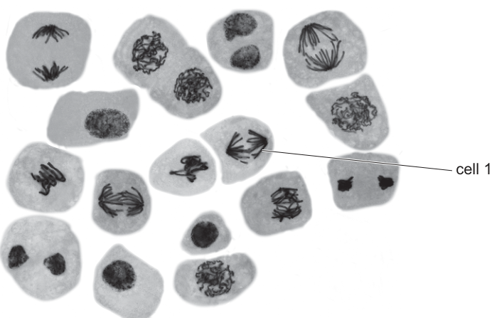
Which row is correct for this photomicrograph?
Options
| the role of a centromere in cell 1 | the proportion of the cells in the photomicrograph in telophase | |
|---|---|---|
| A | attaches daughter chromosomes to spindle fibres | |
| B | holds sister chromatids together | |
| C | attaches daughter chromosomes to spindle fibres | |
| D | holds sister chromatids together |
Working
Role of the centromere in cell 1
Cell 1 is in anaphase — the two sets of chromosomes have separated and are moving towards opposite poles of the spindle. Once sister chromatids separate, each is called a daughter chromosome. The centromere (with its kinetochore) is the structure that attaches each daughter chromosome to the spindle fibres. "Holds sister chromatids together" is the role of the centromere before anaphase (prophase and metaphase), so options B and D are wrong.
Proportion of cells in telophase
Telophase cells are recognised by two reforming nuclei within one cell (chromosomes decondensing, nuclear envelope re-forming). Counting such cells in the micrograph gives a number ≥ 4 out of 17:
so the proportion is > 0.2.
Answer
A
A
Background Concept
Chromosome structure and the centromere. Before DNA replication (in S phase of interphase), each chromosome is a single DNA molecule. After replication, each chromosome consists of two identical sister chromatids joined at a centromere (the constricted region, with associated kinetochore proteins that bind microtubules). Until anaphase, the centromere's job is to hold the two sister chromatids together.
Mitosis stages in brief.
- Prophase: chromosomes condense; nuclear envelope breaks down; spindle forms.
- Metaphase: chromosomes (still as paired sister chromatids) line up at the equator.
- Anaphase: the centromeres split and sister chromatids separate, becoming independent daughter chromosomes that are pulled to opposite poles by shortening spindle fibres attached at the kinetochore.
- Telophase: chromosomes decondense at each pole; nuclear envelopes re-form around the two sets, producing two nuclei within one cell before cytokinesis.
So the centromere has two distinct roles depending on stage:
- Prophase and metaphase: holds sister chromatids together.
- Anaphase onwards (and in each daughter cell): attaches a daughter chromosome to the spindle fibre (via the kinetochore) so it can be moved.
Recognising stages in a micrograph. Interphase cells are large with a clear, intact nucleus and uncondensed chromatin. Mitotic cells are smaller, denser, and lack a clear nuclear envelope. Within mitosis: prophase shows condensing thread-like chromosomes; metaphase shows a line of chromosomes at the equator; anaphase shows two distinct groups moving apart; telophase shows two reforming nuclei in one cell.
Understanding the Question
The question shows a light-micrograph of 17 cells (typical of an onion root tip preparation) at various stages of the cell cycle and asks which statement is correct. There are two parts to each option:
- The role of the centromere in cell 1 — we must first identify the stage of cell 1, then match the centromere's role to that stage.
- The proportion of cells in telophase — we must count telophase cells in the image and decide whether the proportion is greater than, or less than, 0.2 (i.e. whether there are more or fewer than about 3.4 cells in telophase).
The command word "Which row is correct" is typical of an MCQ; only one of A–D is fully correct.
Approach
- Identify cell 1's stage from chromosome arrangement in the photomicrograph.
- Match the centromere's role to that stage (anaphase → attaches daughter chromosomes to spindle fibres; pre-anaphase → holds sister chromatids together).
- Count cells showing the two-nuclei, decondensing-chromatin appearance of telophase.
- Compare that count with the 0.2 threshold (3.4 cells out of 17).
Step-by-Step Reasoning
Step 1 — Identify cell 1. Cell 1 shows two separated groups of chromosomes within a single cell with no nuclear envelope around them. This is anaphase, not metaphase (no equatorial plate) and not telophase (no reforming nuclei).
Step 2 — Role of the centromere in anaphase. The centromere (with its kinetochore) is the attachment point for spindle fibres. By anaphase, sister chromatids have separated and each is now a daughter chromosome. The centromere therefore attaches daughter chromosomes to spindle fibres, allowing them to be pulled polewards. This eliminates options B and D, which state the prophase/metaphase role.
Step-by-step verification: "Holds sister chromatids together" is only correct up to the start of anaphase. From anaphase onwards, once the centromere has divided, the chromatids are no longer held together and the centromere's function is to attach a chromosome to the spindle.
Step 3 — Count telophase cells. Telophase cells are recognised by two compact, dark chromatin masses within a single cell boundary, where the chromosomes have reached the poles and begun to decondense as the nuclear envelope re-forms. Looking carefully across all 17 cells, at least 4 cells show this two-nuclei appearance.
Step 4 — Compare the proportion to 0.2.
So the proportion is greater than 0.2, eliminating option C.
Step 5 — Choose the row. Only option A has both the correct centromere function for an anaphase cell and the correct proportion threshold. The answer is A.
Key Takeaways
- The centromere's role changes with stage: it holds sister chromatids together in prophase and metaphase, and attaches daughter chromosomes to spindle fibres in anaphase and beyond.
- A cell is in anaphase when two separate groups of chromosomes are moving towards opposite poles; in telophase when two reforming nuclei appear in one cell.
- A threshold of 0.2 for 17 cells corresponds to about 3.4 cells, so any count of 4 or more exceeds the threshold.
- When answering MCQs of this type, tackle each column independently: it is often easier to eliminate options by working on one statement at a time.
Common Mistakes
- Confusing the stages of centromere action. A common error is to say the centromere "holds sister chromatids together" throughout mitosis. It does this only until anaphase begins; once chromatids separate, that is no longer its function.
- Confusing anaphase with telophase. Cells with two chromosome groups are anaphase only if the chromosomes are still condensed and there is no nuclear envelope. The moment two distinct nuclear envelopes begin to re-form, the cell is in telophase.
- Misidentifying interphase as telophase. Large cells with one big pale nucleus and visible nucleolus are in interphase, not telophase. Telophase cells are smaller, denser, and have two dark masses.
- Off-by-one counting. With 17 cells, a proportion of 0.2 equals 3.4 cells. Rounding incorrectly to 3 (below 0.2) when 4 are present will reverse the answer.
Things to Be Careful About
- "Sister chromatids" and "daughter chromosomes" refer to the same physical structures at different times — before and after centromere splitting in anaphase. The terminology change matters because the centromere's functional description depends on it.
- The image must be examined for every cell, not just the obvious ones — small, dense cells at the edges can be telophase.
- For MCQs, always evaluate both parts of the option before committing; an option that is half-right is still wrong.
The flow diagram shows some of the stages in the replication of a lagging strand of DNA.
Which stage of DNA replication uses the enzyme DNA ligase?
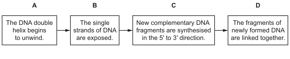
Options
A Stage A
B Stage B
C Stage C
D Stage D
Working
DNA ligase joins (seals) the sugar–phosphate backbones between adjacent DNA fragments. On the lagging strand these are the Okazaki fragments, which must be linked together to form a continuous strand. In the flow diagram, this corresponds to the final box where the fragments of newly formed DNA are linked together.
Answer
D
D
Background Concept
DNA replication is semi-conservative: each new double helix contains one parental strand and one newly synthesised strand. Because DNA polymerase can only add nucleotides to a free 3′-OH, it synthesises DNA strictly in the 5′ → 3′ direction. On the leading strand this proceeds continuously towards the replication fork. On the lagging strand, however, synthesis runs away from the fork and must be carried out in short pieces — the Okazaki fragments — each of which is started by a short RNA primer laid down by primase and then extended by DNA polymerase.
Several enzymes cooperate at the replication fork:
- Helicase unwinds the parental double helix, breaking the hydrogen bonds between complementary bases.
- Single-strand binding proteins (SSBs) stabilise the exposed single strands and prevent them from re-annealing.
- Primase synthesises short RNA primers so that DNA polymerase has a 3′-OH to extend from.
- DNA polymerase extends each primer by adding complementary DNA nucleotides in the 5′ → 3′ direction.
- DNA ligase seals the nicks between adjacent Okazaki fragments by forming phosphodiester bonds between their sugar–phosphate backbones, producing a continuous lagging strand.
Understanding the Question
The question supplies a four-box flow diagram of lagging-strand replication and asks the candidate to identify which box represents the action of DNA ligase. The command word is implicit ("which stage…") and only one option is correct. The student must know that DNA ligase joins separate DNA fragments, not that it unwinds DNA, exposes single strands, or synthesises new nucleotides.
Approach
For each box, identify which enzyme is responsible, then match DNA ligase to the appropriate step:
- Box A — "The DNA double helix begins to unwind" → helicase.
- Box B — "The single strands of DNA are exposed" → the result of helicase/SSB action, no single named enzyme.
- Box C — "New complementary DNA fragments are synthesised in the 5′ to 3′ direction" → DNA polymerase (on the lagging strand these are Okazaki fragments).
- Box D — "The fragments of newly formed DNA are linked together" → DNA ligase, which forms the phosphodiester bonds that join adjacent Okazaki fragments into one continuous strand.
Step-by-Step Reasoning
- The diagram depicts events on the lagging strand, where DNA is made in discontinuous pieces (Okazaki fragments) because DNA polymerase works only 5′ → 3′.
- Helicase performs stage A by separating the two parental strands at the replication fork.
- The exposed single strands (stage B) are the substrate for the next enzyme; they are held open by single-strand binding proteins.
- DNA polymerase carries out stage C, synthesising each new complementary DNA fragment in the 5′ → 3′ direction, producing a series of separated Okazaki fragments.
- Each Okazaki fragment still has nicks in the sugar–phosphate backbone where it meets the next fragment. DNA ligase performs stage D, catalysing the formation of phosphodiester bonds that seal these nicks and produce a continuous DNA strand.
- Therefore the box that uses DNA ligase is D.
Key Takeaways
- DNA ligase seals nicks between adjacent DNA fragments by forming phosphodiester bonds.
- On the lagging strand, ligase joins Okazaki fragments into a continuous strand.
- Each box in a replication flow diagram corresponds to a specific enzyme(s): unwind → helicase; synthesise → DNA polymerase; join → DNA ligase.
- DNA polymerase can only add nucleotides in the 5′ → 3′ direction, which is why the lagging strand is made discontinuously.
Common Mistakes
- Choosing C because students remember that "DNA polymerase makes new DNA" but forget that, on the lagging strand, this produces separate fragments that still need to be joined.
- Choosing A or B because helicase (which unwinds DNA) is wrongly attributed to DNA ligase.
- Confusing DNA ligase with RNA ligase or with restriction enzymes.
Things to Be Careful About
- Read each option against the enzyme's function, not just the keyword "DNA".
- "Linking" / "joining" fragments is the defining verb for DNA ligase; "synthesising" new nucleotides is the role of DNA polymerase.
- The question is about the lagging strand specifically, but the same ligase action applies anywhere two DNA fragments must be joined (e.g. during DNA repair).
In a gene, the 20 common amino acids are each coded for by three bases in adjacent nucleotides.
Why is there a difference between the number of amino acids and the number of possible triplet codes?
Options
A 44 triplets have no functions.
B Some amino acids are coded for by more than one triplet.
C Some triplets have unknown functions.
D There are 44 ‘start’ and ‘stop’ triplets.
Working
There are 4 bases, so the number of possible triplet codes is . Only 20 amino acids are coded for, leaving "extra" triplets.
These 44 extra triplets are accounted for by the fact that most amino acids are specified by more than one codon — the code is degenerate. (Three of the 64 codons are stop signals, so degeneracy still leaves more than 20 sense codons distributed across 20 amino acids.)
- A: Wrong — the 44 "extra" triplets are not functionless; they either code for amino acids or act as stop codons.
- B: Correct — degeneracy means several triplets specify the same amino acid, so 64 triplets can code for only 20 amino acids.
- C: Wrong — all 64 codons have defined functions (61 sense codons + 3 stop codons).
- D: Wrong — there are 3 stop codons and 1 start codon, not 44.
Answer
B
B
Background Concept
The genetic code is the set of rules by which the sequence of bases in DNA/mRNA is translated into a sequence of amino acids during protein synthesis. Each amino acid is specified by a sequence of three consecutive bases called a codon (or triplet). Because there are 4 different bases (A, U/T, G, C) and codons are three bases long, the total number of possible triplet codes is .
However, only 20 common amino acids occur in proteins, and 64 is much larger than 20. The code is therefore described as degenerate — most amino acids are encoded by more than one codon. For example, leucine is coded by six different codons (UUA, UUG, CUU, CUC, CUA, CUG), and serine by four. Only two amino acids (methionine and tryptophan) have a single codon each, and methionine's codon (AUG) doubles as the start signal.
Of the 64 codons, 61 are sense codons (they code for amino acids) and 3 are stop codons (UAA, UAG and UGA) that signal the end of translation. The degeneracy is the reason the number of codons (64) exceeds the number of amino acids (20).
Understanding the Question
The stem gives the key fact: each of the 20 common amino acids is encoded by three adjacent bases. The question asks why the number of amino acids (20) is smaller than the total number of possible triplet codes (64). The candidate must explain the biological reason for this numerical mismatch.
The command word is "Why" — a short conceptual explanation is required, and here the answer is presented as a single MCQ option.
Approach
To answer, calculate the number of possible triplet codes (), compare it with the number of amino acids (20), and recall that the code is degenerate. The 44 "extra" codons are not waste — they are used to specify the same amino acid as other codons (degeneracy) or to act as stop signals.
Step-by-Step Reasoning
- Total possible triplets = .
- Number of amino acids = 20.
- Difference = .
- The code is degenerate: many amino acids have more than one codon, so 64 codons can map onto only 20 amino acids.
- Option A claims 44 triplets have no function — false, all 64 codons are either sense codons or stop codons.
- Option B states that some amino acids are coded for by more than one triplet — true, and this is the definition of degeneracy.
- Option C says some triplets have unknown functions — false; every codon has been assigned a function.
- Option D says there are 44 start and stop codons — false; there are only 3 stop codons and 1 start codon, totalling 4.
Key Takeaways
- The genetic code is degenerate: multiple codons (triplets) can code for the same amino acid.
- 64 possible codons arise from combinations of four bases.
- Of the 64 codons, 61 are sense codons (specifying amino acids) and 3 are stop codons (UAA, UAG, UGA).
- Only methionine (AUG) and tryptophan have a single codon; most amino acids have 2–6 synonymous codons.
Common Mistakes
- Confusing degeneracy with redundancy — the code is degenerate, not redundant; every codon still has a unique sequence.
- Thinking many codons are "unused" or have no biological role — in fact, every codon has a defined function.
- Forgetting that degeneracy is the reason 64 > 20, not the existence of start/stop codons alone (only 4 codons are start/stop, not 44).
Things to Be Careful About
- Be precise with numbers: 4 bases → codons, 61 sense codons, 3 stop codons.
- The start codon (AUG) is also a sense codon for methionine — it is not in addition to the 61 sense codons.
- The word "degenerate" in this context is the CIE term for the genetic code; it does not mean the code is broken or inefficient.
How is a primary transcript modified to form mRNA?
Options
A It becomes longer as exons are joined together.
B It becomes shorter as exons are removed.
C It becomes longer as introns are joined together.
D It becomes shorter as introns are removed.
Working
The primary transcript (pre-mRNA) contains both exons (coding sequences) and introns (non-coding sequences). During processing, introns are excised and the remaining exons are joined together. Because introns are removed, the mature mRNA is shorter than the primary transcript.
Answer
D
D
Background Concept
In eukaryotes, transcription produces a primary transcript (pre-mRNA) that is an exact copy of the DNA template strand, including both coding and non-coding regions. The coding regions are called exons (expressed sequences) and the non-coding regions are called introns (intervening sequences). Before the mRNA leaves the nucleus and is translated, it must be processed.
The key modification is splicing: introns are removed and exons are joined end-to-end to produce a continuous coding sequence. The mature mRNA is therefore shorter than the primary transcript. Other modifications include the addition of a 5' cap and a 3' poly-A tail, but these do not change the core question here — splicing is the step that alters the length.
Understanding the Question
This is a straightforward MCQ. The candidate must identify what happens to the primary transcript during its conversion to mature mRNA, specifically in terms of length and which sequences are removed. The four options pair two ideas: (1) which sequences are involved — exons or introns — and (2) the effect on length — longer or shorter. Only one pairing is biologically correct.
Approach
Recall the definitions:
- Exons = coding sequences, retained in mature mRNA
- Introns = non-coding sequences, removed during splicing
Therefore the removal of introns makes the molecule shorter.
Step-by-Step Reasoning
- Option A is wrong on two counts: exons are NOT removed (they are kept and joined), and the molecule does not become longer.
- Option B is wrong because exons are not removed — they are the sequences that are joined together to form the mature mRNA.
- Option C is wrong because introns are removed, not joined. A transcript that retained introns would be the unprocessed primary transcript, which is not what mature mRNA looks like.
- Option D correctly states that the mature mRNA is shorter than the primary transcript because introns have been removed by splicing. This matches the mark scheme.
Key Takeaways
- Splicing removes introns and joins exons in the primary transcript to form mature mRNA.
- Mature mRNA is therefore shorter than the primary transcript.
- Introns are described as 'non-coding' because they do not code for amino acids in the final protein product; exons are the 'expressed' sequences that do.
Common Mistakes
- Confusing introns and exons — students sometimes think exons are the non-coding parts and introns are the coding parts, leading them to choose B or C.
- Forgetting that splicing shortens the transcript; the mature mRNA is shorter, not longer, than the primary transcript.
Things to Be Careful About
- The 5' cap and poly-A tail are added during processing, but they do not involve removing introns. The question specifically asks about length change due to the joining/removal of sequences, which is splicing.
- Splicing happens in the nucleus before the mRNA exits to be translated at a ribosome in the cytoplasm.
The diagram shows protein synthesis in a prokaryote.
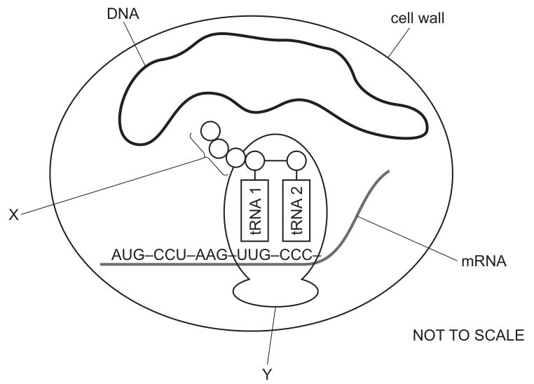
Which row is correct for molecule X, structure Y and the anticodon of tRNA 1?
Options
| molecule X | structure Y | anticodon of tRNA 1 | |
|---|---|---|---|
| A | amino acid | 80S ribosome | AAC |
| B | polypeptide | 80S ribosome | UUG |
| C | polypeptide | 70S ribosome | AAC |
| D | protein | 70S ribosome | UUG |
Working
Molecule X — X is drawn as a chain of several circles (amino acids) joined together. A single circle would be an amino acid; a folded, functional chain is a protein. An unfinished chain of amino acids linked by peptide bonds is a polypeptide, so options A and D are wrong on this row.
Structure Y — Y is the ribosome. The question states the diagram shows a prokaryote. Prokaryotic ribosomes are 70S (a 50S large subunit + 30S small subunit); eukaryotic ribosomes are 80S. This rules out A and B.
Anticodon of tRNA 1 — tRNA 1 is base-pairing with an mRNA codon in an antiparallel fashion. The anticodon given (AAC) is the complementary sequence to the codon tRNA 1 is sitting on, read in the 3′→5′ direction. (UUG pairs antiparallel: 3′-U-U-G-5′ with 3′-A-A-C-5′.)
Only row C satisfies all three: polypeptide, 70S ribosome, and an anticodon consistent with antiparallel pairing to the mRNA codon.
Answer
C
C
Background Concept
Translation is the second stage of protein synthesis. After transcription has produced an mRNA, the mRNA is read by the ribosome in codons of three bases, always in the 5′ → 3′ direction. Each codon specifies either an amino acid or a stop signal.
tRNA molecules act as adaptors. At one end each tRNA carries a specific amino acid; at the other end it carries a three-base anticodon that base-pairs with the mRNA codon. Crucially, the codon–anticodon pairing is antiparallel: the first base of the codon (5′ end) pairs with the third base of the anticodon (3′ end).
The ribosome has three tRNA-binding sites:
- A site (aminoacyl) – holds the incoming tRNA carrying the next amino acid.
- P site (peptidyl) – holds the tRNA attached to the growing polypeptide chain.
- E site (exit) – the empty tRNA leaves from here.
The ribosome translocates 5′ → 3′ along the mRNA; the P-site tRNA carries the polypeptide, so the tRNA drawn attached to the chain of amino acids must be in the P site.
Ribosome size is a useful diagnostic:
- Prokaryotic ribosome = 70S (made up of a 50S + a 30S subunit).
- Eukaryotic ribosome = 80S (made up of a 60S + a 40S subunit).
A polypeptide is a chain of amino acids linked by peptide bonds. A protein is one (or more) polypeptide(s) folded into a functional 3-D shape. A single amino acid is just a monomer.
Understanding the Question
The diagram shows a snapshot of translation inside a prokaryotic cell. We are given three unknowns:
- X – the chain of circles attached to tRNA 1 (the growing product of translation).
- Y – the large oval structure holding the two tRNAs in place on the mRNA (the ribosome).
- The anticodon of tRNA 1 – the three bases at the bottom of tRNA 1, which must be complementary to whichever mRNA codon tRNA 1 is currently paired with.
The command word is "which row is correct" – we have to pick the single option in which all three statements are biologically accurate for what is shown.
Approach
Treat each column of the table as a separate mini-question, eliminate options column by column, then confirm the surviving row.
- Decide what X must be (amino acid, polypeptide or protein).
- Decide what Y must be (70S or 80S), using the "prokaryote" cue in the stem.
- Read the codon tRNA 1 is sitting on in the diagram, then apply antiparallel base pairing to get its anticodon.
- The only row that matches all three is the answer.
Step-by-Step Reasoning
Column 1 – molecule X
X is shown as several circles joined by a line – i.e. more than one amino acid. So X is not a single amino acid (eliminates A). A protein, in the strict sense used at A-level, is a polypeptide that has folded into its functional 3-D shape; the diagram shows a linear chain, not a folded molecule, so X is best described as a polypeptide (eliminates D on this column).
Column 2 – structure Y
Y is the ribosome. The stem explicitly tells us this is happening in a prokaryote. Prokaryotic ribosomes are 70S; 80S ribosomes are found in eukaryotic cytoplasm. So Y = 70S ribosome (eliminates A and B on this column).
Column 3 – anticodon of tRNA 1
tRNA 1 is base-pairing with the mRNA codon it sits over. Because codon–anticodon pairing is antiparallel:
- The 5′ base of the codon pairs with the 3′ base of the anticodon.
- A pairs with U, C pairs with G.
Working through the antiparallel pairing to obtain the anticodon 3′→5′, the sequence given in row C, AAC, is the antiparallel complement of the codon tRNA 1 is reading (i.e. UUG read antiparallel gives 3′-AAC-5′). The other option in the table, UUG, is actually a codon – it is the sequence on the mRNA, not the anticodon on the tRNA. That distinction is what eliminates rows B and D.
Combining the three: only row C has polypeptide AND 70S ribosome AND a valid anticodon. Rows A, B and D each fail on at least one column.
Key Takeaways
- A growing chain of amino acids attached to a tRNA is a polypeptide, not a protein (a protein is folded and functional) and not an amino acid (a single monomer).
- Prokaryotes have 70S ribosomes; eukaryotes have 80S. This single fact distinguishes many translation questions.
- The codon–anticodon interaction is antiparallel and complementary: A–U, C–G. Always read the codon 5′→3′ and the anticodon 3′→5′ when writing the pair on paper.
- A useful trap to recognise: the codon (mRNA) and the anticodon (tRNA) look superficially similar but the tRNA sequence is the reverse complement of the codon, not the direct complement.
Common Mistakes
- Writing the anticodon as the direct complement of the codon (e.g. UUC for codon AAG) instead of the reverse complement (CUU). Forgetting the antiparallel orientation gives the wrong letters.
- Calling X a "protein" because it will eventually become one. During synthesis it is a polypeptide; the term "protein" is reserved for the folded, functional product.
- Choosing 80S for Y because the cell is drawn at large scale, or because ribosomes are commonly remembered as 80S from eukaryotic biology. The stem explicitly says prokaryote – use 70S.
- Picking the codon (e.g. UUG) when the question asks for the anticodon. The two sequences are related but live on different molecules.
Things to Be Careful About
- Direction matters: 5′–3′ of the mRNA is fixed. When writing an anticodon, it is conventional to give it 3′→5′ as it pairs with the codon, but always check the convention used in the source you are studying.
- "S" in 70S/80S is a Svedberg unit (a measure of how a particle sediments in a centrifuge), not a simple sum of the subunits – 50S + 30S gives 70S, not 80S, because sedimentation rate depends on shape as well as mass.
- The chain attached to the P-site tRNA always represents a polypeptide, regardless of how many amino acids have so far been joined – even a dipeptide or tripeptide is still called a polypeptide in this context.
The photomicrographs show sections of two organs of a plant.
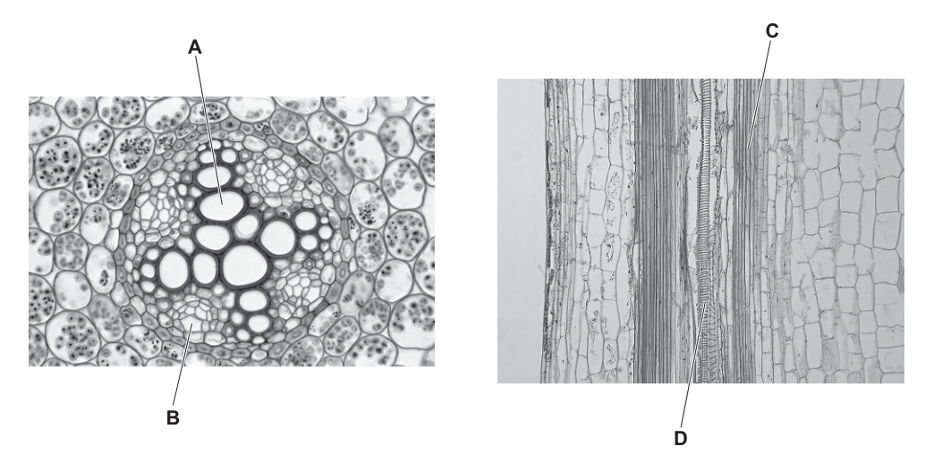
Which letter identifies a cell found with companion cells in a tissue in a root?
Options
A A
B B
C C
D D
Working
Phloem tissue is composed of sieve tube elements and companion cells, so the cell found with companion cells is a sieve tube element. In a transverse section of a dicotyledonous root, phloem is located between the arms of the X-shaped xylem. In the left photomicrograph, label A points to a large thick-walled xylem vessel at the centre of the stele, and label B points to the small cells of the phloem between the xylem arms. Label C (longitudinal section) is a xylem vessel with spiral thickening, and label D is parenchyma.
Answer
B
B
Background Concept
In vascular plants, the two main transport tissues are xylem and phloem.
- Xylem carries water and mineral ions from roots to shoots. It is composed of dead, hollow, lignified xylem vessel elements (and tracheids). In transverse section they appear as large, thick-walled, empty-looking cells.
- Phloem carries assimilates (mainly sucrose) from sources to sinks. It is composed of living sieve tube elements joined end-to-end into sieve tubes, together with their companion cells. Companion cells carry out the metabolism for the enucleate sieve tube elements and load/unload sugars at source and sink.
Because companion cells are a defining feature of phloem, the cell that is always found with a companion cell is a sieve tube element.
Understanding the Question
The question shows two photomicrographs. The left one is a transverse section (TS) of a young dicotyledonous root, with an X-shaped arrangement of xylem at the centre of the vascular cylinder (stele). The right one is a longitudinal section (LS) showing xylem vessels with spiral/helical secondary wall thickening. The four labels point to different cell types, and we must pick the one that is a sieve tube element in the phloem of the root.
Approach
- Recall that companion cells occur only alongside sieve tube elements in phloem, so we need to locate the phloem in the root TS.
- In a dicot root, phloem sits in small groups between the arms of the X-shaped xylem, while the xylem itself forms the central X.
- Match each label to its tissue type and pick the one that is phloem in the root.
Step-by-Step Reasoning
- Label A is in the centre of the stele in the root TS. It points to a very large, thick-walled, empty cell — characteristic of a xylem vessel in TS. Xylem does not have companion cells, so A is wrong.
- Label B is in the root TS, between the arms of the X-shaped xylem. This is exactly the position of phloem in a dicot root, and the small cells indicated are sieve tube elements together with their companion cells. This matches the description in the question.
- Label C is in the LS photomicrograph and points to a tubular structure with a spiral/helical band of thickening — a xylem vessel in longitudinal view. No companion cells here.
- Label D is in the LS photomicrograph pointing to a thin-walled, undifferentiated parenchyma cell next to the xylem. Parenchyma is not associated with companion cells.
Only B represents the sieve tube element/companion cell complex of phloem in a root, so B is correct.
Key Takeaways
- Companion cells are diagnostic of phloem; the cell found with a companion cell is a sieve tube element.
- In a dicot root TS, xylem forms a central X and phloem sits in small groups between the arms of the X.
- In LS, xylem vessels are recognised by their lignified, often spirally thickened, walls; companion cells and sieve tubes are much smaller, thin-walled, and have cytoplasmic contents.
Common Mistakes
- Choosing A because it is a large prominent cell — but it is a xylem vessel, and xylem has no companion cells.
- Choosing C because of its striking spiral thickening — but spiral thickening is a feature of xylem, not phloem.
- Confusing parenchyma (D) with phloem — parenchyma cells are general packing tissue, not associated with companion cells.
Things to Be Careful About
- Always read the question carefully: it specifies a tissue in a root, so the LS (right-hand image) is included as a distractor and is not the source of the answer.
- Companion cells are a phloem-specific feature; no other plant cell type is described as being "found with companion cells".
Which row correctly describes the pressure in the xylem and the water potential in the root hair cells used in the transport of water in a transpiring plant?
Options
| pressure in the xylem | water potential in root hair cells | |
|---|---|---|
| A | positive | negative |
| B | positive | positive |
| C | negative | negative |
| D | negative | positive |
Working
In a transpiring plant, evaporation of water from the mesophyll cell walls of the leaf creates a pull (tension) on the continuous column of water in the xylem. This pull generates a negative pressure in the xylem — the driving force described by the cohesion-tension theory.
Root hair cells contain cell sap with dissolved solutes (mineral ions, sugars), so their water potential is negative (lower than that of soil water). This water potential gradient allows water to enter the root hairs from the soil by osmosis.
Answer
C
C
Background Concept
Two key concepts govern water movement in a transpiring plant: water potential (Ψ) and xylem hydrostatic pressure.
- Water potential is the tendency of water to move from one place to another. Pure water has Ψ = 0 kPa, and any solution has Ψ < 0 kPa (negative). The more dissolved solutes, the more negative the water potential. Water always moves down a water potential gradient (from less negative to more negative) by osmosis if a selectively permeable membrane is present, or by bulk flow if not.
- Xylem pressure is the hydrostatic pressure exerted by the water column inside the xylem vessels. It can be positive (e.g. root pressure, which can produce guttation) or negative (tension generated by transpiration).
In the cohesion–tension theory of water transport, evaporation from mesophyll cell walls pulls water out of the xylem at the leaf end, creating tension (negative pressure) that is transmitted all the way down the continuous water column to the roots. Water molecules cohere to each other by hydrogen bonding and adhere to the xylem walls, so the column does not break.
Understanding the Question
The question tests two specific facts about a transpiring plant:
- The sign of the pressure inside the xylem.
- The sign of the water potential of the root hair cell contents.
The key word is transpiring: water is being lost from the leaves, so the cohesion-tension mechanism dominates (root pressure, which is positive, is not the main driver during transpiration).
Approach
- For the xylem in a transpiring plant, recall that transpiration pulls the water column under tension → negative pressure.
- For root hair cells, recall that their cytoplasm/vacuole contains dissolved solutes, so the cell's water potential must be negative (lower than that of soil water, which is also slightly negative but less so).
- Match both facts to the correct row of the table.
Step-by-Step Reasoning
- Xylem pressure in a transpiring plant is negative. As water evaporates from the mesophyll into the air spaces and out through the stomata, the curved meniscus in each mesophyll cell wall retreats, pulling on the water in the nearest xylem vessel. This tension (a pulling force) registers as a negative hydrostatic pressure. The negative pressure is transmitted down through the continuous water column to the roots because of cohesion between water molecules (hydrogen bonds) and adhesion to the lignin in xylem walls.
- Water potential of root hair cells is negative. Root hair cells absorb mineral ions (e.g. K⁺, NO₃⁻, Ca²⁺, Mg²⁺) from the soil, often by active transport, so their vacuolar sap is a concentrated solution. Because Ψ = Ψs + Ψp (solute potential is negative), and turgor pressure inside the cell only makes Ψ less negative (not positive overall), the net water potential remains negative. This negative Ψ is essential: it is more negative than soil water, so water flows into the root hair by osmosis.
- The row that gives negative pressure in the xylem AND negative water potential in root hair cells is row C.
Why the other rows are wrong:
- A — positive xylem pressure describes root pressure, which is only significant at night or in very humid conditions when transpiration is minimal; it is not the situation in a transpiring plant. The second column (negative Ψ in root hairs) is correct here, but the first column is wrong.
- B — both values listed as positive. Xylem pressure during transpiration is not positive, and root hair cell water potential cannot be positive because the cell sap contains solutes.
- D — positive water potential in root hair cells would mean the cell sap is more dilute than pure water, which is impossible for a living cell containing dissolved ions and sugars.
Key Takeaways
- In a transpiring plant, xylem water is under tension (negative pressure) — the basis of the cohesion-tension theory.
- Root hair cell water potential is negative because of dissolved solutes; this gradient drives water uptake from the soil by osmosis.
- The sign of xylem pressure can switch from negative (during transpiration) to slightly positive (root pressure, e.g. guttation) depending on conditions, so always note the physiological state of the plant.
Common Mistakes
- Confusing root pressure (positive) with the dominant xylem pressure during transpiration (negative). The question specifies a transpiring plant — the cohesion-tension pull is what matters.
- Thinking water potential is the same as solute concentration. Water potential is the combined effect of solute potential (Ψs) and pressure potential (Ψp); the cell as a whole has a negative Ψ even though turgor pressure makes it less negative than Ψs alone.
- Selecting A because students remember "water moves from roots to leaves" and assume positive pressure pushes it up; in reality, transpiration pulls the column up.
Things to Be Careful About
- Read the word transpiring carefully — it specifies that evaporation-driven tension is operating, not root pressure.
- Water potential and pressure are different quantities: pressure is a mechanical force per unit area, water potential is a thermodynamic quantity (energy per unit volume). Both can be positive or negative, but they are not interchangeable.
- The units are different: pressure in kPa, water potential also in kPa, but the scales and signs are independent.
Which row correctly identifies the definitions for each of the key terms used in transpiration?
Options
| attraction between water molecules and walls of the xylem vessel | effect created by evaporation of water from the surface of the leaf | |
|---|---|---|
| A | cohesion | adhesion |
| B | adhesion | tension |
| C | cohesion | tension |
| D | tension | cohesion |
Working
- Adhesion = the attraction between water molecules and the walls of the xylem vessel (water sticks to the vessel wall). This matches the first column.
- Cohesion = the attraction between water molecules for each other (water sticks to water) — it is not described in the table.
- Tension = the pulling/suction effect generated as water evaporates from the mesophyll cell walls and out through the stomata, pulling the water column up the xylem. This matches the second column.
- Option B places adhesion in column 1 and tension in column 2, which is the only row that is fully correct.
Answer
B
B
Background Concept
Transpiration is the loss of water vapour from a plant, mainly through the stomata in the leaves. As water evaporates from the wet cell walls of the mesophyll and diffuses out of the leaf, it creates a pulling force on the continuous column of water inside the xylem. This pulling force draws more water up from the roots to replace what was lost. Three specialised terms describe the physical forces that keep this water column intact and moving:
- Adhesion — the attractive force between water molecules and the wall of the xylem vessel. Water is slightly polar, so it forms hydrogen bonds with the hydrophilic cellulose of the vessel wall. This force helps water "cling" to the sides of the xylem and counteracts gravity.
- Cohesion — the attractive force between water molecules themselves, again due to hydrogen bonding. It is cohesion that allows water to form a continuous, unbroken column up the entire height of the xylem without the column snapping.
- Tension — the pulling (suction) force generated at the top of the water column by evaporation of water from the leaf. Because the water column is cohesive, tension at the top is transmitted all the way down to the roots, pulling water up. This is the essence of the cohesion–tension theory of water transport.
Students often confuse adhesion and cohesion because both are intermolecular attractions; the key is to remember what each water is interacting with — itself (cohesion) or the vessel wall (adhesion).
Understanding the Question
The question gives two definitions in a table and asks which row correctly identifies the corresponding key terms:
- Column 1: "attraction between water molecules and walls of the xylem vessel"
- Column 2: "effect created by evaporation of water from the surface of the leaf"
The command word "identifies" simply means match each definition to the correct term. The distractors use combinations of cohesion, adhesion and tension in the wrong slots, so a confident grasp of each definition is needed.
Approach
Match each definition directly to its term using the biological meaning:
- Water ↔ xylem wall = adhesion
- Evaporation-generated pull at the leaf = tension
Then look for the row that places these two terms in the correct order.
Step-by-Step Reasoning
- Column 1 — "attraction between water molecules and walls of the xylem vessel": this is the textbook definition of adhesion. The water molecules are being attracted to something other than themselves (the cellulose walls of the xylem), so it cannot be cohesion.
- Column 2 — "effect created by evaporation of water from the surface of the leaf": as water evaporates from the mesophyll into the air spaces and out through the stomata, the loss of water molecules at the top of the xylem column creates a pulling (suction) effect. This is tension, the central concept of the cohesion–tension theory. It is not adhesion or cohesion, because the definition describes a consequence of evaporation, not a force between water molecules.
- Check each option:
- A: cohesion | adhesion — both wrong; cohesion is water-to-water, adhesion is not what evaporation produces.
- B: adhesion | tension — both correct. ✓
- C: cohesion | tension — first term wrong; the definition explicitly mentions the xylem wall.
- D: tension | cohesion — both wrong; tension is a pulling effect, not an attraction to walls, and cohesion is water-to-water, not what evaporation creates.
- The correct answer is therefore B.
Key Takeaways
- Adhesion = water ↔ xylem wall.
- Cohesion = water ↔ water (keeps the column continuous).
- Tension = the pulling force generated by evaporation of water from the leaf — the engine that drives the ascent of xylem sap in the cohesion–tension theory.
Common Mistakes
- Swapping adhesion and cohesion: the easiest trap. Remember that "ad-hesion" contains "ad-" (to/onto) — water sticks onto the wall — while "co-hesion" means "sticking together" — water sticks to other water.
- Choosing "cohesion" for column 1 because both definitions mention "attraction". Read each definition carefully: the wall is mentioned only in the adhesion definition.
- Choosing "tension" for column 1: tension is a pulling effect, not an attraction between two things.
Things to Be Careful About
- Do not confuse transpiration (loss of water vapour) with transpiration pull (= tension, the resulting upward force).
- Adhesion alone is not enough to lift water to the top of a tall tree; the cohesion–tension combination is required. Make sure you can describe each component's role clearly in extended-response questions.
Which combination of features is characteristic of a phloem sieve tube element immediately after it is loaded from a source?
Options
| water potential of the phloem sieve tube element | lignification of the cell wall | |
|---|---|---|
| A | higher than source | not present |
| B | higher than source | present |
| C | lower than source | not present |
| D | lower than source | present |
Working
When a phloem sieve tube element is loaded with sucrose at a source:
- Sucrose is actively loaded into the sieve tube element via a proton/sucrose co-transporter, raising the solute concentration inside. A higher solute concentration means a lower (more negative) water potential. Therefore the water potential inside the sieve tube element becomes lower than that of the surrounding source cells, so water enters by osmosis and generates the high hydrostatic pressure that drives mass flow.
- Phloem sieve tube elements have thin cellulose cell walls; they are not lignified. (Lignification is a feature of xylem vessels, which require rigid, waterproofed walls to withstand the tension of the transpiration pull.)
Answer
C
C
Background Concept
Phloem translocates assimilates (mainly sucrose, but also amino acids and other organic solutes) from sources (e.g. photosynthetic leaves, storage organs being mobilised) to sinks (e.g. roots, fruits, growing shoots, meristems). The mature conducting cells of phloem are sieve tube elements, joined end-to-end by perforated sieve plates. They are living but have lost most organelles, including the nucleus, to minimise resistance to flow. Each sieve tube element is closely associated with a companion cell, which carries out the metabolic work (e.g. loading) on behalf of the enucleate sieve tube element.
The mechanism of phloem transport is the mass-flow (pressure-flow) hypothesis:
- At a source, sucrose is actively loaded into the companion cell / sieve tube element via a proton–sucrose co-transporter (a secondary active transport mechanism, driven by H⁺ gradients established by a proton pump).
- The increased solute concentration inside the sieve tube element lowers its water potential (makes it more negative).
- Water therefore moves in from the surrounding cells by osmosis, generating a high hydrostatic (turgor) pressure inside the sieve tube element at the source end.
- At the sink, sucrose is actively or passively unloaded, the water potential inside the sieve tube element rises, water leaves by osmosis, and the hydrostatic pressure falls.
- The resulting pressure gradient between source and sink drives bulk flow of sap through the sieve tubes.
It is essential to distinguish phloem from xylem:
- Xylem vessels are dead, hollow, and have their walls thickened and lignified to withstand the strong negative pressures (tensions) generated by the transpiration pull without collapsing.
- Phloem sieve tube elements are living, have thin primary cellulose cell walls, and are not lignified.
Understanding the Question
The question asks about the state of a phloem sieve tube element immediately after it has been loaded with solutes at a source, and asks for two features:
- Its water potential relative to the source cells.
- Whether its cell wall is lignified.
You must select the combination that correctly describes both features.
Approach
- Recall that loading a sieve tube element with solutes lowers its water potential (osmotic entry of water then creates the turgor pressure needed for mass flow).
- Recall that only xylem walls are lignified; phloem sieve tube element walls are not.
- Match these two facts to one of the four option combinations.
Step-by-Step Reasoning
Step 1 — Water potential after loading.
The defining event at the source is the active accumulation of sucrose inside the sieve tube element. Solute potential (Ψs) becomes more negative; total water potential (Ψ = Ψs + Ψp) inside the sieve tube element falls below that of the neighbouring source mesophyll cells. This gradient is precisely what causes water to enter the sieve tube element by osmosis, raising the turgor (hydrostatic) pressure that pushes sap towards the sink. So the water potential is lower than the source.
Step 2 — Lignification of the wall.
Sieve tube elements must remain flexible and permeable to allow loading and to permit mass flow. Their cell walls are thin and composed mainly of cellulose and pectin. Lignin, a rigid, waterproof polymer, is absent. Therefore lignification is not present.
Step 3 — Match to the options.
- A: higher water potential, not lignified — wrong (loading makes it lower, not higher).
- B: higher water potential, lignified — wrong on both counts.
- C: lower water potential, not lignified — correct on both counts.
- D: lower water potential, lignified — wrong on lignification.
Key Takeaways
- Phloem loading at a source lowers the water potential of the sieve tube element by raising the solute (sucrose) concentration inside it.
- Phloem sieve tube element walls are unlignified; lignification is a xylem feature.
- The pressure-flow hypothesis depends on this water-potential-driven entry of water at the source to create the turgor gradient that moves sap to sinks.
Common Mistakes
- Confusing phloem and xylem walls, and writing that sieve tube elements are lignified (they are not — only xylem vessels and tracheids are).
- Thinking that loading a sieve tube element with solutes raises its water potential; in fact, more dissolved solute means a more negative water potential.
- Forgetting that sieve tube elements are living cells (unlike xylem vessel elements), which is why they can be loaded at all and why they cannot have thick lignified walls like xylem.
Things to Be Careful About
- "Immediately after loading" is the key phrase — the question is about the moment the sieve tube element has just received its sucrose load but before significant flow has occurred.
- Water potential is always quoted as a more negative number when solutes are concentrated; do not confuse this with hydrostatic (pressure) potential, which is positive and rises as water enters.
- Lignification is specifically the deposition of lignin in the cell wall — phloem sieve tube elements retain thin, flexible, primary cellulose walls, and are typically surrounded by a layer of callose at the sieve plates.
Which blood vessel carries blood with the lowest pressure?
Options
A aorta
B pulmonary artery
C pulmonary vein
D vena cava
Working
Blood pressure is highest in arteries leaving the ventricles and falls progressively as blood passes through arterioles, capillaries, venules and veins. The vena cava returns deoxygenated blood to the right atrium at very low pressure (typically only a few mmHg higher than the right atrium itself), whereas the aorta leaves the left ventricle at high pressure and the pulmonary artery, although lower than systemic, is still higher than the vena cava. The pulmonary vein returns blood from the lungs to the left atrium at low pressure, but pressure in the vena cava is the lowest of the four.
Answer
D
D
Background Concept
The mammalian circulatory system is a closed double circulation: blood passes through the heart twice for every complete circuit. The right side of the heart pumps deoxygenated blood to the lungs via the pulmonary artery, and oxygenated blood returns to the left side of the heart via the pulmonary veins. The left side of the heart then pumps oxygenated blood through the aorta to the rest of the body, with deoxygenated blood returning to the right atrium via the superior and inferior venae cavae.
Blood pressure varies markedly along this route. It is highest in the arteries leaving the ventricles (because ventricular contraction forces blood out under pressure) and falls as blood flows through the resistance offered by arterioles and capillaries. By the time blood reaches the veins, pressure is very low — often only a few mmHg. Veins rely on residual pressure, skeletal-muscle pump and one-way valves to return blood to the heart, rather than on their own propulsive force.
Understanding the Question
The question asks for the vessel carrying blood at the lowest pressure. The four options represent one artery and one vein from each side of the circulation: aorta (systemic artery), pulmonary artery, pulmonary vein and vena cava (systemic vein).
Approach
Compare the typical blood pressures in each vessel and select the one that is lowest. Pressure can be ordered from the data on the cardiac cycle:
- Aorta: ~120 mmHg (systolic) / ~80 mmHg (diastolic)
- Pulmonary artery: ~25 mmHg (systolic) / ~10 mmHg (diastolic)
- Pulmonary vein: ~15 mmHg
- Vena cava: ~2–5 mmHg
The vena cava has the lowest pressure because it sits at the very end of the systemic circulation, just before the blood re-enters the heart. The right atrium is a low-pressure chamber, and the vena cava joins it almost passively.
Step-by-Step Reasoning
- The aorta leaves the left ventricle, which generates the highest pressures in the systemic circulation, so it is eliminated.
- The pulmonary artery leaves the right ventricle, which generates lower pressure than the left ventricle, but is still an artery carrying blood away from the heart under ventricular contraction — not the lowest.
- The pulmonary vein returns blood from the lung capillaries to the left atrium and is at low pressure, but pulmonary capillary pressure (and therefore the post-capillary pulmonary vein) is higher than systemic venous pressure because the pulmonary circulation is a lower-resistance circuit but still has measurable pressure.
- The vena cava is the terminal systemic vein joining the right atrium. By the time blood has passed through systemic capillaries, hydrostatic pressure has dropped dramatically, so the vena cava carries blood at the lowest pressure of the four options.
Key Takeaways
- Arterial pressure > venous pressure, with the aorta the highest.
- The pulmonary artery has lower pressure than systemic arteries because the right ventricle wall is thinner than the left.
- Veins operate at low pressure and rely on valves and the skeletal-muscle pump.
- The vena cava, returning blood to the right atrium, is at the lowest pressure in the circulation.
Common Mistakes
- Confusing the pulmonary artery (a low-pressure artery) with a vein and selecting it as the answer.
- Choosing the pulmonary vein because veins are "low pressure", overlooking that the systemic vena cava operates at even lower pressure than the pulmonary vein.
- Forgetting that blood pressure refers to the pressure inside the vessel and not vessel wall thickness.
Things to Be Careful About
- Always distinguish arteries (away from the heart) from veins (towards the heart); oxygenation status of the blood is irrelevant to the question.
- Pressure in the vena cava is close to right-atrial pressure (central venous pressure), which is a small positive value, not zero.
- The pulmonary artery is unusual: although it carries deoxygenated blood, it is structurally and functionally an artery.
The photomicrograph shows cells found in mammalian blood.
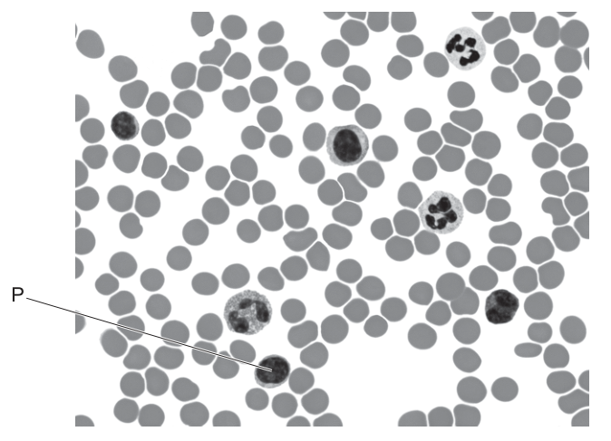
What is cell P?
Options
A lymphocyte
B monocyte
C neutrophil
D red blood cell
Working
Cell P is small with a single, large, round nucleus that occupies almost the whole cell, leaving only a thin rim of cytoplasm. This nuclear morphology (round, unlobed, taking up most of the cell) is characteristic of a lymphocyte.
Answer
A
A
Background Concept
A blood smear viewed under a light microscope shows three main cellular components: red blood cells (erythrocytes), white blood cells (leucocytes), and platelets. The white blood cells are classified by their nuclear shape and cytoplasmic appearance, and there are several types a CIE candidate is expected to recognise in a photomicrograph:
- Red blood cell (erythrocyte): a small, pale, biconcave disc with no nucleus — appears as a grey ring with a lighter centre.
- Lymphocyte: a small-to-medium cell with a single, large, round, darkly-stained nucleus that occupies most of the cell, leaving only a thin rim of pale cytoplasm.
- Monocyte: the largest white blood cell, with a large kidney-shaped or horseshoe-shaped (indented) nucleus and abundant cytoplasm.
- Neutrophil: a cell with a distinctive multi-lobed nucleus (usually 2–5 lobes joined by thin strands) and granular cytoplasm.
Understanding the Question
The question asks the candidate to identify a single cell (labelled P) on a photomicrograph of a blood smear. The other leucocytes are also visible for comparison: a multi-lobed neutrophil is at the top of the field, and a larger cell with a kidney-shaped nucleus (monocyte) sits near the centre. The arrow from P points to a cell that is small, with a single round nucleus filling most of the cell.
The command word is what is — a simple identification. The only clue is the appearance of the cell on the micrograph, so the decision rests on nuclear shape and the relative amount of cytoplasm.
Approach
Compare cell P with each of the four options using observable features:
- Is it a red blood cell? No — P has a clearly stained nucleus, while red cells are anucleate.
- Is it a neutrophil? No — P's nucleus is a single, round mass, not divided into lobes.
- Is it a monocyte? No — P is much smaller than the monocyte visible in the centre of the field, and its nucleus is round rather than kidney-/horseshoe-shaped.
- Is it a lymphocyte? Yes — small size, single round nucleus filling most of the cell, with only a thin rim of cytoplasm. This matches the cell P.
Step-by-Step Reasoning
- Cell P is roughly 1.5–2× the diameter of the surrounding red cells — too small to be a monocyte, which is the largest leucocyte.
- The nucleus is unlobed and round, ruling out the neutrophil (visible nearby with its multi-lobed nucleus) and the monocyte (with its indented, kidney-shaped nucleus).
- The nucleus occupies most of the cell, with only a thin rim of cytoplasm visible — the classic appearance of a lymphocyte.
- Therefore cell P is a lymphocyte, option A.
Key Takeaways
- Lymphocytes are identified by a single, large, round, darkly-stained nucleus that fills most of the cell.
- Neutrophils have a multi-lobed nucleus; monocytes are the largest leucocytes with a kidney-shaped nucleus; red blood cells have no nucleus.
- When asked to identify a cell on a micrograph, use both size and nuclear shape as the primary distinguishing features.
Common Mistakes
- Confusing the lymphocyte with the monocyte: both have a single, unlobed nucleus region, but the monocyte is much larger and has a kidney-shaped (indented) nucleus, while the lymphocyte has a perfectly round nucleus and a small cell size.
- Confusing the lymphocyte with a neutrophil: the neutrophil's nucleus is divided into distinct lobes connected by thin chromatin strands — quite different from the single round nucleus of the lymphocyte.
- Selecting a red blood cell (D): red cells lack any nucleus at all.
Things to Be Careful About
- Distinguishing a monocyte from a large lymphocyte on a micrograph can be subtle. The decisive features are: monocyte = kidney-shaped / indented nucleus and abundant cytoplasm; lymphocyte = perfectly round nucleus and minimal cytoplasm.
- Do not judge a white cell by cytoplasm colour alone; rely on nuclear morphology, which is far more distinctive.
The diagram shows the Bohr shift in actively respiring cells.
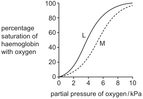
What causes the shift from L to M?
Options
A Carbonic acid dissociates to release protons that bind to oxyhaemoglobin, affecting the conformation of haemoglobin to reduce its affinity for oxygen.
B Hydrogencarbonate ions produced by carbonic anhydrase alter the charge of oxyhaemoglobin, reducing its ability to bind oxygen.
C Hydrogen ions combine with chloride ions in tissue fluid to produce hydrochloric acid, distorting oxyhaemoglobin and causing it to lose bound oxygen.
D A low concentration of oxygen in actively respiring cells causes oxyhaemoglobin to release oxygen more quickly down the concentration gradient.
Working
In actively respiring cells, CO2 diffuses into red blood cells and is hydrated by carbonic anhydrase to form carbonic acid (H2CO3), which dissociates into H+ and HCO3− ions. The H+ ions bind to oxyhaemoglobin, causing a conformational change in the haemoglobin molecule that lowers its affinity for O2, so the dissociation curve shifts to the right (L → M).
Answer
A
A
Background Concept
The oxygen dissociation curve plots the percentage saturation of haemoglobin against the partial pressure of oxygen. A rightward shift (the Bohr shift) indicates that haemoglobin has a lower affinity for oxygen at any given pO2, so it releases O2 more readily.
In respiring tissues:
- CO2 enters red blood cells and combines with water: CO2 + H2O ⇌ H2CO3 (carbonic acid).
- Carbonic anhydrase catalyses this reaction.
- H2CO3 dissociates into H+ and HCO3− (hydrogencarbonate).
- The H+ ions bind to amino acid residues on the globin chains of oxyhaemoglobin, stabilising the deoxygenated (T) conformation and lowering O2 affinity.
- HCO3− diffuses out into the plasma (the chloride shift maintains electrical balance).
Understanding the Question
The figure shows two sigmoidal curves: L (left) representing normal haemoglobin saturation, and M (right) representing haemoglobin in actively respiring tissue. At any given pO2, curve M shows a lower percentage saturation, meaning more O2 has been released. The question asks what chemical event causes this rightward shift.
Approach
Identify the cause of the Bohr shift: it is specifically the H+ ions (from carbonic acid dissociation) binding to haemoglobin, not the HCO3− ions, not chloride ions, and not a low pO2 itself. Match this to the option that names protons (H+) binding to oxyhaemoglobin and producing a conformational change.
Step-by-Step Reasoning
- Option A states: carbonic acid dissociates to release protons that bind to oxyhaemoglobin, affecting the conformation of haemoglobin to reduce its affinity for oxygen. This is the textbook mechanism of the Bohr shift. ✓
- Option B blames hydrogencarbonate ions. The HCO3− does not directly lower haemoglobin's O2 affinity; it is the H+ that does. ✗
- Option C describes H+ combining with Cl− to make HCl. This is not what happens in the red cell; Cl− is involved in the chloride shift (ion exchange for HCO3−), not in producing HCl. ✗
- Option D describes simple unloading due to a low pO2 gradient. That is normal dissociation, not the Bohr shift. The Bohr shift is defined by the rightward movement of the curve caused by CO2/H+, independent of the pO2 gradient. ✗
Key Takeaways
- The Bohr shift is caused by H+ (from CO2 via carbonic anhydrase) binding to oxyhaemoglobin.
- This lowers haemoglobin's affinity for O2, favouring release in respiring tissues.
- A rightward shift on the dissociation curve = reduced O2 affinity = more O2 released to tissues.
Common Mistakes
- Confusing the role of H+ with that of HCO3−; the proton is the active species, not the hydrogencarbonate ion.
- Thinking the Bohr shift is simply about a low pO2 in tissues — that is ordinary concentration-gradient unloading, not a shift of the curve.
- Believing HCl is produced; in fact H+ binds directly to histidine residues on haemoglobin.
Things to Be Careful About
- The Bohr shift is about a CHANGE in the curve's position, not the normal release of O2 down its gradient.
- Carbonic anhydrase is the enzyme that accelerates the CO2 + H2O ⇌ H2CO3 reaction inside red blood cells.
- The chloride shift (Hamburger shift) refers to Cl− moving into red cells as HCO3− moves out; it is not the Bohr shift itself.
The bar chart shows the mean thickness of the walls of the main chambers in a mammalian heart.
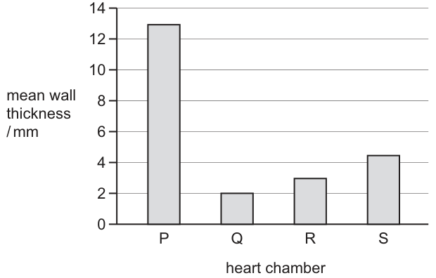
Which row correctly identifies the heart chambers?
Options
| right atrium | left atrium | right ventricle | left ventricle | |
|---|---|---|---|---|
| A | R | Q | S | P |
| B | Q | S | R | P |
| C | R | Q | P | S |
| D | Q | R | S | P |
Working
The wall thickness of each heart chamber reflects the pressure it must generate:
- P (≈ 13 mm, thickest) → left ventricle. It pumps blood into the systemic circulation, requiring the highest pressure, so its wall (myocardium) is by far the thickest.
- Q (≈ 2 mm, thinnest) → right atrium. It only pumps blood the short distance into the right ventricle at low pressure, so its wall is the thinnest.
- R (≈ 3 mm) → left atrium. Slightly thicker than the right atrium because the left ventricle (thicker, less compliant) requires a marginally higher atrial pressure to fill.
- S (≈ 4.5 mm) → right ventricle. Pumps blood to the lungs (pulmonary circulation) at moderate pressure — thicker than either atrium but much thinner than the left ventricle.
So the identification is: right atrium = Q, left atrium = R, right ventricle = S, left ventricle = P.
Answer
D
D
Background Concept
The mammalian heart is a double pump with four chambers: right atrium (RA), right ventricle (RV), left atrium (LA) and left ventricle (LV). Blood enters each atrium from the veins, is passed into the ventricle below, and is then ejected either to the lungs (RV → pulmonary artery) or to the rest of the body (LV → aorta).
The thickness of the muscular wall (myocardium) of each chamber is directly related to the pressure that chamber must generate when it contracts:
- Left ventricle — pumps blood through the systemic circulation (every organ except the gas-exchange surface), so it works against a large volume of vessels and high resistance. It generates the highest pressure (≈ 120 mmHg systolic) and therefore has the thickest wall.
- Right ventricle — pumps blood only to the lungs via the pulmonary circulation, a much shorter, lower-resistance circuit. It generates only ≈ 25 mmHg systolic, so its wall is moderate in thickness.
- Left atrium — pumps blood into the left ventricle, whose thick, less-compliant wall needs a slightly higher filling pressure, so the LA wall is slightly thicker than the RA wall.
- Right atrium — pumps blood only into the right ventricle, which is thin-walled and easy to fill. The RA therefore has the thinnest wall.
A useful rule of thumb: atria thin, ventricles thick; the chamber on the left is thicker than its right-side counterpart.
Understanding the Question
The bar chart (Fig. 35.1) shows the mean wall thickness of four unnamed heart chambers, labelled P, Q, R and S. The candidate must match each letter to the correct chamber name. The y-axis is mean wall thickness in mm; the x-axis lists the four chambers.
Reading the chart:
- P ≈ 13 mm — very tall bar
- Q ≈ 2 mm — shortest bar
- R ≈ 3 mm — second shortest
- S ≈ 4.5 mm — third tallest
The command word is implicit ("Which row correctly identifies the heart chambers?"), so a single correct option is required, with reasoning.
Approach
Rank the bars from thinnest to thickest, then map that ranking onto the four chambers using the pressure/thickness principle:
Expected anatomical ranking from thinnest to thickest wall:
Aligning the two sequences gives Q = RA, R = LA, S = RV, P = LV.
Step-by-Step Reasoning
- Identify the thickest wall (P, ≈ 13 mm). Only one chamber generates the very high pressure required to drive blood through the entire systemic circulation — the left ventricle. So P = LV.
- Identify the thinnest wall (Q, ≈ 2 mm). Atria only need to push blood a short distance into the adjacent ventricle at low pressure. Of the two atria, the right atrium has the slightly thinner wall because the right ventricle is the easier of the two to fill. So Q = RA.
- Distinguish the two middle bars (R ≈ 3 mm, S ≈ 4.5 mm). R is thinner than S, so R must be the remaining atrium and S the remaining ventricle. The left atrium is marginally thicker than the right atrium (it must fill the thick-walled, less-compliant left ventricle), so R = LA. The right ventricle pumps blood to the lungs at moderate pressure, giving a wall thicker than either atrium but far thinner than the LV, so S = RV.
- Check against the options. Right atrium = Q, left atrium = R, right ventricle = S, left ventricle = P — this matches option D exactly.
A second check using the "left > right" rule: in any matched pair, the left side of the heart is thicker. Q < R (atria: RA < LA ✓) and S < P (ventricles: RV < LV ✓), so the assignment is consistent.
Key Takeaways
- Wall thickness tracks the pressure a chamber must generate, not its size.
- The order of wall thickness is: right atrium < left atrium < right ventricle < left ventricle.
- A standard mnemonic: Pump Loudest = Left Ventricle (the chamber that needs the most muscle).
- For bar-chart questions, always read the values from the axes (with units!) before applying a biological principle, and use the values to rank, not just to compare visually.
Common Mistakes
- Confusing atrium and ventricle thicknesses. Atria have much thinner walls than ventricles because they are "booster pumps" for venous return, not the main systemic pump. A candidate who treats P and S as both ventricles but mixes up the atria can still arrive at D, but if they swap an atrium and a ventricle they will get the wrong option (A or C).
- Forgetting that the left side is thicker than the right. Students sometimes assume both atria are identical in wall thickness and both ventricles are identical, then guess. In fact RA < LA < RV < LV, and this ordering is what makes the question unambiguous.
- Misreading the y-axis. The values are in mm, not cm. Always check the units before interpreting; a wall of "13" sounds modest in cm but is substantial in mm.
Things to Be Careful About
- The question supplies the labels P, Q, R, S on the x-axis; do not assume the chambers appear in any anatomical order. The arrangement is alphabetical (P, Q, R, S), which is not the order of wall thickness.
- The numerical estimates from the chart are approximate; do not over-interpret small differences (e.g. between Q and R). The principle of relative ranking (atria < ventricles; left > right) is what is being tested.
- Answer only the letter (D). On multiple-choice papers, full reasoning is not required in the answer space, but it must be done on the question paper or in the head before selecting an option.
The diagram shows parts of the gas exchange system in humans and the associated cells or structures.
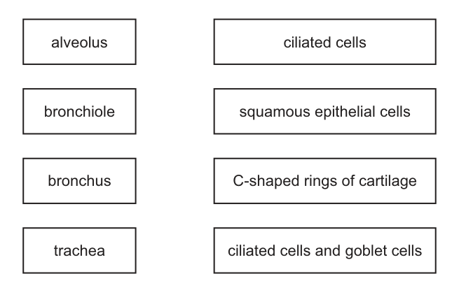
Which diagram shows the lines that correctly match the parts to their cells or structures?
Options
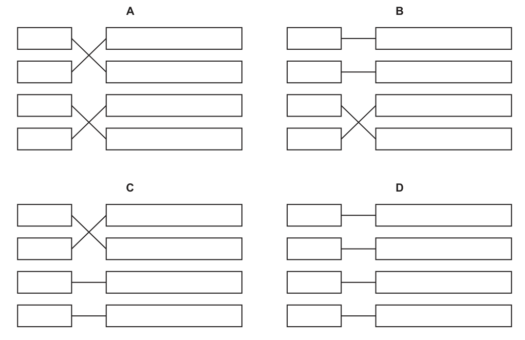
Working
Each part of the gas exchange system is lined by a characteristic epithelium or supported by a characteristic tissue:
- Alveoli have very thin walls to allow rapid diffusion of gases → squamous epithelial cells.
- Bronchioles are small airways whose walls are dominated by ciliated cells (with some smooth muscle); they lack cartilage and have few goblet cells.
- The trachea is held open by C-shaped rings of cartilage in its wall.
- The bronchi are larger airways lined by a ciliated epithelium containing goblet cells (and with cartilage plates, not rings).
Placing these matches in the boxes of Fig. 36.1 requires both pairs of lines to cross (alveolus ↔ squamous, bronchiole ↔ ciliated, bronchus ↔ ciliated + goblet, trachea ↔ cartilage rings).
Answer
A
A
Background Concept
The human gas exchange system is a branching "respiratory tree" that carries air from the external environment deep into the lungs. As you pass from the trachea → bronchi → bronchioles → alveoli, the diameter of each passage decreases but the total cross-sectional area and the surface area for gas exchange increase enormously. The structure of the wall changes at each level to match the function of that region:
- Trachea: a large, flexible tube reinforced by C-shaped rings of hyaline cartilage that prevent the airway from collapsing when the air pressure inside falls during inhalation. The open part of the "C" faces the oesophagus so the trachea can slightly deform when food is swallowed.
- Bronchi: the first branches off the trachea. Their walls still contain cartilage (as plates rather than complete rings) and they are lined by a pseudostratified ciliated epithelium with many goblet cells. The cilia beat in a coordinated manner to move mucus (trapping dust and microbes) upwards towards the pharynx, while goblet cells secrete the mucus itself.
- Bronchioles: smaller branches with no cartilage and progressively fewer goblet cells. The dominant cell type lining them is still the ciliated cell, and the wall contains a relatively large amount of smooth muscle (important in conditions such as asthma).
- Alveoli: the tiny blind-ended sacs where gas exchange actually occurs. They are lined by extremely thin squamous (pavement) epithelial cells, often only one cell thick, so that O₂ and CO₂ can diffuse rapidly between the alveolar air and the blood in the surrounding capillaries.
Understanding the Question
The question presents two columns of boxes. The left column lists four parts of the gas exchange system (alveolus, bronchiole, bronchus, trachea) and the right column lists four cells/structures (ciliated cells, squamous epithelial cells, C-shaped rings of cartilage, ciliated cells and goblet cells). The candidate must decide which of options A–D draws the correct matching lines. The answer therefore requires knowing the characteristic cell or structural feature of each region of the respiratory tract.
The command word is implicit ("which diagram … shows the lines that correctly match …") and so the response is a single letter. No working is shown on the exam paper; the reasoning is done in the head.
Approach
Work down the left column and ask, for each structure, "what is the single most characteristic cell or supporting tissue in its wall?" Then check that the right-hand cell/structure you chose appears exactly once among the four matches. Once the four pairings are fixed, look at the four options to see which arrangement of crossing/straight lines reproduces them.
Step-by-Step Reasoning
- Alveolus → the air sac must be thin for gas exchange → squamous epithelial cells.
- Bronchiole → a small airway without cartilage, dominated by ciliated cells (with relatively little mucus production) → ciliated cells.
- Bronchus → a larger airway with cartilage plates and lots of mucus-secreting cells → ciliated cells and goblet cells.
- Trachea → the largest conducting airway, kept open by the distinctive cartilage rings → C-shaped rings of cartilage.
Now translate these four pairings into the box layout. The left column reads (top to bottom): alveolus, bronchiole, bronchus, trachea. The right column reads: ciliated cells, squamous epithelial cells, C-shaped rings of cartilage, ciliated cells and goblet cells. The four pairings are therefore:
- alveolus (1st L) → squamous epithelial cells (2nd R)
- bronchiole (2nd L) → ciliated cells (1st R)
- bronchus (3rd L) → ciliated cells and goblet cells (4th R)
- trachea (4th L) → C-shaped rings of cartilage (3rd R)
This means the first two lines must cross (1↔2) and the last two lines must also cross (3↔4). Looking at the four options, only option A shows both pairs of lines crossing; B and C each have only one crossing pair, and D has no crossings at all.
Key Takeaways
- The respiratory tract changes structure along its length: cartilage rings (trachea) → cartilage plates + ciliated/goblet epithelium (bronchi) → ciliated epithelium with smooth muscle (bronchioles) → squamous epithelium (alveoli).
- Cartilage is present only in the trachea and bronchi — not in bronchioles or alveoli.
- Goblet cells are abundant in the trachea and bronchi but sparse in bronchioles and absent from alveoli.
- Squamous epithelial cells are a defining feature of the alveoli, giving the very short diffusion distance required for gas exchange.
Common Mistakes
- Matching trachea → ciliated cells and goblet cells instead of trachea → C-shaped rings of cartilage. The trachea is indeed lined by that epithelium, but in this question the characteristic (most distinguishing) structural feature being tested is the cartilage rings, which are unique to the trachea in this list.
- Matching bronchiole → ciliated cells and goblet cells and bronchus → C-shaped rings of cartilage. Bronchioles lack cartilage rings; only the trachea has them as complete rings.
- Confusing bronchioles and bronchi. The bronchus is larger, has cartilage plates in its wall and many goblet cells; the bronchiole is smaller, has no cartilage and few goblet cells.
Things to Be Careful About
- The question is testing the single best match for each structure from the four options given, not every cell type that is present in the wall.
- The right-hand column has two similar-sounding entries ("ciliated cells" and "ciliated cells and goblet cells"). The distinction matters: goblet cells are characteristic of bronchi, not bronchioles.
- Cartilage in the trachea is specifically in the form of C-shaped rings; in the bronchi it is in irregular plates (so "C-shaped rings of cartilage" can only match the trachea here).
The photomicrograph shows the wall of a bronchus as seen with a light microscope.
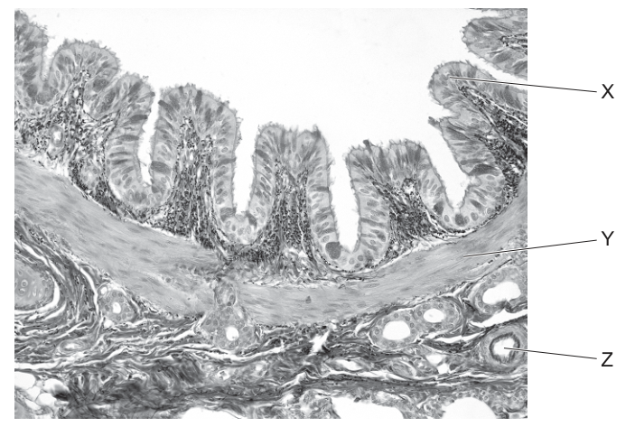
Which row correctly identifies the structures labelled?
Options
| X | Y | Z | |
|---|---|---|---|
| A | epithelium | elastic fibres | bronchiole |
| B | endothelium | smooth muscle | bronchiole |
| C | epithelium | smooth muscle | blood vessel |
| D | endothelium | elastic fibres | blood vessel |
Working
- X points to the inner lining of the bronchus. The folded, finger-like projections show a pseudostratified ciliated columnar epithelium (with goblet cells), not an endothelium (endothelium lines blood vessels, not airways).
- Y points to a thick, fairly uniform band of tissue beneath the epithelium. This is smooth muscle (which contracts to narrow the bronchus during bronchoconstriction). Elastic fibres would appear as thin, wavy, dark-staining strands scattered in the connective tissue, not as a solid sheet.
- Z points to a small circular profile in the surrounding connective tissue. It is a blood vessel cut in cross-section (a small artery/vein). A bronchiole would have its own epithelial lining, smooth muscle and surrounding connective tissue — it is a complete airway, not a simple circular structure embedded in the wall of a larger airway.
Answer
C
C
Background Concept
The wall of a bronchus is built up in layers, each with a characteristic appearance under the light microscope. From the lumen outwards, the typical arrangement is:
- Pseudostratified ciliated columnar epithelium with goblet cells — the inner lining. The nuclei sit at different levels, giving a falsely "stratified" appearance, and the apical surface bears cilia that beat mucus (with trapped particles) upwards. Goblet cells secrete the mucus.
- Lamina propria — loose connective tissue just beneath the epithelium, containing blood vessels, nerves and lymphatics.
- Smooth muscle — a substantial layer that constricts and dilates the airway (bronchoconstriction/bronchodilation). This is the layer that contracts excessively in asthma.
- Submucosa with seromucous glands.
- Hyaline cartilage — C-shaped (incomplete) rings that hold the airway open (only in bronchi, not in bronchioles).
- Adventitia — outer connective tissue that blends with surrounding lung tissue.
Elastic fibres are also present, but they appear as thin, wavy, darkly-stained threads running within the connective tissue layers rather than as a thick continuous band.
Endothelium is a single squamous layer lining the inside of blood and lymph vessels. It is NOT the lining of an airway, so seeing an "inner lining" label on the bronchus lumen cannot be endothelium.
Bronchioles differ from bronchi: they have no hyaline cartilage and no submucosal glands, and their wall is dominated by smooth muscle with a simple cuboidal/columnar epithelium. A bronchiole is a complete small airway, not a single circular hole inside the wall of a larger airway.
Understanding the Question
The question shows a light micrograph of the wall of a bronchus (the question stem makes this clear) and asks the candidate to identify three labelled structures:
- X — the folded inner lining projecting into the lumen.
- Y — a thicker band of tissue immediately beneath the lining.
- Z — a small circular profile sitting in the connective tissue below the smooth muscle.
The candidate must read each label and pick the row of options whose three identifications are all correct.
Approach
For each label, decide what tissue type it is by appearance and position, then eliminate options whose identification of that label is wrong.
- Look at X first: it is a layer lining an air-filled lumen with surface folds. The only correct identification is epithelium (ciliated pseudostratified columnar). Endothelium is wrong because it lines blood/lymph vessels, not airways. This eliminates options B and D.
- Look at Y: it is a thick, fairly uniform layer under the epithelium. The classic bronchus-wall layer here is smooth muscle. Elastic fibres appear as thin wavy lines scattered through connective tissue, not as a solid sheet. This confirms option A is wrong (Y = elastic fibres) and supports C.
- Look at Z: it is a circular profile with a clear lumen embedded in connective tissue outside the smooth muscle layer. It is a small blood vessel cut in cross-section. A bronchiole would itself have its own epithelium, smooth muscle and connective tissue and would be a complete airway, not a hole in the wall of another airway. So Z = blood vessel, eliminating A and B.
Only option C is consistent: X = epithelium, Y = smooth muscle, Z = blood vessel.
Step-by-Step Reasoning
- X — epithelium (not endothelium). The image shows characteristic luminal folding of the respiratory epithelium, with cilia visible on the apical surface. Endothelium is restricted to the inside of blood and lymph vessels. The respiratory tract lining is always called epithelium (specifically pseudostratified ciliated columnar epithelium with goblet cells).
- Y — smooth muscle (not elastic fibres). Beneath the epithelium is a continuous, substantial layer of tissue. This is the smooth muscle of the bronchus, which is responsible for bronchoconstriction (e.g. during an asthma attack). Elastic fibres, although present in the wall, look very different: thin, dark, wavy threads running through the connective tissue — never a thick continuous sheet.
- Z — blood vessel (not bronchiole). A bronchiole is an entire small airway with its own epithelial lining, smooth muscle, and (sometimes) elastic tissue. The labelled structure is just a circular profile in the connective tissue with a hollow centre — that is the cross-section of a small artery or vein supplying the wall. Hence it is a blood vessel.
- Putting the three identifications together gives row C.
Key Takeaways
- The inner lining of any airway (trachea, bronchus, bronchiole) is epithelium, never endothelium.
- A thick band of tissue immediately under the airway epithelium is smooth muscle.
- Small circular profiles with a lumen, found in the connective tissue outside the muscle, are blood vessels (or possibly lymph vessels, but never whole airways).
- A bronchiole is a complete airway in its own right and would not be seen as a single labelled structure inside the wall of a larger airway.
Common Mistakes
- Confusing epithelium with endothelium. Endothelium lines blood and lymphatic vessels only; the airway lining is always epithelium.
- Misidentifying smooth muscle as elastic fibres (or vice versa). Smooth muscle forms a thick, continuous band; elastic fibres appear as thin, dark, wavy threads scattered in connective tissue.
- Calling Z a bronchiole. A bronchiole is a small airway with its own wall layers; the labelled Z is a single circular profile (a vessel) embedded in the connective tissue, not an airway.
- Confusing this section with a blood vessel. Some students try to make X = endothelium, mistaking the bronchus lumen for a vessel lumen. The presence of cilia and goblet cells, plus the folding pattern, identify it as a respiratory epithelium.
Things to Be Careful About
- Always check what the label is pointing to, not just where it sits in the picture. The position alone (inner lining, middle layer, outer structure) is a strong hint, but the texture of the tissue confirms it.
- Remember that the question stem tells you the tissue is the wall of a bronchus, not a bronchiole. This tells you cartilage should be present in a real bronchus, although it is not one of the structures being asked about here.
- "Epithelium" is the umbrella term; you do not need to specify "pseudostratified ciliated columnar" to score the mark, but recognising the full name helps confirm the identification.
- Use process of elimination: even if you are unsure about one label, you can usually eliminate enough rows by confidently identifying one or two labels.
Which factors affect the global pattern of distribution of malaria?
1 the disease is mainly restricted to tropical and subtropical environments
2 the effective vaccine has eradicated the disease in many countries
3 the vector required for transmission of malaria parasites
Options
| 1 | 2 | 3 | |
|---|---|---|---|
| A | ✓ | ✓ | ✗ |
| B | ✓ | ✓ | ✓ |
| C | ✓ | ✗ | ✓ |
| D | ✗ | ✓ | ✓ |
key
✓ = effect
✗ = not an effect
Working
- Statement 1: Malaria is restricted to tropical and subtropical regions because the Anopheles mosquito vector requires warm, humid conditions to breed. ✓
- Statement 2: No effective vaccine for malaria exists, and the disease has certainly not been eradicated by vaccination. ✗
- Statement 3: The Anopheles mosquito (the vector) is essential for transmission, so its geographic range directly shapes malaria's global distribution. ✓
Therefore: 1 ✓, 2 ✗, 3 ✓.
Answer
C
C
Background Concept
Malaria is a parasitic disease caused by Plasmodium species (mainly P. falciparum and P. vivax) and transmitted between humans by the bite of an infected female Anopheles mosquito. The global distribution of malaria is therefore constrained by the ecology and geography of its vector. Anopheles mosquitoes thrive in warm (typically 20–30 °C), humid conditions with standing water for larval development, which is why malaria is concentrated in tropical and subtropical regions of sub-Saharan Africa, South-East Asia, South America and parts of the Middle East.
Despite decades of effort, no highly effective vaccine against malaria exists. The RTS,S vaccine (Mosquirix), approved by WHO in 2021, has only modest efficacy (around 30–50 % in children) and is used as a supplementary tool in limited regions — it has not eradicated malaria anywhere. Eradication efforts instead rely on insecticide-treated bed nets, indoor residual spraying, antimalarial drugs and prompt diagnosis.
Understanding the Question
The question presents three statements and asks which actually affect the global distribution of malaria. The correct option identifies which statements are real contributing factors (✓) and which are not (✗).
Approach
Evaluate each statement against established malaria biology and epidemiology:
- Does the statement describe a genuine factor shaping where malaria is found worldwide?
- If false (either because the premise is wrong or the factor does not actually operate), mark it ✗.
Step-by-Step Reasoning
Statement 1 — "the disease is mainly restricted to tropical and subtropical environments": True. The temperature and humidity requirements of Anopheles mosquitoes restrict malaria to warm regions. Outside the tropics the parasite cannot complete its development cycle within the mosquito (sporogony requires ~21 days at 25 °C, much longer at lower temperatures). ✓
Statement 2 — "the effective vaccine has eradicated the disease in many countries": False. There is no highly effective malaria vaccine, and no country has had malaria eradicated by vaccination. (Smallpox is the only human disease eradicated by a vaccine, in 1980.) ✗
Statement 3 — "the vector required for transmission of malaria parasites": True. Without Anopheles mosquitoes, Plasmodium cannot be transmitted between humans. The mosquito's geographic range is therefore a direct determinant of malaria's distribution. ✓
The pattern (✓ ✗ ✓) matches option C.
Key Takeaways
- The global distribution of malaria is governed by the ecology of the Anopheles mosquito vector (climate, breeding sites) and by socio-economic factors (healthcare, vector control).
- No effective vaccine currently exists for malaria — control depends on prevention (bed nets, insecticides) and treatment (artemisinin-based combination therapy).
- The only human disease eradicated by vaccination is smallpox.
Common Mistakes
- Selecting A or B (ticking statement 2): this wrongly assumes an effective malaria vaccine exists. Candidates often confuse ongoing vaccine research and pilot rollouts of RTS,S with established, effective vaccination programmes.
- Confusing the term "vector" with "pathogen": the Plasmodium parasite is the pathogen; the Anopheles mosquito is the vector. Both are needed, but they play different roles in transmission.
- Forgetting that tropical/subtropical climates favour mosquito breeding — some candidates reject statement 1 thinking malaria is a "worldwide" disease.
Things to Be Careful About
- Read the wording of statement 2 carefully: it makes TWO claims (an effective vaccine exists AND it has eradicated malaria). Both are wrong, but only one is needed to mark the statement ✗.
- Keep malaria distinct from other infectious diseases in the syllabus (cholera, TB, HIV) — each has its own epidemiology and control measures.
- On tick-box style MCQs, transcribing the ✓/✗ pattern correctly is easy to muddle; double-check the option letter against the 1/2/3 columns before committing.
Which molecule could be a self antigen?
Options
A a viral capsid protein
B a phospholipid in a viral envelope
C a toxin released by a pathogen
D a glycoprotein on the surface of a macrophage
Working
Self antigens are molecules on the surface of the body's own cells that the immune system recognises as "self" and does not attack. They are glycoproteins (MHC molecules) present on the surface of all nucleated cells, including macrophages.
- A. A viral capsid protein is foreign (non-self).
- B. A phospholipid in a viral envelope is foreign (non-self).
- C. A toxin released by a pathogen is foreign (non-self).
- D. A glycoprotein on the surface of a macrophage is a self antigen (an MHC marker on a body cell).
Answer
D
D
Background Concept
An antigen is any molecule that can be recognised by the immune system and provoke a response. Antigens are typically large, complex molecules (often proteins or glycoproteins) on the surface of cells or pathogens.
A self antigen is a molecule found on the surface of the body's own cells. These are mainly the MHC (Major Histocompatibility Complex) glycoproteins, which are present on virtually all nucleated cells in the body. The immune system learns to recognise these self antigens during development and does not normally mount an attack against them — this is the basis of immunological tolerance.
A non-self antigen is any molecule foreign to the body, such as components of pathogens (viruses, bacteria, fungi) or toxins. Non-self antigens trigger an immune response, with B-lymphocytes producing antibodies and T-lymphocytes attacking infected cells.
Macrophages are white blood cells that belong to the body; their surface carries MHC glycoproteins, which serve as self antigens.
Understanding the Question
The question asks which of the four options could be a self antigen — i.e., a molecule originating from the body's own cells, recognised as "self" by the immune system. Three options involve components of pathogens or foreign substances; one involves a molecule on a host cell.
The command word is "could be" — we need to identify the option that is biologically consistent with being a self antigen.
Approach
Eliminate the options that describe foreign (non-self) molecules and select the one that describes a host-cell surface molecule (specifically a glycoprotein, since MHC molecules are glycoproteins).
Step-by-Step Reasoning
- Option A — viral capsid protein: Viruses are pathogens and are foreign to the body. Capsid proteins are non-self antigens that stimulate an immune response. ❌
- Option B — phospholipid in a viral envelope: This is a component of a virus. It is foreign and would be recognised as non-self. ❌
- Option C — toxin released by a pathogen: Toxins are produced by bacteria and other pathogens and are foreign molecules that provoke antibody production. ❌
- Option D — glycoprotein on the surface of a macrophage: Macrophages are host cells. Their surfaces display MHC glycoproteins, which act as self antigens, marking the cell as belonging to the body. ✔
Key Takeaways
- Self antigens are typically MHC glycoproteins on the surface of the body's own cells.
- Non-self antigens come from pathogens (viruses, bacteria) or toxins.
- The immune system distinguishes self from non-self to avoid attacking the body's own tissues.
Common Mistakes
- Confusing self antigens with non-self antigens — picking a pathogen-derived molecule because it "triggers the immune system" without checking whether it is from the body or from a foreign source.
- Forgetting that self antigens are usually glycoproteins (the wording of option D matches MHC molecules exactly).
- Choosing phospholipids as self antigens — phospholipids are abundant in the body's own membranes, but on their own they are not the markers that distinguish self cells; recognition depends on the protein/glycoprotein components.
Things to Be Careful About
- The term "self antigen" specifically refers to identifiable surface markers on the body's own cells, not just any molecule present in the body.
- Pathogen components (capsids, envelopes, toxins) are always classified as non-self antigens because they originate from outside the body.
The diagram shows an antibody molecule with some parts labelled.
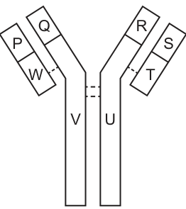
Which row is correct about the parts that form an antigen-binding site and have identical amino acid sequences?
Options
| form an antigen-binding site | have identical amino acid sequences | |
|---|---|---|
| A | P and Q | R and S |
| B | Q and V | P and S |
| C | R and S | U and V |
| D | U and V | Q and R |
Working
An antibody (immunoglobulin) is a Y-shaped molecule made of four polypeptide chains: two identical heavy chains and two identical light chains, held together by disulfide bonds.
- Antigen-binding sites are located at the tips of the Y. Each site is formed by the variable region of one light chain together with the variable region of one heavy chain. In the figure, one binding site is formed by P and Q, the other by R and S.
- The constant regions of the two heavy chains (labelled U and V in the stem of the Y) have the same amino acid sequence because the two heavy chains are identical polypeptides.
Checking the options:
- A: P+Q do form a binding site ✓, but R and S are variable regions and are not identical to each other ✗
- B: Q and V do not form a binding site ✗
- C: R+S do form a binding site ✓, and U and V are the constant regions of the identical heavy chains ✓
- D: U and V are constant regions in the stem — they do not form a binding site ✗
Answer
C
C
Background Concept
An antibody (immunoglobulin) is a globular protein with a characteristic Y-shape. It is built from four polypeptide chains:
- Two heavy chains — the longer polypeptides that run the full length of the Y (forming the arms and the stem).
- Two light chains — the shorter polypeptides that sit alongside the upper halves of the heavy chains (forming only the arms).
The chains are held together by disulfide bonds and the overall molecule has two identical antigen-binding sites, one at the tip of each arm of the Y.
Each chain has a variable region (amino acid sequence differs between antibodies, giving specificity for a particular antigen) and a constant region (sequence is the same for all antibodies of the same class, e.g. all IgG). An antigen-binding site is formed by the variable region of a light chain paired with the variable region of a heavy chain.
A key consequence of this quaternary structure:
- The two heavy chains are identical to each other → their variable regions are identical, and their constant regions are identical.
- The two light chains are identical to each other → same logic.
- Therefore the two antigen-binding sites are identical (paratopes the same), and the two constant regions of the heavy chains are identical in sequence.
Understanding the Question
The diagram (Fig. 40.1) labels eight regions of an antibody:
- P, Q — variable regions of the light and heavy chains at the tip of the left arm → together form one antigen-binding site.
- R, S — variable regions of the light and heavy chains at the tip of the right arm → together form the other antigen-binding site.
- W, T — constant regions of the two light chains (on the arms).
- V, U — constant regions of the two heavy chains (forming the stem of the Y).
The question asks which option correctly identifies BOTH:
- A pair that forms an antigen-binding site (variable light + variable heavy at one tip).
- A pair that has identical amino acid sequences (the two heavy chains, or the two light chains, since each pair is identical).
Approach
Apply two rules:
- A binding site needs a light-chain variable region + heavy-chain variable region at the same tip — so look for adjacent regions on one arm of the Y.
- Identical sequences occur between the two heavy chains (so U ↔ V) or the two light chains (so W ↔ T) — the two arms are mirror images of each other.
Then scan the four options and reject any that fail either condition.
Step-by-Step Reasoning
Option A — P and Q (binding site); R and S (identical sequences).
- P and Q are at the same tip (left arm): correctly form a binding site. ✓
- R and S are the variable regions of the right arm. They are NOT identical in sequence — variable regions differ in amino acid sequence to provide antigen specificity, and in any case R is part of a light chain while S is part of a heavy chain. ✗
- Reject.
Option B — Q and V (binding site); P and S (identical sequences).
- Q is the variable region of a heavy chain; V is the constant region of a heavy chain. They are in different parts of the molecule and do not form a binding site. ✗
- Reject.
Option C — R and S (binding site); U and V (identical sequences).
- R and S sit together at the tip of the right arm (variable regions of the light and heavy chains): correctly form a binding site. ✓
- U and V are the constant regions of the two heavy chains in the stem. Because the two heavy chains are identical polypeptides, U and V have the same amino acid sequence. ✓
- Both conditions satisfied. Correct.
Option D — U and V (binding site); Q and R (identical sequences).
- U and V are constant regions in the stem, not at the tips, so they do not form an antigen-binding site. ✗
- Reject.
Key Takeaways
- An antibody has two identical antigen-binding sites, each made of one light-chain variable region + one heavy-chain variable region.
- The molecule is built from two identical heavy chains and two identical light chains, so corresponding regions across the two halves (e.g. U and V, or W and T) share their amino acid sequence.
- Variable regions differ between antibodies and are not identical even within the same antibody (the light-chain variable differs from the heavy-chain variable, though the two binding sites are identical to each other).
Common Mistakes
- Thinking any two adjacent regions form a binding site — both must be variable regions (one from a light chain, one from a heavy chain) AND they must be on the same arm of the Y.
- Assuming the two binding sites have different sequences — they are in fact identical, because the two halves of the antibody are mirror-image copies.
- Picking U and V as a binding site — they are the constant regions of the heavy chains in the stem, not at the tips, so they cannot bind antigen.
- Confusing which regions are constant vs variable — variable regions are at the tips, constant regions are lower down the arms and in the stem.
Things to Be Careful About
- The diagram labels must be matched carefully to which chain (light or heavy) and which region (variable or constant) they represent. U and V specifically are the two heavy-chain constant regions in the stem.
- "Identical amino acid sequences" applies between the two heavy chains (so U and V match) and between the two light chains (so W and T match), but not between a heavy-chain region and a light-chain region, and not between variable and constant regions.
- The two binding sites (P+Q and R+S) are themselves identical to each other, but the individual regions P, Q, R, S are not all identical to one another — only the two heavy-chain variable regions match each other and the two light-chain variable regions match each other.
Your score so far
Answer a question to start scoring
Your marks add up here as you work through the paper.