Biology 9700/11 — October/November 2025
Cambridge AS Level · Multiple Choice (AS Level) · answer key with instant marking and worked solutions
Topics Biological Molecules · Cell Structure · Cell Membranes and Transport · Nucleic Acids and Protein Synthesis · Transport in Plants · Transport in Mammals · +5 more
Tap an option under each question to check it — your score builds as you go.
In a photomicrograph of magnification , a chloroplast measures in diameter.
What is the actual diameter of the chloroplast?
Options
A
B
C
D
Working
Answer
D
D
Background Concept
When a specimen is viewed under a microscope (light or electron), it appears larger than its true size. The number of times it is enlarged is the magnification. The triangle of relationships is:
Two units of length matter here:
- (so )
A real chloroplast in a plant mesophyll cell is typically across, so an answer in the low micrometre range is biologically sensible — a useful check before committing to an option.
Understanding the Question
The candidate is given a photomicrograph (a photographed image taken down a microscope) and told:
- the magnification of the image is , and
- a chloroplast on the image measures in diameter.
The task is to find the real diameter of that chloroplast. Because the image is times larger than the object, dividing the measured image size by the magnification recovers the real size.
The command word is implicit in a multiple-choice question, but the underlying skill is "calculate the actual size of a structure given the magnification and the measured size in the image".
Approach
- Rearrange the magnification formula to solve for actual size.
- Substitute the image size () and magnification ().
- Convert the answer from to so it can be compared with the options (which are all in ).
- Pick the matching option.
Step-by-Step Reasoning
Converting to (multiply by ):
So the chloroplast's actual diameter is , which corresponds to option D.
A quick reality check: a chloroplast of is a realistic size for a higher-plant chloroplast (roughly ), so the answer is biologically credible.
Key Takeaways
- The core relationship is ; getting the rearrangement wrong is the most common error.
- Always match units. Here, the image size is in but the answers are in , so a conversion is required.
- A sense of scale is useful: real chloroplasts are a few micrometres across, so an answer of or (the size of small viruses or ribosomes) should be immediately suspicious.
Common Mistakes
- Multiplying instead of dividing (i.e. calculating ): this gives , an absurd size, and would not match any option — usually a sign the candidate used the wrong rearrangement.
- Forgetting to convert units and selecting (a distractor that has the right number but the wrong unit, hidden by giving it as after a misplaced factor-of-10 conversion, or by choosing the answer in ).
- Off-by-10³ conversions: , not and not . Get this wrong and the answer drifts by a factor of or .
- Choosing option A () by misplacing a decimal after a conversion (e.g. converting to before dividing, an error that confuses the image size with the answer).
Things to Be Careful About
- Watch the units: image size is in here, but it is often given in or in other questions. Always write out the conversion explicitly.
- Significant figures: the measured image size is (two sig figs), so quoting the answer as (one sig fig) is reasonable, but would be acceptable too.
- The magnification in a photomicrograph refers to the final printed image, not the objective lens alone — this is given as and should be used as such.
- If a question gives a scale bar instead of stating magnification, you can still use the same triangle by treating the printed scale-bar length as the "image size" of a structure whose "actual size" is written next to the bar.
What is a function of Golgi bodies?
Options
A formation of vesicles for endocytosis
B modification of proteins for secretion
C synthesis of ATP
D synthesis of polypeptides
Working
The Golgi apparatus receives proteins from the rough endoplasmic reticulum, modifies them (e.g. by adding carbohydrate groups to form glycoproteins), and packages them into vesicles for secretion from the cell by exocytosis.
- A — vesicles for endocytosis are formed at the cell surface membrane, not the Golgi.
- B — modification of proteins for secretion is the defining role of the Golgi. ✓
- C — synthesis of ATP is carried out by mitochondria.
- D — synthesis of polypeptides is carried out by ribosomes on the rough endoplasmic reticulum.
Answer
B
B
Background Concept
The Golgi apparatus (Golgi body / Golgi complex) is a stack of flattened, membrane-bound sacs called cisternae. It is part of the cell's endomembrane system and sits between the rough endoplasmic reticulum (RER) and the plasma membrane. Proteins synthesised on ribosomes attached to the RER are packaged into transport vesicles that bud off the RER and fuse with the cis face of the Golgi. Inside the cisternae, the proteins undergo post-translational modification — most commonly glycosylation (sugar groups are added or trimmed) and sometimes phosphorylation or sulfation. The finished products are then sorted at the trans face, packaged into secretory vesicles, and either held in the cytosol until needed or dispatched to the plasma membrane for release by exocytosis. The Golgi also produces lysosomes, whose hydrolytic enzymes are sorted and packaged here.
Understanding the Question
This is a single-best-answer multiple choice question. The stem asks for a function of Golgi bodies, so any one correct, well-established role scores the mark. The four options test knowledge of the Golgi alongside three other organelles/cell processes, so the candidate must distinguish the Golgi from the cell surface membrane, mitochondria and ribosomes.
Approach
Recall the principal function of the Golgi apparatus, then mentally eliminate each distractor by identifying which organelle actually carries out the role described.
Step-by-Step Reasoning
- Option A — formation of vesicles for endocytosis. Endocytosis describes material being taken into the cell; the vesicle is formed by invagination and pinching-off of the plasma membrane, not the Golgi. The Golgi makes vesicles for the outgoing (exocytotic / secretory) route. Reject.
- Option B — modification of proteins for secretion. Proteins arriving from the RER are chemically modified in the Golgi cisternae (e.g. carbohydrates added to form glycoproteins), then packaged into vesicles for secretion. This is the textbook function of the Golgi. Accept.
- Option C — synthesis of ATP. ATP synthesis by oxidative phosphorylation and, in some cells, substrate-level phosphorylation in the Krebs cycle occurs in the mitochondria (and a small amount in the cytoplasm during glycolysis). Reject.
- Option D — synthesis of polypeptides. Polypeptide chains are assembled on ribosomes, which may be free in the cytosol or bound to the RER. The Golgi does not translate mRNA. Reject.
The single correct option is therefore B.
Key Takeaways
- The Golgi apparatus modifies, sorts and packages proteins (and lipids) received from the RER.
- It is the source of secretory vesicles that fuse with the plasma membrane to release their contents by exocytosis.
- Other major cellular roles are distributed across distinct organelles: mitochondria = ATP synthesis; ribosomes = polypeptide synthesis; plasma membrane = endocytic vesicle formation.
Common Mistakes
- Confusing the direction of vesicle traffic: endocytosis is inward (plasma membrane) while Golgi-derived vesicles move outward (secretion).
- Attributing ATP synthesis to the Golgi because it "produces energy-rich molecules" — energy is stored in the modified secretory products, but the ATP itself is made in mitochondria.
- Attributing protein synthesis to the Golgi because it "handles proteins" — the Golgi only modifies proteins made elsewhere; it does not translate mRNA.
Things to Be Careful About
- The question only needs one correct function; you do not need to recall every role of the Golgi.
- "Modification of proteins for secretion" is the precise wording; vague answers such as "processes proteins" or "packages things" are insufficient if they do not make clear the modification and secretion aspects.
A cell in the human body is specialised for synthesis and secretion of lipids.
Which row shows the organelles required by the cell for synthesis and secretion of lipids?
Options
| mitochondria | smooth endoplasmic reticulum | vesicles | |
|---|---|---|---|
| A | ✓ | ✓ | ✓ |
| B | ✓ | ✗ | ✗ |
| C | ✗ | ✓ | ✓ |
| D | ✗ | ✓ | ✗ |
key
✓ = required
✗ = not required
Working
- Smooth endoplasmic reticulum (SER) is the site of lipid synthesis, so it is required.
- Vesicles transport lipids from the SER to the Golgi apparatus and then to the plasma membrane for secretion, so they are required.
- Mitochondria produce ATP by aerobic respiration; ATP is needed to power the active steps of synthesis and secretion, so they are required.
All three organelles are required.
Answer
A
A
Background Concept
Eukaryotic cells contain membrane-bound organelles, each with a specialised function. For a cell that synthesises and secretes lipids (for example, a Leydig cell in the testis producing steroid hormones, or a cell in the adrenal cortex), three organelles are critical:
- Smooth endoplasmic reticulum (SER): an interconnected network of membranes lacking ribosomes. It is the site of synthesis of lipids, including phospholipids, steroids, and fatty acids. Enzymes embedded in its membranes catalyse these reactions.
- Vesicles: small membrane-bound sacs that bud off from one organelle and fuse with another. They shuttle newly synthesised lipids from the SER to the Golgi apparatus for further processing, and then from the Golgi to the plasma membrane, where exocytosis releases the lipid product from the cell.
- Mitochondria: the site of aerobic respiration, producing ATP. ATP supplies the energy for the active steps of vesicle budding, vesicle movement along cytoskeletal tracks, and exocytosis.
Understanding the Question
This is a multiple-choice question asking which combination of organelles a lipid-synthesising and -secreting cell needs. The stem presents a table with three organelles (mitochondria, SER, vesicles), and each row of the table shows whether that organelle is required (✓) or not required (✗).
The command word is implicit ("which row shows..."), and there is one mark for the correct option.
Approach
Consider each organelle in turn and decide whether it plays a direct role in (1) synthesising lipids, (2) secreting them, or (3) supplying energy for the process. An organelle is "required" if it contributes to any of these roles.
Step-by-Step Reasoning
- Smooth endoplasmic reticulum: Lipid synthesis occurs on the cytoplasmic face and within the membranes of the SER. Without SER, the cell could not make lipids. → required (✓).
- Vesicles: Newly made lipids must be packaged and moved to the Golgi and then to the plasma membrane. Vesicles perform this transport. → required (✓).
- Mitochondria: Budding of vesicles, vesicle transport, and exocytosis are energy-requiring processes. ATP is supplied by mitochondria. → required (✓).
Only option A has all three ticked as required. The other options each leave out an organelle that is needed.
Key Takeaways
- The SER is the site of lipid synthesis.
- Vesicles are required to move lipids between organelles and out of the cell.
- Mitochondria provide ATP for the active steps of synthesis, transport, and secretion.
- When asked which organelles a cell needs for a process, consider both the direct machinery and the energy supply.
Common Mistakes
- Choosing C or D because SER "makes the lipids" — this overlooks that secretion also needs vesicles and energy from mitochondria.
- Confusing rough ER (studded with ribosomes; synthesises proteins) with smooth ER (synthesises lipids).
- Thinking mitochondria are only needed for "energy-demanding" muscle cells, forgetting that vesicle trafficking and exocytosis are ATP-dependent in all secretory cells.
Things to Be Careful About
- "Required" means the organelle contributes to synthesis OR secretion, not only to the chemical synthesis step itself.
- Vesicles are distinct from the SER itself; budding transport vesicles are how materials leave the SER.
- The question says "synthesis and secretion" — both processes must be supported, so a correct answer must include organelles for each.
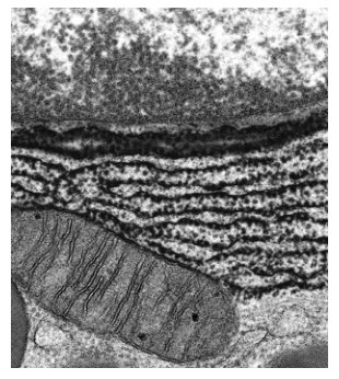
The photomicrograph shows part of a cell.
Which row is correct?
Options
| this cell will contain 70S ribosomes | this could be part of a plant cell | the organelles shown could be used to make antibodies | |
|---|---|---|---|
| A | ✓ | ✓ | ✓ |
| B | ✓ | ✗ | ✗ |
| C | ✗ | ✓ | ✗ |
| D | ✗ | ✗ | ✓ |
key
✓ = correct
✗ = not correct
Working
The electron micrograph shows a mitochondrion (lower left, with visible cristae) and parallel stacks of rough endoplasmic reticulum (RER, studded with ribosomes).
- 70S ribosomes: Mitochondria contain their own ribosomes, which are 70S (like those of prokaryotes) — a relic of their endosymbiotic origin. So this cell does contain 70S ribosomes (inside the mitochondrion). ✓
- Could be part of a plant cell: Plant cells contain both mitochondria and RER, so this could indeed be part of a plant cell. ✓
- Organelles used to make antibodies: Antibodies are proteins secreted by plasma cells. RER is the site of synthesis of secretory proteins, so these organelles could be used to make antibodies. ✓
All three statements are correct.
Answer
A
A
Background Concept
The transmission electron micrograph (TEM) shows two membrane-bound eukaryotic organelles:
- Mitochondrion (lower-left, oval-shaped with internal folds): the inner membrane is thrown into cristae, which increase surface area for the electron transport chain and ATP synthase.
- Rough endoplasmic reticulum (RER) (the parallel dark, ribosome-studded flattened sacs across the upper part of the image): a continuous extension of the nuclear envelope whose surface is covered in ribosomes. RER is the site of synthesis of secretory proteins, membrane proteins and proteins destined for organelles such as lysosomes.
Two key biological principles are also tested:
- 70S ribosomes in mitochondria. Cytoplasmic eukaryotic ribosomes are 80S. However, mitochondria (and chloroplasts) contain their own ribosomes, which are 70S — the same size as bacterial ribosomes. This is one of the main lines of evidence for the endosymbiotic theory, which proposes that mitochondria evolved from an engulfed aerobic prokaryote.
- RER and antibody production. Plasma cells (activated B-lymphocytes) have an unusually extensive RER dedicated to synthesising and secreting large quantities of antibody (immunoglobulin) molecules.
Understanding the Question
The question presents a TEM of part of a cell and asks the candidate to evaluate three statements about it. The mark scheme expects:
- The cell will contain 70S ribosomes — depends on whether mitochondria/chloroplasts are present (here, a mitochondrion is visible).
- The structure could be part of a plant cell — depends on whether these organelles are compatible with a plant cell.
- The organelles shown could be used to make antibodies — depends on whether the organelles visible (RER) are capable of producing secretory proteins such as antibodies.
The command word is implicit: identify which row has the correct combination of ticks and crosses.
Approach
- Identify the organelles in the micrograph — mitochondrion (with cristae) and RER (parallel cisternae studded with ribosomes).
- Test each statement against the biology:
- Do eukaryotic cells with mitochondria contain 70S ribosomes? Yes — inside the mitochondria.
- Are mitochondria and RER found in plant cells? Yes — both are present in plant cells.
- Is RER used to synthesise antibodies? Yes — plasma cells have extensive RER for this purpose.
- Select the option where all three statements are correct.
Step-by-Step Reasoning
Statement 1: "This cell will contain 70S ribosomes."
The micrograph clearly shows a mitochondrion. Inside every mitochondrion are 70S ribosomes (and a small circular DNA molecule). These 70S ribosomes are a vestige of the prokaryotic ancestor from which mitochondria evolved. The cell's cytoplasm contains 80S ribosomes, but the cell as a whole still contains 70S ribosomes (inside the mitochondrion). The statement is therefore correct.
Statement 2: "This could be part of a plant cell."
Plant cells possess both mitochondria and rough ER. Nothing in the micrograph excludes it from being plant — there is no cell wall visible, but the section may simply not include that region. So it is reasonable to say this could be part of a plant cell. The statement is correct.
Statement 3: "The organelles shown could be used to make antibodies."
Antibodies are glycoproteins that are synthesised on the ribosomes attached to the RER, then folded, modified and packaged for secretion. Plasma cells, which secrete antibodies, are packed with RER. The RER visible in the micrograph is exactly the organelle responsible for this function. The statement is correct.
All three statements are correct, so the answer is the option with three ticks — A.
Key Takeaways
- Always identify the organelles in a TEM before evaluating statements: here, mitochondrion + RER.
- Eukaryotic cells contain both 80S (cytoplasm) and 70S (mitochondria/chloroplasts) ribosomes — the question hinges on the word "contain", not "are made of".
- RER is the site of synthesis of secretory proteins, including antibodies in plasma cells.
- Plant cells share the basic eukaryotic organelle complement (mitochondria, RER) with animal cells — they differ by having chloroplasts, a large central vacuole and a cell wall, not by lacking these organelles.
Common Mistakes
- Rejecting statement 1 because "the cell is eukaryotic, so it has 80S ribosomes" — forgetting that eukaryotic cells also contain 70S ribosomes inside their mitochondria and (in plants) chloroplasts. The question asks whether the cell will contain 70S ribosomes, not whether the bulk of its ribosomes are 70S.
- Rejecting statement 2 because no cell wall or chloroplast is visible — but the section may not include these features, and the statement says "could be", not "must be".
- Rejecting statement 3 because mitochondria are not the site of antibody production — the statement refers to the organelles shown, and RER (also visible) is exactly where antibodies are made.
- Confusing rough ER with smooth ER. Only RER (with ribosomes) synthesises secretory proteins; smooth ER is involved in lipid synthesis, detoxification and Ca²⁺ storage.
Things to Be Careful About
- The word "contain" in statement 1 is critical: a eukaryotic cell contains 70S ribosomes within its mitochondria even though most of its ribosomes are 80S.
- The word "could" in statement 2 makes it a possibility, not a certainty — a single TEM section rarely shows every organelle, so absence of a cell wall or chloroplast does not rule out a plant cell.
- The organelles visible are mitochondrion AND RER — the question's third statement specifically applies to the RER; the mitochondrion's role is in ATP production, not antibody synthesis.
- Antibody synthesis occurs in plasma cells (effector B-lymphocytes), not in all cells with RER. The statement only requires that the organelles could be used to make antibodies, which is true.
Which statement about viruses is correct?
Options
A They are cells with a capsid made of phospholipids.
B They are cells with a capsid made of protein.
C They are particles with a capsid made of phospholipids.
D They are particles with a capsid made of protein.
Working
Viruses are not cells — they are non-cellular particles, consisting only of a nucleic acid core (DNA or RNA) surrounded by a protein coat (the capsid) made of protein subunits called capsomeres. (Some viruses also have a lipid envelope outside the capsid, but the capsid itself is protein.)
Evaluating the options:
- A — wrong on both counts: viruses are not cells, and the capsid is not phospholipid.
- B — wrong because viruses are not cells (they are particles).
- C — wrong because the capsid is made of protein, not phospholipid.
- D — correct: viruses are particles, and their capsid is made of protein.
Answer
D
D
Background Concept
A virus is an extremely small, non-cellular infectious agent. A typical virus consists of:
- a nucleic acid core — either DNA or RNA, which carries the genetic information;
- a capsid — a protein coat built from repeating protein subunits called capsomeres, which protects the nucleic acid and helps the virus attach to host cells;
- in some viruses, a lipid envelope (derived from a host-cell membrane) surrounding the capsid, studded with glycoprotein spikes.
Because viruses lack cytoplasm, organelles, a plasma membrane (in the cellular sense) and ribosomes, they do not satisfy the basic definition of a cell. They can only replicate by entering a living host cell and using its machinery — outside a host they are metabolically inert particles.
Understanding the Question
The command word is implicit here — it is a "which statement is correct?" MCQ. The candidate must apply TWO facts:
- Whether a virus is a cell or a particle (it is a particle);
- What the capsid is chemically made of (it is protein).
Only the option that gets both facts right is correct.
Approach
Filter each option through the two facts in turn:
- First, does the option correctly classify a virus as a cell or a particle?
- Second, does it correctly name the chemical nature of the capsid?
Both must be correct for the option to be the answer.
Step-by-Step Reasoning
- Option A ("cells with a capsid made of phospholipids"): fails on both criteria — viruses are not cells, and the capsid is protein, not phospholipid. Phospholipid is the correct description of a cell's plasma membrane (or of a virus's envelope, if present), not the capsid. Reject.
- Option B ("cells with a capsid made of protein"): gets the chemistry right (capsid = protein ✓) but wrongly calls a virus a cell. Reject.
- Option C ("particles with a capsid made of phospholipids"): correctly calls them particles, but the capsid is protein, not phospholipid. Reject.
- Option D ("particles with a capsid made of protein"): correctly states that viruses are non-cellular particles AND that the capsid is made of protein. Accept.
Key Takeaways
- Viruses are particles, not cells — they have no cytoplasm, organelles, or metabolism of their own.
- The capsid is a protein coat (built from capsomeres); it is not phospholipid.
- Phospholipid is the material of biological membranes, including the lipid envelope some viruses acquire from a host.
Common Mistakes
- Calling viruses "cells" because they replicate and carry genetic information — replication alone does not make something a cell.
- Confusing the envelope (lipid, present in some viruses like HIV and influenza) with the capsid (always protein). Mark schemes will reject "phospholipid capsid".
- Believing viruses contain cytoplasm or organelles just because micrographs show internal structure — what looks internal is usually the nucleic acid coiled inside the capsid.
Things to Be Careful About
- The phrase "capsid made of phospholipid" is a classic CIE distractor — watch for it.
- Some textbooks mention that viruses can have a phospholipid envelope; this is correct but is a separate structure from the capsid.
- The terms "virus particle" and "virion" are interchangeable and emphasise the non-cellular nature of the entity.
Solution X was tested for the presence of non-reducing sugars. It did not contain reducing sugars.
Some steps that can be used to test for the presence of biological molecules are listed.
1 Add Benedict’s solution to the test-tube.
2 Add dilute hydrochloric acid to the test-tube.
3 Add sodium hydrogencarbonate to the test-tube.
4 Heat the test-tube in a water-bath.
Which order of steps to identify the presence of non-reducing sugars in solution X is correct?
Options
A 1 4
B 2 3 1 4
C 2 4 3 1 4
D 3 2 4 1
Working
The test for non-reducing sugars requires first hydrolysing the glycosidic bond to release the component reducing sugars, then carrying out the standard Benedict's test. The steps must be:
- Add dilute hydrochloric acid (step 2) to hydrolyse the non-reducing sugar into its reducing-sugar monomers.
- Heat in a water bath (step 4) to speed up the hydrolysis reaction.
- Add sodium hydrogencarbonate (step 3) to neutralise the acidic solution, because Benedict's reagent must be alkaline to work.
- Add Benedict's solution (step 1).
- Heat in a water bath (step 4) again to carry out the Benedict's test for reducing sugars.
This gives the sequence 2 → 4 → 3 → 1 → 4.
Answer
C
C
Background Concept
Sugars are classified as reducing or non-reducing depending on whether they have a free aldehyde (–CHO) or ketone group that can reduce copper(II) ions. Glucose, fructose, maltose and lactose are reducing sugars. Sucrose is the classic non-reducing sugar because its glycosidic bond joins the two anomeric carbons, leaving no free reducing group.
The standard test for a reducing sugar is the Benedict's test:
- Add Benedict's reagent (alkaline copper(II) sulfate solution).
- Heat in a boiling water bath.
- A positive result is a colour change from blue → green → yellow → orange → brick-red precipitate of copper(I) oxide.
A non-reducing sugar gives a negative result in this test, so a different procedure is needed.
Understanding the Question
The question tells us that solution X does not contain reducing sugars, and asks which sequence of steps will identify a non-reducing sugar. The candidate must know the standard non-reducing sugar test and pick the option whose step numbers reflect that procedure in the right order.
The command word here is essentially "identify"; the candidate must select the correct procedural sequence from four options.
Approach
The non-reducing sugar test has two phases:
- Phase 1 — hydrolysis: break the glycosidic bond to release reducing-sugar monomers.
- Phase 2 — Benedict's test: detect the now-present reducing sugars.
For phase 1 the bond is broken by boiling with dilute hydrochloric acid. For phase 2 the acid must be neutralised with sodium hydrogencarbonate (because Benedict's reagent only works in alkaline conditions), then Benedict's is added and the mixture is heated again.
Matching each step to its role:
- Step 2 = add dilute HCl → hydrolysis reagent
- Step 4 = heat in water bath → required both during hydrolysis and during the Benedict's test
- Step 3 = add sodium hydrogencarbonate → neutralise the acid
- Step 1 = add Benedict's solution → carry out the reducing-sugar test
Step-by-Step Reasoning
- Step 2 first: add dilute hydrochloric acid to hydrolyse any non-reducing sugar present into its reducing-sugar constituents.
- Step 4 (first time): heat the acidified solution in a boiling water bath so the hydrolysis is fast and complete.
- Step 3 next: add sodium hydrogencarbonate. It reacts with the remaining HCl, fizzing off CO₂, and raises the pH. Benedict's reagent is alkaline and will not work in acid, so neutralisation is essential.
- Step 1: add Benedict's solution to the now-neutral solution.
- Step 4 (second time): heat again in a boiling water bath. If reducing sugars are now present (because hydrolysis was successful), the solution turns from blue to green/yellow/orange/brick-red.
This is exactly 2 → 4 → 3 → 1 → 4, which is option C.
Key Takeaways
- A non-reducing sugar must be hydrolysed before it can be detected by Benedict's reagent.
- The hydrolysis uses dilute HCl + heat.
- The HCl must then be neutralised with sodium hydrogencarbonate; otherwise Benedict's will not give a valid result because it is alkaline.
- The full sequence is: HCl → heat → NaHCO₃ → Benedict's → heat.
Common Mistakes
- Skipping the neutralisation step and going straight from HCl to Benedict's (the Benedict's reagent will be destroyed by the acid — no colour change occurs even if reducing sugars are present).
- Forgetting the second heat, so any reducing sugars produced by hydrolysis are never detected.
- Confusing this with the iodine test or the biuret test — those are for starch and protein respectively.
Things to Be Careful About
- Benedict's reagent must be added after the acid has been neutralised.
- The test requires two separate heating stages: one for hydrolysis and one for the Benedict's reaction.
- A brick-red precipitate is a positive Benedict's result; this is the same colour change as in a standard reducing-sugar test once the non-reducing sugar has been hydrolysed.
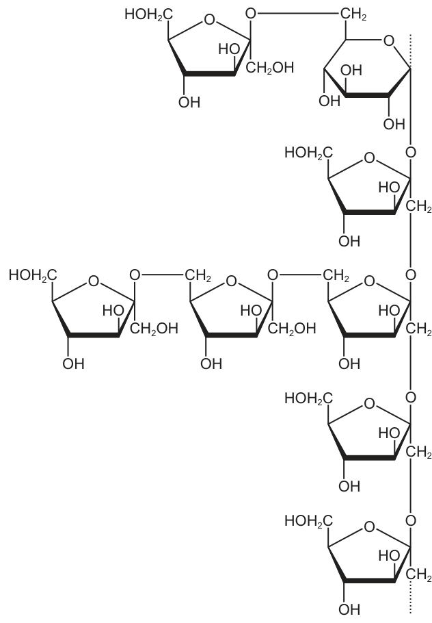
The diagram shows the structure of part of a biological molecule.
Which statements about this biological molecule are correct?
1 It has monomers joined by ester bonds.
2 It is a macromolecule.
3 It is a polysaccharide.
Options
A 1, 2 and 3
B 1 and 2 only
C 1 and 3 only
D 2 and 3 only
Working
The diagram shows a branched chain of glucose monomers linked by glycosidic bonds (the branched structure with α(1→4) and α(1→6) linkages is characteristic of glycogen or amylopectin).
Statement 1: Glucose monomers in a polysaccharide are joined by glycosidic bonds, not ester bonds. Ester bonds join fatty acids to glycerol in lipids. → False.
Statement 2: A polysaccharide such as glycogen is a large polymer and therefore a macromolecule. → True.
Statement 3: The structure shows many monosaccharide (glucose) units joined together — by definition a polysaccharide. → True.
Only statements 2 and 3 are correct.
Answer
D
D
Background Concept
Polysaccharides are large carbohydrates made from many monosaccharide monomers linked together. The most biologically important polysaccharides at AS level are starch (a storage polysaccharide in plants, consisting of amylose and amylopectin), glycogen (the storage polysaccharide in animals and fungi), and cellulose (a structural polysaccharide in plant cell walls). All are built from glucose monomers.
The bond formed between two sugar monomers is a glycosidic bond, which is a type of covalent bond created by a condensation reaction (releasing one molecule of water). In amylopectin and glycogen, two kinds of glycosidic bond occur:
- α(1→4) glycosidic bonds form the straight-chain links.
- α(1→6) glycosidic bonds occur at branch points, where a new chain leaves the main chain.
Ester bonds are an entirely different type of linkage. They form between an acid group (–COOH) and an alcohol group (–OH), releasing water. In biology, ester bonds are characteristic of lipids — they join fatty acids to glycerol in triglycerides and phospholipids. They do not occur between sugar monomers in a polysaccharide.
A macromolecule is simply a very large molecule built from many smaller repeating subunits (monomers). Proteins, nucleic acids, polysaccharides and some lipids (e.g. triglycerides) all qualify. Because polysaccharides such as glycogen contain thousands of glucose units, they comfortably meet the definition of a macromolecule.
Understanding the Question
The diagram (Fig. 7.1) shows a small section of a branched polysaccharide. Each ring is a glucose monomer, identified by the characteristic six-membered ring with a CH₂OH group, hydroxyl (–OH) groups, and an oxygen in the ring. The monomers are joined by oxygen linkages, and the diagram clearly shows a branch point — a hallmark of glycogen or amylopectin. The candidate must judge which of three statements about this molecule are correct.
Approach
For each statement, decide whether it describes a branched polysaccharide of glucose:
- Statement 1 — what kind of bond joins the monomers? (glycosidic, not ester)
- Statement 2 — is the molecule large enough to be called a macromolecule?
- Statement 3 — is a polymer of many monosaccharides a polysaccharide by definition?
Step-by-Step Reasoning
- Statement 1 — "It has monomers joined by ester bonds." The oxygen bridges between glucose rings in the figure are glycosidic bonds, formed by condensation between the –OH on C1 of one glucose and the –OH on C4 (or C6 at a branch point) of the next. Ester bonds require a carboxyl group, which sugars do not possess in this linkage. The mark scheme therefore rejects this statement. → False.
- Statement 2 — "It is a macromolecule." Although the diagram shows only a small fragment, the dotted lines and "CH₂…" continuations indicate the chain extends far beyond what is drawn. The full molecule (glycogen or amylopectin) contains thousands of glucose units and is unambiguously a macromolecule. → True.
- Statement 3 — "It is a polysaccharide." A polysaccharide is, by definition, a polymer composed of many monosaccharide units. The diagram shows many glucose units linked together, so the molecule is a polysaccharide. → True.
Only statements 2 and 3 are correct, which corresponds to option D.
Key Takeaways
- Monosaccharides link to form polysaccharides via glycosidic bonds (never ester bonds).
- Ester bonds are the linkages found in lipids (triglycerides and phospholipids), not in carbohydrates.
- A polysaccharide is, by definition, a polymer of many monosaccharides and is therefore a macromolecule.
- A branched chain of α-glucose units with both α(1→4) and α(1→6) linkages is characteristic of glycogen or amylopectin.
Common Mistakes
- Confusing glycosidic with ester bonds — a very common error. Remember: sugars → glycosidic; lipids → ester.
- Thinking the small fragment shown cannot be a macromolecule — diagrams of polymers always show only a section, with the rest implied.
- Confusing polysaccharide (many sugars) with disaccharide (two sugars) or monosaccharide (one sugar unit).
Things to Be Careful About
- Read the bond type in the diagram, not from memory of a generic carbohydrate. The figure specifically shows C–O–C linkages, which are glycosidic, not ester.
- "Macromolecule" is a size/design description, not a chemical class — both polysaccharides and proteins qualify.
- On a branched structure, identify both kinds of glycosidic bond (1→4 in the chain, 1→6 at the branch point) to confirm it is glycogen/amylopectin rather than a straight-chain polysaccharide like cellulose.
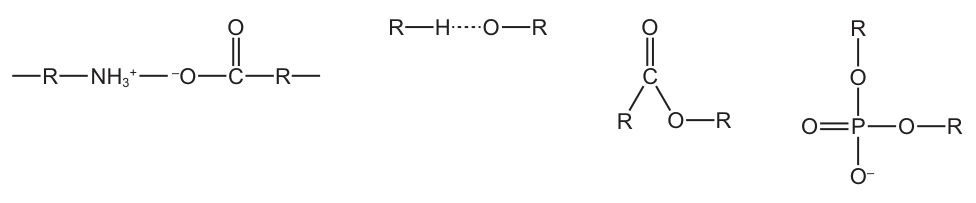
Which diagram has a covalent bond that joins monomers together to form a biological polymer in eukaryotes?
Options
Working
A covalent bond is formed by the sharing of a pair of electrons between two atoms. To form a biological polymer, monomers must be linked by a covalent bond.
- A: shows an –NH₃⁺ group interacting with a carboxylate (–COO⁻); this is an ionic interaction, not a covalent peptide bond.
- B: shows a hydrogen bond (R–H···O–R); hydrogen bonds are intermolecular forces, not covalent, and do not link monomers.
- C: shows an ester bond (R–CO–O–R); ester bonds join glycerol to fatty acids in lipids, but lipids are not polymers of repeating monomers.
- D: shows a phosphodiester bond (R–O–P(=O)(O⁻)–O–R); this covalent bond joins nucleotides together to form nucleic acids (DNA and RNA), which are biological polymers found in eukaryotes.
Answer
D
D
Background Concept
Biological polymers are large molecules built by linking many small monomer subunits together. The linkage between monomers is always a covalent bond — a bond in which two atoms share a pair of electrons. Once such covalent bonds are made, the resulting polymer is stable and the monomers cannot easily separate.
In eukaryotic cells the three major classes of biological polymer are:
- Polysaccharides (e.g. starch, glycogen, cellulose) — monomers are monosaccharides joined by glycosidic bonds.
- Polypeptides/proteins — monomers are amino acids joined by peptide bonds.
- Nucleic acids (DNA, RNA) — monomers are nucleotides joined by phosphodiester bonds.
It is essential to distinguish these covalent polymer-forming bonds from the many other types of bond or interaction drawn in diagrams. Hydrogen bonds, ionic (salt-bridge) interactions, and van der Waals' forces are all non-covalent and are therefore weak enough to break and reform — useful for stabilising the 3-D shape of a molecule, but not strong enough to fix monomers into a polymer backbone.
Understanding the Question
The question shows four bond representations labelled A–D and asks which one is a covalent bond joining monomers to form a biological polymer found in eukaryotes. Two things must be true of the correct answer:
- The bond drawn must be a true covalent bond (shared electron pair).
- The monomers being joined must be repeating units of a biological polymer present in eukaryotic cells.
The command word is "which diagram", so this is a multiple-choice question: only one option is correct.
Approach
Read each structure carefully, identify the bond type, then check whether that bond type links repeating monomers into a polymer. The two key filters are:
- Is the bond covalent? (Look for shared pairs, not dots/dashes representing partial charges, dashes, or ionic arrows.)
- Does it join repeating monomers into a polymer? (Glycosidic, peptide, phosphodiester — yes. Ester in lipid, hydrogen bond, ionic interaction — no.)
Step-by-Step Reasoning
Option A depicts a protonated amino group (–NH₃⁺) sitting next to a carboxylate (–COO⁻). The attraction between a positively charged ammonium and a negatively charged carboxylate is an ionic (electrostatic) interaction, not a covalent peptide bond. A genuine peptide bond would be drawn as R–NH–C(=O)–R′ with a direct N–C linkage and loss of water. So A is rejected.
Option B shows R–H···O–R with a dotted line. The dots explicitly indicate a hydrogen bond — an intermolecular attraction between a slightly δ⁺ H and a slightly δ⁻ O. Hydrogen bonds are non-covalent and do not join monomers into polymers; they instead stabilise secondary and tertiary structures (e.g. the two strands of DNA, or α-helices and β-sheets in proteins). So B is rejected.
Option C shows R–C(=O)–O–R, an ester bond. Ester bonds occur where a carboxylic acid reacts with an alcohol (e.g. glycerol + fatty acids → triglyceride; glycerol + phosphate + fatty acid → phospholipid). However, lipids are not polymers of repeating identical monomers; a triglyceride has three (often different) fatty acids attached to one glycerol, and that is the whole molecule. So C is rejected.
Option D shows R–O–P(=O)(O⁻)–O–R, the characteristic phosphodiester bond. This covalent bond is formed when the 3′-OH of one nucleotide attacks the phosphate of the next, releasing water. Phosphodiester bonds link nucleotides into the long chains of DNA and RNA, both of which are biological polymers present in every eukaryotic cell. So D is correct.
Key Takeaways
- Only covalent bonds form the backbone of a biological polymer; hydrogen bonds and ionic interactions are non-covalent and merely stabilise shape.
- The three polymer-forming bonds to recognise are: glycosidic (sugars), peptide (amino acids) and phosphodiester (nucleotides).
- Ester bonds (option C) are covalent but join the components of a lipid, which is not a polymer of repeating monomers.
- The way a bond is drawn matters: an –NH₃⁺ next to a –COO⁻ indicates an ionic pair, not a peptide bond.
Common Mistakes
- Choosing A because it "looks like a peptide bond". The –NH₃⁺ group is fully protonated and is shown separately from the C=O; this represents an ionic salt bridge, not the covalent C–N peptide linkage.
- Choosing B because hydrogen bonds are important in biology. They are not polymer-forming.
- Choosing C because ester bonds are covalent. Covalent alone is not enough — the monomers must be repeating units of a polymer.
Things to Be Careful About
- Read the bonds, not just the atoms. A dotted line means hydrogen bonding; a charge symbol (⁺, ⁻) suggests an ionic interaction; only a single line between two atoms normally represents a covalent bond.
- Remember the polymer criterion: the monomers must repeat many times along a chain. Lipids fail this test because a triglyceride has only three fatty-acid units on a single glycerol.
- Phosphodiester bonds appear in both DNA and RNA; the question specifies eukaryotes, but the same bond also occurs in prokaryotes — it is simply true for all cellular life that has nucleic acids.
Which feature of storage polysaccharides means they do not change the water potential of cells?
Options
A They are easily hydrolysed.
B They are compact.
C They are insoluble.
D They are branched molecules.
Answer
C
C
Background Concept
Water potential () of a cell is determined mainly by its solute potential (): the more dissolved solute particles present, the lower (more negative) the solute potential, and therefore the lower the water potential. Anything that dissolves in the cytoplasm contributes osmotically active particles. Anything that remains as an undissolved solid does not.
Storage polysaccharides such as starch (in plants) and glycogen (in animals) are built from many -glucose monomers joined by glycosidic bonds. A key structural feature is that the long chains coil and pack tightly together, so the huge molecules cannot be surrounded and stabilised by water molecules. As a result, starch grains and glycogen granules are insoluble in water — they sit in the cytoplasm as discrete solid inclusions rather than dissolving into it.
Understanding the Question
The question is asking which property of storage polysaccharides allows them to be stored in large quantities inside cells without pulling water into the cell by osmosis. The command word is implied ("which feature"), and a single option must be selected from A–D.
Approach
The water potential of a cell is lowered by dissolved solutes only. Insoluble substances do not contribute to solute potential because they do not produce free particles in solution. Therefore the relevant feature is the one that prevents the polysaccharide from dissolving.
Step-by-Step Reasoning
- A — Easily hydrolysed: Hydrolysis is what breaks the polysaccharide down into glucose for use. It does not determine whether the stored polymer is currently dissolved. Even an easily hydrolysed polymer is stored in insoluble form.
- B — Compact: Being compact is true (and biologically useful for storage), but compactness does not by itself prevent dissolution. A compact but hydrophilic molecule could still dissolve.
- C — Insoluble: Correct. Because starch and glycogen are insoluble, they do not contribute dissolved particles to the cytoplasm, so they do not affect the cell's water potential. The cell can therefore stockpile very large amounts of carbohydrate without causing water to be drawn in osmotically.
- D — Branched: Branching (e.g. linkages in glycogen) affects how quickly the molecule can be mobilised by enzymes, but it does not in itself determine solubility.
Key Takeaways
- Water potential of a cell depends on dissolved solutes (solute potential).
- Storage polysaccharides (starch, glycogen) are insoluble, so they can accumulate in large amounts without lowering and without causing osmotic water uptake.
- Structure–function links for these molecules: insoluble → osmotically inactive; compact → efficient storage; branched (glycogen) → rapid mobilisation.
Common Mistakes
- Choosing D (branched) because glycogen is the most familiar branched polymer — branching is real but is not the reason these polymers are osmotically inactive.
- Choosing B (compact) because compactness is associated with good storage, but it is the insolubility that matters for water potential.
- Confusing the property that makes a good storage molecule (insoluble, compact, branched) with the property that specifically affects water potential (insolubility).
Things to Be Careful About
- "Insoluble" is the property that matters; "compact" and "branched" are true features but do not, by themselves, prevent osmotic effects.
- Do not equate hydrolysis (a property of the molecule) with solubility (a property of how it sits in the cytoplasm).
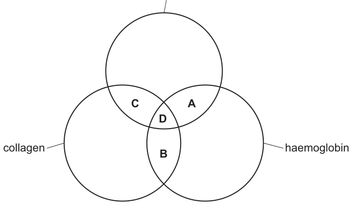
Which molecules contain at least four double bonds?
Options
A A
B B
C C
D D
Working
The three categories are saturated triglyceride, collagen and haemoglobin. Count the double bonds each contains:
- Saturated triglyceride: 3 ester linkages, each with one C=O → 3 double bonds (less than 4). By definition, "saturated" means 0 C=C double bonds in the fatty acid chains.
- Collagen: a fibrous protein of many amino acids linked by peptide bonds. Each peptide bond has one C=O → many double bonds (more than 4).
- Haemoglobin: a globular protein (4 polypeptide chains) → many C=O double bonds in peptide bonds, plus the porphyrin haem group which has multiple C=C and C=N double bonds → many double bonds (more than 4).
The category of molecules containing at least 4 double bonds is therefore the one shared by collagen and haemoglobin but excluding saturated triglyceride (which only has 3). This is region B.
Answer
B
B
Background Concept
Three biological molecules are involved here, each with a distinctive set of covalent bonds.
Triglyceride structure. A triglyceride is a glycerol molecule esterified to three fatty acids. The three ester linkages each contain one carbon–oxygen double bond (C=O). The fatty acid chains of a saturated triglyceride contain only single C–C bonds — "saturated" means fully hydrogenated, with no C=C double bonds. So a saturated triglyceride contains exactly 3 C=O double bonds and no C=C double bonds.
Collagen structure. Collagen is a fibrous protein built from long polypeptide chains (mostly glycine, proline and hydroxyproline, with characteristic hydroxyproline residues). Each peptide bond in the backbone contains one C=O double bond. A single collagen molecule has hundreds of amino acids, giving it hundreds of C=O double bonds. Its amino acid side chains contain no C=C double bonds.
Haemoglobin structure. Haemoglobin is a globular protein with four polypeptide chains (2α, 2β) and four haem prosthetic groups. The globin chains contribute the same kind of C=O double bonds as collagen — many, in the peptide bonds. Each haem group contains a porphyrin ring system with multiple conjugated C=C and C=N double bonds (the alternating double bonds that give haem its colour and its ability to bind oxygen). So haemoglobin contains many C=O, C=C and C=N double bonds.
Understanding the Question
The Venn diagram shows three overlapping sets: saturated triglyceride, collagen, and haemoglobin. Each region (A, B, C, D) represents molecules that share features with the categories whose circles overlap there. We are asked to identify the region whose molecules contain at least four double bonds of any kind.
The threshold of four is the key clue: a single saturated triglyceride has only three double bonds (the three ester carbonyls), so any region that includes "saturated triglyceride" features alone cannot reach four. We need a region where the molecules are protein-like (rich in peptide-bond C=O) and so exceed four double bonds by a wide margin.
Approach
Count the double bonds for each of the three categories, identify which ones clear the threshold of four, and then locate the region of the Venn diagram corresponding to those categories.
Step-by-Step Reasoning
- Saturated triglyceride: 3 C=O (ester) + 0 C=C = 3 double bonds → fewer than 4.
- Collagen: hundreds of C=O (peptide) + 0 C=C = many double bonds → ≥4.
- Haemoglobin: hundreds of C=O (peptide) + multiple C=C/C=N (porphyrin) = many double bonds → ≥4.
- The categories that pass the "at least four double bonds" test are collagen and haemoglobin. The intersection of these two circles (excluding saturated triglyceride) is region B.
A molecule lying in region B therefore has the protein features of both collagen and haemoglobin — it is proteinaceous — and so has far more than four double bonds.
Key Takeaways
- A saturated triglyceride has only the three C=O double bonds of its ester linkages — never any C=C.
- Proteins are rich sources of C=O double bonds because every peptide bond contains one; a peptide of just 5 amino acids already has 4 C=O bonds.
- Haemoglobin additionally carries the conjugated double-bond system of its porphyrin haem groups, giving many more C=C/C=N bonds on top of the peptide-bond C=O.
- "At least 4 double bonds" is a counting question — work out how many each category actually has before choosing the region.
Common Mistakes
- Counting only C=C bonds in a saturated triglyceride and concluding it has 0 double bonds. Saturated refers specifically to the fatty acid chains; the three C=O ester carbonyls are still double bonds. This makes the saturated triglyceride have 3, not 0, which matters for the threshold of 4.
- Forgetting the C=O of the peptide bond. Many students count only C=C and decide that "proteins have no double bonds", which leads to ruling out region B incorrectly.
- Overlooking the haem group's double bonds. Haemoglobin is not just a protein; the porphyrin ring contributes many additional C=C/C=N bonds.
- Confusing the regions of the Venn diagram. Region B is the lens between collagen and haemoglobin, not the central triple overlap (D).
Things to Be Careful About
- The threshold "at least four" is precise; 3 is not enough. The saturated triglyceride sits at exactly 3, so any region that includes it cannot be the answer.
- "Double bond" here includes C=O and C=N as well as C=C — all are double bonds by definition.
- In Venn-diagram questions, read the labels of every circle and identify which intersections are being asked about; a careless glance at the letter can cost the mark.
In some people a mutation can affect the beta chains in their haemoglobin molecules. As a result polar amino acids on the outer surfaces of the chains are replaced by non-polar amino acids.
What will be the effect on haemoglobin?
Options
A Haemoglobin will become less soluble.
B Haemoglobin will become more soluble.
C Hydrophobic interactions between amino acids will decrease.
D More hydrogen bonds will form in the molecule.
Working
Polar (hydrophilic) R-groups on the outer surface of haemoglobin form favourable interactions with water, keeping the molecule soluble in the cytoplasm. Replacing them with non-polar (hydrophobic) R-groups removes these water interactions and exposes hydrophobic groups to the aqueous environment, decreasing haemoglobin's solubility.
Answer
A
A
Background Concept
The 20 standard amino acids differ only in their side chain (R-group). These R-groups are classified by their chemistry:
- Polar (hydrophilic) R-groups carry partial or full charges and form hydrogen bonds with water. Examples: serine, threonine, asparagine, glutamine, lysine, arginine, histidine, aspartate, glutamate.
- Non-polar (hydrophobic) R-groups are mostly hydrocarbon chains or rings. Examples: alanine, valine, leucine, isoleucine, phenylalanine, tryptophan, methionine.
In a globular protein like haemoglobin, which functions in an aqueous cellular environment, the surface is dominated by hydrophilic R-groups (so the protein stays dissolved), while hydrophobic R-groups are tucked into the interior (driven together by the hydrophobic effect, which is entropically favourable because it releases ordered water molecules).
The question describes exactly such a protein: haemoglobin is a soluble, globular protein whose beta chains normally have polar amino acids facing the surrounding water.
Understanding the Question
The stem describes a point mutation that swaps polar amino acids on the OUTER surface of the beta chain for non-polar ones. The command word "what will be the effect" requires linking this single change in R-group chemistry to a property of the whole haemoglobin molecule. We must pick the option that correctly predicts the consequence.
Key facts to anchor the reasoning:
- The affected amino acids are on the outside of the protein (i.e. in contact with water).
- The change is polar → non-polar (hydrophilic → hydrophobic).
- Haemoglobin normally dissolves in the cytoplasm of red blood cells.
Approach
Ask: "What does water 'see' on the outside of the modified haemoglobin?" — it now sees hydrophobic R-groups instead of hydrophilic ones. By definition, hydrophobic groups do not interact favourably with water, so the protein's ability to remain dissolved in aqueous cytosol will fall. That points directly to option A.
Step-by-Step Reasoning
Why A is correct:
Replacing surface polar R-groups with non-polar R-groups exposes hydrophobic surfaces to the aqueous solvent. The protein can no longer form hydrogen bonds with water at these positions, and water molecules around the exposed hydrophobic groups become more ordered (a thermodynamically unfavourable state). The result is reduced solubility; in vivo this drives abnormal haemoglobin molecules to aggregate — the molecular basis of sickle-cell disease, in which Glu6 on the beta chain is replaced by Val6, a non-polar residue. The mutant haemoglobin (HbS) polymerises and is far less soluble than normal HbA.
Why B is wrong:
Non-polar groups repel water, so a more non-polar surface makes a protein LESS, not more, soluble. Option B states the opposite of the chemistry.
Why C is wrong:
The statement "hydrophobic interactions between amino acids will decrease" is incorrect on two counts. First, the change INTRODUCES more non-polar R-groups, which INCREASES the potential for hydrophobic interactions. Second, the affected residues are on the outer surface, so the relevant interactions are with water, not between amino acids. Even if we considered internal packing, the hydrophobic effect would intensify, not weaken.
Why D is wrong:
Polar R-groups are the ones that form hydrogen bonds. Replacing them with non-polar R-groups removes hydrogen-bond donors and acceptors, so hydrogen bonding DECREASES, not increases.
Key Takeaways
- Soluble globular proteins have polar/hydrophilic R-groups on their surface and non-polar/hydrophobic R-groups buried inside (the hydrophobic effect).
- A mutation that puts hydrophobic groups on the outside of a protein reduces its solubility in water.
- This is precisely the molecular basis of sickle-cell anaemia: Glu → Val at position 6 of the beta chain produces haemoglobin S, which polymerises inside red blood cells.
Common Mistakes
- Choosing B because "amino acids have been added" — confusion over the chemistry; the type of amino acid matters, not the count.
- Choosing C, thinking that fewer polar groups mean fewer interactions overall, without distinguishing between hydrophobic interactions (which would actually increase) and hydrogen bonds.
- Choosing D, thinking that more amino acids means more bonds, without recognising that non-polar R-groups cannot hydrogen-bond.
- Forgetting that "outside" means in contact with water, so the relevant comparison is to water, not to other amino acids.
Things to Be Careful About
- The R-group property is what matters: the same backbone and peptide bonds exist in both polar and non-polar amino acids, so the change is in side-chain chemistry only.
- "Hydrophobic interactions" are not true bonds between R-groups; they are an entropic effect arising from the release of ordered water when non-polar groups cluster. This is a frequent point of confusion.
- The clinical link (sickle-cell) is a useful memory aid but is not required to answer the question; the answer follows from first principles of R-group chemistry.
The graph shows the effect of substrate concentration on an enzyme-catalysed reaction with and without a competitive inhibitor.
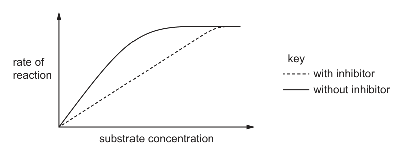
What is the effect of the competitive inhibitor on and ?
Options
A decreases and decreases.
B stays the same and decreases.
C stays the same and increases.
D decreases and stays the same.
Working
The two curves on the graph both plateau at the same maximum rate, so is unchanged by the inhibitor. However, the 'with inhibitor' curve rises more slowly and only reaches half of at a higher substrate concentration, so is increased. This pattern (same , higher ) is the signature of a competitive inhibitor, which is overcome at saturating substrate levels.
Answer
C
C
Background Concept
Enzyme kinetics describes how the rate of an enzyme-catalysed reaction varies with substrate concentration. As substrate concentration increases, the rate rises hyperbolically and levels off at — the maximum rate — when all active sites are continuously occupied. The Michaelis constant, , is the substrate concentration at which the rate is half of ; it is inversely related to the apparent affinity of the enzyme for its substrate.
A competitive inhibitor is a molecule that resembles the substrate and binds reversibly to the active site, physically blocking substrate binding. Because the binding is reversible and competitive, raising the substrate concentration can out-compete the inhibitor and restore full activity. A non-competitive inhibitor binds to a different (allosteric) site, lowering the effective amount of functional enzyme so falls and cannot be recovered by adding more substrate; is typically unchanged.
Understanding the Question
The figure plots rate of reaction (y-axis) against substrate concentration (x-axis) for two conditions:
- solid line: reaction without inhibitor
- dashed line: reaction with a competitive inhibitor
The question asks how and change in the presence of the inhibitor. These two quantities are read from the curve: is the height of the plateau; is the substrate concentration at half of that plateau height.
Approach
Compare the two curves at two features:
- Compare the height of the plateaux ().
- Compare the substrate concentrations at half the plateau height ().
Then match the pattern to the standard signature of a competitive inhibitor (same , higher ).
Step-by-Step Reasoning
- : Both curves in Fig. 12.1 level off at the same height. With enough substrate, the inhibitor is displaced from the active site and the reaction reaches the same maximum rate as the uninhibited reaction. So stays the same.
- : The 'with inhibitor' curve reaches half its plateau further to the right (at a higher substrate concentration) than the 'without inhibitor' curve. Because the inhibitor is occupying some active sites, more substrate is needed to drive the enzyme into a half-saturated state. So increases.
- Combining these: unchanged, increased → answer C.
Key Takeaways
- Competitive inhibitor: same , higher (substrate can out-compete the inhibitor at high [S]).
- Non-competitive inhibitor: lower , same (substrate cannot overcome an allosteric block).
- Always read from the plateau and from the [S] at half ; do not confuse with the substrate concentration at .
Common Mistakes
- Saying decreases because the dashed curve is below the solid curve in the middle of the graph — this is wrong because at very high [S] the two curves meet.
- Saying decreases because the curve appears to start lower — is defined by the half-saturation point on the x-axis, not by the low-[S] region.
- Confusing competitive with non-competitive inhibition and choosing D.
Things to Be Careful About
- The graph's dashed curve approaches the same asymptote — do not assume the lines have different plateaux.
- is a concentration, not a rate. It is read off the x-axis.
- 'Stays the same' is a valid description even when the inhibited curve is lower at intermediate [S]; what matters is the asymptote.
The graph shows energy changes in a chemical reaction.
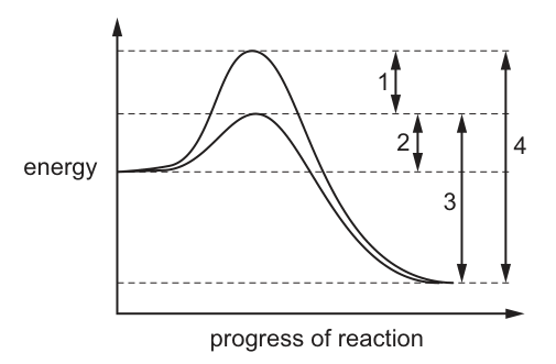
What is the activation energy when an enzyme is added?
Options
A 1 + 2
B 2 only
C 3 – 2
D 4
Working
An enzyme is a biological catalyst. Catalysts (and therefore enzymes) lower the activation energy of a reaction without being consumed. On the energy profile, the catalysed reaction is the curve with the lower peak.
Activation energy is defined as the minimum energy that reactants must possess to start the reaction, i.e. the energy difference between the reactants and the peak of the catalysed curve.
On the diagram:
- The reactants sit at the energy level on the left.
- The lower (catalysed) peak is reached by the vertical distance labelled 2 from the reactants.
- Therefore the activation energy with the enzyme present is distance 2.
Answer
B
B
Background Concept
Enzymes are biological catalysts. Like all catalysts, they speed up a chemical reaction by providing an alternative reaction pathway with a lower activation energy (Ea), while themselves remaining unchanged at the end of the reaction. Activation energy is the minimum amount of kinetic energy that the reactant molecules must have at the moment of collision in order for the reaction to proceed — i.e. the size of the energy "hump" that must be overcome for reactants to be converted into products.
Understanding the Question
Fig. 13.1 is an energy profile for an exothermic reaction. The horizontal lines mark key energy levels:
- the top dashed line = the peak of the uncatalysed reaction (higher hump);
- the lower dashed line = the peak of the catalysed reaction (lower hump, where an enzyme would operate);
- the line on the far left = the energy of the reactants;
- the line on the far right = the energy of the products (lower than reactants because the reaction is exothermic).
Four vertical distances are labelled: 1, 2, 3 and 4. The question asks which of these represents the activation energy when an enzyme is added.
Approach
- Identify which curve corresponds to the enzyme-catalysed reaction. Catalysts lower the activation energy, so the catalysed curve has the lower peak.
- Locate the reactants' energy level on the left of the diagram.
- Measure (or read off) the vertical distance from the reactants to the peak of the catalysed curve. That distance is the activation energy in the presence of the enzyme.
Step-by-Step Reasoning
- The vertical distance from the reactants up to the lower (catalysed) peak is labelled 2 on the diagram.
- By definition, this distance (reactants → catalysed peak) is the activation energy with the enzyme present.
- Therefore the activation energy when an enzyme is added = 2 only, which corresponds to option B.
Why the other options are wrong:
- A (1 + 2): Adding 1 and 2 gives the distance from the reactants to the higher peak — the activation energy without the enzyme (the uncatalysed activation energy).
- C (3 − 2): 3 is the overall energy change of the reaction (reactants to products, the enthalpy change ΔH for the exothermic reaction). Subtracting 2 from 3 has no biological meaning as an activation energy.
- D (4): 4 is the distance from the products up to the higher (uncatalysed) peak — this is not the activation energy of either reaction.
Key Takeaways
- An enzyme lowers the activation energy; it does not change the energy of the reactants or the products, and it does not change the overall energy change (ΔH) of the reaction.
- On an energy profile, activation energy is always measured from the reactants to the peak of the relevant curve (catalysed for an enzyme, uncatalysed for the reaction without it).
- The activation energy in the presence of an enzyme is the smaller of the two humps measured from the reactants.
Common Mistakes
- Confusing the catalysed and uncatalysed peaks and choosing A (1 + 2), which is the uncatalysed activation energy.
- Picking D (4) by thinking activation energy is measured from the products; activation energy is always measured from the reactants.
- Treating 3 as if it were activation energy; 3 represents the overall energy change (ΔH) of the reaction, not the activation energy.
Things to Be Careful About
- Read the labels carefully: in this diagram 2 is the distance from the reactants to the lower (catalysed) peak, not the catalysed peak to the products.
- "Activation energy with an enzyme" specifically refers to the smaller hump, because the enzyme-catalysed route has the lower Ea.
- A catalyst does not alter the position of the reactants or products on the y-axis; only the height of the energy barrier is reduced.
The diagram shows an enzyme, its substrate and an enzyme–substrate complex.
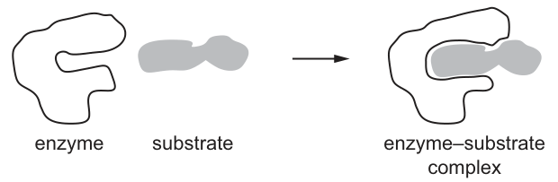
Which statement explains how this substrate is able to enter the active site of this enzyme?
Options
A Contact between the substrate and the enzyme causes a change in the enzyme shape.
B The shape of the active site and the shape of the substrate are complementary.
C The substrate within the active site forms hydrogen bonds with amino acids.
D When the enzyme–substrate complex forms, the tertiary structure of the enzyme changes.
Working
The diagram shows the substrate initially not perfectly matching the active site; on contact, the enzyme's outline changes to wrap around the substrate. This is the induced-fit model, in which contact with the substrate causes a conformational change in the enzyme that allows the substrate to enter the active site.
- B describes the lock-and-key model, which assumes the shapes are already complementary — this is contradicted by the diagram and does not explain how the substrate enters.
- C describes a feature of binding that occurs after the substrate is already in the active site, so it cannot explain entry.
- D states that the tertiary structure changes when the ES complex forms; this is a consequence, not the mechanism that permits the substrate to enter.
Answer
A
A
Background Concept
Enzymes are biological catalysts (almost always proteins) that speed up metabolic reactions by binding their substrates at a region called the active site. Two historical models describe how a substrate fits into this site:
- Lock-and-key hypothesis — the active site and substrate have rigid, pre-formed complementary shapes, like a key fitting a lock.
- Induced-fit hypothesis (Koshland, 1958) — the active site is not a perfect fit initially. Contact with the substrate induces a conformational change in the enzyme, moulding the active site around the substrate. This also helps the enzyme apply strain to the substrate, lowering the activation energy.
CIE Biology recognises the induced-fit model as the more accurate description of enzyme–substrate interaction, and many diagrams deliberately show a slight change in the enzyme's outline when the ES complex forms.
Understanding the Question
The diagram (Fig. 14.1) depicts the enzyme, the separate substrate, and the enzyme–substrate complex. The question asks specifically how the substrate is able to enter the active site — that is, what mechanism permits entry, not what stabilises the complex once formed.
The command word here is "explains how", so the correct option must describe the process that allows the substrate to fit, not the resulting state.
Approach
- Look at the diagram: does the active site appear pre-complementary to the substrate, or does the enzyme's outline change on binding?
- Decide which model the diagram is illustrating.
- Match that mechanism to the option that explains entry (not post-binding effects).
- Eliminate options that describe consequences of binding, or describe lock-and-key instead.
Step-by-Step Reasoning
- The diagram shows the enzyme's outline adapting to the substrate when the complex forms — the active site closes around / moulds to the substrate. This visual cue is the hallmark of induced fit.
- Option A — "Contact between the substrate and the enzyme causes a change in the enzyme shape." This is precisely the induced-fit mechanism: initial contact triggers a conformational change that lets the substrate sit properly within the active site. ✓
- Option B — "The shape of the active site and the shape of the substrate are complementary." This is the lock-and-key model. The diagram contradicts it (the active site is not initially complementary), and even if true, it would not explain how the substrate enters — it would simply assert that they already fit.
- Option C — "The substrate within the active site forms hydrogen bonds with amino acids." Hydrogen bonds and other weak interactions help stabilise the ES complex, but they form only after the substrate is already inside. They do not explain how entry occurs.
- Option D — "When the enzyme–substrate complex forms, the tertiary structure of the enzyme changes." This is true, but the change in tertiary structure is described as occurring when the complex forms — i.e. as a result of, not a cause of, binding. It therefore does not explain how the substrate is able to enter the active site in the first place.
Only option A describes a causal mechanism that permits entry.
Key Takeaways
- Distinguish the lock-and-key model (static complementarity) from the induced-fit model (dynamic change in enzyme shape on contact).
- Read the command word carefully: "explains how the substrate enters" demands a mechanism of entry, not a description of the resulting complex.
- Beware of statements that are biologically true but address the wrong stage of the process (e.g. hydrogen bonds form after binding; tertiary-structure changes are a consequence, not a cause, of entry under this wording).
Common Mistakes
- Choosing B because "complementary shapes" is a familiar phrase — but this is lock-and-key, and the question asks about entry.
- Choosing D because it sounds correct — the tertiary structure does change, but the option places the change after the complex has formed, so it cannot explain entry.
- Choosing C because hydrogen bonding is a real feature of enzyme–substrate interaction — but it describes what holds the complex together, not how the substrate gets in.
Things to Be Careful About
- The induced-fit wording is specific: contact causes a change in the enzyme shape that enables the substrate to bind. The change must be presented as the cause of binding, not as a consequence.
- If a future option states that the active site changes shape so that it is complementary after binding, that is also induced-fit and would be credited. The key is direction of cause and effect.
- The diagram in CIE questions often deliberately shows a non-complementary initial fit to push candidates toward induced-fit. Always look at the figure before reading the options.
What are the main roles of glycolipids in cell surface membranes?
1 to help cells to attach to each other to form tissues
2 to act as antigens for cell-to-cell recognition
3 to increase fluidity for the cell surface membrane
Options
A 1, 2 and 3
B 1 and 2 only
C 1 and 3 only
D 2 and 3 only
Working
- Statement 1: correct. Glycolipids (and glycoproteins) on the outer surface of the membrane are involved in cell adhesion, allowing cells to attach to one another to form tissues.
- Statement 2: correct. The carbohydrate chains of glycolipids act as antigens / recognition sites, allowing cell-to-cell recognition (e.g. the ABO blood group antigens).
- Statement 3: incorrect. Increasing membrane fluidity is the role of cholesterol, not glycolipids. Glycolipids have a relatively small effect on fluidity; cholesterol is the component credited with regulating fluidity.
Only statements 1 and 2 are roles of glycolipids.
Answer
B
B
Background Concept
The fluid mosaic model describes the cell surface membrane as a phospholipid bilayer in which various proteins and other components are embedded. Among the non-protein components are glycolipids and glycoproteins: lipids or proteins with short carbohydrate chains attached on the outer surface of the membrane only. These carbohydrate chains are the key to many cell-surface functions.
Cholesterol is a separate lipid component inserted among the phospholipid tails. Its main role is to regulate membrane fluidity — at higher temperatures it restrains phospholipid movement, and at lower temperatures it prevents the membrane from becoming too rigid. Students often confuse cholesterol's function with that of glycolipids because both are membrane lipids.
Understanding the Question
This is a multiple-choice question offering three statements about glycolipids, and asking which combination correctly describes their main roles. The question requires the candidate to know what glycolipids do (and, by elimination, to recognise the one statement that is not about glycolipids at all).
The command word is implicit: "What are the main roles" — so the answer must list functions actually performed by glycolipids, not by other membrane components.
Approach
Treat each statement independently, decide whether it is a recognised function of glycolipids, then match the combination to one of the answer letters. The likely trap is statement 3, because "membrane fluidity" is a real membrane concept — but it belongs to cholesterol, not glycolipids.
Step-by-Step Reasoning
- Statement 1 — cell adhesion to form tissues. The carbohydrate portion of glycolipids (and glycoproteins) on adjacent cells can bind to one another, helping cells recognise and adhere to like cells. This is essential during embryonic development and in maintaining tissue architecture. ✓
- Statement 2 — antigens for cell-to-cell recognition. The specific sugar sequences of the carbohydrate chains differ between cell types and between individuals. The immune system uses them as antigens to distinguish self from non-self (e.g. ABO blood group antigens on red blood cell membranes are glycolipids). ✓
- Statement 3 — increasing membrane fluidity. This is the role of cholesterol. Glycolipids are present in much smaller quantities and are not credited with controlling fluidity. ✗
Therefore only statements 1 and 2 are correct, which corresponds to option B.
Key Takeaways
- Glycolipids are membrane lipids with a short carbohydrate chain on the outer leaflet.
- Their main roles: cell–cell recognition (acting as antigens/receptors) and cell adhesion (forming tissues).
- Cholesterol, not glycolipids, regulates membrane fluidity.
Common Mistakes
- Selecting A or D because statement 3 was wrongly attributed to glycolipids instead of cholesterol.
- Confusing glycolipids with glycoproteins; both share the recognition/adhesion roles, but only glycolipids are lipids with attached carbohydrate.
- Forgetting that the carbohydrate chains project only on the outer surface of the membrane, which is why they can interact with the external environment and other cells.
Things to Be Careful About
- "Increase fluidity" is a tempting distractor because glycolipids are membrane components — always check which component actually performs each named function.
- The carbohydrate portion, not the lipid portion, is responsible for the recognition and adhesion functions.
A sample of healthy plant cells taken from the same tissue is placed in a beaker containing distilled water.
The cells change in size.
What would explain this change in size?
Options
A Water will leave the cells by active transport.
B Water will enter the cells by osmosis.
C Solutes will enter the cells by active transport.
D Solutes will leave the cells by osmosis.
Distilled water has a higher (less negative) water potential than the cell sap of the plant cells. Net water therefore moves into the cells by osmosis down the water potential gradient, and the cells swell (and become turgid).
Answer
B
B
Background Concept
Water potential (Ψ) is the tendency of water to move from one place to another. Pure water has the highest water potential (Ψ = 0 kPa), and any solution of solutes has a lower (more negative) water potential. Water moves by osmosis — a special case of diffusion — across a partially permeable membrane from a region of higher water potential to a region of lower water potential. Osmosis only refers to the movement of water (never solutes), and it is a passive process (it does not require ATP, so it is not active transport).
Inside a healthy plant cell, the central vacuole contains cell sap rich in dissolved solutes (sugars, salts, organic acids), giving it a water potential of around −200 to −1000 kPa, depending on the tissue. The cell surface membrane is a partially permeable membrane, so when such a cell is placed in distilled water (Ψ ≈ 0 kPa), a steep water potential gradient exists across the membrane.
Understanding the Question
The scenario is a classic plant-cell osmosis setup:
- Given: healthy plant cells from the same tissue placed in distilled water.
- Observation: the cells change in size.
- Asked: which option correctly explains the change in size.
The command word is implicit but it is an "explain" type: pick the statement that correctly describes what is moving and how. Distilled water outside the cell is the key piece of information — it has a higher water potential than the cytoplasm/vacuole inside.
Approach
- Identify the direction of the water potential gradient between the external distilled water and the internal cell sap.
- Decide which substance moves (water or solute) and by which process (osmosis, diffusion, or active transport).
- Match these to one of the four options.
Step-by-Step Reasoning
- Direction of gradient: distilled water outside (Ψ ≈ 0) has a higher water potential than cell sap inside (Ψ is negative). Water therefore moves into the cell.
- Process: water crosses the partially permeable membrane by osmosis, which is passive. It does not require carrier proteins or ATP, so it is not active transport.
- Eliminate options:
- A — wrong on two counts. Water is not moved by active transport, and in this scenario water moves into, not out of, the cell.
- B — water moves into the cells by osmosis. ✓
- C — solutes do not enter the cell in this scenario because there are essentially no solutes in distilled water, and active transport is irrelevant here.
- D — osmosis only describes the movement of water, not solutes. The wording is biologically meaningless.
- Result: water entering the cells by osmosis causes the protoplast to swell, pressing the cell membrane against the cell wall, and the cells become larger and turgid.
Key Takeaways
- Osmosis is the passive movement of water (never solutes) across a partially permeable membrane down a water potential gradient.
- Distilled water has Ψ = 0; any solution has a more negative Ψ. Water always moves from 0 → negative.
- Plant cells placed in distilled water take up water and become turgid; in a concentrated solution they lose water and become plasmolysed.
- Distinguish carefully between the substance moving (water vs solute) and the process (osmosis vs active transport).
Common Mistakes
- Choosing A because it mentions "water" — but water is never moved by active transport; ions, sugars and other solutes are.
- Choosing C because cells "change size" — but distilled water contains no solutes to enter, and active transport would require ATP, which is irrelevant to a cell simply sitting in water.
- Choosing D because it includes the word "osmosis" — but osmosis strictly refers to water movement, never solute movement.
- Confusing osmosis (water, passive) with active transport (solutes, requires energy).
Things to Be Careful About
- "Osmosis" only ever refers to the movement of water. If a statement says "solutes move by osmosis" it is automatically wrong.
- "Active transport" only ever refers to the movement of solutes (ions/molecules), against a concentration gradient, using ATP. Water is never actively transported across a cell membrane in this syllabus.
- Remember the sign convention: higher Ψ = less negative; pure water has the highest possible Ψ.
A student studying surface area to volume ratio and diffusion made a cuboid, S1, using agar stained blue with a pH indicator. The dimensions of S1 are shown in the diagram.
The student made a second agar cuboid, S2. Each dimension of S2, (the length, the width and the height), was half that of S1.
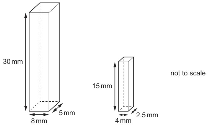
The student placed each cuboid in a test-tube and covered it in acid. The time taken for each cuboid to completely change colour was recorded. All variables other than the size of the cuboids were standardised.
Which row shows the surface area to volume ratio of S1 and the time taken for S1 to change colour completely in acid compared to the time taken for S2 to change colour completely?
Options
| surface area to volume ratio of S1 | time taken for S1 to change colour completely in acid compared to S2 | |
|---|---|---|
| A | 0.72 : 1 | S1 takes less time than S2 |
| B | 0.72 : 1 | S1 takes more time than S2 |
| C | 1.4 : 1 | S1 takes less time than S2 |
| D | 1.4 : 1 | S1 takes more time than S2 |
Working
For S1 (30 mm × 8 mm × 5 mm):
For S2 (half each dimension of S1):
S2 has the higher SA:Vol ratio, so acid diffuses through it (relative to its size) more quickly, and S2 changes colour completely in less time than S1. Therefore S1 takes more time than S2.
Answer
B
B
Background Concept
Diffusion is the net movement of particles (molecules or ions) from a region of higher concentration to a region of lower concentration, down a concentration gradient. The rate at which a substance can diffuse into or out of a cell or a block of tissue depends on two geometric quantities:
- The surface area available for exchange (through which particles cross).
- The distance the particles must travel to reach the centre of the object (which scales with the object's size, i.e. the cube root of its volume).
This is summarised by the surface area to volume ratio (SA:Vol). For any solid shape, as the size increases, the volume grows faster than the surface area (volume ∝ length³, area ∝ length²), so the SA:Vol ratio falls as the object gets larger. A high SA:Vol ratio means a relatively large surface for exchange relative to the bulk that must be supplied — so diffusion can service the whole object more quickly.
Understanding the Question
The question describes a practical demonstration of this principle using agar cuboids stained blue with a pH indicator. When immersed in acid, the acid diffuses in and changes the colour of the indicator. The experimenter times how long the whole cuboid takes to change colour.
- S1 has dimensions 30 mm × 8 mm × 5 mm.
- S2 has each dimension halved: 15 mm × 4 mm × 2.5 mm.
We are asked to identify the row that correctly states both:
- The SA:Vol ratio of S1 (a number).
- How the time for S1 to fully change colour compares with the time for S2.
The command word "Which row" tells us we are choosing between four pre-stated combinations.
Approach
- Compute the surface area and volume of S1 to get its SA:Vol ratio.
- Recognise that halving every linear dimension reduces surface area by a factor of 4 and volume by a factor of 8, so S2's SA:Vol ratio is exactly twice that of S1 — useful as a check.
- Use the principle that a higher SA:Vol ratio → faster diffusion relative to the object's size → the whole object changes colour sooner.
- Match these conclusions to the four options.
Step-by-Step Reasoning
Step 1 — Surface area of S1
A cuboid has six faces in three pairs. Using S1's dimensions 30 mm, 8 mm, 5 mm:
Step 2 — Volume of S1
Step 3 — SA:Vol of S1
This eliminates options C and D, both of which give 1.4 : 1.
Step 4 — Compare SA:Vol of S2
Halving every linear dimension of a cuboid scales surface area by (½)² = ¼ and volume by (½)³ = ⅛. So:
This confirms S2 has roughly double the SA:Vol ratio of S1 — the geometric check is consistent.
Step 5 — Time to change colour
S2 has the larger SA:Vol ratio, so acid diffuses into S2 more efficiently relative to its size. Even though S2 is smaller (so the absolute distance is shorter, also helping), the reasoning the exam wants is the SA:Vol comparison: S2's higher ratio means the whole cuboid is serviced by diffusion faster. Therefore S2 changes colour completely in less time than S1, i.e. S1 takes more time than S2.
Step 6 — Match to the options
- SA:Vol of S1 = 0.72 : 1 ✓
- S1 takes more time than S2 ✓
This is row B.
Key Takeaways
- For a cuboid, halving each linear dimension doubles the SA:Vol ratio (because area scales as length², volume as length³).
- A higher SA:Vol ratio means faster diffusion relative to the object's volume, so smaller objects of similar shape reach equilibrium sooner.
- This is the geometric reason why cells are small and why large organisms need specialised exchange surfaces (e.g. alveoli, villi, gills) and circulatory systems.
- When asked about "time for the whole object to change", reason about SA:Vol — the option with the higher ratio changes colour faster.
Common Mistakes
- Mixing up SA:Vol of S1 and S2. The question asks specifically about S1's ratio (0.72 : 1), not S2's. Choosing 1.4 : 1 inverts the calculation.
- Reversing the time comparison. Students sometimes think "bigger object = bigger surface area = faster change". In fact, what matters is the ratio of surface area to volume; S2's ratio is higher, so S2 changes colour sooner, and S1 takes more time.
- Confusing absolute size with rate. S1 is physically larger, but the question is about how quickly acid penetrates the whole object, not the absolute rate at molecules cross a face.
- Forgetting to halve all three dimensions. "Each dimension of S2 was half that of S1" — students occasionally halve only one dimension, which gives the wrong ratio.
Things to Be Careful About
- Always state SA:Vol as a ratio with the larger number first when convenient (e.g. "1.43 : 1" rather than "1 : 0.7"), but be consistent with the option's form. Here 0.72 : 1 and 1.4 : 1 are both legitimate — just be sure you are computing the ratio of S1, not S2.
- The mark scheme accepts either surface area or surface area to volume ratio as the answer form, but this question explicitly asks for the ratio.
- The phrase "completely change colour" means the acid has reached the centre of the cuboid. This is exactly the scenario the SA:Vol principle describes — diffusion distance is the limiting factor.
- Standardising all other variables (acid concentration, temperature, agar type) is what allows a fair comparison purely on size.
Two test-tubes, labelled X and Y, were set up containing equal volumes of solution X or solution Y respectively.
A large number of type P cells and a large number of type Q cells were added into test-tube X and also into test-tube Y.
After a few minutes, samples of the solutions were taken and the cells were observed with a microscope.
● In solution X, all of cell type P had burst and cell type Q had not burst.
● In solution Y, no cells had burst.
Which row correctly identifies cell type Q and solutions X and Y?
Options
| cell type Q | solution X | solution Y | |
|---|---|---|---|
| A | red blood cells | 5% NaCl solution | distilled water |
| B | goblet cells | 5% glucose solution | distilled water |
| C | liver cells | distilled water | 5% glucose solution |
| D | root hair cells | distilled water | 5% NaCl solution |
Working
In solution X, cell type P bursts, so solution X is hypotonic to P (water enters P by osmosis). Cell type Q does not burst, so solution X is either isotonic to Q, or Q is protected by a cell wall that resists bursting.
In solution Y, no cells burst, so solution Y is either isotonic to both cell types or hypertonic to both.
- A: 5% NaCl is hypertonic to animal cells; they would shrink, not burst. ✗
- B: Goblet cells are animal cells with no cell wall; they would burst in a hypotonic 5% glucose solution. ✗
- C: Distilled water is hypotonic to both; liver cells (animal) would burst. ✗
- D: Distilled water is hypotonic — animal cell type P bursts, but root hair cell type Q does not burst because its cell wall prevents lysis. 5% NaCl is hypertonic to both, so no bursting. ✓
Answer
D
D
Background Concept
Osmosis is the net movement of water molecules across a partially permeable membrane from a region of higher water potential (Ψ, less negative) to a region of lower water potential (more negative). When a cell is placed in a solution:
- Hypotonic solution (higher Ψ outside than inside): water enters the cell by osmosis. Animal cells, which have only a plasma membrane, swell and eventually burst (lyse). Plant cells, which have a rigid cellulose cell wall outside the membrane, take in water until the protoplast pushes against the wall; the wall prevents further expansion, so the cell becomes turgid but does NOT burst.
- Isotonic solution (Ψ equal inside and out): no net water movement; cells stay the same size.
- Hypertonic solution (lower Ψ outside than inside): water leaves the cell. Animal cells shrivel (crenate); plant cells become plasmolysed (protoplast pulls away from the cell wall).
The presence of a cell wall is the key structural difference that allows plant cells to survive in hypotonic conditions where animal cells lyse.
Understanding the Question
We are told the outcome of two parallel experiments in which two cell types (P and Q) are placed in two different solutions (X and Y). The command word is essentially "identify"/"deduce" — we must use the observed behaviour (P bursts in X, Q does not burst in X, nothing bursts in Y) to work out which row in the table is biologically consistent.
Three pieces of information must be explained simultaneously:
- Why P bursts in X → X must be hypotonic to P, and P must lack a cell wall (animal cell).
- Why Q does NOT burst in X → either X is isotonic to Q, or Q has a cell wall that prevents lysis (plant cell).
- Why nothing bursts in Y → Y must not be strongly hypotonic to either cell type; it is either isotonic or hypertonic to both.
Approach
The first filter is to ask: in any given option, would the proposed cell type behave the way described? Specifically, can cell type Q survive the proposed solution X without bursting, given the structural features of Q? Then check whether the proposed solution Y is consistent with no cell bursting in it. The single distractor-elimination rule is: the only cell type that can sit in a strongly hypotonic solution without bursting is a plant cell with a cell wall.
Step-by-Step Reasoning
Option A — Q = red blood cells, X = 5% NaCl, Y = distilled water
- 5% NaCl has a much lower water potential than cytoplasm, so it is hypertonic. P (an animal cell) would lose water and shrivel, not burst. Observation violated. ✗
Option B — Q = goblet cells, X = 5% glucose, Y = distilled water
- 5% glucose is hypotonic to typical animal cells, so P would burst (OK), but goblet cells are also animal cells with no cell wall, so Q would also burst in 5% glucose. The observation says Q did NOT burst, so this is inconsistent. ✗
Option C — Q = liver cells, X = distilled water, Y = 5% glucose
- Distilled water is the most hypotonic solution possible; both P and Q (liver cells are animal) would burst. Q is described as not bursting, which is inconsistent. ✗
Option D — Q = root hair cells, X = distilled water, Y = 5% NaCl
- Distilled water is strongly hypotonic: P (an animal cell, e.g. red blood cell) bursts ✓. Q (root hair cell) is a plant cell with a cellulose cell wall; the wall prevents the membrane from stretching beyond the wall's limit, so the cell becomes turgid but does not burst ✓.
- 5% NaCl is strongly hypertonic: P loses water and shrivels (no burst) ✓; Q loses water and becomes plasmolysed (no burst) ✓.
- All observations are satisfied. ✓
Key Takeaways
- Cells burst (lyse) only when they take in water AND have no cell wall to resist expansion — i.e. animal cells in hypotonic solutions.
- Plant cells, protected by the cellulose cell wall, become turgid in hypotonic solutions rather than bursting.
- Hypertonic solutions cause animal cells to crenate and plant cells to plasmolyse; neither bursts.
- The mark scheme rewards linking the behaviour (burst / not burst) to water potential relationships and to the presence or absence of a cell wall.
Common Mistakes
- Assuming "no burst" means "isotonic". It can also mean "has a cell wall" — this is the key insight of the question.
- Choosing B or C because "5%" sounds like it must be a strong/concentrated solution that would protect cells, without checking that animal cells still lack a wall.
- Confusing the direction of water movement in hypertonic vs hypotonic solutions (water moves from high Ψ to low Ψ, i.e. into a hypertonic-looking cell from outside actually means… it leaves — re-check the gradient each time).
- Forgetting that "distilled water" is the most extreme hypotonic solution — guaranteed to burst any unprotected cell.
Things to Be Careful About
- The strength of a solution (e.g. 5%) only has meaning relative to the cell's cytoplasm; the exam assumes a typical animal cell Ψ of about −0.9 kPa and a plant cell around −1 to −3 kPa (varies with cell type and vacuolar solutes).
- Cell type P is not specified in the question, so it must be inferred: P bursts in distilled water → P is an animal cell. The answer choices confirm this by giving a plant cell for Q in the correct option.
- Read the table carefully: the question asks which ROW is correct — the cell type, solution X, and solution Y must all be consistent together, not individually.
A human muscle cell contains 46 chromosomes.
How many centromeres will be present in a cell during the stages of the mitotic cell cycle?
Options
| early prophase | metaphase | anaphase | |
|---|---|---|---|
| A | 46 | 46 | 92 |
| B | 46 | 92 | 92 |
| C | 46 | 46 | 46 |
| D | 92 | 46 | 46 |
Working
- Each chromosome has one centromere, regardless of whether it consists of one or two chromatids.
- After S phase, the 46 chromosomes have each been replicated into two sister chromatids, but the chromatids of each pair are still joined at a single centromere.
- Early prophase: 46 chromosomes → 46 centromeres.
- Metaphase: 46 chromosomes (each with two chromatids joined at one centromere) → 46 centromeres.
- Anaphase: sister chromatids separate; each former chromatid is now an individual chromosome with its own centromere → 92 centromeres.
Answer
A
A
Background Concept
A centromere is a specialised region of DNA on a chromosome that holds the two sister chromatids together and serves as the attachment point for spindle microtubules during cell division. The crucial point is the one centromere = one chromosome rule: a single, unreplicated chromosome has one centromere, and a replicated chromosome (two sister chromatids) also has one centromere, because the two chromatids are joined at that same centromere.
During interphase, the cell goes through G1, S and G2:
- G1: each chromosome is a single chromatid.
- S phase: DNA replicates, producing two identical sister chromatids per chromosome, joined at the centromere.
- G2: the cell prepares for mitosis; the chromosomes are still in their two-chromatid form.
When sister chromatids separate at anaphase, each becomes a daughter chromosome in its own right, and each carries one centromere. So the centromere count doubles at this moment.
Understanding the Question
The question gives a human muscle cell with 46 chromosomes (the diploid number) and asks how many centromeres are present at three specific stages of mitosis: early prophase, metaphase and anaphase. We need to track whether the chromosomes are in their unreplicated (one-chromatid) or replicated (two-chromatid) form at each stage, and remember that the centromere count always equals the chromosome count.
Approach
Apply the one-chromosome-one-centromere principle to each stage. The trick is to recognise that, although the amount of DNA per chromosome doubles after S phase, the number of centromeres only doubles when the sister chromatids physically separate at anaphase.
Step-by-Step Reasoning
- Start in a human muscle cell: 46 chromosomes before DNA replication.
- During S phase of interphase, every chromosome is replicated. The cell still has 46 chromosomes, but each now consists of two sister chromatids joined at a single centromere. So the centromere count is still 46.
- Early prophase: chromosomes condense and become visible. Each is still a two-chromatid structure held together at one centromere. Centromeres = 46.
- Metaphase: chromosomes line up at the equator. They are still in their two-chromatid form, so the centromere count remains 46.
- Anaphase: the centromeres split (or, more precisely, the cohesion between sister chromatids is released) and the sister chromatids are pulled to opposite poles. Each former chromatid is now regarded as a separate chromosome, and it carries one centromere. Centromeres = 46 × 2 = 92.
- Matching the options: 46 / 46 / 92 corresponds to option A.
Key Takeaways
- One chromosome = one centromere, no matter how many chromatids it has.
- Replication in S phase doubles chromatids but not centromeres.
- The centromere number only doubles at anaphase, when sister chromatids separate and become independent chromosomes.
Common Mistakes
- Counting the two sister chromatids as having two centromeres each, which would give 92 centromeres at metaphase. In reality, the two chromatids share one centromere until anaphase.
- Forgetting that the chromosome number temporarily doubles at anaphase (from 46 to 92) because each former chromatid becomes a chromosome.
- Confusing the number of DNA molecules (which doubles in S phase) with the number of centromeres (which only doubles at anaphase).
Things to Be Careful About
- The wording "early prophase" is important: in late prophase/prometaphase the nuclear envelope breaks down and the spindle attaches, but the centromere count is unchanged because chromatids are still joined.
- At telophase, when the chromosomes decondense and a nuclear envelope reforms around each set, the centromere count returns to 46 per daughter cell once cytokinesis is complete. Don't carry 92 forward past anaphase.
Telomeres become shorter during each cell cycle of a specialised cell.
In which phase of the cell cycle do telomeres shorten?
Options
A cytokinesis
B
C S
D
Working
Telomeres shorten because DNA polymerase cannot fully replicate the lagging strand at the very end of a linear chromosome (the end-replication problem). DNA replication occurs in the S phase of interphase, so this is when telomeres become shorter.
Answer
C
C
Background Concept
Telomeres are short, repetitive, non-coding DNA sequences (TTAGGG in humans) located at the ends of linear chromosomes, together with associated proteins. They act as protective caps, preventing the chromosome ends from being recognised as DNA damage, from fusing with neighbouring chromosomes, or from losing coding sequences when the chromosome is shortened.
DNA replication is carried out by DNA polymerase. Because the two strands of DNA are antiparallel and DNA polymerase can only synthesise in the 5' → 3' direction, the lagging strand is made as a series of short Okazaki fragments, each requiring an RNA primer. When the final RNA primer at the chromosome end is removed, there is no upstream 3' end available for DNA polymerase to fill in the gap, so a small section at the chromosome end cannot be copied. This is the end-replication problem and is the reason telomeres shorten each time a cell divides.
Understanding the Question
The question describes a "specialised cell" in which telomeres become shorter with each cell cycle, and asks at which phase of the cell cycle this shortening occurs. The phases listed are cytokinesis, G₁, S and G₂. The clue is that the shortening is tied to DNA replication, so we must identify the phase in which DNA is replicated.
Approach
- Recall that DNA replication is the molecular event that causes telomere shortening (end-replication problem).
- Identify the cell-cycle phase during which DNA replication occurs.
- Match that phase to the option provided.
Step-by-Step Reasoning
- Cytokinesis is the physical division of the cytoplasm; no DNA synthesis occurs, so telomeres do not shorten here. (Option A is wrong.)
- G₁ is a growth phase during which the cell increases in mass and produces the proteins and organelles needed for DNA synthesis. The DNA is not yet being replicated, so telomeres do not shorten in G₁. (Option B is wrong.)
- S phase (the synthesis phase) is when the entire genome is replicated. Because of the end-replication problem on the lagging strand, the telomeric repeats at the chromosome ends cannot be fully copied and become slightly shorter. (Option C is correct.)
- G₂ is a second growth phase during which the cell continues to grow, synthesises proteins (including those needed for mitosis) and replaces telomere DNA through the enzyme telomerase if it is present. There is no bulk DNA replication in G₂, so telomere shortening as a consequence of replication does not occur here. (Option D is wrong.)
Key Takeaways
- DNA replication takes place in the S phase of interphase.
- The end-replication problem means that telomeres get a little shorter with every round of S-phase DNA replication in cells that lack sufficient telomerase activity.
- In germ cells, stem cells and most cancer cells, telomerase adds back telomeric repeats, largely preventing shortening.
Common Mistakes
- Choosing cytokinesis or mitosis: shortening happens at the molecular level during DNA replication, not when the cell physically divides.
- Choosing G₁ or G₂: these are gap phases of growth and preparation; no new DNA copies are being made, so the end-replication problem does not apply.
- Confusing telomere shortening (a feature of most somatic cells) with the role of telomerase, which lengthens telomeres in stem cells, germ cells and many cancer cells.
Things to Be Careful About
- The question is about when shortening occurs, not why cells age or what telomerase does.
- The end-replication problem only affects the lagging strand; the leading strand can in principle be replicated to the very end, but the last primer on the lagging strand is the source of the shortening.
The photomicrographs show stages of mitosis.
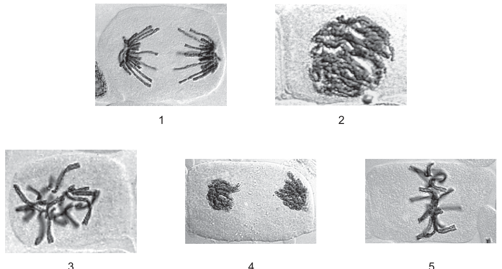
What is the correct order for the stages of mitosis?
Options
A 2 3 5 1 4
B 2 4 1 5 3
C 3 2 5 4 1
D 5 1 4 3 2
Working
Identify each numbered image:
- 1 — anaphase (two sets of chromosomes being pulled to opposite poles)
- 2 — prophase (condensed chromosomes visible within the nuclear area, no equatorial alignment)
- 3 — late telophase / cytokinesis (two separate daughter nuclei with a cell plate between them)
- 4 — early telophase (chromosomes have reached the poles and are beginning to decondense, but no cell plate yet)
- 5 — metaphase (chromosomes aligned along the equatorial plate)
The canonical sequence of mitosis is: prophase → metaphase → anaphase → telophase (early) → cytokinesis (late telophase).
Mapping the numbers: 2 → 5 → 1 → 4 → 3.
Answer
A
A
Background Concept
Mitosis is a continuous process, but biologists divide it into a series of stages that describe the changing appearance and position of the chromosomes. In the standard ordering the four named stages are:
- Prophase — the chromatin condenses into visible chromosomes (each made of two sister chromatids joined at a centromere); the nucleolus disappears and the nuclear envelope begins to break down.
- Metaphase — the chromosomes, now attached to spindle fibres at their centromeres, line up along the equator (metaphase plate) of the cell.
- Anaphase — the sister chromatids separate at the centromere and are pulled to opposite poles of the cell by the shortening spindle fibres, so two identical sets of chromosomes are seen moving apart.
- Telophase — a new nuclear envelope forms around each set of chromosomes at the poles, the chromosomes decondense, and in plant cells a cell plate forms across the middle of the cell (cytokinesis), producing two genetically identical daughter cells.
Because telophase is itself a gradual process, images of "early telophase" (chromosomes at the poles, beginning to decondense) and "late telophase / cytokinesis" (two distinct nuclei with a cell plate) are both legitimate stages to place in the sequence, with cytokinesis necessarily being the last event shown.
Understanding the Question
The question presents five photomicrographs of plant cells, numbered 1–5, and asks the candidate to put them in the correct order. The command word is implicit but the task is to identify each stage from the image and then order the stages into the sequence prophase → metaphase → anaphase → telophase. Only one of the four answer sequences is biologically correct.
Approach
For each image, look at the key diagnostic features:
- Are the chromosomes condensed and visible but still within an intact-looking nuclear area? → prophase.
- Are the chromosomes arranged in a single line across the middle (equator) of the cell? → metaphase.
- Are the chromosomes visibly separated into two groups moving towards opposite poles? → anaphase.
- Are the chromosomes at the poles, beginning to decondense, but a single cell still visible? → early telophase.
- Are there two distinct daughter nuclei with a cell plate between them, inside what is recognisably the original cell wall? → late telophase / cytokinesis.
Then place the five stages into the canonical order: prophase → metaphase → anaphase → telophase (early) → telophase (late / cytokinesis).
Step-by-Step Reasoning
Image 1 — Two clearly separated groups of chromosomes are seen at opposite sides of the cell, with spindle-like strands between them. This is anaphase, the stage at which sister chromatids are pulled to opposite poles.
Image 2 — Thick, darkly-stained, condensed chromosomes fill the nuclear area but are not aligned and not yet pulled apart. The nuclear envelope is still recognisable. This is prophase.
Image 3 — Two small, condensed but distinct nuclei are visible at opposite ends of the cell, with a clear cell plate forming between them. This is late telophase / cytokinesis, the final stage of mitosis.
Image 4 — Two groups of chromosomes have reached the poles and are beginning to decondense, but a new cell plate has not yet formed. This is early telophase, which must come before late telophase.
Image 5 — Chromosomes are aligned along a single line (the equatorial plate / metaphase plate) across the middle of the cell. This is metaphase, which sits between prophase and anaphase.
Putting them in order: prophase (2) → metaphase (5) → anaphase (1) → early telophase (4) → late telophase / cytokinesis (3).
That gives the sequence 2 → 5 → 1 → 4 → 3, which is option A.
Key Takeaways
- A cell in prophase has condensed chromosomes still within the original nuclear area.
- A cell in metaphase has chromosomes lined up along the equator.
- A cell in anaphase has two visibly separated sets of chromosomes moving to opposite poles.
- A cell in early telophase has chromosomes at the poles starting to decondense.
- A cell in late telophase / cytokinesis has two distinct daughter nuclei and (in plant cells) a cell plate.
- The correct order is prophase → metaphase → anaphase → telophase → cytokinesis.
Common Mistakes
- Confusing prophase and metaphase: in prophase the chromosomes are condensed but scattered, whereas in metaphase they are lined up along the equator.
- Confusing anaphase and late telophase: in anaphase the chromosomes are still condensed and individual; in telophase they are at the poles and starting to decondense into chromatin, and a nuclear envelope is reforming.
- Placing telophase before anaphase: chromosomes cannot reach the poles until the sister chromatids have separated in anaphase.
- Treating the cell plate (image 3) as a structure of prophase or metaphase: a cell plate only appears in cytokinesis, at the very end of mitosis.
Things to Be Careful About
- Use the canonical sequence prophase → metaphase → anaphase → telophase as the anchor when reading the photomicrographs; do not guess from the picture alone.
- Distinguish early from late telophase carefully: a cell plate (image 3) indicates cytokinesis, which is the latest event and must come after image 4.
- In plant cells, cytokinesis is achieved by the formation of a cell plate across the middle of the cell — a feature you will not see in animal cells, where a cleavage furrow forms instead.
- The chromosomes in image 1 are clearly in two opposing groups, confirming anaphase rather than metaphase, where they would be in a single line.
The gene that codes for protein R is long.
Protein R is in mass.
The typical mass of an amino acid is .
(, )
Which row is correct?
Options
| approximate length of introns in this gene/ base pairs | approximate length of mRNA for this gene/ base pairs | |
|---|---|---|
| A | 6000 | 6000 |
| B | 6000 | 130 000 |
| C | 124 000 | 6000 |
| D | 124 000 | 130 000 |
Working
- Number of amino acids in protein R:
- Length of the coding (exon) sequence in the gene, and therefore the approximate length of the mature mRNA (introns are spliced out):
- Length of introns in the gene:
Answer
C
C
Background Concept
Eukaryotic genes are not continuous stretches of coding sequence. A typical gene consists of:
- Exons – the sequences that remain in the mature mRNA and are translated into protein.
- Introns – non-coding sequences that are transcribed into pre-mRNA but are removed (spliced out) before the mRNA leaves the nucleus.
Only the exons are joined together in the mature mRNA, so the length of the mRNA corresponds to the sum of the exon sequences in the gene, not the total gene length. Stop and start codons contribute only marginally to length and are ignored in this approximation.
The genetic code is read in triplets: every amino acid is specified by 3 bases (a codon) in the mRNA. Because the template (antisense) strand of DNA has the same number of bases as the mRNA it is transcribed into, 3 DNA bases code for each amino acid.
Understanding the Question
The question gives three pieces of numerical information about a single gene and the protein it encodes:
- The whole gene (including introns + exons) is long.
- The protein product, protein R, has a mass of .
- An average amino acid residue has a mass of .
It asks us to work out (i) the approximate length of the introns in the gene and (ii) the approximate length of the mature mRNA. We must pick the row that matches both numbers. The conversion factors (, ) are provided.
The command word is implied: choose the correct option, having calculated the two quantities.
Approach
The strategy has three short steps:
- Convert protein mass to number of amino acids using the average amino-acid mass.
- Convert number of amino acids to the length of the coding sequence (and hence the mature mRNA) using the 3-bases-per-amino-acid rule.
- Subtract the coding (exon) length from the total gene length to get the intron length.
Because introns are removed during RNA processing, the mature mRNA is much shorter than the gene — often by an order of magnitude or more.
Step-by-Step Reasoning
Step 1 – Number of amino acids in protein R
(Strictly, a polypeptide of amino acids has peptide bonds and so loses ~18 Da per bond as water; for an order-of-magnitude estimate the question intends we ignore this, otherwise the answer would be ~2000 either way.)
Step 2 – Length of the mature mRNA
Each amino acid is coded by a codon of 3 bases, so the coding region of the gene is:
Because introns are spliced out of pre-mRNA to form mature mRNA, the mature mRNA is approximately bases long.
Step 3 – Length of introns
The total gene length is . Subtract the coding (exon) length:
So the introns together make up about , and the mature mRNA is about .
Looking at the table:
- A – introns, mRNA → wrong (introns cannot be equal to coding length in this gene).
- B – introns, mRNA → wrong (mRNA cannot equal gene length, introns are spliced out).
- C – introns, mRNA → correct.
- D – introns, mRNA → wrong (mRNA is the spliced product, not the whole gene).
Key Takeaways
- Gene length (DNA) > pre-mRNA length = mature mRNA length (because introns are removed during splicing).
- Coding length of a gene ≈ bases.
- Approximate number of amino acids in a protein = (here ~110 Da).
- Many eukaryotic genes are dominated by introns — protein-coding sequence can be only a small fraction of the gene.
Common Mistakes
- Forgetting that mRNA is the spliced product. Choosing B or D — which give mRNA = gene length — confuses pre-mRNA with mature mRNA.
- Forgetting the triplet code. Dividing amino acids by instead of multiplying, giving ~667 bp for mRNA.
- Calculating introns incorrectly. Adding instead of subtracting the exon length from the gene length.
- Ignoring the conversion factors. Using kDa where Da are needed (or vice versa) and getting a factor-of-1000 error.
Things to Be Careful About
- Use the same units throughout: convert to and to before dividing.
- This is an order-of-magnitude / approximation question, so do not be put off by small discrepancies from stop/start codons or water loss during peptide bond formation.
- Remember the direction of the subtraction: introns = total gene − coding (exon) length, not the other way round.
- The mature mRNA length is given in bases, not base pairs (mRNA is single-stranded). In this question the options use "base pairs" loosely, but the numerical value is the same as the corresponding number of bases in the mRNA.
The diagram shows a section of DNA. Two of the bases in this section are labelled 1 and 2.
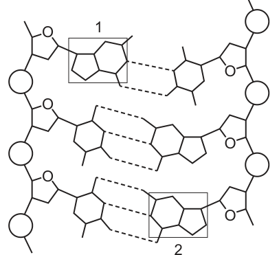
Which row correctly identifies the bases 1 and 2?
Options
| base 1 | base 2 | |
|---|---|---|
| A | purine | adenine |
| B | purine | guanine |
| C | pyrimidine | adenine |
| D | pyrimidine | guanine |
Working
Both bases 1 and 2 are shown as double-ringed structures, so they are both purines (purines have two rings; pyrimidines have one ring).
Base 2 is part of a base pair connected by three hydrogen bonds (shown as three dashed lines). A–T pairs have two hydrogen bonds, while G–C pairs have three. Therefore base 2 is guanine (paired with cytosine).
Answer
B
B
Background Concept
The nitrogenous bases in DNA fall into two structural categories:
- Purines — double-ringed bases. In DNA these are adenine (A) and guanine (G).
- Pyrimidines — single-ringed bases. In DNA these are cytosine (C) and thymine (T).
Because the DNA double helix has a constant width, every base pair must consist of one purine paired with one pyrimidine. The two types of pair can also be distinguished by their hydrogen bonding:
- A–T pairs are held together by two hydrogen bonds.
- G–C pairs are held together by three hydrogen bonds.
So from a labelled diagram you can identify a specific base either by its ring count (purine/pyrimidine) or by the number of hydrogen bonds drawn between the two bases of a pair.
Understanding the Question
The question shows a short section of the DNA double helix with two boxed bases labelled 1 and 2. The candidate is asked to pick the row that correctly names base 1 and base 2. The options combine a structural class (purine/pyrimidine) with a specific base identity (adenine/guanine), so the candidate has to read the diagram for two pieces of information: the shape of the base (rings) and, where possible, which specific base it is (hydrogen bonds).
Approach
- Look at the shape of each boxed base. Count the rings: one ring = pyrimidine; two fused rings = purine.
- If the base is a purine, decide between A and G by inspecting its partner in the base pair: two H-bonds → A; three H-bonds → G.
- Match the findings to the correct option row.
Step-by-Step Reasoning
- Base 1: Drawn as a fused double-ring structure on the left strand. Two rings → it is a purine. The mark scheme accepts "purine" as the answer for base 1, and the correct option (B) does not require a specific identity for base 1.
- Base 2: Drawn as a fused double-ring structure on the right strand — so it is also a purine — and its base pair is connected by three dashed lines (three hydrogen bonds). Three H-bonds between the two bases of the pair identify it as a guanine–cytosine pair; base 2, being the purine, must therefore be guanine.
- Checking the options:
- A: purine / adenine — wrong, the H-bond count shows G, not A.
- B: purine / guanine — correct.
- C: pyrimidine / adenine — wrong, base 1 has two rings, so it is a purine; and base 2 is a purine, not adenine.
- D: pyrimidine / guanine — wrong, the purine identification of base 1 is correct but here it is called a pyrimidine, and base 2 is correctly named but the class is wrong.
Key Takeaways
- Ring count = base class: 1 ring = pyrimidine (C, T); 2 rings = purine (A, G).
- H-bond count = specific base pair: 2 H-bonds = A–T; 3 H-bonds = G–C.
- In a DNA double helix, a purine is always paired with a pyrimidine, so two purines are never paired together.
- Reading either feature (ring shape or hydrogen bonds) is usually enough; using both removes any ambiguity.
Common Mistakes
- Confusing purines and pyrimidines by ring number (e.g. calling a double-ringed base a pyrimidine because it looks "smaller" in a different orientation).
- Assuming the labelled base must be a pyrimidine just because it sits in a particular strand — DNA strands alternate purines and pyrimidines along each strand but there is no rule that one strand is all purines.
- Reading two H-bonds instead of three and choosing A instead of G.
- Selecting an option where the class (purine/pyrimidine) and the named base are inconsistent.
Things to Be Careful About
- Count the rings carefully on the labelled base itself, not on its partner in the base pair.
- Count the dashed lines (hydrogen bonds) between the two bases of the pair, not between unrelated bases above or below.
- Adenine and guanine are both purines, so an option of the form "purine / X" can only have X = A or G. The H-bond count distinguishes them.
- Cytosine and thymine are both pyrimidines; the H-bond count again distinguishes which is which within a pair.
Which row describes the role of the enzymes DNA polymerase and DNA ligase?
Options
| DNA polymerase | DNA ligase | |
|---|---|---|
| A | adds nucleotides in a 5′ to 3′ direction | joins sections of DNA together on the lagging strand |
| B | adds nucleotides in a 3′ to 5′ direction | joins sections of DNA together on the leading strand |
| C | adds nucleotides in a 3′ to 5′ direction | joins sections of DNA together on the lagging strand |
| D | adds nucleotides in a 5′ to 3′ direction | joins sections of DNA together on the leading strand |
Working
DNA polymerase can only add free nucleotides to the free 3′ end of a growing DNA strand, so it synthesises DNA in the 5′ to 3′ direction. The lagging strand is synthesised discontinuously as short Okazaki fragments, and DNA ligase joins these sections of DNA together on the lagging strand.
Answer
A
A
Background Concept
DNA replication is semi-conservative: each new DNA molecule contains one original (template) strand and one newly synthesised strand. Because the two strands of the parent DNA double helix are antiparallel (one runs 5′ → 3′, the other 3′ → 5′), the two new strands cannot be made in the same way.
- DNA polymerase catalyses the formation of phosphodiester bonds between the 3′-OH of the last nucleotide on the growing strand and the 5′-phosphate of the incoming nucleotide. It can therefore only add nucleotides to a free 3′ end, meaning synthesis proceeds in the 5′ to 3′ direction.
- On the strand whose 3′ end faces the replication fork (the leading strand), synthesis can continue smoothly in the 5′ → 3′ direction as the fork opens.
- On the other strand (the lagging strand), the 3′ end faces away from the fork, so synthesis must occur in short, discontinuous pieces (Okazaki fragments) that grow 5′ → 3′ away from the fork.
- DNA ligase seals the nicks between adjacent Okazaki fragments by forming phosphodiester bonds, joining sections of DNA on the lagging strand.
Understanding the Question
This is a multiple-choice question testing recall of two specific facts about DNA replication: (1) the direction in which DNA polymerase adds nucleotides, and (2) the role and location of DNA ligase activity. Each option combines a polymerase statement with a ligase statement, so both halves of the chosen row must be correct.
Approach
Recall the direction of DNA polymerase (5′ → 3′) and the substrate on which DNA ligase acts (Okazaki fragments on the lagging strand). The correct option must contain both of these facts.
Step-by-Step Reasoning
- Options B and C both say DNA polymerase adds nucleotides 3′ → 5′ — this is wrong; polymerase can only add to a free 3′-OH, so the new strand grows 5′ → 3′.
- This eliminates B and C, leaving A and D.
- Options A and D agree on the polymerase direction (5′ → 3′) but disagree on DNA ligase.
- DNA ligase joins Okazaki fragments on the lagging strand (where synthesis is discontinuous), not on the leading strand (where synthesis is continuous and no joining is needed).
- Therefore, the row in which ligase "joins sections of DNA together on the lagging strand" is correct — that is option A.
Key Takeaways
- DNA polymerase always synthesises DNA in the 5′ → 3′ direction.
- The leading strand is synthesised continuously; the lagging strand is synthesised as Okazaki fragments.
- DNA ligase joins Okazaki fragments on the lagging strand.
Common Mistakes
- Saying or believing that DNA polymerase adds nucleotides in the 3′ → 5′ direction (confusing the direction of polymerase movement along the template with the direction in which the new strand grows).
- Attributing DNA ligase activity to the leading strand — it is needed only where there are nicks to seal, which is the lagging strand.
Things to Be Careful About
- The 5′ and 3′ labels refer to the carbons on the deoxyribose sugar; "5′ to 3′ direction" describes the new strand's growth, not the template strand's orientation.
- DNA ligase does not add nucleotides; it joins pre-existing sections by forming phosphodiester bonds between them.
The table shows some of the DNA triplet codes for some amino acids.
| amino acid | DNA triplet code | amino acid | DNA triplet code |
|---|---|---|---|
| arginine | GCA | glycine | CCA |
| arginine | GCC | glycine | CCG |
| arginine | GCG | glycine | CCT |
| asparagine | TTA | lysine | TTC |
| asparagine | TTG | lysine | TTT |
| cysteine | ACA | proline | GGA |
| cysteine | ACG | proline | GGC |
| STOP | ATC | valine | CAC |
The base sequence on the template DNA strand coding for part of a polypeptide is shown.
CCA TTC ACG GCG TTA GCA
Two mutations occur in this sequence during DNA replication.
Which mutated DNA would result in two different amino acids?
Options
A CCA ATC ACG GCG TTG GCA
B CCA TTC ACA GCA TTA GCA
C CCA TTC ACG CCG TTA GCC
D CCA TTC ACG GCG TTC GGA
Working
Translate the original sequence first:
CCA TTC ACG GCG TTA GCA
glycine – lysine – cysteine – arginine – asparagine – arginine
Compare each option codon by codon:
-
A CCA ATC ACG GCG TTG GCA
- Position 2: TTC → ATC = lysine → STOP (one change, plus a premature stop)
- Position 5: TTA → TTG = asparagine → asparagine (no change)
- Only 1 amino-acid change (and a stop codon).
-
B CCA TTC ACA GCA TTA GCA
- Position 3: ACG → ACA = cysteine → cysteine (silent)
- Position 4: GCG → GCA = arginine → arginine (silent)
- 0 amino-acid changes.
-
C CCA TTC ACG CCG TTA GCC
- Position 4: GCG → CCG = arginine → glycine
- Position 6: GCA → GCC = arginine → arginine (silent)
- 1 amino-acid change.
-
D CCA TTC ACG GCG TTC GGA
- Position 5: TTA → TTC = asparagine → lysine
- Position 6: GCA → GGA = arginine → proline
- 2 amino-acid changes.
Answer
D
D
Background Concept
DNA is read three bases at a time as codons, each of which specifies one amino acid (or a stop signal) during translation. The full set of codon–amino-acid assignments is called the genetic code. A key property of the code is that it is degenerate: most amino acids are coded for by more than one codon. For example, arginine is coded by GCA, GCC and GCG, and asparagine by TTA and TTG.
A point (substitution) mutation swaps one base for another. Depending on which base is changed, the substitution can have one of three effects:
- Silent (synonymous) – the new codon still codes for the same amino acid, so the polypeptide is unchanged.
- Missense – the new codon codes for a different amino acid, so one residue in the polypeptide is altered.
- Nonsense – the new codon becomes a STOP codon, terminating translation early.
Understanding the Question
We are given the original template-strand DNA sequence:
CCA TTC ACG GCG TTA GCA
Using the supplied table, the original polypeptide segment is:
glycine – lysine – cysteine – arginine – asparagine – arginine
Four mutated versions are offered. The question asks which one produces two different amino acids compared with the original. The trap is that the code is degenerate, so some substitutions look like a change but in fact code for the same amino acid. We must check every codon, not just glance at the changed letters.
Approach
The systematic method is:
- Translate the original sequence using the table.
- For each option, mark the codons that differ from the original.
- Translate the changed codons in the option.
- Count how many of the substitutions produce a different amino acid.
- Choose the option that yields exactly two such changes.
Step-by-Step Reasoning
Original translation (using the table):
- CCA → glycine
- TTC → lysine
- ACG → cysteine
- GCG → arginine
- TTA → asparagine
- GCA → arginine
Option A – CCA ATC ACG GCG TTG GCA
- Position 2: TTC → ATC. From the table, ATC = STOP. This is a nonsense mutation, not a different amino acid; it would also truncate the polypeptide.
- Position 5: TTA → TTG. The table lists both TTA and TTG as asparagine, so this is a silent substitution.
- Net: one stop signal, not two different amino acids.
Option B – CCA TTC ACA GCA TTA GCA
- Position 3: ACG → ACA. Both ACG and ACA code for cysteine → silent.
- Position 4: GCG → GCA. Both GCG and GCA code for arginine → silent.
- Net: zero amino-acid changes. This option exists to test whether you notice that the third base of a codon often wobbles without changing the amino acid (degeneracy).
Option C – CCA TTC ACG CCG TTA GCC
- Position 4: GCG → CCG. GCG = arginine, CCG = glycine. This is a missense change (arginine → glycine).
- Position 6: GCA → GCC. Both code for arginine → silent.
- Net: one amino-acid change.
Option D – CCA TTC ACG GCG TTC GGA
- Position 5: TTA → TTC. TTA = asparagine, TTC = lysine. This is a missense change (asparagine → lysine).
- Position 6: GCA → GGA. GCA = arginine, GGA = proline. This is a missense change (arginine → proline).
- Net: two amino-acid changes.
Only option D produces two different amino acids in the resulting polypeptide.
Key Takeaways
- The genetic code is degenerate: the third base of a codon often varies without changing the amino acid. This means a substitution mutation is not guaranteed to alter the protein.
- Substitution mutations are classified as silent, missense or nonsense depending on the effect on the polypeptide.
- When comparing DNA sequences, always re-translate every changed codon using the codon table — do not assume that a different base means a different amino acid.
Common Mistakes
- Choosing A because the letters look "more different". A STOP codon is not "a different amino acid"; it terminates translation and would be described as a nonsense mutation.
- Choosing B because two bases are visibly changed. Both changes are silent owing to the degeneracy of the code, so the protein is unchanged.
- Choosing C because it contains two visible changes. Only one of the two changes (GCG → CCG) alters the amino acid; the other (GCA → GCC) is silent.
- Translating using the wrong strand. The question gives the template strand, and the table provided is the DNA triplet code, so each triplet in the sequence already corresponds to an amino acid directly — no transcription step is required.
Things to Be Careful About
- Always read the codon table exactly as given; this question uses a non-standard, simplified table (e.g. ATC = STOP, GCA/GCC/GCG = arginine) rather than the full universal code, so a memorised table could mislead you.
- Re-check every codon in the mutated sequence, not just the obviously changed ones; distractors rely on silent third-base changes.
- "Two different amino acids" means two codons in the new sequence each code for an amino acid that differs from the original — not two bases changed, and not a stop codon.
The diagram shows a vertical section through part of the stem of a dicotyledonous plant. Three types of structures are labelled.
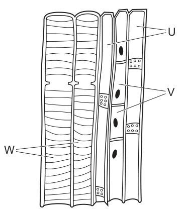
What are the structures U, V and W?
Options
| companion cells | phloem sieve tube elements | xylem vessel elements | |
|---|---|---|---|
| A | U | W | V |
| B | V | U | W |
| C | U | V | W |
| D | W | V | U |
Working
- W points to wide tubes on the left with spiral/annular wall thickening. The lignin-impregnated, patterned thickening and large diameter are diagnostic of xylem vessel elements.
- U points to a wider tube on the right with no visible nucleus, containing a sieve plate (perforated end wall). The absence of a nucleus and the presence of a sieve plate identify a phloem sieve tube element.
- V points to the small, narrow cells lying alongside the sieve tube, with dense cytoplasm and a prominent nucleus. These are companion cells, which are always associated with sieve tube elements and provide them with metabolic support.
Matching this to the table:
| companion cells | phloem sieve tube elements | xylem vessel elements | |
|---|---|---|---|
| U | ✓ | ||
| V | ✓ | ||
| W | ✓ |
Answer
B
B
Background Concept
Vascular tissue in dicotyledonous plants is organised into two conducting tissues:
- Xylem carries water and mineral ions from the roots to the leaves. The conducting cells are xylem vessel elements — wide, dead, hollow tubes formed by the end-to-end fusion of cells. Their walls are heavily lignified, which is what gives the characteristic spiral, annular, reticulate or pitted patterns seen in longitudinal section.
- Phloem carries assimilates (mainly sucrose) from sources to sinks. The conducting cells are sieve tube elements — living but enucleate tubes joined end to end, with perforated end walls called sieve plates. Because sieve tube elements lack a nucleus and most organelles, they depend on a closely associated companion cell (which is nucleate and densely cytoplasmic) for ATP, proteins and the loading/unloading of sucrose.
The three cell types are therefore easy to tell apart in a longitudinal section:
| Feature | Xylem vessel element | Sieve tube element | Companion cell |
|---|---|---|---|
| Diameter | Wide | Moderate | Narrow |
| Living/dead | Dead | Living (no nucleus) | Living |
| Nucleus | None | None | Yes, prominent |
| Wall | Thickened, lignified (spiral/annular etc.) | Thin cellulose, sieve plates at ends | Thin cellulose |
| Position | Often central in vascular bundle | Adjacent to companion cell | Next to sieve tube |
Understanding the Question
A longitudinal section through part of a dicot stem vascular bundle is shown. Three structures U, V and W are labelled, and you must match each to one of: companion cell, phloem sieve tube element, or xylem vessel element. The figure gives the candidate three independent visual clues — wall pattern, presence/absence of a nucleus, and relative diameter — and the answer is the option that uses all three consistently.
The command word is essentially "identify"; no further explanation is required once the structures are correctly named.
Approach
- Use the diagnostic features of each cell type listed above.
- Examine each label in the figure and decide which cell type it most closely resembles.
- Cross-check with the position in the tissue (xylem tends to be the wider, patterned tubes; phloem tubes are next to small nucleate cells).
- Match to the table of options to find the one in which every label is correctly placed.
Step-by-Step Reasoning
- W has arrows pointing to the two large tubes on the left of the figure. Their walls show clear spiral / annular thickening — the appearance produced by rings or a helix of lignin laid down inside the wall of a young, still-elongating xylem vessel. This is the unmistakable signature of an xylem vessel element.
- U points to a tube on the right of the figure that lacks any patterned wall thickening and has no visible nucleus. Its end wall (where it meets the next element) shows the sieve plate — a perforated cross wall characteristic of mature sieve tube elements. Hence U = phloem sieve tube element.
- V points to a small, narrow cell that sits alongside the sieve tube, has dense cytoplasm and a clearly drawn dark oval nucleus. The combination of small size + nucleus + close association with a sieve tube is the defining feature of a companion cell.
Matching to the options in the table:
- U = phloem sieve tube element
- V = companion cell
- W = xylem vessel element
Only option B places U, V and W in exactly these three categories.
Key Takeaways
- Lignified, patterned wall thickening → xylem vessel element.
- Enucleate tube with sieve plates → phloem sieve tube element.
- Small, nucleate, dense-cytoplasm cell adjacent to a sieve tube → companion cell.
- Always use more than one feature when identifying a cell (e.g. wall pattern AND diameter) — single features can occasionally be ambiguous.
- Companion cells and sieve tube elements are functionally inseparable: the sieve tube element cannot survive without its companion cell because it has lost its nucleus.
Common Mistakes
- Confusing a sieve tube element with a xylem vessel element because both look like long tubes. The differentiator is wall pattern: lignified thickening in xylem, thin cellulose wall with sieve plates in phloem.
- Calling the narrow cell next to the sieve tube a "sieve tube element" because it is small. The nucleus is the giveaway — sieve tube elements are enucleate at maturity.
- Confusing the xylem parenchyma or other cells for the labelled structure — stick strictly to the three cell types in the option table.
- Selecting an option because one label is "right" — every label in the chosen option must be correct, not just one.
Things to Be Careful About
- In a longitudinal section, xylem vessel elements can appear with several different lignin patterns (annular, spiral, reticulate, pitted) depending on whether the vessel was still elongating when sectioned; all of them are still xylem.
- A mature sieve tube element is described as having no nucleus — but the section is a snapshot of the tissue and the absence of a drawn nucleus in the figure is what matters.
- Companion cells are typically much narrower than the sieve tube element they support, but they are always present as a pair. In the figure, the two V labels emphasise that the same cell type appears repeatedly in the tissue.
- Read the question table carefully: the option chosen must match all three labels simultaneously; partial matches do not score.
Which features of xylem vessels help to reduce the resistance to water flow?
1 Lignin forms an incomplete secondary wall.
2 There are no cross walls between the vessel elements.
3 The xylem vessels form narrow tubes.
Options
A 1, 2 and 3
B 1 and 2 only
C 2 only
D 3 only
Working
Water flows through xylem vessels as a continuous column under tension (transpiration pull). Anything that makes that column smoother and more continuous reduces resistance.
- Statement 1: Lignin waterproofs and strengthens the wall so the vessel does not collapse inward under the negative pressure of the transpiration stream. The incomplete nature of the secondary wall leaves pits for lateral water movement between vessels — it is not a feature that reduces resistance to longitudinal flow.
- Statement 2: Vessel elements are joined end-to-end and their end walls break down, so there are no cross walls to obstruct the water column. This provides a continuous, smooth tube and therefore reduces resistance. ✓
- Statement 3: Resistance to flow through a tube is inversely proportional to the fourth power of its radius (Poiseuille's law). Narrow tubes therefore increase resistance. Xylem vessels are narrow to reduce the risk of cavitation (air embolism), not to reduce resistance.
Only statement 2 reduces resistance to water flow.
Answer
C
C
Background Concept
Xylem vessels are dead, hollow tubes made of vessel elements stacked end-to-end. Their structure is specialised for moving large volumes of water from roots to leaves under tension generated by transpiration. Three structural features matter for how they function:
- Lignin in the secondary cell wall makes the wall rigid, waterproof and resistant to collapse under the strong negative pressure (tension) inside the vessel. The secondary wall is laid down in a pattern that leaves pits — areas with only the primary wall — so that water can move laterally from one vessel to another if air bubbles block the main tube.
- End-wall dissolution — during vessel maturation the end walls between successive vessel elements break down, so each vessel element is essentially an open cylinder fused to its neighbours into a single continuous tube.
- Diameter — vessel diameters vary widely. Wider vessels carry more water per unit time but are more vulnerable to cavitation (formation of air emboli that break the water column); narrower vessels are safer but carry less water.
Understanding the Question
This is an MCQ testing whether the candidate can match each structural feature to its actual contribution to reducing flow resistance. The command word is implicit: which of statements 1, 2 and 3 correctly describe features that lower the resistance water meets as it moves up the xylem? The candidate must read each statement carefully — features that aid other functions (support, lateral bypass, cavitation prevention) do not score.
Approach
For each statement, ask: does this feature lower hydraulic resistance inside the vessel?
- Statement 1 → look at lignin's role. It supports and waterproofs. Its incompleteness enables lateral flow through pits. Neither reduces longitudinal resistance.
- Statement 2 → absence of cross walls = continuous open tube. Yes, this lowers resistance.
- Statement 3 → recall Poiseuille's law: resistance ∝ 1/r⁴. Narrower tubes increase resistance. The narrowness exists for a different reason (cavitation safety), so this does not score.
Only statement 2 satisfies the criterion.
Step-by-Step Reasoning
Statement 1 — Lignin forms an incomplete secondary wall. Lignin strengthens and waterproofs the wall so the vessel does not collapse under the suction of the transpiration stream. The "incomplete" part means pits remain, which lets water move sideways into a neighbouring vessel if one tube becomes blocked by an air bubble (embolism). This is essential for the safety of the water column, but neither the lignin nor the pits reduce the frictional resistance water encounters as it flows up the vessel. Reject.
Statement 2 — No cross walls between vessel elements. Each vessel element starts as a living cell with end walls. As it matures, those end walls disintegrate, leaving a continuous hollow pipe many centimetres long. With no barriers to interrupt the flow, water passes smoothly from element to element. This does reduce resistance to flow. Accept.
Statement 3 — Xylem vessels form narrow tubes. Resistance to flow through a tube is given by Poiseuille's law, , so resistance increases very steeply as the radius decreases. Narrower tubes therefore increase, not reduce, frictional resistance. Xylem vessels are narrow primarily to limit the chance of cavitation (a wide vessel is more likely to break the water column when under high tension). The statement misattributes the purpose of narrowness. Reject.
Therefore only statement 2 is correct → C.
Key Takeaways
- The adaptations of xylem fall into different functional categories: support/strength (lignin), continuous flow (no end walls), and cavitation safety (narrow diameter, bordered pits). Each structural feature has a specific role, and MCQs often test whether you can assign the correct role.
- Flow resistance through a tube decreases sharply with increasing radius (∝ r⁴), so wider tubes move water more easily. Narrowness in xylem exists to limit embolism risk, not to aid flow.
- Always check what a question is actually asking — here, "reduce resistance to flow" — rather than the general function of the structure.
Common Mistakes
- Choosing B because statements 1 and 2 both describe real features of xylem. The trap is that being a true feature of xylem is not enough; it must also serve the specific function the question names.
- Choosing D because students associate "narrow tube" with efficient capillary rise. Capillary rise is relevant to initial water movement in fine tubes but is not the same as resistance to bulk flow under tension; in the transpiration stream, narrow tubes impede, not aid, flow.
- Choosing A — the most inclusive option — by assuming that "more features must be better". Selection pressure on xylem balances several demands, not just low resistance.
Things to Be Careful About
- Distinguish between resistance to flow (a function of tube continuity, smoothness and diameter) and prevention of collapse/cavitation (a function of lignin and narrow diameter). The exam deliberately conflates them in distractors.
- "Incomplete secondary wall" describes the pattern of lignin deposition (with pits); it is not a feature that smooths flow.
- Remember the inverse-fourth-power dependence of resistance on radius — it is a recurring principle in any biology question involving fluid flow (xylem, blood flow, airways).
What causes water to move from the root hair cells to the endodermis?
Options
A diffusion through cell walls and osmosis down a water potential gradient in the cytoplasm
B diffusion through the symplast and osmosis and root pressure through the apoplast
C osmosis from cell vacuole to cell vacuole and active transport into the endodermis
D osmosis through the intercellular spaces and diffusion in cell walls and cytoplasm
Working
Water moves from root hair cells to the endodermis by two pathways:
- Apoplast pathway – water moves through the cell walls and intercellular spaces by diffusion/mass flow (no membrane crossing, so no osmosis).
- Symplast pathway – water moves through the cytoplasm of cells (connected by plasmodesmata) by osmosis down a water potential gradient (a partially permeable membrane, the tonoplast/plasma membrane, is crossed at the root hair cell surface).
Evaluating the options:
- A correctly describes the apoplast as diffusion through cell walls AND the symplast as osmosis down a water potential gradient through the cytoplasm. ✓
- B wrongly attributes the symplast to diffusion and the apoplast to osmosis/root pressure. ✗
- C wrongly states water moves vacuole to vacuole and that active transport is involved. ✗
- D wrongly states osmosis occurs through intercellular spaces (no membrane there). ✗
Answer
A
A
Background Concept
Water entering a plant at the root hair cells must cross the cortex to reach the endodermis and ultimately the xylem. There are two parallel pathways for this movement:
-
Apoplast pathway – water moves through the continuous network of cell walls and intercellular spaces. Because water here does not cross any plasma membrane, it is not osmosis; movement occurs by diffusion and mass flow driven by a water potential gradient and the pulling force generated by transpiration at the leaves (cohesion–tension).
-
Symplast pathway – water moves through the cytoplasm of cells, passing from cell to cell via plasmodesmata (the cytoplasmic channels that connect adjacent cells). Because water crosses the plasma membrane (and tonoplast) to enter the symplast at the root hair cell, its movement across each membrane is by osmosis, driven by a water potential gradient (more negative ψ inside the cell than in the soil solution).
Both pathways converge at the endodermis, where the Casparian strip in the endodermal cell walls blocks the apoplast route, forcing water to cross the plasma membrane of an endodermal cell (i.e. into the symplast) before reaching the xylem.
Understanding the Question
The question is a multiple-choice item testing recall of how water physically moves from the root hair cell layer to the endodermis. The command is "What causes water to move..." so the candidate must identify the correct mechanism for each pathway — not just the pathways themselves.
Approach
For each option, check two things:
- Which pathway is being described (apoplast = cell walls/intercellular spaces; symplast = cytoplasm via plasmodesmata)?
- Which mechanism is being claimed (osmosis requires crossing a partially permeable membrane; diffusion occurs without membrane crossing in the cell wall network)?
An answer is correct only if BOTH the pathway AND the mechanism are right for each component.
Step-by-Step Reasoning
Option A — "diffusion through cell walls and osmosis down a water potential gradient in the cytoplasm"
- "Diffusion through cell walls" → apoplast pathway, mechanism = diffusion/mass flow (correct).
- "Osmosis down a water potential gradient in the cytoplasm" → symplast pathway, mechanism = osmosis across a membrane down a ψ gradient (correct).
- This is fully accurate. ✓
Option B — "diffusion through the symplast and osmosis and root pressure through the apoplast"
- The symplast does not involve "diffusion through cell walls" — it is movement through cytoplasm.
- The apoplast is not driven primarily by osmosis (no membrane) and root pressure is a minor contributor compared with transpiration pull.
- Inaccurate. ✗
Option C — "osmosis from cell vacuole to cell vacuole and active transport into the endodermis"
- Water does not move directly from vacuole to vacuole — it must pass through cytoplasm and across membranes.
- Water is never moved by active transport; ATP-powered solute pumps create the water potential gradient, but water itself moves passively by osmosis.
- Inaccurate. ✗
Option D — "osmosis through the intercellular spaces and diffusion in cell walls and cytoplasm"
- Intercellular spaces are air gaps with no membrane, so osmosis cannot occur there.
- Inaccurate. ✗
Key Takeaways
- Apoplast = cell walls + intercellular spaces → movement by diffusion/mass flow (no membrane crossed).
- Symplast = cytoplasm connected by plasmodesmata → movement by osmosis down a water potential gradient (membrane crossed).
- These are the two parallel routes from root hair to endodermis, converging at the Casparian strip.
- Water is never moved by active transport.
Common Mistakes
- Confusing diffusion with osmosis: diffusion describes movement of molecules down a concentration gradient (e.g. through cell walls), while osmosis is specifically the diffusion of water across a partially permeable membrane.
- Attributing osmosis to the apoplast: there is no membrane in the cell wall network, so osmosis cannot occur there.
- Saying "active transport of water": water moves passively; ATP is used to pump solutes (e.g. into the xylem) which then draws water in osmotically.
- Confusing root pressure with the main driving force: root pressure is a small contribution from ion accumulation; the dominant force for water movement up the plant is the transpiration pull generated at the leaves.
Things to Be Careful About
- "Cytoplasm" in option A refers to the symplast; the wording is correct even though the full word "symplast" is not used.
- Osmosis specifically requires a water potential gradient across a partially permeable membrane — both conditions must be stated for a mark-scheme level answer.
- The two pathways operate simultaneously; they are not alternative options for the plant.
Which changes occur as carbohydrate is moved into a sink?
Options
| water potential of the sieve tube element becomes | volume of liquid of the sieve tube element | |
|---|---|---|
| A | lower | decreases |
| B | lower | increases |
| C | higher | decreases |
| D | higher | increases |
Working
At a sink, sucrose is actively unloaded from the sieve tube element into the surrounding cells. Removing solute raises the water potential of the sieve tube sap, so water leaves the sieve tube down the water potential gradient by osmosis. Loss of water reduces the volume of liquid inside the sieve tube element (and so lowers the hydrostatic pressure there).
Answer
C
C
Background Concept
Assimilates such as sucrose are translocated in the phloem from a source (e.g. a photosynthesising leaf) to a sink (e.g. a root, fruit, growing shoot). The widely accepted explanation is the mass flow hypothesis (Münch):
- At the source, companion cells use ATP from proton pumps to actively load sucrose into the sieve tube element. The solute concentration of the sap rises, the water potential () falls, and water enters the sieve tube from the xylem by osmosis. The volume of liquid increases, raising the hydrostatic pressure at the source end.
- At the sink, sucrose is actively unloaded into surrounding cells. The solute concentration of the sap inside the sieve tube falls, the water potential rises, and water leaves the sieve tube by osmosis back into the surrounding tissue. The volume of liquid decreases, lowering the hydrostatic pressure at the sink end.
- The resulting pressure gradient between source and sink pushes the sap along the sieve tubes.
The single principle linking every option in this question is therefore: whatever the solute does to concentration, water does to volume (and therefore to pressure) by osmosis, in the opposite direction.
Understanding the Question
The question presents a table with two columns: the change in water potential of the sieve tube element, and the change in volume of liquid in the sieve tube element, as carbohydrate is moved INTO a sink (i.e. as it leaves the sieve tube at the sink). The command word is implicit — the candidate must select the correct combination.
Approach
Think about what happens at the sink in the sequence sucrose removal → osmotic effect → water movement → volume change. Then translate each step into the language of the options.
Step-by-Step Reasoning
- Carbohydrate moves INTO a sink means sucrose is being unloaded from the sieve tube element. This is an active process driven by proton pumps and co-transporters in the sink cell membranes.
- Removing sucrose from the sap decreases the solute concentration of the sap inside the sieve tube element.
- A lower solute concentration means a higher (less negative) water potential of the sieve tube sap. So the sieve tube water potential becomes higher.
- Because the sieve tube sap is now less negative (higher ) than the surrounding tissue, water moves out of the sieve tube element by osmosis (down the water potential gradient).
- Loss of water means the volume of liquid inside the sieve tube element decreases, and the hydrostatic pressure inside it falls.
- Matching this with the table gives: water potential higher, volume decreases → option C.
The other options describe what happens at the source (A and B) — where sucrose is loaded in, lowering the water potential and pulling water in by osmosis, increasing volume — or are internally inconsistent (D — a higher water potential would drive water out, not in).
Key Takeaways
- Source: sucrose loaded in → falls → water enters → volume (and pressure) rises.
- Sink: sucrose unloaded out → rises → water leaves → volume (and pressure) falls.
- The pressure gradient (high at source, low at sink) is what drives mass flow in the phloem.
- The direction of the water potential change is always opposite to the direction of the solute change, and the volume change follows the water movement.
Common Mistakes
- Confusing the sink with the source: many candidates correctly identify that carbohydrate loading lowers water potential and answer A or B. That is the source story, not the sink story.
- Forgetting that water follows solute by osmosis, so the volume must change in the same direction as the water movement — not in the same direction as the solute movement.
- Choosing D because "more carbohydrate is in the sink cell so water potential should fall" — but the question asks about the sieve tube element, not the sink cell. It is the sieve tube that has lost sucrose and so has a higher .
Things to Be Careful About
- "Carbohydrate moved INTO a sink" describes sucrose leaving the sieve tube at the sink end, not entering a sink cell from outside the plant. Keep the compartment (sieve tube element) clear in your head.
- Water potential becomes "higher" — i.e. less negative / closer to zero. Do not say "water potential increases" if the option explicitly says "becomes higher"; both are acceptable but stay consistent with the question's wording.
- The volume change is a consequence of osmotic water movement, not a cause of anything else. Do not describe the volume decrease as driving the water out.
Which statement correctly compares blood plasma and tissue fluid in a healthy person?
Options
A Blood plasma contains more protein than tissue fluid.
B Both blood plasma and tissue fluid contain red blood cells.
C Tissue fluid contains white blood cells whereas blood plasma does not contain lymphocytes.
D Tissue fluid is formed from blood plasma and is not returned to blood plasma.
Working
Blood plasma and tissue fluid have very similar compositions because tissue fluid is formed by filtration of plasma through the capillary walls. However:
- The capillary endothelium is impermeable to large plasma proteins (e.g. albumin, globulins), so blood plasma contains significantly more protein than tissue fluid.
- Red blood cells are too large to leave the capillaries, so they remain in blood plasma and are not found in tissue fluid.
- Some white blood cells (e.g. neutrophils, monocytes) can squeeze between capillary wall cells (diapedesis) and are present in tissue fluid; lymphocytes are also present in blood plasma.
- The majority of tissue fluid is returned to the blood plasma at the venous end of the capillary (and the remainder enters the lymph). It is incorrect to say it is not returned.
Only statement A is correct.
Answer
A
A
Background Concept
Blood plasma is the liquid component of blood, in which red blood cells, white blood cells and platelets are suspended. It is approximately 90% water, with dissolved solutes including plasma proteins (albumin, globulins, fibrinogen), glucose, amino acids, hormones, urea, and ions such as Na⁺, Cl⁻ and HCO₃⁻.
Tissue fluid is the fluid that bathes the cells of the body. It is formed from blood plasma by ultrafiltration at the arterial end of a capillary: the hydrostatic pressure of the blood forces water and small solutes out through gaps between the endothelial cells that form the capillary wall. Plasma proteins and blood cells are normally too large to pass through these gaps and remain inside the capillary.
At the venous end of the capillary, the hydrostatic pressure has fallen and the osmotic (oncotic) pressure of the retained plasma proteins draws most of the tissue fluid back into the blood. The remaining ~10% is drained into the lymphatic system and eventually returned to the blood via the thoracic duct.
Understanding the Question
The question is a multiple-choice item (one mark). It asks which of four statements is a correct comparison of blood plasma and tissue fluid in a healthy person. Each option makes a specific claim about composition or fate, and only one is biologically accurate.
Approach
Test each option against the established composition and formation route of tissue fluid:
- Option A: compare protein content of plasma vs tissue fluid.
- Option B: check whether red blood cells appear in tissue fluid.
- Option C: check whether white blood cells and lymphocytes are correctly placed.
- Option D: check the fate of tissue fluid after formation.
Step-by-Step Reasoning
-
Option A is correct. Blood plasma contains around 60–80 g dm⁻³ of protein (mostly albumin), whereas tissue fluid contains very little — typically only 1–3 g dm⁻³. The difference arises because plasma proteins are too large to be filtered through the capillary endothelium. Therefore the statement "blood plasma contains more protein than tissue fluid" is true.
-
Option B is wrong. Red blood cells (erythrocytes) are about 7–8 µm in diameter and cannot squeeze through the small gaps between capillary endothelial cells. They are confined to the blood plasma inside the capillaries. Red blood cells are therefore present in blood plasma but not in tissue fluid.
-
Option C is wrong. Some white blood cells — particularly neutrophils and monocytes — can perform diapedesis, squeezing between capillary endothelial cells to enter the tissues, so they are found in tissue fluid. Crucially, blood plasma does contain lymphocytes (and indeed all white cell types), so the second half of the statement is also false.
-
Option D is wrong. Although tissue fluid is indeed formed from blood plasma, the great majority of it is returned to the blood plasma: roughly 90% is reabsorbed at the venous end of the capillary by the osmotic effect of the retained plasma proteins, and the remaining 10% enters the lymphatic system and is eventually returned to the bloodstream. It is therefore incorrect to say that tissue fluid is not returned to blood plasma.
Only Option A is a correct comparison.
Key Takeaways
- Tissue fluid is formed by ultrafiltration of blood plasma at the arterial end of capillaries.
- Plasma proteins (especially albumin) are too large to cross the capillary wall, so blood plasma has a much higher protein concentration than tissue fluid.
- Red blood cells remain in the blood; some white blood cells (e.g. neutrophils, monocytes) can leave the blood and enter the tissue fluid.
- Most tissue fluid returns to the plasma at the venous end of the capillary; the remainder enters the lymph and is later returned to the blood.
Common Mistakes
- Assuming red blood cells leak into tissue fluid — they are too large to pass the capillary endothelium.
- Believing that tissue fluid is permanently lost to the tissues — in reality, almost all of it is returned to the plasma directly or via the lymph.
- Forgetting that lymphocytes, like all white cells, are normally present in blood plasma — they are not excluded from it.
Things to Be Careful About
- The distinction is about what can and cannot pass through the gaps in the capillary endothelium: small solutes and water pass freely, plasma proteins and red blood cells do not.
- "Tissue fluid" is sometimes confused with "lymph"; lymph is the small portion of tissue fluid that has entered the lymphatic vessels, not all of the tissue fluid.
- The protein gradient between plasma and tissue fluid is the basis of the osmotic reabsorption of fluid at the venous end of the capillary (Starling forces).
The body maintains an average normal blood pH of 7.4.
Which row describes the conditions that would increase the dissociation rate of haemoglobin the most during periods of intense exercise?
Options
| blood pH | blood partial pressure/ kPa | |
|---|---|---|
| A | 7.2 | 5.6 |
| B | 7.2 | 9.5 |
| C | 7.6 | 5.6 |
| D | 7.6 | 9.5 |
Working
During intense exercise, respiring muscle produces extra . In red blood cells, carbonic anhydrase catalyses:
The extra lowers blood pH and raises partial pressure. Both low pH and high cause the Bohr shift: the oxygen dissociation curve moves to the right, so haemoglobin releases (dissociates) more at a given partial pressure.
To maximise dissociation, the row must show:
- low pH (7.2, not 7.6) and
- high partial pressure (9.5 kPa, not 5.6 kPa).
Only row B (pH 7.2 and 9.5 kPa) satisfies both.
Answer
B
B
Background Concept
Haemoglobin is the oxygen-carrying protein in red blood cells. Its affinity for oxygen is not fixed — it depends on the chemical environment around the molecule. Two environmental factors that change this affinity are the concentration of hydrogen ions (pH) and the partial pressure of carbon dioxide ().
The Bohr shift describes how an increase in and a decrease in pH (i.e. an increase in ) reduce the affinity of haemoglobin for oxygen, shifting the oxygen dissociation curve to the right. At any given on the right-shifted curve, haemoglobin is less saturated — meaning it has released more to the surrounding tissues. This is exactly what respiring muscle needs during exercise: more delivered, more carried away.
The molecular basis: combines with water (catalysed by the enzyme carbonic anhydrase inside red blood cells) to form carbonic acid, which dissociates into and . The ions bind to haemoglobin and stabilise the deoxygenated (T) state, lowering its affinity. In addition, can bind directly to the N-terminal amino groups of haemoglobin to form carbaminohaemoglobin, which also stabilises the T state.
Understanding the Question
The question describes "periods of intense exercise" and asks which row of conditions would increase the dissociation rate of haemoglobin the most — in other words, which conditions would cause the Bohr shift to the right, releasing the most from haemoglobin to respiring tissues.
The table gives two variables that change together: blood pH and blood partial pressure. The candidate must select the combination of values that most strongly promotes release.
Approach
Identify the direction of change that increases release:
- pH — lower pH (more acidic) → greater Bohr shift → more released. So the desired pH is 7.2, not 7.6.
- partial pressure — higher → greater Bohr shift → more released. So the desired is 9.5 kPa, not 5.6 kPa.
Select the row that combines the low pH and the high .
Step-by-Step Reasoning
- Option A (pH 7.2, 5.6 kPa): Low pH is right for the Bohr shift, but the partial pressure is low — this is the resting direction, not the exercising one. The right-shift is only partial.
- Option B (pH 7.2, 9.5 kPa): Both conditions favour the Bohr shift — the most acidic pH and the highest partial pressure. This is what active muscle produces. The oxygen dissociation curve shifts furthest to the right, so haemoglobin releases the most .
- Option C (pH 7.6, 5.6 kPa): Both conditions move the curve to the left — haemoglobin holds on to more tightly. This is the opposite of what exercising tissue needs.
- Option D (pH 7.6, 9.5 kPa): The high would push the curve right, but the alkaline pH (7.6) pushes it left. These two effects partially oppose each other, and the right-shift is not as large as in option B.
Therefore B is the row that produces the greatest increase in the rate of dissociation from haemoglobin.
Key Takeaways
- The Bohr shift = a rightward shift of the oxygen dissociation curve caused by high and/or low pH.
- High and low pH are linked: .
- During intense exercise, respiring muscle produces more and more , so the local environment at the muscle has low pH and high — exactly the conditions that make haemoglobin dump more .
- In the lungs the opposite occurs: diffuses out, pH rises, and the curve shifts left, allowing haemoglobin to load readily.
Common Mistakes
- Confusing the direction of the Bohr shift. Some students think "low pH means more and so haemoglobin holds more tightly". This is backwards — stabilises the deoxygenated form, releasing .
- Treating the two variables independently. A row with a high pH and high might be chosen by someone who notices that exercise raises but forgets that exercise also lowers pH. Both must move in the Bohr-shift direction together.
- Misreading the table. Make sure 7.2 is the lower (more acidic) pH than 7.6, and 9.5 kPa is the higher than 5.6 kPa.
Things to Be Careful About
- The Bohr shift concerns the rate of release to tissues, not the carrying capacity of blood in the lungs. The two effects (release at the muscle, loading at the lungs) are both useful and both depend on the same curve shifting back and forth.
- The question uses the word "dissociation rate" loosely to mean "how much comes off haemoglobin" — this corresponds to a rightward shift of the curve and a lower percentage saturation at the tissue .
- pH 7.2 is realistic for very active muscle (arterial pH rarely falls this low, but venous blood leaving active muscle can approach it), and a of 9.5 kPa reflects high metabolic production.
The graph shows changes in blood pressure during one cardiac cycle.
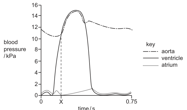
What is happening at time X?
Options
| aortic semilunar valve | atrium | |
|---|---|---|
| A | closing | emptying |
| B | closing | filling |
| C | opening | emptying |
| D | opening | filling |
Working
At time X the ventricular pressure (solid line) rises and crosses above the aortic pressure (dashed-dotted line). A valve opens when the pressure behind it exceeds the pressure in front of it, so the aortic semilunar valve opens at this moment.
At the same time the atrial pressure curve (dotted line) is low and the atrium is in diastole, passively receiving blood from the vena cavae and pulmonary veins — i.e. the atrium is filling.
Answer
D
D
Background Concept
The cardiac cycle consists of alternating phases of contraction (systole) and relaxation (diastole) of the atria and ventricles. Blood flows down a pressure gradient, and each of the four heart valves opens or closes passively in response to the pressure difference across it:
- The atrioventricular (AV) valves (bicuspid and tricuspid) separate the atria from the ventricles.
- The semilunar valves (aortic and pulmonary) separate the ventricles from the aorta and pulmonary artery.
During ventricular systole the ventricle contracts, its pressure rises sharply, and once ventricular pressure exceeds aortic pressure the aortic semilunar valve opens so blood can be ejected into the aorta. Meanwhile, the atria are relaxed (atrial diastole) and blood returning from the venae cavae and pulmonary veins passively fills them — this is the period of atrial filling.
Understanding the Question
Fig. 32.1 shows three pressure curves over one cardiac cycle: aorta (dashed-dotted), ventricle (solid) and atrium (dotted), all in kPa on the y-axis against time in seconds. Time X is marked at the exact point on the time axis where the rising ventricular curve meets and exceeds the aortic curve. We need to identify (i) the state of the aortic semilunar valve at this point, and (ii) what the atrium is doing at the same time.
The command word is "what is happening", so the answer must identify both events occurring simultaneously at time X.
Approach
Two pieces of information are needed:
-
Valve state — A valve opens when downstream pressure falls below upstream pressure, and closes when downstream pressure rises above upstream pressure. At X the ventricle is still pressurising and has just overtaken aortic pressure; ventricular pressure is therefore higher than aortic pressure, pushing blood into the aorta — so the aortic semilunar valve has just opened.
-
Atrial activity — Look at the dotted atrial curve at time X. It is low and relatively flat, characteristic of atrial diastole. Because the AV valves are closed (the ventricle is contracting and pressurising), blood cannot pass from atria into ventricles, so blood returning from the veins accumulates in the relaxing atria — the atria are filling.
Step-by-Step Reasoning
- Around time X the ventricular curve rises steeply from ~10 kPa to >15 kPa, crossing the aortic curve.
- Until this crossover, aortic pressure was higher than ventricular pressure, keeping the aortic semilunar valve closed and preventing backflow.
- The moment ventricular pressure exceeds aortic pressure, blood is forced through the aortic semilunar valve, which therefore opens.
- Just after this point the two curves track each other closely as blood is ejected into the aorta during ventricular ejection.
- The dotted atrial curve at X sits near its baseline (~0.5 kPa) and is essentially flat or slightly rising — characteristic of the atria passively filling with venous return during ventricular systole. The atria are therefore filling, not emptying (emptying occurs earlier, during atrial systole, before time X).
- Putting the two answers together: aortic semilunar valve opening, atrium filling → option D.
The distractors fail because:
- A and B: the aortic semilunar valve is closing only when ventricular pressure falls below aortic pressure later in the cycle (not at the rising crossover at X).
- C: the atrium is not emptying at X — atrial emptying (atrial systole) happens earlier, before the AV valves close and the ventricle begins to contract.
Key Takeaways
- Valves respond passively to pressure differences — they are not actively pulled open or shut.
- The crossover of ventricular and aortic pressures on a pressure–time trace marks the opening of the aortic semilunar valve and the start of ventricular ejection.
- Atrial filling occurs during ventricular systole, not during atrial systole — a frequently confused point.
Common Mistakes
- Confusing atrial systole with ventricular systole — students sometimes think the atrium is contracting (emptying) at the same time as the ventricle, but atrial systole actually precedes ventricular systole.
- Assuming a valve is "closing" at any pressure crossover — it closes only when pressure behind the valve (downstream) exceeds pressure in front.
- Misreading the curves: the solid line is the ventricle (which changes most dramatically), the dashed-dotted line is the aorta (which rises and falls with ventricular ejection), and the dotted line is the atrium (small, low-pressure variations).
Things to Be Careful About
- Identify each curve from the key before reading values — mixing up the ventricle and aorta gives the wrong valve state.
- Time X sits on the rising part of the ventricular curve, where ventricular pressure is increasing past aortic pressure. The later crossover (on the falling phase) would correspond to valve closure, not opening.
- The atrial pressure curve stays near 0–1 kPa throughout; small fluctuations in atrial pressure are normal and do not indicate atrial contraction.
How many times must a carbon dioxide molecule pass through a cell surface membrane to travel from the tissue fluid into the blood plasma?
(Assume there are no pores between the cells the carbon dioxide molecule must pass through.)
Options
A 1
B 2
C 3
D 4
Working
The capillary wall consists of a single layer of endothelial cells. To move from the tissue fluid into the blood plasma, a CO₂ molecule must:
- Diffuse into the endothelial cell by passing through its cell surface membrane (1st membrane).
- Diffuse out of the endothelial cell on the blood side by passing through its cell surface membrane again (2nd membrane).
Total = 2 cell surface membranes.
Answer
B
B
Background Concept
Carbon dioxide is a small, uncharged, lipid-soluble molecule produced continuously by aerobic respiration in living cells. It diffuses down its concentration gradient from sites of high concentration (respiring cells) to sites of lower concentration (the blood, and ultimately the lungs for exhalation).
Blood is carried away from the tissues in capillaries. A capillary is the narrowest type of blood vessel and its wall is extremely thin — it consists of a single layer of squamous (flattened) endothelial cells sitting on a thin basement membrane. This thinness is essential for efficient and rapid exchange of gases, nutrients and metabolic wastes between the blood and the surrounding tissue fluid.
Understanding the Question
The question is asking about the membrane-crossing steps required for a CO₂ molecule to move from the tissue fluid (the fluid bathing the cells) into the blood plasma (the liquid component of blood inside the capillary). Crucially:
- The starting point is the tissue fluid, not the inside of the original respiring cell. The molecule has already left the cell that produced it.
- The question explicitly tells us to assume there are no pores between the cells, so the CO₂ cannot slip between the endothelial cells — it must go through them.
The command is essentially "trace the path and count the membranes." Each time the CO₂ crosses a cell surface membrane counts as one.
Approach
- Identify the structure that separates the tissue fluid from the blood plasma — the capillary wall.
- Recognise that the capillary wall is one cell thick (single endothelial layer).
- To traverse one cell, a molecule must cross two cell surface membranes: one to enter, one to exit.
Step-by-Step Reasoning
Step 1 — Where is the CO₂ coming from and going to?
The CO₂ is in the tissue fluid, the thin film of fluid that surrounds body cells. It needs to reach the blood plasma inside the capillary lumen.
Step 2 — What stands between the tissue fluid and the plasma?
The only barrier is the capillary wall. This wall is composed of a single layer of endothelial cells (with a basement membrane, but basement membrane is freely permeable and does not constitute a cell surface membrane in the CIE sense).
Step 3 — How many membranes must the CO₂ cross to pass through one cell?
- It must first enter the endothelial cell from the tissue-fluid side: that is crossing the outer cell surface membrane → 1.
- It must then leave the endothelial cell on the blood-plasma side: that is crossing the inner cell surface membrane → 2.
Step 4 — Final count.
2 cell surface membranes. The answer is B (2).
Key Takeaways
- Capillary walls are one cell thick, which is why they are the principal site of exchange between blood and tissues.
- To pass through one cell, a molecule must cross two cell surface membranes (one at each face of the cell), assuming no pores are present.
- The same logic explains why a substance travelling from a respiring cell to the blood crosses 3 membranes (cell membrane of the respiring cell + two membranes of the capillary endothelial cell).
Common Mistakes
- Saying "1": a common error is to assume the capillary wall is "just a wall" with one membrane. The wall is a cell, and any substance going through that cell must enter and leave it, costing two membrane crossings.
- Saying "3": this would only be correct if the starting point were the inside of the respiring cell that produced the CO₂. The question clearly specifies tissue fluid as the starting point, so the original cell's membrane is not counted.
- Saying "4": this would apply if the capillary were two cells thick, or if the question were asking about the entire path including an alveolus at the other end.
Things to Be Careful About
- Read the question's specified starting and ending points very carefully — they determine how many membranes are in scope.
- "No pores" is a clue: it forces you to route the CO₂ through the endothelial cell rather than between adjacent endothelial cells.
- Cell surface membranes are phospholipid bilayers with proteins; they are the unit being counted. The basement membrane and glycocalyx are not cell surface membranes and do not count.
Two types of epithelial cells, 1 and 2, occur in the mammalian gas exchange system in a healthy human.
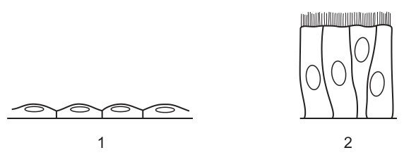
Which row shows the function and distribution of these types of epithelial cells?
Options
| cell type 1 | cell type 2 | |
|---|---|---|
| A | absorption in bronchi | secrete mucus in alveoli |
| B | exchange surface in alveoli | secrete mucus in bronchi |
| C | absorption in bronchi | move mucus in alveoli |
| D | exchange surface in alveoli | move mucus in bronchi |
Working
Cell type 1 is a squamous (pavement) epithelial cell — very thin and flat. Its thinness minimises the diffusion distance for respiratory gases, so it forms the exchange surface in the alveoli.
Cell type 2 is a ciliated columnar epithelial cell — tall cells with cilia on their apical (luminal) surface. The cilia beat in a coordinated wave to move mucus (secreted by goblet cells) upwards, out of the bronchi and towards the pharynx. Cilia themselves do not secrete mucus.
- A: Wrong — squamous cells are not for absorption, and mucus is not secreted in alveoli.
- B: Wrong — ciliated cells move mucus; goblet cells secrete it. Ciliated cells are in the bronchi, not the alveoli.
- C: Wrong — cilia move, rather than secrete, mucus; they are not found in the alveoli.
- D: Correct — squamous cells form the alveolar exchange surface; ciliated cells move mucus up the bronchi.
Answer
D
D
Background Concept
The mammalian gas exchange system is lined by different types of epithelium in different regions, each adapted to its specific role.
- Alveoli are tiny, thin-walled air sacs where gas exchange occurs. They are lined by an extremely thin squamous (pavement) epithelium — just one cell thick. Oxygen and carbon dioxide diffuse across this layer between the alveolar air and the pulmonary capillaries. The very small diffusion distance is essential for rapid gas exchange.
- Bronchi and bronchioles are larger airways that conduct air to and from the alveoli. They are lined by ciliated columnar epithelium together with goblet cells. Goblet cells secrete sticky mucus that traps inhaled particles (dust, microbes, smoke particles). The cilia on the columnar cells beat in a synchronised, wave-like rhythm that propels this mucus — and the trapped debris — upwards towards the pharynx, where it is swallowed or coughed out. This is the mucociliary escalator.
Crucially, the cilia move mucus; they do not secrete it, and alveoli have neither cilia nor goblet cells — they are purely an exchange surface.
Understanding the Question
The question presents two drawings of epithelial cells and asks you to identify each cell type from its structure and then match it to its function (what it does) and distribution (where it is found) in the gas exchange system. The four options each pair cell type 1 with cell type 2.
From the figure:
- Cell type 1 is flat with a central nucleus — a squamous epithelial cell.
- Cell type 2 is tall and column-shaped with hair-like projections (cilia) on the apical surface — a ciliated columnar epithelial cell.
The command word here is essentially "identify and match" — the difficulty lies in correctly assigning function (not confusing secretion with movement of mucus) and distribution (not confusing alveoli with bronchi).
Approach
For each cell type, ask two questions:
- What does its structure allow it to do (function)?
- Where in the gas exchange system does this function need to occur (distribution)?
- Thin flat cells → diffusion / gas exchange → alveoli.
- Tall cells with cilia → movement of mucus → conducting airways (bronchi/bronchioles).
Then eliminate any option that swaps these, places either cell in the wrong region, or confuses mucus secretion with mucus movement.
Step-by-Step Reasoning
Cell type 1 — squamous epithelium:
- Structure: very thin, flat, single layer, central nucleus.
- Functional implication: minimal diffusion distance.
- Location: alveoli — the gas exchange surface.
- Correct pairing: exchange surface in alveoli.
Cell type 2 — ciliated columnar epithelium:
- Structure: tall, column-shaped, with cilia on the apical surface.
- Functional implication: cilia beat to move material along the surface.
- Location: bronchi (and bronchioles/trachea) — the conducting airways, where mucus is produced by goblet cells and needs to be cleared.
- Correct pairing: move mucus in bronchi.
Eliminating the options:
- A — wrong on both counts. Squamous cells are not for absorption (they're for gas exchange), and mucus is not secreted in alveoli.
- B — half right. Squamous cells are correctly placed in alveoli, but ciliated cells do not secrete mucus (goblet cells do) and they are in the bronchi, not the alveoli.
- C — cilia move mucus rather than absorbing anything in bronchi, and they are not in the alveoli.
- D — fully correct: squamous cells form the alveolar exchange surface, and ciliated columnar cells move mucus up the bronchi.
Key Takeaways
- Squamous epithelium = thin = short diffusion distance = gas exchange in alveoli.
- Ciliated columnar epithelium = cilia on surface = movement of mucus = conducting airways (trachea, bronchi, bronchioles).
- Goblet cells (not shown here) are the cells that secrete mucus; cilia are what move it.
- Alveoli contain only squamous epithelium; they have no cilia and no goblet cells.
Common Mistakes
- Confusing secretion with movement of mucus — the most common error. Cilia move mucus; goblet cells secrete it. The cells shown in cell type 2 only have cilia, so their function is to move mucus.
- Placing ciliated cells in the alveoli — alveoli have no cilia because cilia would interfere with gas exchange and there is no mucus layer to clear there.
- Calling squamous cells "absorption cells" — absorption is a function of, for example, gut epithelium, not alveolar epithelium. In the lungs, the thin squamous layer exists for diffusion of gases.
- Reversing function and distribution — squareness of cell 1 (flat/thin) is the key clue to its location in alveoli, and the cilia on cell 2 are the key clue to its function in bronchi.
Things to Be Careful About
- Read the option carefully: the function column and the distribution column must both be correct for each cell type.
- "Move mucus" and "secrete mucus" are not interchangeable — they refer to different cell types (ciliated cells vs goblet cells).
- "Alveoli" appear in every option, but only options B and D place the right cell there. This is a useful pattern to spot when eliminating answers.
Which tissues would be seen in a photomicrograph of the wall of the trachea?
Options
| elastic fibres | smooth muscle | |
|---|---|---|
| A | ✗ | ✓ |
| B | ✓ | ✗ |
| C | ✓ | ✓ |
| D | ✗ | ✗ |
key
✓ = present
✗ = not present
Working
The trachea wall is held open by C-shaped rings of hyaline cartilage, and the open ends of each C are bridged by the trachealis muscle (smooth muscle). Elastic fibres are also woven through the tracheal wall, allowing it to stretch and recoil during breathing while resisting over-expansion.
So both elastic fibres ✓ and smooth muscle ✓ are present.
Answer
C
C
Background Concept
The trachea is a tube that conducts air between the larynx and the bronchi. Its wall must stay permanently patent (open) so air can flow freely, but it must also be able to stretch and recoil slightly with the pressure changes of breathing. These two functional demands are met by a combination of tissues embedded in the wall:
- Hyaline cartilage in incomplete (C-shaped) rings keeps the lumen open and prevents the trachea from collapsing during inspiration when intrathoracic pressure falls.
- Smooth muscle (trachealis muscle) bridges the open posterior ends of each cartilage C. It can contract to narrow the airway (e.g. in the cough reflex or during bronchoconstriction).
- Elastic fibres are distributed throughout the wall (in the lamina propria and between the muscle and cartilage). They recoil after stretching, helping to restore the tracheal diameter during expiration and preventing over-distension during deep inspiration.
- Pseudostratified ciliated columnar epithelium with goblet cells lines the lumen and traps/removes inhaled particles.
Understanding the Question
This is a structured multiple-choice question. The candidate is given a 2 × 2 table of tissues (elastic fibres, smooth muscle) and tick/cross options, and must decide which combination of tissues is present in a photomicrograph of the tracheal wall. Each of the four answer options (A, B, C, D) tests whether the student knows whether each of the two tissues is present. The distractors are partial combinations — one tissue present but not the other, or neither present.
Approach
For each of the two tissues in the table, decide independently whether it occurs in the tracheal wall. Then match the answer to the option whose ticks and crosses correspond.
- Elastic fibres: present ✓
- Smooth muscle: present ✓ (as the trachealis muscle completing the cartilage C)
This combination corresponds to option C.
Step-by-Step Reasoning
- Smooth muscle in the trachea. The trachea is not a rigid pipe — it must be able to constrict. The posterior gap in each C-shaped cartilage ring is closed by a band of smooth muscle called the trachealis muscle. Smooth muscle is therefore definitely present in a transverse section of the tracheal wall, and would be visible in a photomicrograph as a band of spindle-shaped, unstriated cells with single central nuclei.
- Elastic fibres in the trachea. The trachea is constantly being pushed and pulled by movement of the neck, swallowing, and pressure changes during the respiratory cycle. Elastic fibres are woven through the connective tissue layers of the wall (in the lamina propria and the submucosa). They would appear in a stained section as dark, wavy, branching fibres. They are not always easy to pick out in routine H&E sections, but with elastic stains (e.g. Verhoeff's) they are unmistakable, and the question lists them as a recognised component of the tracheal wall.
- Matching to the table. Elastic fibres = ✓; smooth muscle = ✓ → option C.
A common reason for ruling out option A or B is forgetting one of the two tissues; ruling out D requires believing the trachea wall has neither — which is wrong because both are fundamental wall components.
Key Takeaways
- The tracheal wall is built around C-shaped hyaline cartilage rings; the posterior gap is bridged by smooth muscle (trachealis), so smooth muscle is present.
- Elastic fibres are present throughout the connective tissue of the tracheal wall and allow recoil after stretching.
- Cartilage keeps the airway open; smooth muscle adjusts its diameter; elastic fibres provide recoil — three complementary roles in one wall.
Common Mistakes
- Choosing A or B — forgetting that the trachea contains BOTH tissues. Some students only remember cartilage and overlook the smooth muscle, or only remember the smooth muscle and overlook the elastic fibres.
- Choosing D — wrongly assuming the trachea wall is just cartilage and epithelium. The wall is a layered structure with connective tissue, muscle and elastic components, not just a rigid tube.
- Confusing the trachea with the oesophagus. The oesophagus wall contains smooth muscle (in its muscularis externa) but no cartilage, and would not appear in a section of the trachea.
Things to Be Careful About
- "Smooth muscle" must be distinguished from skeletal muscle (striated, multinucleate) and from cardiac muscle — the trachea is under autonomic control via smooth muscle only.
- Elastic fibres are often not obvious on H&E-stained slides; do not assume they are absent simply because they are hard to see — they are a recognised wall component.
- The question asks about tissues seen in a photomicrograph; both tissues are visible (with appropriate staining) in such a preparation, so a "not visible" argument does not apply.
The graph shows the decrease in cases of tuberculosis (TB) in a country between 1910 and 2000.
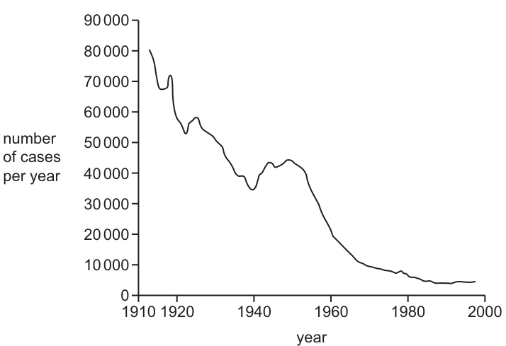
Which factors could have contributed to the fall over this period?
1 pasteurisation of milk
2 the provision of new housing to reduce overcrowding
3 removing large pools of stagnant water
4 identification of contacts of people infected with TB
Options
A 1, 2 and 4
B 1 and 3
C 1 and 4 only
D 2, 3 and 4
Working
Tuberculosis (TB) is caused by Mycobacterium tuberculosis (and historically Mycobacterium bovis from cattle).
- Statement 1 — Pasteurisation of milk: ✓ M. bovis is transmitted in unpasteurised cow's milk. Pasteurisation kills the bacterium, removing this transmission route.
- Statement 2 — New housing to reduce overcrowding: ✓ TB spreads via respiratory droplets/aerosols; overcrowding increases transmission. Reducing overcrowding lowers spread.
- Statement 3 — Removing large pools of stagnant water: ✗ This targets mosquito breeding sites to control malaria, not TB.
- Statement 4 — Identification of contacts of infected people: ✓ Contact tracing identifies and treats those exposed, breaking the chain of transmission.
Correct statements: 1, 2 and 4.
Answer
A
A
Background Concept
Tuberculosis (TB) is caused by bacteria of the Mycobacterium genus — most cases by Mycobacterium tuberculosis, but historically a significant number of human cases, especially of bovine TB, were caused by Mycobacterium bovis. TB is primarily a respiratory disease: it spreads when an infected person coughs or sneezes and releases droplet nuclei containing the bacterium, which are then inhaled by a susceptible person. M. bovis was historically an additional transmission route via unpasteurised milk from infected cattle.
Because TB has two main transmission routes (respiratory droplets and contaminated milk), effective control requires measures that interrupt both:
- Respiratory route: reduce overcrowding (less close contact), improve ventilation, isolate infectious cases, and trace/chemoprophylax contacts.
- Milk-borne route: pasteurisation of milk, and later test-and-slaughter of infected cattle.
Removing stagnant water has no role in TB control — it is a measure aimed at Anopheles mosquito breeding sites to control malaria, a completely different disease with a vector-borne transmission cycle.
Understanding the Question
The question gives a graph showing TB cases falling steadily from about 80,000/year in 1910 to near zero by 2000, and asks which of four public health measures could have contributed to this fall. The four statements must each be evaluated against TB biology — its pathogen and its transmission routes — to decide which are credible explanations.
Approach
For each statement, ask two questions:
- Does it interrupt a known route of TB transmission?
- Is it biologically relevant to TB, or is it a control measure for a different disease?
Then select the option that lists all the correct statements and no incorrect ones.
Step-by-Step Reasoning
- Statement 1 (pasteurisation of milk): M. bovis infected cattle and was passed to humans in unpasteurised milk. The introduction of compulsory pasteurisation (and TB testing of dairy herds) removed this route, so statement 1 is a valid contributor. ✓
- Statement 2 (new housing, less overcrowding): TB spreads most efficiently where people share poorly ventilated indoor air. Slum clearance, rehousing, and better ventilation reduce droplet transmission, so statement 2 is a valid contributor. ✓
- Statement 3 (removing pools of stagnant water): Stagnant water is the breeding habitat of Anopheles mosquitoes, the vector of Plasmodium (malaria). It has no link to TB transmission, so statement 3 is rejected. ✗
- Statement 4 (identification of contacts of infected people): Contact tracing finds people who have been exposed to an active case, allowing them to be tested and offered treatment or chemoprophylaxis (e.g. isoniazid), which breaks chains of transmission. Statement 4 is a valid contributor. ✓
Statements 1, 2 and 4 are correct, so the answer is A (1, 2 and 4).
Key Takeaways
- TB has two key transmission routes: respiratory droplets and (historically) contaminated milk.
- Pasteurisation tackles the milk-borne route; reduced overcrowding, ventilation, and contact tracing tackle the respiratory route.
- Public health measures are disease-specific — always check that a control measure actually targets the relevant pathogen or vector.
Common Mistakes
- Selecting an option containing statement 3 because "removing stagnant water sounds like a sensible public health measure". Stagnant water control is for malaria, not TB.
- Forgetting that M. bovis was historically important in human TB, leading students to doubt the relevance of milk pasteurisation.
- Confusing contact tracing (TB, HIV, STIs) with vector control (malaria, dengue).
Things to Be Careful About
- The fall in TB began before antibiotics and vaccination were widely available, so the early decline (1910s–1940s) is largely attributable to social and public health measures: pasteurisation and improved housing/nutrition. The steep fall after the 1950s reflects antibiotics (streptomycin, isoniazid) and BCG vaccination. All four statements concern non-pharmaceutical control, but they are still valid contributors over the whole 90-year period.
- "Identification of contacts" is contact tracing — a core TB control activity, not a treatment in itself; the mark is for the surveillance/epidemiological action, which the statement clearly describes.
The pathogens for some diseases are transmitted by vectors.
Which disease is transmitted with the help of a vector that could be controlled by the removal of pools of water?
Options
A cholera
B HIV/AIDS
C malaria
D tuberculosis
Working
Malaria is caused by Plasmodium and transmitted by the female Anopheles mosquito. The mosquito lays its eggs in still water, so removing pools of standing water destroys its breeding sites.
- A — cholera is water-borne (contaminated drinking water), not vector-borne.
- B — HIV/AIDS is transmitted by direct exchange of body fluids, not by a vector.
- D — tuberculosis is airborne (droplet infection), not vector-borne.
- C — malaria is transmitted by a mosquito that breeds in pools of water. ✓
Answer
C
C
Background Concept
A vector in the context of infectious disease is a living organism (typically an arthropod) that carries a pathogen from one host to another. The pathogen may multiply or develop inside the vector, or the vector may simply transport it mechanically. The classic Cambridge syllabus examples of vector-borne disease all involve mosquitoes or similar blood-sucking insects.
The four diseases named in this question each have a different mode of transmission:
- Cholera — caused by the bacterium Vibrio cholerae. Transmitted by the faecal–oral route through drinking water contaminated with infected faeces. It is a water-borne disease, but water here is the vehicle, not a vector's habitat.
- HIV/AIDS — caused by the human immunodeficiency virus. Transmitted by direct exchange of certain body fluids (blood, semen, vaginal secretions, breast milk). No arthropod vector is involved.
- Malaria — caused by protoctistan parasites of the genus Plasmodium (mainly P. falciparum and P. vivax). Transmitted by the bite of an infected female Anopheles mosquito, which injects Plasmodium sporozoites from its salivary glands into the host's bloodstream while feeding.
- Tuberculosis (TB) — caused by the bacterium Mycobacterium tuberculosis. Transmitted by inhaled droplets/aerosols from an infected person coughing or sneezing. No vector.
The life cycle of the Anopheles mosquito is critical here. After taking a blood meal from an infected person, the mosquito harbours the parasite while it develops into sporozoites in its gut and salivary glands. The female must then lay her eggs in still, fresh water — puddles, ponds, blocked drains, irrigation channels, discarded tyres and similar collections of water. The larvae and pupae are aquatic, so the entire aquatic phase of the mosquito's life cycle depends on these water bodies.
Understanding the Question
The question is asking us to identify which of the four diseases is transmitted by a vector whose population could be reduced by removing pools of water. This is a multi-step linking question: (1) identify the vector for each disease, (2) determine where that vector breeds, and (3) match the breeding habitat to "pools of water".
Approach
- Eliminate diseases that are not vector-borne at all (HIV/AIDS, TB).
- Of those that are vector-borne (cholera? malaria?), check whether the transmission involves a vector. Cholera is water-borne, not vector-borne, so it falls away too.
- The only disease that involves an arthropod vector whose breeding site is stagnant water is malaria.
Step-by-Step Reasoning
- A — Cholera: Eliminate. Cholera is transmitted by drinking water contaminated with Vibrio cholerae from infected human faeces. Removing pools of water would not help; in fact clean water supplies and good sanitation are the control measures.
- B — HIV/AIDS: Eliminate. HIV is transmitted by direct exchange of body fluids — sexual contact, contaminated needles, mother-to-child (across the placenta, during birth, or in breast milk), and blood transfusion. There is no arthropod vector.
- C — Malaria: Accept. Malaria is transmitted by the bite of an infected female Anopheles mosquito. The mosquito breeds in stagnant pools of water. Draining, filling or removing these pools (an anti-larval measure) reduces mosquito populations and therefore malaria transmission. ✓
- D — Tuberculosis: Eliminate. TB is transmitted by inhaling respiratory droplets containing Mycobacterium tuberculosis. No vector; control is by vaccination (BCS), antibiotics and case-finding.
Key Takeaways
- Only malaria (and a handful of less common diseases) is vector-borne among the four options.
- The vector is the female Anopheles mosquito, which requires still water for its aquatic larval and pupal stages.
- Removing standing water is a core public-health intervention against malaria (alongside insecticide-treated bed nets and indoor residual spraying).
- A common confusable is cholera — cholera is associated with water, but as a vehicle of transmission, not as a vector's breeding ground.
Common Mistakes
- Choosing A (cholera) because it has the word "water" in its transmission story. Cholera is water-borne, not vector-borne; destroying pools of water does not control cholera.
- Confusing the vehicle of transmission (water, air, body fluids) with the vector's habitat. The question specifically asks about a vector, which is a living organism.
- Thinking that because TB affects the lungs it might be transmitted by an insect — TB is droplet-borne, not vector-borne.
Things to Be Careful About
- The question says "with the help of a vector" — this is the giveaway that the correct disease must be one whose transmission involves a living carrier organism.
- "Removal of pools of water" specifically targets the aquatic immature stages of the mosquito. It does not affect adult flying mosquitoes directly; it prevents the next generation.
- The mosquito is only a vector; the pathogen is Plasmodium (a protoctist), and only female mosquitoes feed on blood (males feed on nectar).
How does penicillin affect bacteria?
Options
A It inhibits DNA replication by binding to nucleotides.
B It inhibits translation by preventing tRNA binding to ribosomes.
C It is a competitive inhibitor of an enzyme in cell wall synthesis.
D It is a competitive inhibitor of an enzyme in protein synthesis.
Answer
Penicillin inhibits cell wall synthesis in bacteria by acting as a competitive inhibitor of the transpeptidase enzyme that cross-links peptidoglycan chains. The bacterial cell wall is weakened and the cell undergoes lysis. Options A (DNA replication), B (translation) and D (protein synthesis) describe mechanisms of other antibiotics, not penicillin.
C
C
Background Concept
Antibiotics are antimicrobial substances that kill bacteria (bactericidal) or inhibit their growth (bacteriostatic). Different classes of antibiotics target different bacterial structures or processes:
- Cell wall synthesis inhibitors (e.g. penicillin, amoxicillin, cephalosporins) — interfere with peptidoglycan cross-linking in the bacterial cell wall.
- Protein synthesis inhibitors (e.g. tetracyclines, streptomycin, chloramphenicol) — bind to bacterial ribosomes (70S) and block translation.
- DNA replication inhibitors (e.g. quinolones such as ciprofloxacin) — inhibit DNA gyrase / topoisomerase.
- Nucleic acid synthesis inhibitors (e.g. rifampicin) — block RNA polymerase.
Bacterial cell walls are made of peptidoglycan, a mesh of alternating NAG (N-acetylglucosamine) and NAM (N-acetylmuramic acid) sugar units, cross-linked by short peptide chains. The enzyme transpeptidase (also called DD-transpeptidase or a penicillin-binding protein, PBP) catalyses the peptide cross-links that give the wall its tensile strength.
Understanding the Question
The MCQ asks specifically about penicillin's mode of action. The student must identify which cellular process penicillin disrupts. The four options refer to four distinct targets: DNA replication, translation (tRNA/ribosomes), cell wall synthesis and protein synthesis — only one of which is correct for penicillin.
Approach
Recall the well-established mechanism: penicillin is structurally similar to the D-alanyl-D-alanine terminus of the peptidoglycan precursor, so it binds to the active site of transpeptidase and acts as a competitive inhibitor. With cross-linking prevented, the growing cell wall is mechanically weak, water enters the cell by osmosis, and the bacterium lyses. This immediately rules out any option that names DNA replication, translation, or protein synthesis.
Step-by-Step Reasoning
- Penicillin belongs to the β-lactam class of antibiotics. The β-lactam ring mimics the D-alanyl-D-alanine substrate of transpeptidase.
- The drug binds reversibly to the transpeptidase active site, blocking peptidoglycan cross-linking — this is the definition of competitive inhibition at the active site.
- Without cross-links, the cell wall cannot withstand the internal turgor pressure of the bacterium, so the cell bursts (lysis).
- Option A is incorrect: nothing about penicillin concerns DNA replication or nucleotide binding.
- Option B is incorrect: tRNA/ribosome binding is the target of antibiotics such as tetracyclines (block tRNA at the A site) or aminoglycosides (cause misreading).
- Option D is wrong on two counts — protein synthesis is the wrong target, and penicillin's action is on cell wall enzymes, not protein synthesis.
- Option C correctly identifies competitive inhibition of an enzyme (transpeptidase) in cell wall synthesis.
Key Takeaways
- Penicillin is a competitive inhibitor of transpeptidase, an enzyme involved in bacterial cell wall (peptidoglycan) synthesis.
- It works because animal cells have no peptidoglycan wall, giving penicillin selective toxicity against bacteria.
- Antibiotics are not a single class — each family targets a specific bacterial structure; confusing their mechanisms is a common exam trap.
- Antibiotics do not affect viruses, which lack all of the structures listed (no cell wall, no ribosomes, no DNA replication machinery of their own).
Common Mistakes
- Confusing penicillin's target (cell wall) with that of streptomycin or tetracyclines (ribosomes/protein synthesis).
- Thinking of penicillin as a non-competitive inhibitor — it binds at the active site by mimicking the natural substrate, so it is competitive.
- Choosing option B because "tRNA" and "ribosomes" sound biologically important — they are, but they are not penicillin's target.
Things to Be Careful About
- "Competitive" vs "non-competitive" inhibition: competitive means the inhibitor resembles the substrate and binds the active site; non-competitive binds elsewhere and changes the enzyme's shape. Penicillin is competitive.
- The wording must say cell wall synthesis, not just "cell wall" — the mechanism is the inhibition of synthesis, leading to a weakened wall.
- Distinguish between bactericidal (kills, like penicillin) and bacteriostatic (stops growth) — though this distinction is not tested in this particular option.
- Remember that viruses have no cell wall, ribosomes, or metabolism of their own; that is why antibiotics cannot treat viral infections.
The hybridoma method is used for the production of monoclonal antibodies.
Which two types of cell are used in this method?
Options
A stem cell and B-lymphocyte
B stem cell and T-lymphocyte
C tumour cell and B-lymphocyte
D tumour cell and T-lymphocyte
Working
The hybridoma method fuses a B-lymphocyte (which produces a single specific antibody but cannot divide indefinitely in culture) with a myeloma/tumour cell (which divides continuously but does not produce a specific antibody). The resulting hybridoma combines both properties: it produces one specific antibody and can be cultured indefinitely.
Answer
C
C
Background Concept
Monoclonal antibodies are identical antibodies that all recognise the same single epitope (antigen-binding site). To produce them in useful quantities, scientists need cells that can both:
- make one specific antibody, and
- keep dividing indefinitely in culture.
No single normal cell does both. B-lymphocytes make highly specific antibodies but die out after a few divisions, while myeloma (tumour) cells divide endlessly but produce no useful antibody. The hybridoma technique solves this by fusing the two cell types.
Understanding the Question
This is a straightforward MCQ asking which two cell types are combined in the hybridoma method. The answer must be the pairing that brings together antibody specificity and unlimited division.
Approach
Think about what each cell contributes:
- The cell that gives antibody specificity must be a B-lymphocyte (plasma cells derived from B-cells are the antibody factories of the immune system).
- The cell that gives immortality in culture must be a tumour (myeloma) cell, because tumour cells divide uncontrollably.
The other pairings fail because stem cells do not produce one specific antibody, and T-lymphocytes do not secrete antibodies at all (they act on infected cells directly).
Step-by-Step Reasoning
- Option A (stem cell + B-lymphocyte): A B-lymphocyte contributes the antibody, but stem cells do not provide the immortal-division property; they would not give a continuously dividing cell line.
- Option B (stem cell + T-lymphocyte): T-lymphocytes do not secrete antibodies — they are involved in cell-mediated immunity, so even a successful hybrid would not produce antibodies.
- Option C (tumour cell + B-lymphocyte): Correct. The myeloma (tumour) cell divides continuously; the B-lymphocyte produces one specific antibody. Fusion gives a hybridoma that does both, and individual hybridomas are screened so that only those producing the desired antibody are cloned.
- Option D (tumour cell + T-lymphocyte): T-lymphocytes do not secrete antibodies, so the hybridoma would not produce a useful monoclonal antibody.
Key Takeaways
- The hybridoma method always uses a B-lymphocyte (for antibody specificity) and a myeloma/tumour cell (for continuous division).
- T-lymphocytes and stem cells are not used because they do not provide the required combination of features.
Common Mistakes
- Choosing B-lymphocyte and T-lymphocyte — confusing humoral (B-cell, antibody-based) and cell-mediated (T-cell) immunity.
- Choosing stem cell — assuming any dividing cell will do; stem cells do not confer the specific antibody property nor the uncontrolled division of a cancer cell line.
Things to Be Careful About
- "Tumour cell" in this context means a myeloma (B-cell cancer) cell line, but for the purposes of the question any answer pairing B-lymphocyte with a tumour cell is correct.
- Remember that only B-lymphocytes (and plasma cells derived from them) secrete antibodies; T-lymphocytes do not.
What describes a non-specific immune response?
Options
A activation of killer T-lymphocytes by infected cells
B cloning of B-lymphocytes to form plasma cells
C ingestion of a bacterial cell by a neutrophil
D recognition of antigens on the cell surface of macrophages
Working
Non-specific (innate) immune responses act against any pathogen without needing prior exposure or recognition of a specific antigen. Phagocytosis of bacteria by neutrophils is a classic example of such a response.
A — Killer T-lymphocytes are activated only after recognising a specific antigen presented by an infected cell; this is a specific (cell-mediated) response.
B — Cloning of B-lymphocytes into plasma cells occurs only after a specific antigen binds to a complementary B-cell receptor; this is a specific (humoral) response.
C — Neutrophils engulf any bacteria they encounter at a site of infection; phagocytosis is a non-specific response.
D — Macrophages presenting antigens on their surface is part of antigen presentation, which initiates the specific immune response.
Answer
C
C
Background Concept
The immune system has two broad branches:
- Non-specific (innate) immunity — defences that act against any pathogen in the same way, regardless of what the pathogen is. They are present from birth, do not require prior exposure to the pathogen, and do not involve antigen-specific recognition. Examples include physical barriers (skin, mucus), phagocytosis by neutrophils and macrophages, inflammation, and the action of antimicrobial proteins such as lysozyme and interferon.
- Specific (adaptive) immunity — defences that target a particular antigen. They involve B-lymphocytes (humoral immunity, producing antibodies via plasma cells) and T-lymphocytes (cell-mediated immunity, including helper T and killer/cytotoxic T cells). Specific responses have memory, which is the basis of long-lasting protection after infection or vaccination.
A useful way to remember the distinction: if the response is the same the first time and the hundredth time, it is non-specific. If it becomes faster and stronger on re-exposure (because of memory cells), it is specific.
Understanding the Question
This is a single-best-answer multiple-choice question. The stem simply asks which of the four listed events describes a non-specific immune response. The candidate must pick the option that represents a generalised defence — one that does not depend on recognising a particular antigen.
Approach
Examine each option and decide whether it depends on antigen-specific recognition (specific) or whether it is a generalised defence that occurs against any pathogen (non-specific).
Step-by-Step Reasoning
- Option A — activation of killer T-lymphocytes by infected cells: Killer (cytotoxic) T-cells are part of specific cell-mediated immunity. They only kill cells that display a particular antigen (e.g. a viral peptide) on MHC class I molecules. Specific — not the answer.
- Option B — cloning of B-lymphocytes to form plasma cells: A B-lymphocyte divides and differentiates into plasma cells only after its surface antibody has bound a matching antigen. This is the central event of the specific humoral response. Specific — not the answer.
- Option C — ingestion of a bacterial cell by a neutrophil: Neutrophils are short-lived phagocytes that engulf and digest bacteria at sites of infection using lysosomal enzymes. They perform this action on any bacterium they encounter, without needing to recognise a specific antigen. This is a textbook example of non-specific immunity. Correct.
- Option D — recognition of antigens on the cell surface of macrophages: Macrophages act as antigen-presenting cells. When they display antigen fragments on MHC class II molecules, this triggers helper T-cells — initiating the specific immune response. Specific — not the answer.
Key Takeaways
- Non-specific (innate) responses are the same against every pathogen; they include phagocytosis by neutrophils and macrophages, inflammation, and physical/chemical barriers.
- Specific (adaptive) responses involve B-cells, plasma cells, antibodies, helper T-cells, and killer T-cells, and depend on recognition of particular antigens.
- Phagocytosis by neutrophils is the canonical example of a non-specific response.
Common Mistakes
- Confusing macrophages (which do act in both non-specific phagocytosis AND antigen presentation) with neutrophils (which act almost exclusively in non-specific phagocytosis). In this question, the neutrophil performing ingestion is the non-specific clue.
- Treating any cell of the immune system as automatically non-specific. T- and B-lymphocytes are exclusively specific responders.
Things to Be Careful About
- "Recognition of antigens" is essentially the defining feature of specific immunity, so any option mentioning antigen recognition (D) is a red flag that the response is not non-specific.
- Do not be misled by the word "activation" (A) or "cloning" (B) — both describe lymphocyte events, which are always specific.
Your score so far
Answer a question to start scoring
Your marks add up here as you work through the paper.