Biology 9700/24 — May/June 2025
Cambridge AS Level · AS Level Structured Questions · worked solutions for every part, with the mark scheme
Topics The Mitotic Cell Cycle · Cell Structure · Biological Molecules · Cell Membranes and Transport · Transport in Plants · Enzymes · +4 more
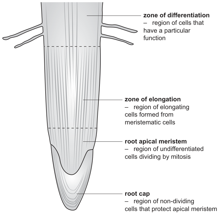
In the roots of plants, specialised tissues are formed from a region in the root tip known as the root apical meristem.
Fig. 1.1 shows the different regions of a root tip.
Cells in all stages of mitosis will be visible in the root apical meristem by using the high power of a light microscope.
Name the stage of mitosis during which the chromosomes are arranged at the spindle equator of the cell.
Answer
Metaphase.
Metaphase
Background Concept
Mitosis is a continuous process but is divided into four named stages for ease of study and identification under the microscope. The stages are, in order: prophase, metaphase, anaphase and telophase. Each stage is defined by a specific chromosome behaviour:
- Prophase — chromosomes condense and become visible; the nuclear envelope breaks down into small vesicles; the spindle forms.
- Metaphase — chromosomes (each consisting of two sister chromatids joined at the centromere) line up along the equator of the spindle, attached to spindle fibres at their centromeres.
- Anaphase — sister chromatids separate at the centromere and are pulled to opposite poles of the cell by shortening spindle fibres.
- Telophase — chromatids arrive at the poles, decondense; vesicles derived from the broken-down nuclear envelope fuse around each set of chromosomes to form two new nuclear envelopes; the spindle breaks down; cytokinesis follows.
In a root apical meristem, all of these stages can be observed because cells are actively and continuously dividing.
Understanding the Question
The question asks for the stage of mitosis during which chromosomes are arranged at the spindle equator. The spindle equator is the imaginary plane in the middle of the cell, equidistant from the two spindle poles. Chromosomes only occupy this position briefly before being pulled apart.
Approach
Recall the definition of each mitotic stage and match the description ("chromosomes arranged at the spindle equator") to the stage whose hallmark this is.
Step-by-Step Reasoning
During metaphase, spindle fibres from opposite poles attach to the centromeres of the chromosomes and pull them with equal force from each side. The chromosomes line up across the middle of the cell — the spindle equator. The key word in the question is "arranged at the spindle equator"; this is the textbook definition of metaphase.
Key Takeaways
- Metaphase = chromosomes lined up at the spindle equator.
- The order of mitosis stages is prophase → metaphase → anaphase → telophase.
Common Mistakes
- Confusing metaphase (chromosomes at the equator) with anaphase (chromatids moving away from the equator toward the poles). The question specifies "arranged at" the equator, not "moving away from" it.
- Spelling: "methaphase" is not credited.
Things to Be Careful About
The mark scheme expects the exact term "metaphase" — be careful with spelling. No descriptive reasoning is needed for a one-mark "name the stage" question.
Early in mitosis, the nuclear envelope breaks up into vesicles and so will not be seen in some cells in the root apical meristem.
Name the stage of mitosis during which the vesicles fuse to form new nuclear envelopes.
Answer
Telophase.
Telophase
Background Concept
Early in mitosis (during prophase), the nuclear envelope disassembles into small membrane-bound vesicles. This allows the spindle to access and attach to the chromosomes. Once mitosis is nearly complete, the cell must rebuild a nuclear envelope around each new set of chromosomes so that two separate daughter nuclei form.
Understanding the Question
The question asks for the stage during which the vesicles produced from the broken-down nuclear envelope fuse together to form new nuclear envelopes. This nuclear-reassembly event marks the end of mitosis.
Approach
Match the description ("vesicles fuse to form new nuclear envelopes") to the stage in which this occurs.
Step-by-Step Reasoning
At the end of mitosis, once the chromatids have reached opposite poles, the spindle breaks down and the vesicles derived from the old nuclear envelope gather around each set of chromatids and fuse with one another. This forms two new nuclear envelopes, one around each new set of chromosomes. This event defines telophase.
Key Takeaways
- Telophase = nuclear envelope reforms around the two new sets of chromosomes; spindle breaks down; cytokinesis is completed.
- The mark scheme explicitly ignores ("I") any reference to early/late telophase — just "telophase" is enough.
Common Mistakes
- Saying "interphase" — this is incorrect; the nuclear envelope reforms during telophase of mitosis, not during interphase (which is the non-dividing phase).
- Confusing telophase with prophase (the opposite process of envelope breakdown).
Things to Be Careful About
The mark scheme awards the mark only for "telophase" — a reference to "late prophase" or "early telophase" is ignored. Be concise and use the precise term.
In the zone of elongation, shown in Fig. 1.1, the newly formed cells expand and elongate.
Two main events that occur in these cells are:
- the entry of water into the cells
- the formation of the large vacuole.
Answer
- Osmosis.
- Water (molecules) move down a water potential gradient, from a higher (less negative) to a lower (more negative) water potential.
- This occurs across the (cell surface) membrane.
- The movement is passive / does not require ATP.
Osmosis; water moves down a water potential gradient across the cell surface membrane; passively / no ATP required.
Background Concept
Osmosis is a special case of diffusion in which water molecules move across a partially permeable membrane from a region of higher (less negative) water potential to a region of lower (more negative) water potential. The water potential () of a solution is influenced by the concentration of solutes it contains — the more solute dissolved, the more negative the water potential, and the more strongly water is "drawn into" that solution.
Key features of osmosis:
- It is the movement of water molecules only (not solutes).
- It takes place across a partially permeable membrane (e.g. the cell surface membrane / tonoplast).
- It moves down a water potential gradient (from high to low; from less negative to more negative ).
- It is a passive process — no ATP is required.
Understanding the Question
In the zone of elongation, newly formed cells take up water so that they can expand and elongate. The question asks you to outline the mechanism by which water enters these cells. The key is to use the correct biological terminology (osmosis, water potential gradient, partially permeable membrane, passive) rather than vague language like "water flows into the cell".
Approach
State the name of the process, then describe what moves, in which direction, across what, and with what energy requirement.
Step-by-Step Reasoning
- Name the process: Osmosis.
- What moves: water (molecules).
- In which direction: down a water potential gradient, i.e. from a higher (less negative) water potential outside the cell to a lower (more negative) water potential inside the cell. The cell sap in the developing vacuole contains dissolved solutes (sugars, mineral ions, organic acids), making the inside more negative.
- Across what: the cell (surface) membrane, which is partially permeable — it allows water but not solutes to pass freely.
- Energy: the movement is passive; no ATP is needed.
Any two of points 2, 3, 4 and 5 (in addition to naming osmosis) earn the remaining two marks.
Key Takeaways
- Osmosis = passive movement of water across a partially permeable membrane, down a water potential gradient.
- Always use "water potential gradient", not "concentration gradient" — the latter is rejected by the mark scheme.
Common Mistakes
- Writing "water moves down a concentration gradient" — rejected; the term must be water potential gradient.
- Saying "water is sucked into the cell" — vague; use precise terminology.
- Implying active transport / ATP requirement — osmosis is passive.
- Omitting the partially permeable membrane / cell surface membrane.
Things to Be Careful About
The mark scheme accepts the symbol for water potential. The phrase "down a water potential gradient" is non-negotiable. Do not credit "down a concentration gradient".
Answer
- The vacuole is surrounded by a single (partially permeable) membrane called the tonoplast.
- It is filled with cell sap — a solution containing water and dissolved substances such as sugars, mineral ions, organic acids and pigments.
A single membrane (tonoplast); contains cell sap with dissolved substances.
Background Concept
A mature plant cell contains a large central vacuole that can occupy up to 90% of the cell volume. The vacuole has two main structural components to learn:
- Tonoplast — the single, partially permeable membrane that bounds the vacuole. It controls which substances pass into and out of the vacuole and is important for maintaining the cell's turgor.
- Cell sap — the fluid contents inside the vacuole: an aqueous solution containing dissolved sugars, mineral ions (e.g. K⁺, Cl⁻), organic acids, pigments (e.g. anthocyanins in red/purple petals) and sometimes waste products.
Understanding the Question
This is a "state/describe" question asking you to outline the structure of a mature plant vacuole. The mark scheme requires two specific pieces of information: the bounding membrane and the contents.
Approach
Think about what surrounds the vacuole (a membrane — what is its special name?) and what fills it (a fluid — what is this fluid called and what does it contain?).
Step-by-Step Reasoning
- Bounding membrane: the vacuole is enclosed by a single, partially permeable membrane called the tonoplast. The membrane is single, not double, and is not the same as the cell surface membrane — it is a distinct organelle membrane.
- Contents: the inside of the vacuole is filled with cell sap, an aqueous solution containing a variety of dissolved organic and inorganic substances — sugars, salts, acids, pigments and waste products.
Key Takeaways
- Tonoplast = single membrane around the vacuole.
- Cell sap = aqueous solution inside the vacuole containing dissolved substances.
- The vacuole is a membrane-bound organelle, not a "bag" without a boundary.
Common Mistakes
- Calling it a "double membrane" or "envelope" — rejected; the tonoplast is a single membrane.
- Describing the tonoplast as a "wall" — rejected.
- Omitting any detail of the cell sap contents (e.g. just saying "water") — this does not earn the second mark; you need to mention the dissolved substances.
- Confusing the tonoplast with the cell surface membrane.
Things to Be Careful About
The mark scheme rejects "envelope", "membranes" (plural) and "double-membrane bound" descriptions. It also rejects treating the tonoplast as a wall. Just one dissolved-substance example is enough — "salts, sugars, pigments" is sufficient detail.
In the zone of differentiation, shown in Fig. 1.1, two types of vascular (transport) tissue form: xylem and phloem.
Draw a diagram showing the distribution of the vascular tissue as seen in a transverse section of a root in the zone of differentiation.
Answer
The vascular tissue is arranged centrally within the root. The xylem forms a star (or cross) shape in the middle of the section. The phloem is found as separate strands between the arms of the xylem star.
Xylem as a central star/cross with phloem between the arms of the star — drawn centrally within the root section.
Background Concept
The arrangement of vascular tissues in a transverse section (TS) is a fundamental diagnostic feature distinguishing roots from stems:
- Root TS: xylem and phloem are found in the centre of the root, not at the edges. The xylem typically forms a star-shaped (or cross-shaped) mass in the very centre, with separate strands of phloem lying between the arms (points) of the xylem star. This arrangement is described as a radial vascular bundle, and the whole vascular cylinder is called the stele.
- Stem TS: vascular bundles are arranged in a ring near the edge of the section, with xylem on the inside of each bundle and phloem on the outside (a collateral arrangement). The mark scheme rejects any answer showing vascular bundles around the edge for a root.
Understanding the Question
The question asks for a diagram showing the distribution of vascular tissue in a TS of a root in the zone of differentiation. You need to draw the typical root vascular pattern: a central xylem star with phloem between the arms.
Approach
Think of the classic textbook image of a dicotyledonous root TS: a roughly circular outline, with a star-shaped (or X-shaped) xylem in the middle and small phloem strands wedged between the points of the star. This is not a stem — do not put the bundles near the edge.
Step-by-Step Reasoning
- Draw a roughly circular outline representing the TS of the root (the epidermis/cortex can be omitted or simply suggested).
- In the centre, draw the xylem as a star or cross (typically with 4–6 arms radiating outwards).
- Between each pair of arms of the xylem star, draw a phloem strand.
- Make sure the whole vascular tissue is shown centrally within the root, not at the periphery.
The mark scheme awards one mark for the correct shape and relative position of xylem and phloem, and one mark for the whole vascular cylinder being shown centrally.
Key Takeaways
- Root vascular tissue is central, in a radial arrangement.
- Xylem forms a star; phloem lies between the arms of the star.
- This is the opposite of a stem, where vascular bundles are at the periphery.
Common Mistakes
- Drawing vascular bundles around the edge of the root (this is the stem arrangement) — rejected.
- Drawing phloem on the outside and xylem on the inside of a single ring (still a stem-like arrangement) — rejected.
- Drawing the xylem as a complete circle with phloem just outside it — this is not the root arrangement and is not credited.
Things to Be Careful About
This is a drawing question: the candidate must show the arrangement, not just describe it in words. A plan diagram is appropriate (no individual cells needed) — clear lines, correct proportions, and correct labels are what score marks.
Starch can be stored in the roots of plants. This can be converted to sucrose, which is loaded into phloem sieve tubes for transport within the phloem sap to growing areas in the stem.
After loading of sucrose into phloem sieve tubes, the transport of phloem sap occurs by mass flow.
Explain how the phloem sap is moved within the sieve tubes by mass flow from the root storage areas to the growing areas of the stem.
Answer
- At the source (root storage area), sucrose is loaded into the phloem sieve tubes, so the water potential inside the sieve tubes becomes lower (more negative).
- Water therefore enters the sieve tubes from the surrounding cells by osmosis, down the water potential gradient.
- The influx of water causes the volume of the sieve tube contents to increase at the source.
- This generates a high hydrostatic pressure inside the sieve tube at the source end.
- At the sink (growing areas of the stem), sucrose is unloaded from the sieve tubes, lowering the hydrostatic pressure at that end.
- The phloem sap therefore flows down the pressure gradient, from high(er) to low(er) hydrostatic pressure, from source to sink — this is mass flow.
Sucrose loading lowers sieve-tube water potential; water enters by osmosis; volume and hydrostatic pressure rise at the source; unloading at the sink lowers pressure there; sap flows down the pressure gradient (mass flow).
Background Concept
The pressure-flow hypothesis (originally proposed by Ernst Münch, 1930) explains how organic assimilates such as sucrose move through the phloem from a source (where they are made or stored — here, the starch-storing roots after conversion to sucrose) to a sink (where they are used or stored — here, the growing areas of the stem).
The mechanism depends on four interlinked ideas:
- Sucrose loading at the source (often via a proton-sucrose co-transporter, but the AS syllabus does not require the molecular detail). Sucrose is actively loaded into the sieve tubes, raising the solute concentration inside them.
- Lower water potential at the source. More solute means a more negative , so water enters the sieve tubes by osmosis from the surrounding xylem water.
- High hydrostatic (turgor) pressure at the source. Incoming water raises the volume and hence the pressure inside the sieve tube at the source end.
- Sucrose unloading at the sink. At a growing region, sucrose is removed from the sieve tubes (used in respiration or converted to other compounds). Water also leaves osmotically. Pressure at the sink end is therefore low.
- Bulk flow / mass flow down the pressure gradient. Because pressure is high at the source and low at the sink, the phloem sap moves en masse from source to sink through the sieve tubes.
Understanding the Question
The question asks you to explain how phloem sap is moved within the sieve tubes from the root storage areas (source) to the growing areas of the stem (sink) by mass flow. The command word "explain" means each point must include the reason (the link between cause and effect), not just a list of events. The stem already tells you that sucrose loading happens and that movement is by mass flow — your job is to explain the mechanism that drives the mass flow.
Approach
Build the chain of cause-and-effect in the order in which the events happen: sucrose loading → water potential change → osmotic water entry → volume/pressure increase at source → unloading at sink → pressure decrease at sink → mass flow down the pressure gradient.
Step-by-Step Reasoning
Point 1 — Loading lowers the water potential.
When sucrose (and other assimilates) is loaded into the sieve tube at the source, the solute concentration in the sieve tube rises, so the water potential inside the sieve tube falls (becomes more negative).
Point 2 — Water enters the sieve tube by osmosis.
Because the sieve tube now has a lower (more negative) water potential than the surrounding cells/xylem water, water moves down the water potential gradient into the sieve tube by osmosis across the partially permeable sieve tube membrane.
Point 3 — Volume increases inside the sieve tube.
The entry of water increases the volume of the fluid inside the sieve tube at the source end.
Point 4 — Hydrostatic pressure rises at the source.
The increased volume cannot be accommodated indefinitely (the sieve tube walls are rigid and the plant cells around resist expansion), so a high hydrostatic (turgor) pressure builds up inside the sieve tube at the source.
Point 5 — Unloading at the sink lowers the pressure there.
At the sink (the growing areas of the stem), sucrose is unloaded and used (in respiration or for making new cell materials). As solute is removed, water also leaves. The hydrostatic pressure at the sink end of the sieve tube is therefore lower than at the source end.
Point 6 — Mass flow down the pressure gradient.
Because pressure is high at the source and low at the sink, the phloem sap (water + dissolved sucrose) moves in bulk from source to sink — this is mass flow, driven by the pressure gradient.
Key Takeaways
- The driving force for phloem transport is a pressure gradient (not a water potential gradient in the sieve tube itself, although a water potential gradient is the mechanism by which that pressure gradient is created).
- Osmosis sets up the pressure; the pressure gradient then drives mass flow.
- Source = where assimilates are loaded (roots in this case); sink = where they are unloaded (growing stem in this case).
- The sieve tubes must be alive but have reduced cytoplasm to minimise resistance to flow; the mature sieve tube element has lost its nucleus and most organelles, and the end walls are perforated to form sieve plates.
Common Mistakes
- Saying "water moves down a concentration gradient" — rejected; the correct term is water potential gradient.
- Saying "water potential gradient decreases" — ignored; the water potential itself decreases, not the gradient.
- Describing mass flow as "diffusion" — diffusion is too slow to account for the rapid transport observed in phloem; mass flow is bulk movement driven by a pressure difference.
- Implying that sucrose itself moves by osmosis — only water moves by osmosis; sucrose is loaded/unloaded by other mechanisms.
- Saying pressure "creates" or "forms" (instead of "builds up" or "increases") — the mark scheme ignores "creates" / "forms" and accepts "builds up" or "high(er)".
Things to Be Careful About
- Hydrostatic pressure must be mentioned by name at least once (mark scheme: "must have hydrostatic once").
- "Mass flow" or "bulk flow" must be linked to the pressure gradient (from high to low pressure).
- The unloading step must explicitly lower the pressure at the sink — not just "sucrose is used".
- The word "down a water potential gradient" is preferred over "from high to low water potential gradient" (the latter is ignored).
- Ecf applies: if a candidate gives a wrong direction but consistent reasoning, marks can still be awarded for the consequence steps.
The human pancreas synthesises and secretes a number of different digestive enzymes. These enzymes have their effect in the small intestine.
Pancreatic lipase acts by breaking the bond between glycerol and fatty acids in triglycerides.
State the term used to describe any enzyme that is secreted to the outside of the cell where it has its effect.
Answer
extracellular (enzyme)
extracellular (enzyme)
Background Concept
Enzymes are biological catalysts and are classified by where they act:
- Intracellular enzymes are made and used inside the same cell — e.g. the enzymes of glycolysis, the Krebs cycle or DNA replication.
- Extracellular enzymes are synthesised inside a cell but secreted across the plasma membrane to act on substrate outside the cell — e.g. digestive enzymes such as pancreatic lipase, amylase and trypsin, which are released into the small intestine.
Understanding the Question
The stem tells us that pancreatic lipase is made in the pancreas but has its effect in the small intestine — i.e. it leaves the cell that made it. The question asks for the single term that describes an enzyme secreted out of the cell to do its work.
Approach
This is a one-word recall item. The accepted CIE term is extracellular. The mark scheme explicitly rejects exocellular, so the standard word must be used.
Step-by-Step Reasoning
- The enzyme works in the small intestine, outside the pancreatic cells that secreted it.
- Enzymes that are secreted from a cell and act externally are called extracellular enzymes.
- The single required answer is extracellular.
Key Takeaways
- Extracellular = synthesised inside a cell, secreted, and active outside the cell.
- Intracellular = made and used inside the same cell.
Common Mistakes
- Writing exocellular or exoenzyme — neither is the accepted CIE term.
- Confusing with endoplasmic (a subcellular organelle, not an enzyme class).
Things to Be Careful About
- One mark only — give the single word; no explanation is needed or credited.
One of the triglycerides found in olive oil is shown in Fig. 2.1.
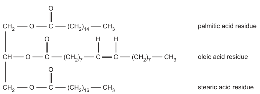
Draw an arrow on Fig. 2.1 to show where the bond between the glycerol residue and the stearic acid residue is broken by lipase digestion.
Answer
Draw an arrow on the lower (stearic acid) fatty acid residue, with the arrowhead between the O of the glycerol side and the C of the C=O group of the ester linkage on the bottom fatty acid chain.
Arrow between the O and the C=O of the ester linkage on the lower (stearic acid) residue.
Background Concept
A triglyceride is built from one glycerol molecule and three fatty acids, joined by three ester bonds of the form -O-C(=O)-. Each ester bond is formed by condensation between an -OH on glycerol and a -COOH on a fatty acid, with the loss of a water molecule. Hydrolysis (the reverse) adds water back across the same bond, regenerating glycerol and free fatty acids — this is what pancreatic lipase catalyses in the small intestine.
In Fig. 2.1 the glycerol backbone is on the left (three carbons: CH2 at the top, CH in the middle, CH2 at the bottom), each carbon being linked via -O-C(=O)- to a fatty acid chain. The bottom chain is the stearic acid residue.
Understanding the Question
The candidate is asked to mark, on the printed structure, exactly where lipase cleaves the bond between glycerol and the stearic acid residue. The mark scheme requires an arrow between the O and the C of the C=O group on the lower (stearic acid) ester linkage.
Approach
- Locate the lower fatty acid residue in Fig. 2.1 — it is the stearic acid residue, -(CH2)16-CH3.
- Identify the ester linkage: -O-C(=O)- joining the bottom CH2 of glycerol to the fatty acid chain.
- Draw the arrow across the O-C single bond, with the arrowhead between the O and the carbonyl C, indicating the bond being broken.
Step-by-Step Reasoning
- An ester bond has the form R-O-C(=O)-R′. The two atoms joined by the single bond that hydrolysis cleaves are the O (from glycerol) and the C (of the carbonyl).
- The C also carries a double bond to a second oxygen (=O), but that double bond is part of the carbonyl group and is not the bond broken by hydrolysis.
- The arrow must be on the lower residue (stearic acid) — not on the palmitic or oleic linkages.
Key Takeaways
- The bond hydrolysed by lipase is the O-C single bond of the ester linkage, not the C=O double bond.
- Read the question carefully: it specifies the stearic acid residue, so the arrow must be on the bottom linkage.
Common Mistakes
- Drawing the arrow on the C=O double bond — this is a carbonyl, not the bond broken by hydrolysis.
- Drawing the arrow on the wrong fatty acid residue (e.g. the palmitic or oleic linkage instead of the stearic one).
- Drawing the arrow between the O and the CH2 of the glycerol — that is not the ester bond.
Things to Be Careful About
- The arrow is an annotation on a printed figure, not a free-hand drawing. It must clearly cross the -O-C(=O)- single bond of the bottom (stearic acid) ester linkage.
State the type of reaction that occurs to produce stearic acid from this triglyceride and name the type of bond that is broken.
type of reaction = ______
type of bond = ______
Answer
type of reaction = hydrolysis
type of bond = ester
type of reaction = hydrolysis; type of bond = ester
Background Concept
Triglycerides are formed by condensation between glycerol and three fatty acids. Each condensation produces an ester bond and releases a molecule of water. The reverse reaction — adding water back across the bond to split the molecule into glycerol and free fatty acids — is hydrolysis and is the reaction catalysed by lipases.
The reaction is reversible:
The forward direction is condensation (esterification); the reverse direction is hydrolysis (with the C-O single bond of the ester linkage cleaved).
Understanding the Question
The candidate is told that lipase breaks a bond in a triglyceride to release a fatty acid (stearic acid). They must name the type of reaction producing the free fatty acid and the type of bond that is broken.
Approach
This is a two-term recall item. The reaction is hydrolysis (water-adding breakdown); the bond is the ester bond between glycerol and the fatty acid.
Step-by-Step Reasoning
- The triglyceride is being split into glycerol and a free fatty acid, with water being added across the broken bond. This is hydrolysis.
- The bond that joins a fatty acid to glycerol in a triglyceride is an ester bond (the same bond formed originally by condensation between an -OH of glycerol and a -COOH of the fatty acid).
- Therefore: type of reaction = hydrolysis; type of bond = ester.
Key Takeaways
- Ester bonds are formed by condensation and broken by hydrolysis.
- A triglyceride contains three ester bonds, one per fatty acid residue.
Common Mistakes
- Writing condensation for the type of reaction — that is the reverse (formation) direction.
- Writing hydrogen bond or covalent bond for the type of bond — too general; ester is the precise term.
- Writing ester linkage or ester bond instead of just ester — usually accepted, but the mark-scheme wording is the single word ester.
Things to Be Careful About
- One mark for each correct term; both must be present and unambiguous.
Investigations were carried out into the activity of a lipase extracted from a strain of bacterium that lives in hot springs.
The activity of the bacterial lipase was measured at using different concentrations of olive oil as the substrate.
The results were used to derive a Michaelis–Menten constant () of .
Explain what is meant by a of .
Answer
A of is the substrate (olive oil) concentration at which the bacterial lipase works at half the maximum rate of reaction () at .
of 91.76 mmol dm⁻³ = the substrate (olive oil) concentration at which the rate of reaction is half the maximum rate (½ Vmax) at 37 °C.
Background Concept
The Michaelis–Menten model describes how the rate of an enzyme-catalysed reaction depends on substrate concentration. As [S] rises, the rate rises and approaches a maximum value, , at which all active sites are saturated. The substrate concentration at which the rate is exactly half of is defined as the Michaelis–Menten constant, .
is often used as an inverse measure of the apparent affinity of the enzyme for its substrate: a low means the enzyme reaches half its maximum rate at a low substrate concentration (high affinity), while a high means a higher substrate concentration is required (lower affinity).
Understanding the Question
The question gives a specific value (with units) for a bacterial lipase acting on olive oil at . The candidate must explain what this number actually means in a biological context, not just recite the textbook definition.
The mark scheme requires two ideas:
- Identification of the variable: substrate (olive oil) concentration.
- The condition under which this concentration applies: the rate of reaction is half the maximum rate, i.e. .
- The context (temperature of ) is part of the experimental setup.
Approach
- Identify the variable from the units ( are concentration units): the substrate (olive oil) concentration.
- Identify the operational meaning: at this concentration, the enzyme works at half its maximum rate, .
- Reference the experimental temperature: (as stated in the stem).
Step-by-Step Reasoning
- The value is given in , which are units of concentration. This tells us is a concentration of the substrate.
- The value is the concentration at which the rate of reaction = — i.e. half the maximum rate achievable when substrate is saturating.
- The investigation was carried out at , so this value applies at that temperature only.
- The mark scheme rejects a statement that is half the maximum rate — is a concentration, not a rate.
Key Takeaways
- has units of concentration (e.g. ), not units of rate.
- is the substrate concentration at which rate = .
- is a property of a particular enzyme-substrate pair at a particular temperature and pH.
Common Mistakes
- Stating " is half the maximum rate" — this is a rate, not a concentration; the mark scheme rejects it.
- Omitting the substrate — saying only "the concentration at which rate is half Vmax" without specifying that it is the substrate (olive oil) concentration.
- Forgetting the experimental temperature context.
- Confusing with (which is a maximum rate, not a concentration).
Things to Be Careful About
- Two separate ideas are required: (1) the quantity being measured (substrate concentration), and (2) the rate condition (). A single sentence that omits either half scores at most one mark.
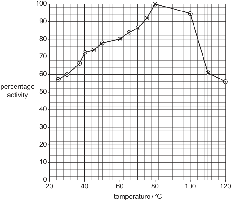
Fig. 2.2 shows the results of an investigation into the effect of temperature on the activity of the bacterial lipase.
Answer
- The optimum temperature is , where the activity peaks at .
- From to , the activity rises (from approximately to ). Increased temperature gives substrate and enzyme molecules greater kinetic energy, so the frequency of successful enzyme–substrate collisions per unit time rises and more enzyme–substrate complexes form per unit time.
- Above , the activity falls (from to approximately at ).
- This decrease is due to (partial / progressive) denaturation of the lipase: the increased thermal vibration breaks some of the bonds (hydrogen, ionic and disulfide) that hold the tertiary structure, distorting the active site so substrate can no longer bind efficiently. The enzyme is not fully denatured at because it is a thermostable enzyme isolated from a bacterium that lives in hot springs — its tertiary structure is more compact and is stabilised by additional bonds, allowing it to retain substantial activity even at very high temperatures.
Optimum 80 °C; activity rises with temperature to 80 °C because of greater kinetic energy and more enzyme–substrate collisions; activity falls above 80 °C because of partial denaturation, but the enzyme is thermostable (from a hot-spring bacterium) so it retains substantial activity at 120 °C.
Background Concept
Enzyme activity is strongly temperature-dependent:
- Below the optimum: as temperature rises, molecules gain kinetic energy. This increases the frequency of successful collisions between enzyme and substrate, so the rate of reaction increases.
- At the optimum: the rate is at its maximum — kinetic energy is high but the enzyme's tertiary structure is still intact.
- Above the optimum: increased vibration begins to disrupt the hydrogen, ionic and disulfide bonds that hold the tertiary structure of the protein. The active site loses its specific shape and can no longer bind substrate efficiently — the enzyme is denaturing. The rate falls sharply, and eventually the enzyme is fully denatured.
Enzymes from organisms that live in extreme environments (thermophiles, hyperthermophiles) are often thermostable: they have additional disulfide bonds, more compact tertiary structures or other adaptations that resist thermal denaturation. Their optimum temperature is correspondingly much higher than that of enzymes from mesophilic organisms such as mammals.
Understanding the Question
Fig. 2.2 shows a temperature–activity curve for a bacterial lipase isolated from a hot spring. The candidate must read the graph, describe the trend, and then explain it in terms of kinetic theory (the rising limb) and denaturation (the falling limb). The unusually high optimum and the high residual activity at must be explained by thermostability of the enzyme.
The four marks come from four distinct points: optimum, rise + explanation, fall + explanation, and the thermostable explanation.
Approach
- Read the optimum: , activity.
- Describe the rise: from to , activity rises from about to .
- Explain the rise: kinetic energy increases → more frequent enzyme–substrate collisions (per unit time) → more enzyme–substrate complexes form per unit time.
- Describe the fall: above activity falls to about at .
- Explain the fall: increased thermal vibration breaks the bonds maintaining tertiary structure, so the active site distorts (partial / progressive denaturation).
- Explain why activity is still substantial at : the enzyme is thermostable, with extra bonds and a more compact structure, adapted to its hot-spring habitat.
Step-by-Step Reasoning
- The graph shows percentage activity (y-axis) vs temperature in (x-axis). The maximum is at .
- Below the optimum the curve rises. The kinetic-theory explanation is needed: more kinetic energy → more successful collisions per unit time → more enzyme–substrate complexes form → higher rate.
- Above the curve falls. The denaturation explanation is needed: heat disrupts the tertiary structure, the active site is lost, so substrate can no longer bind effectively.
- A normal enzyme would be completely denatured well before , so the fact that the bacterial lipase still has activity at must be explained by its thermostable nature — extra disulfide bridges, a more compact tertiary structure, and origin from a hot-spring bacterium.
- The mark scheme explicitly accepts "partial / progressive denaturation" and the ORA (only partial because of thermostability). It also accepts "fewer enzyme–substrate collisions" as an alternative to a denaturation explanation if collision frequency was missed earlier.
Key Takeaways
- Temperature affects enzymes in two opposing ways: kinetic (more collisions) up to the optimum, and structural (denaturation) above it.
- An enzyme's optimum is the temperature at which these two effects balance to give the maximum rate.
- Enzymes from extreme environments (e.g. hot springs) are thermostable, with high temperature optima and resistance to denaturation.
Common Mistakes
- Saying only "the enzyme is more active at higher temperature" — the mark scheme rejects "enzyme more active" because it is a tautology.
- Describing a constant, linear increase right across the whole range (ignoring the fall after the optimum).
- Omitting the kinetic-theory explanation of the rising limb (kinetic energy → collisions → complexes).
- Saying "more peptide bonds" make the enzyme thermostable — peptide bonds are the backbone of all proteins, so adding more cannot make an enzyme more stable. The mark scheme rejects this.
- Stating that the enzyme is fully denatured at — the curve shows it still has about activity, so the denaturation is only partial.
Things to Be Careful About
- The mark scheme requires that the kinetic-theory explanation of the rising limb be in the context "only up to " — do not extend it past the optimum.
- Quoting both the optimum temperature and the residual activity at makes the answer concrete and evidence-based.
An investigation using human pancreatic lipase at the same range of temperature will produce different results from those shown in Fig. 2.2.
Predict how the trend will be different from that shown in Fig. 2.2.
Answer
Human pancreatic lipase would be active over a much narrower range of temperature, with its optimum (peak activity) at approximately to . The rise from to the optimum would be steeper, and the fall after the optimum would be much steeper too, with the activity decreasing to (complete denaturation) at temperatures only a little above the optimum.
Narrower temperature range, optimum at 35–40 °C, steeper rise and fall, activity falls to zero above the optimum (complete denaturation).
Background Concept
Mammalian enzymes are adapted to function at body temperature, which for humans is around . The optimum temperature of a human enzyme is therefore typically between and , and the enzyme is much more sensitive to heat than a thermostable bacterial enzyme. Outside a narrow band around the optimum, mammalian enzymes denature quickly and lose all activity. This is one reason why a sustained high fever is dangerous — vital enzymes begin to denature.
Understanding the Question
The question asks the candidate to predict how a graph of percentage activity vs temperature for human pancreatic lipase would differ from the bacterial lipase graph in Fig. 2.2. The prediction must be specific and comparative.
The mark scheme accepts any two of:
- a narrower range of temperatures over which the enzyme is active;
- a peak / optimum between and ;
- a steeper rise before the optimum and a steeper fall after it;
- a fall to activity (i.e. complete denaturation) above the optimum.
Approach
- Place the optimum: (because the enzyme is human and operates at body temperature).
- Comment on the width of the active range: much narrower than the bacterial lipase.
- Comment on the shape: steeper rise to the optimum, steeper fall after the optimum.
- Comment on the outcome at high temperature: complete denaturation, activity = (this is why the curve would end at zero rather than at ).
Step-by-Step Reasoning
- Human pancreatic lipase has evolved to work at ; its tertiary structure is stable only within a narrow band around this temperature.
- Below the rate still rises with temperature (kinetic theory), but the window of useful temperatures is small.
- Above about the rate falls steeply as denaturation sets in — the bonds in the tertiary structure are much less heat-resistant than those in the bacterial enzyme.
- By about a mammalian enzyme is usually completely denatured, so the predicted curve would hit activity well before the end of the bacterial curve.
Key Takeaways
- Mammalian enzymes have a much lower optimum temperature than thermostable bacterial enzymes.
- Mammalian enzymes have a narrower active range and denature completely a small distance above their optimum.
- The contrast between Fig. 2.2 and the human-pancreatic-lipase prediction is a classic example of enzyme adaptation to environmental temperature.
Common Mistakes
- Predicting the same optimum as the bacterial lipase () — the human enzyme would denature at this temperature.
- Predicting an optimum below (e.g. ) — human body temperature is higher, so the optimum is higher.
- Saying the curve would be "the same but lower" — the shape is fundamentally different.
- Saying the activity would still be "around " at high temperatures — mammalian enzymes are completely denatured, not partially.
Things to Be Careful About
- Use comparative language: narrower range, steeper rise, steeper fall, lower optimum, complete denaturation.
- Two clear comparative points are required for the two marks.
Fig. 3.1 is a scanning electron micrograph of a pair of human chromosomes in a stage of the mitotic cell cycle.
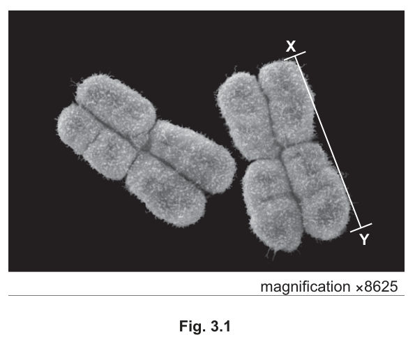
Calculate the actual length, to the nearest , of the chromosome in Fig. 3.1 indicated by the line X–Y.
Write the formula you used to make your calculation.
formula
actual length = ______
Working
Measured length of X–Y on the micrograph = =
Answer
actual length =
5.2 µm
Background Concept
Magnification describes how much larger (or smaller) an image is than the actual specimen. The relationship is:
Rearranged to find the actual size of a specimen from an electron micrograph:
The image size must always be measured in the same units as the desired answer (or converted to those units). For a chromosome typically measured in micrometres (), the image length taken from the micrograph (in mm) must be converted to µm by multiplying by 1000 before dividing by the magnification. Alternatively, the actual size can be expressed in mm by dividing the image size (in mm) by the magnification, and then converted to µm.
Understanding the Question
The question asks the candidate to find the real (actual) length of the chromosome highlighted by line X–Y in Fig. 3.1, a scanning electron micrograph at a stated magnification of . The image provides the magnified length, the magnification is printed on the micrograph, and the candidate must use these to back-calculate the true length of one chromatid in µm to the nearest .
Approach
- Measure the length of line X–Y on the printed micrograph in millimetres (this depends on the candidate's measurement of the printed page).
- Convert mm into µm so units match the required answer.
- Divide the converted image size by the magnification stated on the micrograph ().
- Round to the nearest .
Step-by-Step Reasoning
- A representative measurement of X–Y on the printed micrograph is . Converting to µm: .
- The magnification is (printed on the figure).
- Substituting into the rearranged formula:
- Rounded to the nearest , this is .
- The mark scheme accepts answers from to to allow for small differences in measurement of the printed line X–Y.
Key Takeaways
- The magnification formula and its rearrangement are essential tools in any microscopy-based question.
- Unit conversion (mm ↔ µm) is a common source of error and must always be shown.
- The number of significant figures / decimal places in the final answer must match what the question requests.
Common Mistakes
- Forgetting to convert mm to µm before dividing by magnification — this would give a tiny answer in the wrong units.
- Multiplying instead of dividing (using the image size magnification), which is a common error when the candidate is unsure which way round the formula works.
- Not writing the formula explicitly, even though the question demands it.
- Quoting an unrounded answer (e.g. ) when one decimal place is requested, or rounding to the wrong number of decimal places.
Things to Be Careful About
- The line X–Y is along a single chromatid, not the full chromosome. The candidate must measure the line as drawn, not the whole chromosome.
- Always write the formula before the substitution — the mark scheme awards a mark for the formula alone.
- Be consistent with units: if the question asks for µm, the numerator must also be in µm.
Outline one feature of Fig. 3.1 that confirms the microscope used to obtain the image is a scanning electron microscope and not a transmission electron microscope.
Answer
The chromosomes appear three-dimensional / show surface contours (topography); SEM scans the surface of specimens, whereas TEM produces flat 2D images of internal structures from thin sections.
The image is three-dimensional / shows surface topography, which is characteristic of SEM (TEM produces 2D images of thin sections).
Background Concept
Electron microscopes use a beam of electrons (rather than light) to image specimens, allowing much higher resolution than light microscopes. There are two main types:
- Transmission electron microscope (TEM): electrons pass through an ultra-thin section of the specimen. The image is 2D and shows internal structures as varying shades according to how many electrons pass through.
- Scanning electron microscope (SEM): electrons scan across the surface of the specimen, and secondary electrons emitted from the surface are detected. The image shows 3D surface topography (contours, bumps, texture) but no internal detail.
Understanding the Question
The candidate is shown a scanning electron micrograph and asked to identify one observable feature of Fig. 3.1 that tells us the microscope used was a SEM and not a TEM. The image is provided, so the answer must be something visible in the figure itself, not a general fact about SEMs.
Approach
Look at Fig. 3.1 and decide which characteristic of SEM (and not TEM) is apparent. The figure shows two chromosomes that look solid, with shadowed surfaces, depth, and visible texture on the outer surface of each chromatid.
Step-by-Step Reasoning
- Each chromosome in Fig. 3.1 has visible 3D shape: light and shadow on the chromatid surfaces give a strong impression of depth.
- The surface texture (small bumps, fuzzy projections from the chromosome surface) is visible — this is surface topography, exactly what SEM reveals.
- No internal structure is visible (no dark/light contrast showing the inside of the chromatid), which is consistent with SEM (a TEM would show internal detail in a thin section).
- Any one of these observations is sufficient to score the mark.
Key Takeaways
- SEM = 3D, surface, opaque, depth of field, no internal detail.
- TEM = 2D, internal, thin sections, dark/light contrast from electron transmission.
- When asked to identify the type of EM from an image, pick an observable visual feature — not an invisible property.
Common Mistakes
- Stating a fact about SEMs that is not actually visible in the figure (e.g. "it has a higher resolution than a light microscope") — this would not be evident from the image and would not score.
- Confusing the two types: describing a SEM feature as if it were a TEM feature (e.g. "you can see internal structure") — this is the opposite of the truth and scores zero.
- Vague wording such as "it looks more detailed" or "it looks better" — the mark scheme wants specific features (3D, surface contours, depth of field, no internal detail).
Things to Be Careful About
- The image is small, so the candidate must look carefully for the 3D / surface appearance. The fuzzy outline and the shading on the chromatids are the strongest visual clues.
- The mark scheme accepts several equivalent phrasings (e.g. "3D image", "good depth of field", "surface contours visible", "no internal detail"); any one of these scores the mark.
With reference to Fig. 3.1, explain how it is possible to deduce that DNA replication has already occurred.
Answer
Each chromosome is made up of two sister chromatids joined at a centromere. Sister chromatids are produced by DNA replication during S phase of interphase, so the presence of two chromatids per chromosome confirms that replication has already occurred.
Each chromosome consists of two sister chromatids, which are the result of DNA replication during S phase.
Background Concept
During the cell cycle, DNA is replicated during S phase (the synthesis phase) of interphase, before mitosis begins. The result of replication is that each chromosome now consists of two identical sister chromatids held together at the centromere. Each chromatid contains one DNA double helix (with associated histone proteins), and the two chromatids are genetically identical because they are copies of the same original DNA molecule.
This two-chromatid structure persists through prophase, metaphase and into anaphase of mitosis, when the sister chromatids finally separate and are pulled to opposite poles of the cell.
Understanding the Question
The candidate is shown a micrograph of two chromosomes, each clearly made of two chromatids joined at a centromere. The question asks for the observable feature in the figure that shows DNA replication has already taken place. The key is to link what is visible (two chromatids) to the event that must have produced it (S phase of interphase).
Approach
Identify the most direct piece of evidence visible in the figure: the presence of two sister chromatids per chromosome. Connect this to the event that produces sister chromatids — DNA replication during S phase.
Step-by-Step Reasoning
- A unreplicated chromosome (e.g. in G1 phase) consists of a single DNA molecule and appears as a single chromatid under the microscope.
- In Fig. 3.1, each chromosome clearly shows two sister chromatids joined at a centromere.
- Sister chromatids are formed only when the original DNA molecule is copied, which happens in S phase.
- Therefore, the two-chromatid appearance is direct evidence that DNA replication has already occurred.
- Alternative acceptable answers (from the mark scheme) are: a centromere is visible in each chromosome; or the cell is at late prophase / prometaphase / metaphase — all stages that occur after S phase has been completed.
Key Takeaways
- The two-chromatid structure of a chromosome is the visible signature of completed DNA replication.
- S phase (synthesis) occurs in interphase, before any of the visible stages of mitosis.
- Linking visible structure to an invisible molecular event is a key skill in cell biology questions.
Common Mistakes
- Saying "two DNA molecules are present" without mentioning that they are the result of replication. The mark scheme explicitly ignores "two identical DNA molecules" as a standalone answer — the candidate should refer to the visible chromatids.
- Confusing the cell cycle stage: the candidate may say "the cell is in anaphase" or "the chromosomes are separating" — but in Fig. 3.1 the chromatids are still joined at the centromere, so the cell is in prophase / metaphase, not anaphase.
- Vague answers such as "it has chromosomes" or "the nucleus has divided" — these do not specifically indicate that DNA replication has occurred.
Things to Be Careful About
- Use the term sister chromatids (the two identical copies joined at the centromere), not "two chromosomes" — chromosomes and chromatids are distinct.
- DNA replication is the copying of the DNA; the appearance of two chromatids is the consequence of that copying.
- The centromere is a feature of an already-replicated chromosome; its presence is also accepted by the mark scheme as evidence.
A number of species in the genus Plasmodium are known to cause malaria in humans.
Plasmodium has a complex life cycle with a number of different structural forms. The merozoite is one form of the pathogen that is present in human hosts.
Answer
Protoctist (Protoctista)
Protoctist
Background Concept
Plasmodium is the genus of single-celled parasites responsible for malaria in humans. Although Plasmodium is eukaryotic (it has a true nucleus and membrane-bound organelles such as mitochondria and an endoplasmic reticulum), it does NOT belong to the kingdoms Animalia, Plantae, Fungi, Prokaryotae or Virus. The kingdom to which it is assigned at AS Biology is the Protoctista (sometimes written Protoctista or Protista) — the group containing mostly single-celled eukaryotes that are not fungi, animals or plants (e.g. Amoeba, Paramecium, Plasmodium and Trypanosoma). Other important diseases caused by protoctists include sleeping sickness (Trypanosoma) and amoebic dysentery (Entamoeba).
Understanding the Question
The command word is "state" — give a single, factual, mark-scheme term. The question asks for the type (i.e. the kind of organism) that causes malaria, not a description of what it does.
Approach
Recognise that Plasmodium is not a bacterium (so not Prokaryotae), not a fungus, not an animal in the multicellular sense, and not a virus. Recall its kingdom and give the single word/term expected.
Step-by-Step Reasoning
- "Type of organism" at AS Biology level means the kingdom (or equivalent grouping) of the pathogen.
- Plasmodium is a single-celled eukaryote, so it is neither prokaryote nor virus.
- The kingdom is Protoctista (also acceptable: Protoctista, with the capital P, or Protoctistae). The mark scheme explicitly accepts "Protoctist" and "Protoctista".
- The mark scheme also IGNORES species names (so writing Plasmodium is not enough) and IGNORES "eukaryote" alone (too vague — a protoctist IS a eukaryote but the question asks for the type of organism, i.e. the kingdom).
Key Takeaways
- Malaria is caused by protoctists of the genus Plasmodium (mainly P. falciparum, P. vivax, P. ovale, P. malariae and P. knowlesi).
- At CIE AS, write the kingdom, not the species or the cell type.
Common Mistakes
- Writing Plasmodium (the genus) — credit is for the type/kingdom, not the species.
- Writing "parasite" — true but too vague; it does not name the kingdom.
- Writing "protozoan" — this is a colloquial sub-group within protoctists; "protoctist" is the accepted term in this syllabus.
- Writing "animal" — Plasmodium belongs to Kingdom Protoctista, not Animalia.
Things to Be Careful About
Spell "protoctist" with a 'c' (proto-ctist), not "protist". The mark scheme accepts both spellings but "protoctist" is the form used in the CIE syllabus.
Fig. 4.1 is a scanning electron micrograph of a red blood cell infected with merozoites. Debris (waste particles) from the preparation of the electron micrograph is also shown.
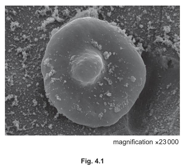
State the difference between the appearance of the red blood cell shown in Fig. 4.1 and a healthy red blood cell.
Answer
The infected red blood cell has lost its (full) biconcave shape — it has a bulge / is swollen in the centre (rather than the central dimple of a healthy red blood cell).
The red blood cell has a bulge in the centre / is swollen in the centre / has lost its (full) biconcave shape.
Background Concept
A healthy human red blood cell (erythrocyte) is a biconcave disc — a flattened disc with a central depression (dimple) on each face, and no nucleus. The biconcave shape gives a high surface area to volume ratio for gas exchange and allows the cell to deform and squeeze through capillaries. When Plasmodium merozoites invade and develop inside a red blood cell, the cell is changed both in shape and contents. The parasite digests haemoglobin and uses red blood cell proteins for its own growth, and the cell often swells as the parasite develops.
Understanding the Question
You are given a scanning electron micrograph (SEM) of a single red blood cell at ×23 000 magnification. The cell shows one large rounded bulge in its centre and a generally lumpy/irregular outline, plus some small debris particles from specimen preparation. The question asks for a single difference between the appearance of this cell and that of a healthy red blood cell.
Approach
- Recall the appearance of a normal red blood cell (biconcave disc, central dimple, smooth round outline).
- Compare that to the image: there is a clear rounded bulge in the centre and the cell is misshapen/lumpy overall.
- State the single most obvious visible difference, in the precise wording the mark scheme credits.
Step-by-Step Reasoning
- Healthy red blood cell: smooth circular outline, biconcave (a clear depression/dimple on each side).
- Image of infected cell: the centre is bulged out (not dimpled); a large spherical bulge is obvious in the middle of the cell, and the cell is also irregular/misshapen.
- The mark scheme credits: "bulge in centre / swollen in centre / lost (full) biconcave shape" — and accepts "misshapen / shape not regular" as alternatives.
- A single, clear visible difference is all that is required for the 1 mark.
Key Takeaways
- Merozoite-infected red blood cells change shape: the central dimple is lost and replaced by a bulge or an irregular outline.
- SEM images show 3-D surface topography, so a bulge in the centre is the most obvious feature.
Common Mistakes
- Stating the difference is the presence of merozoites on the surface — they are not visibly sitting on the surface in this image; the visible feature is the bulge.
- Stating vague things like "the cell looks different" or "it is infected" — the mark requires a specific visible feature.
- Confusing the cellular bulge with the central dimple of a normal cell.
Things to Be Careful About
The question asks for one difference, so a single clear point is enough. The mark scheme rejects anything that is not directly visible in the image.
When Plasmodium is present within mature red blood cells there is a decrease in concentration of haemoglobin in these infected cells.
Suggest why the concentration of haemoglobin within the infected red blood cells decreases.
Answer
The merozoites break down (digest) the haemoglobin to release amino acids, which they use to grow / to synthesise their own (Plasmodium) proteins.
Merozoites break down haemoglobin to produce amino acids, which they use to synthesise other proteins / for growth.
Background Concept
Haemoglobin is the oxygen-carrying protein that fills a healthy red blood cell. Plasmodium merozoites, once inside the red blood cell, cannot make all the amino acids they need — they obtain many of them by digesting the host's haemoglobin. The parasite digests haemoglobin inside a food vacuole, releasing free amino acids (and toxic haem, which the parasite sequesters as haemozoin). The released amino acids are then used by the parasite to make its own proteins and to grow. As a result, the concentration of haemoglobin in the infected red blood cell falls over the parasite's intra-erythrocytic cycle.
Understanding the Question
The stem says haemoglobin concentration decreases inside infected red blood cells. The command word is "suggest" — this means propose a reasonable biological explanation. Only 1 mark is available, so one clear point is needed.
Approach
Think about what the parasite needs (amino acids for protein synthesis) and what resource is plentiful inside the red blood cell (haemoglobin, which is a protein). Connect the two: the parasite breaks down haemoglobin to obtain the amino acids it needs.
Step-by-Step Reasoning
- Haemoglobin is a protein — when broken down, it yields amino acids.
- The parasite must grow and synthesise new proteins (e.g. MSP1, membrane proteins) inside the host cell.
- Therefore, the parasite digests haemoglobin → amino acids → used to build Plasmodium proteins / for parasite growth.
- The net result is a falling concentration of haemoglobin inside the infected cell.
- A mark-scheme-acceptable point is therefore: "(merozoites) break down (haemoglobin) to produce amino acids" AND/OR "(merozoites) use the amino acids for growth / to synthesise other proteins".
Key Takeaways
- Plasmodium is a haemoglobin-consumer inside the red blood cell.
- A fall in host protein is a direct consequence of the parasite's feeding strategy.
Common Mistakes
- Writing only "the parasite uses haemoglobin for energy" — rejected by the mark scheme. The protein part of haemoglobin is the source of amino acids; Plasmodium does not significantly oxidise haemoglobin for ATP — it digests the globin for amino acids.
- Writing "the parasite destroys the red blood cell" — too vague, and haemoglobin falls well before the cell bursts.
- Writing "haemoglobin leaks out" — the cell membrane is intact during most of the intra-erythrocytic stage, so leakage is not the main cause.
Things to Be Careful About
Only 1 mark is available, so give a single tight point. "Suggest" questions often reward any biologically reasonable idea but in this case the mark scheme lists specific accepted answers — phrase yours as either "break down haemoglobin to amino acids" or "use amino acids to make proteins / for growth".
A protein known as merozoite surface protein 1 (MSP1) is found as part of the cell surface membrane of merozoites. All species of Plasmodium that cause malaria have MSP1. The protein is used to help Plasmodium enter human red blood cells.
MSP1 is composed of a single polypeptide, which is coded for by gene MSP1.
Some of the steps occurring in the synthesis of MSP1 by Plasmodium are described in Table 4.1. They are not listed in the correct sequence.
Table 4.1
| step | description |
|---|---|
| A | tRNA, following amino acid activation, attaches to ribosome |
| B | mRNA passes through nuclear pores |
| C | a stop codon is reached and the polypeptide is released |
| D | DNA double helix unwinds |
| E | codon–anticodon binding occurs |
| F | RNA polymerase forms phosphodiester bonds |
| G | peptide bond formation occurs |
| H | mRNA attaches to ribosome |
Use the steps described in Table 4.1 to complete Table 4.2 to show the correct sequence of events as they would occur in the synthesis of MSP1.
Two of the steps have been added to Table 4.2 for you.
Table 4.2
| correct sequence | step |
|---|---|
| 1 | |
| 2 | |
| 3 | |
| 4 | H |
| 5 | |
| 6 | |
| 7 | |
| 8 | C |
Answer
| correct sequence | step |
|---|---|
| 1 | D — DNA double helix unwinds |
| 2 | F — RNA polymerase forms phosphodiester bonds |
| 3 | B — mRNA passes through nuclear pores |
| 4 | H — mRNA attaches to ribosome (given) |
| 5 | A — tRNA, following amino acid activation, attaches to ribosome |
| 6 | E — codon–anticodon binding occurs |
| 7 | G — peptide bond formation occurs |
| 8 | C — a stop codon is reached and the polypeptide is released (given) |
D, F, B, A, E, G (steps 1, 2, 3, 5, 6, 7)
Background Concept
Protein synthesis occurs in two stages:
- Transcription in the nucleus: a section of the DNA double helix unwinds; RNA polymerase synthesises a complementary pre-mRNA strand by forming phosphodiester bonds between free RNA nucleotides; after splicing, the mature mRNA leaves the nucleus through a nuclear pore and enters the cytoplasm.
- Translation in the cytoplasm: the mRNA binds to a ribosome; aminoacyl-tRNAs (tRNAs that have been "charged" with their amino acid by aminoacyl-tRNA synthetases, a process called amino acid activation) enter the ribosome; the codon on the mRNA pairs with the anticodon on the tRNA (codon–anticodon binding); the ribosome catalyses peptide bond formation between adjacent amino acids; this repeats along the mRNA until a stop codon is reached, when the completed polypeptide is released.
MSP1 is a single polypeptide, so once it is released, the chain folds into the secondary and tertiary structure described in part (e).
Understanding the Question
Table 4.1 lists eight steps (A–H) of protein synthesis out of order. Two of the steps (H and C) are already placed in the sequence table (Table 4.2). You need to place the remaining six steps (A, B, D, E, F, G) in their correct positions. There are 4 marks — one mark per correct line that is filled in, and the mark scheme notes a paired credit: ";(for steps 2 and 3)" and ";(for steps 5 and 6)", meaning if you place BOTH 2 and 3 correctly you get one of the four marks.
Approach
Mentally split the steps into two phases — transcription (in the nucleus) and translation (in the cytoplasm). Then arrange each group chronologically:
- Transcription: DNA unwinds (D) → phosphodiester bonds form as RNA polymerase makes the mRNA (F) → mRNA leaves the nucleus (B).
- Translation, after the mRNA binds the ribosome (H, given): activated tRNA attaches (A) → codon–anticodon binding (E) → peptide bond forms (G) → stop codon releases the chain (C, given).
Step-by-Step Reasoning
- The mRNA has to be made before it can leave the nucleus, and it leaves the nucleus before it binds a ribosome in the cytoplasm.
- Transcription steps in the nucleus, in order: D (unwind DNA) → F (RNA polymerase forms phosphodiester bonds to make the mRNA) → B (mRNA leaves the nucleus through a nuclear pore).
- Step 4 is given as H (mRNA binds the ribosome), so step 3 must end transcription.
- After H, the first cytoplasmic event is an activated tRNA arriving at the ribosome (A). Note that tRNAs are charged with their amino acid by aminoacyl-tRNA synthetase in the cytoplasm BEFORE they bind the ribosome — "amino acid activation" is the energy-requiring step that attaches the correct amino acid to its tRNA.
- Once the tRNA is in place, its anticodon pairs with the codon on the mRNA (E — codon–anticodon binding).
- The ribosome then catalyses the peptide bond between the new amino acid and the growing chain (G).
- The cycle of A → E → G repeats as the ribosome moves along the mRNA until a stop codon is reached (C, given).
- Mark-scheme check: steps 2 & 3 form one linked credit, and steps 5 & 6 form another linked credit — so getting each pair correct together is the route to all 4 marks.
Key Takeaways
- Transcription order: DNA unwinds → RNA polymerase joins RNA nucleotides by phosphodiester bonds → mRNA leaves through nuclear pores.
- Translation order: mRNA binds ribosome → aminoacyl-tRNA enters → codon–anticodon pairing → peptide bond formation → stop codon releases the polypeptide.
- The amino acid is activated (charged onto its tRNA) BEFORE the tRNA enters the ribosome, so step A precedes E.
Common Mistakes
- Placing A before B (i.e. before the mRNA is in the cytoplasm) — translation cannot begin until mRNA has left the nucleus.
- Putting E before A — codon–anticodon pairing only happens once a charged tRNA is in the ribosome.
- Putting B after H — the mRNA must reach the cytoplasm before binding a ribosome, so B is the last transcription event and comes before H.
- Treating F (phosphodiester bonds) as a translation step — it is the mRNA being synthesised in the nucleus, not peptide bonds.
- Forgetting that mRNA has to be exported from the nucleus to the cytoplasm in a eukaryote like Plasmodium.
Things to Be Careful About
The mark scheme groups steps 2–3 and 5–6 as paired marks: 1 mark for both correct, 1 mark for both correct. Therefore, if you swap the two steps in either pair (e.g. write B then F, or E then A), you lose that pair's mark. Within the pairs the order is fixed because RNA polymerase must act on the unwound DNA BEFORE the finished mRNA can leave the nucleus, and a charged tRNA must be in the ribosome BEFORE its anticodon can pair with the codon.
The MSP1 protein has three levels of protein structure: primary, secondary and tertiary.
Outline the changes that occur after translation to a polypeptide, such as the MSP1 polypeptide, that result in a protein showing secondary and tertiary structure.
Answer
- The polypeptide chain folds / coils upon itself, giving regions of secondary structure such as α-helices and β-pleated sheets (stabilised by hydrogen bonds between the –N–H and –C=O groups of the polypeptide backbone).
- The chain then folds further into a specific 3-D tertiary structure, held together by interactions / bonds between the R-groups (side chains) of different amino acids.
- Named examples of these R-group interactions include: hydrogen bonds, ionic / electrostatic bonds (between –NH₃⁺ and –COO⁻ groups), disulfide bridges (between –SH groups of cysteine), and hydrophobic interactions (between non-polar R-groups).
Secondary structure: folding of the polypeptide into α-helices and β-pleated sheets. Tertiary structure: further folding held by bonds between R-groups (hydrogen, ionic, disulfide bridges, hydrophobic interactions).
Background Concept
A newly synthesised polypeptide is a linear chain of amino acids joined by peptide bonds. To become a functional protein it must fold into a specific 3-D shape.
- Primary structure = the linear sequence of amino acids held by peptide bonds.
- Secondary structure = regular, repeating patterns of folding of the polypeptide backbone, stabilised by hydrogen bonds between the H of an N–H group and the O of a C=O group further along the chain. The two main motifs are the α-helix (a right-handed coil, with H-bonds every 3.6 amino acids) and the β-pleated sheet (polypeptide strands lying side-by-side, held by H-bonds between them).
- Tertiary structure = the further folding of an already-folded region into a specific 3-D shape, held by interactions between the R-groups (side chains) of amino acids. The four main types of R-group interaction are:
- Hydrogen bonds between polar R-groups (e.g. –OH groups).
- Ionic / electrostatic bonds between charged R-groups (e.g. –NH₃⁺ and –COO⁻).
- Disulfide bridges / bonds between two –SH groups of cysteine residues (covalent S–S bonds).
- Hydrophobic interactions between non-polar R-groups, which cluster in the interior of soluble proteins.
(Quaternary structure, when several polypeptide chains combine, is not relevant for MSP1 because it is a single polypeptide — the question already notes this.)
Understanding the Question
The stem tells you MSP1 has three levels of structure (primary, secondary, tertiary). The command word is "outline" — give the main, organised points without lengthy detail. 4 marks means four clear, distinct points. The mark scheme specifically REJECTS R-group bonds when linked to secondary structure, and REJECTS peptide bonds when describing the bonds that hold tertiary structure together.
Approach
Plan your answer in two clean blocks:
- A block on secondary structure — folding, the named motifs, and that they are stabilised by H-bonds in the backbone.
- A block on tertiary structure — further folding, bonds between R-groups, and at least two of the four named R-group bond types.
Step-by-Step Reasoning
- Point 1 (1 mark): the polypeptide folds / coils — and is therefore now in secondary / tertiary structure. "Folding" is the headline idea.
- Point 2 (1 mark): the secondary structure includes named motifs — α-helices OR β-pleated sheets. Either is enough; the mark scheme accepts both and any equivalent spelling (β-pleated sheets / β-pleats; R "beta-plates").
- Point 3 (1 mark): in tertiary structure, bonds / interactions form between the R-groups (side chains) of different amino acids. The mark scheme rejects answers that put R-group bonds into secondary structure.
- Point 4 (1 mark): at least two named bonds from the list — hydrogen, disulfide, ionic/electrostatic, hydrophobic. Don't include peptide bonds or "covalent bonds" as a generic answer.
- A bonus point (AVP) is available for additional correct detail, e.g. describing exactly where H-bonds in the α-helix form (between the H of N–H and the O of C=O) or the pattern of H-bonds (every 3.6 residues).
Key Takeaways
- Secondary structure is about the backbone; tertiary structure is about the R-groups.
- The four R-group interactions are: hydrogen, ionic, disulfide, hydrophobic.
- "Folding" is the one-word headline for what happens after translation.
Common Mistakes
- Calling the secondary-structure H-bonds "R-group bonds" — they are backbone H-bonds.
- Including "peptide bonds" in the list of tertiary-structure interactions — peptide bonds hold the primary structure, not the tertiary.
- Writing "van der Waals forces" or "hydrophilic interactions" — both are ignored in the mark scheme for this question.
- Spelling "β-pleated sheet" as "β-plates" or "β-pleats" alone is acceptable (β-pleated sheets is safer).
- Confusing secondary and tertiary structure — the question explicitly asks for both, so two distinct blocks of answer are needed.
Things to Be Careful About
- Use the technical terms (R-group, hydrogen bond, disulfide bridge, ionic bond, hydrophobic interaction). "Bonding" alone is too vague.
- The mark scheme REJECTS bonds credited to the wrong level — get the level right.
- If you only give one named R-group bond, you only score the bond point for the first one named (you need two for the mark).
Subunit vaccines are vaccines that contain non-self antigens, but do not contain whole organisms. The aim is to stimulate a primary immune response after the vaccine is given so that the person gains artificial active immunity.
One trial that has been carried out on human volunteers has used MSP1 from Plasmodium falciparum in a subunit vaccine against malaria.
Explain why a vaccine containing MSP1 provides artificial active immunity to malaria.
Answer
- The MSP1 antigen is recognised as non-self and triggers a primary immune response in which specific B- and T-lymphocytes are activated (clonal selection).
- These selected lymphocytes undergo clonal expansion (many mitoses) to form a large clone, including memory cells (long-lived B- and T-memory cells that remain in the circulation).
- On subsequent exposure to the Plasmodium antigen (the actual merozoite), the memory cells trigger a secondary immune response — faster, larger and with higher levels of antibody produced in a shorter time — which destroys the pathogen before symptoms of malaria develop.
- Because the antigen is injected deliberately as a vaccine rather than caught naturally, and the response is the body's own (lymphocytes make the antibodies), the immunity is artificial (deliberately induced) and active (the body makes its own antibodies and memory cells).
The vaccine contains the non-self MSP1 antigen, which stimulates a primary immune response producing memory cells; on natural exposure the memory cells produce a rapid, large secondary response, giving artificial active immunity.
Background Concept
Active immunity is immunity produced by the body's own immune system, in response to antigens. The body makes its own antibodies and memory cells.
- Natural active immunity follows infection (e.g. catching measles gives life-long immunity).
- Artificial active immunity follows vaccination — the body is deliberately exposed to a harmless form of the antigen.
Passive immunity is immunity "borrowed" from another source, e.g. maternal antibodies crossing the placenta, or injection of antivenom. The body does NOT make its own memory cells, so protection is short-lived.
A vaccine is a preparation of antigen (a whole weakened/killed organism, or part of an organism — e.g. a surface protein) that stimulates a primary immune response:
- Antigen-presenting cells (e.g. macrophages) take up and present the antigen.
- Specific B-lymphocytes (with complementary surface receptors) bind the antigen, are activated with T-helper-cell help, and undergo clonal selection.
- Selected lymphocytes divide many times (clonal expansion) by mitosis, producing a large clone of effector cells (plasma cells that secrete antibody) and long-lived memory cells.
- On a later encounter with the real pathogen (which has the same antigen), the memory cells are quickly reactivated — this is the secondary immune response. It is faster, larger and produces much more antibody than the primary response, so the pathogen is destroyed before it causes disease.
Understanding the Question
The stem tells you that the vaccine contains MSP1 — a non-self protein — and that the resulting immunity is "artificial active". The command word is "explain" — give reasons, not just definitions. 4 marks are available for explaining the mechanism by which a vaccine produces artificial active immunity.
Approach
Structure your answer around the sequence of immune events caused by the vaccine, and finish by linking the speed/size of the secondary response to the absence of symptoms:
- Antigen recognition + clonal selection (the trigger).
- Clonal expansion (the production of effectors and memory cells).
- Memory cells are long-lived (so protection persists).
- On real infection, the secondary response is faster/larger — pathogen is cleared before disease (this is what immunity means in practice).
Step-by-Step Reasoning
- Mark point 1 (memory cells formed) — the headline outcome of vaccination: long-lived B- and T-memory cells are produced and persist in the body.
- Mark point 2 (clonal selection details) — the antigen is processed/antigen presented; specific B- and T-lymphocytes recognise and bind the antigen; this triggers the lymphocytes to be activated. ("MSP1 stimulates B-cell to divide" alone counts as point 2 not point 3 — credit it as the activation step.)
- Mark point 3 (clonal expansion) — once activated, the specific lymphocytes divide many times by mitosis, making a large clone of plasma cells (which secrete antibody) and memory cells.
- Mark point 4 (memory cells are long-lived) — the B- and T-memory cells remain in circulation for years ("remain in the body" alone is rejected; "long-lived" or "in circulation" is needed).
- Mark point 5 (secondary response on real exposure) — when the person is later bitten by an infected mosquito, the real Plasmodium antigen (MSP1 on the merozoite) is recognised, triggering a secondary immune response.
- Mark point 6 — because the clone of specific lymphocytes is already present and very large, the response is faster / greater; more plasma cells, more antibody, more T-helper cells. The mark scheme also accepts "increased chance of fast recognition of antigen".
- Mark point 7 — therefore higher levels / faster production of antibody, which neutralises the merozoite before it can establish a malaria infection.
- Mark point 8 (AVP) — e.g. antibodies binding MSP1 prevent the merozoite entering the red blood cell (the precise protective mechanism in this case).
- Finally, tie the chain back to the type of immunity: because the antigen is deliberately injected as a vaccine (not caught naturally) and the body makes its own antibodies and memory cells, the immunity is artificial and active.
Key Takeaways
- A vaccine works by generating memory cells in a primary response, so that the real pathogen triggers a larger, faster secondary response.
- Artificial = the antigen is administered deliberately (vaccination).
- Active = the body makes its own antibodies and memory cells.
- The MSP1 protein in the subunit vaccine is a surface antigen of the merozoite, so antibodies raised against it should block merozoite entry into red blood cells.
Common Mistakes
- Confusing artificial active with artificial passive — passive would mean injecting ready-made antibodies, which the vaccine does NOT do.
- Saying "vaccines give you antibodies" — the vaccine makes the body produce its own antibodies, that is the active component.
- Failing to mention memory cells — these are the essential output of vaccination and the basis of long-term protection.
- Saying the immune system "kills the pathogen immediately on vaccination" — the vaccine contains only a non-self protein (subunit), not the whole pathogen, so it does not cause disease; protection only becomes visible on later exposure.
- Stating that antibodies are produced by T-lymphocytes — antibodies are produced by plasma cells, which are differentiated B-lymphocytes.
Things to Be Careful About
- The mark scheme rejects "kept in the body" alone — memory cells must be described as long-lived or remaining in circulation.
- "Live longer" is rejected unless it is made clear that they are present when the real antigen arrives (i.e. they persist long enough).
- Make sure the link from memory cells to faster / more antibody on re-exposure is explicit — the question asks for an explanation of immunity, not just a list of events.
The female Anopheles mosquito is the vector of Plasmodium.
Discuss the ways in which the vector is controlled to help prevent the transmission of malaria.
Answer
- Prevent the female mosquito from feeding on human blood (she needs a blood meal to mature her eggs), so the Plasmodium life cycle is broken. Examples: insecticide-treated bed nets (ITNs / LLINs) used while sleeping; long-sleeved clothing covering bare skin; insect repellents on skin or clothes; indoor residual spraying (IRS) of insecticides on walls where mosquitoes rest.
- Kill adult mosquitoes with insecticides / pesticides, e.g. spraying inside homes, treating bed nets with insecticide, or aerial spraying of wetlands.
- Disrupt breeding by attacking the aquatic stages (eggs, larvae, pupae): remove or cover standing water (e.g. cover water tanks, drain ponds); spray oil on the surface of small bodies of water to suffocate larvae; introduce biological control agents such as mosquitofish that eat the larvae.
- AVP — e.g. release sterile (irradiated) male mosquitoes so that matings produce no offspring; treat the water with drugs that kill Plasmodium inside the mosquito, preventing onward transmission.
Adult control: bed nets, repellents, indoor residual spraying, insecticides. Larval control: remove/cover standing water, oil on ponds, biological control (e.g. mosquitofish). AVP: sterile males, drugs inside the mosquito.
Background Concept
Malaria is transmitted by the bite of an infected female Anopheles mosquito. The mosquito is therefore the vector. The life cycle of the mosquito has four stages: egg → larva → pupa → adult, with the first three in water and only the adult flying. The female needs a blood meal to mature her eggs. Plasmodium itself completes its sexual reproduction inside the mosquito, so the mosquito is essential to the parasite's life cycle.
Vector control aims to break the chain of transmission. It can be attacked at different points:
- Before the mosquito bites a person — by repelling or killing adults, or by physical barriers.
- Before the mosquito breeds — by removing or treating the water where eggs are laid and larvae develop.
- By making matings unproductive — e.g. releasing sterile males.
- By killing the parasite inside the mosquito — e.g. endectocides (drugs that kill Plasmodium in the mosquito when the mosquito feeds on a treated person).
Understanding the Question
The command word is "discuss" — at AS Biology this means present the main approaches with an example each. 3 marks are available, so the mark scheme expects three distinct points, each usually paired with a named example.
Approach
Group the methods by what they target:
- The adult mosquito before it bites (barriers/repellents).
- The adult mosquito with chemicals (insecticides).
- The aquatic stages (eggs, larvae, pupae) by removing or treating the water.
- Optional AVP — biological/genetic methods (sterile males, drug-treated water).
Step-by-Step Reasoning
- Mark point 1 (prevent blood meals) — only the female mosquito takes blood (to mature eggs), and Plasmodium is transmitted by her bite. Stop her feeding, and the life cycle is broken. Examples that earn the linked mark: mosquito nets (used at night / while sleeping); long-sleeved clothing (covering bare skin as a barrier); insect repellents (qualified — on body or clothes); indoor residual spraying of walls.
- Mark point 2 (use of insecticides / pesticides) — kill adult mosquitoes. Examples: insecticide-treated bed nets (ITNs / LLINs), indoor residual spraying (IRS), aerial sprays over wetlands. Mosquito coils and insect repellents count as repellents, not as insecticides, so they are credited under mark point 1 only once.
- Mark point 3 (treat water to disrupt egg-laying, eggs, larvae or pupae) — the aquatic stages are particularly vulnerable because they are concentrated and unable to fly away. Examples: remove or cover standing water (water tanks, ponds — NOT rivers); spray oil on ponds / small lakes / swamps to suffocate larvae; use biological control such as mosquitofish (Gambusia) that eat mosquito larvae; release Bti (a bacterial larvicide).
- Mark point 4 (AVP) — more advanced methods: release sterile (irradiated) males so matings produce no offspring; treat the mosquito's water with drugs that kill Plasmodium inside the mosquito.
Key Takeaways
- Vector control hits the mosquito at different points: adult (before she bites), aquatic (eggs, larvae, pupae), and reproduction (sterile males).
- The most effective measures target the adult female because she is the only one that bites and transmits Plasmodium.
- Insecticide-treated bed nets (ITNs / LLINs) are one of the most cost-effective public-health interventions against malaria.
- Standing water is the breeding ground — environmental management that removes it is a cheap, long-term control method.
Common Mistakes
- Mentioning only one method (e.g. "use a mosquito net") — "discuss" expects breadth, so cover both adult and aquatic stages.
- Calling the male mosquito the one that transmits the disease — only the female takes a blood meal and therefore only the female transmits Plasmodium.
- Confusing vectors (the mosquito) with pathogens (the Plasmodium protoctist). Vector control targets the mosquito; treatments that target the parasite (e.g. artemisinin) are not vector control.
- Suggesting the use of antibiotics — antibiotics kill bacteria, not mosquitoes.
- Releasing sterile females (ignored by the mark scheme); it must be sterile males.
- Suggesting "covering rivers" (ignored) — mosquitoes do not lay in flowing water; only still/stagnant water.
Things to Be Careful About
- For each control method, give a named example or qualification — the mark scheme marks the example alongside the principle. "Use a net" alone is borderline; "mosquito net at night" is safer.
- Mosquito coils and insect repellents count as repellents/barriers (point 1), not as insecticides (point 2) — credit once, not twice.
- "Aerial sprays" must be qualified (where, e.g. over wetlands) to earn the mark.
The human gas exchange system is responsible for the efficient uptake of oxygen into the blood and for the excretion of carbon dioxide.
The different tissues of the human gas exchange system are each adapted to their specific function.
Ciliated epithelium is a lining tissue found in the human gas exchange system. The tissue is composed of ciliated epithelial cells and goblet cells.
Squamous epithelium is a lining tissue found in the walls of the alveoli.
Squamous epithelial cells do not have cilia, unlike ciliated epithelial cells.
Suggest why ciliated epithelial cells are not suitable as the lining tissue in the walls of alveoli.
No worked solution for this part yet — the official mark scheme is linked at the top of the page.
Goblet cells and mucous glands function to maintain the health of the gas exchange system. They produce mucus that traps pathogens and other particles, such as dust.
Explain why ciliated epithelial cells are also important in maintaining the health of the gas exchange system.
No worked solution for this part yet — the official mark scheme is linked at the top of the page.
The statements in Fig. 5.1 refer to the saturation of haemoglobin with oxygen in blood leaving the alveolar capillaries.
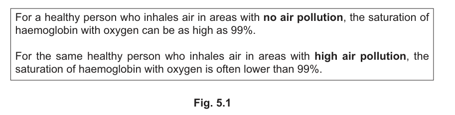
Suggest explanations for the differences stated in Fig. 5.1.
No worked solution for this part yet — the official mark scheme is linked at the top of the page.
The sinoatrial node (SAN), the atrioventricular node (AVN) and the Purkyne fibres have a role in the initiation and control of heart action.
The sinoatrial nodal artery (SN artery) is a branch of one of the main arteries serving the cardiac muscle. Partial blockage of the SN artery as a result of cardiovascular disease can cause the SAN to malfunction.
Answer
Coronary artery
Coronary artery
Background Concept
The muscular wall of the heart (cardiac muscle / myocardium) is far too thick to be supplied by diffusion from the chambers. It has its own dedicated blood supply delivered by the coronary arteries, which branch from the aorta just above the aortic valve. The right and left coronary arteries and their branches run in grooves on the heart surface and penetrate into the muscle to deliver oxygen and glucose to cardiomyocytes.
Understanding the Question
This is a one-mark factual recall. The stem tells you that the sinoatrial nodal (SN) artery is a branch of a main artery serving the cardiac muscle, and asks you to name that parent artery.
Approach
Identify the main artery whose branches deliver oxygenated blood to cardiac muscle, including the specialised conducting tissue of the SAN.
Step-by-Step Reasoning
The SN artery is a small branch that supplies the SAN. As a vessel supplying cardiac muscle, it must arise from the arterial system that perfuses the myocardium — the coronary artery. The coronary arteries arise from the base of the aorta and ramify over and through the cardiac muscle; one of their small branches (the sinoatrial nodal artery) reaches and supplies the SAN.
Key Takeaways
- The coronary arteries supply cardiac muscle.
- The SAN has its own dedicated arterial branch (the SN artery) from this system.
- Blockage of this branch directly compromises the SAN's function.
Common Mistakes
- Writing "aorta" — rejected because the SN artery is a branch of the coronary artery, not directly of the aorta.
- Writing "pulmonary artery" — this carries deoxygenated blood to the lungs and does not supply cardiac muscle.
Things to Be Careful About
Be specific: the marking point is "coronary (artery)", not just any artery.
With reference to the role of the SAN, suggest how a slow rate of ventricular contraction could indicate that the SAN is not functioning correctly.
Answer
- The SAN sets the rate of the heartbeat by initiating impulses that travel as a wave of excitation across the atria and then through the AVN to the ventricles, causing them to contract.
- A slow ventricular contraction rate suggests the SAN is malfunctioning because it is generating impulses at a slower than normal rate (it is no longer setting the correct heart rate as a pacemaker).
The SAN's role as pacemaker is impaired; impulses are generated more slowly so the ventricles, normally stimulated via the AVN after each atrial wave of excitation, contract less frequently.
Background Concept
The sinoatrial node (SAN) is a small patch of specialised cardiac muscle in the wall of the right atrium. It is myogenic — it generates its own electrical impulses (waves of excitation) without needing nerve stimulation. This makes it the heart's natural pacemaker: each impulse spreads across the atria, causing atrial contraction (systole). The impulse reaches the atrioventricular node (AVN), which delays it briefly before passing it down the bundle of His, Purkyne fibres, and ventricular walls to trigger ventricular contraction. The rate at which the SAN fires therefore directly sets the rate at which the ventricles contract.
Understanding the Question
The stem says partial blockage of the SN artery (which supplies the SAN) can cause the SAN to malfunction, and asks you to use this information to suggest — using your knowledge of the SAN's role — why a slow ventricular contraction rate indicates that the SAN is not working correctly. Two marks are available: one for the link to SAN function, and one for describing the conduction pathway to the ventricles.
Approach
First state the SAN's role (pacemaker that initiates impulses). Then describe how those impulses normally reach the ventricles (across atria → AVN → Purkyne fibres → ventricles). Finally, link a slow ventricular rate to a slow/faulty impulse output from the SAN.
Step-by-Step Reasoning
- The SAN acts as the heart's pacemaker, initiating the heartbeat by setting the rate of impulse generation.
- Each impulse spreads as a wave of excitation across the atria, causing atrial contraction, and then passes through the AVN to the ventricles to cause ventricular contraction.
- If the SAN is malfunctioning, it generates impulses at a slower (or abnormal) rate. Because the ventricles can only contract once per arriving impulse, the ventricles will contract at the same slower rate.
- Therefore, observing a slow ventricular contraction rate is consistent with the SAN generating impulses too slowly — i.e. the pacemaker function is faulty.
Key Takeaways
- The SAN is the pacemaker; ventricular rate is set by SAN firing rate.
- The conduction pathway is SAN → atria → AVN → Purkyne fibres → ventricles.
- If the SAN is hypoxic/ischaemic (e.g. from a blocked SN artery), it fires abnormally.
Common Mistakes
- Saying "the SAN sends nerve impulses" — rejected: conduction in the heart is electrical (myogenic), not nervous.
- Saying "the SAN controls the ventricles directly" — oversimplified; it sets the rate but impulses reach the ventricles via the AVN.
- Not making the explicit link between slow SAN firing and slow ventricular contraction.
Things to Be Careful About
Use the precise term wave of excitation or electrical impulses; reject "signals" or "nerve impulses". Make the link between SAN output and ventricular rate explicit — the mark scheme rewards stating that the SAN's pacemaker action is faulty/abnormal.
In people with Wolff–Parkinson–White syndrome, there is a bundle of fibres known as the bundle of Kent. These fibres can conduct impulses from the atria to the ventricles. This means that impulses do not always take the normal route through the AVN to the ventricles.
With reference to the role of the AVN, suggest and explain the change that occurs in the heart rate when impulses pass down the bundle of Kent to the ventricles instead of passing through the AVN.
Answer
- The heart rate / rate of ventricular contraction increases.
- Normally the AVN delays the impulse on its way from the atria to the ventricles, ensuring the atria finish contracting before the ventricles contract. The bundle of Kent bypasses this AVN delay, so impulses reach the ventricles more quickly and the ventricles contract sooner (while the atria are still contracting), producing a faster heart rate.
Heart rate increases because impulses bypass the AVN's normal delay; ventricles contract prematurely/atria contract while ventricles contract.
Background Concept
The atrioventricular node (AVN) sits between the atria and ventricles. Its role is twofold: (1) it is the only electrical bridge between atria and ventricles (the fibrous skeleton of the heart insulates the rest), and (2) it introduces a short delay (~0.1 s) between atrial contraction and ventricular contraction. This delay allows the atria to empty their blood fully into the ventricles before the ventricles contract. If this delay is removed, the atria and ventricles contract almost simultaneously, which is inefficient for pumping.
Understanding the Question
The stem describes Wolff–Parkinson–White syndrome: an extra bundle of fibres (bundle of Kent) allows impulses to pass directly from atria to ventricles, bypassing the AVN. Using your knowledge of the AVN's role, you must (1) state the change in heart rate, and (2) explain why that change happens. Two marks are available: one for identifying the change (heart rate increases), and one for explaining the mechanism (AVN delay is bypassed → premature ventricular contraction).
Approach
First identify what the AVN does. Then consider what removing it from the conduction route (via the bundle of Kent) would do to the timing of ventricular contraction relative to atrial contraction. Finally, translate that earlier ventricular contraction into an increase in heart rate.
Step-by-Step Reasoning
- The AVN's normal role is to delay the impulse travelling from atria to ventricles so that the ventricles contract after the atria have finished contracting.
- In Wolff–Parkinson–White syndrome, impulses from the atria can travel down the bundle of Kent directly to the ventricles, without going through the AVN.
- Because the AVN's delay is bypassed, impulses reach the ventricular muscle earlier than they would normally.
- The ventricles therefore contract too soon — while the atria are still contracting — shortening the cardiac cycle.
- A shorter cardiac cycle means more contractions per minute: heart rate is increased (the ventricles contract at a faster rate).
Key Takeaways
- The AVN delays the impulse to allow sequential atrial-then-ventricular contraction.
- Bypassing the AVN causes premature ventricular contraction and an increased heart rate.
- This illustrates why the AVN delay is essential for efficient, coordinated cardiac action.
Common Mistakes
- Saying the heart rate decreases — wrong; bypassing a delay speeds up the cycle.
- Just saying "the ventricles contract too soon" without linking it to a faster heart rate.
- Describing the bundle of Kent as "another AVN" — it is an abnormal accessory pathway, not a duplicate AVN, and crucially it does not provide the AVN's delay.
Things to Be Careful About
The question says "suggest and explain the change that occurs in the heart rate" — so you must explicitly state the direction of change (increase) AND explain it. Use precise language: "AVN delays the impulse", "bypasses the AVN", "premature ventricular contraction". Avoid vague phrases like "the rhythm is disrupted".