Biology 9700/23 — May/June 2025
Cambridge AS Level · AS Level Structured Questions · worked solutions for every part, with the mark scheme
Topics The Mitotic Cell Cycle · Cell Membranes and Transport · Immunity · Cell Structure · Enzymes · Transport in Plants · +5 more
Ranunculus is a group (genus) of dicotyledonous plants that includes more than 1600 species.
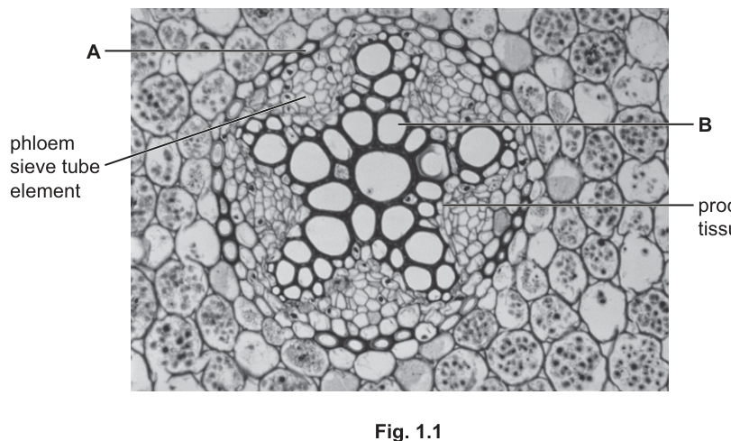
Fig. 1.1 is a photomicrograph of a transverse section through part of a root of a Ranunculus species.
Answer
A = endodermal (cell)
B = xylem vessel element
A = endodermal (cell); B = xylem vessel element
Background Concept
In a transverse section (TS) of a young dicot root, the vascular cylinder (stele) sits in the centre of the root, surrounded by the cortex and the outer epidermis. From the outside in, the stele is bounded by a single layer of tightly packed cells called the endodermis, with a Casparian strip running through the anticlinal walls that regulates the apoplastic movement of water and minerals into the stele. Inside the endodermis lies the pericycle, then the vascular tissues themselves.
In Ranunculus, the xylem occupies the centre of the stele as a star-shaped (or central) mass of large, thick-walled, lignified xylem vessel elements — these are dead at maturity and appear as the biggest, darkest-walled, empty lumens in the centre of the section. Between the arms of the xylem sit small groups of phloem sieve tube elements (smaller, thinner-walled living cells), and between the xylem and phloem lie the small, densely cytoplasmic cells of the procambium.
Understanding the Question
The stem shows a TS photomicrograph of a Ranunculus root. Cell A is labelled on the single ring of cells that encloses the vascular cylinder, and cell B is labelled on one of the large, thick-walled empty cells in the centre of the stele. The candidate simply has to name each cell type. This is a "name" command word question: one correct biological term per cell.
Approach
Recognise that the ring of cells forming the outer boundary of the stele is the endodermis (A), and the large, lignified, empty cell in the centre of the vascular cylinder is a xylem vessel element (B). Use the precise CIE-accepted names from the mark scheme.
Step-by-Step Reasoning
- A: The line points to a single cell forming the innermost layer of the cortex, immediately outside the procambium. This is the endodermis (a single endodermal cell). The mark scheme accepts "endodermal (cell)" as the named answer; bare "endodermis" is also credited as referring to the cell/tissue.
- B: The line points to one of the very large, thick-walled, hollow cells in the centre of the stele. These are lignified, dead at maturity, and conduct water and mineral ions. This is a xylem vessel element. The mark scheme accepts "xylem vessel element", "vessel element" or "xylem element"; bare "xylem" is ignored.
Key Takeaways
- The endodermis is the boundary layer of the stele; it controls which substances enter the xylem.
- The large, thick-walled, empty cells in the centre of a young dicot root TS are xylem vessel elements (lignified, dead at maturity).
- Always use the full tissue-level name, not just the organ name ("xylem vessel element", not "xylem").
Common Mistakes
- Calling B a "tracheid" or a "xylem fibre": Ranunculus is a dicot and its xylem contains vessel elements, not just tracheids.
- Calling A "cortex" (A is the innermost layer of cells, forming a discrete ring — the endodermis, not the broader cortex).
- Writing just "xylem" for B: the mark scheme explicitly ignores this and requires "xylem vessel element" (or equivalent).
Things to Be Careful About
- The endodermis and the pericycle are easy to confuse: pericycle sits just inside the endodermis and gives rise to lateral roots; endodermis sits at the very outer edge of the stele.
- Note the orientation: the question is a TS through a root, not a stem — in a root the xylem is typically central, in a stem it is peripheral (in bundles).
The procambium tissue shown in Fig. 1.1 consists of stem cells.
Suggest a role of the procambium tissue in the roots of this plant.
Answer
The procambium consists of stem cells that divide continuously by mitosis, producing cells that differentiate into xylem and phloem for the growth and repair of the vascular tissue.
Meristematic stem cells divide by mitosis to produce cells that differentiate into xylem and phloem (for growth / repair of vascular tissue).
Background Concept
Procambium (also called procambial tissue or vascular cambium precursor) is a primary meristem — a tissue made up of undifferentiated, actively dividing cells. Meristems are the source of all new cells in a plant: their cells retain the capacity to divide by mitosis indefinitely, and the daughter cells produced can differentiate into the mature cell types of a particular tissue.
In a young root, the procambium lies between the developing xylem and phloem. Its cells divide and the inner derivatives differentiate into more xylem elements while the outer derivatives differentiate into more phloem sieve elements and companion cells. This allows the vascular cylinder to grow thicker and to repair damaged vessels.
Understanding the Question
The question says the procambium "consists of stem cells" and asks the candidate to suggest a role of this tissue in the root. The command word "suggest" allows a reasonable deduction, but the mark-scheme answers are tightly constrained around two ideas: (1) the cells divide by mitosis, and (2) they produce cells that differentiate into xylem/phloem for growth/repair.
Approach
Recognise procambium as a meristem and apply the standard role of a meristem: mitosis → new cells → differentiation into the tissue types of the surrounding organ. Tie this to Ranunculus root context — growth of the vascular cylinder and repair of xylem/phloem.
Step-by-Step Reasoning
- The cells of the procambium are unspecialised and divide continuously by mitosis, generating a pool of new cells.
- These new cells differentiate into cells of the vascular tissues — most importantly xylem vessel elements and phloem sieve tube elements.
- This supplies the root with new vascular tissue for growth of the root and for repair of damaged xylem/phloem.
Any one of these points is sufficient to earn the single mark, but combining the division idea with a function (e.g. "divide to form new xylem for growth") is the cleanest answer.
Key Takeaways
- Procambium is a primary meristem; its defining feature is continuous mitotic division.
- Daughter cells of a meristem differentiate into the functional cell types of the surrounding tissue.
- Meristems sustain growth and enable repair of plant tissues.
Common Mistakes
- Stating only "growth" without linking it to new vascular tissue: too vague for the mark.
- Saying the procambium "transports water" — it does not; it produces the cells that later become water-transporting xylem.
- Confusing procambium with the vascular cambium of a woody root: in a young herbaceous root the procambium is the relevant meristem producing primary vascular tissue.
Things to Be Careful About
- "Suggest" questions still demand precise biological vocabulary — name the tissue formed (xylem / phloem) and the process (mitosis + differentiation); do not give a hand-wavy answer.
Some species in the Ranunculus genus are xerophytes.
State and explain two adaptations of the leaves of xerophytic plants that reduce water loss.
adaptation ______
explanation ______
adaptation ______
explanation ______
Answer
Adaptation 1: Thick (waxy) cuticle
Explanation 1: Increases the diffusion distance for water vapour leaving the leaf (and is largely impermeable to water), so transpiration is reduced.
Adaptation 2: Sunken stomata (stomata in grooves / crypts / surrounded by trichomes)
Explanation 2: Traps a layer of still, humid air around the stomata, reducing the water potential gradient between the leaf interior and the outside air and so reducing transpiration.
(Alternative valid pairs: needle-shaped/narrow leaves → low surface area to volume ratio → less transpiration; low stomatal density → fewer sites of water-vapour loss → less transpiration; multilayered epidermis/hypodermis → increased diffusion distance → less transpiration; rolled/curled leaves → traps humid air near stomata → reduced gradient.)
- Thick waxy cuticle — increases diffusion distance / is impermeable, so reduces water-vapour loss. 2. Sunken stomata (or stomata in crypts / with hairs) — traps humid air, reducing the water potential gradient and so transpiration.
Background Concept
Xerophytes are plants adapted to live in environments where water is scarce (e.g. deserts, salt marshes, exposed rocky outcrops). Their leaves show a suite of anatomical adaptations that reduce transpiration — the loss of water vapour from the leaf surface. Transpiration is driven by a water potential (Ψ) gradient from the moist inner air spaces of the leaf to the drier outside air; water vapour diffuses out mainly through open stomata, and to a lesser extent across the cuticle.
The main ways a leaf can reduce transpiration are:
- Reduce the driving gradient (make the air outside the leaf more humid, or cooler).
- Increase the diffusion pathway (thicken the cuticle, add a hypodermis, fold/roll the leaf so the path is longer).
- Reduce the area available for diffusion (fewer/smaller stomata, smaller leaf surface area).
- Trap a boundary layer of still, humid air around the stomata (sunken stomata, crypts, trichomes, rolled leaves).
Each adaptation must be paired with the physical mechanism by which it lowers water loss — this is what "state AND explain" requires.
Understanding the Question
The candidate must give two xerophytic leaf adaptations, and for each one give a mechanistic explanation of how it reduces water loss. The mark scheme marks each pair (A + E) as a unit, max two pairs = 4 marks. The most commonly credited pairs are thick waxy cuticle, sunken stomata, rolled leaves, needle-shaped leaves, low stomatal density, multilayered epidermis/hypodermis.
Approach
Pick two adaptations whose mechanisms are well understood, state each as a single short phrase, then for each write a one-sentence mechanism in terms of the diffusion pathway, the water potential gradient, or the surface area available for evaporation.
Step-by-Step Reasoning
Pair 1 — Thick waxy cuticle
- Adaptation: Many xerophytes deposit a thick layer of cutin (a waxy polymer) on top of the epidermal cells.
- Explanation: Water vapour must diffuse through this extra layer, so the diffusion distance from the mesophyll to the outside air is greatly increased. Cutin is also largely impermeable to water, so very little water escapes through the cuticle. Net effect: reduced cuticular transpiration.
Pair 2 — Sunken stomata (or stomata in crypts / surrounded by hairs / in grooves)
- Adaptation: The stomata sit in pits below the general leaf surface, often with trichomes (leaf hairs) further slowing the air movement.
- Explanation: Air inside the crypt becomes saturated with water vapour, creating a layer of still, humid air immediately around the stomatal pores. This reduces the water potential gradient between the inside of the leaf and the air just outside the stomata, so the rate of diffusion of water vapour out of the leaf falls. (Note: the mark scheme explicitly ignores "concentration gradient" — the correct term is "water potential gradient".)
Other admissible pairs include:
- Needle-shaped / narrow leaves — small surface area to volume ratio → less area exposed for transpiration.
- Low stomatal density / fewer stomata — fewer pores for water vapour to escape through.
- Multilayered epidermis / hypodermis — extra cell layers → longer diffusion pathway.
- Rolled / curled leaves — encloses humid air around the stomata → reduces water potential gradient.
Key Takeaways
- Xerophytic adaptations are best answered as adaptation + mechanism pairs.
- Mechanisms must invoke either the diffusion distance, the water potential gradient, the surface area available, or the trapping of humid air.
- "Stomata close at midday" or "stomata open at night" are valid but describe behavioural, not structural, adaptations.
Common Mistakes
- Stating the adaptation without a mechanism (e.g. "sunken stomata" alone) — the second mark of the pair is lost.
- Saying "traps water" or "traps CO₂" instead of trapping humid air — the explanation must refer to water vapour saturation of the boundary layer.
- Using "concentration gradient" instead of "water potential gradient" — explicitly rejected by the mark scheme.
- Saying "less evaporation" without naming water vapour or transpiration: evaporation from a free water surface is not the same as transpiration through stomata.
- Giving two adaptations but only one explanation (or vice versa) — the marks are paired.
Things to Be Careful About
- A common misconception is that a thick cuticle "prevents transpiration": the precise wording is that it increases the diffusion distance and is largely impermeable to water vapour.
- Sunken stomata do not "trap water"; they trap water-vapour-saturated air, which is what lowers the gradient.
- "Reduced surface area" alone is not enough — qualify it as "reduced surface area to volume ratio" or "less area exposed for transpiration".
Bees are insects that produce venom as a means of self-defence.
Melittin is a polypeptide present in the venom of bees.
Lysine is one of the amino acids present in melittin.
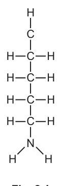
Fig. 2.1 shows an incomplete diagram of the structure of lysine.
Answer
4
4
Background Concept
All amino acids share a common backbone: a central α-carbon (alpha carbon) bonded to four different groups — an amino group (), a carboxyl group (), a hydrogen atom () and a variable side chain called the R group. The R group is the only part that differs between amino acids, and it is what gives each amino acid its unique chemical properties (size, charge, polarity, ability to form bonds).
Lysine is one of the 20 standard amino acids. Its R group has the structure , which is a chain of four groups ending in a primary amine (). Lysine is therefore classified as a basic, positively-charged amino acid at physiological pH.
Understanding the Question
Fig. 2.1 shows a partially-drawn molecule. The vertical chain below the topmost carbon is the R group of lysine (the side chain). The topmost carbon — the one with a single H drawn above it — is the α-carbon position. The candidate must count how many carbon atoms are in the R group itself, NOT including the α-carbon.
The command word is "state", so a single number is the entire required answer.
Approach
Identify the boundary of the R group and count only the carbon atoms inside it. The α-carbon is the carbon that would carry the amino group, the carboxyl group and an H; the R group is everything attached to the α-carbon except those three groups.
Step-by-Step Reasoning
Looking at Fig. 2.1 from top to bottom below the α-carbon position:
- The first carbon below the α-carbon is shown as .
- The second carbon is shown as .
- The third carbon is shown as .
- The fourth carbon is shown as .
- This fourth is bonded to the nitrogen of the terminal .
There are four units in the side chain. None of them is the α-carbon (the α-carbon is the one to which the and of the main amino acid backbone would be attached). Therefore, the R group of lysine contains four carbon atoms.
Key Takeaways
- The R group of an amino acid is everything attached to the α-carbon except , and .
- Lysine's R group is — four carbons long.
- Always be careful not to count the α-carbon as part of the R group.
Common Mistakes
- Counting the α-carbon (the topmost C in the diagram) as part of the R group, which would give 5 instead of 4.
- Counting the nitrogen as a carbon.
Things to Be Careful About
The mark scheme accepts either "4" or "four". Do not include any units; this is a count, not a measurement.
Answer
Add an group and a group, both bonded to the topmost carbon (the α-carbon position). The H already shown above the top carbon remains. The α-carbon is then bonded to four groups: , , and the R group.
Add an -NH2 group and a -COOH group to the topmost carbon.
Background Concept
Every amino acid has the same general structure: a central α-carbon bonded to four different groups — , (amino), (carboxyl) and an R group. In a properly drawn amino acid, all four substituents appear on the α-carbon.
In Fig. 2.1, only the R group () and a single H are drawn. The α-carbon is shown as the topmost C, but it is missing the two functional groups that define an amino acid.
Understanding the Question
The candidate must complete Fig. 2.1 by adding the two missing functional groups to the topmost carbon. The mark scheme awards one mark for the group and one mark for the group; both must be bonded to the same topmost carbon.
Approach
Identify the α-carbon position (the topmost C in Fig. 2.1) and attach the two missing functional groups — amino and carboxyl — to it. The single H drawn above this carbon is retained.
Step-by-Step Reasoning
- The α-carbon is the topmost C in the diagram. It already has one bond shown going up to H and one going down to the first of the R group.
- Two further bonds need to be drawn on this α-carbon: one to and one to .
- The completed α-carbon therefore has four substituents: , , and the R group.
The marking scheme accepts the and with or without explicit bond lines within the functional groups (e.g. the C=O of the carboxyl may be drawn or implied). Atoms other than C and N bonded to the top carbon are not allowed.
Key Takeaways
- The general amino acid formula has four groups on the α-carbon: , , and R.
- Lysine is a basic amino acid because its R group contains an extra , but the backbone amino and carboxyl groups are still required.
Common Mistakes
- Attaching the new groups to the wrong carbon (e.g. to the first of the R group instead of the α-carbon).
- Drawing only one of the two required groups.
- Adding an extra group such as or another atom not in the amino acid general formula.
Things to Be Careful About
The mark scheme explicitly rejects any diagram that shows atoms other than C and N bonded to the α-carbon, and the and must both be present. Writing rather than is fine, and the explicit double bond of the carboxyl group is not required.
Descriptions of the structure of melittin are shown in Table 2.1.
Complete Table 2.1 by writing the level of protein structure that applies to each description.
Table 2.1
| description | level of protein structure |
|---|---|
| in some conditions, four melittin polypeptides can bind to each other | |
| a melittin polypeptide consists of a sequence of 26 amino acids | |
| alpha helices are formed at each end of a melittin polypeptide |
Answer
| description | level of protein structure |
|---|---|
| in some conditions, four melittin polypeptides can bind to each other | quaternary |
| a melittin polypeptide consists of a sequence of 26 amino acids | primary |
| alpha helices are formed at each end of a melittin polypeptide | secondary |
Quaternary; Primary; Secondary
Background Concept
Proteins can be described at four structural levels:
- Primary (1°): the linear sequence of amino acids linked by peptide bonds.
- Secondary (2°): regular local folding patterns of the polypeptide backbone, the most common being the α-helix and the β-pleated sheet, stabilised by hydrogen bonds between the backbone N–H and C=O groups.
- Tertiary (3°): the overall 3D shape of a single polypeptide chain, produced by folding driven by R-group interactions (hydrogen bonds, ionic bonds, disulfide bridges, hydrophobic interactions).
- Quaternary (4°): the association of two or more polypeptide subunits into a functional protein (e.g. haemoglobin's four globin chains).
Understanding the Question
The candidate is given three short descriptions of melittin and must place each into the correct structural level. The mark scheme awards one mark per correct entry.
Approach
Read each description and decide which structural level it implies. Key trigger words/phrases are: "sequence of amino acids" → primary; "α-helices" → secondary; "multiple polypeptides binding" → quaternary.
Step-by-Step Reasoning
- "in some conditions, four melittin polypeptides can bind to each other" — more than one polypeptide chain associating together defines quaternary structure.
- "a melittin polypeptide consists of a sequence of 26 amino acids" — the linear sequence of amino acids is the primary structure.
- "alpha helices are formed at each end of a melittin polypeptide" — α-helices are the textbook example of secondary structure.
Key Takeaways
- A linear amino-acid sequence = primary.
- α-helices and β-sheets = secondary.
- Interactions involving multiple polypeptide chains = quaternary.
- Tertiary would describe the overall 3D shape of a single folded polypeptide — not present in any of the three given statements.
Common Mistakes
- Confusing secondary with tertiary: α-helices are a regular, repeating local pattern of the backbone, which is by definition secondary, not tertiary.
- Choosing tertiary for the four-polypeptide description — a single folded chain is tertiary, but more than one chain associating is quaternary.
Things to Be Careful About
The mark scheme demands the exact level names (primary, secondary, quaternary). Abbreviations such as "1°, 2°, 4°" are not accepted unless the words are also given.
When a bee stings a person, venom enters the body.
Melittin in the venom interacts with cell surface membranes of body cells, as shown in Fig. 2.2.
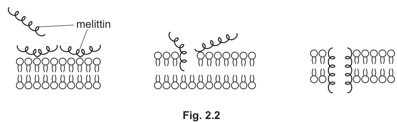
Outline how melittin affects the structure of a cell surface membrane, as shown in Fig. 2.2.
Answer
- Melittin molecules insert into the phospholipid bilayer and span it, producing a pore (channel/opening) through the cell surface membrane.
- The melittin molecules displace/disrupt the regular arrangement of the phospholipids, separating the phospholipid tails in the bilayer.
Melittin inserts into the bilayer, displacing phospholipids and forming a transmembrane pore.
Background Concept
The cell surface membrane is a fluid phospholipid bilayer in which the phospholipid molecules can move laterally. The bilayer is held together by hydrophobic interactions between the fatty acid tails. Any molecule that interferes with these interactions can disturb the integrity of the membrane.
Melittin is a small amphipathic peptide — it has both hydrophobic and hydrophilic regions. This allows it to interact with phospholipids: the hydrophobic parts of the peptide associate with the lipid tails, while the hydrophilic parts associate with the aqueous environment and the phospholipid heads.
Understanding the Question
Fig. 2.2 shows three stages: (1) melittin molecules floating outside the bilayer, (2) the peptides starting to insert into the outer leaflet, and (3) the peptides spanning the bilayer and forming a clear pore. The candidate is asked to outline what the figure shows — what melittin does to the membrane structure. The mark scheme awards 2 marks: one for "produces a pore/channel" and one for "disrupts/displaces phospholipids".
Approach
Look at the final stage of the figure. Identify what new structure appears in the membrane (a pore) and what has happened to the phospholipids (they are pushed apart / displaced by the inserted melittin molecules).
Step-by-Step Reasoning
- In the third panel of Fig. 2.2, several melittin molecules have come together within the bilayer to create a water-filled pore/channel that passes right through the membrane. This is the structural change in the membrane.
- Where the melittin has inserted, the regular, tidy rows of phospholipid heads and tails are broken — the phospholipids are displaced/disrupted/separated by the peptide.
The two ideas together — pore formation and phospholipid disruption — are the two mark-scheme points. A common equivalent of "pore" accepted by the mark scheme is "forms a channel protein" (although melittin is not strictly an integral membrane protein, the wording is accepted).
Key Takeaways
- Pore-forming peptides are amphipathic and disrupt membranes by spanning the bilayer.
- The structural effect on the membrane is twofold: formation of a pore, and displacement of phospholipids from their normal arrangement.
- Disrupted membranes leak ions and small molecules, which is why melittin is cytotoxic.
Common Mistakes
- Describing only the pore without mentioning the phospholipids (or vice versa) — only one of the two marks would be awarded.
- Saying the membrane "breaks" or "is destroyed" — the mark scheme wants a specific term such as pore or channel, and the displacement of phospholipids.
- Confusing melittin with an integral channel protein — although the mark scheme accepts "forms a channel protein" as an alternative wording, melittin is not actually a transmembrane protein; it is a free peptide that self-assembles into a pore.
Things to Be Careful About
The mark scheme says "produces a pore/gap/channel/opening" (not just "pore"), and the phospholipid reference should explicitly name the phospholipids or the bilayer.
Cells that have been affected by melittin break down into cell fragments.
These cell fragments are taken in by phagocytes for further breakdown.
Describe the process by which phagocytes take in and break down these cell fragments.
Answer
- The cell fragment binds to receptors on the phagocyte cell surface membrane.
- The phagocyte extends pseudopodia that surround and engulf the cell fragment.
- The cell-surface membrane fuses behind the fragment, pinching off to form a phagocytic vacuole (phagosome) inside the cell.
- A lysosome containing hydrolytic (digestive) enzymes fuses with the phagocytic vacuole, releasing enzymes such as proteases that hydrolyse the proteins of the cell fragment into amino acids. The soluble products are absorbed into the cytoplasm and any undigested residue is expelled by exocytosis.
Phagocytosis: receptor binding → engulfment by pseudopodia → phagosome formation → lysosome fusion → hydrolysis by enzymes (e.g. proteases → amino acids).
Background Concept
Phagocytosis is a form of endocytosis in which a cell engulfs a solid particle (a pathogen, a cell fragment or debris) and digests it internally. It is carried out by phagocytes — a group of white blood cells that includes neutrophils and monocytes/macrophages.
The process relies on:
- Receptor–ligand binding at the cell surface, which lets the phagocyte recognise and adhere to the target.
- Membrane fluidity and remodelling, which allows pseudopodia (cytoplasmic extensions) to wrap around the particle.
- Membrane fusion, which seals the engulfed material inside a vesicle.
- Lysosomes, membrane-bound organelles that contain hydrolytic enzymes working at an acidic internal pH. When a lysosome fuses with a phagosome, the hydrolytic enzymes digest the contents.
Understanding the Question
Part (d) gives context: cells damaged by melittin break into fragments and are taken in by phagocytes. The candidate must describe the full phagocytosis process — from binding at the cell surface, through engulfment and vesicle formation, to enzymatic digestion inside the phagocyte. The mark scheme offers 4 marks from a list of 6 creditable ideas, so the candidate needs at least four clearly-stated points.
Approach
Walk through phagocytosis in the order in which it happens, picking the most concrete and biologically specific points to mention. Avoid vague answers such as "the phagocyte digests the cell" — the mark scheme explicitly rejects "lysosome digests" because the lysosome alone does not do anything; the enzymes inside it do the digesting.
Step-by-Step Reasoning
The mark scheme lists the following six creditable points (any 4 of which earn 4 marks):
- Binding: the cell fragment binds to receptors on the phagocyte cell surface membrane (or attaches to the membrane).
- Engulfment: the cell-surface membrane of the phagocyte surrounds the cell fragment, often with pseudopodia forming around it.
- Vesicle formation: the membrane fuses behind the fragment and pinches off, releasing a phagocytic vacuole / phagosome into the cytoplasm. The terms "phagosome" and "vesicle" are accepted alternatives for "vacuole".
- Lysosome fusion: a lysosome (containing hydrolytic enzymes) fuses with the phagocytic vacuole. The combined structure may be called a phagolysosome.
- Hydrolysis: the cell fragment is broken down by hydrolytic / digestive enzymes (e.g. proteases, lipases, carbohydrases).
- Named enzyme and product OR two named enzymes: for example, "protease → amino acids", or naming both protease and lipase. This converts the candidate's generic "digestion" claim into a specific, credit-worthy statement.
A valid alternative point is opsonisation — antibodies or complement proteins coating the cell fragment, which speeds up recognition by the phagocyte.
The answer above puts these in a logical order, picking the four most concrete points and including a named enzyme + product for the final mark.
Key Takeaways
- Phagocytosis is receptor-mediated endocytosis of solid material.
- Lysosomes deliver hydrolytic enzymes that work at low pH to digest the engulfed material.
- Specific enzymes act on specific macromolecules: proteases on proteins, lipases on lipids, carbohydrases on carbohydrates.
- Vague terms like "digests" or "breaks down" are insufficient — the enzymes do the work, and the names matter.
Common Mistakes
- Saying the lysosome "digests" the cell fragment without naming enzymes — the mark scheme explicitly rejects this.
- Skipping the receptor-binding step, which is essential for specific recognition.
- Omitting the vesicle (phagosome) formation step — without it, the material is not internalised.
- Forgetting the membrane-fusion event that releases the phagosome into the cytoplasm.
Things to Be Careful About
The mark scheme requires the word "lysosome" to appear (it does not accept "vesicle of enzymes" or similar), and digestion must be attributed to the enzymes inside the lysosome, not the lysosome itself. Naming one specific enzyme with its product (or two enzymes with their substrates) is what unlocks the final mark.
Staphylococcus epidermidis is a species of bacterium that lives on human skin.
Staphylococcus aureus is a pathogenic bacterium that can infect humans.
S. epidermidis and S. aureus are prokaryotes.
Table 3.1 shows some cell features that could apply to typical prokaryotic cells or to typical eukaryotic cells or to both types of cell.
Complete Table 3.1 by using a tick (✓) if the feature applies to the type of cell or a cross (✗) if the feature does not apply to the type of cell.
Put a tick (✓) or a cross (✗) in every box.
Table 3.1
| feature | prokaryotic cell | eukaryotic cell |
|---|---|---|
| circular DNA | ||
| 80S ribosomes | ||
| a cell diameter of |
Answer
| feature | prokaryotic cell | eukaryotic cell |
|---|---|---|
| circular DNA | ✓ | ✓ |
| 80S ribosomes | ✗ | ✓ |
| a cell diameter of | ✗ | ✓ |
(One mark per correct row.)
See table — circular DNA ✓/✓, 80S ribosomes ✗/✓, 20 µm diameter ✗/✓
Background Concept
Prokaryotic cells (bacteria and archaea) and eukaryotic cells differ in several fundamental ways:
- DNA: Prokaryotes carry a single circular molecule of DNA in the nucleoid region. Eukaryotes have linear chromosomes in a nucleus, BUT they also possess small circular DNA molecules inside mitochondria (and chloroplasts in plants), so eukaryotic cells do contain circular DNA in addition to their nuclear linear DNA.
- Ribosomes: Prokaryotic ribosomes are 70S (composed of a 50S and a 30S subunit). Eukaryotic ribosomes in the cytoplasm are 80S (60S + 40S). This is one of the targets of antibiotics such as those used to treat S. aureus.
- Size: Prokaryotic cells are typically 0.1–5 µm in diameter, whereas most eukaryotic cells are 10–100 µm. A diameter of 20 µm is therefore far too large for a typical prokaryote and is consistent with a eukaryotic cell.
Understanding the Question
The stem establishes that S. epidermidis and S. aureus are prokaryotes and presents Table 3.1 with three features. The candidate must decide, for each feature, whether it applies to prokaryotes, eukaryotes, or both, and place a tick or a cross in every box (no blanks).
Approach
Read each feature, recall which type of cell it describes, and remember that both types can sometimes share a feature (this is the trap for the circular DNA row). The mark scheme awards one mark per fully correct row.
Step-by-Step Reasoning
- Circular DNA: Bacteria have one main circular chromosome. Eukaryotes have linear nuclear DNA plus small circular mtDNA (and ctDNA in plants). Both cell types therefore possess circular DNA — ✓ / ✓.
- 80S ribosomes: Only eukaryotic cytosolic ribosomes are 80S; prokaryotes have 70S. So only eukaryotic — ✗ / ✓.
- Cell diameter of 20 µm: Prokaryotes are typically 0.1–5 µm, well below 20 µm. A 20 µm cell is in the eukaryotic size range — ✗ / ✓.
Key Takeaways
- Don't assume "both have X" means it applies to only one type — circular DNA is a classic shared feature.
- The "S" in ribosome size (70S vs 80S) is a key prokaryote/eukaryote discriminator and the basis of several antibiotics.
- Typical cell size is a strong, quick diagnostic between the two cell types.
Common Mistakes
- Ticking circular DNA for prokaryote only — forgetting mitochondrial/chloroplast DNA in eukaryotes.
- Putting ✓ in the prokaryote column for 80S ribosomes or 20 µm diameter.
- Leaving boxes blank — the instruction says "every box" must be filled.
Things to Be Careful About
The mark scheme explicitly notes "if no marks gained, check correct column for 1 mark," so a single correctly-completed column on the eukaryotic side still earns a mark. Always complete the table — no empty cells.
One way that S. aureus can infect humans is through wounds (breaks) in the skin.
Populations of S. aureus develop on human skin as part of a biofilm. The biofilm contains cells of S. aureus within a mixture of polymers that have been secreted by the cells.
S. epidermidis produces a protease enzyme that prevents the growth of S. aureus populations on human skin. Proteases catalyse the breakdown of proteins.
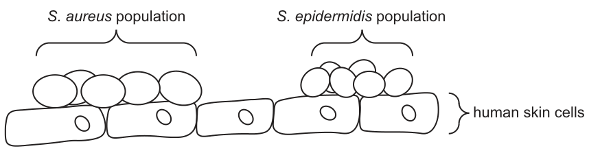
Fig. 3.1 is a diagram showing populations of S. epidermidis and S. aureus on human skin cells.
Suggest and explain how the protease produced by S. epidermidis cells prevents the growth of an S. aureus population on human skin.
Answer
Any three of:
- The protease is secreted / released by S. epidermidis (it is an extracellular enzyme) and acts on the S. aureus population.
- The protease has an active site complementary to proteins (its substrate) that are specific to S. aureus; it forms enzyme–substrate complexes and hydrolyses them.
- The protease breaks down the proteins / polymers in the S. aureus biofilm, so the S. aureus cells can no longer remain attached to the skin cells.
- (Alternative) The protease breaks down S. aureus cell-surface proteins (e.g. receptors / binding sites) that the cells need to attach to the skin.
Extracellular protease is secreted; it has an active site specific to S. aureus proteins; it breaks down biofilm polymers so the S. aureus cannot remain attached to the skin.
Background Concept
A biofilm is a community of microorganisms embedded in a self-produced matrix of extracellular polymeric substances (polysaccharides, proteins, eDNA). The matrix holds the cells together and adheres them to a surface. Removing the matrix therefore dislodges the population.
Enzymes are biological catalysts. Proteases hydrolyse peptide bonds in proteins. The active site of a protease is complementary in shape and chemistry to its substrate, so a given protease typically acts only on certain proteins — this is enzyme specificity.
Staphylococcus epidermidis living on the skin is an example of a commensal bacterium (it benefits but does not harm the host). One of its secretions is a protease that interferes with the growth of the pathogen Staphylococcus aureus on the same surface — an example of microbial competition.
Understanding the Question
Fig. 3.1 shows a population of S. aureus embedded in a biofilm on human skin cells, alongside a population of S. epidermidis. The question asks for a suggested mechanism (so any biologically plausible idea is fine) and an explanation (so each point must be justified) of how the S. epidermidis protease prevents the S. aureus population from growing. The command word is "suggest and explain", so the answer must do both.
Approach
- State that the protease is released into the surrounding environment (it is extracellular), so it can reach the S. aureus population.
- Apply enzyme specificity — the protease's active site fits proteins found in/around the S. aureus cells.
- Identify which proteins: those in the biofilm (so the cells can no longer cling to the skin) — or alternatively, surface proteins that the cells need to attach.
- Conclude that, without attachment / without the protective and anchoring matrix, the S. aureus population cannot establish or grow.
Step-by-Step Reasoning
- Point 1 — secretion: The protease is an extracellular enzyme, so S. epidermidis secretes it; it can then diffuse through the biofilm and reach S. aureus cells. (Mark-scheme reward: "protease is secreted / released"; "AV — extracellular enzyme".)
- Point 2 — specificity: The protease's active site is complementary to proteins in / on S. aureus; substrate binds to form an enzyme–substrate complex and is then hydrolysed. (Mark-scheme reward: "active site complementary to substrate of S. aureus" / "forms ES complexes".)
- Point 3 — target and consequence: The proteins broken down are the (biofilm) polymers that hold the S. aureus cells together and anchor them to the skin. With the biofilm degraded, the S. aureus population cannot stay attached to the skin cells, so it cannot grow there. (Mark-scheme reward: "breaks down protein in biofilm" and "without biofilm S. aureus cannot remain attached".)
- Alternative 3 (AVP): Could also be cell-surface proteins (membrane proteins, receptors, binding sites) needed by S. aureus to attach to / colonise the skin, or substances the cells need from the biofilm for survival.
Key Takeaways
- Extracellular enzymes are secreted to act on substrates outside the cell.
- Enzyme specificity means one protease will only cleave proteins with matching sequences/conformations.
- Biofilms protect and anchor bacteria; destroying the biofilm is one effective way to control a bacterial population without directly killing the cells.
Common Mistakes
- Saying the protease "kills" S. aureus — it breaks down proteins; it is not described as bactericidal here.
- Not mentioning secretion — the enzyme must reach the substrate.
- Vague "it attacks the bacteria" without specifying which protein target (biofilm polymers, surface receptors, etc.) and why this stops the population growing.
Things to Be Careful About
The mark scheme is generous with alternatives ("other example of S. aureus protein broken down … e.g. cell membrane proteins / receptors / binding sites"). Any one specific, well-explained protein target is acceptable, but the explanation must link the broken-down protein to the loss of attachment / survival.
S. aureus can infect many tissues in the human body, including tissues in the gas exchange system.
Describe the role of goblet cells in the protection of tissues in the trachea from infection by S. aureus.
Answer
Any two of:
- Goblet cells secrete / produce mucus (mucin).
- The mucus traps S. aureus cells so they cannot reach the underlying tracheal tissue.
- (The mucus acts as a physical / chemical barrier preventing the bacteria from infecting the cells lining the trachea.)
Goblet cells secrete mucus; the mucus traps S. aureus cells and forms a barrier preventing them from reaching the underlying tissue.
Background Concept
The trachea is lined by a pseudostratified ciliated columnar epithelium with goblet cells scattered among it. Goblet cells are unicellular glands whose function is to secrete mucus — a sticky glycoprotein-rich fluid. Mucus sits on top of the epithelium and traps inhaled particles, dust and microorganisms (a non-specific, innate defence). The cilia of the neighbouring ciliated epithelial cells then beat in a coordinated rhythm to move the mucus and trapped debris upwards towards the pharynx, where it is swallowed (the mucociliary escalator).
Understanding the Question
Part (c)(i) is restricted to the role of goblet cells (not the ciliated cells) in protecting tracheal tissue from infection by S. aureus specifically. The question requires two creditable points.
Approach
Recall the structure–function relationship: goblet cells → secrete mucus. Then link the property of mucus (sticky / viscous) to its effect on bacteria (trapping them) and therefore to the protection of the underlying cells.
Step-by-Step Reasoning
- Mark 1 — secretion: Goblet cells secrete (produce / release) mucus (or mucin, the principal glycoprotein of mucus). (Mark scheme: "secrete / produce / AW, mucus / mucin".)
- Mark 2 — function: This mucus traps S. aureus cells and forms a barrier between the bacteria and the cells lining the trachea, so the bacteria cannot reach and infect those cells. (Mark scheme: "mucus traps S. aureus / acts as a barrier".)
- Optional elaboration: The mucus is then moved by cilia away from the lungs, removing the trapped bacteria.
Key Takeaways
- Goblet cells are a key component of the non-specific / innate defence of the gas exchange system.
- "Mucus traps pathogens" is the core functional point and is the one the mark scheme expects.
- Goblet cells and ciliated cells have separate but cooperating roles; the question deliberately splits them between (c)(i) and (c)(ii).
Common Mistakes
- Saying the goblet cells "trap" S. aureus without first stating that they secrete the mucus that does the trapping.
- Writing "goblet cells prevent infection by S. aureus" — too vague; the mark scheme credits the mechanism.
- Confusing goblet cells with ciliated cells (which move the mucus rather than produce it).
Things to Be Careful About
The mark scheme allows "bacteria / pathogens / microorganisms" in place of S. aureus for the second mark, so phrasing such as "mucus traps pathogens" is acceptable. Do not credit answers about viruses, because the mark scheme explicitly says R virus here.
S. aureus cells can infect tissues by passing in between cells that line the lumen of the trachea.
State the name of a cell type, other than goblet cells, that lines the lumen of the trachea.
Answer
Ciliated epithelial cell.
Ciliated epithelial cell
Background Concept
The lumen of the trachea is lined by a pseudostratified ciliated columnar epithelium (sometimes referred to as "respiratory epithelium"). Among these cells are scattered goblet cells and basal cells. The dominant cell type other than goblet cells is the ciliated epithelial cell. Each ciliated cell bears numerous cilia on its apical surface; the cilia beat in synchrony to move mucus (and trapped debris) upwards — the mucociliary escalator.
Understanding the Question
The question is a single-mark recall: name a cell type, other than goblet cells, that lines the lumen of the trachea.
Approach
Pick the most prominent and obvious lining cell of the tracheal lumen — the ciliated epithelial cell. The stem of the question (3(c)) even mentions that S. aureus can pass in between cells lining the lumen, hinting at an epithelium made up of multiple cell types.
Step-by-Step Reasoning
- The two principal cell types in the tracheal lining are goblet cells and ciliated epithelial cells. Since goblet cells are excluded, the answer is ciliated epithelial cell.
Key Takeaways
- The trachea is lined by ciliated epithelium plus mucus-secreting goblet cells.
- Knowing the cell types of a typical gas-exchange epithelium is a standard CIE requirement.
Common Mistakes
- Writing just "ciliated cell" — the mark scheme explicitly ignores this wording.
- Writing "epithelial cell" — too vague, doesn't capture the ciliated feature that distinguishes it.
- Naming an immune cell such as a phagocyte — these patrol the tissue but are not part of the lining of the lumen.
Things to Be Careful About
Use the full name: ciliated epithelial cell (the scheme accepts "ciliated epithelium cell"). "Ciliated cell" alone is ignored.
Vancomycin and penicillin are antibiotics that are used to treat infectious diseases caused by S. aureus.
Fig. 3.2 shows the mechanism of action of vancomycin.
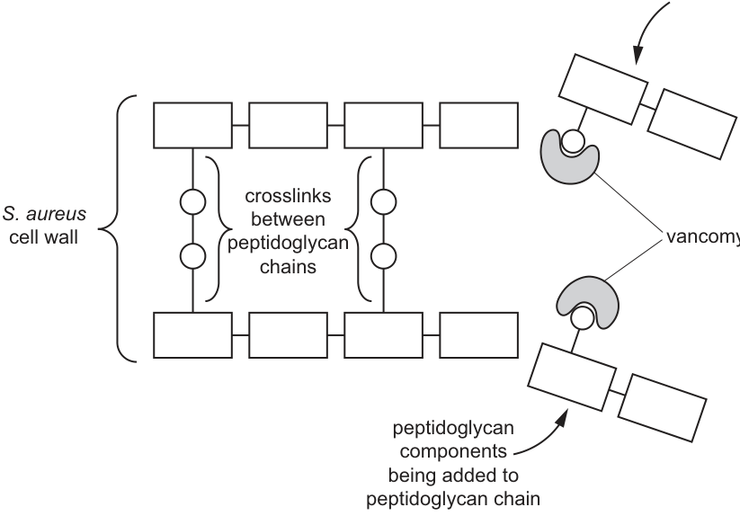
Vancomycin and penicillin act on the cell wall of bacterial cells.
With reference to Fig. 3.2, describe the similarities and differences between the mechanism of action of vancomycin and the mechanism of action of penicillin.
Answer
Similarities:
- Both antibiotics prevent the formation of crosslinks (cross-bridges) between peptidoglycan chains in the bacterial cell wall.
- As a result, both stop / prevent the synthesis and repair of the cell wall.
Differences:
- Penicillin binds to and inhibits the enzyme(s) (e.g. transpeptidases) that catalyse the formation of crosslinks between peptidoglycan chains.
- Vancomycin binds to the peptidoglycan (subunit) components themselves, blocking their access to the growing peptidoglycan chain so they cannot be incorporated (and so the enzyme that would join them cannot act).
Similarities: both prevent peptidoglycan crosslinks and so stop cell-wall synthesis/repair. Difference: penicillin inhibits the crosslinking enzymes (transpeptidases); vancomycin binds to the peptidoglycan subunits themselves, preventing their incorporation into the wall.
Background Concept
Bacterial cell walls contain peptidoglycan — long glycan chains crosslinked by short peptide bridges. The crosslinks are made by enzymes such as transpeptidases (also called penicillin-binding proteins, PBPs). Without crosslinks the wall is weak, the bacterium cannot withstand osmotic pressure, and it lyses.
Penicillin is a structural analogue of the D-Ala-D-Ala terminus of the peptidoglycan precursor. It binds irreversibly to the active site of the transpeptidase, blocking it. The enzyme can no longer form crosslinks, so the wall is not properly assembled. Penicillin is therefore described as an enzyme inhibitor.
Vancomycin works differently (Fig. 3.2). It binds directly to the D-Ala-D-Ala terminus of the peptidoglycan subunit (the free precursor), physically blocking the subunit so that the transpeptidase cannot use it. The crosslink is therefore not formed, but the blockage is at the substrate, not at the enzyme.
Understanding the Question
Part (d)(i) is a structured compare-and-contrast ("similarities and differences"). The candidate must consult Fig. 3.2 for vancomycin's mechanism and recall penicillin's mechanism from prior knowledge. The mark scheme allocates three marks to be drawn from any of the credit-worthy points; the answer must be clearly organised into similarities and differences.
Approach
- Similarities — ask: what is the net effect of both antibiotics on the cell? Both ultimately prevent the formation of crosslinks between peptidoglycan chains, and therefore stop / prevent the synthesis (and repair) of the cell wall.
- Differences — ask: how does each antibiotic achieve this? Penicillin binds to the enzyme (transpeptidase) that makes the crosslinks; vancomycin binds to the peptidoglycan subunit so the enzyme has nothing to work on.
- Keep the language tight and the contrast explicit. Use terms such as "enzyme" and "substrate / peptidoglycan component" to make the difference unambiguous.
Step-by-Step Reasoning
- Similarity 1 (mark-scheme point): Both prevent the formation of crosslinks (cross-bridges) between peptidoglycan chains. This is the mechanistic similarity.
- Similarity 2 (mark-scheme point): Both therefore stop / prevent the synthesis and repair of the cell wall. (Without new crosslinks the wall is not made, and existing damage cannot be patched — the bacterium is doomed by osmotic lysis.)
- Difference 1 (mark-scheme point): Penicillin binds to and inhibits the enzyme that catalyses crosslink formation (transpeptidase / PBP). Mark scheme language: "penicillin binds to / is an inhibitor of enzymes / transpeptidases that catalyse formation of crosslinks".
- Difference 2 (mark-scheme point): Vancomycin binds to / acts on the peptidoglycan (subunit / crosslink component) itself. Mark scheme language: "vancomycin binds to / acts on peptidoglycan / crosslink components".
- Optional elaboration (also worth a mark, AVP): Vancomycin blocks access of the peptidoglycan subunit to the growing chain, or it may prevent the enzyme that joins subunits together from binding.
Key Takeaways
- "Prevents crosslinks" is the common end-point of both antibiotics; the means differ — enzyme inhibition (penicillin) vs. substrate binding (vancomycin).
- A clear compare-and-contrast structure makes it obvious to the examiner that both halves of the question have been addressed.
- Reading Fig. 3.2 carefully tells you exactly what vancomycin is binding to (the free peptidoglycan subunit), which is the key piece of information for the difference.
Common Mistakes
- Saying only that both "break down" or "destroy" the cell wall — the action is on crosslink formation, not on direct wall destruction.
- Failing to distinguish the levels of action (enzyme vs. substrate) when describing the difference.
- Skipping the similarities and listing only differences (or vice-versa) — the question requires both.
- Confusing penicillin with vancomycin: e.g. saying penicillin "binds to peptidoglycan components" (it binds to the enzyme).
Things to Be Careful About
The mark scheme permits ecf (error carried forward): if a candidate mis-states penicillin's mechanism, an answer that correctly describes vancomycin from Fig. 3.2 may still earn one mark. So even with a partial misconception, some credit is recoverable. The phrase "prevent formation of crosslinks" must come from a correct understanding of both antibiotics for that similarity mark to be awarded.
Some strains of S. aureus are resistant to vancomycin and penicillin.
Describe the steps that can be taken to reduce the impact of antibiotic resistance.
Answer
Any three of:
- Only prescribe / take antibiotics when they are absolutely necessary (e.g. not for viral infections; not as a preventative).
- Make sure the correct / effective antibiotic is prescribed and used for the specific infection.
- Patients should complete the course and follow the instructions (e.g. DOTS for TB).
- Develop new antibiotics or use other antibacterials.
- Reduce / control the use of antibiotics in agriculture and in animals used for food.
- Break the transmission cycle of resistant strains, e.g. through vaccination programmes, good hospital hygiene, isolation / quarantine of infected individuals.
- (Other valid points: training and updating healthcare professionals; reporting patterns of resistance; monitoring whether an antibiotic is still effective; limiting over-the-counter sales; the WHO Global Plan to End TB.)
Use antibiotics only when necessary; ensure the correct antibiotic is used; complete the course; develop new antibiotics; reduce antibiotic use in agriculture; break transmission (vaccines, hygiene, isolation).
Background Concept
Antibiotic resistance arises when random mutations or horizontal gene transfer give individual bacteria the ability to survive an antibiotic. In the presence of the antibiotic these resistant cells are strongly selected for and come to dominate the population. Resistant strains of S. aureus (notably MRSA — meticillin-resistant Staphylococcus aureus, and VRSA — vancomycin-resistant) are major clinical problems.
Reducing the impact of resistance relies on two complementary strategies:
- Slowing the emergence and spread of resistance (stewardship, hygiene, vaccination).
- Replenishing the armoury (new antibiotics, alternative antibacterials, combination therapy).
The mark scheme provides a long menu of acceptable points, so any three of them earn the three marks.
Understanding the Question
Part (d)(ii) asks for steps that can be taken to reduce the impact of antibiotic resistance. This is broader than just "stop using antibiotics" — it covers the full set of clinical, public-health and research actions.
Approach
Group the ideas into three or four natural categories and pick the three most concrete, distinct points:
- Prescribing behaviour (when and what to prescribe).
- Patient behaviour (completing the course).
- Drug development (new antibiotics / alternatives).
- Agricultural use (reducing non-medical antibiotic exposure).
- Transmission control (vaccines, hygiene, isolation).
Step-by-Step Reasoning
- Point 1 — Only when necessary: Prescribing / taking antibiotics only when absolutely required; not for viral infections and not prophylactically. This reduces selection pressure.
- Point 2 — Correct antibiotic: Make sure the correct, effective antibiotic is used for the diagnosed bacterial infection (use sensitivity testing where possible). This avoids low-dose / wrong-drug exposure that breeds resistance.
- Point 3 — Complete the course: Patients must complete the full prescribed course so that partially-resistant survivors are not selected for. DOTS (Directly Observed Treatment, Short-course) is a structured example for TB.
- Point 4 (alternative): Develop new antibiotics or use other antibacterials, so there is always an effective drug available against resistant strains.
- Point 5 (alternative): Reduce or control the use of antibiotics in agriculture and in animals reared for food, since resistant organisms can spread from farms to humans.
- Point 6 (alternative): Break the transmission cycle of resistant strains through vaccination, good hospital hygiene (especially hand-washing between patients), and isolation / quarantine.
- Point 7 (AVP): Train and update healthcare professionals and the public; report patterns of antibiotic resistance; monitor whether an antibiotic remains effective; limit over-the-counter antibiotic sales; support initiatives such as the WHO Global Plan to End TB.
Key Takeaways
- Antibiotic resistance is driven by selection pressure; reducing unnecessary antibiotic use is the single most important intervention.
- Combinations of stewardship (Point 1), correct prescribing (Point 2), patient adherence (Point 3), new drugs (Point 4), agricultural controls (Point 5) and transmission control (Point 6) are all needed; no single measure is sufficient.
- A public-health-style answer (vaccines, hygiene, quarantine) is as relevant as a clinical-style answer (prescribing, courses).
Common Mistakes
- Vague answers like "be careful with antibiotics" or "use less medicine" — too non-specific to earn marks.
- Only listing clinical measures and ignoring agriculture, transmission control, or drug development.
- Confusing antibiotic resistance with vaccine resistance or antiviral resistance.
Things to Be Careful About
The mark scheme is generous (three points from a long list) and explicitly allows AVP — so any plausible, distinct, well-stated measure earns a mark. However, the wording must be specific to antibiotic resistance rather than generic infection control (e.g. "wash hands" alone is too broad; "hospital hand-hygiene to prevent spread of MRSA" is on-target).
Lysosomes are membrane-bound organelles found in mammalian cells.
Scientists measured the concentration of cholesterol in the membranes of lysosomes in a mammalian cell.
The concentration of cholesterol in the lysosome membranes was found to be lower than the concentration in other membranes inside mammalian cells.
State how a lower concentration of cholesterol would make the properties of lysosome membranes different from other membranes in mammalian cells.
Answer
The (lysosome) membrane would be more fluid / the (lateral) movement of phospholipids in the bilayer would be greater.
Membrane more fluid / greater lateral movement of phospholipids
Background Concept
Cholesterol is a small, planar lipid molecule that sits within the phospholipid bilayer of eukaryotic plasma and organelle membranes. Its polar hydroxyl (-OH) group hydrogen-bonds with the phospholipid head groups, while its rigid four-ring hydrocarbon body nestles among the fatty acid tails of the inner bilayer. Through these interactions, cholesterol modulates membrane fluidity:
- At higher temperatures it restrains the movement of the fatty acid tails, reducing membrane fluidity.
- At lower temperatures it prevents the tails from packing too closely, maintaining some fluidity.
In mammalian cells at body temperature (~37 °C), the net effect of cholesterol is to reduce membrane fluidity and to make the membrane less permeable to small water-soluble molecules and ions.
Understanding the Question
The stem tells you that lysosome membranes contain a lower concentration of cholesterol than other membranes in the same cell. The question asks you to state how this lower cholesterol level makes the lysosome membrane's properties different. The command word "state" means a single, clearly-worded fact.
Approach
Apply the principle above in reverse: if cholesterol reduces fluidity, then less cholesterol means the membrane loses some of that restraint and becomes more fluid.
Step-by-Step Reasoning
- Cholesterol normally reduces the lateral movement of phospholipids in the bilayer.
- With less cholesterol, this restraining effect is reduced.
- The membrane therefore becomes more fluid and phospholipids show greater (lateral) movement.
- The mark scheme also accepts "increased passage of polar molecules/ions" or "easier fusion with other membranes" as alternative consequences, but the core, easiest point is the increase in fluidity.
Key Takeaways
- Cholesterol is a fluidity regulator: less cholesterol → more fluid membrane.
- A physical property (fluidity) follows directly from membrane composition.
- This principle sets up the structure–function link tested in (a)(ii).
Common Mistakes
- Saying the membrane becomes "weaker", "thinner" or "less stable" — these are not properties directly affected by cholesterol.
- Saying the membrane becomes "less fluid" — the opposite of the correct answer.
- Vague answers like "membrane changes" without specifying what changes.
Things to Be Careful About
- The word "fluidity" is the precise term expected in CIE mark schemes; synonyms such as "runny" or "loose" do not score.
- The answer is one point only; do not pad with extra (unrewarded) biology.
Suggest why the lower concentration of cholesterol in lysosome membranes would help lysosomes carry out their function.
Answer
Greater fluidity makes it easier for the lysosome membrane to fuse with, phagosomes / vesicles / vacuoles (so that the hydrolytic enzymes inside the lysosome can mix with, and digest, the substrate).
Easier for the lysosome membrane to fuse with phagosomes / vesicles / vacuoles
Background Concept
Lysosomes are roughly spherical, membrane-bound organelles that contain hydrolytic (digestive) enzymes — proteases, lipases, nucleases, and glycosidases such as α-galactosidase. They work at an internal pH of about 4.5–5.0, maintained by V-type H⁺-ATPases (proton pumps) in the lysosomal membrane, while the cytoplasm sits at about pH 7.2.
A lysosome does not "eat" substrates directly. Instead, it fuses with another membrane-bounded compartment that contains material to be digested:
- Phagosomes / endosomes carrying material taken in by endocytosis or phagocytosis.
- Autophagosomes containing worn-out organelles from the cell itself.
Fusion mixes the lysosomal hydrolases with the substrate, breaking it down inside the resulting vesicle.
Understanding the Question
You have just established that lysosome membranes are more fluid than other cellular membranes. The question now asks you to suggest why this greater fluidity would help the lysosome perform its function. The command word "suggest" allows you to propose a reasoned idea rather than state a textbook fact.
Approach
Connect the physical property (fluidity) to the lysosome's mode of action (membrane fusion). More fluid bilayers bend, deform and merge with other bilayers more easily.
Step-by-Step Reasoning
- A lysosome's job is to fuse with vesicles (phagosomes, endosomes, autophagosomes) that contain material to be digested.
- Two membranes can fuse only if their phospholipid bilayers can deform, come into very close contact, and merge.
- A more fluid membrane is more deformable; its phospholipids can rearrange more readily.
- Therefore, the lysosome can more easily fuse with these vesicles, allowing the hydrolytic enzymes to be delivered to the substrate.
- Note the mark scheme's ecf concession: if you wrongly said fluidity DECREASED in (i), then the explanation here would be that the membrane becomes more impermeable, preventing the hydrolytic enzymes from leaking out of the lysosome.
Key Takeaways
- Lysosomes function by fusion, not by direct digestion.
- Membrane fluidity is the physical property that determines ease of fusion.
- A structural feature (low cholesterol) is adapted to a function (fusion).
Common Mistakes
- Saying the lysosome "digests material" without specifying the fusion step.
- Conflating fluidity with permeability — they are related but distinct; the mark scheme wants the fusion link.
- Repeating the answer from (i) without going further into the functional link.
Things to Be Careful About
- "Suggest" lets you give a reasonable proposal; it does not require quoting a textbook sentence.
- The mark scheme wants at least one of: vacuoles, vesicles, phagosomes, endosomes, or "membranes of other organelles".
The enzyme -galactosidase is present in lysosomes.
Students investigated the effect of substrate concentration on the rate of reaction catalysed by -galactosidase at pH 4.5 and at pH 5.9.
The results are shown in Fig. 4.1.
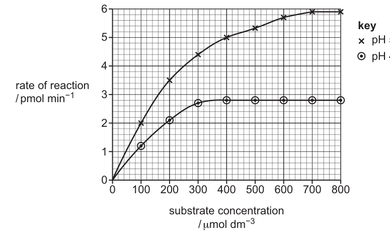
Determine the Michaelis–Menten constant, , for -galactosidase at pH 4.5 using the data in Fig. 4.1.
State the unit for the value in your answer.
= ______ unit ______
Working
From the pH 4.5 curve (open circles), the plateau value is:
Half of :
Reading horizontally from on the y-axis to the pH 4.5 curve, then vertically down to the x-axis, the substrate concentration is:
Answer
120 µmol dm⁻³
Background Concept
The Michaelis–Menten equation describes the relationship between the rate () of an enzyme-catalysed reaction and the substrate concentration :
Two key constants are:
- — the maximum rate, reached at saturating substrate concentration when essentially every active site is occupied by substrate.
- (the Michaelis constant) — the substrate concentration at which . It is an inverse measure of enzyme–substrate affinity: a low means high affinity.
On a Michaelis–Menten curve, is read off by:
- Reading the plateau (which is ).
- Halving it.
- Drawing a horizontal line from the half-rate to the curve, then a vertical line down to the substrate axis.
Understanding the Question
You are given Fig. 4.1, a Michaelis–Menten-style curve for α-galactosidase at pH 4.5. You must determine for that pH and quote the unit. The unit of is always the unit of substrate concentration (the same as the x-axis).
Approach
Identify the pH 4.5 curve (open circles, the lower curve), read its plateau as , halve that value, and read the substrate concentration at which the curve crosses that half-rate.
Step-by-Step Reasoning
- The pH 4.5 curve plateaus at .
- Half of : .
- Locate on the y-axis and draw a horizontal line to the right until it meets the pH 4.5 curve.
- From that point, drop a vertical line to the x-axis; the reading is .
- Therefore at pH 4.5.
Key Takeaways
- is read off a Michaelis–Menten curve at half .
- The unit of is the same as the unit of substrate concentration.
- A small indicates a high-affinity enzyme.
Common Mistakes
- Reading at the substrate concentration equal to half the x-axis range (i.e. 400) — wrong; you must halve the y-axis value first.
- Forgetting to state the unit.
- Reading the wrong curve (the pH 5.9 curve).
- Saying the unit is "mol" instead of "".
Things to Be Careful About
- The x-axis in the figure is in , not .
- The question wants the value AND the unit; both are credited separately.
- The mark scheme accepts the 110–130 range; 120 is the intended reading.
With reference to Fig. 4.1, describe the differences in the results at pH 4.5 and pH 5.9 and suggest explanations for the differences.
Answer
Differences between pH 4.5 and pH 5.9:
- The rate of reaction is lower at pH 4.5 than at pH 5.9 at every substrate concentration.
- is lower at pH 4.5 () than at pH 5.9 ().
- is reached at a lower substrate concentration at pH 4.5 () than at pH 5.9 ().
Explanations:
- pH 5.9 is closer to the optimum pH of -galactosidase (since it gives the higher ); pH 4.5 is further from the optimum.
- The (higher) H⁺ concentration at pH 4.5 alters the ionisation of R-groups in the active site and disturbs the tertiary structure of the enzyme, so the shape of the active site changes.
- The active site becomes less complementary to the substrate, so fewer enzyme–substrate (E–S) complexes form per unit time, lowering the rate at every substrate concentration and reducing .
Rate and Vmax are lower at pH 4.5; the active site is distorted so fewer E-S complexes form
Background Concept
Enzyme activity is highly sensitive to pH. Each enzyme has an optimum pH at which:
- The ionisation state of the active-site R-groups is correct for substrate binding and catalysis.
- The ionisation state of the substrate is also correct.
- The tertiary structure (held by ionic, hydrogen and disulfide bonds) is undisturbed.
Away from the optimum, the altered H⁺ concentration disrupts these bonds, partially distorts the active site, reduces the rate of E–S complex formation, and therefore reduces . The enzyme is not necessarily fully denatured; it is simply less catalytically efficient.
For the α-galactosidase curves shown in Fig. 4.1, pH 5.9 gives the higher and so is closer to the optimum, while pH 4.5 is further from the optimum and gives the lower .
Understanding the Question
The question provides two Michaelis–Menten curves (pH 4.5 and pH 5.9) and asks you to:
- Describe the differences between the two sets of results (data extraction).
- Suggest explanations for those differences (mechanistic reasoning).
The command word "describe AND suggest explanations" tells you that both halves of the answer carry marks.
Approach
First, systematically compare the two curves on the graph:
- The overall rate at any given substrate concentration.
- The plateau height ().
- The substrate concentration at which the plateau is reached.
- The apparent steepness of the initial rise.
Then turn each observation into a mechanism: what does the lower pH do to the enzyme's structure and active site?
Step-by-Step Reasoning
Differences (data extraction):
- The rate is lower at pH 4.5 than at pH 5.9 at every substrate concentration shown.
- at pH 4.5 is while at pH 5.9 it is — about half.
- is reached at a lower substrate concentration at pH 4.5 () than at pH 5.9 ().
Explanations (mechanism):
- pH 5.9 is closer to the optimum pH of α-galactosidase; pH 4.5 is further from the optimum, which is why the latter gives the lower .
- The higher H⁺ concentration at pH 4.5 alters the ionisation of the R-groups of amino acids in the active site, disturbing ionic and hydrogen bonds and slightly changing the enzyme's tertiary structure and active-site shape.
- The active site becomes less complementary to the substrate, so fewer successful enzyme–substrate complexes form per unit time.
- Consequently, the rate is lower at every substrate concentration and is reduced.
Key Takeaways
- pH influences enzyme activity by altering the ionisation of R-groups and the active-site shape.
- The curve with the higher is closer to the optimum pH.
- A systematic comparison should cover rate, , substrate concentration at and the shape of the initial rise.
- An enzyme is not "all or nothing" — moving away from the optimum lowers gradually rather than switching the enzyme off.
Common Mistakes
- Describing only the differences and forgetting the explanations.
- Saying pH 4.5 is the optimum — wrong, because it has the lower .
- Saying the enzyme is denatured at pH 4.5 — the enzyme is still working (Vmax is reached); it is just less active.
- Vague explanations like "pH affects the enzyme" without specifying how.
- Mixing up pH 4.5 and pH 5.9 in either the description or the explanation.
Things to Be Careful About
- The mark scheme rewards up to 3 explanation marks; you don't need every detail point to score well.
- The data extraction marks require specific numerical comparisons; vague language like "lower" alone is not enough — quote values.
- An "or reverse argument" (ora) is acceptable on the differences, but you must state it the right way round for the explanation.
Telomeres are lengths of DNA that consist of repetitive nucleotide sequences. Telomeres are present in eukaryotic chromosomes.
Answer
- Telomeres allow DNA replication to occur many times at the ends of chromosomes.
- Telomeres allow cells to carry out many / repeated mitoses (cell divisions) without loss of essential genetic information.
- Telomeres prevent the loss of genes / coding sequences from the ends of chromosomes during DNA replication.
See working
Background Concept
Telomeres are specialised repetitive DNA sequences (in humans, the repeat is TTAGGG) found at the ends of linear eukaryotic chromosomes. They are coated by shelterin proteins and do not usually code for proteins. Linear chromosomes have a problem during DNA replication: because DNA polymerase can only add nucleotides to an existing 3′-OH group and can only synthesise the lagging strand as short Okazaki fragments, the very end of the lagging strand cannot be fully replicated. Each round of replication therefore shortens the chromosome by a small amount at each end.
Telomeres solve this problem by acting as sacrificial, non-coding buffer DNA. The enzyme telomerase (active in germ cells, stem cells and most cancer cells) adds extra telomere repeats to chromosome ends using its own RNA template, restoring telomere length. Because the lost DNA comes from the non-coding telomeric region, no genes or coding sequences are lost, and the cell can continue to divide many times.
Understanding the Question
This is an outline question worth 3 marks, asking for the role of telomeres in eukaryotic chromosomes. 'Outline' means a short, structured description of the main points — not a single sentence. The mark scheme rewards three distinct ideas: the role in repeated DNA replication, the role in allowing multiple cell cycles, and the protection of coding/genetic information from being lost.
Approach
Identify the mechanism by which telomeres protect the chromosome end and link it to the consequence for the cell (continued division without loss of genes). The key idea is that the telomere is sacrificial / non-coding, so any shortening that occurs during replication does not erode functional genetic information.
Step-by-Step Reasoning
- Marking point 1: state that telomeres allow DNA replication to occur many times. This links the structure (repetitive, non-coding) to the mechanism (buffer against the end-replication problem).
- Marking point 2: extend this to the cellular level — because the chromosome end is preserved through many replication rounds, the cell can carry out many / repeated mitoses or cell cycles. The mark scheme explicitly ignores 'cell replication' as too vague, so 'mitoses' or 'cell divisions' is required.
- Marking point 3: state the consequence for genetic information — the loss of DNA at chromosome ends only removes the non-coding telomeric repeats, so genes / coding sequences further in are not lost. The mark scheme requires 'genes / genetic information' rather than the vaguer 'DNA / genetic material'.
Optional AVP: telomeres also prevent the free ends of chromosomes from being recognised as damaged DNA (which would trigger cell-cycle arrest or DNA-repair responses) and prevent the fusion of different chromosome ends.
Key Takeaways
- Telomeres are non-coding, repetitive DNA at the ends of linear chromosomes.
- They buffer against the end-replication problem so that coding sequences are not lost.
- They permit many rounds of mitosis in cells that retain telomerase activity (stem cells, germ cells and most cancer cells).
Common Mistakes
- Saying that telomeres 'protect the chromosome' without specifying what is protected (genes / coding sequences, not 'DNA' generally). 'Prevents loss of DNA' is rejected by the mark scheme because it is too vague.
- Saying 'cell replication' instead of 'mitosis / cell division'.
- Confusing telomeres with centromeres, which hold sister chromatids together.
- Saying telomeres code for proteins — they are non-coding.
Things to Be Careful About
- Use 'mitoses / cell divisions', not 'cell replication'.
- Be specific: 'prevents loss of genes / genetic information' is credited; 'prevents loss of DNA' is not.
- Do not claim that telomeres 'replicate themselves' — the mechanism (telomerase) does, but this is not the role of the telomeric DNA itself.
Fig. 5.1 shows part of the telomere nucleotide sequence in one of the DNA strands.
A A T C C C A A T C C C A A T C C C
Fig. 5.1
Scientists have found that the DNA in telomeres can be transcribed to produce RNA known as TERRA.
TERRA is transcribed from the DNA nucleotide sequence shown in Fig. 5.1.
Complete Fig. 5.2 to show the six bases in the RNA sequence of TERRA.

Answer
RNA sequence: U U A G G G
| DNA template | A | A | T | C | C | C |
|---|---|---|---|---|---|---|
| RNA (TERRA) | U | U | A | G | G | G |
U U A G G G
Background Concept
Transcription is the synthesis of an RNA molecule using one strand of DNA (the template / transcribed strand) as a guide. RNA polymerase reads the template 3′→5′ and synthesises a complementary RNA strand 5′→3′. Complementary base pairing in transcription follows the rules: A (DNA) pairs with U (RNA), T (DNA) pairs with A (RNA), C (DNA) pairs with G (RNA), and G (DNA) pairs with C (RNA). The only difference from DNA replication is that uracil (U) replaces thymine (T) in the new RNA strand.
Understanding the Question
The question states explicitly that TERRA is transcribed from the DNA sequence shown in Fig. 5.1. The strand given is therefore the template strand, and the student must write out the six complementary RNA bases underneath. One mark is awarded for the correct six-letter RNA sequence.
Approach
Apply the transcription base-pairing rules position by position across the six DNA bases shown: A A T C C C. Each DNA base has only one correct RNA partner.
Step-by-Step Reasoning
- Position 1: DNA base A → RNA base U.
- Position 2: DNA base A → RNA base U.
- Position 3: DNA base T → RNA base A (T pairs with A in RNA, not with U).
- Position 4: DNA base C → RNA base G.
- Position 5: DNA base C → RNA base G.
- Position 6: DNA base C → RNA base G.
Resulting RNA: 5′-UUAGGG-3′. This is the six-base sequence written into Fig. 5.2.
Key Takeaways
- Transcription uses complementary base pairing, but with U replacing T in the RNA product.
- The DNA strand shown is the template; do not write the same sequence back (that would be the non-template strand, not the RNA product).
Common Mistakes
- Writing the complementary DNA strand instead of RNA: AAU is wrong; the RNA must contain U, not T.
- Writing AATTCC or copying the DNA sequence with T instead of producing the RNA complement.
- Pairing DNA-T with DNA-T (using replication rules instead of transcription rules).
Things to Be Careful About
- RNA contains uracil, never thymine. Every A on the DNA template must be read as U on the RNA.
- Check the orientation: the question asks for the RNA 'transcribed from' the given sequence, so the given sequence is the template.
Fig. 5.3 is a diagram showing DNA triplet codes on the non-transcribed strand of a gene and the amino acids coded by the triplets.
The bases in the centre of the diagram represent the first base in a triplet.
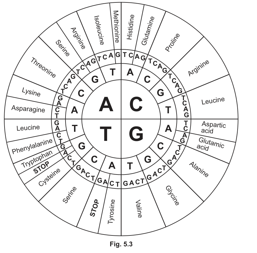
Dipeptides are sometimes translated from TERRA RNA.
Use Fig. 5.3 to state the two amino acids in the dipeptide translated from TERRA RNA.
1 ______
2 ______
Working
The TERRA RNA sequence is 5′-UUAGGG-3′. Group the bases into triplets starting from the first base:
Using the genetic code:
- UUA → Leucine
- GGG → Glycine
Answer
- Leucine
- Glycine
- Leucine; 2. Glycine
Background Concept
The genetic code is read in triplets (codons), each codon specifying one amino acid. On an mRNA molecule the codons are read 5′→3′, in non-overlapping groups of three. A dipeptide consists of two amino acids joined by a single peptide bond and is therefore the product of two codons (six bases) of mRNA.
In a double-stranded DNA gene, the strand that is not used as the template during transcription is called the non-transcribed (or sense / coding) strand. Its sequence matches the mRNA exactly except that T is present where the mRNA has U. The circular genetic code chart in Fig. 5.3 lists DNA triplets on this non-transcribed strand, so the codon for the first amino acid can be read directly by converting the mRNA codon back to its DNA equivalent (U → T) and using the chart.
Understanding the Question
The TERRA RNA is UUAGGG (from part (b)(i)). The question asks for the two amino acids of the dipeptide translated from TERRA. The figure provides a circular code with DNA triplets on the non-transcribed strand, read from the centre (first base) outwards. Two marks are available, one per amino acid.
Approach
- Group the six RNA bases into two non-overlapping triplets starting at the 5′ end: UUA then GGG.
- Convert each mRNA triplet to its equivalent DNA triplet on the non-transcribed strand (replace U with T): TTA and GGG.
- Use the chart, starting from the centre, to read off the amino acid for each triplet.
Step-by-Step Reasoning
- Step 1 — split the mRNA into codons:
TERRA = UUA GGG → two codons: UUA and GGG. - Step 2 — convert to the DNA form used by the chart:
UUA → TTA, and GGG → GGG (no change, since there is no U in this codon). - Step 3 — read the chart:
- TTA: start with T in the centre, then T as the second base (on the T side), then A as the third base → Leucine.
- GGG: start with G in the centre, then G as the second base, then G as the third base → Glycine.
- The dipeptide is therefore leucine–glycine (or glycine–leucine depending on which is read as amino acid 1, but the mark scheme credits 'leucine; glycine' as the two answers).
The mark scheme offers error carried forward (ecf) for the alternative reading of asparagine and proline, which corresponds to reading the codons in a different frame or a different interpretation, but the standard correct reading is leucine and glycine.
Key Takeaways
- mRNA is read in non-overlapping triplets of three bases from the 5′ end.
- A dipeptide is the product of two codons (six bases).
- The non-transcribed (sense) DNA strand has the same sequence as the mRNA except T replaces U, which lets a DNA-based code chart be used directly.
- A circular genetic code chart is read from the centre (1st base) outwards (2nd, then 3rd base).
Common Mistakes
- Reading the chart with the wrong base first (using the 3′ end instead of the 5′ end) — codons are read 5′→3′.
- Forgetting to convert U to T before using a DNA-based code chart, leading to wrong amino acids.
- Reading overlapping triplets (UAG, GGG…) instead of non-overlapping triplets (UUA, GGG).
- Confusing the non-transcribed strand with the transcribed (template) strand, which would be the complement of the mRNA and would give a different amino acid sequence.
Things to Be Careful About
- The figure shows DNA triplets on the non-transcribed strand, so U in the mRNA must be converted to T before using the chart.
- The reading direction is 5′→3′ on the mRNA, which corresponds to a fixed direction around the circular chart (centre → middle ring → outer ring).
- The ecf in the mark scheme accepts 'asparagine and proline' if a different reading frame is used; only credit the standard 'leucine; glycine' if the correct frame is used.
Scientists have discovered that TERRA interacts with genes in stem cells.
Increased concentrations of TERRA in stem cells result in a large increase in the number of genes that are transcribed.
Suggest the result of the large increase in the number of genes that are transcribed in stem cells.
Answer
A greater variety of proteins is produced, so the stem cell can differentiate / become specialised into different cell types.
Increase in number of different proteins produced, allowing the stem cell to differentiate / become specialised.
Background Concept
Stem cells are unspecialised cells that retain the ability to divide and to differentiate into a range of specialised cell types. Differentiation depends on selective gene expression: different cells transcribe different subsets of their genes, producing different mRNAs and therefore different proteins (especially transcription factors, enzymes and structural proteins) that give each cell type its identity and function.
If many more genes are transcribed at once, the cell gains access to a wider repertoire of mRNAs, and therefore a wider repertoire of proteins, than would otherwise be possible. In a stem cell, this expanded protein repertoire provides the raw material from which the cell can choose a particular differentiation pathway.
Understanding the Question
The question states that TERRA interacts with genes in stem cells and that a large increase in TERRA concentration leads to a large increase in the number of genes transcribed. The student must suggest ONE plausible biological consequence for the stem cell. One mark is available, so a single well-articulated point is sufficient.
Approach
Trace the flow of information from DNA → mRNA → protein → cellular function. An increase in the number of transcribed genes means an increase in the variety of mRNAs and therefore proteins the cell can make. In a stem cell, the most biologically significant consequence is the ability to differentiate into a wider range of specialised cell types.
Step-by-Step Reasoning
- More genes transcribed → more different mRNA molecules available in the cell.
- More different mRNAs → more different proteins can be translated (the mark scheme accepts 'enzymes' as an alternative specific form of 'protein').
- In a stem cell, this expanded protein set provides the molecular machinery needed for the cell to differentiate (become specialised / take on a particular function) or to grow and produce organelles in preparation for mitosis.
The mark scheme credits any one of:
- increase in number of different proteins (or enzymes) produced;
- reference to the cell's ability to differentiate / become specialised / take on a particular function;
- increase in growth of the cell in preparation for mitosis;
- production of cell organelles in preparation for mitosis.
Key Takeaways
- Gene expression is the link between the genome and cellular phenotype.
- A wider range of transcribed genes gives a wider range of proteins, which in stem cells supports differentiation.
- This is an 'outline' / 'suggest' question, so a single well-articulated downstream consequence is sufficient for the mark.
Common Mistakes
- Saying 'more protein is produced' without specifying 'different' kinds — quantity alone is not the right idea here, the point is variety.
- Saying 'more mRNA is produced' instead of more different types of mRNA / protein.
- Suggesting that the cell becomes cancerous — the question is about stem cells, not uncontrolled division.
- Confusing transcription with translation: the question explicitly says 'genes that are transcribed', so the first consequence is at the mRNA level.
Things to Be Careful About
- The command word is 'suggest', so a plausible biological consequence is required — there is no single 'correct' answer, but the mark scheme lists the creditable options.
- 'Differentiation' or 'becoming specialised' is a high-yield answer that links gene expression to stem-cell function.
Monoclonal antibodies can be used in the treatment of disease.
Answer
- Inject a (non-self / foreign / specific) antigen into a small mammal (e.g. a mouse).
- Leave time (over several weeks) for an immune response to occur.
- Remove splenocytes (B-lymphocytes / plasma cells) from the spleen.
- Fuse the splenocytes with myeloma (tumour) cells using a fusogen (e.g. polyethylene glycol) to form hybridoma cells.
- Screen and select the hybridoma cells that produce the desired antibody.
- Clone the selected hybridoma cells to produce large quantities of identical (monoclonal) antibodies.
See working.
Background Concept
Monoclonal antibodies are identical antibody molecules produced by a single clone of B-lymphocytes, all specific for one antigenic determinant (epitope). In the body, however, when an antigen is encountered many different B-cell clones are activated, each producing a slightly different antibody (polyclonal response). To obtain a single, pure antibody in large amounts, scientists exploit the fact that:
- B-lymphocytes / plasma cells make the desired specific antibody, but cannot divide indefinitely in culture.
- Myeloma cells (a type of cancerous B-cell tumour) divide rapidly in culture indefinitely, but produce the wrong antibody (or none).
Fusing the two cell types produces a hybridoma — a hybrid cell that combines the antibody-producing ability of the B-lymphocyte with the immortal, rapidly dividing property of the myeloma. Each hybridoma is a clone producing one specific antibody; growing it in culture yields large quantities of that monoclonal antibody.
Understanding the Question
Part (a) is a 5-mark 'describe' question on the hybridoma method. The command word describe requires a sequenced account of the key steps; up to seven marking points are available and any five earn full credit. The figure for part (b) and the passage for part (c) are independent of this part.
Approach
Lay out the procedure in the order it is actually carried out in the lab:
- Stimulate the immune response in a mammal.
- Isolate the antibody-producing cells.
- Fuse them with immortal myeloma cells.
- Select the successful hybrids (hybridomas).
- Grow the chosen hybridomas in bulk.
This logical sequence mirrors the mark scheme and shows the examiner you understand the purpose of each step.
Step-by-Step Reasoning
- Antigen injection: A specific (non-self) antigen is injected into a mouse (or other small mammal). This is necessary to trigger an immune response so the animal produces B-lymphocytes specific to that antigen. Mark point 1.
- Time for immune response: The animal is left for several weeks so that B-lymphocytes can be activated, proliferate and undergo clonal selection. Mark point 2.
- Harvest splenocytes: B-lymphocytes / plasma cells are removed from the spleen (which is a major site of lymphocyte activation and proliferation). Mark point 3.
- Cell fusion: The B-lymphocytes are fused with myeloma (cancerous) cells, usually using polyethylene glycol (PEG) as a fusogen, to make hybridoma cells. Mark points 4 and 7 (AVP — fusogen).
- Screening and selection: The mixture of cells is cultured in a selective medium (e.g. HAT medium) so that only hybridomas survive. They are then screened to identify those producing the desired antibody. Mark points 5 and 7 (AVP — HAT).
- Cloning: Selected hybridoma cells are cloned (e.g. by limiting dilution into separate wells) so each well contains a clone producing identical antibody molecules. Mark points 6 and 7 (AVP — separate wells).
- AVP — humanising: For therapeutic use in humans, the mouse antibody genes are often combined with human constant regions ('humanised') to reduce immune rejection. Mark point 7.
The simplest five-point answer covers: antigen injection → time for response → harvest splenocytes → fuse with myeloma → screen and select → clone. Most candidates hit five marks with these.
Key Takeaways
- The hybridoma technique solves the problem of producing unlimited quantities of a single, specific antibody.
- The procedure combines the antibody specificity of B-lymphocytes with the immortality of myeloma cells.
- Selective media (e.g. HAT) and screening are essential to isolate the rare hybridoma that produces the antibody of interest.
Common Mistakes
- Saying 'take antibodies from the mouse' — the antibodies themselves are not harvested; the B-lymphocytes (which make them) are removed so they can be fused.
- Omitting the fusion step or confusing which cells fuse with which.
- Skipping the screening step — without it, you would not know which hybridomas make the right antibody.
- Describing only the in-vivo part (antigen injection) and forgetting the in-vitro fusion and culture steps.
Things to Be Careful About
- 'Splenocytes' can be credited as 'plasma cells' or 'B-lymphocytes', but not simply 'blood cells' or 'white blood cells'.
- The myeloma cells must be described as cancerous / tumour cells (not just any dividing cell).
- 'Humanising' is bonus AVP credit and not required for the basic 5 marks.
Abciximab is a drug developed from a monoclonal antibody. Abciximab is used to prevent blood clotting in the coronary arteries in people with coronary heart disease.
Abciximab prevents blood clotting by stopping structures called platelets from binding together.
Fig. 6.1 shows platelets and abciximab in a coronary artery.
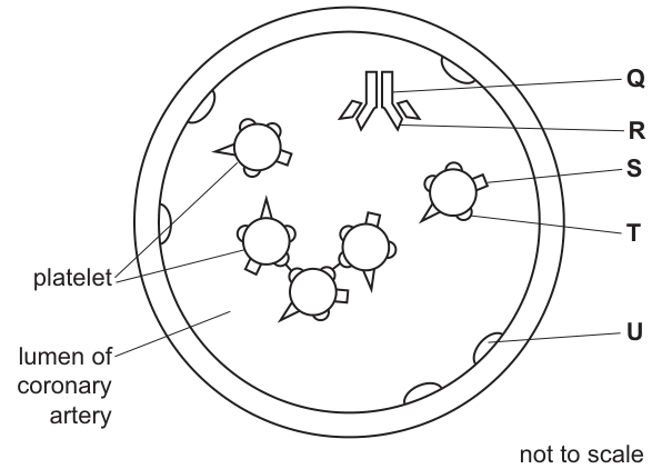
State the letter in Fig. 6.1 that represents:
• the target antigen for abciximab ______
• the constant region of abciximab ______
• one of the antigen binding sites of abciximab ______ .
Answer
- Target antigen for abciximab: S
- Constant region of abciximab: R
- One of the antigen binding sites of abciximab: Q
S, R, Q
Background Concept
An antibody (immunoglobulin) has a characteristic Y-shape made of four polypeptide chains — two heavy chains and two light chains — held together by disulfide bonds. It has two functionally distinct regions:
- Variable region: the tips of the two arms of the Y. The amino-acid sequence here varies between antibodies and forms the antigen-binding site (two per antibody molecule, one on each arm tip). This is the region that is complementary to, and binds, a specific epitope on the antigen.
- Constant region: the stem of the Y and the lower parts of the arms. Its amino-acid sequence is the same for all antibodies of the same class. It determines the antibody's class (e.g. IgG) and how the immune system destroys the bound antigen (e.g. by recruiting complement or phagocytes).
Abciximab is a monoclonal antibody whose antigen-binding sites are complementary to a specific receptor (an antigenic protein) on the surface of platelets. By binding this receptor, it prevents platelets from sticking together, so it acts as an anti-platelet-aggregation drug in coronary heart disease.
Understanding the Question
Fig. 6.1 shows a coronary artery in cross-section with several platelets floating in the lumen and Y-shaped abciximab molecules. The diagram labels five structures: Q (tip of an abciximab arm), R (the stem of an abciximab molecule), S (a small structure on a platelet surface — the receptor/antigen), T (an entire platelet) and U (the artery wall).
The question asks you to name the letter for:
- The target antigen for abciximab — i.e. the molecule on the platelet that the antibody binds.
- The constant region of abciximab — i.e. the part of the antibody that is the same in every molecule of this antibody.
- One of the antigen binding sites of abciximab — i.e. one of the two tips of the Y, where the variable region contacts the antigen.
Approach
For each blank, match the description to what each label points to:
- Q points to a tip of the Y-shaped abciximab — the antigen-binding site (variable region tip).
- R points to the stem of the Y — the constant region.
- S points to a small receptor sticking out of a platelet — the target antigen (the platelet surface receptor that abciximab binds).
- T points to the whole platelet (the cell, not a part of it).
- U points to the artery wall — irrelevant here.
Step-by-Step Reasoning
- Target antigen (S): Abciximab is specific for a receptor on the platelet surface; this is the antigen the antibody recognises. The small structure on the platelet labelled S is that receptor, so S is the target antigen. (T — the whole platelet — is also accepted by the mark scheme because platelets as a whole expose the antigen, but S is the more precise answer.)
- Constant region (R): The stem of the Y is identical in every abciximab molecule, so the arrow at R indicates the constant region.
- Antigen binding site (Q): Each tip of the Y forms one antigen-binding site. The arrow at Q points to such a tip — it sits directly over the platelet-receptor S, illustrating binding.
Key Takeaways
- An antibody's variable region (tips of the Y) determines antigen specificity; its constant region (stem) determines class and effector function.
- The target of a therapeutic monoclonal antibody is a specific molecular antigen (here, a platelet surface receptor), not the whole cell.
- Reading a biological diagram is often a matter of matching a description to the labelled structure and recalling the basic structure of an antibody.
Common Mistakes
- Picking T (the platelet) for the target antigen — platelets are cells, not antigens; the receptor on the platelet is the antigen. T is accepted on this mark scheme, but S is the more accurate answer.
- Confusing the variable and constant regions of the antibody: the variable region is at the tips, the constant region is the stem.
- Mixing up the antigen binding site with the antigen itself — the binding site is on the antibody (Q), the antigen is on the platelet (S).
Things to Be Careful About
- The mark scheme accepts 'T / S' for the target antigen — so T (the whole platelet) is allowed because the antigen is on the platelet. S is preferred.
- 'Q' could plausibly be the tip of the variable region or the variable region as a whole; either way it is the antigen binding site.
Coronary arteries supply oxygenated blood to the cells of the heart.
The passage outlines the cardiac cycle of the heart and the structures in the heart that control the cycle.
Complete the passage by using the most appropriate scientific terms.
The sinoatrial node is located in the wall of the ______,
one of four chambers of the mammalian heart. Electrical impulses from the sinoatrial node
reach the atrioventricular node, which transmits impulses towards the apex of the heart along
a series of specialised muscle fibres called ______.
Pressure increases in the ventricles when they contract in the stage of the cardiac cycle
known as ______.
Answer
The sinoatrial node is located in the wall of the right atrium, one of four chambers of the mammalian heart. Electrical impulses from the sinoatrial node reach the atrioventricular node, which transmits impulses towards the apex of the heart along a series of specialised muscle fibres called Purkyne fibres (Purkyne tissue). Pressure increases in the ventricles when they contract in the stage of the cardiac cycle known as ventricular systole.
Right atrium; Purkyne fibres/tissue; ventricular systole
Background Concept
The mammalian heart is a myogenic organ — it generates its own electrical impulses rather than relying on nervous stimulation. The rhythmic contractions are coordinated by a specialised conducting system made of modified cardiac muscle:
- Sinoatrial node (SAN) — a small patch of pacemaker tissue in the wall of the right atrium, near the entry of the superior vena cava. It sets the basic rate of contraction by spontaneously depolarising fastest.
- Atrioventricular node (AVN) — in the septum between the atria; receives the wave of excitation from the SAN and delays it briefly so the atria finish contracting before the ventricles do.
- Bundle of His / Purkyne fibres — specialised muscle fibres that carry the impulse from the AVN down through the ventricular septum, around the apex and up the ventricular walls, causing the ventricles to contract from the apex upwards (efficiently squeezing blood out into the arteries).
The cardiac cycle has three named stages:
- Atrial systole — atria contract and push blood into the ventricles.
- Ventricular systole — ventricles contract; pressure inside rises sharply, the AV valves snap shut ('lub' sound) and blood is forced out through the semilunar valves into the pulmonary artery and aorta.
- Diastole — whole heart relaxes; blood flows into the atria and ventricles; semilunar valves close ('dup' sound).
Understanding the Question
The passage in part (c) is a fill-in-the-blanks exercise testing three core facts:
- The exact location of the sinoatrial node (which chamber wall).
- The name of the specialised muscle fibres that conduct impulses from the AVN to the apex.
- The name of the cardiac cycle stage in which ventricular contraction raises ventricular pressure.
The question is a 'state' / completion task — the exact scientific term is required for each mark.
Approach
For each blank, reach for the precise anatomical / physiological term:
- Wall of the … (chamber containing the SAN): the SAN is in the right atrium (some animals have it associated with the left atrium, but in humans and most mammals it is the right atrium).
- Specialised muscle fibres (between AVN and apex): Purkyne fibres (or Purkinje fibres), also creditable as Bundle of His.
- Stage when ventricles contract: ventricular systole.
Step-by-Step Reasoning
- Right atrium: The SAN sits in the upper posterior wall of the right atrium. This is the most common point of origin of the heartbeat, so the right atrium is correct.
- Purkyne fibres: Once the impulse has been delayed at the AVN, it travels down the interventricular septum via the Bundle of His, which then branches into Purkyne fibres that distribute the impulse rapidly through the ventricular myocardium. The Bundle of His is an alternative credit-worthy answer; the question asks for the fibres along which the impulse reaches the apex, so Purkyne fibres is the most precise answer.
- Ventricular systole: When the ventricular muscle contracts, the ventricular volume decreases sharply and intraventricular pressure rises above atrial pressure (closing the AV valves) and then above arterial pressure (opening the semilunar valves). This is ventricular systole — distinct from atrial systole (which only raises atrial and then early ventricular pressure as blood is forced in) and diastole (relaxation).
Key Takeaways
- The heart is myogenic; the SAN in the wall of the right atrium is the natural pacemaker.
- The conducting pathway is SAN → AVN → Bundle of His → Purkyne fibres → ventricular muscle.
- Ventricular systole is the stage of the cardiac cycle when ventricular pressure rises as the muscle contracts.
Common Mistakes
- Writing 'left atrium' for the SAN location — the SAN is on the right.
- Spelling 'Purkyne' as 'Purkinje' (an alternative spelling is allowed by the mark scheme, but the CIE-preferred spelling is 'Purkyne'); the examiner also credits 'Bundle of His', but not just 'nerve' or 'muscle'.
- Writing 'systole' alone without specifying 'ventricular' — the question asks for the stage when the ventricles contract, so 'ventricular systole' is required.
- Confusing systole (contraction) with diastole (relaxation).
Things to Be Careful About
- The mark scheme accepts 'Purkyne tissue / fibres' or 'Purkinje tissue / fibres' or 'Bundle of His' as alternative answers for the second blank.
- 'Right atrium' must be fully stated — 'atrium' alone is too vague (the heart has two atria).
- The phrase 'ventricular systole' is two words; do not collapse them into 'ventricularsystole' or 'ventricle systole'.