Biology 9700/22 — May/June 2025
Cambridge AS Level · AS Level Structured Questions · worked solutions for every part, with the mark scheme
Topics Immunity · Transport in Plants · Biological Molecules · Transport in Mammals · Nucleic Acids and Protein Synthesis · Cell Structure · +5 more
Root hair cells are specialised plant cells located in the outer layer of young roots of plants. Root hair cells have an essential role in the uptake of water and dissolved mineral ions from the soil solution.
The transport of water across the root to reach the central xylem tissue can occur by the symplast pathway or apoplast pathway.
Fig. 1.1 is a diagram of a photomicrograph of a root hair cell.
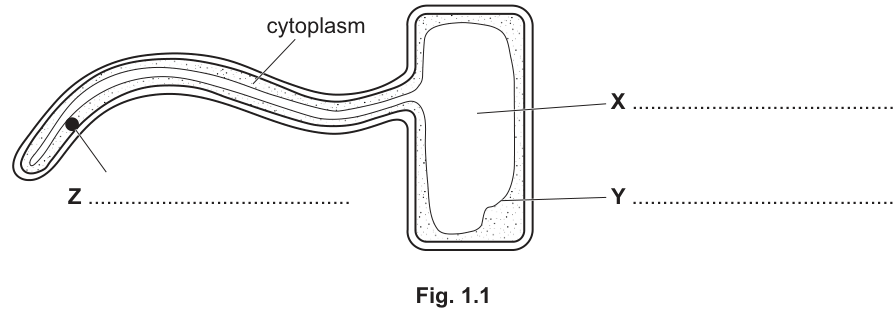
On Fig. 1.1, name cell structures X, Y and Z.
Z = ______
X = ______
Y = ______
Answer
- X = (large / permanent) vacuole
- Y = tonoplast (vacuolar membrane)
- Z = nucleus
X = vacuole, Y = tonoplast, Z = nucleus
Background Concept
A root hair cell is a highly specialised epidermal cell whose long, thin extension greatly increases the surface area in contact with soil water. As a plant cell, it possesses all the typical eukaryotic organelles, three of which are highlighted in Fig. 1.1:
- The nucleus is the largest organelle and contains the cell's DNA; it appears as a small, dense, often darker-stained body inside the cytoplasm.
- The vacuole (more correctly, the permanent/large central vacuole) is a fluid-filled sac of cell sap that occupies most of the volume of a mature plant cell. Because it is mostly water with dissolved solutes, it often looks like an empty space in a micrograph.
- The tonoplast is the single membrane that bounds the vacuole and separates the cell sap from the surrounding cytoplasm. Like the cell surface membrane, it is selectively permeable.
Understanding the Question
The question shows a photomicrograph-style diagram of a root hair cell with three pointer labels (X, Y and Z) and asks the candidate to name each. From the position of each label on the diagram:
- Z points to a small dark oval near the tip of the root hair extension.
- X points to the large pale area filling the main cell body.
- Y points to a line bounding the central pale area.
Approach
Match each label to the most likely plant cell organelle based on size, position and appearance in the diagram. The dark small body must be the nucleus, the large empty region must be the vacuole, and the surrounding membrane of the vacuole must be the tonoplast (not the outer cell wall, which is the outermost line of the diagram).
Step-by-Step Reasoning
- Z sits at the tip of the root hair as a small, darkly stained body — this is the nucleus, whose appearance reflects the dense chromatin it contains.
- X lies inside the main cell body, occupying the bulk of the cell — this is the large vacuole, which appears empty because it is filled with watery cell sap.
- Y is a distinct line forming the boundary of that large empty area — this is the tonoplast (the membrane that encloses the vacuole), not the outer cell wall.
Key Takeaways
- The root hair cell has the same three basic features as other mature plant cells: nucleus, large central vacuole, and a tonoplast.
- The large central vacuole is a defining feature of plant cells; it pushes the cytoplasm into a thin layer against the cell wall.
- The tonoplast is a distinct membrane from the cell surface membrane; both are selectively permeable, but the cell wall is freely permeable.
Common Mistakes
- Calling Y the cell wall or cell surface membrane — Y is the membrane around the vacuole, i.e. the tonoplast.
- Confusing the cytoplasm (which is labelled) with the vacuole.
- Spelling "tonoplast" as "tonoplast membrane" or "tonoplasm" — only "tonoplast" or "vacuolar membrane" is credited.
Things to Be Careful About
- The diagram is in 2-D; the large pale area is the vacuole and the line around it is the tonoplast.
- "Vacuole" alone is credited; "large vacuole" or "permanent vacuole" is also accepted but "food vacuole" or "contractile vacuole" is wrong in this context.
In the symplast pathway, water passes through the cells of the different tissues in the root before entering the xylem vessels of xylem tissue.
Name the tissues of the root, in the correct sequence, through which water passes in the symplast pathway.
Answer
The tissues, in the order water passes through them, are:
epidermis (epidermal tissue) → cortex (cortical tissue) → endodermis (endodermal tissue) → pericycle → xylem
Any three of these named in the correct sequence is sufficient for full credit (e.g. epidermis, cortex, endodermis).
epidermis, cortex, endodermis, pericycle, xylem (in this sequence)
Background Concept
A young root is organised into concentric tissue layers, beginning at the outside and moving towards the centre:
- Epidermis — the outermost single cell layer; root hair cells are modified epidermal cells.
- Cortex — several layers of loosely packed parenchyma cells with many intercellular spaces.
- Endodermis — a single layer of tightly packed cells with a waterproof Casparian strip in their walls.
- Pericycle — a layer of meristematic cells just inside the endodermis; gives rise to lateral roots and part of the vascular cambium.
- Xylem — the central water-conducting tissue, whose vessels are dead, hollow tubes reinforced with lignin.
In the symplast pathway water moves from cell to cell through the cytoplasm, passing through plasmodesmata (cytoplasmic channels that connect adjacent cells). It does not cross any cell surface membrane or cell wall, so it remains inside the continuous cytoplasm of every cell it traverses.
Understanding the Question
The question asks for the tissues (not the cell types) through which water passes in the symplast pathway, given in the correct order. The mark scheme credits any three tissues in the correct sequence for two marks; only two in correct sequence earns one mark.
Approach
Visualise a root in cross-section and trace the path of water from the soil into the xylem, naming each tissue layer in turn. Remember that the symplast route takes water through the living cytoplasm, so it follows the living tissues of the root from outside to inside.
Step-by-Step Reasoning
- Water enters via a root hair cell, which is part of the epidermis.
- From the root hair cell it moves through plasmodesmata into adjacent cortical cells of the cortex.
- It then reaches the endodermis (forced into the symplast here because the Casparian strip blocks the apoplast route).
- It continues into the pericycle.
- Finally it enters the xylem vessels of the vascular tissue, where bulk flow up the plant begins.
Any three of these five tissues, written in this order, scores both marks. The most common short answer is: epidermis, cortex, endodermis.
Key Takeaways
- The symplast route passes through living tissues only: epidermis → cortex → endodermis → pericycle → xylem.
- The Casparian strip at the endodermis blocks the apoplast pathway and forces water into the symplast.
- Once water enters the xylem, it leaves the symplast and moves by bulk flow driven by transpiration pull.
Common Mistakes
- Including the phloem in the sequence — phloem transports assimilates, not water, and is not part of the symplast water pathway.
- Listing cell types (e.g. "root hair cell", "xylem vessel element") rather than tissues — the mark scheme prefers the tissue names.
- Stating only two tissues in the correct sequence (only 1 mark).
- Reversing the order (e.g. "xylem, cortex, epidermis").
Things to Be Careful About
- "Cortex" is credited; "cortical cells" or "parenchyma" are also accepted but "phloem" is rejected.
- The xylem is the final tissue, not the starting one.
- The endodermis is the layer that channels water from the apoplast into the symplast.
In the apoplast pathway, water passes along the cell walls of adjacent plant cells and through the intercellular spaces. This is more efficient than the symplast pathway.
Explain the structural features of plant cell walls that make the apoplast pathway an efficient pathway for the transport of water.
Answer
Any three of the following points:
- Cellulose (molecules / fibres) are hydrophilic, allowing adhesion of water molecules to the cell wall.
- The arrangement of cellulose fibres creates numerous small spaces / gaps through which water can flow.
- Cell walls are freely permeable to water (i.e. they offer the path of least resistance, with no selective barrier to water movement).
- Cellulose molecules form hydrogen bonds with water molecules, holding water within the wall and aiding its movement.
- Hemicellulose and pectin are also hydrophilic, further attracting and holding water in the wall matrix.
Cellulose is hydrophilic; cellulose arrangement gives water-filled spaces; the cell wall is freely permeable to water; hydrogen bonds form between cellulose and water; hemicellulose and pectin are also hydrophilic (any three).
Background Concept
The apoplast is the continuous network of cell walls and intercellular spaces that runs through a plant tissue without crossing any cell surface membrane. Because the cell surface membrane (and especially its hydrophobic phospholipid bilayer core) is the main barrier to water movement, the apoplast offers a much lower-resistance route for water than the symplast.
The plant cell wall is built from:
- Cellulose — long, unbranched chains of β-glucose linked by β-1,4 glycosidic bonds. Many chains lie parallel and are held together by hydrogen bonds to form cellulose microfibrils. Each glucose monomer carries three –OH groups, so the surface of a microfibril is densely decorated with hydroxyl groups.
- Hemicellulose — branched polysaccharides that cross-link microfibrils.
- Pectin — a gel-like matrix of acidic polysaccharides rich in carboxyl groups, which trap water.
- Lignin (in some walls) — a hydrophobic polymer for strength; absent from primary walls.
The hydroxyl groups on cellulose (and carboxyl groups on pectin) are hydrophilic and form hydrogen bonds with water. Between the microfibrils and through the pectin matrix there are numerous sub-microscopic spaces, giving a sponge-like structure through which water can flow freely.
Understanding the Question
The question explains that the apoplast pathway carries water along cell walls and through intercellular spaces, and is more efficient than the symplast route. The candidate must explain why the structure of the cell wall makes it efficient for water transport. Three marking points are needed.
Approach
Think about two things:
- The chemistry of the wall components (hydrophilic? capable of hydrogen bonding?).
- The physical structure of the wall (does it allow water through? are there spaces? is it selective?).
Then express each as a separate credit-worthy point.
Step-by-Step Reasoning
- Hydrophilicity: cellulose molecules carry many –OH groups, so they are hydrophilic and allow water to adhere to the wall surface. (1 mark)
- Spaces for flow: cellulose microfibrils are not packed tightly; the gaps between them, and the pectin matrix, provide channels through which water can flow continuously. (1 mark)
- Freely permeable: the cell wall is not a selective barrier — unlike the cell surface membrane, it does not restrict water. This means water moves with the minimum of resistance along the wall. (1 mark)
- (Alternative) Hydrogen bonding: the –OH groups on cellulose form hydrogen bonds with water molecules, drawing water into and along the wall. (1 mark)
- (Alternative) Hemicellulose/pectin hydrophilic: these additional wall components are also hydrophilic and so attract and retain water in the wall. (1 mark)
Any three of the above earn full credit.
Key Takeaways
- Cellulose is a β-1,4-glucose polymer whose many hydroxyl groups make the wall strongly hydrophilic.
- The cell wall is a porous, mesh-like network of microfibrils and matrix; water moves through it by bulk flow because there is no membrane barrier.
- Hydrogen bonding between cellulose (and pectin) and water holds water in the wall and supports its movement.
- The freely permeable nature of the wall is the main reason the apoplast pathway is faster than the symplast.
Common Mistakes
- Saying the cell wall is "impermeable" or "selectively permeable" — the cell wall is freely permeable.
- Confusing the cell wall with the cell surface membrane.
- Vague answers like "the wall is porous" or "it has gaps" without naming the structure that produces the gaps (cellulose microfibrils / pectin matrix).
- Mentioning lignin — primary cell walls, which are what water moves through in the apoplast, contain little or no lignin.
Things to Be Careful About
- Use the precise term "freely permeable" for the cell wall, not "selectively permeable" (that is the cell surface membrane).
- Hydrophilic = "water-attracting"; can also be expressed as forming hydrogen bonds with water.
- The apoplast route is blocked at the endodermis by the Casparian strip — so it is not the only pathway, but it is the most efficient within a tissue.
Root hair cells also synthesise and secrete substances into the soil.
Electron microscopy of the structure of root hairs has identified endoplasmic reticulum (ER), a number of small Golgi bodies, and numerous vesicles.
• Root hair cells of the barley plant secrete enzymes known as acid phosphatases, which catalyse the release of inorganic phosphate ions from organic phosphates in the soil.
• Root hair cells of the sorghum plant secrete a hydrophobic, lipid compound known as sorgoleone, which slows down the growth of neighbouring plants.
Explain why the proportion of rough ER to smooth ER may be different in the root hair cells of barley plants compared with the root hair cells of sorghum plants.
Answer
- Rough ER is the site of protein (enzyme) synthesis, whereas smooth ER is the site of lipid (e.g. sorgoleone) synthesis.
- Therefore, the barley root hair cells (which secrete the enzyme acid phosphatase) will have a higher proportion of rough ER and a lower proportion of smooth ER, while the sorghum root hair cells (which secrete the lipid sorgoleone) will have a higher proportion of smooth ER and a lower proportion of rough ER.
Rough ER synthesises proteins/enzymes; smooth ER synthesises lipids. Barley (enzyme-secreting) has more rough ER; sorghum (lipid-secreting) has more smooth ER.
Background Concept
The endoplasmic reticulum (ER) is a continuous network of flattened sacs (cisternae) and tubules that runs through the cytoplasm. There are two functionally distinct regions:
- Rough ER has ribosomes on its outer surface, giving it a rough appearance in electron micrographs. These ribosomes synthesise proteins that enter the ER lumen, where the proteins are folded and may be modified (e.g. by glycosylation). Rough ER is therefore abundant in cells that produce large amounts of protein for secretion — for example, the cells of the pancreas that secrete digestive enzymes, or plasma cells that secrete antibodies.
- Smooth ER lacks ribosomes and so has a smooth appearance. It is the site of lipid synthesis (including phospholipids and steroids), and is also involved in lipid metabolism, carbohydrate metabolism and detoxification of drugs. Cells that secrete lipid-based products (e.g. sebaceous gland cells, or root hair cells secreting sorgoleone) contain large amounts of smooth ER.
Understanding the Question
The question tells us that barley root hair cells secrete acid phosphatase (an enzyme, i.e. a protein) while sorghum root hair cells secrete sorgoleone (a hydrophobic lipid). It then asks why the proportion of rough ER to smooth ER may differ between the two cell types. Two marking points are available.
Approach
For each cell type, identify the type of secretory product, link it to the type of ER that synthesises it, and then state the consequence for the ER composition of that cell.
Step-by-Step Reasoning
- Rough ER is the site of protein (enzyme) synthesis; smooth ER is the site of lipid synthesis (1 mark).
- Barley cells secrete acid phosphatase, which is a protein/enzyme — these cells therefore have a higher proportion of rough ER and a lower proportion of smooth ER.
- Sorghum cells secrete sorgoleone, which is a lipid — these cells therefore have a higher proportion of smooth ER and a lower proportion of rough ER (1 mark).
Key Takeaways
- The dominant secretory product of a cell determines the relative amounts of rough vs smooth ER it contains.
- Protein-secreting cells (e.g. barley root hairs, pancreatic acinar cells, plasma cells) have lots of rough ER.
- Lipid-secreting cells (e.g. sorghum root hairs, sebaceous gland cells) have lots of smooth ER.
- "Rough" refers to the ribosomes on the surface; "smooth" ER lacks ribosomes.
Common Mistakes
- Stating the wrong ER type for the wrong product (e.g. saying barley has more smooth ER).
- Vague answers like "barley has more ER" without specifying the type.
- Saying "barley cells have more ribosomes" without mentioning rough ER specifically.
- Confusing the function of the ER with that of the Golgi apparatus.
Things to Be Careful About
- The mark scheme requires the link between ER type and the type of molecule being secreted (protein vs lipid), not just a statement that one ER type is more abundant.
- Use the precise terms "rough ER" and "smooth ER" — "granular" and "agranular" ER are not standard CIE terminology.
- The question is about proportion; either stating barley has more rough ER or sorghum has more smooth ER is enough for the second mark.
Student X stated that acid phosphatases and sorgoleone could be transported out of root hair cells using the same process.
Student Y stated that acid phosphatases and sorgoleone are transported out of root hair cells using different processes.
Suggest the reasons given by student X and by student Y to support their statements.
student X = ______
student Y = ______
Answer
Student X — same process (exocytosis for both):
- Both acid phosphatase and sorgoleone could leave the cell by exocytosis.
- Exocytosis allows bulk transport of large quantities and molecules that are too large to exit by other means.
- The presence of Golgi bodies and vesicles in root hair cells supports this: molecules are packaged into Golgi (secretory) vesicles that move to and fuse with the cell surface membrane, releasing their contents outside.
Student Y — different processes:
- Acid phosphatase is a hydrophilic protein and so cannot pass through the hydrophobic core of the phospholipid bilayer directly. It must cross the cell surface membrane by facilitated diffusion through transport (channel / carrier) proteins, down its concentration gradient.
- Sorgoleone is a hydrophobic lipid and so dissolves in the phospholipid bilayer; it can cross the cell surface membrane by simple diffusion down its concentration gradient, without the need for transport proteins.
- Because the two molecules have opposite solubilities, they are transported by different mechanisms.
Student X: both are released by exocytosis (bulk transport, via Golgi vesicles fusing with the cell surface membrane). Student Y: enzymes (hydrophilic) use facilitated diffusion through transport proteins; sorgoleone (hydrophobic) uses simple diffusion through the phospholipid bilayer.
Background Concept
Molecules cross the cell surface membrane by different mechanisms depending on their size, polarity and concentration gradient:
- Simple diffusion is the passive movement of small, non-polar (hydrophobic) molecules directly through the phospholipid bilayer, down a concentration gradient. No proteins and no ATP are required. Examples: O₂, CO₂, steroid hormones, and other lipid-soluble molecules such as sorgoleone.
- Facilitated diffusion is the passive movement of larger or polar (hydrophilic) molecules through specific transport proteins (channel or carrier proteins), still down a concentration gradient and without ATP. Examples: glucose, ions, amino acids, water (via aquaporins), and hydrophilic proteins that can still fit through a channel.
- Exocytosis is the active, ATP-requiring process by which membrane-bound vesicles (typically derived from the Golgi apparatus) fuse with the cell surface membrane and release their contents to the outside. It is used for the bulk secretion of molecules that are too large or too polar to pass through the bilayer or through transport proteins, e.g. enzymes, hormones and mucins.
The phospholipid bilayer has a hydrophilic outer surface (phosphate heads) but a hydrophobic core (fatty acid tails). Hydrophobic molecules dissolve in this core and pass through easily; hydrophilic molecules are repelled by the core and must use a protein route.
Understanding the Question
The question presents two opposing student views:
- Student X: both molecules use the same process.
- Student Y: the two molecules use different processes.
Both views can be supported to some extent, but the most accurate biology is captured by student Y's argument, because the two molecules have opposite solubilities. The candidate must give reasoned arguments for both students. The mark scheme awards up to 3 marks for the X argument and up to 3 marks for the Y argument; the question is out of 4, so the examiner selects the best 4 marks across the two answers.
Approach
For each student, decide which mechanism best fits and back it up with the cellular machinery and the chemical properties of the molecules:
- Student X: argue for exocytosis as a common bulk-transport mechanism, citing the observed Golgi bodies and vesicles.
- Student Y: separate the two molecules on the basis of polarity and match each to its appropriate transport mechanism (facilitated diffusion for the hydrophilic enzyme; simple diffusion for the hydrophobic lipid).
Step-by-Step Reasoning
Student X (max 3 marks):
- States that both molecules leave the cell by exocytosis (1 mark).
- Adds that exocytosis is suitable for bulk transport / large quantities and/or that the molecules may be too large to exit by other means (1 mark).
- Provides a detail about the cell's machinery: presence of (small) Golgi bodies and vesicles that move to and fuse with the cell surface membrane, releasing the contents outside (1 mark).
Student Y (max 3 marks):
- Recognises that the two molecules have different solubilities: acid phosphatase is hydrophilic (polar / not hydrophobic) and sorgoleone is hydrophobic (non-polar) (1 mark).
- Acid phosphatase therefore needs facilitated diffusion through a transport (channel or carrier) protein because it cannot pass through the hydrophobic core of the bilayer (1 mark).
- Sorgoleone, being hydrophobic, can dissolve in the phospholipid bilayer and cross the membrane by simple (passive) diffusion down its concentration gradient, with no protein required (1 mark).
- A further point is that the concentration gradient drives the movement of sorgoleone (1 mark, alternative to above).
(An alternative version of student Y's argument would pair sorgoleone with exocytosis and acid phosphatase with facilitated diffusion; the principle is the same — different mechanisms for different molecules.)
Key Takeaways
- The polarity of a molecule determines how it crosses a membrane: hydrophobic molecules can diffuse through the bilayer; hydrophilic molecules need a protein route or a vesicle.
- Exocytosis is used for bulk secretion of large molecules such as proteins and lipids that cannot cross the membrane one molecule at a time.
- The presence of abundant Golgi bodies and vesicles in an electron micrograph is a strong clue that the cell is actively secreting by exocytosis.
- A single, well-evidenced argument is worth more marks than several vague statements.
Common Mistakes
- Saying sorgoleone uses channel or carrier proteins (it is hydrophobic and does not need them).
- Saying acid phosphatase diffuses directly through the phospholipid bilayer (it is hydrophilic and cannot).
- Confusing exocytosis (out of the cell) with endocytosis (into the cell).
- Stating only "diffusion" without specifying simple or facilitated — these are different mechanisms with different requirements.
- Failing to mention the vesicles and Golgi when arguing for exocytosis.
- Confusing the phospholipid bilayer (hydrophobic core) with the cell wall (freely permeable).
Things to Be Careful About
- Use the exact terms: simple diffusion, facilitated diffusion and exocytosis — they are different and the mark scheme distinguishes them.
- "Hydrophilic" means water-soluble / polar; "hydrophobic" means water-insoluble / non-polar. These adjectives explain why each mechanism is needed.
- The question awards marks for reasoning as well as for naming the mechanism. A bare statement "both use exocytosis" with no supporting detail earns only one mark.
- Note that an alternative version of student Y is also accepted: sorgoleone leaves by exocytosis (because it is made in vesicles) and the enzyme leaves by facilitated diffusion. The biology is consistent — the mark scheme accepts multiple combinations as long as the reasoning is sound.
The transport of respiratory gases involves blood plasma and red blood cells. Red blood cells contain the globular protein, haemoglobin.
Describe features of a haemoglobin molecule that are typical of a globular protein, other than having an approximately spherical shape.
Answer
Any two from:
- Amino acids with hydrophilic R groups positioned on the outside of the molecule, facing the aqueous (watery) cytosol / cytoplasm; amino acids with hydrophobic R groups are located in the interior of the molecule.
- The molecule is soluble in water / in the cytoplasm of the red blood cell.
- It has a (physiological / metabolic / dynamic) role, e.g. carrying oxygen (and carbon dioxide).
- The amino acid sequence is non-repetitive / irregular (unlike a fibrous protein such as collagen).
- The 3D shape (tertiary / quaternary structure) is maintained by two or more different types of bonds (e.g. hydrogen, ionic, disulfide, hydrophobic interactions); not by peptide bonds.
Any two of the features listed above.
Background Concept
Proteins are built from amino acids linked by peptide bonds. The primary structure is the linear sequence of amino acids; the secondary, tertiary and (when present) quaternary structures are produced by folding driven by the side chains (R groups) of the amino acids. CIE classifies proteins into two broad structural categories: fibrous and globular.
- Fibrous proteins (e.g. collagen, keratin) have long, parallel polypeptide chains, are insoluble in water, and play a mainly structural role. Their amino acid sequence is often repetitive.
- Globular proteins (e.g. haemoglobin, enzymes, antibodies, some hormones) are folded into compact, approximately spherical shapes. The folding places amino acids with hydrophilic (polar) R groups on the outside, in contact with the aqueous cytosol or extracellular fluid, and amino acids with hydrophobic (non-polar) R groups in the interior, away from water. This makes the molecule soluble in water and gives it a physiological / metabolic (dynamic) role such as transport, catalysis or signalling.
The 3D shape of a globular protein is held in place by several different types of bonds acting together: hydrogen bonds, ionic bonds, disulfide bridges and hydrophobic interactions (not the peptide bonds of the primary structure, which simply link amino acids along the chain). In addition, the amino acid sequence in a globular protein is typically non-repetitive / irregular — a key contrast with the regular, repeating sequences seen in fibrous proteins such as collagen (Gly–X–Y repeats).
Haemoglobin is the classic globular protein example on the AS syllabus. It is a conjugated protein with four polypeptide chains (2 α, 2 β), each carrying a haem group, and its tertiary and quaternary structure shows the hydrophilic-outside / hydrophobic-inside arrangement that makes it soluble in the cytoplasm of the red blood cell.
Understanding the Question
Part (a) gives 2 marks. The command word is "describe features ... other than having an approximately spherical shape". The question is explicitly not asking the candidate to say that haemoglobin is spherical — that feature is excluded. The candidate must pick two other features that haemoglobin shares with globular proteins in general, and which distinguish globular from fibrous proteins.
The stem supplies the biological setting: haemoglobin is a globular protein inside red blood cells involved in the transport of respiratory gases.
Approach
Recall the standard set of "globular protein" features taught at AS level:
- Hydrophilic R groups on the outside; hydrophobic R groups in the interior.
- Soluble in water / cytoplasm.
- Has a metabolic / physiological / dynamic role (not purely structural).
- Non-repetitive / irregular amino acid sequence.
- Several different bond types maintain the tertiary / quaternary structure (not peptide bonds).
Pick any two of these and write each as one clear, mark-scheme-style point. Avoid writing only the R-group idea without the word "amino acids" — the mark scheme insists on "amino acids" appearing somewhere for credit.
Step-by-Step Reasoning
- Point 1 (hydrophilic/hydrophobic arrangement): Globular proteins fold so that polar (hydrophilic) R groups point outward to interact with the surrounding water molecules, while non-polar (hydrophobic) R groups are buried in the centre, away from water. This arrangement is what makes the protein soluble. The mark scheme requires the word "amino acids" to be present, so a good phrasing is "amino acids with hydrophilic R groups face the aqueous exterior while amino acids with hydrophobic R groups are in the interior".
- Point 2 (solubility): Because the outside is hydrophilic, globular proteins dissolve in water and in the cytoplasm/cytosol of the cell. Haemoglobin is freely soluble in the red blood cell cytosol — essential for it to function as a transport protein.
- Point 3 (physiological / dynamic role): Globular proteins carry out dynamic functions — transport, catalysis, signalling, defence. Haemoglobin transports O₂ (and CO₂) — a clear physiological role. The mark scheme explicitly rejects the unqualified word "functional"; a qualifier such as "metabolic" or "physiological" is needed, ideally with a brief example.
- Point 4 (non-repetitive sequence): Unlike collagen, whose Gly–X–Y repeat gives it a fibrous structure, the amino acid sequence of each globin chain is irregular / non-repetitive, which permits the chain to fold up into a compact globule rather than a fibre.
- Point 5 (multiple bond types): The tertiary (and quaternary) structure is held by two or more of: hydrogen bonds, ionic bonds, disulfide bridges, hydrophobic interactions. Peptide bonds only hold the primary structure and are explicitly rejected by the mark scheme.
The mark scheme awards 2 marks, so any two of these five points scored cleanly will earn full credit.
Key Takeaways
- A globular protein = roughly spherical + hydrophilic outside / hydrophobic inside + soluble + dynamic/physiological role + non-repetitive sequence + multiple bond types holding tertiary/quaternary shape.
- When describing "globular" features in an exam, always include the word amino acids with the hydrophilic/hydrophobic R-group point.
- Distinguish dynamic (physiological) roles from purely structural roles — "functional" alone is not enough; add a qualifier or example.
- Peptide bonds maintain primary structure, not the 3D shape; the 3D shape needs hydrogen / ionic / disulfide / hydrophobic bonds.
Common Mistakes
- Saying only "it is round" or "it is spherical" — explicitly excluded by the question, so scores 0.
- Writing "hydrophilic outside, hydrophobic inside" without mentioning amino acids — the mark scheme will not credit the point.
- Writing "functional" without a qualifier — rejected by the mark scheme ("functional, for example haemoglobin carries oxygen" is fine because it is a specific functional example rather than a vague claim).
- Listing "peptide bonds" as one of the bonds that hold the 3D shape — explicitly rejected.
- Confusing globular with fibrous: claiming globular proteins are insoluble or structural — these describe fibrous proteins.
Things to Be Careful About
- The 2 marks here are independent — there is no requirement to write a continuous paragraph; two short bullet points are sufficient.
- "Approximately spherical shape" must NOT appear in the answer; the question has already excluded it.
- The "amino acids" wording is important for the R-group credit. The mark scheme explicitly states "must see amino acids somewhere in text to allow hydrophobic / hydrophilic ideas".
A number of substances are involved in the transport of respiratory gases.
Complete Table 2.1 by stating the name of the substance that matches the description of its role in the transport of respiratory gases.
The first row has been completed for you.
Table 2.1
| substance | role in the transport of respiratory gases |
|---|---|
| carbon dioxide | In the capillaries of respiring tissues, this combines with water to form carbonic acid. |
| In the capillaries of respiring tissues, this enters red blood cells through a membrane transport protein. | |
| In red blood cells, this is formed when carbon dioxide binds to haemoglobin. | |
| In alveolar capillaries, this combines with a hydrogen ion in red blood cells to form carbonic acid. | |
| In alveolar capillaries, this is formed in red blood cells as a result of oxygen binding to a haem group. | |
| In red blood cells, this combines with haemoglobin to form haemoglobinic acid. |
Answer
| substance | role in the transport of respiratory gases |
|---|---|
| carbon dioxide | In the capillaries of respiring tissues, this combines with water to form carbonic acid. |
| chloride ion (Cl⁻) | In the capillaries of respiring tissues, this enters red blood cells through a membrane transport protein. |
| carbaminohaemoglobin | In red blood cells, this is formed when carbon dioxide binds to haemoglobin. |
| hydrogencarbonate ion (HCO₃⁻) | In alveolar capillaries, this combines with a hydrogen ion in red blood cells to form carbonic acid. |
| oxyhaemoglobin | In alveolar capillaries, this is formed in red blood cells as a result of oxygen binding to a haem group. |
| hydrogen ion (H⁺) | In red blood cells, this combines with haemoglobin to form haemoglobinic acid. |
Cl⁻; carbaminohaemoglobin; HCO₃⁻; oxyhaemoglobin; H⁺.
Background Concept
Respiratory gas transport between the alveoli of the lungs and the respiring tissues involves both oxygen and carbon dioxide, and uses a set of carrier molecules and ions in the plasma and inside the red blood cells.
The relevant core pathways are:
-
Oxygen transport: O₂ diffuses from alveolar air into the red blood cell, where it binds reversibly to the Fe²⁺ in each haem group, forming oxyhaemoglobin (HbO₂).
-
Carbon dioxide transport (three routes):
- Dissolved in plasma (a small fraction).
- As hydrogencarbonate ions (HCO₃⁻) in plasma — the main route. Inside the red blood cell, CO₂ + H₂O ⇌ H₂CO₃ (catalysed by carbonic anhydrase), which then dissociates to HCO₃⁻ + H⁺. The HCO₃⁻ diffuses out into the plasma; to maintain electrical balance, chloride ions (Cl⁻) move from the plasma into the red blood cell. This exchange is the chloride shift (sometimes called the Hamburger shift). The Cl⁻ enters the red blood cell through a specific membrane transport protein.
- Bound to haemoglobin as carbaminohaemoglobin (HbCO₂) — CO₂ binds to the globin (protein) part of haemoglobin, not the haem group.
- Hydrogen ion buffering: The H⁺ released when H₂CO₃ dissociates is mopped up by haemoglobin acting as a buffer, forming haemoglobinic acid (HHb). This is essential because a build-up of H⁺ would lower the pH inside the red blood cell and shift the oxygen dissociation curve to the right (Bohr effect).
In the lungs (alveolar capillaries) the reactions reverse: O₂ binds to haem, H⁺ is released from HHb, HCO₃⁻ re-enters the red blood cell, combines with H⁺ to reform H₂CO₃ (and then CO₂ + H₂O), and Cl⁻ moves back out into the plasma. CO₂ then diffuses from the blood into the alveolar air.
Understanding the Question
The stem tells the student that respiratory gas transport involves blood plasma and red blood cells and centres on haemoglobin. Part (b) gives a five-row Table 2.1 to complete; the first row (carbon dioxide + water → carbonic acid) is filled in. The remaining five rows each describe a role in the transport process, and the student must name the substance that fulfils that role. Each correct answer is worth 1 mark, for a total of 5 marks.
Approach
Work through each row in turn, identify the direction (tissues vs alveolar capillaries) and the chemical event described, and then match it to the species that is doing it.
| Row | Setting | Event described | Substance |
|---|---|---|---|
| 2 | respiring tissues | enters red blood cell through a membrane transport protein | chloride ion, Cl⁻ (the chloride shift) |
| 3 | inside red blood cell | formed when CO₂ binds to haemoglobin | carbaminohaemoglobin |
| 4 | alveolar capillaries | combines with H⁺ in red blood cells to form carbonic acid | hydrogencarbonate ion, HCO₃⁻ (reverse of the chloride shift) |
| 5 | alveolar capillaries | formed when O₂ binds to a haem group | oxyhaemoglobin |
| 6 | inside red blood cell | combines with haemoglobin to form haemoglobinic acid | hydrogen ion, H⁺ |
The candidates most easily confused are HCO₃⁻ and H⁺, and carbaminohaemoglobin vs oxyhaemoglobin; the row context and the precise wording ("binds to haemoglobin", "binds to a haem group", "combines with a hydrogen ion", "combines with haemoglobin") are what disambiguate them.
Step-by-Step Reasoning
- Row 2 — chloride ion (Cl⁻): In respiring tissues, CO₂ enters red blood cells and (via carbonic anhydrase) is converted to HCO₃⁻ + H⁺. HCO₃⁻ diffuses out into the plasma, leaving the red blood cell with a net positive charge. To balance this, Cl⁻ moves from the plasma into the red blood cell through a specific Cl⁻/HCO₃⁻ antiporter — a membrane transport protein. Hence the row answer is chloride ion.
- Row 3 — carbaminohaemoglobin: About 20–25% of CO₂ in the blood is carried bound directly to the protein (globin) part of haemoglobin, forming carbaminohaemoglobin. This is not the same as oxyhaemoglobin (O₂ on the haem group) — note the question says "binds to haemoglobin", not "binds to a haem group".
- Row 4 — hydrogencarbonate ion (HCO₃⁻): In the alveolar capillaries the reactions reverse. HCO₃⁻ diffuses back into the red blood cell (this is the key clue — it is being moved into the cell, and combines with a hydrogen ion to reform carbonic acid, which then splits to CO₂ + H₂O). The substance is the hydrogencarbonate ion (sometimes written as bicarbonate, but CIE prefers hydrogencarbonate).
- Row 5 — oxyhaemoglobin: In the alveolar capillaries O₂ binds to the Fe²⁺ in each haem group, producing oxyhaemoglobin (HbO₂). The wording "as a result of oxygen binding to a haem group" is decisive.
- Row 6 — hydrogen ion (H⁺): The H⁺ generated inside the red blood cell when carbonic acid dissociates is buffered by haemoglobin, which acts as a base. The product is haemoglobinic acid (HHb); the species combining with haemoglobin is the hydrogen ion (H⁺).
Key Takeaways
- CO₂ is carried in three forms: dissolved, as HCO₃⁻ in plasma, and as carbaminohaemoglobin in red blood cells.
- The chloride shift maintains electrical neutrality as HCO₃⁻ leaves and re-enters the red blood cell; Cl⁻ crosses via a membrane transport protein.
- O₂ binds to the haem group → oxyhaemoglobin; CO₂ binds to the globin (protein) part → carbaminohaemoglobin. Do not mix these up.
- H⁺ produced from H₂CO₃ dissociation is buffered by haemoglobin → haemoglobinic acid (HHb); this also explains the Bohr shift.
Common Mistakes
- Writing bicarbonate instead of hydrogencarbonate — the CIE mark scheme accepts both, but the syllabus name is hydrogencarbonate.
- Confusing carbaminohaemoglobin with carboxyhaemoglobin — the latter is CO bound to the haem group, formed in CO poisoning; it is not on this syllabus under normal gas transport.
- Putting oxygen in row 5 — but the row asks for the substance formed, which is oxyhaemoglobin, not oxygen itself.
- Putting water in row 6 or 4 — water is a reactant/product, not the species that combines with haemoglobin or with H⁺.
- Putting carbonic anhydrase somewhere — this is the enzyme, not a transported substance; it does not score.
- Writing HCl for row 6 — the substance is the H⁺ ion, not hydrochloric acid.
Things to Be Careful About
- Read each row very carefully: the same substance can appear in different roles in different settings (e.g. HCO₃⁻ in tissues vs in the lungs). It is the role in this specific row that determines the answer.
- "Binds to haemoglobin" → carbaminohaemoglobin; "binds to a haem group" → oxyhaemoglobin. The distinction between the protein and the prosthetic group is crucial.
- "Combines with a hydrogen ion in red blood cells to form carbonic acid" is the reverse direction (lungs), so the substance is the one moving into the red blood cell — hydrogencarbonate ion.
- "Through a membrane transport protein" is a specific hint that the answer is an ion crossing a membrane via a carrier (or via a specific channel) — the chloride shift, not diffusion of CO₂.
Carrots are root vegetables of the carrot plant. The carrot plant is an important food crop that is grown throughout the world. Carrots have a sweet taste because sugars form a proportion of the total carbohydrate present.
Plant breeding has produced many different varieties of carrot, with different levels of sweetness.
One of the sugars found in carrots is galactose.
Galactose has the same molecular formula, , as -glucose.
Fig. 3.1 shows the molecular structure of galactose found in carrots. This is similar, but not identical, to the molecular structure of -glucose.
In Fig. 3.1, the six carbon atoms are numbered 1 to 6.
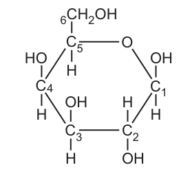
State the differences between the molecular structure of galactose, shown in Fig. 3.1, and the molecular structure of -glucose.
Answer
At C4, the –OH is above the ring and the –H is below in galactose, whereas in α-glucose the –OH is below the ring and the –H is above. (The positions of H and OH at C1 are the same in both sugars — OH below, H above in each case.)
At C4, the –OH is above the ring and –H below in galactose, whereas in α-glucose the –OH is below the ring and –H above.
Background Concept
Glucose and galactose are both aldohexoses with the molecular formula C₆H₁₂O₆. When drawn as pyranose ring structures (Haworth projections), each carbon in the ring carries either an –H or an –OH group, and the spatial position of these groups (above or below the plane of the ring) defines the identity of the sugar.
α-glucose and galactose are epimers — they differ in the configuration of –H and –OH at only one carbon atom. Galactose is the C4 epimer of glucose, so the only structural difference between α-glucose and α-galactose is the arrangement of the –H and –OH at C4.
In α-D-glucose:
- C1: –OH below, –H above (α-configuration)
- C2: –OH below, –H above
- C3: –OH above, –H below
- C4: –OH below, –H above
- C5: –CH₂OH above, –H below
In α-D-galactose (standard structure):
- C1: –OH below, –H above
- C2: –OH below, –H above
- C3: –OH above, –H below (same as glucose)
- C4: –OH above, –H below (DIFFERENT from glucose — this is the epimeric carbon)
- C5: –CH₂OH above, –H below
Understanding the Question
Fig. 3.1 is a Haworth projection of galactose, with the carbons numbered 1 to 6. The question asks the student to identify how the structure shown differs from that of α-glucose. The single mark is for a clear statement of the specific structural difference.
Approach
Compare each numbered carbon in the galactose figure with the corresponding carbon in α-glucose, looking for any carbon(s) where the positions of –H and –OH are different. The most likely place for a difference is C4, because galactose is the C4 epimer of glucose.
Step-by-Step Reasoning
- At C1, both the galactose shown in Fig. 3.1 and α-glucose have –OH below the ring and –H above. No difference here — both are α-sugars at C1.
- At C4, the figure shows –OH ABOVE the ring and –H below. In α-glucose, the –OH at C4 is BELOW the ring and –H is above.
- Therefore the structural difference is at C4: in galactose the –OH is on the opposite side of the ring compared to α-glucose.
- All other carbons have the same configuration in both sugars (galactose is the C4 epimer of glucose, so only C4 should differ).
Key Takeaways
- Hexose sugars with the same molecular formula can differ structurally — these are isomers (and, in the case of differing at one carbon, epimers).
- Galactose is the C4 epimer of glucose: the two sugars differ only at C4.
- Haworth projections allow direct visual comparison of the spatial arrangement of –H and –OH groups around the ring.
- When asked to compare sugar structures, always identify the specific carbon number(s) where the difference occurs.
Common Mistakes
- Stating the difference is at C1 (incorrect — C1 has –OH below in both α-glucose and the α-galactose figure).
- Saying "the H and OH are in different positions" without specifying which carbon — this is too vague to earn the mark.
- Listing too many differences (most of the structure is identical).
- Confusing galactose with β-glucose or β-galactose — the α/β designation refers only to the position of –OH at C1.
Things to Be Careful About
- Always give the carbon number when comparing sugar structures.
- A mirror image (enantiomer) is not the same as an epimer — galactose is an epimer, not a mirror image, of glucose.
- A clear answer names the carbon and the side of the ring on which the –OH sits in each sugar.
An investigation was carried out to determine the sugar content of mature carrots produced by different local varieties of carrot plants that are grown in Tunisia, North Africa.
Carrot plants were grown from seed under standardised conditions. When mature, the carrots were harvested.
The different sugars present in the carrots were identified. Measurements of sugar content for each of the sugars were made.
Table 3.1 shows the results for five different local varieties of carrot plant, A to E.
Table 3.1
| local variety | sugar content of carrots / of dry weight | |||
|---|---|---|---|---|
| fructose | galactose | glucose | sucrose | |
| A | 183.04 | 1.43 | 173.28 | 73.73 |
| B | 194.43 | 1.13 | 188.18 | 46.20 |
| C | 200.15 | 5.99 | 125.19 | 85.93 |
| D | 157.35 | 4.88 | 137.28 | 89.57 |
| E | 170.38 | 4.54 | 133.57 | 60.27 |
After studying the results shown in Table 3.1, a student concluded that the carrots from the local varieties contain the same four sugars.
The student made three other conclusions from the data in Table 3.1.
conclusion 1: There are non-reducing and reducing sugars in the carrots.
conclusion 2: There are monosaccharide and disaccharide sugars in the carrots.
conclusion 3: The carrots have the same pattern of results.
Explain the evidence in Table 3.1 that supports these three other conclusions.
conclusion 1 = ______
conclusion 2 = ______
conclusion 3 = ______
Answer
Conclusion 1 (non-reducing and reducing sugars):
Fructose, galactose and glucose are all present in every variety, and these three sugars are reducing sugars. Sucrose is also present in every variety, and sucrose is a non-reducing sugar. Therefore, both reducing and non-reducing sugars are present.
Conclusion 2 (monosaccharide and disaccharide sugars):
Fructose, galactose and glucose are monosaccharides (all present in every variety). Sucrose is a disaccharide (present in every variety). Therefore, both monosaccharide and disaccharide sugars are present.
Conclusion 3 (same pattern of results):
In every variety A–E, the order of sugar content from highest to lowest is: fructose > glucose > sucrose > galactose. The same ranking is observed in all five varieties.
Conclusion 1: fructose/galactose/glucose are reducing, sucrose is non-reducing. Conclusion 2: fructose/galactose/glucose are monosaccharides, sucrose is a disaccharide. Conclusion 3: in every variety the ranking is fructose > glucose > sucrose > galactose.
Background Concept
Sugars can be classified in two important ways relevant to this question:
-
Reducing vs non-reducing sugars. A reducing sugar has a free aldehyde (–CHO) or ketone group, or can open up to form one in solution; it can reduce copper(II) ions in Benedict's reagent. Glucose, fructose and galactose are all reducing sugars. Sucrose is non-reducing because the glycosidic bond joins the two anomeric carbons (C1 of glucose and C2 of fructose), so neither ring can open to expose a free aldehyde/ketone.
-
Monosaccharides vs disaccharides. Monosaccharides are single sugar units (e.g. glucose, fructose, galactose). Disaccharides consist of two monosaccharide units joined by a glycosidic bond (e.g. sucrose = glucose + fructose).
Understanding the Question
Table 3.1 gives the content (in mg per g dry weight) of four sugars in five different carrot varieties. The student must write a short justification for each of three given conclusions. Each justification must be evidence from the table, combined with the relevant chemistry.
Approach
For each conclusion, identify which sugars in the table support it, and combine the data observation with the chemical property to write a single, complete sentence.
Step-by-Step Reasoning
Conclusion 1 (reducing and non-reducing):
- The four sugars listed are fructose, galactose, glucose, sucrose.
- Fructose, galactose and glucose are reducing sugars.
- Sucrose is a non-reducing sugar.
- All four appear in every variety, so each variety contains both types.
- Justification: the presence of fructose/galactose/glucose (reducing) AND sucrose (non-reducing) in every variety supports the conclusion.
Conclusion 2 (monosaccharide and disaccharide):
- Fructose, galactose and glucose are monosaccharides.
- Sucrose is a disaccharide.
- All four appear in every variety.
- Justification: the presence of three monosaccharides AND a disaccharide in every variety supports the conclusion.
Conclusion 3 (same pattern):
Inspect the data row by row:
- A: 183.04 (F) > 173.28 (G) > 73.73 (S) > 1.43 (Ga) → order F > G > S > Ga
- B: 194.43 (F) > 188.18 (G) > 46.20 (S) > 1.13 (Ga) → order F > G > S > Ga
- C: 200.15 (F) > 125.19 (G) > 85.93 (S) > 5.99 (Ga) → order F > G > S > Ga
- D: 157.35 (F) > 137.28 (G) > 89.57 (S) > 4.88 (Ga) → order F > G > S > Ga
- E: 170.38 (F) > 133.57 (G) > 60.27 (S) > 4.54 (Ga) → order F > G > S > Ga
In every variety, the order from highest to lowest content is: fructose > glucose > sucrose > galactose. This consistent pattern supports the conclusion.
Key Takeaways
- A reducing sugar has a free anomeric carbon; sucrose is non-reducing because both anomeric carbons are tied up in its glycosidic bond.
- Monosaccharides are single units; disaccharides are pairs joined by a glycosidic bond.
- When supporting a conclusion from a data table, cite both the specific feature in the data AND the underlying chemistry.
- "Same pattern" conclusions are supported by a consistent ranking across all samples, not by quoting single values.
Common Mistakes
- Stating that another sugar besides sucrose is non-reducing (only sucrose is non-reducing in this list — the mark scheme rejects this).
- Stating that another sugar besides sucrose is a disaccharide (only sucrose is a disaccharide in this list).
- For conclusion 3, quoting individual values rather than describing the consistent ranking.
- Noting a numerical coincidence (e.g. "B has the highest glucose") rather than the overall pattern.
Things to Be Careful About
- The mark scheme rejects answers that name an additional sugar as non-reducing or disaccharide — be careful to name only sucrose for these properties.
- "Reducing sugar" requires the chemical property, not just a list of names.
- For the pattern conclusion, the supporting evidence is the consistent ranking, not the magnitude of the values.
Carbohydrates that are not sugars are also present in carrots.
Name one carbohydrate that is present in carrots and that is not a sugar.
Answer
Starch (any of: starch / amylose / amylopectin / cellulose / hemicellulose / pectin / inulin).
Starch
Background Concept
Carbohydrates include both sugars (mono-, di- and oligosaccharides) and non-sugars (polysaccharides such as starch, glycogen, cellulose, pectin and inulin). Polysaccharides are polymers of many monosaccharide units joined by glycosidic bonds; they are not sweet, do not crystallise, and do not give a positive Benedict's test.
Carrots are root storage organs. They accumulate:
- sugars (glucose, fructose, sucrose, galactose — see Table 3.1);
- storage polysaccharides (notably starch in many plants, and the fructose polymer inulin — carrot is well known to store inulin, especially in earlier growth);
- structural polysaccharides in the cell wall (cellulose, hemicellulose, pectin).
Understanding the Question
The question asks for ONE carbohydrate that is present in carrots but is NOT a sugar. Any plant polysaccharide found in carrot tissue is acceptable.
Approach
Recall the main non-sugar carbohydrates found in plants (especially in storage roots), and pick one that is creditable. All of the following are accepted by the mark scheme: starch, amylose, amylopectin, cellulose, hemicellulose, pectin, inulin.
Step-by-Step Reasoning
- Carrots store carbohydrate as both sugars (Table 3.1) and polysaccharides.
- Polysaccharides are built from many monosaccharide units linked by glycosidic bonds and are not classified as sugars.
- A clearly credited example is starch (the main storage polysaccharide in many plants, including carrots as they mature).
- Other acceptable answers include cellulose (plant cell wall), pectin (plant cell wall/middle lamella) and inulin (a fructose polymer found in carrot roots).
Key Takeaways
- Carbohydrates ≠ sugars; polysaccharides are carbohydrates but not sugars.
- Plant storage roots typically contain a mixture of sugars (for transport and immediate metabolism) and storage polysaccharides (for long-term energy storage).
- A wide range of polysaccharide names is acceptable for this style of question.
Common Mistakes
- Naming a sugar (e.g. sucrose, glucose) — this does not answer the question because the question explicitly says NOT a sugar.
- Naming a non-carbohydrate (e.g. protein, lipid) — also not what is asked.
Things to Be Careful About
- The answer must be a named non-sugar carbohydrate found in carrots.
- "Starch" alone is sufficient; specifying amylose or amylopectin is also acceptable but not required.
Each carrot plant produces a carrot in the first year of growth.
If the carrot is not removed from the plant after it matures, the plant passes through a dormant period and shoots develop from the carrot in the second year of growth. This allows flowers to be produced and seed formation to occur before the plant dies.
Explain, with reference to the life cycle of the carrot plant, when the carrot acts as a source and when the carrot acts as a sink.
source = ______
sink = ______
Answer
Source (in the second year of growth, after the dormant period):
The carrot acts as a source. Stored assimilates (e.g. starch) are mobilised / converted to soluble sugars and loaded into the phloem, providing assimilates for the developing shoots, flowers and seeds.
Sink (in the first year of growth):
The carrot acts as a sink. It receives assimilates (e.g. sucrose) transported in the phloem from the photosynthetic leaves and stores them (e.g. as starch) as it grows.
Source: in the second year of growth (after dormancy), the carrot mobilises stored assimilates and loads them into the phloem to supply the developing shoots, flowers and seeds. Sink: in the first year, the carrot receives assimilates from the leaves via the phloem and stores them as it grows.
Background Concept
In phloem transport, a source is any region that produces (or releases) assimilates — typically photosynthesising leaves, but also storage organs that re-mobilise their reserves. A sink is any region that imports and uses or stores assimilates — typically roots, fruits, developing leaves, seeds and storage organs when they are accumulating reserves.
Assimilates (mainly sucrose, but also amino acids and other small organic molecules) move from source to sink through the phloem by mass flow, driven by an osmotic pressure gradient set up by active loading at the source and unloading at the sink.
The carrot is a biennial plant:
- Year 1: the plant grows vegetatively; the taproot (carrot) accumulates reserves. The carrot is therefore a sink.
- Late year 1 / winter: the plant becomes dormant.
- Year 2: the plant re-grows from the carrot; shoots, flowers and seeds are produced using reserves from the carrot. The carrot is therefore a source.
Understanding the Question
The question describes the carrot plant's biennial life cycle. The student must use the source–sink terminology to identify when the carrot is acting in each role, and give one piece of supporting detail for each (3 marks total: 1 for identifying source time + 1 detail; 1 for identifying sink time + 1 detail — actually structured as 1 mark for timing, 1 mark for source detail, 1 mark for sink detail).
Approach
Identify the carrot's role in each of the two growing seasons, and link the role to the direction of phloem transport of assimilates. For each role, give a specific detail (mobilisation/loading for source; receiving/storing for sink).
Step-by-Step Reasoning
Carrot as a source (year 2, after dormancy):
- In the second year, the carrot is no longer growing — the new shoots, flowers and seeds need energy and materials.
- The reserves stored in the carrot (e.g. starch) are broken down to soluble sugars.
- These sugars are loaded into the phloem of the carrot and transported to the developing sinks (shoots, flowers, seeds).
- The carrot therefore acts as a source, providing assimilates for the new growth.
Carrot as a sink (year 1):
- In the first year, the carrot is growing and accumulating reserves.
- It imports assimilates (mainly sucrose) from the photosynthesising leaves via the phloem.
- These assimilates are used for growth and converted to storage compounds (e.g. starch) for overwintering.
- The carrot therefore acts as a sink.
(Strictly, during the dormant period itself there is little active transport, so the carrot is neither an active source nor an active sink. The mark scheme specifically says "ignore 'during dormant period'".)
Key Takeaways
- Source = net exporter of assimilates; sink = net importer.
- A plant organ can switch between source and sink depending on its developmental role — a storage root is a sink when it accumulates reserves and a source when it re-mobilises them.
- Biennial plants show this switch dramatically: vegetative growth in year 1 (storage organ as sink) and reproductive growth in year 2 (storage organ as source).
Common Mistakes
- Saying the carrot is a source in year 1 and a sink in year 2 — this is the wrong way round.
- Saying the carrot is "always a sink" — the carrot is a sink only in year 1; it becomes a source in year 2.
- Failing to give a specific detail for source or sink — the mark scheme requires a detail such as "mobilises starch" or "receives assimilates from the leaves".
- Using the word "nutrients" instead of "assimilates" — "nutrients" is ignored by the mark scheme (assimilates is the precise term).
Things to Be Careful About
- The carrot is not an active source or sink during the dormant period — this is ignored by the mark scheme.
- A complete answer needs both a clear identification of source/sink timing AND at least one supporting detail for each.
- The terms source and sink refer to roles in phloem transport, not to the function of the organ as food for animals.
Carrot virus Y is a pathogen of carrot plants. The virus, which belongs to a group known as Potyvirus, replicates its viral nucleic acid and proteins within host carrot cells.
The general structure of potyviruses is shown in Fig. 3.2.

The synthesis of viral proteins in host carrot cells only involves the process of translation. The process of transcription does not occur.
Suggest why translation occurs in host carrot cells during the synthesis of viral proteins, but transcription does not occur.
Answer
- The potyvirus has RNA (not DNA) as its genetic material, so its nucleic acid does not need to be transcribed; transcription does not occur.
- The viral RNA itself acts as mRNA / carries the codon sequence that can be read directly by host ribosomes during translation, so the host cell uses it directly to synthesise viral proteins.
The virus has RNA, not DNA, so transcription is not required. The viral RNA itself acts as mRNA and is translated directly by host ribosomes to make viral proteins.
Background Concept
In cellular organisms, the central dogma describes the flow of genetic information: DNA → RNA → protein. The two key processes are:
- Transcription — DNA is used as a template to make mRNA.
- Translation — mRNA is read by ribosomes to make a polypeptide.
In a host cell infected by a virus, the cell's ribosomes, tRNAs, amino acids and other translation machinery are used to make viral proteins. Whether transcription is needed depends on the nature of the viral genome.
Viruses are non-cellular obligate intracellular parasites. Their genome can be DNA or RNA. Potyviruses (the group to which carrot virus Y belongs) have a single-stranded RNA genome (shown in Fig. 3.2 as the coiled strand inside the protein capsid).
Understanding the Question
The question states that the synthesis of viral proteins in the host carrot cells involves translation but NOT transcription. The student is asked to explain why. The answer requires knowledge of (i) the type of nucleic acid in the virus and (ii) the role of that nucleic acid in protein synthesis.
Approach
Identify the genome type (RNA), and then explain how this allows the genetic information to be used directly by the host's ribosomes without a transcription step.
Step-by-Step Reasoning
- The potyvirus has RNA (not DNA) as its genetic material. (Fig. 3.2 shows the viral RNA inside the capsid.)
- Because the genetic material is already RNA, there is no DNA template that needs to be transcribed into mRNA — so transcription does not occur.
- The viral RNA itself can act as mRNA: it carries the codon sequence that the host ribosomes can read directly.
- The host cell's ribosomes, tRNAs and amino acids therefore translate the viral RNA directly, producing viral proteins.
Any two of these points earns the two marks on the mark scheme.
Key Takeaways
- The central dogma (DNA → RNA → protein) applies to cellular organisms but must be modified for RNA viruses.
- RNA viruses bypass transcription because their genetic material is already in the form of RNA that can act as mRNA.
- Retroviruses (with RNA genomes) go one step further: they use reverse transcription to make DNA from RNA, but potyviruses do not — they translate their RNA directly.
- The fact that a virus cannot make its own proteins (no ribosomes of its own) is why it must use the host's translation machinery.
Common Mistakes
- Stating that the virus has DNA (incorrect — potyviruses have RNA).
- Saying that the virus "doesn't have the enzymes for transcription" — while this is true, it is not the reason rewarded by the mark scheme; the mark scheme rewards the genome-type argument.
- Confusing the direction of information flow (saying that translation makes RNA, or that transcription makes protein).
- Saying that transcription "doesn't happen in carrot cells" — transcription does happen in carrot cells for the plant's own genes; the point is that it is not needed for the virus.
Things to Be Careful About
- The question is about the viral RNA being translated directly, not about whether host cell transcription happens at all.
- Whether the viral RNA is single- or double-stranded is not relevant to this question (and the mark scheme says single/double stranded references are neutral — they earn no marks either way).
- Use the precise terms "transcription" and "translation" rather than informal descriptions.
Tuberculosis (TB) is an infectious disease that is caused by a bacterial pathogen.
The pathogen has mechanisms to avoid digestion by phagocytes. Macrophages may engulf the bacteria, but in some cases the bacteria remain alive within the cells instead of being killed.
Although there are two main species of bacterium causing TB, it is rare for the species that causes bovine TB in cattle to infect humans.
Name the species of bacterium that is the main cause of TB in humans.
Answer
Mycobacterium tuberculosis
Mycobacterium tuberculosis
Background Concept
Tuberculosis (TB) is a chronic infectious disease of the lungs caused by bacteria of the genus Mycobacterium. Two species are most commonly associated with the disease:
- Mycobacterium tuberculosis (also called M. tuberculosis or MTB) — the main cause of TB in humans. It is sometimes called the "human" or "true" tubercle bacillus.
- Mycobacterium bovis — the main cause of TB in cattle (bovine TB). It can occasionally infect humans, typically through drinking unpasteurised milk or eating undercooked contaminated meat.
Both are slow-growing, acid-fast, rod-shaped (bacillus) bacteria with a waxy mycolic acid cell wall that resists Gram staining and protects them from digestion inside phagocytes.
Understanding the Question
The stem explicitly distinguishes between two species of TB-causing bacterium and says the one that causes bovine TB rarely infects humans. The candidate is asked for the species that is the main cause of TB in humans — a one-word (well, two-word italic) recall of the scientific name.
Approach
The question is a direct recall of the binomial name. Spelling matters: this is a genus + species name in italics, and the marking scheme explicitly states that it "must be spelled correctly".
Step-by-Step Reasoning
The bacterium that causes TB in humans is Mycobacterium tuberculosis. The word Mycobacterium is the genus; tuberculosis is the specific epithet. Both must be written in italics (or underlined) to follow binomial nomenclature convention, and the genus is capitalised while the species is lower case.
Key Takeaways
- The main human TB pathogen is Mycobacterium tuberculosis.
- M. bovis primarily infects cattle and only rarely jumps to humans.
- Binomial names must be italicised, with the genus capitalised.
Common Mistakes
- Writing M. tuberculosis without italics, or with incorrect capitalisation (e.g. Mycobacterium Tuberculosis).
- Confusing the two species and writing Mycobacterium bovis.
- Misspelling — Microbacterium, Mycobacterium tubercolosis, etc.
Things to Be Careful About
The mark scheme states the spelling "must be spelled correctly". Get the genus and species spellings exactly right and format as italics (or underline).
Explain how the pathogen named in (a) is transmitted from a person with the disease to a person who is uninfected.
Answer
- An infected person coughs / sneezes / talks / breathes out, releasing droplets / an aerosol containing the pathogen.
- An uninfected person then inhales the droplets / aerosol, so the bacteria are transmitted by droplet infection / aerosol infection.
Droplet / aerosol infection: infected person coughs/sneezes/talks, releasing bacteria in airborne droplets which are then inhaled by an uninfected person.
Background Concept
TB is a respiratory disease spread by airborne particles. When a person with active pulmonary TB coughs, sneezes, speaks or even breathes out, tiny droplets of mucus and saliva are released into the surrounding air. If these droplets contain M. tuberculosis and are small enough to remain suspended (often called droplet nuclei or an aerosol), they can be breathed in by another person. Once inhaled, the bacteria reach the alveoli, where they are engulfed by alveolar macrophages. This route is called droplet infection, aerosol infection or airborne transmission.
Understanding the Question
The question asks the candidate to explain how M. tuberculosis is transmitted from an infected to an uninfected person. "Explain" here means stating the source action, the medium of transmission, and the route into the new host.
Approach
The two marking points target:
- The infected person's action (cough / sneeze / talk / breathe out) — releasing the pathogen.
- The transmission term or description — droplets in air, and the uninfected person inhaling them.
Step-by-Step Reasoning
Marking point 1: An infected person coughs / sneezes / talks / breathes out, expelling droplets that contain the bacteria.
Marking point 2: These droplets form an aerosol / cloud of airborne droplets. An uninfected person then inhales (inspires, breathes in) the droplets, and the bacteria enter their lungs. The whole process is described as droplet infection, aerosol infection, droplet transmission or airborne transmission.
The mark scheme rejects "infected droplets" or "droplets of air" because droplets cannot themselves be infected — they carry the infection.
Key Takeaways
- TB is spread by droplet / aerosol transmission.
- The infected person releases bacteria in respiratory droplets when they cough, sneeze, talk or breathe out.
- The uninfected person acquires the infection by inhaling those droplets.
Common Mistakes
- Stating only the term ("airborne transmission") without describing the source action or the route of entry — fails to earn the first mark.
- Writing "infected droplets" — droplets are not infected; they carry the pathogen. The mark scheme rejects this wording.
- Including additional incorrect transmission routes (e.g. touching, contaminated food) — caps the answer at 1 mark.
- Describing how M. bovis is transmitted (contaminated milk/meat) instead of the airborne route for M. tuberculosis — caps the answer at 1 mark.
Things to Be Careful About
- Use the term "droplet infection" or "aerosol infection" (or similar) — a bare description without the term is acceptable only if it is correct in detail (organism in airborne droplets).
- Do not add any other transmission mode alongside the correct one or the mark is capped at 1.
Answer
Antibiotic
Antibiotic
Background Concept
TB is caused by a bacterium (M. tuberculosis). Diseases caused by bacteria are treated with antibiotics — drugs that kill bacteria (bactericidal) or prevent them from multiplying (bacteriostatic). Common antibiotics used in TB include isoniazid, rifampicin, ethambutol and pyrazinamide, usually given in combination over many months to prevent the development of antibiotic resistance.
Note that antibiotics have no effect on viruses, so they cannot be used to treat viral diseases such as influenza, measles, HIV/AIDS, or COVID-19.
Understanding the Question
The question is a single-word / single-term recall of the type of drug used to treat TB. It is testing whether the candidate knows that TB is a bacterial infection and that bacterial infections are treated with antibiotics.
Approach
Provide the precise drug class. The mark scheme says "I examples" (ignore examples) and "I antibacterial" (reject "antibacterial" — it is not the standard term).
Step-by-Step Reasoning
The mark scheme accepts only the term antibiotic. Naming a specific antibiotic (e.g. "rifampicin" or "penicillin") is ignored — the question asks for the type of drug, not a specific drug. "Antibacterial" is rejected because the precise term is "antibiotic".
Key Takeaways
- TB is a bacterial disease, so it is treated with antibiotics.
- The drug class is "antibiotic", not "antibacterial", and not the name of a specific antibiotic.
Common Mistakes
- Writing "antibacterial" — wrong term; the mark scheme rejects it.
- Naming a specific antibiotic (e.g. "streptomycin", "rifampicin") — the question asks for the type, and the mark scheme ignores examples.
- Writing "vaccine" or "antiviral" — TB is bacterial, not viral, and is not treated with a vaccine.
Things to Be Careful About
The accepted answer is the single word "antibiotic". Do not add qualifiers that the mark scheme will not credit.
Fig. 4.1 is a photomicrograph of a section of lung tissue taken from a person who has not been infected with the bacterial pathogen and who does not have TB.
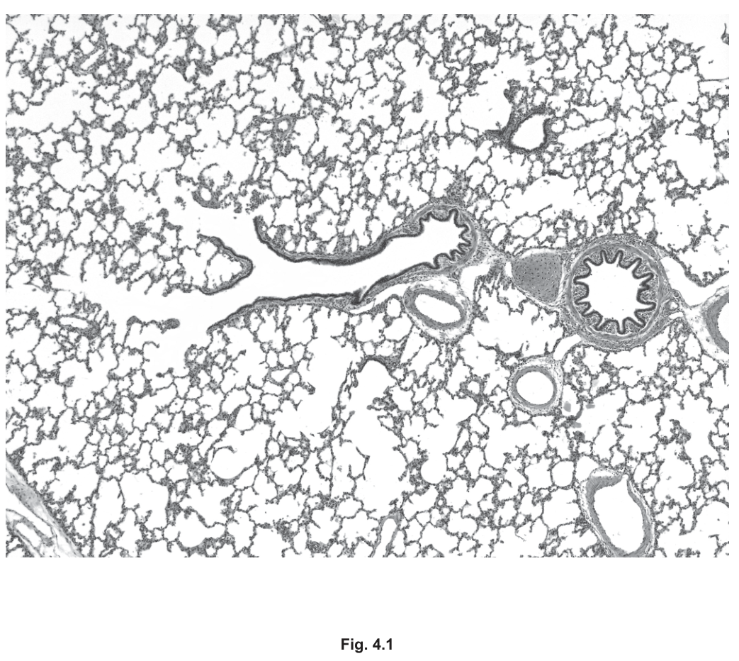
Blood vessels and some structures of the gas exchange system are visible in Fig. 4.1.
On Fig. 4.1, use a label line and label:
• a bronchus
• a bronchiole
• a blood vessel.
Answer
- Bronchus — label the largest airway (right-hand side of Fig. 4.1), with the highly folded/wavy inner epithelium and a thick wall containing cartilage.
- Bronchiole — label one of the smaller airways (central area of Fig. 4.1) with a folded inner lining but a thinner wall and no cartilage plates.
- Blood vessel — label one of the rounded structures with a relatively thick smooth wall but with an unfolded (smooth) luminal surface and blood cells inside (must not be labelled a capillary).
Three correct label lines added to Fig. 4.1: one to a bronchus (largest, folded, cartilage-containing airway), one to a bronchiole (smaller, folded, no cartilage) and one to a blood vessel (rounded, smooth lining, not a capillary).
Background Concept
A histological section of the lung shows three broad categories of structure:
- Bronchi — the larger conducting airways. They have a folded (rugae-like) pseudostratified ciliated columnar epithelium thrown into folds because the wall is relaxed in the section. The wall contains irregular plates of hyaline cartilage, smooth muscle and seromucous glands. The lumen is relatively large.
- Bronchioles — the smaller branches of the airway tree (diameter < 1 mm). They have no cartilage and no submucosal glands. The epithelium is still folded and is mostly ciliated columnar or cuboidal. Smooth muscle is relatively more prominent in the wall.
- Blood vessels — pulmonary arteries run alongside the bronchi and have a thick, muscular wall but a smooth (unfolded) inner lining and often contain visible red blood cells. Veins have thinner walls than arteries. Capillaries are the very thin-walled exchange vessels; in a light micrograph at this magnification they are not resolved as discrete structures and cannot be reliably labelled.
Understanding the Question
The candidate is given a photomicrograph of healthy lung tissue (Fig. 4.1) and asked to add three labelled lines pointing to: (1) a bronchus, (2) a bronchiole and (3) a blood vessel. This tests recognition of lung micro-anatomy from real tissue, not from an idealised diagram.
Approach
Look for the structure with the most distinctive features first:
- The largest airway with cartilage in its wall and the most dramatic luminal folding is the bronchus.
- A smaller airway (folded epithelium, no cartilage) is the bronchiole.
- A round structure with a thick wall and a smooth inner surface (no folding) is the blood vessel. Do not label a capillary — at this magnification the alveolar walls are the capillaries and they cannot be picked out as individual vessels.
Step-by-Step Reasoning
Walking around the image:
- The very large structure on the right with the most pronounced wavy inner lining and a thick wall is a bronchus.
- One of the smaller round/oval structures (e.g. near the centre of the image) with a folded inner lining but a thinner wall and no cartilage is a bronchiole.
- A round structure with a thick, smooth muscle wall and an unfolded interior — for example, one of the medium-sized circular profiles between the alveoli — is a blood vessel (pulmonary artery branch).
- The label line for each must clearly end on the structure being named.
The mark scheme rejects a label of "capillary" for the blood vessel. This is because individual capillaries are not distinguishable at this magnification — the thin alveolar walls are the capillary beds, but a label line cannot sensibly be drawn to a single capillary here.
Key Takeaways
- Bronchus = large airway + folded epithelium + cartilage plates in the wall.
- Bronchiole = smaller airway + folded epithelium + no cartilage.
- Blood vessel (artery/vein) = round + thick wall + smooth (unfolded) lining, distinct from airways.
- Capillaries cannot be reliably labelled on a light micrograph of this magnification.
Common Mistakes
- Labelling the wrong structure as the blood vessel (e.g. a small bronchiole with a thick wall) — the smooth unfolded lining is the key feature.
- Labelling a capillary — rejected by the mark scheme.
- Putting the label line on a fold of epithelium rather than on the wall — the marker for the structure is its wall, not its lining.
- Confusing the bronchus and bronchiole labels — the size difference and the presence/absence of cartilage are the deciding features.
Things to Be Careful About
- A label line must end on or just outside the wall of the named structure with the text written at the other end.
- Only one label per structure, and each label must be the correct word — no vague terms like "airway" in place of "bronchus".
In some people with TB, areas known as granulomas may form in lung tissue as part of an immune response to the pathogen.
Fig. 4.2 is a photomicrograph of a granuloma in lung tissue.
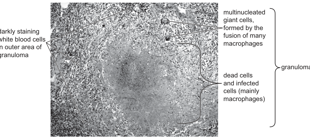
With reference to Fig. 4.1 and Fig. 4.2, describe and explain how the changes that occur as a result of granuloma formation:
• can affect gas exchange and harm the health of an infected person
• may help to prevent TB developing in other parts of the body.
Answer
Effect on gas exchange and health:
- Granuloma formation damages lung tissue, reducing the number of bronchioles / alveoli / alveolar capillaries, so the surface area for gas exchange decreases and less oxygen enters the blood.
- The dense mass of immune cells, dead cells and debris in the granuloma physically hinders gas exchange across the affected region.
- Less oxygen reaches the rest of the body in the circulation, leading to tiredness / fatigue / breathlessness / strain on the heart.
Prevention of TB elsewhere:
- The granuloma confines / localises / isolates the pathogen, so infected cells and bacteria cannot easily enter the bloodstream and reach other organs.
- Many immune cells (macrophages, multinucleated giant cells, lymphocytes) surround the pathogen; continued phagocytosis and cytokine release limit bacterial reproduction and spread.
Granulomas reduce alveolar surface area and produce a dense cellular mass that impairs gas exchange (causing tiredness / breathlessness), while at the same time walling off the pathogen in a localised mass of immune cells, preventing it from entering the bloodstream and reaching other tissues.
Background Concept
Gas exchange surface. The lungs are built around millions of alveoli, each a tiny, thin-walled air sac wrapped in a network of pulmonary capillaries. The huge combined surface area and the very short diffusion distance (alveolar epithelium + capillary endothelium, both one cell thick) are what allow the rapid equilibration of O₂ and CO₂ between air and blood.
Granuloma formation. A granuloma is a focal, organised collection of activated macrophages and other immune cells that forms when the immune system cannot quickly eliminate an intracellular pathogen such as M. tuberculosis. The centre of a mature granuloma in TB often contains caseous (cheese-like) necrotic debris — dead macrophages and bacteria. Surrounding this core are epithelioid macrophages, multinucleated giant cells (Langhans giant cells, formed by fusion of activated macrophages), and an outer rim of lymphocytes and fibroblasts. The granuloma is essentially a wall the body builds to keep the bacteria in one place.
Understanding the Question
The candidate is given two photomicrographs:
- Fig. 4.1 — healthy lung tissue with normal alveoli, bronchioles and blood vessels.
- Fig. 4.2 — a granuloma: a large, dense, circular mass of cells with a central area of dead and infected cells, multinucleated giant cells, and an outer rim of darkly staining white blood cells.
The question asks the candidate to compare these and produce two explanations, each linking structure to consequence:
- How the changes caused by the granuloma affect gas exchange and harm health.
- How those same changes may help to prevent TB from developing in other parts of the body.
The question says "describe and explain", so the answer needs both the observable change and the biological consequence.
Approach
Split the answer into two halves (as the question does):
- First half (gas exchange / harm to health) — focus on the destruction of normal lung architecture (alveoli, alveolar capillaries, bronchioles) and the physical barrier the granuloma creates. Link the loss of surface area to a fall in O₂ uptake, then to systemic symptoms.
- Second half (containment) — focus on the function of the wall of immune cells, the role of macrophages and giant cells, and the prevention of haematogenous spread.
The mark scheme marks any 5 from a pool of 10, with a maximum of 4 from either half. So a strong answer covers both halves.
Step-by-Step Reasoning
Gas exchange and health (mp 1–5):
- Loss of gas-exchange surface. A granuloma replaces normal lung parenchyma with a dense cellular mass. Fewer alveoli, fewer alveolar capillaries and (where bronchi/bronchioles are involved) smaller airway lumens all mean a reduced surface area for gas exchange and a longer diffusion path. The mark scheme also credits "takes up lung space" as a description of mp 1.
- Consequence. Less air enters the affected alveoli, fewer red blood cells per unit time are oxygenated, and the net rate of O₂ uptake falls. A blocking lesion in a bronchiole reduces ventilation to the alveoli it serves.
- Physical hindrance by cells. The thick layer of immune cells, dead cells and debris in the centre and rim of the granuloma adds a significant barrier to gas diffusion between alveolar air and capillary blood (this is distinct from the loss-of-surface-area point in mp 1).
- Systemic effect. With less O₂ reaching the circulation, less O₂ is delivered to body tissues.
- Symptoms. The systemic hypoxia manifests as fatigue / tiredness, breathlessness on exertion, increased breathing rate, raised blood pressure and increased strain on the heart.
Containment of TB (mp 6–10):
- No bloodstream spread. The granuloma walls off the infected cells and bacteria, preventing them from entering the bloodstream. This is the key to stopping disseminated (miliary) TB.
- Localisation. The structure confines / isolates / localises the pathogen to one site. The wall of cells and fibrous tissue acts as a physical barrier.
- Many immune cells around the pathogen. A dense population of macrophages, multinucleated giant cells, lymphocytes and other leucocytes surrounds the bacteria. Note: "white blood cells" alone is not credited (mp 8 needs a named cell type, e.g. macrophage, lymphocyte, plasma cell, phagocyte, neutrophil).
- Active immune response. Continued phagocytosis of infected cells and bacteria, antigen presentation, cytokine release and antibody formation by plasma cells all help to keep the bacterial population in check.
- Multinucleated giant cells. These very large fused macrophages are more effective phagocytes — they have a bigger surface in contact with the centre of the granuloma and a higher chance of encountering the pathogen. The immune response also limits bacterial reproduction.
Key Takeaways
- Granulomas harm the lung by destroying gas-exchange surface and by adding a physical barrier to diffusion — this lowers O₂ uptake and causes systemic hypoxia symptoms (fatigue, breathlessness, cardiac strain).
- Granulomas protect the rest of the body by walling off the pathogen in a localised mass of activated macrophages, giant cells and lymphocytes, preventing it from entering the bloodstream and seeding other organs.
- The same structural change (a dense mass of immune cells) is therefore both harmful (locally) and protective (systemically) — a useful example of the trade-offs in immune defence.
Common Mistakes
- Saying granulomas "block the airways" — the mark scheme ignores this; the credit is for the loss of surface area or the physical mass of cells.
- Saying only "white blood cells surround the bacteria" — too vague; name a cell type (macrophage, lymphocyte, etc.).
- Confusing cause and effect: a granuloma is a consequence of the immune response, not the response itself.
- Claiming granulomas directly kill the patient — they usually cause chronic, progressive damage rather than acute death; symptoms are chronic (tiredness, breathlessness) not collapse.
- Omitting one of the two halves of the question — the question explicitly asks about both gas exchange and containment, and the mark scheme splits into two pools (max 4 from each).
Things to Be Careful About
- Use precise language: "multinucleated giant cells", "macrophages", "lymphocytes" — not vague "white blood cells".
- When describing the harm, link the structural change to the consequence ("fewer alveoli → less surface area → less O₂ uptake → fatigue"). The mark scheme gives credit for both the change and its consequence.
- When describing the protection, emphasise the localisation and the prevention of bloodstream spread — these are the two key ideas the mark scheme rewards.
- Quote Fig. 4.2 features (multinucleated giant cells, dead cells in the core, darkly staining immune cells in the rim) to demonstrate that you have used the photomicrograph.
There is a global shortage of blood for transfusions. Researchers can culture bone marrow stem cells in the laboratory to manufacture red blood cells for potential use as an artificial blood product.
The researchers collect bone marrow stem cells that are present in small quantities in blood, rather than extracting them from bone marrow.
• Antibodies, specific to bone marrow stem cells, are attached to tiny magnetic beads.
• The beads are added to a sample of blood.
• An electric field is applied to immobilise the beads so that the bone marrow stem cells can be collected.
The antibodies are attached to the beads so that the sites used for binding to specific molecules on the bone marrow stem cells are left exposed.
Name the term given to the specific molecules on the bone marrow stem cells that attach to the antibody binding sites.
Answer
Antigens
Antigens
Background Concept
Antibodies are Y-shaped proteins produced by B-lymphocytes that bind specifically to other molecules. Each antibody has two identical binding sites at the tips of the Y, and the shape of these sites is complementary to a particular target molecule. The molecule on the surface of a cell (or virus, bacterium or free in solution) that an antibody binds to is called an antigen. Antigens are typically proteins, glycoproteins or polysaccharides, and the immune system uses the huge diversity of antibody binding sites to recognise an enormous range of different antigens. The immune system must learn not to attack the body's own antigens — this is described as distinguishing self from non-self.
Understanding the Question
The stem describes antibodies attached to magnetic beads being used to capture bone marrow stem cells from a blood sample. Antibodies only bind to specific target molecules, so the molecules they recognise on the bone marrow stem cells are the answer. The mark scheme explicitly says to ignore the terms 'self' and 'non-self' as answers — it is looking only for the name of the type of molecule.
Approach
Recognise that an antibody's binding site is shaped to fit a particular molecular shape on a target cell. Recall the single correct term for that target molecule.
Step-by-Step Reasoning
- The antibodies on the magnetic beads must recognise a specific molecule on the surface of bone marrow stem cells — that is what makes the technique work.
- The general term for any molecule that is recognised and bound by an antibody is an antigen.
- Each antibody recognises one specific antigen (or a small set of structurally similar antigens), which is why antibodies can be used to pull out particular cell types from a mixed sample.
Key Takeaways
- Antigens are the molecular targets of antibodies; they are the molecules recognised, not the recognition proteins themselves.
- Antibody specificity is the basis of many laboratory techniques including this magnetic-bead cell-sorting method.
Common Mistakes
- Writing 'antibody' or 'receptor' instead of antigen — the antibody is on the bead, the antigen is on the stem cell.
- Writing 'self' or 'non-self' — these are descriptive terms but the mark scheme explicitly ignores them; the precise name of the molecule type is 'antigen'.
Things to Be Careful About
- The question asks for the term for the molecules on the bone marrow stem cells, not the molecules on the bead. The antibodies are the recognisers, the antigens are the recognised.
Suggest and explain the advantages of using bone marrow stem cells from the blood sample to manufacture artificial red blood cells.
Answer
- (Using bone marrow stem cells) Bone marrow stem cells are haematopoietic — they only differentiate into blood cell types, so they can be directed to produce red blood cells.
- (Using bone marrow stem cells) They retain the ability to divide continuously by mitosis, so large numbers of (red blood cell) cells can be produced.
- (From the blood sample) Collecting the stem cells from a blood sample is less invasive than extracting them from bone marrow.
See working
Background Concept
Stem cells are unspecialised cells that can both self-renew (produce more stem cells by mitosis) and differentiate into one or more specialised cell types. Bone marrow contains haematopoietic (blood-forming) stem cells, which are multipotent — they give rise to all the different blood cell types, including red blood cells, white blood cells and platelets. Because they keep dividing throughout life, the bone marrow is a constant source of new blood cells. A small number of these haematopoietic stem cells are also normally present in circulating blood, where they can be harvested without the need for a bone marrow biopsy. The small number of antibodies on magnetic beads exploit the principle of antibody–antigen specificity: only the cells bearing the matching antigen (the bone marrow stem cells) stick to the beads and are held in place when a magnetic field is applied, while everything else is washed away.
Understanding the Question
Part (b) has the command word 'suggest and explain' — this means each marking point needs both the suggested advantage AND a reason why it is an advantage. The marks are split into two groups by the mark scheme: advantages that come from using bone marrow stem cells themselves, and advantages that come specifically from collecting those cells from a blood sample rather than from bone marrow directly. To score three marks, the candidate must address at least one point from each group, and ideally cover both.
Approach
First identify the desirable properties of bone marrow stem cells as a starting material: what makes them biologically suitable for manufacturing red blood cells. Then identify the practical advantages of obtaining them from blood instead of bone marrow. Each suggested advantage should be coupled to a clear explanation of why that matters for making an artificial blood product.
Step-by-Step Reasoning
- Bone marrow stem cells are haematopoietic (a mark-scheme term — it means 'blood-forming'). Because they are committed to the blood cell lineage, when they differentiate they will give rise to blood cell types, including red blood cells. This avoids the need to redirect cells from another lineage.
- Stem cells retain the ability to undergo mitosis repeatedly, so a small starting population can be expanded into the very large numbers of red blood cells required for a transfusion product. This is a key practical requirement because a single unit of blood contains roughly 2.5 × 10¹² red blood cells.
- Because the cells are isolated from a blood sample rather than drilled out of bone, the donor procedure is far less invasive — no surgical bone marrow extraction is needed. This makes the procedure safer and more acceptable to donors, allowing more cells to be collected and a more sustainable supply.
- A valid alternative in the same group: the magnetic-bead method is far easier to carry out on a liquid blood sample than on solid bone marrow tissue, where the cells are embedded in a complex matrix.
- Other credit-worthy points include: the cells are genetically identical (so the product is uniform), and the screened blood sample is less likely to pass on disease than cells taken directly from bone marrow.
Key Takeaways
- Haematopoietic stem cells are multipotent for blood lineages, are mitotic, and can self-renew — exactly the properties needed for mass-producing red blood cells in the lab.
- Sampling blood is much less invasive than aspirating bone marrow, which is both a donor-safety and supply-scalability advantage.
- 'Suggest and explain' questions require both the suggestion (the advantage) and the explanation (why it matters) on each line.
Common Mistakes
- Writing only the suggestion without the explanation — the mark scheme rewards the reason, not the bare idea.
- Saying 'they can replicate' — the mark scheme explicitly rejects 'replicate' or 'reproduce' and requires 'divide' or 'mitosis' or 'cell cycle'.
- Saying 'they can differentiate into stem cells' — the mark scheme rejects this; stem cells self-renew (make more stem cells), they do not differentiate into stem cells.
- Confusing multipotency with pluripotency: bone marrow stem cells are multipotent (limited to blood lineages), not pluripotent (which would be embryonic stem cells).
- Vague 'no disease' — the mark scheme wants a specific phrase such as 'avoid passing on diseases' or equivalent.
Things to Be Careful About
- The question asks for advantages of using bone marrow stem cells FROM the blood sample. Marks are split between properties of the stem cells themselves and properties of obtaining them from blood. To score full marks, address both.
- The mark scheme ignores 'multipotent' as a credit-bearing point on its own, so don't write it as the entire answer — combine it with the consequence (only producing blood cell types, including red blood cells).
One desirable feature of artificial blood products, such as artificial red blood cells, is that they should be economical to produce.
Suggest other desirable features of artificial blood products.
Answer
- Should not have any toxic / harmful effects in the recipient.
- Should not carry pathogens / should not cause infection in the recipient.
- Should have a long shelf life / should be easy to store.
See working
Background Concept
A transfusion product is introduced directly into a patient's bloodstream, so any imperfection in the product can have immediate, serious effects on the recipient. The criteria for an ideal blood product are therefore a combination of:
- Safety: nothing toxic, no infectious agents, no triggering of the patient's immune system (which would cause a transfusion reaction), and ideally compatible with all blood groups so that supply is simple.
- Function: it must actually do the job of blood — most importantly transport oxygen around the body — and it must remain soluble in plasma long enough to circulate.
- Practicality: a real product must keep on a shelf for a reasonable time, must be producible in large quantities, and ideally must be acceptable to people whose religion or culture forbids donor blood transfusions.
A normal unit of donor red blood cells, for comparison, has a shelf life of about 35–42 days at 2–6 °C, must be matched for ABO and Rh blood group, and carries a small but real residual risk of transmitting infection despite screening.
Understanding the Question
Part (c) has the command word 'suggest' and explicitly says 'other' desirable features — i.e. features BEYOND being economical to produce, which is the one already given. The mark scheme offers around seven commonly credited points plus two AVP (any valid point) marks, so any three reasonable, distinct suggestions from the list will earn the marks. The explanations are not required to score, but a short justification of why each feature is desirable strengthens the answer.
Approach
Think about what a recipient needs the artificial blood to do and what risks a transfusion carries. Run through the categories: safety to the patient (toxicity, infection, immune reaction), functional performance (oxygen transport, solubility, circulation), and supply logistics (shelf life, blood-group compatibility, mass production). Pick three distinct points.
Step-by-Step Reasoning
- Toxicity: an artificial blood product must not poison or damage the recipient's tissues. Any breakdown products must be safely metabolised or excreted.
- Pathogen safety: donor blood must be screened for HIV, hepatitis B and C, syphilis and other infections. An artificial product made in the laboratory should be free of these pathogens, removing the screening requirement and the residual risk of infection.
- Shelf life: natural red blood cells only last about six weeks even when refrigerated, which limits supply. An artificial product with a long shelf life (and easy storage, ideally at room temperature) would solve a major supply problem.
- Other credit-worthy suggestions include: not stimulating the recipient's immune system (so it is not rejected); being soluble in plasma so it can circulate; being usable for all blood groups; and being acceptable on religious/cultural grounds (e.g. to Jehovah's Witnesses, who refuse donor blood).
Key Takeaways
- A good artificial blood product must be safe (non-toxic, non-infectious, non-immunogenic), functional (carries oxygen, soluble in blood) and practical (long shelf life, universal blood group).
- 'Suggest' questions on design criteria reward any reasonable, biology-grounded idea — the mark scheme lists many alternatives and includes 'AVP' slots, so several different valid answers are possible.
Common Mistakes
- Restating that it should be economical to produce — the question explicitly says 'OTHER' features, so this gains no credit.
- Vague answers such as 'safe' or 'good quality' — the mark scheme wants a specific feature (e.g. 'not toxic', 'no pathogens', 'long shelf life').
- Saying 'compatible' without saying compatible with what (all blood groups) — be specific.
- Writing 'no immune response' alone when the mark scheme specifically rewards phrasing such as 'should not stimulate an immune/allergic response'.
Things to Be Careful About
- Each marking point in this style of question is essentially independent — three clearly distinct suggestions are usually sufficient for three marks; there is no need to explain or link them.
- Keep the suggestion to ONE feature per line — the mark scheme lists each feature as a separate point.
Horseradish peroxidase (HRP) is an enzyme that can be extracted from the roots of the horseradish plant, Armoracia rusticana. HRP is used extensively in industry and technology.
In the reaction catalysed by HRP, hydrogen peroxide () is used to oxidise an organic substrate. This is summarised in Fig. 6.1.

Inhibitors can have an effect on , the maximum rate of reaction, and , the Michaelis–Menten constant, of HRP.
Fig. 6.2 shows the effect of substrate concentration on the rate of reaction of HRP.
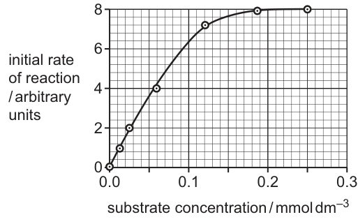
Complete Fig. 6.2 by drawing a curve to show how the presence of a non-competitive inhibitor will affect the rate of reaction of HRP.
Use the curve you have drawn to obtain an estimate of .
= ______
Answer
A new curve is drawn on Fig. 6.2 starting at the origin, with the same (hyperbolic) shape as the original, rising initially close to the original, branching away before the third plotted point (at 2 arbitrary units) and plateauing at a lower maximum rate (a lower than 8 arbitrary units). The new curve crosses half of its own lower at the same substrate concentration as the original curve crosses half of its (0.055 to 0.06 ).
= 0.055 to 0.06
Km = 0.055 to 0.06 mmol dm^-3
Background Concept
The rate of an enzyme-catalysed reaction depends on substrate concentration in a characteristic hyperbolic way (Michaelis–Menten kinetics). Two constants describe the curve:
- : the maximum rate, reached when all active sites are saturated with substrate. On the graph, is the plateau height.
- : the Michaelis–Menten constant, defined as the substrate concentration at which the reaction rate is half of . It is a rough measure of the enzyme's affinity for its substrate — a low means high affinity, a high means low affinity.
Inhibitors alter these constants in characteristic ways:
- A competitive inhibitor resembles the substrate and binds to the active site. Increasing the substrate concentration can out-compete it, so the curve eventually reaches the same as the uninhibited reaction. However, more substrate is needed to reach half , so increases (curve shifts to the right but at the same plateau).
- A non-competitive inhibitor binds to a site other than the active site (an allosteric site). It does not compete with substrate, so the effective concentration of functional enzyme is reduced and the plateau is lower ( decreases). The remaining active enzymes still bind substrate with the same intrinsic affinity, so is unchanged.
Understanding the Question
Fig. 6.2 plots initial rate against substrate concentration for HRP, with a smooth curve that rises steeply from the origin and plateaus at = 8 arbitrary units around 0.20 . The question asks the student to add a second curve for HRP in the presence of a non-competitive inhibitor and then read from the new curve.
Approach
- Draw a new curve that is the same hyperbolic shape, but plateaus at a lower than 8.
- The new curve must start to deviate from the original before the third plotted point (at 2 arbitrary units on the y-axis).
- The new curve must reach half of its own (lower) at the same x-value as the original curve reaches half of 8. Reading the original graph, this x-value is 0.055–0.06 .
- Then read off the new curve at its own half- — which is the same x-value.
Step-by-Step Reasoning
- From the original graph, is 8 arbitrary units, so half is 4 arbitrary units. Reading the curve at rate = 4 gives ≈ 0.055–0.06 for the uninhibited enzyme.
- A non-competitive inhibitor reduces the effective concentration of functional enzyme, so the new plateau must be drawn lower (e.g. at 6 arbitrary units), but the new curve must still pass through the same x-value at its own half plateau. So the new curve must pass through approximately (0.055, half of new ) on the graph.
- Draw the new curve starting at the origin, rising initially close to the original (because at very low [S] the inhibitor effect is small), then branching away below the original before the third plotted point, and flattening at the new lower .
- Because the new curve passes through (0.055–0.06, half of new ), the x-value at half of the new is 0.055–0.06 — i.e. is unchanged.
Key Takeaways
- Non-competitive inhibition: ↓, unchanged.
- Competitive inhibition: unchanged, ↑.
- is read off a Michaelis–Menten curve at the substrate concentration that gives half the plateau rate.
Common Mistakes
- Drawing a competitive curve instead of a non-competitive one — a competitive curve has the same plateau as the original but shifts to the right. The mark scheme explicitly warns: drawing a competitive curve earns only 1 mark even if is read off it correctly (via ecf).
- Drawing an S-shaped/sigmoid curve, which the mark scheme rejects.
- Reading off the original (uninhibited) curve instead of the new one.
- Failing to branch the new curve away from the original early enough (must deviate before the third plot point at 2 arbitrary units).
- Drawing the new curve with a higher (not lower) plateau.
Things to Be Careful About
- The shape must remain a rectangular hyperbola (asymptotic to a lower plateau), not sigmoid.
- The value 0.055–0.06 is read by eye; quoting any value inside that narrow band is acceptable.
- The mark scheme allows ecf (error carried forward): if the candidate does not draw a curve but reads off the printed curve, the read-off mark can still be awarded.
Scientists can design synthetic DNA nucleotide sequences to produce a synthetic HRP gene. These sequences will include a start codon and a stop codon so that translation of messenger RNA (mRNA) can occur.
Explain what is meant by a start codon and a stop codon.
Answer
- Start codon: a triplet of three bases on mRNA (AUG) that codes for methionine and signals the ribosome to begin translation.
- Stop codon: a triplet of three bases on mRNA (UAA, UAG or UGA) that does not code for an amino acid; it signals the ribosome to terminate translation, ending polypeptide chain elongation and allowing the polypeptide to detach from the ribosome.
Start codon = AUG, codes for methionine, begins translation. Stop codon = UAA/UAG/UGA, does not code for an amino acid, ends translation.
Background Concept
The genetic code is read in non-overlapping triplets of bases called codons. Each codon of three mRNA bases corresponds either to one of the 20 amino acids or to a stop signal. Translation is the process by which ribosomes read the mRNA codon by codon and assemble a polypeptide chain.
Two codons have special roles in defining the reading frame and its end:
- The start codon is AUG. It codes for the amino acid methionine (in eukaryotes) and also sets the reading frame for translation: the ribosome assembles at AUG and begins to add amino acids.
- Stop codons are UAA, UAG and UGA. They do not code for any amino acid. When a ribosome reaches a stop codon, no tRNA binds, and releasing factors instead cause the completed polypeptide to detach from the ribosome. Translation then ends.
If more than one stop codon is given in an answer, all of them must be correct. If a DNA sequence is given instead of RNA, the non-transcribed (coding/sense) strand matches the mRNA (T instead of U), and the transcribed (template/antisense) strand is the complement.
Understanding the Question
The question asks for an explanation of what is meant by a start codon and a stop codon. The question is 2 marks — one for each — so the student should give a clear, separate statement for each.
Approach
Define each codon in terms of three ideas: (1) it is a triplet of three bases, (2) it has a specific sequence, and (3) it has a specific function in translation (start or stop). The mark scheme accepts either a description in words or the explicit codon sequences (AUG, and UAA/UAG/UGA).
Step-by-Step Reasoning
- Start codon — a codon is a triplet of three RNA bases. The start codon is AUG; it codes for methionine (the first amino acid in the polypeptide) and signals the ribosome to begin translation.
- Stop codon — a codon is a triplet of three RNA bases. The stop codons are UAA, UAG or UGA; they do not code for an amino acid and signal the ribosome to terminate translation, so that the completed polypeptide is released.
Key Takeaways
- Start codon: AUG, codes for methionine, begins translation.
- Stop codons: UAA, UAG, UGA — do not code for an amino acid, terminate translation.
- Both are triplets of three nucleotide bases in mRNA.
Common Mistakes
- Confusing the start codon (AUG) with the stop codons (UAA, UAG, UGA) — note they all start with U but the second base differs.
- Stating that a stop codon codes for an amino acid (it does not).
- Stating that a stop codon 'stops DNA replication' (it stops translation, not replication).
- Quoting only one stop codon when more than one is asked for, or quoting a non-existent codon like 'UTA'.
- Confusing mRNA and DNA sequences (T instead of U for the non-transcribed strand).
Things to Be Careful About
- The mark scheme accepts the codon sequence as a sufficient definition. AUG for start; UAA, UAG or UGA for stop.
- If the candidate gives the DNA equivalents, ATG (non-transcribed/coding strand) is acceptable for the start codon, and TAG, TAA, TGA (non-transcribed) for stop codons.
- If more than one stop codon is given, all must be correct.
HRP is used in an immunological test known as a sandwich ELISA. One use of the test is to diagnose disease.
Fig. 6.3 outlines the main steps in a sandwich ELISA in which a toxin released by a pathogen is detected in a sample of body fluid taken from a person who is ill.
The test involves two types of monoclonal antibody that can bind to the toxin, a capture antibody and a detection antibody.
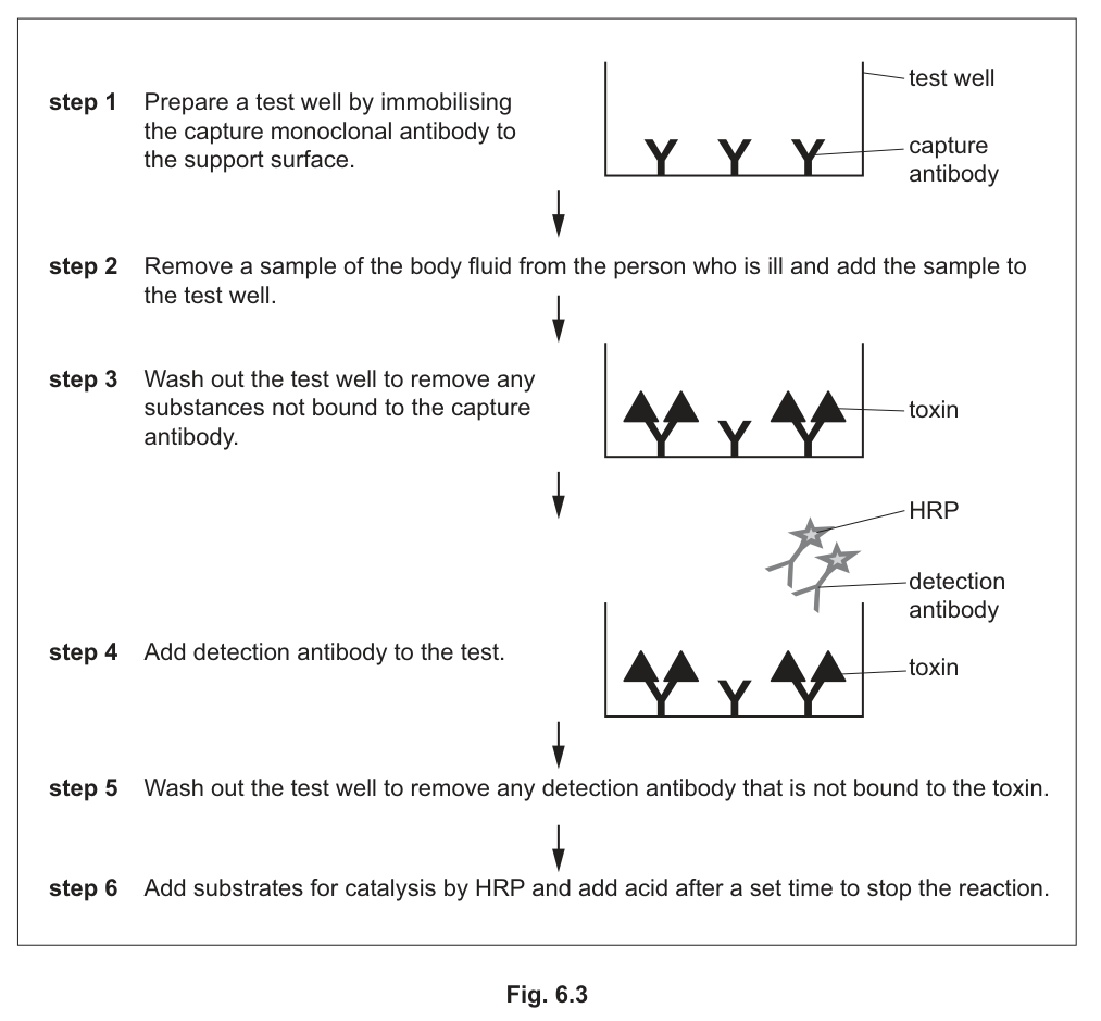
Answer
In Fig. 6.4, each detection antibody (drawn as a Y-shape with a small star representing HRP) is shown bound to the top of a captured toxin molecule. There are no unbound detection antibodies and no detection antibodies bound directly to the capture antibodies or to the well surface.
Detection antibodies (with HRP) are drawn bound to the tops of the toxin molecules.
Background Concept
A sandwich ELISA uses two different monoclonal antibodies that recognise different epitopes (binding sites) on the same antigen. The first — the capture antibody — is immobilised on the surface of the well. The second — the detection antibody — is linked to a reporter enzyme (here, HRP) and is added after the sample.
When the sample is added, the antigen in it is 'captured' by the capture antibody. After a wash, the detection antibody is added; it binds to a different site on the captured antigen, so each antigen molecule is sandwiched between a capture antibody below and a detection antibody above. A second wash removes any detection antibody that failed to bind. The result of step 5 is therefore a 'sandwich' of capture antibody – antigen – detection antibody, with no free detection antibody remaining.
Understanding the Question
Fig. 6.4 already shows the well after steps 1–3 of Fig. 6.3: capture antibodies (Y-shapes) attached to the bottom of the well, with some of them binding toxin molecules (triangles). The question asks the student to complete the picture after step 5 — i.e. after the detection antibody has been added and the well washed.
Approach
- The student must add detection antibodies, each with a small star (or other label) representing the conjugated HRP enzyme.
- Each detection antibody should be drawn bound to a captured toxin (not to the well surface, not to a capture antibody, and not free in solution).
- Unbound detection antibodies must not appear, because step 5 is the wash that removes them.
Step-by-Step Reasoning
- The capture antibodies (Y) are already drawn at the bottom of the well, with toxin triangles bound to some of them.
- Each captured toxin has a second binding site (epitope) on its upper surface, free to bind the detection antibody.
- Draw a Y-shaped detection antibody on top of each captured toxin. Attach a small star to the detection antibody to represent the conjugated HRP.
- The mark scheme allows a single detection antibody binding astride two captured toxin molecules, but the simpler configuration is one detection antibody per toxin.
- Reject detection antibodies drawn bound to the support surface, bound directly to capture antibodies, or floating free (since they would have been washed away in step 5).
Key Takeaways
- The configuration after step 5 is a sandwich: capture antibody – toxin – detection antibody.
- Detection antibody carries the enzyme (HRP) that produces the colour change in step 6.
- Free detection antibody is removed by the wash in step 5.
Common Mistakes
- Drawing detection antibodies directly on the well surface (incorrect — they bind only to captured toxin).
- Drawing detection antibodies attached to the capture antibody itself (incorrect — they bind the toxin, not the capture antibody).
- Drawing free detection antibodies still floating in the well (these should have been washed away).
- Forgetting to add the HRP label (star) on the detection antibody.
Things to Be Careful About
- The diagram must be consistent with Fig. 6.3: capture antibody Y at the bottom, toxin triangles in the middle, detection antibody Y with HRP star on top.
- Unbound detection antibodies should not be present after step 5.
With reference to Fig. 6.3, describe one difference between the capture antibody and the detection antibody.
Answer
The detection antibody has HRP (the enzyme) attached to it, whereas the capture antibody does not. (Or, equivalently, the capture antibody is immobilised on the support surface, whereas the detection antibody is free in solution and binds to a different site on the toxin.)
The detection antibody is conjugated to HRP (enzyme), whereas the capture antibody is not.
Background Concept
In a sandwich ELISA, two monoclonal antibodies are used. Each recognises a different epitope on the same antigen (toxin). Their structural roles differ:
- The capture antibody is fixed to the well surface. It is the 'anchor' that pulls the antigen out of the sample. It is unlabelled.
- The detection antibody is free in solution. It binds to a second, non-overlapping epitope on the captured antigen and is conjugated to a reporter enzyme (here, HRP) that produces a colour change when the substrate is added.
Understanding the Question
The question asks for one difference between the capture antibody and the detection antibody, with reference to Fig. 6.3. One mark is awarded, so a single clear point is needed.
Approach
Compare what each antibody is bound to, where it sits, or what is attached to it. The mark scheme accepts any of:
- They bind to different sites (epitopes) on the toxin.
- The detection antibody has HRP (the enzyme) conjugated to it (the capture antibody does not).
- The capture antibody is immobilised on the well surface; the detection antibody is added in solution.
Step-by-Step Reasoning
- The capture antibody is immobilised on the well (step 1 of Fig. 6.3); the detection antibody is added free in solution (step 4).
- The detection antibody is shown in Fig. 6.3 with a star representing HRP, while the capture antibody is shown unlabelled.
- The two antibodies must bind to different sites on the toxin, otherwise they would sterically block each other. Hence the binding sites (epitopes) are different.
- Any one of these differences is sufficient for the mark.
Key Takeaways
- The two monoclonal antibodies in a sandwich ELISA bind to different epitopes on the antigen.
- The detection antibody carries the reporter (enzyme), so it can produce the colour change.
- The capture antibody anchors the antigen to the well surface.
Common Mistakes
- Stating that they are different 'types' of antibody without giving a structural or functional reason.
- Stating that they bind to different antigens (incorrect — both bind the same toxin, but to different epitopes).
- Saying that the detection antibody is fluorescent or radioactive (incorrect in this ELISA — it is conjugated to HRP, an enzyme).
Things to Be Careful About
- Be specific: 'detection antibody has HRP conjugated to it' is better than 'the detection antibody is labelled'.
- 'Different epitopes' is the precise term for the different binding sites on the same antigen.
Fig. 6.5 outlines the reaction catalysed by HRP in step 6. The organic substrate, TMB, is in excess and changes colour when it is oxidised. This indicates a positive test result.

A student suggested that:
• a low concentration of toxin may by diagnosed as a negative result instead of a positive result
• using a colorimeter after step 6 in the sandwich ELISA test would provide a quantitative result and help to avoid this error.
With reference to Fig. 6.5, explain why using a colorimeter would provide a quantitative measurement for detection of a low concentration of the toxin and help to confirm a positive result.
Answer
- A colorimeter measures the absorbance (or transmission) of light by the reaction mixture — this gives an objective numerical reading, not a subjective judgement.
- A low concentration of toxin results in a low quantity of detection antibody (and therefore HRP) being bound, so only a small amount of TMB is oxidised and the solution is only very pale blue-green. By eye this pale colour may look indistinguishable from the colourless negative control, leading to a false negative.
- A pale blue-green solution has a low(er) absorbance reading (or higher transmission) in a colorimeter, whereas a colourless negative gives a different reading (e.g. close to the blank).
- The colorimeter therefore produces different absorbance values for a low-toxin positive sample and a negative sample, distinguishing them objectively. With a calibration curve of known toxin concentrations, the absorbance can be converted to an actual toxin concentration, giving a fully quantitative result and confirming a positive even at low toxin levels.
A colorimeter measures absorbance objectively; pale colour gives a low but measurable absorbance, distinguishing a low-toxin positive from a true negative (which by eye would look the same), so the result is quantitative and avoids the false-negative error.
Background Concept
In a sandwich ELISA, the intensity of the colour produced in step 6 is proportional to the amount of antigen (toxin) originally present in the sample:
- More toxin → more captured on the well → more detection antibody bound → more HRP in the well → more TMB oxidised per unit time → more intense blue-green colour.
Two practical problems arise at low toxin concentrations:
- The colour is very pale and may be invisible or indistinguishable from a colourless negative by eye.
- The eye cannot reliably rank different shades of pale colour, so the result is qualitative (positive/negative) rather than quantitative.
A colorimeter overcomes both problems. It passes light of a defined wavelength through the sample and measures the absorbance () or transmittance (). According to the Beer–Lambert law, absorbance is directly proportional to the concentration of the absorbing (coloured) species:
where is the molar absorptivity, is the concentration, and is the path length. A pale solution has a low absorbance; a colourless (or blank) solution has an absorbance close to zero. Different absorbances can be measured objectively and used with a calibration curve to read off the original toxin concentration.
Understanding the Question
The student has proposed that a low toxin concentration could be misread as a negative result by eye, and that using a colorimeter after step 6 would give a quantitative result and avoid the error. The question asks the student to explain this with reference to Fig. 6.5 (TMB + H₂O₂ → oxidised blue-green TMB + H₂O, catalysed by HRP). 4 marks are available.
Approach
Build a chain of reasoning:
- Colorimeter measures absorbance (or transmission) — a numerical, objective measurement.
- Low toxin → small amount of HRP captured → small amount of TMB oxidised → pale blue-green colour.
- A pale solution still absorbs some light, so the colorimeter records a low (but non-zero) absorbance — distinguishable from the colourless negative control.
- Different absorbances can be obtained for different toxin concentrations, giving a quantitative (rather than just yes/no) result.
- Compare explicitly with by-eye judgement, which the mark scheme requires for full marks.
Step-by-Step Reasoning
- Colorimeter measures absorbance of light by the reaction mixture. (Mark scheme point 1.)
- Low colour intensity gives a low absorbance reading (or higher transmission). The colourless negative gives a different reading, so the two are objectively distinguishable. (Points 2 and 4.)
- The intensity of the colour reflects the amount of oxidised TMB, which in turn reflects how much HRP (and therefore how much detection antibody and how much toxin) was present. So a low absorbance corresponds to a low concentration of toxin. (Point 3.)
- With a calibration curve of known toxin concentrations, the absorbance can be converted into an actual concentration, giving a quantitative measurement. (Point 5/6, AVP.)
- Compare with by-eye: by eye, 'colourless' and 'very pale blue-green' are hard to tell apart — judgement is subjective and may miss a slight colour change. The colorimeter is not subjective, so it avoids the false-negative error the student described.
Key Takeaways
- Colorimetry converts colour intensity into an objective, numerical absorbance.
- A pale coloured solution still has a measurable absorbance that differs from a colourless blank.
- Absorbance is proportional to the concentration of the coloured species, so colorimetry gives a quantitative result.
- A calibration curve of known concentrations can be used to read off the original antigen concentration.
Common Mistakes
- Stating simply that 'a colorimeter detects a slight colour change' without comparing with by-eye — the mark scheme explicitly rejects this.
- Confusing absorbance and transmission (a darker solution has high absorbance and low transmission).
- Stating that colorimetry gives a 'yes/no' answer — it gives a continuous numerical value.
- Failing to link the absorbance back to the toxin concentration (i.e. explaining why a low absorbance means a low toxin level).
- Saying that colorimetry is 'more sensitive' without explaining how.
Things to Be Careful About
- The mark scheme awards marks only if the explanation explicitly compares the colorimeter with the by-eye judgement at some point (otherwise the candidate only restates that the colorimeter 'detects' small changes).
- Beer–Lambert law is assumed; quoting it is good but not essential at AS level — what matters is the idea that absorbance is proportional to concentration.
- The reaction in step 6 is catalysed by HRP; the substrate (TMB) is in excess, so the amount of product (oxidised TMB) is limited by the amount of HRP (and therefore by the amount of captured toxin).