Biology 9700/14 — May/June 2025
Cambridge AS Level · Multiple Choice (AS Level) · answer key with instant marking and worked solutions
Topics Cell Structure · Biological Molecules · Transport in Mammals · Enzymes · Transport in Plants · Cell Membranes and Transport · +5 more
Tap an option under each question to check it — your score builds as you go.
Plant cells are stained and then viewed with a simple light microscope, using daylight as the only light source.
Which cell structures are clearly visible at a magnification of ?
Options
A chloroplast grana
B lysosomes
C nucleoli
D ribosomes
Working
A light microscope has a maximum resolution of about (200 nm), so only structures at or above this size can be seen clearly. Staining makes the nucleus stand out, and inside it the nucleoli appear as one or more dark, rounded bodies — these are roughly – across and are clearly visible at .
- A. chloroplast grana — grana are stacks of thylakoid membranes, each thylakoid disc is about , and individual grana stacks are at the limit of light-microscope resolution and are not clearly seen; whole chloroplasts are visible, but the grana inside them are not.
- B. lysosomes — roughly – and not distinguishable as separate structures in a routine stained preparation.
- C. nucleoli — large, densely stained, inside the nucleus, easily seen at . ✓
- D. ribosomes — about – in diameter, far below the resolution of a light microscope; an electron microscope is needed.
Answer
C
C
Background Concept
A light microscope (the kind used in school and university practical classes) has a maximum useful magnification of about , but the more important limit is its resolving power — the smallest distance between two points at which they can still be seen as separate. The best light microscopes can resolve down to about (200 nm). Anything smaller than this appears as an indistinct blur, no matter how much you magnify the image.
To make structures more visible, specimens are stained (e.g. with iodine, methylene blue, or toluidine blue). Stains bind to particular chemicals in the cell, making features such as the nucleus, nucleolus, cell wall, starch grains and chloroplasts stand out as coloured regions against a paler background.
To decide whether a structure is "clearly visible" at a given magnification, you need to compare its size with the resolution of the microscope:
- The nucleolus is a dense, rounded body inside the nucleus where ribosomes are assembled. It is typically – in diameter, far above the resolution limit, and it stains very darkly because it is rich in RNA and protein. It is one of the most obvious features in any stained cell viewed at .
- Chloroplast grana are stacks of thylakoid discs within a chloroplast. Individual grana are only – wide — right at, or below, the resolution of a light microscope. The whole chloroplast (–) is easily seen, but the grana inside it are not.
- Lysosomes are small, roughly spherical organelles (–), and no routine stain specifically reveals them in a temporary preparation of plant cells.
- Ribosomes are about – in diameter, an order of magnitude below the resolution of any light microscope. Only an electron microscope (which can resolve a few nanometres) can show individual ribosomes.
Understanding the Question
The question asks which structure is clearly visible in stained plant cells at magnification using a light microscope. The four options test whether you can:
- Distinguish resolution from magnification.
- Know the relative sizes of common sub-cellular structures.
- Recognise that staining (especially of the nucleus) makes nucleoli stand out.
The command word "clearly visible" is the key — a structure must be well above the resolution limit and easily identifiable, not just theoretically in the field of view.
Approach
The strategy is to use the resolution limit of the light microscope () as a threshold and compare it with the size of each structure in turn. Any structure that is well above this threshold and that takes up a stain (or is naturally coloured) will be clearly visible. The candidate that fits both criteria — large enough and stained — is the answer.
Step-by-Step Reasoning
- Establish the resolution limit. A good light microscope resolves to about . To be "clearly visible", a structure should be at least several times this size — say, above .
- Test each option against the threshold and the staining context.
- A — chloroplast grana: the whole chloroplast is easily seen (it is green), but grana are stacks of thylakoids only across — at or below the resolution limit. They cannot be resolved as separate features. Reject.
- B — lysosomes: about –, near or below the resolution limit, and they are not highlighted by any common stain used on plant cells. Reject.
- C — nucleoli: – across, several times the resolution limit, and they stain intensely because of their high RNA/protein content. Inside the lighter-stained nucleus they appear as one or more prominent dark bodies. This is the answer.
- D — ribosomes: – (–) — about ten times smaller than the resolution limit. Definitely not visible with a light microscope. Reject.
- Conclusion: the nucleoli are the only structure that is both large enough to be resolved and darkly stained at . The answer is C.
Key Takeaways
- Resolution, not magnification, determines what you can see. Turning the magnification up beyond on a light microscope does not reveal new detail — the limit is set by the wavelength of visible light (–).
- At with a light microscope and routine stains, the most reliably seen features in a plant cell are: the cell wall, the nucleus (and within it the nucleoli), chloroplasts (as green ovals), large vacuoles (clear region), and starch grains.
- Sub-cellular detail (thylakoid grana, ribosomes, internal membrane systems, viral particles) requires the higher resolution of a transmission electron microscope (resolving power ).
Common Mistakes
- Choosing A (chloroplast grana) because chloroplasts are visible. Whole chloroplasts are obvious (they are green even without staining), but the grana inside them are not resolved by a light microscope.
- Confusing resolution with magnification. Increasing magnification enlarges the image but does not improve the resolution; you cannot see ribosomes by simply zooming in more.
- Choosing B (lysosomes) because they appear in textbook diagrams. Lysosomes are much more prominent in animal cells, and even there they are at the very limit of light-microscope resolution; they are essentially invisible in a stained plant cell preparation.
- Choosing D (ribosomes) because they are described as "small but present in all cells" — they are simply too small for a light microscope.
Things to Be Careful About
- "Visible" in microscopy means clearly distinguishable as a separate feature, not merely "in the field of view". A structure at or below the resolution limit will only ever appear as a blur.
- Always quote the resolution limit of a light microscope as approximately (200 nm). The electron microscope's much better resolution () is what allows us to see ribosomes, grana and viral coats.
- Remember that staining makes a difference: an unstained nucleolus might be hard to spot, but a stained one is one of the most prominent features in the cell. Where a question specifies staining, take that into account.
The diagram shows an image of a cell with a scale bar.
The scale bar (Z) represents an actual size of .
Distance Y represents the diameter of the cell image.
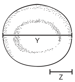
Which calculation to find the actual diameter of the cell is correct?
Options
A
B
C
D
Working
The scale bar Z on the image represents an actual length of . The image length of the cell diameter is Y.
By proportion, the actual diameter is:
Answer
C
C
Background Concept
A scale bar is a line of known actual length drawn on (or below) a micrograph or drawing. It allows the viewer to convert any other measured distance on the same image into an actual size. The principle is simple proportion: every length on the image has been magnified by the same factor, so the ratio of image length to actual length is constant across the whole image.
Formally:
Rearranging to find actual size:
Because the magnification is the same everywhere on the image, you can use the scale bar to find it:
and then apply it to the cell:
Understanding the Question
The figure shows a roughly circular cell. A horizontal line Y spans the diameter of the cell on the image. Below the cell, a scale bar Z is printed, and the question tells us that Z corresponds to an actual length of . We are asked which of the four algebraic expressions correctly converts the image diameter Y into the real diameter of the cell.
The command word is effectively "identify the correct calculation" — we do not need a numerical answer, just the right formula.
Approach
Set up the proportion image-length : actual-length using the scale bar (Z : 15 µm) and apply the same ratio to the cell diameter Y. The answer must keep the units consistent: the image lengths (Y and Z) cancel, leaving the actual length in µm.
Step-by-Step Reasoning
- Step 1 — Use the scale bar to find how many µm each unit of image length represents: per Z image units.
- Step 2 — Multiply that "actual length per image unit" by the cell's image diameter, Y: actual diameter .
- Step 3 — Compare with the options:
- A: — adds image lengths to a real length; meaningless.
- B: — inverted ratio; would give an answer in image units, not µm.
- C: — correct proportion.
- D: — treats 15 as an image length rather than a real length; wrong units.
- Therefore the correct option is C.
Key Takeaways
- A scale bar lets you convert any measured image length into an actual length by simple proportion.
- The correct structure is .
- Keep image lengths with image lengths and actual lengths with actual lengths; do not mix them.
Common Mistakes
- Inverting the ratio (option B) — this gives a number with units of image length, not µm.
- Adding image and actual quantities (option A) — dimensionally invalid.
- Forgetting the scale bar entirely and dividing by 15 as if it were an image length (option D) — units are wrong.
Things to Be Careful About
- Always check that your formula cancels the image-length unit (mm or cm on the page) and leaves a real-world length (µm).
- The same proportional method works whether you measure Y and Z in mm on a printout, in cm on a screen, or in eyepiece-graticule units — only the ratio matters.
- The general magnification formula is ; getting this upside down is a very common error.
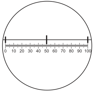
The diagram shows a stage micrometer, with divisions of , viewed using an eyepiece graticule.
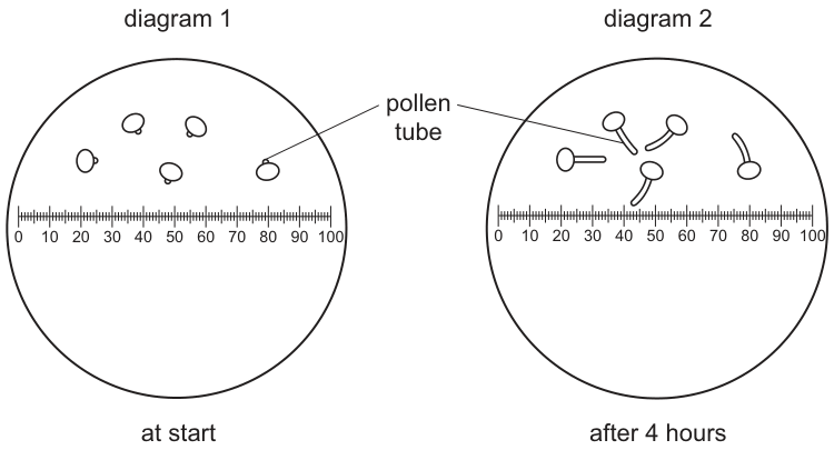
Pollen grains were grown in a sugar solution and viewed using the eyepiece graticule.
Diagram 1 shows the pollen grains at the start. Diagram 2 shows the pollen grains after 4 hours.
What is the growth rate of the pollen tubes?
Options
A
B
C
D
Working
1. Calibrate the eyepiece graticule (Fig. 3.1)
stage micrometer divisions align with eyepiece graticule units.
2. Measure the pollen tube (Fig. 3.2)
The pollen tube in Diagram 2 has grown by graticule units.
3. Calculate the growth rate
Answer
A
A
Background Concept
An eyepiece graticule is a small glass disc with a finely divided scale (typically units) that sits inside the eyepiece of a microscope. The graticule is superimposed on the image of the specimen, so the apparent size of a structure can be read off in graticule units. The drawback is that the real length of one graticule unit depends on the magnification of the objective lens in use — change the objective and the same graticule unit represents a different real length.
A stage micrometer is a microscope slide on which an accurately known scale has been etched, most commonly in divisions of (). To use a graticule you first place the stage micrometer on the stage, line up its scale with the graticule scale, and read off the calibration. For example, " graticule units coincide with of stage micrometer" means one graticule unit at that magnification. The stage micrometer is then removed and the actual specimen is measured using the calibrated graticule.
Understanding the Question
Fig. 3.1 is the calibration image: a stage micrometer (top scale, divisions of ) aligned with the eyepiece graticule (bottom scale, –).
Fig. 3.2 shows pollen grains at the start (Diagram 1) and after 4 hours (Diagram 2), both viewed through the same calibrated graticule. The grains have extended pollen tubes. The question asks for the growth rate of the tubes — that is, the change in length per unit time — so the candidate must (1) work out the calibration, (2) measure how much the tube has grown, and (3) divide by the 4-hour interval.
Approach
- Compare the stage micrometer and the graticule in Fig. 3.1 to find the real length of one graticule unit (and convert to µm).
- In Fig. 3.2, read the length the tube has grown in graticule units and convert to µm.
- Growth rate length grown time elapsed, with the unit .
Step-by-Step Reasoning
Step 1 — Calibrate
In Fig. 3.1, stage micrometer divisions of (so in total) line up exactly with the full graticule units.
Converting to micrometres (microscope measurements are conventionally quoted in µm):
So at this magnification, one graticule unit is in real length.
Step 2 — Measure the tube
In Fig. 3.2, Diagram 2, the pollen tube has grown by graticule units in 4 hours.
Step 3 — Calculate the rate
A rate is a change in quantity divided by the time over which that change occurred:
Key Takeaways
- An eyepiece graticule must be calibrated with a stage micrometer at the magnification being used; the calibration is not transferable between objective lenses.
- Convert mm to µm (); biological microscope measurements are quoted in µm.
- A rate is always a change in quantity divided by the time over which that change occurred: .
- Make sure the unit in the final answer is appropriate to the size of the structure (µm h here, not mm h).
Common Mistakes
- Wrong unit: forgetting to convert mm to µm gives or (distractors C and D). A pollen tube of this size should be reported in µm, not mm.
- Misreading the calibration: thinking one graticule unit equals instead of makes the tube appear too large and the rate becomes (not an option here, but a common trap in similar questions).
- Measuring the wrong feature: measuring the diameter of the grain, or measuring from the start position rather than the change in length.
- Forgetting the time: reporting just the length grown () instead of the rate per hour.
Things to Be Careful About
- Show the calibration and the conversion explicitly — the mark scheme credits the working, not just the final number.
- Use the time given in the question (4 h), not minutes or days.
- Quote the final answer with its unit (); biology marks are lost when the unit is wrong or missing.
- The graticule scale must be re-calibrated every time the objective lens is changed.
What are found in chloroplasts and also in mitochondria?
1 DNA
2 70S ribosomes
3 mRNA
Options
A 1, 2 and 3
B 1 and 2 only
C 1 only
D 2 and 3 only
Both chloroplasts and mitochondria are semi-autonomous organelles:
-
- DNA — both contain their own (circular) DNA.
-
- 70S ribosomes — both contain ribosomes of the 70S (prokaryotic) type.
-
- mRNA — both transcribe their DNA to produce mRNA for protein synthesis.
All three are present in both organelles, so 1, 2 and 3 are correct.
Answer
A
A
Background Concept
Chloroplasts and mitochondria are described as semi-autonomous organelles because each carries some of the machinery needed to make its own proteins. Specifically, both contain:
- their own DNA (a small, circular molecule, separate from the nuclear DNA),
- ribosomes that are 70S (the same size and type as those found in prokaryotes such as bacteria), and
- the mRNA transcribed from that DNA, which those ribosomes then translate into protein.
The presence of 70S ribosomes and circular DNA is a key piece of evidence for the endosymbiotic theory, which proposes that chloroplasts and mitochondria originally evolved from free-living prokaryotes that were engulfed by an ancestral eukaryotic cell. The 70S ribosomes distinguish them from the 80S ribosomes of the eukaryotic cytoplasm.
Understanding the Question
The question asks which items from a list of three are found in both chloroplasts and mitochondria. It is a multiple-combination question (the type that uses option lines such as "1, 2 and 3" / "1 and 2 only" / etc.), so each of the three statements must be considered independently and then the correct combination chosen.
The key command is "found in chloroplasts and also in mitochondria" — the answer must satisfy both organelles, not just one.
Approach
Go through statements 1, 2 and 3 in turn and decide whether each is true of both organelles. Then match the three "yes" decisions to one of the four option combinations.
Step-by-Step Reasoning
- Statement 1 — DNA: True for both. Mitochondrial DNA and chloroplast DNA are both small, circular molecules carrying genes for some of the organelle's own proteins (and for tRNA/rRNA used in their protein synthesis).
- Statement 2 — 70S ribosomes: True for both. Both organelles synthesise some of their own proteins on 70S ribosomes — the prokaryotic type, smaller than the 80S ribosomes of the eukaryotic cytoplasm.
- Statement 3 — mRNA: True for both. Transcription of the organelle's own DNA produces mRNA, which is then translated on the organelle's 70S ribosomes. So mRNA is present in both.
All three statements are true of both organelles, giving the combination 1, 2 and 3 — option A.
Why the distractors are wrong:
- B (1 and 2 only) — would be correct only if 70S ribosomes were missing, which is not the case.
- C (1 only) — would require 70S ribosomes and mRNA to be absent, which they are not.
- D (2 and 3 only) — would require DNA to be absent, which is incorrect: both organelles definitely contain DNA.
Key Takeaways
- Chloroplasts and mitochondria are semi-autonomous: they each have their own DNA, 70S ribosomes and mRNA, and so can carry out their own transcription and translation for a subset of their proteins.
- The 70S ribosome is the prokaryotic type — distinct from the 80S ribosomes of the eukaryotic cytoplasm.
- The shared features (circular DNA + 70S ribosomes) underpin the endosymbiotic theory of organelle origin.
Common Mistakes
- Confusing 70S (prokaryotes, mitochondria, chloroplasts) with 80S (eukaryotic cytoplasm, rough endoplasmic reticulum). Writing that mitochondria/chloroplasts have 80S ribosomes is incorrect.
- Forgetting that mRNA is present in the organelle because the organelle transcribes its own DNA.
- Thinking that DNA is only in the nucleus — many students miss that mitochondria and chloroplasts carry their own small genome.
Things to Be Careful About
- "S" in 70S/80S is a Svedberg unit, a measure of how fast a particle sediments in a centrifuge; it correlates with size but is not strictly additive from subunits.
- "Both" means every item in the chosen combination must apply to both organelles — re-check each organelle for every statement before selecting an answer.
A long-distance runner has become adapted to run faster for longer after regular training.
Which cell structure in a muscle cell has increased in number to allow this adaptation?
Options
A lysosome
B mitochondrion
C nucleus
D smooth endoplasmic reticulum
Working
Long-distance running requires sustained aerobic respiration in muscle cells to release the large amounts of ATP needed for continuous contraction. Mitochondria are the site of aerobic respiration, and training increases the number of mitochondria in muscle cells to meet this demand.
Answer
B
B
Background Concept
Mitochondria are double-membrane organelles found in eukaryotic cells. They are the site of aerobic respiration, where glucose (and other respiratory substrates) is oxidised in the presence of oxygen to produce large quantities of ATP. The inner membrane is folded into cristae, which increase the surface area available for the electron transport chain and ATP synthase, and the matrix contains the enzymes of the Krebs cycle.
Different cell types contain different numbers of mitochondria depending on their energy demand. Cells with very high energy requirements — such as muscle cells, liver cells, and sperm cells — have many mitochondria. The number of mitochondria within a cell is not fixed; it can increase in response to sustained metabolic demand.
Understanding the Question
The question describes a long-distance runner who, after regular training, can run faster for longer. It asks which cell structure within a muscle cell has increased in number to bring about this adaptation. The command word implies we should identify a structure whose increase directly enables greater endurance performance.
Approach
The key is to identify what limits sustained muscular activity and which organelle provides the energy for it. Endurance running is powered almost entirely by aerobic respiration, so the relevant organelle is the mitochondrion.
Step-by-Step Reasoning
- Sustained muscle contraction during long-distance running requires a continuous supply of ATP.
- Aerobic respiration in mitochondria produces far more ATP per glucose molecule (~30–32 ATP) than anaerobic respiration in the cytoplasm (2 ATP per glucose).
- Therefore, to run for longer, the muscle cells need a greater capacity for aerobic respiration.
- Training stimulates mitochondrial biogenesis (the production of new mitochondria), so the number of mitochondria in muscle fibres increases.
- With more mitochondria, the muscle cell can carry out a higher rate of aerobic respiration, supplying more ATP and allowing the runner to sustain faster pace for longer before fatigue.
- The other organelles do not provide energy for muscle contraction:
- Lysosomes contain digestive enzymes for breaking down waste materials — not relevant to energy supply.
- The nucleus stores genetic information — its number (one per cell) does not change with training.
- Smooth endoplasmic reticulum is involved in lipid synthesis and detoxification — not in ATP production.
Key Takeaways
- Mitochondria are the site of aerobic respiration and the main source of ATP in muscle cells.
- Endurance training increases the number of mitochondria in skeletal muscle, raising aerobic capacity.
- The number of an organelle within a cell can change in response to functional demand.
Common Mistakes
- Choosing D (smooth endoplasmic reticulum) — confusing its role in lipid/detoxification with energy metabolism.
- Choosing A (lysosome) — confusing it with the energy-providing organelle.
- Choosing C (nucleus) — the nucleus number per cell does not change with training.
Things to Be Careful About
- The question specifies "increased in number" — the answer must be an organelle whose quantity can rise with training, not merely one present in muscle cells.
- "Lysosome", "nucleus" and "smooth endoplasmic reticulum" can all be found in muscle cells, but only mitochondria have a direct role in supplying the ATP needed for sustained contraction.
Some functions carried out within eukaryotic cells are listed.
1 lysosome production
2 polypeptide modification
3 exocytosis
Which functions are carried out by the Golgi body?
Options
A 1, 2 and 3
B 1 and 2 only
C 1 and 3 only
D 2 and 3 only
Working
The Golgi body receives proteins from the rough endoplasmic reticulum, modifies them (e.g. by adding carbohydrate groups to form glycoproteins), and packages them into vesicles.
- Lysosome production ✓ — lysosomes are formed by the Golgi, which packages hydrolytic enzymes into vesicles.
- Polypeptide modification ✓ — the Golgi modifies polypeptides received from the rER (glycosylation, phosphorylation, etc.).
- Exocytosis ✗ — exocytosis is the fusion of a vesicle with the plasma membrane; it is performed by the vesicle/cell surface membrane, not by the Golgi itself.
Answer
B
B
Background Concept
The Golgi body (Golgi apparatus) is a stack of flattened membrane-bound cisternae found in eukaryotic cells. It receives vesicles from the rough endoplasmic reticulum (rER) on its cis face and dispatches vesicles from its trans face. Its principal functions are:
- Modifying polypeptides — newly synthesised polypeptides from the rER are further modified inside the Golgi, e.g. by addition of carbohydrate groups (glycosylation) to form glycoproteins, or by phosphorylation or sulfation.
- Sorting and packaging — finished products are sorted according to their destination and packaged into vesicles that bud off the trans face.
- Producing lysosomes — lysosomes are essentially Golgi-derived vesicles that contain hydrolytic (digestive) enzymes. The Golgi packages these enzymes and the vesicle becomes a lysosome.
A closely related but distinct process is exocytosis, in which a vesicle travels to and fuses with the plasma membrane, releasing its contents to the outside of the cell. The Golgi produces the vesicle, but the fusion event (and the release of contents) is a property of the vesicle and the plasma membrane, not the Golgi itself.
Understanding the Question
The question lists three cellular activities and asks which are carried out by the Golgi body. Each must be judged on whether it is a function intrinsic to the Golgi or a function performed elsewhere (often by structures the Golgi merely produces). The candidate must be alert to the distinction between producing/packaging (a Golgi role) and vesicle fusion with the plasma membrane (not a Golgi role).
Approach
Check each numbered function against the known roles of the Golgi:
- Lysosome production — Yes, this is a Golgi function. Lysosomes bud from the trans face carrying hydrolytic enzymes.
- Polypeptide modification — Yes, this is a central Golgi function. Polypeptides from the rER are glycosylated, phosphorylated or otherwise modified as they pass through the cisternae.
- Exocytosis — No. The Golgi packages material into secretory vesicles, but the vesicles themselves travel to and fuse with the plasma membrane; that fusion step is exocytosis and is performed by the vesicle/plasma membrane, not the Golgi.
Only 1 and 2 are correct → option B.
Step-by-Step Reasoning
- A candidate who simply remembered "Golgi = packaging and modification" would select options that contain functions 1 and 2.
- Option A (1, 2 and 3) is the trap for anyone who broadly associates "vesicles leaving the Golgi" with the Golgi doing the whole secretory pathway, including exocytosis.
- Option C (1 and 3) drops polypeptide modification, which is wrong because modification is a core Golgi function.
- Option D (2 and 3) drops lysosome production, which is wrong because lysosomes are formed at the trans face of the Golgi.
- Option B (1 and 2 only) correctly credits the two processes that occur at the Golgi, and excludes the exocytosis step that occurs after the vesicle has left.
Key Takeaways
- The Golgi modifies polypeptides and packages them into vesicles.
- Lysosomes are produced by the Golgi.
- Exocytosis is performed by secretory vesicles fusing with the plasma membrane; it is not a Golgi function, even though the Golgi produces the vesicle.
- Be alert to "produces / packages" versus "releases / fuses" when classifying organelle functions.
Common Mistakes
- Confusing the Golgi with the secretory pathway as a whole and selecting option A because vesicles from the Golgi eventually undergo exocytosis.
- Thinking lysosomes come from the rER because their hydrolytic enzymes are made on ribosomes attached to the rER; in fact the enzymes are packaged by the Golgi to form the lysosome.
- Attributing exocytosis to any organelle that touches vesicles; exocytosis specifically requires the vesicle and plasma membrane.
Things to Be Careful About
- The wording of the question is "carried out by the Golgi body". A process that the Golgi merely enables (by making the vesicle) is not the same as one the Golgi carries out.
- "Polypeptide modification" is a specific term here — do not substitute "protein synthesis", which occurs on ribosomes, not in the Golgi.
The diagram shows the structure of a unicellular organism.
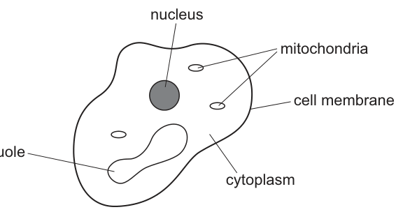
Which statement is correct for this organism?
Options
A It contains a nucleus so it is not prokaryotic.
B It contains mitochondria so it is not a plant.
C It does not have a cell wall so it must be eukaryotic.
D It is unicellular so it must be a bacterium.
Working
The diagram shows a unicellular organism with a nucleus, mitochondria, a vacuole, cytoplasm and a cell membrane — features characteristic of a eukaryote such as Amoeba.
Evaluating each option:
- A — Prokaryotes (bacteria) lack a true, membrane-bound nucleus; their DNA is free in the cytoplasm in a nucleoid. The presence of a nucleus therefore means the cell is not prokaryotic. ✓
- B — Plants do have mitochondria (in all their cells), so the presence of mitochondria cannot rule out being a plant. ✗
- C — Many eukaryotes (e.g. animal cells, Amoeba) lack a cell wall; the absence of a cell wall does not, on its own, define a eukaryote. ✗
- D — Many eukaryotes (e.g. Amoeba, Paramecium, Saccharomyces) are unicellular, so being unicellular does not make an organism a bacterium. ✗
Answer
A
A
Background Concept
Cells are divided into two fundamental types based on their internal organisation:
- Prokaryotes (bacteria and archaea) are small cells (typically 1–5 µm) that lack a true, membrane-bound nucleus. Their DNA is a single circular molecule lying free in the cytoplasm in a region called the nucleoid. They also lack other membrane-bound organelles such as mitochondria, endoplasmic reticulum and Golgi apparatus, and their ribosomes are smaller (70S).
- Eukaryotes (animals, plants, fungi, protists) are larger cells (typically 10–100 µm) that possess a true nucleus — the DNA is enclosed by a double membrane called the nuclear envelope. Eukaryotes also contain many membrane-bound organelles, including mitochondria, and have larger (80S) ribosomes.
Some eukaryotes are unicellular — they live as single cells, e.g. Amoeba proteus, Paramecium caudatum, and the yeast Saccharomyces cerevisiae. Unicellularity is therefore a feature of some eukaryotes, some prokaryotes and some members of many kingdoms — it is not a defining feature of any one group on its own.
Understanding the Question
The diagram (Fig. 7.1) shows a single-celled organism with an irregular outline, enclosing a nucleus, mitochondria, a vacuole, cytoplasm, and bounded by a cell membrane. The task is to identify which of the four statements is a correct logical deduction about this organism.
The command word is implied (the question offers a single correct statement), so the candidate must weigh each option against the biology rather than recall a fact.
Approach
The strategy is:
- Read the features shown in the diagram (nucleus, mitochondria, vacuole, cell membrane, no cell wall labelled).
- Recognise these features as those of a typical eukaryotic cell (resembling an Amoeba).
- Test each option against the accepted definitions of prokaryote, eukaryote, plant, animal and bacterium.
Step-by-Step Reasoning
Option A — "It contains a nucleus so it is not prokaryotic."
This follows directly from the definition: a prokaryote is, by definition, a cell that lacks a true nucleus. Any cell that contains a nucleus is therefore eukaryotic, not prokaryotic. The diagram clearly labels a nucleus, so this statement is correct. ✓
Option B — "It contains mitochondria so it is not a plant."
This is a common misconception. All plant cells contain mitochondria — they are needed for aerobic respiration in every living plant cell, just as in animal cells. Mitochondria are not exclusive to animals. So this statement is wrong. ✗
Option C — "It does not have a cell wall so it must be eukaryotic."
The conclusion is unjustified. While the diagram does not show a cell wall (only a cell membrane), the absence of a cell wall does not make a cell eukaryotic. Animal cells and many protists (e.g. Amoeba) lack cell walls yet are eukaryotic, but the absence of a wall is not itself a defining feature — it is a negative observation that does not lead to that classification on its own. ✗
Option D — "It is unicellular so it must be a bacterium."
This is incorrect because many eukaryotes are unicellular. Amoeba, Paramecium, Euglena, Saccharomyces (yeast) and Chlorella are all single-celled eukaryotes. Unicellularity is therefore not exclusive to bacteria. ✗
Only option A gives a statement that is biologically correct.
Key Takeaways
- The presence of a true nucleus is the single defining feature that distinguishes eukaryotes from prokaryotes.
- Mitochondria are present in plants, animals, fungi and most protists — they are not exclusive to animals.
- Unicellularity is a feature of some members of many groups (bacteria, protists, some fungi) and is not diagnostic of any one group.
- Distinguish between observation (e.g. "no cell wall visible") and definition (e.g. "has a nucleus") when evaluating biological statements.
Common Mistakes
- Assuming that anything with mitochondria must be an animal — plants also respire aerobically and contain many mitochondria.
- Equating "no cell wall" with "eukaryote" — the conclusion is non-sequitur; the cell wall is absent in animals and many protists, but its absence is not the criterion for eukaryotic classification.
- Confusing "unicellular" with "bacterium" — single-celled organisms include both bacteria and many eukaryotes (yeast, Amoeba).
Things to Be Careful About
- The mark rewards a statement that is both true and a valid logical deduction from the diagram. Watch for statements that are true in isolation but do not follow from the features shown.
- "Not prokaryotic" is a weaker claim than "is eukaryotic" — option A's wording is deliberately cautious and therefore correct; the others overgeneralise.
Diastase is an enzyme that breaks down starch into maltose.
A sample of starch is treated with boiled diastase and left for 15 minutes.
Samples of the mixture are then tested with iodine solution and with Benedict’s solution.
What is the correct result?
Options
| iodine solution | Benedict’s solution | |
|---|---|---|
| A | blue-black | blue |
| B | blue-black | red |
| C | brown | blue |
| D | brown | red |
Working
Boiling denatures diastase (destroys its tertiary structure and active site), so the enzyme can no longer catalyse the breakdown of starch. Starch therefore remains in the mixture after 15 minutes, and no maltose (reducing sugar) is produced.
- Iodine solution turns blue-black in the presence of starch.
- Benedict's solution remains blue because no reducing sugar is present (and even after heating, the colour does not change).
Answer
A
A
Background Concept
Diastase is an amylase enzyme that hydrolyses the glycosidic bonds in starch, breaking it down into maltose (a reducing sugar). Like all enzymes, diastase is a globular protein whose active site depends on a specific three-dimensional shape held together by hydrogen bonds, ionic bonds and hydrophobic interactions.
When an enzyme is exposed to high temperatures (such as boiling), the kinetic energy of the molecules increases and these weak bonds are disrupted. The tertiary structure of the protein is destroyed and the active site loses its specific shape. This is called denaturation and it is irreversible — the enzyme can no longer bind its substrate and so cannot catalyse the reaction, even when cooled back to a normal temperature.
The two biochemical tests being used here detect different molecules:
- Iodine solution turns blue-black in the presence of starch (the iodine molecules slot into the helical amylose structure). The original yellow-brown colour of iodine is seen when no starch is present.
- Benedict's solution is a deep blue reagent containing copper(II) sulfate. When heated with a reducing sugar (such as maltose, glucose or fructose), the Cu²⁺ ions are reduced to Cu⁺ ions, forming a red precipitate of copper(I) oxide. In the absence of reducing sugar, Benedict's solution simply remains its original blue colour.
Understanding the Question
The question describes starch being treated with boiled diastase and then left for 15 minutes. The key detail is that the diastase has been boiled before being added to the starch — this is not the same as a high-temperature experiment to test the effect of temperature on reaction rate. The enzyme has already been denatured by the time it contacts its substrate.
We are asked to predict the results of two qualitative tests on this mixture after 15 minutes:
- Iodine solution test — tells us whether starch is still present.
- Benedict's solution test — tells us whether any reducing sugar has been produced.
The command word is implicit in the multiple-choice format: we must select the row whose colours correctly represent what the two tests would show.
Approach
Decide whether the enzyme is functional. If it is not, then no reaction has occurred:
- Substrate (starch) is still present → iodine result is determined by the presence of starch.
- Product (maltose) has not been formed → Benedict's result is determined by the absence of reducing sugar.
Match the predicted colours to the available options.
Step-by-Step Reasoning
- Boiled diastase is denatured. The tertiary structure is destroyed, so the active site can no longer bind starch. The enzyme is permanently inactivated.
- No hydrolysis of starch occurs. Because diastase cannot work, the starch molecules remain intact in the mixture throughout the 15-minute incubation.
- No maltose is produced. Without enzyme activity, the glycosidic bonds in starch are not broken, so the reducing sugar maltose is not formed.
- Iodine test prediction. With starch still present, iodine molecules insert into the amylose helix and the solution turns blue-black (option A or B).
- Benedict's test prediction. With no reducing sugar present, Benedict's reagent does not change colour — it remains its original blue colour (option A or C).
- Combine the two predictions. Blue-black with iodine AND blue with Benedict's corresponds to option A.
Key Takeaways
- Boiling an enzyme causes irreversible denaturation — the enzyme cannot recover its function on cooling.
- The iodine test is specific for starch (blue-black = positive; yellow-brown = negative).
- Benedict's test detects reducing sugars (red/orange precipitate = positive after heating; stays blue = negative).
- A negative Benedict's result keeps the reagent its original blue colour, not a clear or colourless result.
- Always consider whether the enzyme is actually active before predicting product formation.
Common Mistakes
- Choosing B (blue-black, red): This assumes diastase still works after boiling and produces maltose, which Benedict's would detect as a reducing sugar. This is a common error because students forget that boiling destroys enzymes.
- Choosing D (brown, red): This combines two wrong predictions — it assumes starch has been broken down (brown iodine) AND reducing sugar has been produced (red Benedict's). These two outcomes are inconsistent: if starch is gone, then diastase must have worked, and a working enzyme should give B, not D.
- Choosing C (brown, blue): This assumes starch has been hydrolysed (so iodine stays brown/yellow) but no reducing sugar was produced — which is biologically impossible, since maltose is the direct product of starch hydrolysis by amylase.
- Confusing Benedict's reagent with biuret or with the iodine reagent in terms of starting colour.
Things to Be Careful About
- The wording "boiled diastase" is critical: it is the enzyme that has been boiled, not the starch mixture as a whole. The starch is at a normal temperature for the reaction.
- Even at room temperature, denatured enzymes do not spontaneously refold into their active conformation.
- The Benedict's test always requires heating; the blue colour is the colour of the unreacted reagent itself, not an indication that the test was not carried out.
- The original colour of iodine solution is yellow-brown; remember that a negative result on starch looks like the reagent itself, not clear.
Which molecule is required for this reaction to occur?
Options
A catalase
B copper sulfate
C hydrochloric acid
D sodium hydroxide
Working
The equation shows the hydrolysis of a disaccharide (sucrose, ) into two monosaccharide hexoses (glucose, ). In the standard non-reducing sugar test, this hydrolysis is carried out using dilute hydrochloric acid as the catalyst. The acid is later neutralised with sodium hydrogencarbonate before Benedict's reagent is added.
- A – catalase: an enzyme that breaks down hydrogen peroxide; not relevant here.
- B – copper sulfate: present in Benedict's reagent; it does not hydrolyse the glycosidic bond.
- C – hydrochloric acid: hydrolyses the glycosidic bond in sucrose to release the reducing monosaccharides. ✓
- D – sodium hydroxide: used in the biuret test for proteins; not used to hydrolyse disaccharides.
Answer
C
C
Background Concept
A disaccharide such as sucrose is built from two monosaccharides joined by a glycosidic bond, formed by a condensation (dehydration-synthesis) reaction. The reverse process — splitting the bond by adding a molecule of water — is called hydrolysis. The equation in the question is precisely the hydrolysis of sucrose into two molecules of glucose:
In living cells this hydrolysis is catalysed by the enzyme sucrase (an example of an intracellular/extracellular carbohydrate-digesting enzyme). However, in the laboratory — specifically during the non-reducing sugar test — the glycosidic bond is split chemically using dilute hydrochloric acid (HCl), which is then neutralised with sodium hydrogencarbonate before Benedict's reagent is added to test for the newly released reducing sugars.
Understanding the Question
This is a multiple-choice question (Paper 1 style) that presents the hydrolysis of a disaccharide and asks which reagent is required. The equation gives the clue: water is being added across a glycosidic bond. The candidate must pick, from the four options, the substance used to drive this hydrolysis in the standard practical procedure.
Approach
- Recognise the equation as hydrolysis of a disaccharide (the formula and two products of point to sucrose → glucose + glucose, although glucose + fructose would be biologically more accurate; the equation is schematic).
- Recall the non-reducing sugar test sequence: boil the sample with dilute HCl → cool → neutralise with sodium hydrogencarbonate → add Benedict's reagent and re-boil.
- Match the role of each option to this procedure: only HCl is the hydrolytic reagent.
Step-by-Step Reasoning
- Option A (catalase): an enzyme that decomposes hydrogen peroxide into water and oxygen. It is irrelevant to the hydrolysis of a disaccharide.
- Option B (copper sulfate): a component of Benedict's reagent that provides the ions which are reduced to (brick-red precipitate) by reducing sugars. It does not break glycosidic bonds.
- Option C (hydrochloric acid): ✓ dilute HCl supplies ions that catalyse the hydrolysis of the glycosidic bond. This is exactly the step in the non-reducing sugar test that converts a non-reducing sugar such as sucrose into its constituent reducing monosaccharides.
- Option D (sodium hydroxide): used in the biuret test for proteins (with copper sulfate) to maintain alkaline conditions. It is not a hydrolytic agent for disaccharides and would in fact be used afterwards to neutralise the acid, not to perform the hydrolysis.
Key Takeaways
- Hydrolysis of a disaccharide requires water and a catalyst — biologically an enzyme (e.g. sucrase), in the lab dilute hydrochloric acid.
- The non-reducing sugar test uses dilute HCl + heat to hydrolyse the glycosidic bond, then sodium hydrogencarbonate to neutralise, then Benedict's reagent to detect the released reducing sugars.
- Always read the equation before answering: a single substrate plus water giving two products signals a hydrolysis reaction.
Common Mistakes
- Choosing B (copper sulfate) because it is associated with the Benedict's test for sugars — but copper sulfate detects reducing sugars; it does not hydrolyse them.
- Choosing A (catalase) because it is an enzyme that breaks a bond — but catalase is specific for hydrogen peroxide, not carbohydrates.
- Choosing D (sodium hydroxide) because it is also used in food tests (biuret) — it is not the hydrolytic reagent in the non-reducing sugar test.
Things to Be Careful About
- The word "hydrolysis" and the appearance of on the left of the equation are the key visual cues.
- In a biological (in vivo) context, the molecule that drives this reaction would be an enzyme such as sucrase, not an acid. The question is set in a chemical/test context, so the answer is HCl.
- CIE mark schemes expect the specific chemical name hydrochloric acid, not vague terms such as "an acid".
Which molecules contain at least two double bonds?
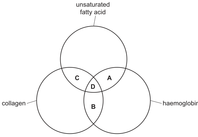
Options
A A
B B
C C
D D
Answer
D
A molecule containing at least two double bonds must be an unsaturated fatty acid (which has double bonds), must have peptide bonds with partial double-bond character (collagen), and must possess the many conjugated double bonds of the haem group (haemoglobin). Only region D lies in the overlap of all three circles.
D
Background Concept
Double bonds in biomolecules are covalent bonds in which two pairs of electrons are shared between two atoms (commonly , , or ). Three biomolecules relevant to this question contain double bonds in different ways:
- Unsaturated fatty acids – their hydrocarbon tails contain one or more carbon–carbon double bonds (). A polyunsaturated fatty acid has two or more such double bonds, while a monounsaturated fatty acid has only one.
- Collagen – a fibrous protein whose polypeptide chains are linked by peptide bonds. The peptide bond has partial double-bond character because the lone pair on nitrogen is delocalised into the carbonyl group, giving the bond some double-bond character. Strictly, however, collagen is not usually classified as containing true double bonds in the structural sense.
- Haemoglobin – a globular protein with four polypeptide chains, each carrying a haem prosthetic group. The haem is a porphyrin ring system with a central ion and contains a large number of conjugated and double bonds, which is why it absorbs visible light and is coloured.
Understanding the Question
The figure is a three-circle Venn diagram with the sets unsaturated fatty acid, collagen, and haemoglobin. Regions A, B, C and D label the four overlaps:
- A = unsaturated fatty acid ∩ haemoglobin (excluding collagen)
- B = collagen ∩ haemoglobin (excluding unsaturated fatty acid)
- C = unsaturated fatty acid ∩ collagen (excluding haemoglobin)
- D = intersection of all three circles
The command word is "Which molecules contain at least two double bonds?" – we must select the region that is the correct home for such a molecule.
Approach
Step through each of the three biomolecules and decide whether it contains at least two conventional double bonds, and therefore belongs in the relevant circle of the Venn diagram. Then identify which region is the overlap that all three of those properties share.
Step-by-Step Reasoning
- Unsaturated fatty acid – the defining feature of an unsaturated fatty acid is the presence of double bonds in the hydrocarbon tail. Polyunsaturated fatty acids contain at least two double bonds, so the molecule belongs inside the "unsaturated fatty acid" circle.
- Collagen – although peptide bonds are formally single bonds, they exhibit partial double-bond character owing to resonance between the carbonyl oxygen and the amide nitrogen. In a Venn-diagram context treating the question of "double bonds", collagen is taken to lie inside its circle because of this resonance character.
- Haemoglobin – each of its four haem groups has a porphyrin ring rich in conjugated and double bonds, far more than two. So haemoglobin lies inside the "haemoglobin" circle.
A molecule that genuinely contains at least two double bonds therefore simultaneously satisfies all three criteria, which on the Venn diagram corresponds to the central region where all three sets overlap – region D.
Key Takeaways
- An unsaturated fatty acid is identified structurally by the presence of double bond(s) along its hydrocarbon chain.
- Collagen is a fibrous protein; its peptide bonds display partial double-bond character through resonance.
- Haemoglobin is a globular protein with a haem prosthetic group whose porphyrin ring contains many conjugated double bonds – this is what makes it red and able to carry .
- On a Venn diagram, a feature that requires the simultaneous presence of all three sets must be placed in the central triple-overlap region.
Common Mistakes
- Confusing monounsaturated (one bond) with polyunsaturated (≥2 bonds) fatty acids and choosing the wrong region.
- Forgetting that haemoglobin's many double bonds reside in the haem group, not the globin chains, and so placing haemoglobin only in the "protein" half of the diagram.
- Treating the peptide bond as a true double bond rather than appreciating its partial double-bond character – on a structural-feature Venn diagram the collagen circle is still relevant.
- Selecting A (only unsaturated fatty acid ∩ haemoglobin) when the question requires the triple overlap because collagen must also be included.
Things to Be Careful About
- Read the wording "at least two" carefully – this is a lower bound, so a molecule with many more double bonds still qualifies.
- On a Venn diagram, a label inside the central triple overlap (D) means the molecule belongs to all three sets simultaneously.
- Mark-scheme style requires using the correct biomolecule terminology: " double bonds", "conjugated double bonds in the haem group", and "partial double-bond character of the peptide bond".
Which description of globular proteins is correct?
Options
A They are only found in cell surface membranes.
B They only contain amino acids with hydrophilic R groups.
C They can change shape by using energy from ATP.
D They always have a quaternary structure of at least three polypeptides.
Working
Globular proteins are roughly spherical and water-soluble. They are found throughout cells (e.g. enzymes, antibodies, haemoglobin), not only in membranes — A is wrong. Their tertiary folding places hydrophobic R groups in the core and hydrophilic R groups on the outside, so they contain both — B is wrong. Some globular proteins (e.g. myosin, active-transport pumps) undergo conformational change powered by ATP hydrolysis — C is correct. Quaternary structure requires two or more polypeptide chains, not three, and many globular proteins (e.g. myoglobin) have only a tertiary structure — D is wrong.
Answer
C
C
Background Concept
Globular proteins are a major class of proteins with compact, roughly spherical (globular) shapes. They are water-soluble because hydrophilic (polar/charged) R groups of their amino acids are positioned on the outer surface, while hydrophobic (non-polar) R groups are tucked into the interior away from the surrounding water. This is the opposite of fibrous proteins, which are long, insoluble and largely structural (e.g. collagen, keratin).
Levels of protein structure relevant to this question:
- Primary – the linear sequence of amino acids linked by peptide bonds.
- Secondary – local folding into α-helices or β-pleated sheets, stabilised by hydrogen bonds.
- Tertiary – the overall 3D folding of a single polypeptide chain, stabilised by interactions between R groups (hydrogen bonds, ionic bonds, disulfide bridges, hydrophobic interactions).
- Quaternary – the association of two or more polypeptide chains (subunits) into a functional protein; not all proteins have this level.
Examples of globular proteins include enzymes (e.g. amylase, catalase), transport proteins (haemoglobin), some hormones (insulin), antibodies (immunoglobulins) and motor/contractile proteins (myosin). Many globular proteins can change their three-dimensional shape — a conformational change — to perform their function. When this shape change is part of an active process (e.g. myosin head movement during muscle contraction, or the pumping action of membrane transport proteins), it is driven by the hydrolysis of ATP.
Understanding the Question
This is a multiple-choice question asking which single statement correctly describes globular proteins. The mark scheme confirms C is correct, so we need to identify which of the four options is biologically accurate and why each of the others is wrong.
Approach
Go through each option in turn and test it against what is known about globular protein structure, location, composition, and function. Reject any option that contains an absolute ("only", "always") that the biology does not support, and confirm C by recalling a specific example of an ATP-driven conformational change in a globular protein.
Step-by-Step Reasoning
- A — "They are only found in cell surface membranes." Globular proteins occur in many cellular locations: enzymes in the cytoplasm and in organelles, antibodies in blood plasma, haemoglobin inside red blood cells, and hormones circulating in blood. Only some globular proteins are integral membrane proteins (e.g. some receptors and transporters). The word only makes this statement false.
- B — "They only contain amino acids with hydrophilic R groups." All 20 standard amino acids are present in proteins generally, including those with hydrophobic (non-polar) R groups (e.g. valine, leucine, phenylalanine). In globular proteins the hydrophobic R groups are buried in the interior, which is precisely what allows hydrophilic R groups to face the aqueous surroundings. The word only makes this false.
- C — "They can change shape by using energy from ATP." Correct. Several globular proteins undergo ATP-driven conformational changes. For example, myosin (a globular motor protein) hydrolyses ATP to ADP + Pi to power the movement of its head, which slides actin filaments during muscle contraction. Similarly, the Na⁺/K⁺-ATPase and other P-type ATPases undergo a phosphorylation-driven shape change as they pump ions across membranes. The word can is appropriate — not all globular proteins do this, but some do.
- D — "They always have a quaternary structure of at least three polypeptides." False for two reasons. First, quaternary structure is defined as two or more polypeptide chains, not three. Second, many globular proteins have only a tertiary structure — a single folded chain with no subunits. Myoglobin is the classic example: it is a globular oxygen-binding protein consisting of a single polypeptide chain with a haem group, and it has only tertiary structure.
Key Takeaways
- Globular proteins are compact, water-soluble proteins with diverse functions (enzymes, transport, antibodies, motor proteins).
- They contain BOTH hydrophilic and hydrophobic R groups; arrangement (hydrophobic inside, hydrophilic outside) determines solubility.
- Some globular proteins use ATP hydrolysis to drive conformational changes essential to their function (e.g. myosin, ATPase pumps).
- Quaternary structure requires ≥2 polypeptide chains; not all globular proteins have it, and "three" is the wrong threshold anyway.
- Watch out for absolute words ("only", "always") in MCQ options — they are often the source of the distractor.
Common Mistakes
- Confusing globular and fibrous proteins and assuming globular ones must be membrane-embedded (leading to picking A).
- Thinking all amino acids in a soluble protein must be hydrophilic (forgetting the hydrophobic core — picking B).
- Forgetting that quaternary structure starts at two subunits, not three (D's threshold is wrong on top of being absolute).
- Overlooking the role of ATP in motor proteins and active transporters, which makes C look like a "too specific" claim.
Things to Be Careful About
- Quaternary structure is defined as two or more polypeptide chains; do not write "three or more" in your own answers.
- "Can" vs "always" matters — a statement saying globular proteins can use ATP for shape change is true, but one saying they always have quaternary structure is false.
- The hydrophobic R groups in the interior of a globular protein are essential for folding; without them the tertiary structure would not form.
Which diagram shows where a hydrogen bond occurs between two water molecules?
Options
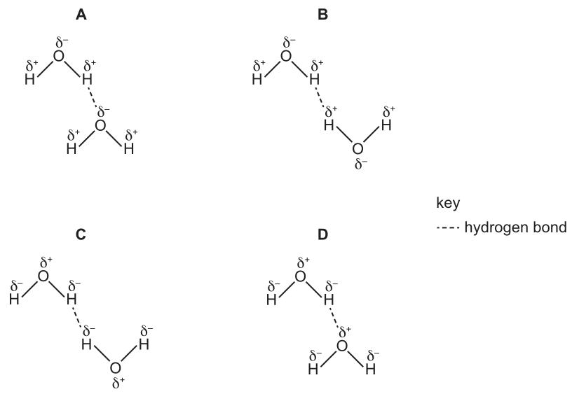
Working
A hydrogen bond forms between the slightly positive hydrogen (δ⁺) of one water molecule and the slightly negative oxygen (δ⁻) of another water molecule, because of the unequal sharing of electrons in the polar O–H bond.
- A — the dashed line joins the H (δ⁺) of the upper molecule to the O (δ⁻) of the lower molecule, and the partial charges on both molecules are correct. This is a valid hydrogen bond.
- B — the dashed line joins two H atoms, both labelled δ⁺. Like charges repel, so this cannot be a hydrogen bond.
- C — the partial charges are reversed (O labelled δ⁺, H labelled δ⁻), and the line again joins two H atoms. The charges are wrong on every atom.
- D — the partial charges are again reversed (O labelled δ⁺, H labelled δ⁻), so even though the line joins H and O, the polarity is wrong.
Only A correctly shows a hydrogen bond between two water molecules.
Answer
A
A
Background Concept
Water (H₂O) is a polar (electrically uneven) molecule. Oxygen is much more electronegative than hydrogen, so the shared electrons in each O–H bond are pulled closer to the oxygen nucleus. As a result:
- The oxygen atom carries a partial negative charge (δ⁻).
- Each hydrogen atom carries a partial positive charge (δ⁺).
Because water is polar, the δ⁺ hydrogen of one molecule is electrostatically attracted to the δ⁻ oxygen of a neighbouring molecule. This weak, non-covalent attraction is a hydrogen bond. In a single hydrogen bond, one atom must be a hydrogen covalently bonded to O, N or F (the δ⁺ donor) and the other must be a lone-pair-bearing N, O or F (the δ⁻ acceptor). In liquid water, each molecule typically forms hydrogen bonds to three or four neighbours, and these bonds are constantly breaking and re-forming.
The key point this question tests: a hydrogen bond always links a δ⁺ hydrogen to a δ⁻ atom with a lone pair (here, oxygen). It cannot link two δ⁺ atoms or two δ⁻ atoms, and the partial charges on water must be δ⁻ on O and δ⁺ on H — never the reverse.
Understanding the Question
The question shows four diagrams of two water molecules each, with a dashed line labelled "hydrogen bond" in the key. Each water molecule is drawn with its O and two H atoms and labelled with partial charges (δ⁺ or δ⁻). The task is to pick the diagram that correctly represents a hydrogen bond between two water molecules — that is, one where:
- the partial charges on every O are δ⁻ and on every H are δ⁺;
- the dashed line runs from a δ⁺ H of one molecule to a δ⁻ O of the other.
Approach
Inspect each option in two stages. First, check whether the partial charges are drawn the right way round (O = δ⁻, H = δ⁺). If the charges are wrong, the diagram cannot represent water correctly and is eliminated. Second, look at the two atoms joined by the dashed line: one must be H (δ⁺) and the other O (δ⁻). A line between two H atoms or two O atoms, or between atoms of the same sign, cannot be a hydrogen bond.
Step-by-Step Reasoning
Option A. Both molecules have O labelled δ⁻ and both H atoms labelled δ⁺ — correct polarisation of water. The dashed line runs from an H (δ⁺) of the upper molecule to the O (δ⁻) of the lower molecule. This matches the definition of a hydrogen bond. A is correct.
Option B. The partial charges on both molecules are correct (O = δ⁻, H = δ⁺), so the molecules themselves are drawn properly. However, the dashed line joins an H (δ⁺) of one molecule to an H (δ⁺) of the other. Two like charges repel, so they cannot form a hydrogen bond. B is wrong.
Option C. The partial charges on both molecules are reversed: O is labelled δ⁺ and H atoms are labelled δ⁻. This is not a real water molecule — oxygen in water is always δ⁻. In addition, the dashed line joins two H atoms (which would here both be δ⁻), failing the charge rule twice over. C is wrong.
Option D. The partial charges are again reversed (O = δ⁺, H = δ⁻), so the molecules are not drawn as real water. Although the dashed line does join an H of one molecule to an O of the other, the charges on those atoms are the wrong way round, so this is not a valid hydrogen bond. D is wrong.
Therefore only A satisfies both conditions.
Key Takeaways
- A hydrogen bond is the electrostatic attraction between a δ⁺ hydrogen (bonded to O, N or F) and a δ⁻ atom with a lone pair (O, N or F) of another molecule.
- In water, oxygen is always δ⁻ and the hydrogens are always δ⁺ because of the electronegativity difference.
- When judging any diagram of a hydrogen bond, check two things: the correct assignment of partial charges and which two atoms the dashed line actually joins.
Common Mistakes
- Reversing the partial charges on water (saying or drawing O as δ⁺ and H as δ⁻) — a frequent slip; remember oxygen is the more electronegative atom.
- Confusing a hydrogen bond with a covalent bond — a hydrogen bond is a weak intermolecular attraction shown as a dashed line, never a solid line.
- Thinking a hydrogen bond can join two H atoms — the H is just the donor of the partial positive; the acceptor must be a lone-pair-bearing electronegative atom.
- Assuming any dashed line between two water molecules must be a hydrogen bond — if the charges drawn are wrong, or the line joins the wrong pair of atoms, it is not.
Things to Be Careful About
- On this type of question, read the sign of every partial charge on every atom, not just the two atoms joined by the dashed line. A diagram with reversed charges is wrong even if the line looks reasonable.
- A hydrogen bond is between molecules, not within the same molecule. The dashed line in these diagrams always runs from one molecule to a different one.
- The "hydrogen" in "hydrogen bond" refers to the hydrogen atom that is covalently bonded to O/N/F; it is not a generic name for any weak attraction.
Some detergents disrupt hydrogen bonds between water molecules.
Which row shows the correct effects of detergent on the specific heat capacity and latent heat of vaporisation of water?
Options
| specific heat capacity | latent heat of vaporisation | |
|---|---|---|
| A | decreases | decreases |
| B | decreases | increases |
| C | increases | decreases |
| D | increases | increases |
Working
Hydrogen bonding between water molecules is responsible for water's unusually high specific heat capacity and high latent heat of vaporisation.
- Specific heat capacity is the energy needed to raise the temperature of 1 kg of water by 1 °C. A large amount of energy is required because incoming heat must first break hydrogen bonds before molecules can move faster. If detergent disrupts hydrogen bonds, less energy is needed to raise the temperature, so specific heat capacity decreases.
- Latent heat of vaporisation is the energy needed to convert 1 kg of liquid water to vapour. Hydrogen bonds must be broken to allow molecules to escape into the gas phase. If detergent disrupts hydrogen bonds, less energy is needed for vaporisation, so latent heat of vaporisation decreases.
Both properties decrease.
Answer
A
A
Background Concept
Water is a small, polar molecule. The partial positive charge on each hydrogen atom is attracted to the partial negative charge on the oxygen atom of a neighbouring molecule, forming a hydrogen bond. Although each individual hydrogen bond is weak, the sheer number of them (each water molecule can form up to four) gives water several anomalous physical properties important in biology:
- A high specific heat capacity (≈ 4.2 kJ kg⁻¹ °C⁻¹): a great deal of energy is needed to raise the temperature of water by 1 °C.
- A high latent heat of vaporisation (≈ 2260 kJ kg⁻¹): a great deal of energy is needed to convert liquid water into water vapour.
- High cohesion and surface tension.
- Lower density as a solid than as a liquid (ice floats).
Both the specific heat capacity and the latent heat of vaporisation are high precisely because of the hydrogen-bond network that must be disrupted or broken before water molecules can move faster (temperature rise) or escape into the gas phase (vaporisation).
Understanding the Question
The stem states that some detergents disrupt hydrogen bonds between water molecules. The candidate is asked to predict how this disruption affects two physical properties of water — specific heat capacity and latent heat of vaporisation — and to choose the row of the table that correctly shows the direction of change for both.
The command word here is implicit: the candidate must decide whether each property increases or decreases in the absence of full hydrogen bonding. The answer is a single letter (A, B, C or D).
Approach
For each property, reason from the definition: identify the role hydrogen bonds play in producing the property, then determine what happens when those bonds are disrupted.
- Specific heat capacity — energy needed to raise temperature → requires breaking hydrogen bonds so molecules can move faster. Fewer/destabilised H-bonds → less energy needed → specific heat capacity falls.
- Latent heat of vaporisation — energy needed to separate molecules into the gas phase → requires fully breaking hydrogen bonds. Fewer/destabilised H-bonds → less energy needed → latent heat of vaporisation falls.
Both should decrease, so the correct row is the one in which both entries read "decreases".
Step-by-Step Reasoning
- Option A (decreases / decreases): Consistent with the reasoning above. Disrupting hydrogen bonds reduces both the energy needed to raise temperature and the energy needed to vaporise the water. ✓
- Option B (decreases / increases): A reduced specific heat capacity is correct, but a higher latent heat of vaporisation would require stronger intermolecular forces, not weaker. ✗
- Option C (increases / decreases): An increased specific heat capacity would require hydrogen bonds to be reinforced, which contradicts the stem. ✗
- Option D (increases / increases): Both increases would imply detergent strengthens the hydrogen-bond network, the opposite of the stated effect. ✗
Therefore the answer is A.
Key Takeaways
- The unusually high specific heat capacity of water is a consequence of its extensive hydrogen-bond network, which must absorb energy before molecules can move faster.
- The high latent heat of vaporisation of water is also a consequence of hydrogen bonding, since all H-bonds must be broken for a molecule to leave the liquid.
- Disrupting hydrogen bonds therefore lowers both properties.
Common Mistakes
- Confusing the direction: students sometimes assume "fewer bonds = less energy to break" applies to specific heat capacity but forget to apply the same logic to latent heat of vaporisation, leading them to pick B.
- Thinking that disrupting hydrogen bonds makes water "harder to heat" because there is "less structure" — in fact, heating becomes easier because there is less intermolecular resistance to overcome.
Things to Be Careful About
- "Specific heat capacity" and "latent heat of vaporisation" are different quantities. Specific heat capacity is about raising the temperature of liquid water; latent heat of vaporisation is about changing the state of water at constant temperature.
- Both, however, are high because of hydrogen bonding, so disrupting the bonds lowers both.
- The question gives no numbers — only the direction of change is being tested, not the magnitude.
An investigation was carried out to see if compound X could improve the thermostability of an enzyme. Thermostable enzymes will function well at high temperatures.
The results of the investigation are shown.
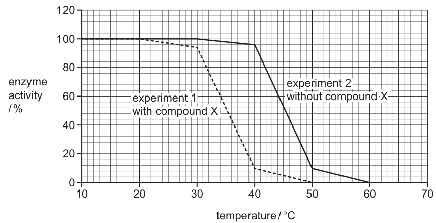
Which row is correct?
Options
| compound X makes the enzyme more thermostable | at in experiment 1 most of the enzyme active sites will no longer be complementary to the substrate | at in experiment 2 approximately half of the enzymes are forming enzyme–substrate complexes | |
|---|---|---|---|
| A | ✓ | ✗ | ✗ |
| B | ✓ | ✓ | ✗ |
| C | ✗ | ✓ | ✓ |
| D | ✗ | ✗ | ✓ |
key
✓ = correct
✗ = not correct
Working
Statement 1 — compound X makes the enzyme more thermostable:
Read from Fig. 14.1, experiment 1 (with compound X, dashed line) drops in activity starting at ~30 °C and reaches ~10% by 40 °C. Experiment 2 (without compound X, solid line) maintains 100% activity until 40 °C and only drops sharply between 40 °C and 50 °C. The enzyme without compound X tolerates higher temperatures, so compound X actually makes the enzyme less thermostable. This statement is incorrect (✗).
Statement 2 — at 40 °C in experiment 1 most active sites are no longer complementary to the substrate:
At 40 °C, experiment 1 shows ~10% activity. The sharp loss of activity above 30 °C is due to denaturation: heat has broken the hydrogen and ionic bonds holding the tertiary structure, distorting the active site so it is no longer complementary to the substrate. This statement is correct (✓).
Statement 3 — at 45 °C in experiment 2 approximately half of the enzymes are forming enzyme–substrate complexes:
At 45 °C in experiment 2, activity has fallen to ~50% (midway between 100% at 40 °C and ~10% at 50 °C). This indicates that about half the enzyme molecules retain a functional active site and are still able to form ES complexes. This statement is correct (✓).
Only the row with ✗ ✓ ✓ is correct.
Answer
C
C
Background Concept
Enzymes are globular proteins whose catalytic activity depends on a precisely shaped active site formed by the folding of the polypeptide chain. The active site is held in its specific 3-D shape by hydrogen bonds, ionic bonds, disulfide bridges and hydrophobic interactions between R-groups of the amino acids that make up the protein.
Because these bonds are relatively weak, the tertiary structure — and therefore the shape of the active site — is sensitive to conditions such as temperature and pH:
- At low/moderate temperatures, raising the temperature increases the kinetic energy of molecules, so enzyme and substrate collide more often and with more force, increasing the rate of formation of the enzyme–substrate (ES) complex and the rate of reaction.
- Above the optimum temperature, the increased vibration of the polypeptide chain begins to break the bonds that maintain the tertiary structure. The active site gradually loses its specific shape and becomes non-complementary to the substrate — the enzyme is denatured. Because the change in shape is permanent, activity cannot be restored by cooling.
- Thermostable enzymes have additional structural features (more disulfide bridges, more ionic interactions, or a more hydrophobic core) that allow them to retain their tertiary structure, and therefore their active-site shape, at much higher temperatures than ordinary enzymes.
Understanding the Question
We are given a graph (Fig. 14.1) of enzyme activity (%) on the y-axis against temperature (°C) on the x-axis. Two curves are plotted:
- Experiment 1 (with compound X) — dashed line.
- Experiment 2 (without compound X) — solid line.
The candidate must decide which of three statements is correct:
- Whether compound X improves thermostability.
- Whether at 40 °C in experiment 1 most active sites are no longer complementary to the substrate.
- Whether at 45 °C in experiment 2 about half of the enzymes can still form ES complexes.
The question is a structured MCQ: the student must read the graph, apply the concept of denaturation, and select the row in which the tick/cross combination matches all three statements.
Approach
- Identify which curve is more thermostable. Thermostability is shown by how far to the right the curve remains high before falling. The curve that keeps 100% activity to a higher temperature is more thermostable.
- Read off values at 40 °C (experiment 1) and 45 °C (experiment 2). Use the y-axis to estimate the enzyme activity at each temperature.
- Translate each percentage into a structural statement — high % activity ≈ many active sites still complementary to substrate, low % activity ≈ most active sites denatured.
- Match the truth values to the answer options.
Step-by-Step Reasoning
Step 1 — Which enzyme is more thermostable?
- Experiment 1 (with compound X): activity begins to fall at ~30 °C and has collapsed to ~10% by 40 °C.
- Experiment 2 (without compound X): activity is still at 100% at 40 °C and only falls to ~10% at 50 °C.
The enzyme without compound X tolerates higher temperatures, so adding compound X actually makes the enzyme less thermostable. Statement 1 is therefore incorrect (✗). This already rules out options A and B (both of which tick statement 1).
Step 2 — Statement 2: 40 °C in experiment 1.
At 40 °C, experiment 1's activity is approximately 10%. The loss of activity above the optimum is due to denaturation: heat has disrupted the bonds holding the enzyme's tertiary structure, so the active site has changed shape and is no longer complementary to the substrate. With activity reduced to ~10%, the great majority of active sites are denatured, so the statement is correct (✓).
Step 3 — Statement 3: 45 °C in experiment 2.
At 45 °C in experiment 2, the activity is approximately 50% — roughly halfway between 100% at 40 °C and ~10% at 50 °C. Interpreting this structurally: around half of the enzyme molecules are still in their native conformation and their active sites remain complementary to the substrate, so they can still form ES complexes. The other half have already been denatured. The statement is correct (✓).
Step 4 — Choose the row.
The pattern of truth values is ✗ (statement 1) ✓ (statement 2) ✓ (statement 3), which matches row C.
Key Takeaways
- An enzyme's thermostability is read off a temperature/activity graph as the temperature range over which the curve remains high before falling.
- A drop in enzyme activity above the optimum reflects denaturation — the active site loses its specific shape and is no longer complementary to the substrate, so ES complexes cannot form.
- A measured activity of, say, 50% can be interpreted as roughly half of the enzyme molecules retaining a functional, substrate-complementary active site, with the other half denatured.
- When a question contains a list of three statements to evaluate, work through them one at a time and use the truth values to narrow down the options — often the first statement alone will eliminate half the answer choices.
Common Mistakes
- Misreading the curve. Confusing which line is experiment 1 and which is experiment 2 leads to the wrong conclusion about whether compound X is helpful. Always read the legend on the figure carefully.
- Confusing 'rate' with 'thermostability'. A thermostable enzyme is not necessarily faster — it just keeps working at higher temperatures. The graph only tells us about tolerance to heat.
- Assuming a drop in activity means less substrate binding rather than denaturation. Below the optimum, a fall in rate is due to fewer collisions; above the optimum, it is due to loss of active-site shape (denaturation). The 40 °C and 45 °C readings in this question are on the high-temperature (denaturing) side of the curve.
- Treating the activity percentage as a direct measure of substrate concentration. Enzyme activity (here, % of maximum rate) reflects how many enzyme molecules are still catalytically competent, not how much substrate is present.
Things to Be Careful About
- Note the direction of the effect: compound X is worsening thermostability in this experiment, even though the question's wording seems to invite the assumption that the compound must be helpful.
- The wording "active sites will no longer be complementary" is the precise phrasing the mark scheme rewards. Avoid loose alternatives such as "the enzyme is broken" or "the active site is destroyed" — denaturation distorts the shape; the site is still there, just no longer the right shape for the substrate.
- When reading between gridlines on a graph, give the value to the nearest reasonable estimate (here, ~10% and ~50%) rather than worrying about reading it to the nearest percent.
- The question's first column asks whether compound X makes the enzyme more thermostable; the answer is no, so this column is crossed (✗). Do not tick it because the compound is described as if it should be helpful.
The table shows the results from an investigation into the effect of temperature on an enzyme-catalysed reaction. All other variables were standardised.
| temperature / | rate of reaction / arbitrary units |
|---|---|
| 10 | 3 |
| 20 | 7 |
| 30 | 16 |
| 40 | 33 |
| 50 | 32 |
| 60 | 14 |
What is the correct conclusion?
Options
A was the optimum temperature.
B The data for was anomalous.
C The optimum temperature was between and .
D All the enzymes denatured at .
Working
The rate peaks at 40 °C (33 a.u.) but is almost the same at 50 °C (32 a.u.). Because no measurements were taken between 30 °C and 50 °C, the true optimum cannot be pinned down — it could lie anywhere in that interval.
- A is wrong: 40 °C is the highest rate recorded, but 50 °C is almost as high, so 40 °C is not shown to be the optimum.
- B is wrong: the 50 °C value (32) follows the expected pattern of a slight fall after the peak; it is not anomalous.
- C is correct: the data only allow the conclusion that the optimum lies between 30 °C and 50 °C.
- D is wrong: the rate at 60 °C is still 14 a.u. (not zero), so the enzymes were not fully denatured.
Answer
C
C
Background Concept
Enzymes are biological catalysts (typically proteins) whose activity depends on the three-dimensional shape of their active site. As temperature rises, kinetic energy increases and more enzyme–substrate collisions have enough energy to react, so the rate rises. Beyond an optimum temperature, the increased vibrational energy disrupts the hydrogen and ionic bonds that maintain tertiary structure; the active site loses its specific shape and the enzyme is denatured — the rate falls sharply. The optimum is therefore the single temperature at which the balance between kinetic effect and structural integrity gives the highest rate.
Understanding the Question
A table of six temperature–rate pairs is supplied. Because the candidate did not run the experiment, the answer must be deduced from these numbers. The command word is "conclusion" — a statement that must be fully supported by the evidence available, with nothing claimed that the data do not justify. The four options test whether the candidate over-interprets the data, recognises what is and is not an anomaly, and avoids absolute claims that the numbers do not support.
Approach
- Locate the maximum rate in the table: 33 a.u. at 40 °C.
- Check the neighbouring value: 32 a.u. at 50 °C — almost identical.
- Ask: does the data prove 40 °C is the exact optimum, or only that the optimum is somewhere near it? With only 10 °C intervals and no finer measurements, we cannot say.
- Examine each option against the numbers.
Step-by-Step Reasoning
Option A — "40 °C was the optimum temperature."
The rate at 40 °C (33) is indeed the highest reading, but 50 °C gives 32 — within ~3 % of the peak. A genuine optimum would show a clear drop on either side. The data do not rule out, for example, 42 °C or 45 °C as the true maximum, because no readings were taken between 30 and 50 °C. Option A claims a precision the experiment does not support.
Option B — "The data for 50 °C was anomalous."
An anomalous reading lies well outside the trend defined by the surrounding values. Here the trend is: rates climb steeply from 10 °C to 40 °C, then begin to fall. A value of 32 at 50 °C, just below the peak of 33, is exactly what is expected. It is not anomalous.
Option C — "The optimum temperature was between 30 °C and 50 °C."
The rate clearly rises between 30 °C (16) and 40 °C (33) and has only just begun to fall by 50 °C (32). The optimum must therefore lie somewhere in this interval. This is exactly what the data show — a conservative, well-supported conclusion.
Option D — "All the enzymes denatured at 60 °C."
The rate at 60 °C is 14 a.u., which is a substantial fraction of the maximum. If all the enzymes had denatured, the rate would be effectively zero. Some denaturation has occurred, but not complete loss of activity. Option D overstates the result.
The correct choice is C.
Key Takeaways
- The "optimum" is the single temperature of maximum rate, but a conclusion drawn from limited data must reflect what the data can actually prove.
- With 10 °C intervals and a near-tie between 40 °C and 50 °C, the safest supported statement is a range, not a single value.
- A reading is only "anomalous" if it lies outside the pattern set by the surrounding data — a small drop after a peak is the expected pattern, not an anomaly.
- "All the enzymes denatured" would require the rate to be essentially zero; a residual rate means denaturation is only partial.
Common Mistakes
- Choosing A because 40 °C gives the largest number, without checking how close the neighbouring value is.
- Choosing B because the rate at 50 °C is lower than at 40 °C, confusing the expected post-peak decline with an outlier.
- Choosing D because temperature was high, overlooking that the rate is still meaningfully above zero.
- Writing a conclusion that is more precise than the data support (a common reason for losing marks in extended-response questions too).
Things to Be Careful About
- "Optimum" means the single best temperature, not "one of the best" — the experiment must have the resolution to identify it.
- Always read every value in the table before judging; the 50 °C value is the key to eliminating A.
- Distinguish between "anomalous" (unexpected given the trend) and "lower than the peak" (expected after the optimum).
- "Denatured" does not mean "rate is reduced"; full denaturation means loss of all activity, i.e. a rate of zero.
The graph shows how the rate of a reaction changes with substrate concentration in the presence of:
- no inhibitor
- inhibitor X
- inhibitor Y.
One of the inhibitors is competitive and the other inhibitor is non-competitive.
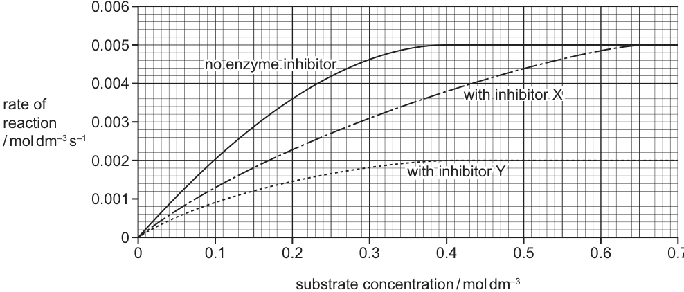
What is the correct estimate of for the reaction shown when a competitive inhibitor is present?
Options
A
B
C
D
Working
-
Identify the type of each inhibitor from :
- Inhibitor X reaches the same (0.0050) as no inhibitor, only at a higher substrate concentration → competitive inhibitor (substrate can outcompete it at high [S]).
- Inhibitor Y reaches a lower (≈ 0.0018) → non-competitive inhibitor (some active sites are permanently lost).
-
Read on the competitive-inhibitor (X) curve.
is the substrate concentration at which the rate = .On the inhibitor X curve, rate = 0.0025 occurs at substrate concentration ≈ 0.23 mol dm⁻³.
Answer
B
B
Background Concept
Enzyme-catalysed reactions follow Michaelis–Menten kinetics. As substrate concentration rises, the rate increases and then plateaus at a maximum value, , when the active sites are saturated. The Michaelis constant, , is the substrate concentration at which the reaction rate is half of . A low means the enzyme reaches half its maximum rate at a low [S] — i.e. the enzyme has high affinity for its substrate.
Competitive inhibitors resemble the substrate and bind reversibly to the active site. They do not lower , because at sufficiently high [S] the substrate outcompetes the inhibitor and the enzyme can still be saturated. However, more substrate is needed to reach any given sub-saturating rate, so increases (apparent rises).
Non-competitive inhibitors bind to a site other than the active site (an allosteric site). They do not change (the remaining functional active sites work normally), but they permanently remove a fraction of enzyme molecules, lowering .
Understanding the Question
The question shows three rate–vs–[S] curves: no inhibitor, inhibitor X, and inhibitor Y. One inhibitor is competitive, the other non-competitive. We are asked for when a competitive inhibitor is present. This is a two-step task: first identify which curve is the competitive one, then read from it.
Approach
- Tell the inhibitors apart by . Whichever curve still reaches the same as the control is competitive; the one with a lower plateau is non-competitive.
- Compute half . = [S] at rate = .
- Read off the [S] at that rate on the competitive-inhibitor curve.
Step-by-Step Reasoning
- From Fig. 16.1, the no-inhibitor curve plateaus at .
- Inhibitor X also plateaus at 0.0050, just at a higher [S] (the curve is shifted right but reaches the same ceiling). Therefore X is the competitive inhibitor.
- Inhibitor Y plateaus at a much lower value (≈ 0.0018), so Y is the non-competitive inhibitor.
- Half of is:
- On the inhibitor X curve, the rate of 0.0025 is reached at [S] ≈ 0.23 mol dm⁻³. That is the in the presence of the competitive inhibitor.
- The no-inhibitor (read the same way on the solid curve) is around 0.11 mol dm⁻³, which is option A — a tempting distractor for those who ignore the inhibitor or mis-identify X. Option C (0.38) is roughly the [S] at half on the no-inhibitor curve is not the answer; option D (0.65) is much too high.
Key Takeaways
- is unchanged by a competitive inhibitor; increases.
- is decreased by a non-competitive inhibitor; is unchanged.
- = [S] at which the rate is half , read directly from the relevant curve.
Common Mistakes
- Confusing the two inhibitors — picking Y because it "looks lower" without thinking about vs. .
- Reading off the no-inhibitor curve instead of the competitive-inhibitor curve, giving the distractor 0.11 mol dm⁻³ (option A).
- Using the wrong horizontal line — taking it at the plateau of inhibitor Y rather than at half of 0.0050.
Things to Be Careful About
- Always quote with its units (mol dm⁻³) and read to the precision of the gridlines on the axis.
- Make sure the half- line corresponds to half of the plateau that is actually reached on the curve you are reading, not half of the control when the curve never gets there (which would not apply here, since X still reaches 0.0050).
- The y-axis label is rate per second; the x-axis is substrate concentration — the value you read is the , an [S] value, so its unit is mol dm⁻³, not mol dm⁻³ s⁻¹.
Which of these substances can pass directly through cell surface membranes without using a carrier protein or channel protein?
1 and
2
3
Options
A 1 and 2
B 1 and 3
C 2 and 3
D 2 only
Working
- and are charged ions; the hydrophobic interior of the phospholipid bilayer repels charged/polar species, so these ions cannot pass through directly — they require channel or carrier proteins (facilitated diffusion or active transport).
- is a small, non-polar molecule; it is freely soluble in the phospholipid bilayer and crosses by simple diffusion without any protein.
- (glucose) is a large, polar molecule; the many -OH groups hydrogen-bond with water and cannot cross the hydrophobic interior, so it requires a carrier protein (e.g. GLUT) for facilitated diffusion.
Only substance 2 () crosses the membrane directly.
Answer
D
D
Background Concept
The cell surface membrane is a phospholipid bilayer with a hydrophobic core (the fatty-acid tails) sandwiched between two hydrophilic surfaces (the phosphate heads). Whether a molecule can pass directly through this bilayer depends on its size, polarity and charge:
- Small, non-polar molecules (e.g. , , steroid hormones) dissolve in the hydrophobic core and diffuse across freely. No protein is needed.
- Small, uncharged polar molecules (e.g. , urea) can also slip through, though more slowly.
- Large, polar molecules (e.g. glucose, , amino acids) and ions (e.g. , , ) cannot cross the hydrophobic core unaided because their charge or polarity is repelled by the fatty-acid tails. They must use channel proteins (which provide a hydrophilic pore) or carrier proteins (which bind the molecule and change shape) — i.e. facilitated diffusion — or be pumped by active transport.
This is the structural reason behind the "fluid mosaic model": only some molecules can take the direct lipid route; everything else needs a protein.
Understanding the Question
The question is a multiple-choice item testing exactly this principle. It offers three numbered substances and asks which of them can pass directly through the membrane without using a carrier protein or a channel protein. The answer is therefore restricted to small, non-polar molecules.
The list:
- 1. and — two ions
- 2. — a small, non-polar gas
- 3. — glucose, a large polar molecule
Only substance 2 fits the criterion. The candidate is expected to classify each one and then select the option that lists only 2.
Approach
Classify each molecule by its physicochemical properties, then match to whether the bilayer alone is sufficient.
- Identify which of the three are ions or large polar molecules (cannot pass directly) and which are small and non-polar (can pass directly).
- Eliminate any numbered item that includes a substance needing a protein.
- Choose the option that contains only the substance(s) that pass through the lipid bilayer unaided.
Step-by-Step Reasoning
Substance 1 — and :
Both are charged. The bilayer interior is hydrophobic and repels charge, so the ions cannot traverse it on their own. They cross the membrane only through ion channels (e.g. voltage-gated channels, ligand-gated channels) or via carrier-mediated transporters. Substance 1 fails the criterion — reject.
Substance 2 — :
Carbon dioxide is a small, linear, non-polar molecule with no permanent dipole. It is highly lipid-soluble and diffuses down its concentration gradient straight through the bilayer at high rates — the basis of gas exchange in the alveoli. Substance 2 passes directly — keep.
Substance 3 — (glucose):
Glucose has six hydroxyl (-OH) groups, making it strongly polar and capable of extensive hydrogen bonding with water. It is also a relatively large molecule. Both features prevent it from passing through the hydrophobic core; it crosses only via GLUT carrier proteins (facilitated diffusion) or the -glucose co-transporter (active transport, e.g. in the ileum). Substance 3 fails the criterion — reject.
Only substance 2 passes directly, so the correct option is D — 2 only.
Key Takeaways
- A substance crosses the phospholipid bilayer directly only if it is small and non-polar (e.g. , , steroid hormones) or small and uncharged polar (e.g. , urea).
- Ions and large polar molecules (e.g. glucose, amino acids, nucleotides) require channel or carrier proteins.
- The "directly through the membrane without a protein" question is a direct test of lipid solubility / hydrophobic-core repulsion principles.
Common Mistakes
- Picking option A (1 and 2) — confusing with an ion or forgetting that ions cannot cross the hydrophobic core. Charged species of any size need a protein route.
- Picking option B (1 and 3) — assuming anything that "diffuses" across membranes needs no protein; this conflates simple diffusion with facilitated diffusion.
- Picking option C (2 and 3) — the most tempting distractor. Students see that and glucose both "diffuse" across membranes and assume they share a mechanism. They do not: diffuses through the lipid; glucose diffuses only through a GLUT carrier.
- Saying "glucose is too big" but missing the more important reason — it is the polarity of glucose (its many -OH groups), not just its size, that blocks direct passage. Credit-worthy biology explains both.
Things to Be Careful About
- The question says "without using a carrier protein or channel protein" — this is a strict exclusion. Any substance that is typically moved via a protein route, even if it can occasionally slip through slowly, must still be excluded.
- Do not be misled by the size of the molecule alone. The decisive property is the interaction of the molecule with the hydrophobic core — charge and polarity matter more than absolute molecular mass.
- is often confused with (bicarbonate) in the chloride shift — the latter is a charged ion and does need a protein (the anion exchanger, AE1), but the that diffuses out of respiring cells and into alveolar blood is the uncharged, freely lipid-soluble gas.
Which of these statements about facilitated diffusion are correct?
1 It is limited by the number of transport proteins.
2 It transports molecules against their concentration gradient.
3 It requires a source of ATP.
Options
A 1, 2 and 3
B 1 and 3 only
C 1 only
D 2 and 3 only
Working
Facilitated diffusion is a passive process that uses transport proteins to move molecules across the membrane.
- Statement 1: True — facilitated diffusion depends on transport proteins (channel or carrier proteins) in the membrane, so the rate is limited by the number of these proteins available.
- Statement 2: False — facilitated diffusion moves molecules down their concentration gradient (high → low), not against it. Movement against a gradient is active transport.
- Statement 3: False — facilitated diffusion is passive and does not require ATP; it is driven by the kinetic energy of the molecules moving down the concentration gradient.
Only statement 1 is correct.
Answer
C
C
Background Concept
Facilitated diffusion is a form of passive transport across a cell membrane. The term facilitated means the movement of molecules across the phospholipid bilayer is helped (facilitated) by transport proteins — either channel proteins (which form a water-filled pore) or carrier proteins (which change shape to shuttle the molecule across). It is used for molecules that cannot cross the phospholipid bilayer directly, such as large polar molecules (e.g. glucose) and ions (e.g. Na⁺, K⁺, Cl⁻).
Key features of facilitated diffusion:
- It is passive — no ATP is required; the driving force is the concentration gradient itself.
- Molecules move down their concentration gradient (from high to low concentration).
- The rate depends on the number of transport proteins in the membrane, so it is saturable — once all transport proteins are in use, increasing the concentration gradient further cannot increase the rate.
- It is specific — each transport protein only binds/allows certain molecules.
By contrast, active transport moves molecules against their concentration gradient and requires ATP (directly from a pump such as the Na⁺/K⁺ pump, or indirectly via a proton gradient).
Understanding the Question
This is a multiple-choice question that lists three statements about facilitated diffusion and asks which are correct. The candidate must evaluate each statement independently and identify the option that includes only the correct ones. The options mix the statements in different combinations, so the candidate must be clear on each feature of facilitated diffusion.
Approach
The cleanest approach is to go through each statement one by one and decide whether it is true or false, based on the defining features of facilitated diffusion. Then select the option that contains only the true statements.
Step-by-Step Reasoning
Statement 1 — "It is limited by the number of transport proteins."
This is correct. Facilitated diffusion relies entirely on transport proteins embedded in the membrane. As the concentration gradient steepens, more molecules pass through, but only up to the point where all available transport proteins are occupied (the Vmax). Beyond that, the rate plateaus because the proteins are the bottleneck. This is what gives a facilitated-diffusion curve its characteristic saturation shape.
Statement 2 — "It transports molecules against their concentration gradient."
This is incorrect. Facilitated diffusion is a passive process: it moves molecules down their concentration gradient (from a region of higher concentration to a region of lower concentration). Movement against the concentration gradient is the defining feature of active transport, not facilitated diffusion. This statement therefore confuses the two processes.
Statement 3 — "It requires a source of ATP."
This is incorrect. Because facilitated diffusion is passive, it does not consume ATP. The energy for the movement comes from the concentration gradient itself (the random kinetic energy of the molecules, biased by their higher concentration on one side). ATP is required for active transport, not facilitated diffusion.
Only statement 1 is correct, so the answer is C (1 only).
Key Takeaways
- Facilitated diffusion = passive transport using transport proteins, moving molecules down their concentration gradient, with no ATP required.
- It is saturable: the rate is limited by the number of available transport proteins.
- The opposite of each of these features describes active transport: uses ATP, moves against the gradient, and (for primary active transport) uses pumps rather than simple channels/carriers.
Common Mistakes
- Confusing facilitated diffusion with active transport — students often wrongly believe both require ATP or both can move substances against a gradient. Remember: facilitated diffusion is passive and downhill.
- Thinking that because facilitated diffusion uses proteins, it must use energy (ATP). The protein simply provides a route; the energy comes from the gradient.
Things to Be Careful About
- The word "facilitated" refers to the protein-assisted nature of the process, not to any energy input.
- Although facilitated diffusion does not use ATP, it is still a selective and controlled form of transport — the specificity and saturation behaviour are what make it so important in cells, especially for glucose uptake and ion movement.
A cell absorbs amino acids. This cell then synthesises and exports a digestive enzyme. Different cell structures are involved with different stages of this process.
Which row shows a possible sequence of cell structures that the amino acids pass through?
Options
| cell surface membrane | Golgi body | rough endoplasmic reticulum | secretory vesicle | |
|---|---|---|---|---|
| A | 1st | 2nd | 3rd | 4th |
| B | 1st | 3rd | 4th | 2nd |
| C | 4th | 2nd | 1st | 3rd |
| D | 4th | 1st | 2nd | 3rd |
Working
After the amino acids have been absorbed across the cell surface membrane, they are built into the enzyme on the rough endoplasmic reticulum (RER). The enzyme is then transported in a vesicle to the Golgi body for modification, packaged into a secretory vesicle, and finally released across the cell surface membrane by exocytosis.
So, tracking the amino acids (now part of the enzyme) from inside the cell to outside:
- RER (synthesis)
- Golgi body (modification)
- Secretory vesicle (transport to membrane)
- Cell surface membrane (exocytosis)
Answer
C
C
Background Concept
Eukaryotic cells contain a system of membrane-bound organelles that work together to make and export proteins such as digestive enzymes. The main components of this secretory pathway are:
- Rough endoplasmic reticulum (RER): a network of flattened sacs studded with ribosomes. It is the site where proteins destined for export are synthesised; the ribosomes assemble amino acids into a polypeptide chain, which is threaded into the lumen of the RER.
- Golgi body (Golgi apparatus): a stack of flattened membrane sacs. Proteins arriving from the RER in transport vesicles are modified here (e.g. by adding carbohydrate groups to form glycoproteins) and sorted according to their destination.
- Secretory vesicles: small membrane-bound sacs that bud off from the trans face of the Golgi body. They carry the finished protein to the cell surface membrane.
- Cell surface membrane: the final destination. When a secretory vesicle fuses with it, the protein is released to the outside by exocytosis.
Understanding the Question
The question describes a cell performing two linked activities: (1) absorbing amino acids from outside, and (2) using them to synthesise and export a digestive enzyme. It then asks which row correctly sequences the four named structures that the amino acids (now built into the enzyme) pass through during the export stage.
The key word is sequence — the rows label the four organelles 1st, 2nd, 3rd or 4th, and we need the order in which the amino acids encounter them on their way out of the cell.
Approach
The trick is to recognise that the amino acids have already been absorbed when the sequence begins, so the cell surface membrane is the last structure, not the first. From there, recall the standard secretory pathway order:
RER → Golgi body → secretory vesicle → cell surface membrane
Then match each organelle to the position number given in the table and check which row is consistent.
Step-by-Step Reasoning
- The amino acids have already crossed the cell surface membrane to enter the cell, so within the cell the amino acids (now part of a polypeptide) are first located on/in the RER, where the ribosomes assembled them.
- The newly synthesised enzyme is pinched off into a transport vesicle that fuses with the Golgi body, where it is modified (e.g. glycosylated) and sorted.
- The finished enzyme is packaged into a secretory vesicle that buds off from the Golgi body and travels to the cell surface.
- The secretory vesicle fuses with the cell surface membrane, releasing the enzyme to the outside by exocytosis.
So the order of passage is: RER (1st) → Golgi body (2nd) → secretory vesicle (3rd) → cell surface membrane (4th).
Reading off the table for option C:
- cell surface membrane = 4th ✓
- Golgi body = 2nd ✓
- rough endoplasmic reticulum = 1st ✓
- secretory vesicle = 3rd ✓
This matches the secretory pathway exactly.
Why the others are wrong:
- A puts the cell surface membrane 1st, which would imply the amino acids start outside the cell — but the question says they have already been absorbed.
- B puts the cell surface membrane 1st (same problem) and the secretory vesicle before the Golgi body, reversing the direction of the pathway.
- D puts the cell surface membrane 4th, which is correct for export, but it places the Golgi body 1st and the RER 2nd, putting the modification step before synthesis — biologically impossible, since the polypeptide must first be made on the RER before it can be modified in the Golgi body.
Key Takeaways
- The secretory pathway runs: RER → Golgi body → secretory vesicle → cell surface membrane (by exocytosis).
- When a question states that a substance has already been absorbed or synthesised, the cell surface membrane is the last step, not the first.
- Proteins are made on ribosomes attached to the RER, so the RER is always the first organelle in any pathway describing the synthesis of a protein for export.
Common Mistakes
- Putting the cell surface membrane first, forgetting that the cell has already absorbed the amino acids before the sequence in question starts.
- Reversing the order of RER and Golgi body, perhaps because the Golgi body is closer to the membrane in the cell.
- Confusing transport vesicles (which carry protein from RER to Golgi body) with secretory vesicles (which carry protein from Golgi body to the cell surface membrane).
Things to Be Careful About
- Read the wording of the question carefully: "a possible sequence" means any biologically valid order — but only one of the four options can match the standard pathway.
- The cell surface membrane appears twice in the export of a protein: once at the start (absorption of amino acids) and once at the end (exocytosis of the enzyme). The question is asking about the second of these visits, so the membrane is the 4th step here, not the 1st.
The diagram shows apparatus set up to investigate the effect of changing the initial concentration of glucose in the surrounding solution on the movement of molecules through a selectively permeable membrane (Visking tubing) in 15 minutes.
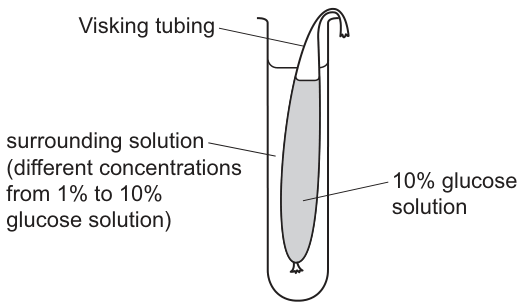
Which statements are correct as the initial concentration of glucose solution in the surrounding solution increases?
1 Net diffusion of water increases.
2 Glucose molecules reach an equilibrium quicker.
3 There is less change in the volume of the surrounding solution.
4 Net diffusion of glucose increases.
Options
A 1, 2, 3 and 4
B 1, 2 and 4 only
C 1 and 3 only
D 2 and 3 only
Working
The Visking tubing contains 10% glucose; the surrounding concentration is increased from 1% to 10%.
- Statement 1 (net diffusion of water increases): FALSE. The water-potential gradient between the inside (10% glucose) and the surrounding solution falls as the surrounding concentration rises. A smaller water-potential difference means a smaller net osmotic movement of water, not more.
- Statement 2 (glucose reaches equilibrium quicker): TRUE. The starting concentrations are closer together, so less net solute movement is needed to equalise them; the system reaches equilibrium faster.
- Statement 3 (less change in volume of surrounding solution): TRUE. With a smaller water-potential difference, less water moves by osmosis into or out of the Visking tubing, so the surrounding solution's volume changes by less.
- Statement 4 (net diffusion of glucose increases): FALSE. As the surrounding concentration rises towards 10%, the concentration gradient for glucose across the membrane decreases, so the net diffusion of glucose decreases.
Only statements 2 and 3 are correct.
Answer
D
D
Background Concept
A selectively permeable membrane such as Visking tubing has pores small enough to let small molecules (water) through freely, but too small for larger solute molecules like glucose to pass easily. Two related but distinct processes operate across it:
- Osmosis is the net movement of water molecules across a selectively permeable membrane from a region of higher water potential () to a region of lower water potential. The more dissolved solute (e.g. glucose) in a solution, the lower (more negative) its water potential.
- Diffusion is the net movement of solute molecules (here, glucose) down their own concentration gradient, from high to low concentration. Net diffusion rate increases with steeper concentration gradient.
In both cases, the magnitude of the gradient (water-potential difference for osmosis, concentration difference for diffusion) controls the rate of net movement, while the direction is set by which side has the higher water potential / higher solute concentration.
Understanding the Question
The set-up fixes the inside of the Visking tubing at 10% glucose. The variable is the concentration of the surrounding solution, which is raised from 1% to 10%. We must predict what happens to (1) net osmosis of water, (2) time taken for glucose to equilibrate, (3) volume change of the surrounding solution, and (4) net diffusion of glucose — and decide which of the four statements is correct.
The command word is implicit but the structure is "Which statements are correct?" — typical of a multiple-completion MCQ where you must evaluate each numbered statement independently.
Approach
Treat each statement as a separate prediction, asking:
- For water movement, compare the water potentials on either side of the membrane at low (1%) and high (10%) surrounding concentrations.
- For glucose, compare the inside concentration (fixed at 10%) to the surrounding concentration at the two extremes.
- A smaller difference (water potential or concentration) → slower net movement → less volume change, but also less solute to move before equality is reached.
Step-by-Step Reasoning
Statement 1 – Net diffusion of water increases.
- At 1% outside, the water-potential difference across the membrane is largest (10% inside is much more negative). Net osmosis of water into the tubing is greatest.
- At 10% outside, the water potentials on both sides are equal, so net osmosis = 0.
- Increasing the outside concentration therefore decreases the net diffusion of water. Statement 1 is FALSE.
Statement 2 – Glucose molecules reach an equilibrium quicker.
- At 1% outside, the starting glucose gradient is 10% – 1% = 9 percentage points; a great deal of net glucose movement is needed before the two sides equalise, so equilibrium is approached slowly.
- At 10% outside, the starting gradient is essentially zero, and the system is already at equilibrium.
- The closer the starting outside concentration is to 10%, the less net glucose transfer is required, and the sooner equilibrium is reached. Statement 2 is TRUE.
Statement 3 – Less change in volume of the surrounding solution.
- Volume change of the surrounding solution is driven by the osmotic movement of water across the Visking tubing.
- As shown for statement 1, this osmotic movement falls as the surrounding concentration rises towards 10%.
- Therefore the change in the volume of the surrounding solution becomes smaller. Statement 3 is TRUE.
Statement 4 – Net diffusion of glucose increases.
- The rate of net diffusion of glucose is proportional to the size of the glucose concentration gradient.
- As the surrounding concentration rises from 1% to 10%, the gradient shrinks from 9 percentage points to 0.
- The net diffusion of glucose therefore decreases, not increases. Statement 4 is FALSE.
Combining these, only statements 2 and 3 are correct, so the correct option is D.
Key Takeaways
- Osmosis depends on water-potential difference, not just concentration. A more concentrated external solution has a lower (more negative) water potential, so the gradient driving water movement is reduced.
- Net diffusion rate scales with concentration gradient. Raising the external solute concentration towards the internal concentration always slows net diffusion of that solute.
- Time to equilibrium is not the same as rate of diffusion. A small starting gradient means less net transfer is needed before equality, so equilibrium is reached sooner even though the diffusion rate is lower at every moment.
- Volume change in the surrounding solution is a direct read-out of net water movement, so it tracks with the size of the water-potential gradient.
Common Mistakes
- Confusing direction with rate. A higher outside concentration reverses the direction of water movement at very high values, but here we move from 1% to 10%, and the key point is that the magnitude of net water movement shrinks across this range.
- Assuming faster diffusion = quicker equilibrium. A steeper gradient gives a faster initial rate of diffusion, but the system has further to travel to reach equilibrium, so equilibrium is reached later, not sooner.
- Reading "increase" as always positive. Statement 1 says "net diffusion of water increases" — students often agree because the concentration (of outside glucose) is increasing, forgetting that the driving gradient (water potential) is decreasing.
- Equating volume change with glucose movement. Glucose cannot cross the Visking tubing freely, so the volume of the surrounding solution changes only because of osmotic water movement, not because of glucose moving in or out.
Things to Be Careful About
- The inside of the Visking tubing is always 10%; the variable is the surrounding solution, not the tubing contents.
- "Net diffusion" refers to the overall one-way excess; it falls as the gradient shrinks even though individual molecules continue to move in both directions.
- The water-potential difference depends on the total solute concentration of the surrounding solution, not just on glucose — but here the surrounding solution is described as glucose solution, so glucose is the only relevant solute.
- Watch for distractors that are individually true under different starting conditions (e.g. glucose diffusing into the tubing when the outside concentration exceeds 10%) — the question restricts us to the range 1%–10%, so glucose always diffuses out of the tubing (or is at equilibrium) and the gradient only ever shrinks.
The epithelium that lines the stomach is damaged by acid and is renewed every two days.
Why is mitosis required to repair the damage?
Options
A Mitosis repairs damaged cells.
B Mitosis produces new genetically similar cells.
C Mitosis provides genetically identical replacement cells.
D Mitosis doubles the original cell number.
Working
Mitosis produces two daughter cells that are genetically identical to the parent cell, so the replacement cells have the same DNA and can carry out the same specialised function as the damaged stomach epithelium. A is wrong because mitosis does not repair existing cells — it produces new ones. B uses the word 'similar' rather than 'identical', which is incorrect for mitosis. D describes a numerical outcome but does not explain why mitosis is required for repair.
Answer
C
C
Background Concept
Mitosis is a type of nuclear division that produces two daughter nuclei, each containing a set of chromosomes that is genetically identical to the parent nucleus. Before division, the DNA is replicated during interphase (S phase), and during mitosis the sister chromatids are separated so each new nucleus receives a complete copy of every chromosome. Because the daughter cells carry identical DNA, they have the same genetic information as the original cell and can differentiate into the same specialised cell type to replace lost or damaged tissue.
The lining of the stomach (a simple columnar epithelium with mucus-secreting cells) is constantly exposed to acidic gastric juice (pH ~1–2) and digestive enzymes, which damage the surface cells. The epithelium is therefore replaced every few days. To replace these cells, pre-existing cells in the gastric pits divide by mitosis, producing genetically identical daughter cells that mature into new epithelial cells to restore the lining.
Understanding the Question
The question is a multiple-choice item asking why mitosis is required to repair the stomach epithelium, given that the epithelium is damaged by acid and renewed every two days. The candidate must identify the correct justification from four similar-sounding options. The command word is implicit in the MCQ format: select the option that correctly explains the role of mitosis in tissue repair.
Approach
The key biological principle is that mitosis produces genetically identical cells (clones). For tissue repair, the replacement cells must be able to perform the same function as the lost cells, which requires the same genes to be active — this is only guaranteed if the replacement cells are genetically identical. A strong answer must therefore include the word "identical" (not "similar") and must refer to replacement of lost cells rather than repair of existing ones.
Step-by-Step Reasoning
- Option A — "Mitosis repairs damaged cells." This is incorrect because mitosis does not repair existing cells; it produces new cells by division. The damaged cells are replaced, not repaired.
- Option B — "Mitosis produces new genetically similar cells." The word "similar" is wrong. Mitosis produces cells that are genetically identical (clones), not merely similar. A subtle but critical distinction: similar implies minor genetic differences (as in meiosis or asexual budding in some organisms), which would not guarantee functional equivalence with the parent tissue.
- Option C — "Mitosis provides genetically identical replacement cells." This correctly captures both the mechanism (genetically identical) and the purpose (replacement of damaged tissue). ✓
- Option D — "Mitosis doubles the original cell number." This is a numerical description of the outcome of one division, but it does not explain why mitosis (as opposed to any other process of producing cells) is needed for repair. It also ignores the genetic identity requirement.
Key Takeaways
- Mitosis produces genetically identical daughter cells — the words "identical" and "similar" are not interchangeable in genetics.
- Mitosis is essential for tissue repair because replacement cells must be functionally equivalent to the cells they replace, which requires identical DNA.
- Mitosis does not repair cells; it creates new ones to replace damaged or lost cells.
Common Mistakes
- Confusing identical with similar: only meiotic products or sexually-recombined cells are "similar but not identical". This single-word distinction decides options B and C.
- Thinking that mitosis repairs cells rather than replaces them (option A is a common trap for students who have not thought carefully about the difference).
- Choosing D because it sounds mathematical/precise, without checking whether it actually answers why mitosis is needed for repair rather than just what mitosis does numerically.
Things to Be Careful About
- In CIE marking, the word identical (not "similar", not "the same", not "copies") is the precise scientific term for the products of mitosis.
- Mitosis has three biological roles: growth, repair, and asexual reproduction. This question tests the repair role specifically.
- Read each option's wording precisely — MCQ distractors often hinge on a single word.
What is the role of telomeres?
Options
A allowing the chromatids to reach the poles during mitosis
B holding sister chromatids together
C making sure that the sister chromatids are of identical length
D preventing loss of genes during DNA replication
Working
Telomeres are repetitive, non-coding DNA sequences at the ends of linear chromosomes. During DNA replication, the lagging strand is synthesised in Okazaki fragments primed by RNA primers; the primer at the very 5′ end of the new strand cannot be replaced with DNA, leaving a small gap. Without telomeres, this would cause the coding regions of the chromosome to be progressively shortened with each round of replication. The telomeric repeats act as a buffer that is lost instead, preserving the genes.
- A is incorrect — spindle fibres (microtubules) pull chromatids to the poles during anaphase.
- B is incorrect — the centromere (and cohesin proteins) hold sister chromatids together until anaphase.
- C is incorrect — identical length of sister chromatids is ensured by semi-conservative replication of the whole DNA molecule, not by telomeres.
- D is correct — telomeres prevent the loss of coding gene sequences from the chromosome ends during DNA replication.
Answer
D
D
Background Concept
A chromosome is a single, very long DNA molecule wrapped around histone proteins and packaged into a compact structure. Every linear chromosome has two ends, and these ends are capped by special repetitive DNA sequences called telomeres. In humans the telomeric repeat is the hexanucleotide TTAGGG, repeated hundreds to thousands of times. Telomeres do not code for proteins; they are essentially protective, disposable buffers.
To understand why telomeres matter, you need to understand the end-replication problem of linear DNA:
- DNA polymerase can only add nucleotides to a free 3′-OH group; it cannot start a new strand from scratch.
- Therefore, an RNA primer is laid down first, and DNA polymerase extends it. On the lagging strand, this happens in short Okazaki fragments, each requiring its own primer.
- The very last primer sits at the 5′ end of the newly synthesised strand. Once that primer is removed, there is no upstream 3′-OH for DNA polymerase to extend from, so the new strand is shorter than the template strand by at least the length of that primer.
If this shortening happened at the start of a gene, vital coding information would be lost every time the cell divided. The telomeric repeats at the chromosome ends are the part that is lost; the important genes are preserved. The enzyme telomerase (active in germ cells, stem cells and many cancer cells) can add telomeric repeats back to the ends, restoring telomere length.
Understanding the Question
This is a single-best-answer MCQ. The stem simply asks "What is the role of telomeres?" — so you are looking for the function that uniquely fits telomeres and not any other chromosomal structure. The four options are designed to test whether you really know what telomeres do, or whether you are confusing them with centromeres, spindle fibres, or other replication machinery. The command word is implicit ("what is the role"), so you need only one correct option.
Approach
- Recall the structure of a eukaryotic chromosome: DNA + histones + centromere + telomeres.
- Recall the function of each component — centromere (attachment to spindle, holds sister chromatids), spindle (moves chromatids), telomeres (protect chromosome ends).
- Match each option to the correct component and eliminate the wrong ones.
- Confirm the surviving answer using the end-replication problem.
Step-by-Step Reasoning
-
Option A — "allowing the chromatids to reach the poles during mitosis."
Chromatids are pulled to opposite poles during anaphase by spindle microtubules that attach to the kinetochore, a protein complex assembled on the centromere. Telomeres play no part in this movement. → Eliminate. -
Option B — "holding sister chromatids together."
Sister chromatids are held together at the centromere (the constricted region) by cohesin protein rings until anaphase, when cohesin is cleaved and the chromatids separate. Telomeres are at the ends and do not hold the chromatids together. → Eliminate. -
Option C — "making sure that the sister chromatids are of identical length."
Identical sister chromatids are produced because DNA replication is semi-conservative: each new double helix contains one parental strand and one new complementary strand, so the two chromatids of a replicated chromosome carry the same base sequence. This has nothing to do with telomeres — and indeed, after replication each new chromatid will be very slightly shorter at its 5′ end, with the loss occurring in the telomere. → Eliminate. -
Option D — "preventing loss of genes during DNA replication."
Because the very ends of linear DNA cannot be fully copied, each round of replication normally shortens the chromosome. The telomeric repeats at the ends act as a non-coding buffer that is eroded first, so the protein-coding genes located internally are preserved. → Correct.
Key Takeaways
- Telomeres are repetitive, non-coding DNA sequences at the tips of linear chromosomes.
- They solve the end-replication problem: lagging-strand synthesis cannot complete the 5′ end of a linear DNA molecule, so without telomeres, genes would be progressively lost.
- Telomeres are maintained by the enzyme telomerase, which is active in stem cells, germ cells and most cancer cells, but largely inactive in adult somatic cells — hence telomere shortening contributes to cellular ageing.
- Do not confuse telomeres with the centromere (spindle attachment, holds sister chromatids) or with spindle fibres/microtubules (move chromatids during anaphase).
Common Mistakes
- Confusing telomeres with centromeres. Both are specialised regions of the chromosome but they have completely different functions: centromeres anchor the spindle and hold sister chromatids together; telomeres cap the chromosome ends.
- Confusing telomeres with the role of the spindle. Spindle microtubules, not telomeres, move chromatids to the poles during anaphase.
- Thinking telomeres "make" sister chromatids identical. Identical sister chromatids result from semi-conservative DNA replication of the whole chromosome, not from telomeres. In fact, telomeres are the very region that becomes shorter after replication.
- Forgetting that the function of telomeres is to protect genes, not to replicate the chromosome. The wording "preventing loss of genes" is the key idea; the mechanism is the end-replication problem.
Things to Be Careful About
- Read the option carefully: "preventing loss of genes" — not "preventing loss of DNA." The point is that the coding sequences are preserved; the non-coding telomeric repeats are the part that is lost.
- Be alert to the CIE convention that on a multiple-choice paper the answer must be the option that is uniquely correct, not merely "true-ish." Options A, B and C are each true descriptions of other structures, which makes them classic distractors.
- Spelling and terminology matter: telomere (the end cap), centromere (the middle constriction), kinetochore (the protein complex on the centromere that attaches to spindle microtubules). Mixing these up costs marks across many topics.
Which features of mitosis help to maintain the genetic composition of the cell?
1 the longitudinal division of the centromeres
2 the DNA of the parent cells replicates before mitosis begins
3 the pulling apart of the chromatids to opposite poles
Options
A 1, 2 and 3
B 1 and 3 only
C 1 only
D 2 and 3 only
Working
For each option, consider whether the event ensures daughter cells receive a full and identical set of chromosomes:
-
Longitudinal division of the centromeres — at the start of anaphase, each centromere splits, allowing the two sister chromatids of every chromosome to be treated as independent chromosomes and move to opposite poles. This guarantees each daughter cell receives one chromatid from each replicated chromosome, preserving chromosome number and genetic identity.
-
DNA of the parent cell replicates before mitosis begins — during S phase of interphase, every chromosome is duplicated to form two genetically identical sister chromatids. Without this prior replication, there would be no complete copy of the genetic material to pass to each daughter cell, so genetic composition could not be maintained.
-
Pulling apart of the chromatids to opposite poles — spindle fibres attached to the centromeres separate the sister chromatids and draw one set to each pole, so that each new nucleus contains a complete and identical set of chromosomes.
All three features act together to ensure daughter cells are genetically identical to the parent cell.
Answer
A
A
Background Concept
Mitosis is the nuclear division that produces two daughter nuclei, each genetically identical to the parent nucleus. To achieve this, three coordinated events must occur:
- DNA replication during S phase of interphase: every chromosome is copied to produce two sister chromatids held together at a centromere. Each sister chromatid is a complete, identical copy of the original DNA molecule.
- Division of the centromeres (at the start of anaphase): the protein ring of the centromere splits longitudinally, so each former sister chromatid becomes a separate, independent chromosome.
- Separation of chromatids (anaphase): spindle microtubules shorten, pulling one chromatid from each pair to each pole of the cell. A new nuclear envelope forms around each set during telophase, so each daughter nucleus contains one full set of chromosomes.
Together these events ensure that the chromosome number is maintained and that each daughter cell has exactly the same alleles as the parent cell — the basis of growth, repair and asexual reproduction.
Understanding the Question
This is a multiple-choice question with four options combining three statements. The candidate must decide which of statements 1, 2 and 3 are correct, then pick the option that lists exactly those correct statements. The mark scheme credits A (1, 2 and 3).
Approach
Treat each statement independently. For each one, ask: "Does this event contribute to daughter cells being genetically identical to the parent cell?" If the answer is yes, the statement is correct. Then match the combination of correct statements to the answer options.
Step-by-Step Reasoning
Statement 1 — Longitudinal division of the centromeres:
Before division, each chromosome consists of two sister chromatids joined at a single centromere. If the centromere did not split, both chromatids would be pulled to the same pole and one daughter cell would receive no copy of that chromosome, while the other received two. Splitting the centromere converts each chromatid into an independent chromosome so that exactly one is delivered to each pole. ✓ This maintains genetic composition.
Statement 2 — DNA of the parent cell replicates before mitosis begins:
Replication during S phase produces the two sister chromatids that will be distributed. Without prior replication there would be only one DNA molecule per chromosome; mitosis could not, on its own, give both daughter cells a full set. The replication step is therefore essential for the genetic fidelity of mitosis. ✓ This maintains genetic composition.
Statement 3 — Pulling apart of the chromatids to opposite poles:
This is the actual segregation event. Spindle fibres attached at the kinetochores shorten during anaphase, dragging one chromatid from each pair to each pole. The result is that each pole receives one complete set of chromatids (now chromosomes), and so each daughter nucleus inherits a full, identical complement of genetic material. ✓ This maintains genetic composition.
All three statements are correct, so the answer is A: 1, 2 and 3.
Key Takeaways
- Genetic stability in mitosis depends on the prior replication of DNA, the longitudinal splitting of centromeres, and the equal segregation of chromatids to opposite poles.
- The centromere is the structural and mechanical link that holds sister chromatids together until anaphase; its division is the trigger that allows them to separate as independent chromosomes.
- Failure at any of these three steps leads to aneuploidy or unequal genetic complements, with consequences such as Down syndrome (trisomy 21) or, in somatic cells, the genomic instability seen in many cancers.
Common Mistakes
- Rejecting statement 1 by thinking the centromere "breaks" rather than divides longitudinally. The longitudinal (rather than transverse) split is what allows the two chromatids to become separate chromosomes that can move to opposite poles.
- Rejecting statement 2 by treating DNA replication as separate from mitosis. Although replication occurs in interphase, it is an essential prerequisite for mitosis to maintain genetic composition and should be credited.
- Selecting B (1 and 3 only) by overlooking that without prior replication, no genetic material could be conserved; or D (2 and 3 only) by forgetting that the centromere split is what physically releases the chromatids.
Things to Be Careful About
- The question asks specifically about maintaining the genetic composition of the cell. All three statements are about the mechanism that delivers identical genetic material, not about unrelated features such as spindle formation or cytokinesis.
- Note that option C (1 only) is a tempting distractor because centromere division is a striking event, but on its own it does not preserve genetic composition — replication must have happened first and segregation must still occur.
- For CIE multiple-choice questions, always check the combination carefully: the wrong option often includes most but not all of the correct statements.
DNA forms a leading strand and a lagging strand during semi-conservative replication.
Which row correctly matches the strands to their properties?
Options
| the strand that is replicated by joining together short sequences of DNA in a 3′ to 5′ direction | the strand that requires DNA ligase | |
|---|---|---|
| A | lagging | lagging |
| B | lagging | leading |
| C | leading | leading |
| D | leading | lagging |
Working
DNA polymerase can only add nucleotides to the 3′ end of a growing strand, so synthesis always proceeds 5′ → 3′. Because the two template strands are antiparallel, only one strand (the leading strand) can be replicated continuously towards the replication fork.
The other template strand runs the opposite way, so the new strand must be built in short Okazaki fragments, each synthesised 5′ → 3′ but overall away from the fork. These short fragments are then joined together by DNA ligase. This is the lagging strand.
- Column 1 (joined from short sequences, 3′ → 5′ direction) = lagging strand
- Column 2 (requires DNA ligase) = lagging strand
Answer
A
A
Background Concept
DNA replication is semi-conservative: each new double helix contains one parental strand and one newly synthesised strand. The two strands of the parent DNA molecule are antiparallel — they run in opposite chemical directions, conventionally written 5′ → 3′ and 3′ → 5′.
DNA polymerase, the enzyme that builds the new strand, can only add a nucleotide to the free 3′ –OH of the growing strand. This means every new DNA strand is synthesised strictly in the 5′ → 3′ direction.
Because the two template strands run antiparallel, the replication machinery cannot treat them identically. At each replication fork:
- The leading strand template runs 3′ → 5′ towards the fork, so the new strand can be built continuously in the 5′ → 3′ direction as the fork opens up.
- The lagging strand template runs 5′ → 3′ towards the fork, so the new strand can only be built 5′ → 3′ by synthesising backwards, in short bursts of ~100–200 nucleotides called Okazaki fragments. Each fragment starts with a short RNA primer laid down by primase, is extended by DNA polymerase, and the fragments are then stitched together.
The "stitching" is done by DNA ligase, which forms the phosphodiester bonds between the 3′ end of one Okazaki fragment and the 5′ end of the next. The leading strand, being one continuous molecule, has no such joins and so does not require DNA ligase for its main synthesis (it may, however, be involved in sealing nicks elsewhere, but this is not the canonical reason ligase is associated with the lagging strand).
Understanding the Question
This is a multiple-choice question with a 2 × 2 grid. The question describes two properties, and each option assigns a strand (leading or lagging) to each property. The correct answer must correctly identify both properties.
- Property 1: "the strand that is replicated by joining together short sequences of DNA in a 3′ to 5′ direction" — this describes the lagging strand, whose Okazaki fragments are joined together (the 3′ → 5′ wording refers to the direction along which successive fragments are added, even though each individual fragment is built 5′ → 3′).
- Property 2: "the strand that requires DNA ligase" — again, the lagging strand, because ligase seals the gaps between Okazaki fragments.
Both columns must therefore point to the lagging strand.
Approach
Match each property to the strand it describes by recalling the mechanism:
- Continuous vs discontinuous synthesis → identifies leading vs lagging.
- Where ligase is needed → identifies lagging.
- The row where both columns give the same (correct) strand is the answer.
Step-by-Step Reasoning
-
Column 1: short sequences joined in a 3′ → 5′ direction
- The lagging strand is built as a series of Okazaki fragments.
- Each fragment is extended 5′ → 3′, but as the fork opens further, a new fragment is started closer to the fork, leaving the previous fragment behind.
- The result is that fragments are joined together in an overall 3′ → 5′ direction (relative to the original template, successive Okazaki fragments are added further towards the 5′ end of the template strand).
- This column therefore describes the lagging strand.
-
Column 2: requires DNA ligase
- After the Okazaki fragments are made, gaps of phosphodiester bonds remain between them.
- DNA ligase seals these nicks, joining the 3′ end of one fragment to the 5′ end of the next.
- The leading strand is synthesised continuously and so does not need ligase to join fragments.
- This column therefore also describes the lagging strand.
-
Match to the options
- A: lagging / lagging ✓ (both correct)
- B: lagging / leading ✗ (second column wrong)
- C: leading / leading ✗ (both wrong)
- D: leading / lagging ✗ (first column wrong)
The correct row is A.
Key Takeaways
- DNA strands are antiparallel; DNA polymerase only synthesises 5′ → 3′.
- The leading strand is made continuously towards the replication fork.
- The lagging strand is made discontinuously as Okazaki fragments that are joined by DNA ligase.
- "Joining short sequences" and "requires DNA ligase" are both defining features of the lagging strand.
Common Mistakes
- Saying the leading strand needs ligase — false; it is one continuous molecule.
- Confusing the direction of synthesis of each Okazaki fragment (5′ → 3′) with the overall direction in which fragments are added (3′ → 5′ along the template). The 3′ → 5′ wording in the question is a deliberate distractor that confuses students who don't carefully read the property.
- Thinking both strands are synthesised the same way — they are not, because of the antiparallel nature of DNA and the unidirectional activity of DNA polymerase.
- Confusing DNA ligase (which joins Okazaki fragments on the lagging strand) with helicase (unwinds the double helix) or primase (lays down RNA primers).
Things to Be Careful About
- The "3′ → 5′" in the question refers to the direction in which the short fragments are joined along the template, not the direction of polymerase activity inside each fragment (which is 5′ → 3′). Be sure to read carefully.
- DNA ligase is sometimes called into action on the lagging strand, but it can also seal nicks during DNA repair and on the leading strand at the end of a round of synthesis; in the context of A-level biology, however, the canonical role is joining Okazaki fragments on the lagging strand — which is what this question tests.
- A common distractor pair is "leading / leading" (option C) — many students assume both strands are made the same way. Don't fall for it: the antiparallel template forces them to be made differently.
How many genes could code for one collagen molecule?
Options
A 1 gene only
B 2 genes only
C 3 genes only
D 1 gene or 2 genes or 3 genes
Working
A collagen molecule is a triple helix of three polypeptide (α) chains. The number of genes required depends on the type of collagen:
- If all three α chains are identical (e.g. Type II collagen, with three α1(II) chains), only 1 gene is needed.
- If two chains are identical and one differs (e.g. Type I collagen, with two α1(I) and one α2(I) chain), 2 genes are needed.
- If all three chains are different, 3 genes are needed.
Answer
D
D
Background Concept
Collagen is a fibrous structural protein and the most abundant protein in mammals. Its basic structural unit, the tropocollagen molecule, is a triple helix composed of three polypeptide (α) chains wound around each other. Each α chain is synthesised from its own mRNA, which is itself transcribed from a single gene. The number of genes required to build one collagen molecule is therefore equal to the number of different α chains in that particular type of collagen.
There are at least 28 known types of collagen in vertebrates, but the syllabus focuses on the principle that different collagen types have different chain compositions. The most common examples are:
- Type I collagen (skin, bone, tendon, dentine, cornea): two α1(I) chains + one α2(I) chain → encoded by 2 different genes (COL1A1 and COL1A2).
- Type II collagen (cartilage, vitreous humour): three identical α1(II) chains → encoded by 1 gene (COL2A1).
- Type III collagen (reticulin in skin, blood vessels): three identical α1(III) chains → 1 gene.
- Other rarer collagens (e.g. Type VI) contain three different chains → 3 genes.
A gene codes for a single polypeptide chain, not for the assembled multi-chain molecule. So you must count distinct polypeptide types, not the total number of chains (which is always three).
Understanding the Question
This is a multiple-choice question testing the link between the molecular composition of a protein and the number of genes required to produce it. The distractors (1, 2 or 3 only) each capture a single possibility; the question wants you to recognise that all three possibilities are valid depending on the collagen type.
Approach
Recall the structure of a collagen molecule: a triple helix of three α chains. Then ask: how many different α chains can be present? The answer is 1, 2 or 3, depending on the type of collagen. Therefore the answer must be the option that includes all three possibilities.
Step-by-Step Reasoning
- A collagen molecule is a triple helix of three α (polypeptide) chains.
- Each α chain is the product of one gene (one gene → one mRNA → one polypeptide).
- For Type I collagen: 2 α1(I) + 1 α2(I) → 2 genes are needed.
- For Type II collagen: 3 identical α1(II) → only 1 gene is needed.
- For some rarer collagens with three distinct chains → 3 genes are needed.
- Since all three numbers are possible depending on the collagen type, the correct option is the one that lists all three.
Key Takeaways
- A gene codes for a polypeptide chain, not for the final assembled protein.
- Collagen is a triple helix of three α chains; the number of distinct chains (and therefore genes) varies by collagen type.
- The most-examined case is Type I collagen, which requires 2 genes (COL1A1 and COL1A2).
Common Mistakes
- Choosing A (1 gene) because a collagen molecule is "one protein" — but it is one protein made of multiple polypeptide chains, each encoded by its own gene.
- Choosing B (2 genes) — this is correct for Type I collagen but ignores other collagen types.
- Choosing C (3 genes) — correct for some collagens, but again ignores the others.
- Forgetting that the three chains may be identical, in which case only one gene is needed.
Things to Be Careful About
- Do not equate the number of chains (always 3) with the number of genes (which depends on whether the chains are identical or different).
- The question is testing breadth of knowledge across collagen types, not just Type I.
The diagram shows some events during the formation of an mRNA molecule at transcription.
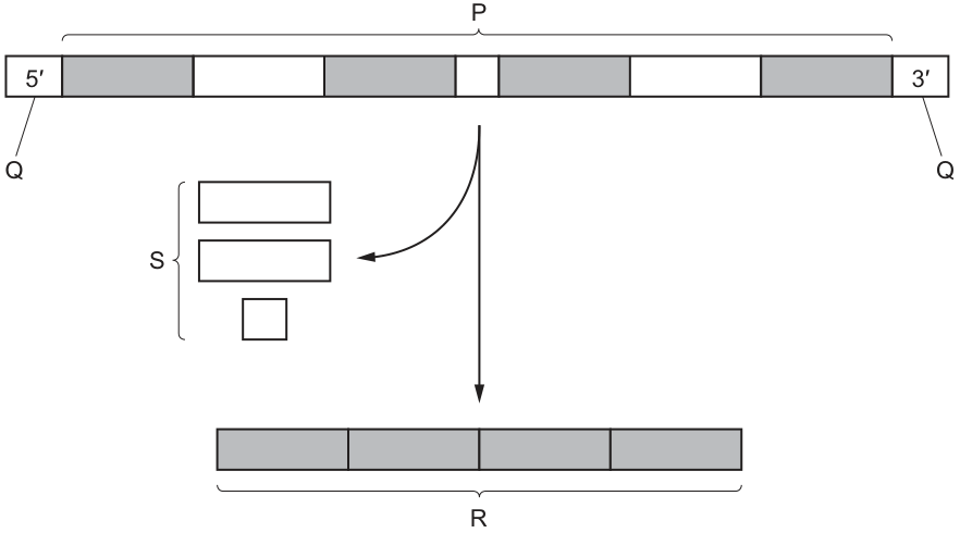
What is correctly identified in the diagram?
Options
A P are introns.
B Q are exons.
C R is a primary transcript.
D S are non-coding sequences.
Working
The diagram shows a DNA strand with alternating grey and white segments. S points to the white segments that are being removed during processing, and R points to the final mRNA consisting of only the joined grey segments.
- P spans the entire length of the strand (both grey and white segments) — it is not a single type of sequence, so it cannot be called introns.
- Q points to the 5' and 3' ends of the DNA — these are the strand termini, not exons.
- R is the spliced mature mRNA (only grey segments joined) — the primary transcript would still contain both grey and white segments before splicing.
- S points to the white segments that are removed — these are the introns, which are non-coding sequences spliced out of the primary transcript.
Answer
D
D
Background Concept
In eukaryotes, a gene is transcribed into a primary transcript (pre-mRNA) that contains both exons (expressed/coding sequences) and introns (intervening/non-coding sequences). Before the mRNA leaves the nucleus, the introns are removed by splicing and the exons are joined together to form a mature mRNA that is translated into protein.
In the diagram, the grey segments represent the exons (coding sequences that are retained) and the white segments represent the introns (non-coding sequences that are removed).
Understanding the Question
The question asks which of the four labels (P, Q, R, S) on the transcription diagram has been correctly identified in the answer options. This requires reading what each label is actually pointing to and matching it to the correct molecular term.
- P spans the entire length of the strand, covering BOTH grey and white segments together. It is not a single type of sequence.
- Q points to the 5' and 3' ends of the DNA — these are simply the directional termini of the DNA strand.
- R points to the final spliced product (only grey/exon segments joined) — this is the mature mRNA, not the primary transcript.
- S points to the white segments being removed from the primary transcript — these are the introns, which are non-coding sequences.
Approach
The key is to identify what each label points to and recall the definitions:
- Introns = non-coding sequences (removed by splicing)
- Exons = coding/expressed sequences (retained in mature mRNA)
- Primary transcript = the unprocessed initial RNA copy (still contains introns)
- Mature mRNA = processed RNA with only exons
Step-by-Step Reasoning
Option A (P are introns): P brackets the entire DNA strand, including both grey and white segments. Since P is not selective for the white segments, it cannot be labelled as introns. Incorrect.
Option B (Q are exons): Q points to the 5' and 3' ends of the DNA. These are strand direction labels, not sequences — and certainly not exons (which would be the grey segments inside the strand). Incorrect.
Option C (R is a primary transcript): R is the final mRNA molecule containing only the grey (exon) segments. The primary transcript would contain BOTH grey and white segments before splicing. R has already been processed, so it is the mature mRNA, not the primary transcript. Incorrect.
Option D (S are non-coding sequences): S brackets the white segments that are being cut out and removed from the primary transcript. These are the introns, which by definition are non-coding sequences. Correct.
Key Takeaways
- Introns are the non-coding sequences spliced out of the primary transcript during mRNA processing.
- Exons are the coding sequences that are retained and joined together in the mature mRNA.
- The primary transcript is the initial RNA product of transcription (contains both exons and introns); mature mRNA is the processed version (exons only).
- Always read what a label is actually pointing to before matching it to a term.
Common Mistakes
- Confusing the primary transcript (pre-mRNA, with introns) with the mature mRNA (post-splicing, exons only).
- Thinking P is introns because it covers a long stretch — but P covers the whole strand, not just the non-coding parts.
- Confusing DNA 5'/3' ends (strand polarity labels) with exons (sequences within the gene).
Things to Be Careful About
- "Non-coding" in this context means the introns do not code for amino acids in the final protein product. Some introns have regulatory functions, but they are removed before translation.
- The 5' and 3' ends are NOT sequences — they refer to the carbon positions on the sugar of the terminal nucleotide that define strand directionality.
Two plants, K and L, are parasites of other plants. Both species grow structures that invade the vascular tissue of plants.
Plant K grows into xylem tissue of other plants.
Plant L grows into phloem tissue of other plants.
Which substances could plants K and L take from the plants that they parasitise?
Options
| plant K | plant L | |
|---|---|---|
| A | water only | water and sucrose only |
| B | water and minerals | water, amino acids and sucrose |
| C | water only | amino acids and sucrose only |
| D | water and minerals | water and sucrose only |
Working
Xylem transports water and dissolved mineral ions (e.g. nitrate, magnesium) from roots to shoots.
Phloem transports assimilates from sources to sinks. The main organic solutes are sucrose, and amino acids are also translocated in phloem sieve tubes, dissolved in water.
So:
- Plant K (xylem feeder): water and minerals
- Plant L (phloem feeder): water, amino acids and sucrose
Answer
B
B
Background Concept
In vascular plants, two distinct tissues move substances around the body:
- Xylem is a dead, hollow tube made of lignified vessel elements. It carries an upward stream of water and dissolved mineral ions (e.g. nitrate, ; magnesium, ; potassium, ) absorbed by the roots up to the leaves and growing points. The driving force is the transpiration pull generated by evaporation from leaves (cohesion–tension), so xylem flow is essentially unidirectional (roots → shoots).
- Phloem is living tissue. Sieve tube elements, joined end-to-end, translocate assimilates — primarily sucrose, but also amino acids and other small organic solutes — from sources (e.g. mature photosynthesising leaves, storage organs) to sinks (e.g. roots, fruits, young leaves, meristems). The flow is driven by a pressure gradient built by proton pumps and mass flow, and can move in any direction depending on where the sources and sinks are.
A parasitic plant that taps into a host's vascular system exploits these streams directly. The set of substances it can obtain depends entirely on which tissue it invades.
Understanding the Question
The stem describes two parasites:
- Plant K penetrates the xylem of its host.
- Plant L penetrates the phloem of its host.
The four-row options table asks which combination of substances each parasite could obtain. This is essentially a test of the contents of xylem sap and phloem sap.
The command word is implicit ("which substances could") — you must select the row that correctly matches the contents of each tissue.
Approach
- List what xylem carries → only water + dissolved mineral ions; sugars and amino acids are not normally in xylem sap in significant amounts.
- List what phloem carries → sucrose + amino acids (+ water as the solvent, plus other small organic molecules).
- Match those lists to the four options; eliminate the rows that mis-state either tissue's contents.
Step-by-Step Reasoning
-
Eliminating options for Plant K (xylem feeder):
- A and C say "water only" — this is wrong because xylem sap does carry dissolved mineral ions. Anything that taps xylem will receive both water and minerals, so A and C are rejected on this point alone.
- B and D both correctly say "water and minerals" for Plant K.
-
Choosing between B and D for Plant L (phloem feeder):
- The phloem sap of most plants contains sucrose as the main transported carbohydrate, plus amino acids (translocated from source leaves to growing or storage tissues), dissolved in water.
- Option D lists only "water and sucrose" for Plant L — this misses the amino acids and is therefore incomplete.
- Option B lists "water, amino acids and sucrose" — this is the full set of major solutes carried in phloem.
So the correct row is B: Plant K takes water and minerals; Plant L takes water, amino acids and sucrose.
Key Takeaways
- Xylem = water + mineral ions (one-way, root to shoot).
- Phloem = sucrose + amino acids + water (multi-directional, source to sink).
- A parasite's nutrient gain is dictated by which vascular tissue its haustorium taps into.
Common Mistakes
- Treating xylem as carrying "water only" and missing the mineral ions — water in xylem is a solution of mineral salts, not pure water.
- Treating phloem as carrying only sucrose and forgetting that amino acids are also translocated — phloem sap is the main long-distance transport route for organic nitrogen, not just for sugars.
- Confusing the roles: thinking xylem carries sugars (it doesn't, except transiently in spring for some trees when stored starch is converted) or that phloem carries mineral ions (it carries some, but the major mineral supply comes from xylem).
Things to Be Careful About
- A phloem parasite can still obtain water — phloem sap is mostly water — but the defining transported solutes are sucrose and amino acids.
- CIE mark schemes typically require the complete correct set; "water and sucrose only" (D) is wrong because it omits amino acids, not because water is wrong.
Which statements are correct for the apoplast pathway?
1 Water enters the cell wall.
2 Water moves by osmosis.
3 Water moves from cell wall to cell wall.
4 Water moves through plasmodesmata.
Options
A 1 and 2
B 1 and 3
C 2 and 4
D 3 and 4
Working
- The apoplast pathway is the movement of water through cell walls and intercellular spaces without crossing any plasma membrane.
- Statement 1: Water enters the cell wall — correct (water fills the porous cell wall as it moves through the apoplast).
- Statement 2: Water moves by osmosis — incorrect; osmosis requires movement across a partially permeable membrane, which the apoplast pathway avoids.
- Statement 3: Water moves from cell wall to cell wall — correct; this describes apoplast flow.
- Statement 4: Water moves through plasmodesmata — incorrect; plasmodesmata are part of the symplast pathway.
Answer
B
B
Background Concept
Water moves from the soil to the xylem in a plant root via three interconnected routes:
- Apoplast pathway — water moves through the porous cellulose cell walls and the intercellular spaces between cells, without ever crossing a plasma membrane.
- Symplast pathway — water moves from cytoplasm to cytoplasm through plasmodesmata (the channels that connect adjacent plant cells through their cell walls).
- Vacuolar pathway — water moves from vacuole to vacuole across the tonoplast and plasma membranes of successive cells (often grouped with the symplast route).
A key distinction: osmosis is defined as the diffusion of water across a partially permeable membrane. If water does not cross a membrane, the movement is not osmosis — it is bulk flow through the porous wall material driven by a water potential gradient.
Understanding the Question
This is a multiple-choice question asking which two of four statements correctly describe the apoplast pathway. The command word is implicit ("which statements are correct"), and the marks come from choosing the single correct combination. The relevant biology is the route water takes through cell walls versus cytoplasm in a plant root.
Approach
Evaluate each statement against the apoplast definition:
- "Water enters the cell wall." — does this fit apoplast flow? Yes, the cell wall is the conduit.
- "Water moves by osmosis." — does apoplast flow cross any membrane? No, so this cannot be osmosis.
- "Water moves from cell wall to cell wall." — is this the literal description of apoplast flow? Yes.
- "Water moves through plasmodesmata." — are plasmodesmata part of the apoplast? No, they connect cytoplasm to cytoplasm and belong to the symplast.
Eliminate statements 2 and 4, leaving 1 and 3, which is option B.
Step-by-Step Reasoning
- Statement 1 (TRUE): The cell wall is composed of cellulose microfibrils with large spaces between them, so water easily enters and travels along these hydrated wall channels. This is exactly what the apoplast pathway exploits.
- Statement 2 (FALSE): Osmosis is a membrane-crossing process. In the apoplast pathway, water stays in the cell wall and never enters the cytoplasm, so it does not pass through the plasma membrane or tonoplast. The driving force is a water potential gradient (more negative inside the root than in the soil), but the mechanism is bulk flow through the wall, not osmosis.
- Statement 3 (TRUE): This is the textbook description of the apoplast pathway: water passes from one cell wall, through intercellular spaces, into the next cell wall, and so on toward the xylem.
- Statement 4 (FALSE): Plasmodesmata are cytoplasmic channels that link the protoplasts of adjacent cells; they are the route for the symplast pathway, not the apoplast.
Selecting the option containing only 1 and 3 gives B.
Key Takeaways
- The apoplast pathway is defined by water moving through cell walls and intercellular spaces without crossing any membrane.
- The symplast pathway uses plasmodesmata; the apoplast does not.
- Osmosis specifically requires a partially permeable membrane, so any water movement that stays entirely within the wall material cannot be called osmosis.
- A common exam trap is to assume that any water movement in a plant must be osmosis; it is not, because osmosis is membrane-dependent by definition.
Common Mistakes
- Crediting statement 2 by assuming "water moving down a water potential gradient = osmosis". It is not — osmosis is only the membrane-crossing component. Water moving through a porous wall is bulk flow.
- Crediting statement 4 because plasmodesmata are associated with water transport in plants. They are, but they belong to the symplast route, not the apoplast.
- Confusing the apoplast with the symplast at the start of revision and selecting an option that mixes the two pathways.
Things to Be Careful About
- Always quote the precise definition of osmosis (water movement across a partially permeable membrane) when justifying why a particular route is or is not osmosis.
- Plasmodesmata are exclusive to the symplast; never describe them as part of the apoplast.
- The apoplast pathway is typically blocked at the endodermis by the Casparian strip, forcing water to enter the symplast to reach the xylem — useful context if a follow-up question asks why apoplast flow does not continue all the way into the stele.
Mass flow is the bulk movement of materials from one place to another.
Which vessels carry fluids by mass flow?
1 artery
2 phloem sieve tube element
3 vein
4 xylem vessel element
Options
A 1, 2, 3 and 4
B 1, 2 and 3 only
C 1 and 3 only
D 2 and 4 only
Working
Mass flow is the bulk movement of fluids (liquids or gases) along a pressure gradient.
- Artery (1): blood is forced along the artery by the high pressure generated by ventricular contraction — mass flow.
- Phloem sieve tube element (2): assimilates (e.g. sucrose) are transported from source to sink by the pressure-driven mass flow mechanism described by the Münch pressure-flow hypothesis.
- Vein (3): blood returns to the heart along veins, aided by residual pressure and skeletal-muscle pumps — mass flow.
- Xylem vessel element (4): water and dissolved mineral ions are pulled up the xylem in a continuous column driven by the transpiration pull at the leaves — mass flow.
All four vessels transport fluids by mass flow.
Answer
A
A
Background Concept
Mass flow describes the bulk, pressure-driven movement of a fluid through a continuous vessel or pipe. In biology, mass flow operates whenever a fluid is pushed or pulled along a pressure gradient inside a hollow tube:
- In animals, the heart generates hydrostatic pressure that drives blood along arteries, capillaries and veins — a single mass-flow system.
- In plants, two separate mass-flow systems exist in parallel:
- the xylem moves water and mineral ions upwards from roots to shoots, driven by the tension created when water evaporates from the leaves (transpiration pull);
- the phloem moves assimilates (mainly sucrose) from "sources" (e.g. photosynthesising leaves, storage organs) to "sinks" (e.g. roots, fruits, growing tips) by the Münch pressure-flow mechanism, in which active loading of sucrose at the source raises the solute potential, drawing water in osmotically, generating a hydrostatic pressure that pushes the sap along the sieve tubes to the sink.
Diffusion, by contrast, is the random net movement of individual particles down their own concentration gradient; it is too slow to account for the rapid transport of blood, sap or transpiration stream over long distances, so biology relies on mass flow for these functions.
Understanding the Question
The stem defines mass flow as the bulk movement of materials from one place to another and lists four candidate vessels. The candidate must decide which of these use mass flow. The correct answer credits every vessel that does — this is not a "best three" or "best two" question, so each item must be evaluated on its own merits.
The command word is implicit ("Which"), and the question is a single-best-answer MCQ worth 1 mark.
Approach
Go through each numbered vessel in turn and check whether fluid is moved through it as a bulk stream driven by a pressure gradient. If yes, it qualifies as mass flow.
Step-by-Step Reasoning
- Artery (1) — Arteries receive blood directly from the ventricles. The contraction of the ventricular muscle produces a high hydrostatic pressure that pushes blood along the artery in a bulk stream. ✔ mass flow.
- Phloem sieve tube element (2) — At the source, companion cells use proton pumps and sucrose–H⁺ symporters to load sucrose into the sieve tube. The raised solute concentration draws water in by osmosis, generating a high hydrostatic pressure at the source end. At the sink, sucrose is unloaded, water leaves by osmosis, and pressure falls. The resulting pressure gradient pushes the phloem sap from source to sink as a continuous bulk stream — the Münch pressure-flow model. ✔ mass flow.
- Vein (3) — Blood returning to the heart in the veins still moves as a bulk stream. The pressure gradient from venules to the right atrium, together with skeletal-muscle pumps, respiratory pumps and one-way valves, sustains the flow. ✔ mass flow.
- Xylem vessel element (4) — When water evaporates from the spongy mesophyll cells in the leaf, a tension (negative pressure) develops in the continuous column of water held together by cohesion and adhesion. This tension pulls the column of water upwards from the roots through the dead, hollow xylem vessel elements in one bulk stream. ✔ mass flow.
Since all four vessels transport fluid by mass flow, the correct combination is 1, 2, 3 and 4 — option A.
Key Takeaways
- Mass flow is the bulk, pressure-driven movement of fluid through a continuous tube.
- Both the animal circulatory system (arteries, capillaries, veins) and the plant vascular system (xylem and phloem) operate by mass flow.
- In plants, xylem transport is driven by a transpiration pull (tension), while phloem transport is driven by a hydrostatic pressure gradient generated by active loading of sucrose at the source (Münch pressure flow).
- Mass flow is fast and efficient over long distances — far faster than diffusion alone could achieve.
Common Mistakes
- Choosing D (2 and 4 only) because the term "mass flow" is most famously associated with phloem translocation; forgetting that the mammalian circulatory system is also a mass-flow system.
- Choosing C (1 and 3 only) by restricting mass flow to blood vessels and overlooking the plant vascular tissues.
- Confusing mass flow with diffusion and incorrectly excluding arteries/veins because their flow is "pulsed" — pulsatile flow is still mass flow.
- Confusing mass flow with the symplast/apoplast pathways of water movement across the root (these describe how water crosses the root cortex, not how it is then transported up the xylem).
Things to Be Careful About
- Xylem vessels and phloem sieve tubes are made of different cell types (dead, lignified vessels vs. living, enucleate sieve tube elements with companion cells), but both are mass-flow conduits.
- "Mass flow" in phloem specifically refers to the bulk flow of sap; the loading/unloading steps that establish the pressure gradient are separate processes (active transport at the source, diffusion/active transport at the sink).
- The question lists "vessels" loosely; in plant anatomy the phloem unit is a sieve tube element and the xylem unit is a vessel element, but the question treats them as "vessels" in the everyday sense of a conducting tube.
Which features are present in companion cells and also in phloem sieve tube elements?
Options
| ribosomes | plasmodesmata | |
|---|---|---|
| A | ✓ | ✗ |
| B | ✓ | ✓ |
| C | ✗ | ✓ |
| D | ✗ | ✗ |
key
✓ = present
✗ = not present
Working
Companion cells are metabolically active cells with a full complement of organelles, so they possess ribosomes (for protein synthesis) and are linked to sieve tube elements via plasmodesmata.
Sieve tube elements, although highly modified at maturity (they lose the nucleus and much of the cytoplasm), retain ribosomes and are connected to their companion cells by plasmodesmata (which are essential for transferring ATP, proteins and other molecules between the two cells).
Both features are therefore present in both cell types.
Answer
B
B
Background Concept
Phloem tissue transports assimilates (mainly sucrose) from sources (e.g. photosynthesising leaves) to sinks (e.g. roots, fruits, growing tips). It is composed of two closely associated cell types:
- Sieve tube elements (sieve tube members) form the conducting tubes. At maturity they are highly modified: the nucleus degenerates, the vacuole breaks down, and most of the cytoplasm is reduced to a thin layer pressed against the cell wall. This minimises resistance to the flow of sap. Sieve plates (perforated end walls) connect adjacent sieve tube elements end-to-end.
- Companion cells are small, living cells that sit alongside each sieve tube element and are connected to it by numerous plasmodesmata. They retain a nucleus and a full set of organelles, including abundant mitochondria, ribosomes, RER and a Golgi apparatus, and they carry out the metabolic work (e.g. loading sucrose) for the enucleate sieve tube element.
The companion cell–sieve tube element pair is often treated as a single functional unit because materials (ATP, proteins, signalling molecules, sucrose) are exchanged across the plasmodesmata that join them.
Understanding the Question
The question is a matrix-style multiple choice. You are given two features (ribosomes and plasmodesmata) and must decide which are present in both companion cells and sieve tube elements. The key is to know the ultrastructure of each cell type — and specifically the less obvious fact that sieve tube elements, although very reduced, do retain ribosomes and plasmodesmata.
The command word is "present in … and also in …" — i.e. you need the intersection of features, not features unique to one cell type.
Approach
- For each cell type, list the two features:
- Companion cells: ribosomes (yes, very active protein synthesis), plasmodesmata (yes, to sieve tube element).
- Sieve tube elements: ribosomes (yes, residual), plasmodesmata (yes, to companion cell).
- Take the intersection — both features are present in both cell types.
- Match to the option that shows ✓ for both columns → B.
Step-by-Step Reasoning
- Plasmodesmata in companion cells: Companion cells are linked to sieve tube elements by many plasmodesmata clustered in special wall regions. ✓
- Plasmodesmata in sieve tube elements: The same plasmodesmata span the wall of the sieve tube element on the companion-cell side, so sieve tube elements also have plasmodesmata. ✓
- Ribosomes in companion cells: Companion cells are metabolically very active (they load sucrose actively, produce ATP, and synthesise proteins for both themselves and the sieve tube element), so they contain abundant ribosomes. ✓
- Ribosomes in sieve tube elements: Although most of the sieve tube element's organelles degenerate, ribosomes are retained so that a small amount of protein synthesis can still occur in the cell. ✓
Both features are present in both cell types → the answer is B (✓, ✓).
Key Takeaways
- Companion cells are the "engine room" of the phloem: full organelles, many mitochondria, and a nucleus.
- Sieve tube elements are reduced for efficient translocation, but they are not completely empty — they keep some ribosomes, and they always retain plasmodesmata linking them to the companion cell.
- The companion cell + sieve tube element complex is a functional unit joined by plasmodesmata.
Common Mistakes
- Choosing A (only ribosomes): A common error is to assume sieve tube elements have no plasmodesmata because they are so reduced. In fact, plasmodesmata to the companion cell are the only way the sieve tube element receives metabolic support.
- Choosing C (only plasmodesmata): Students sometimes wrongly believe that sieve tube elements lose ALL their ribosomes at maturity. A small population of ribosomes is retained for limited protein synthesis.
- Choosing D (neither): This usually comes from overstating how "empty" a mature sieve tube element is. They are reduced, but not organelle-free.
Things to Be Careful About
- The question asks what is present in both cell types, not what is present in either — so look for the intersection.
- Do not confuse sieve tube elements with xylem vessels, which are dead at maturity and have no cytoplasm, no ribosomes and no plasmodesmata at all.
- Remember the specific pairing: plasmodesmata connect companion cell ↔ sieve tube element, not companion cell ↔ companion cell or sieve tube ↔ sieve tube.
Which two terms describe the mammalian circulatory system?
Options
A open and double circulation
B closed and double circulation
C closed and single circulation
D open and single circulation
Working
The mammalian circulatory system is described by two key features:
- Closed circulation — blood is always contained within blood vessels (arteries, veins and capillaries) and never flows freely in open body cavities.
- Double circulation — blood passes through the heart twice during one complete circuit of the body:
- Pulmonary circuit: right side of heart → lungs → left side of heart
- Systemic circuit: left side of heart → body tissues → right side of heart
This matches option B.
Answer
B
B
Background Concept
The circulatory system of an animal can be classified along two independent axes:
- Open vs closed: In a closed circulation, blood is always enclosed within a network of vessels (arteries, capillaries, veins) and is pumped by the heart under pressure. In an open circulation (e.g. insects, most molluscs), the pumping organ empties blood (called haemolymph) into a body cavity (haemocoel) where it bathes the tissues directly.
- Single vs double: In a single circulation, blood passes through the heart only once per complete circuit (e.g. fish: heart → gills → body → heart). In a double circulation, blood passes through the heart twice (e.g. mammals, birds, crocodiles): once on the way to the gas-exchange surface (pulmonary circuit) and once on the way to the rest of the body (systemic circuit).
Mammals have a closed, double circulation: blood is contained in vessels and the heart has two pumps (right side for pulmonary, left side for systemic) working in series.
Understanding the Question
The question gives four combinations of two descriptors and asks which pair correctly describes the mammalian circulatory system. The command word "which" requires selecting the option that contains both correct descriptors.
Approach
Recall the two defining structural features of mammalian circulation and match them to the option that pairs them correctly.
Step-by-Step Reasoning
- Mammalian blood flows inside a continuous system of arteries, capillaries and veins, so it is a closed circulation. This rules out options A and D (both containing "open").
- Mammalian blood passes through the heart twice per circuit — once through the right side to the lungs and back, and once through the left side to the body and back — so it is a double circulation. This rules out option C (which pairs "closed" with "single").
- Only option B pairs the two correct descriptors: closed and double.
Distractor reasoning (why the others are wrong):
- A (open, double): an open system is not compatible with the high-pressure, fast delivery mammals need for their high metabolic rate; this combination does not occur in vertebrates.
- C (closed, single): a single circulation is found in fish, where the heart pumps blood to the gills and then directly to the body without returning to the heart first. Mammals require the higher pressure and separation of oxygenated/deoxygenated blood that only a double circuit provides.
- D (open, single): characteristic of many invertebrates; not found in any vertebrate.
Key Takeaways
- The mammalian circulatory system is closed (blood always in vessels) and double (two circuits: pulmonary and systemic).
- The double circulation separates oxygenated from deoxygenated blood and allows the systemic circuit to be delivered at high pressure, supporting the high metabolic rate of endothermic mammals.
Common Mistakes
- Confusing double with two hearts — a mammal has only one heart, but it functions as two pumps (right and left).
- Saying mammals have a single circulation because "blood goes round once" — every circulation goes round once per circuit; the question is how many times it passes through the heart per circuit.
- Choosing "open" because capillaries are very thin and permeable — permeability does not make the system open; blood still remains inside vessels.
Things to Be Careful About
- Read both descriptors in the option; the question asks for the pair that is correct, so both terms must describe the mammalian system.
- Remember the difference between circuit (the whole loop) and heart passage (what makes circulation single or double).
One function of an arteriole is to increase or decrease the flow of blood to tissues.
Which of these must be present in an arteriole wall to allow this function?
1 collagen
2 endothelium
3 smooth muscle
Options
A 1, 2 and 3
B 1 and 3 only
C 2 only
D 3 only
Working
Arterioles control blood flow to tissues by vasoconstriction and vasodilation — changing their lumen diameter.
- Smooth muscle (3) is present in the arteriole wall and can contract to constrict the lumen or relax to dilate it, directly regulating blood flow. ✓ required
- Endothelium (2) is a single layer of cells lining all blood vessels; it provides a smooth surface but does not change the diameter of the vessel.
- Collagen (1) is found in the outer layers of larger vessels for structural support; it is not the contractile element that changes lumen diameter.
Only smooth muscle is essential for the stated function.
Answer
D
D
Background Concept
Blood vessels are classified by their structure, which is closely tied to their function. Arteries carry blood away from the heart at high pressure and have thick walls containing smooth muscle and elastic fibres. Arterioles are the small branches of arteries that lead into capillaries, and they are the principal site of resistance in the systemic circulation — meaning they are the main regulators of how much blood reaches the capillary beds of individual tissues.
The wall of an arteriole consists of:
- An inner endothelium (a single layer of squamous epithelial cells) lining the lumen
- A layer of smooth muscle wrapped around the endothelium (this is the thickest and most distinctive layer in an arteriole)
- A small amount of collagen and elastic fibres in the outer connective tissue layer
Capillaries, by contrast, consist of endothelium only (plus a basement membrane), which is why they are the site of exchange but cannot regulate flow.
Understanding the Question
The question states one function of an arteriole: to increase or decrease the flow of blood to tissues. It then asks which of the three listed components must be present in the arteriole wall to allow this specific function. We need to evaluate each option independently against the function of actively changing blood flow.
Approach
The key to this question is the word "must." Even if all three tissues are present in an arteriole, only the one directly responsible for changing lumen diameter (and therefore flow) is essential. The function of changing blood flow is achieved by vasoconstriction (smooth muscle contracts, lumen narrows) or vasodilation (smooth muscle relaxes, lumen widens). So we look for the contractile tissue.
Step-by-Step Reasoning
- Smooth muscle (option 3): Smooth muscle is innervated by the autonomic nervous system and can contract or relax in response to signals. When it contracts, the arteriole narrows (vasoconstriction), reducing blood flow to the downstream tissue. When it relaxes, the arteriole widens (vasodilation), increasing blood flow. This is exactly the function described in the question, so smooth muscle is required. ✓
- Endothelium (option 2): Endothelium lines the inside of every blood vessel (arteries, veins, capillaries). It provides a smooth, non-thrombogenic surface and is involved in signalling and exchange, but it does not contract to change the vessel's diameter. The endothelium alone, as in a capillary, cannot regulate flow. ✗
- Collagen (option 1): Collagen is a tough, fibrous protein found in the tunica adventitia (outer layer) of larger vessels, where it resists overexpansion. It is largely inert structurally and does not contract. It cannot actively change the lumen diameter. ✗
Therefore only smooth muscle is essential for the stated function.
Key Takeaways
- Arterioles are the main resistance vessels of the circulatory system.
- The key feature of an arteriole wall is its layer of smooth muscle, which enables vasoconstriction and vasodilation.
- Endothelium is necessary for a non-thrombogenic lining but is not what changes blood flow.
- Collagen provides structural support but cannot actively regulate vessel diameter.
Common Mistakes
- Choosing A (1, 2 and 3) or B (1 and 3) because all three are "present" in an arteriole — the question asks what is required for the specific function, not what is generally found.
- Choosing C (2 only) by confusing endothelium (which is present in capillaries too, despite capillaries not regulating flow) with the contractile tissue.
- Forgetting that the question is about the function of regulating flow, which is performed by smooth muscle alone.
Things to Be Careful About
- Read the stem carefully: "which of these must be present... to allow this function?" — the key word is "must."
- Distinguish between "is present in an arteriole" and "is responsible for the function described."
- Remember that capillaries have endothelium but no smooth muscle, and they cannot change blood flow — this is the logical proof that endothelium alone is insufficient.
The table shows the blood pressures in different parts of the circulatory system of a person sitting at rest.
| parts of the circulatory system | blood pressure / kPa |
|---|---|
| aorta | 16.0 |
| arteriole | 11.3 |
| arterial end of capillary | 4.7 |
| venous end of capillary | 1.3 |
| vein | 0.6 |
What is the percentage decrease in blood pressure between the arteriole and the arterial end of a capillary?
Options
A 42%
B 58%
C 142%
D 240%
Working
Answer
B
B
Background Concept
Blood pressure is highest in the aorta because the left ventricle generates a large pressure to drive blood through the systemic circulation. Pressure falls progressively as blood flows through arteries, arterioles, capillaries, venules and veins, and is lowest in the veins (returning blood to the right atrium). The biggest single drop in pressure occurs across the arterioles, whose muscular walls and narrow lumen create high resistance to flow — this is essential because it protects the delicate capillaries from damagingly high pressures and also helps to control the rate of blood flow into capillary beds.
Understanding the Question
The question gives a table of blood pressures (in kPa) at five points in the circulatory system. It asks for the percentage decrease between two specific points: the arteriole () and the arterial end of the capillary (). The command word is "What is the percentage decrease…", so we must compute percentage change, not simply state a difference.
Approach
The standard percentage change formula uses the starting (initial) value as the denominator, not the average or the final value:
Here the initial value is the arteriole pressure and the final value is the arterial-end-of-capillary pressure.
Step-by-Step Reasoning
- Identify the two values from the table:
- arteriole:
- arterial end of capillary:
- Find the decrease: .
- Divide the decrease by the initial (arteriole) value: .
- Convert to a percentage: , which rounds to .
This matches option B.
Key Takeaways
- Percentage change always uses the starting value in the denominator.
- The arterioles are the site of the largest pressure drop in the systemic circulation because of their high resistance; here the pressure falls by more than half in a very short distance.
- A common trap is to divide by the final value, giving a larger number (≈ 140%), which matches distractor C. Another is to use the simple difference (6.6 kPa) and call it "the decrease" without converting to a percentage.
Common Mistakes
- Dividing by the final value (4.7) instead of the initial value (11.3), which gives — this is option C, a deliberately tempting distractor.
- Adding the two pressures and dividing by 2 (using an average) — produces a meaningless figure close to option D (≈ 240%) and is not how percentage change is defined.
- Reporting the absolute decrease in kPa rather than a percentage.
Things to Be Careful About
- Check which value is the "initial" and which is the "final"; in this question the arteriole comes first in the circulation, so it is the starting value.
- Quote the answer as a percentage and match to the nearest option (58% vs 58.4% — both round to option B).
- Do not be misled by the fact that the pressure is still positive at the capillary — "decrease" here refers to the drop relative to the starting value, not to a complete loss of pressure.
Which reaction is catalysed by carbonic anhydrase?
Options
A the dissociation of carbonic acid
B the formation of carbaminohaemoglobin
C the formation of hydrogencarbonate ions and hydrogen ions
D the association of carbon dioxide and water
Working
Carbonic anhydrase, located in red blood cells (erythrocytes), catalyses the reversible reaction in which carbon dioxide combines with water to form carbonic acid:
This is the association of carbon dioxide and water. The subsequent dissociation of the carbonic acid into hydrogencarbonate (HCO₃⁻) and hydrogen (H⁺) ions occurs spontaneously (without enzyme catalysis) once H₂CO₃ has formed.
Answer
D
D
Background Concept
Carbonic anhydrase is a zinc-containing enzyme found in high concentration inside red blood cells (erythrocytes). It plays a central role in the transport of carbon dioxide from respiring tissues to the lungs. In the tissues, CO₂ diffuses from cells into the blood plasma and then into red blood cells, where it is rapidly converted to carbonic acid (H₂CO₃) by the catalysed reaction:
Carbonic anhydrase dramatically speeds up this reaction — by a factor of about — which would otherwise be far too slow to keep pace with metabolic CO₂ production. The carbonic acid that forms is unstable and almost immediately dissociates (ionises) spontaneously into hydrogencarbonate ions and hydrogen ions:
The hydrogencarbonate ion then diffuses out of the red blood cell into the plasma (exchanged for Cl⁻ via the chloride shift), while the H⁺ is buffered by haemoglobin. In the lungs, the entire process reverses, and CO₂ is released to be exhaled.
A crucial point is that carbonic anhydrase catalyses the association of CO₂ and water (and, being reversible, the reverse dissociation of H₂CO₃ back to CO₂ and water in the lungs). The ionisation of H₂CO₃ into H⁺ and HCO₃⁻ is not catalysed by an enzyme — it is a spontaneous, virtually instantaneous ionic dissociation that occurs because carbonic acid is a weak acid.
Understanding the Question
This is a single-best-answer multiple-choice question asking which specific chemical step carbonic anhydrase catalyses. The candidate must recall the precise reaction the enzyme speeds up and distinguish it from the related (but not enzyme-catalysed) steps that happen in the same overall pathway.
Approach
Recall that carbonic anhydrase catalyses the reversible combination of CO₂ with H₂O to give H₂CO₃. Then check each option against that fact.
- A — describes the breakdown of H₂CO₃ into H⁺ and HCO₃⁻ (an ionic dissociation, not catalysed by carbonic anhydrase).
- B — formation of carbaminohaemoglobin is CO₂ binding directly to the globin chains of haemoglobin; this is non-enzymic and is not the role of carbonic anhydrase.
- C — formation of HCO₃⁻ and H⁺ is the spontaneous dissociation of carbonic acid; this is not catalysed by carbonic anhydrase.
- D — the association of CO₂ and water (to form H₂CO₃) is exactly the reaction catalysed by carbonic anhydrase. ✓
Step-by-Step Reasoning
- Locate carbonic anhydrase in the CO₂ transport pathway. The enzyme is intracellular, in red blood cells, where most CO₂ is converted to a transportable form.
- The enzyme's substrate is CO₂ and H₂O; the product of the catalysed step is H₂CO₃. The reaction is reversible, so the enzyme also accelerates H₂CO₃ → CO₂ + H₂O in the lungs.
- Examine each option in turn:
- Option D explicitly names "the association of carbon dioxide and water" — this matches the catalysed step directly.
- Options A and C describe downstream processes (the breakdown of H₂CO₃) that are not catalysed by an enzyme.
- Option B describes an entirely different mechanism of CO₂ carriage (direct binding to haemoglobin) that does not involve carbonic anhydrase.
- Conclude that D is the correct answer.
Key Takeaways
- Carbonic anhydrase catalyses CO₂ + H₂O ⇌ H₂CO₃.
- The subsequent ionisation H₂CO₃ → H⁺ + HCO₃⁻ is spontaneous and not enzyme-catalysed.
- Carbaminohaemoglobin formation (CO₂ + globin) is also independent of carbonic anhydrase.
- The reaction is reversible, which is essential for CO₂ release in the lungs.
Common Mistakes
- Confusing the enzyme-catalysed step with the spontaneous ionisation and choosing C (formation of H⁺ and HCO₃⁻) or A (dissociation of carbonic acid). These two are often mistakenly thought to be the catalysed reaction.
- Selecting B (carbaminohaemoglobin), confusing the CO₂ transport methods rather than recognising these are separate processes.
- Treating the reaction as one-way; it is in fact reversible, which is what allows CO₂ to be released in the lungs.
Things to Be Careful About
- Use the correct term for what the enzyme does: it catalyses the association of CO₂ and water to give H₂CO₃.
- Distinguish catalysis from the spontaneous ionic dissociation that immediately follows.
- The chloride shift (Hamburger shift) is a separate phenomenon that depends on the products of the carbonic anhydrase reaction but is not itself the catalysed step.
What directly causes the percentage oxygen saturation of haemoglobin to decrease in actively respiring muscles?
Options
A hydrogencarbonate ions
B carbon dioxide
C carbonic acid
D hydrogen ions
Answer
D
D
Background Concept
Haemoglobin is a globular protein with four haem groups, each able to bind one O2 molecule. Its affinity for oxygen is not constant — it depends on the local chemical environment. Two important concepts underlie this question:
- Oxygen dissociation curve: a graph of percentage saturation of haemoglobin against the partial pressure of oxygen (pO2). The S-shaped curve shows that haemoglobin is highly cooperative — binding of the first O2 makes the next subunits bind O2 more readily.
- Bohr shift: a rightward shift of the oxygen dissociation curve caused by an increase in CO2 concentration (and/or a decrease in pH) in respiring tissues. A rightward shift means haemoglobin has a lower affinity for O2, so it releases O2 more readily where it is needed.
The chemistry linking respiration to the Bohr shift runs as follows:
In actively respiring tissues, the high rate of aerobic respiration produces large amounts of CO2. CO2 reacts with water inside red blood cells, catalysed by the enzyme carbonic anhydrase, forming carbonic acid which then dissociates into H+ and hydrogencarbonate (HCO3−) ions.
Understanding the Question
The question asks what directly causes the percentage oxygen saturation of haemoglobin to fall in actively respiring muscles. The key word is directly. Several species are present in the reaction above (CO2, H2CO3, H+, HCO3−), but only one of them is the immediate cause of haemoglobin releasing its bound O2.
Approach
Identify which molecule/ion physically binds to haemoglobin and lowers its O2 affinity. Work backwards from the haemoglobin molecule rather than forwards from respiration.
Step-by-Step Reasoning
- CO2 enters the red blood cell from respiring muscle tissue because respiring cells produce CO2 faster than it can be removed.
- Carbonic anhydrase inside the red blood cell catalyses the hydration of CO2 to carbonic acid:
- Carbonic acid dissociates spontaneously into H+ and HCO3− ions:
- The H+ ions bind to amino acid side chains on the globin (haemoglobin) polypeptide. Specifically, H+ ions bind to histidine residues, stabilising the deoxygenated (T / tense) form of haemoglobin. This lowers its affinity for O2 — the Bohr shift.
- As a result, haemoglobin releases O2 at the same pO2, so its percentage saturation falls.
Eliminating the distractors:
- A — hydrogencarbonate (HCO3−): this is a product of the reaction but does not directly bind to haemoglobin to lower O2 affinity; it diffuses out of the red blood cell in exchange for Cl− (the chloride shift).
- B — CO2: CO2 is the cause of the chain of events, but it does not directly bind to haemoglobin in the Bohr shift mechanism. (A small amount does form carbaminohaemoglobin by binding to N-terminal amino groups, but this is not the mechanism that causes the Bohr shift, and the question asks for the direct cause of decreased O2 saturation.)
- C — carbonic acid (H2CO3): this is an intermediate, present only transiently because carbonic anhydrase rapidly converts it to H+ and HCO3−.
- D — H+ ions: these bind directly to haemoglobin and lower its O2 affinity — the direct cause of the Bohr shift. Correct.
Key Takeaways
- The Bohr shift describes how increased CO2 (and hence increased H+) in respiring tissues causes haemoglobin to release O2 more readily.
- The direct trigger is H+ binding to haemoglobin, even though CO2 production is the ultimate cause.
- Carbonic anhydrase is the enzyme that allows this system to operate fast enough to be physiologically useful.
Common Mistakes
- Choosing CO2 (B) because it is the gas produced by respiration. CO2 is the root cause but does not directly lower haemoglobin's O2 affinity in the Bohr shift mechanism — the H+ does.
- Choosing carbonic acid (C) because it appears in the same reaction. Carbonic acid is short-lived and does not directly interact with haemoglobin to cause the Bohr shift.
- Confusing the chloride shift with the Bohr shift: HCO3− leaves the red blood cell in exchange for Cl−; this is a separate transport mechanism, not the cause of O2 release.
Things to Be Careful About
- The command word "directly" is the discriminator in this question. CO2 → H2CO3 → H+ is a chain; the species that actually binds haemoglobin is H+.
- A small fraction of CO2 does bind directly to haemoglobin to form carbaminohaemoglobin, but this is a minor transport route for CO2, not the mechanism of the Bohr shift that lowers O2 saturation.
- Remember that the Bohr shift is beneficial: it ensures that the tissues respiring most actively (and therefore producing the most CO2 and H+) receive the most O2.
Which row shows the tissues that are present in the wall of the trachea and also the wall of the bronchus?
Options
| ciliated epithelium | squamous epithelium | smooth muscle | |
|---|---|---|---|
| A | ✓ | ✓ | ✓ |
| B | ✓ | ✓ | ✗ |
| C | ✓ | ✗ | ✓ |
| D | ✗ | ✓ | ✓ |
key
✓ = present in trachea and bronchus
✗ = not present in trachea or bronchus
Working
The trachea and primary bronchi are conducting airways. Their walls are lined by ciliated (columnar) epithelium and contain smooth muscle in their walls. Squamous epithelium is not a feature of either airway — it lines the alveoli, where the thinness aids gas exchange.
Row C therefore matches: ciliated epithelium ✓, squamous epithelium ✗, smooth muscle ✓.
Answer
C
C
Background Concept
The human gas exchange system is divided into a conducting zone (trachea, bronchi, bronchioles) and a gas exchange surface (alveoli). The walls of the conducting airways are built from a characteristic set of tissues that match their job of warming, moistening, cleaning and routing the air:
- Ciliated epithelium (specifically, pseudostratified ciliated columnar epithelium) — sweeps mucus and trapped particles upward.
- Goblet cells and mucous glands — secrete sticky mucus to trap dust and pathogens.
- Smooth muscle — constricts or dilates the airway to control airflow.
- Cartilage (C-shaped rings in the trachea, irregular plates in the bronchi) — keeps the airway open.
- Elastic fibres — allow recoil during breathing.
Squamous epithelium (thin, flat, pavement-like cells) is found in the alveoli, not the trachea or bronchi. Its extreme thinness is exactly what makes gas exchange across it efficient, but it is far too delicate and flimsy to line a large conducting tube.
Understanding the Question
This is a multiple-choice item asking you to identify which combination of three tissues — ciliated epithelium, squamous epithelium, smooth muscle — is found in both the tracheal wall and the bronchial wall. The table uses ✓ for "present in both" and ✗ for "absent from both".
The key is to know which of the three tissues genuinely belongs to these conducting airways, and which does not.
Approach
Decide the presence or absence of each of the three tissues in the trachea and bronchi independently, then pick the row that matches all three.
| Tissue | Trachea? | Bronchus? | In both? |
|---|---|---|---|
| Ciliated epithelium | Yes | Yes | ✓ |
| Squamous epithelium | No | No | ✗ |
| Smooth muscle | Yes | Yes | ✓ |
The pattern ✓ ✗ ✓ matches row C.
Step-by-Step Reasoning
- Ciliated epithelium — The trachea and both primary bronchi are lined by ciliated epithelium. Cilia beat in a coordinated wave to move the mucus layer (with trapped particles) up towards the pharynx. This tissue is present in both, so it earns a ✓.
- Smooth muscle — The posterior (open) part of the tracheal wall, between the ends of the C-shaped cartilage rings, contains smooth muscle (the trachealis muscle). Smooth muscle is also wrapped around the bronchi, where it can constrict or dilate the lumen. It is present in both, earning a ✓.
- Squamous epithelium — The trachea and bronchi are NOT lined by squamous epithelium. Their lining is pseudostratified ciliated columnar. Squamous epithelium is reserved for the alveoli, where its very thinness speeds diffusion of O₂ and CO₂. It is absent from both, earning an ✗.
Combining these: ciliated ✓, squamous ✗, smooth ✓ — this is row C.
Key Takeaways
- Conducting airways (trachea and bronchi) share: ciliated epithelium, goblet cells, smooth muscle, cartilage and elastic fibres.
- Squamous epithelium is the hallmark of the gas exchange surface (alveoli), not the conducting tubes.
- When asked to compare tissues across regions of the gas exchange system, separate conducting zone from respiratory zone to avoid confusion.
Common Mistakes
- Confusing alveoli with bronchi — choosing squamous epithelium as ✓ because alveoli have it. The question asks about the trachea and bronchus, not the alveoli.
- Thinking bronchi are "smaller" and therefore must be lined by something different — primary bronchi have very similar tissue composition to the trachea, including ciliated epithelium and smooth muscle; they differ mainly in the shape and amount of cartilage.
- Forgetting that smooth muscle is present in the trachea — students often associate smooth muscle only with bronchioles and overlook the trachealis muscle between the ends of the cartilage rings.
Things to Be Careful About
- The term "squamous" describes shape (flat, scale-like). Do not reject it because it "sounds wrong"; the question is whether it lines these particular tubes.
- The mark scheme uses ✓ / ✗ relative to both the trachea and the bronchus simultaneously. A tissue present in only one of them is, in effect, neither — so it would not earn ✓.
- Stay alert to the precise wording: "present in the wall" excludes the lumen contents (mucus) and considers only the tissues of the wall itself.
Outbreaks of cholera commonly occur in camps that are set up after a major natural disaster.
The list shows some control measures that can be taken to limit the spread of cholera in the camps.
1 treating all drinking water supplies with a high concentration of chlorine
2 setting up an emergency treatment centre to isolate cases of cholera and treat them with antibiotics
3 using concentrated disinfectant to clean sewage disposal areas and infected bedding
4 health workers visiting regularly to detect cases
5 keeping good records of the number of cases and deaths at treatment centres
Which features of these control measures involve an economic factor?
Options
A 1, 2, 3, 4 and 5
B 1, 3 and 5 only
C 2, 3, 4 and 5 only
D 2 and 4 only
Working
Cholera control measures typically involve three types of factor: biological (e.g. killing the pathogen), social (e.g. changing behaviour) and economic (requiring money/resources). Examining each measure:
- Chlorinating drinking water – requires purchasing chlorine and the equipment to apply it. ✓ economic
- Setting up an emergency treatment centre – requires building/equipping the centre and paying staff; antibiotics must be bought. ✓ economic
- Buying concentrated disinfectant and employing people to use it on sewage and bedding. ✓ economic
- Health workers visiting regularly – staff must be employed and transported. ✓ economic
- Keeping good records – requires trained staff, paper/computers and time. ✓ economic
All five measures require funding, so all are economic.
Answer
A
A
Background Concept
When controlling an infectious disease such as cholera (caused by the bacterium Vibrio cholerae), public-health planners consider three categories of factor:
- Biological factors – interventions that act directly on the pathogen or its transmission, e.g. chlorination kills the bacterium in water, antibiotics reduce bacterial load in infected patients, disinfectants destroy V. cholerae on surfaces.
- Social factors – changes in human behaviour, education, or organisation that reduce transmission, e.g. hand-washing campaigns, isolating cases away from the rest of the camp, persuading people to use latrines.
- Economic factors – anything that requires money, equipment, supplies or paid staff. Almost every public-health measure has an economic dimension because none of them can be implemented for free.
The key insight is that "economic" is not a separate category opposed to biological and social; rather, it overlaps with both. Even a strongly biological measure (chlorination) costs money, and even a strongly social measure (health education) requires paid staff to deliver it.
Understanding the Question
The stem lists five control measures for cholera in a post-disaster camp. The question asks which of them involve an economic factor. Because the question is multiple choice, the task is to identify the option that correctly groups together only the economically-loaded measures (and excludes any that are purely biological or purely social with no cost).
Approach
Read each numbered measure and ask: "Does this require money, purchased materials, paid staff, or built infrastructure?" If yes, it has an economic component. Because the measures listed all describe real, on-the-ground activities (not, for example, a public-awareness slogan), each one inevitably involves spending.
Step-by-Step Reasoning
- Chlorination of drinking water (point 1) – Chlorine must be purchased, transported to the camp, and applied with equipment. A clear economic input.
- Emergency treatment centre with antibiotics (point 2) – Building or designating the centre, equipping it, and buying courses of antibiotics are all direct economic costs.
- Concentrated disinfectant for sewage and bedding (point 3) – Disinfectant must be bought, and workers must be paid/trained to use it safely.
- Health workers visiting to detect cases (point 4) – Salaries, transport, and supervision of the workers are economic costs.
- Record-keeping of cases and deaths (point 5) – Requires trained staff time, plus materials (forms, computers, storage) to capture and analyse the data.
Since every measure requires some form of financial or material resource, all five carry an economic factor. That is why the correct option is A (1, 2, 3, 4 and 5).
Key Takeaways
- Economic, biological and social factors in disease control are overlapping categories, not mutually exclusive ones.
- A useful exam technique: when a "which of the following involve X?" question lists actions that all clearly require resources, the answer is usually "all of them".
- Recognising cholera control measures (chlorination, isolation/treatment, disinfection, surveillance, record-keeping) and matching each to its dominant factor is a recurring CIE topic.
Common Mistakes
- Assuming "economic" means only measures that are primarily about money. Almost every practical intervention costs something, so a measure can be primarily biological (chlorination) and still involve an economic factor.
- Confusing social factors (changing behaviour) with economic factors. Social factors would be things like educating the population or changing camp layout to separate latrines from water sources. None of the five listed measures is purely social – they are all activities that have to be paid for.
- Excluding record-keeping (point 5) because it "seems administrative". Trained staff time and materials are economic costs.
Things to Be Careful About
- Read the question carefully: it asks which involve an economic factor, not which are entirely economic. A measure can simultaneously be biological and economic.
- The distractors (B, C, D) each exclude at least one measure that clearly requires funding – a useful self-check is to see whether any excluded measure really could be done at zero cost (it cannot, in the scenarios described).
Antibiotics are used to treat many infections. Each antibiotic has one or more biochemical targets.
Tetracycline inhibits 70S ribosomes.
Rifamycin inhibits prokaryotic RNA polymerase.
Which antibiotics can be used to treat an infection caused by the influenza virus?
Options
| penicillin | tetracycline | rifamycin | |
|---|---|---|---|
| A | ✓ | ✓ | ✓ |
| B | ✓ | ✗ | ✓ |
| C | ✗ | ✗ | ✓ |
| D | ✗ | ✗ | ✗ |
key
✓ = can be used
✗ = cannot be used
Working
Influenza is a virus — it has no cell wall, no ribosomes of its own, and no prokaryotic RNA polymerase. All three antibiotics listed target structures/processes found only in prokaryotes:
- Penicillin inhibits formation of cross-links in bacterial peptidoglycan cell walls → viruses have no cell wall.
- Tetracycline inhibits 70S ribosomes → viruses have no ribosomes (they hijack host cell ribosomes for protein synthesis).
- Rifamycin inhibits prokaryotic RNA polymerase → influenza uses its own viral RNA-dependent RNA polymerase, not a prokaryotic enzyme.
Therefore none of the three antibiotics can be used to treat an influenza infection.
Answer
D
D
Background Concept
Antibiotics are chemicals that kill or inhibit the growth of microorganisms, but each one has a specific biochemical target. The antibiotics named in this question all target features that are characteristic of prokaryotes (bacteria) and are absent from viruses.
- Penicillin belongs to the β-lactam group. It inhibits the enzyme transpeptidase, which catalyses the cross-linking of peptide chains in peptidoglycan — the structural polymer of bacterial cell walls. Human cells have no peptidoglycan, which is why penicillin is selectively toxic to bacteria.
- Tetracycline binds to the 30S subunit of the 70S bacterial ribosome, blocking attachment of aminoacyl-tRNA and so halting bacterial protein synthesis. Eukaryotic cytoplasmic ribosomes are 80S and are unaffected at therapeutic doses.
- Rifamycin (rifampicin) binds to the β-subunit of prokaryotic DNA-dependent RNA polymerase, preventing transcription of bacterial DNA into mRNA.
A virus is a non-cellular particle: a nucleic acid genome (DNA or RNA) enclosed in a protein capsid, sometimes wrapped in a lipid envelope. Viruses have no ribosomes, no cell wall, and no RNA polymerase of their own in the bacterial sense. They replicate only inside a host cell, using host ribosomes (80S in animals) and — for RNA viruses such as influenza — a virus-coded RNA-dependent RNA polymerase that is structurally very different from the prokaryotic enzyme.
The general rule is therefore: antibiotics do not work against viruses, because viruses do not possess the structures or enzymes that antibiotics target.
Understanding the Question
The stem reminds us of the targets of tetracycline (70S ribosomes) and rifamycin (prokaryotic RNA polymerase); penicillin is treated as known (bacterial cell wall synthesis). We are asked to decide, for each of the three, whether it can treat an influenza infection. The table converts each decision into ✓ (can be used) or ✗ (cannot be used), and the four option rows combine those decisions in different ways.
The command word is implicit ("which"), and the task is to apply the "antibiotics don't work on viruses" rule consistently.
Approach
- Confirm that influenza is a virus.
- Check each antibiotic's target against features of a virus: does the virus possess the target structure/enzyme?
- If the target is absent in the virus, the antibiotic cannot be used (✗).
- Combine the three individual ✗/✓ into the option row that matches.
Step-by-Step Reasoning
- Penicillin target = bacterial peptidoglycan cell wall. Influenza virus has a protein capsid and (sometimes) a host-derived lipid envelope — no peptidoglycan, no cell wall → ✗ cannot be used.
- Tetracycline target = 70S ribosome. Influenza has no ribosomes of its own; it uses the host's 80S cytoplasmic ribosomes to make its proteins. Tetracycline does not meaningfully affect 80S ribosomes → ✗ cannot be used.
- Rifamycin target = prokaryotic RNA polymerase. Influenza replicates its RNA using a viral RNA-dependent RNA polymerase, not a prokaryotic DNA-dependent RNA polymerase → ✗ cannot be used.
All three cannot be used, so the row of three ✗ marks is the answer: option D.
Key Takeaways
- Antibiotics are selectively toxic to bacteria because they target features (peptidoglycan cell walls, 70S ribosomes, prokaryotic RNA polymerase) absent from eukaryotic host cells and from viruses.
- Viruses are not treated with antibiotics. Antiviral drugs (e.g. oseltamivir/Tamiflu for influenza) are required for viral infections, and they target virus-specific steps such as viral entry, uncoating, or the viral RNA polymerase.
- A useful quick test when given an antibiotic target: ask "does the infecting agent have this structure?" If no, the drug is useless against that agent.
Common Mistakes
- Choosing B or C because tetracycline's name sounds "broad-spectrum" or because rifamycin's target (an RNA polymerase) is mistakenly assumed to be useful against an RNA virus. The prokaryotic qualifier is the key word; influenza's RNA polymerase is a very different, viral enzyme.
- Choosing A on the assumption that "antibiotic" automatically means "anti-infective". The prefix anti- and the suffix -biotic do not, on their own, imply activity against viruses.
- Confusing 70S with 80S ribosomes. 70S is the bacterial (and mitochondrial/chloroplast) ribosome; 80S is the eukaryotic cytoplasmic ribosome. Viruses use whichever ribosome their host cell provides.
Things to Be Careful About
- Read the qualifier in the stem: "prokaryotic RNA polymerase" — without the word prokaryotic, rifamycin's target would look more relevant to an RNA virus. Always read target descriptions word-for-word.
- Do not infer drug usefulness from the disease name ("flu" → think antibiotic because it sounds like an infection). Match the pathogen type to the drug's mechanism, not the disease to the drug class.
- In a ✓/✗ table, mark each antibiotic independently before combining — this avoids being swayed by a familiar-sounding option (e.g. option A) that lumps all three together as usable.
Which cells involved in the primary immune response are phagocytes?
Options
A monocytes
B plasma cells
C T-helper cells
D T-killer cells
Working
Phagocytes are cells that engulf and digest pathogens. In the primary immune response the phagocytes are neutrophils and monocytes/macrophages.
- A — monocytes: monocytes circulate in the blood and migrate into tissues, where they differentiate into macrophages; macrophages are phagocytes. ✓
- B — plasma cells: plasma cells are activated B-lymphocytes that secrete antibodies; they are not phagocytes. ✗
- C — T-helper cells: T-helper cells release cytokines to activate B-lymphocytes and other T-cells; they are not phagocytes. ✗
- D — T-killer cells: T-killer (cytotoxic) cells induce apoptosis in infected host cells; they are not phagocytes. ✗
Answer
A
A
Background Concept
The primary immune response is the body's first line of specific defence against a newly encountered antigen. It involves two complementary arms:
- Cellular (cell-mediated) immunity — carried out by T-lymphocytes (T-helper cells, T-killer/cytotoxic cells) and macrophages.
- Humoral (antibody-mediated) immunity — carried out by B-lymphocytes, which differentiate into plasma cells that secrete antibodies.
Phagocytes are cells that engulf (phagocytose) and digest pathogens or debris. The two main phagocytes in the immune response are neutrophils (short-lived, arrive first at the site of infection) and macrophages (derived from circulating monocytes, longer-lived, also act as antigen-presenting cells to activate T-helper cells).
Understanding the Question
This is a multiple-choice question asking which of the four listed cell types — all of which play a role in the primary immune response — is a phagocyte. The distractors are the principal non-phagocytic effectors (plasma cells, T-helper cells, T-killer cells).
Approach
Identify, for each option, what the cell type does:
- Does it engulf and digest material? → phagocyte.
- If not, which effector function does it perform (antibody secretion, helper signalling, target-cell killing)?
Then select the option that satisfies the phagocyte criterion.
Step-by-Step Reasoning
- A — monocytes: Monocytes are agranulocytes that circulate in the blood. On entering infected tissue they differentiate into macrophages, which phagocytose pathogens and present antigens to T-helper cells. ✓ phagocytes.
- B — plasma cells: Plasma cells are the antibody-secreting end-stage of activated B-lymphocytes. They release large quantities of immunoglobulins that neutralise and opsonise pathogens — but they do not themselves engulf material. ✗ not phagocytes.
- C — T-helper cells: T-helper (CD4⁺) lymphocytes recognise antigens presented on MHC class II and release cytokines (e.g. interleukins) to stimulate B-cells, cytotoxic T-cells and macrophages. They coordinate the response rather than engulfing. ✗ not phagocytes.
- D — T-killer cells: T-killer / cytotoxic (CD8⁺) lymphocytes recognise infected or abnormal host cells bearing antigen on MHC class I and trigger apoptosis via perforins and granzymes. They kill by signalling, not by engulfment. ✗ not phagocytes.
Only monocytes (option A) are phagocytes.
Key Takeaways
- The principal phagocytes of the immune system are neutrophils and macrophages (monocytes in the blood are the macrophage precursors).
- Plasma cells secrete antibodies (humoral immunity); T-helper and T-killer cells mediate cell-mediated immunity. None of these are phagocytes.
- A common exam trap is to choose T-helper or T-killer cells because of the prefix "T-" suggesting an aggressive, engulfing role — but T-cells never phagocytose.
Common Mistakes
- Choosing B, C, or D because all four cell types are involved in the primary response. The question is specifically about which one is a phagocyte; only monocytes qualify.
- Confusing macrophages (phagocytes, antigen-presenting) with T-helper cells (cytokine-secreting, not phagocytic) — they are distinct lineages.
- Assuming "T-killer" implies engulfing of targets. Cytotoxic T-cells kill by releasing perforins and inducing apoptosis, not by phagocytosis.
Things to Be Careful About
- The term "monocyte" refers to the blood-borne precursor; once in tissue it is called a macrophage. Both forms are phagocytic.
- B-lymphocytes and plasma cells can bind pathogens via surface or secreted antibodies, but binding is not the same as engulfing — they are not classified as phagocytes.
- Watch the wording in mark schemes: "phagocyte" specifically requires engulfment; "engulf" or "ingest" are the precise verbs expected.
Some ways in which different types of monoclonal antibodies can work are described.
1 binding to proteins on cell surfaces and triggering the immune system
2 blocking molecules on cell surfaces that inhibit T-cells
3 blocking cell signalling receptors that trigger cell division
4 blocking cell signalling receptors that trigger the immune response
Which types of monoclonal antibody could be used to treat cancer?
Options
A 1, 2, 3 and 4
B 1, 2 and 3 only
C 1 and 4 only
D 2, 3 and 4 only
Working
Monoclonal antibodies can treat cancer by mechanisms that enhance or direct the immune attack on tumour cells, or that block signals driving tumour growth:
- Statement 1 – Binding to proteins (tumour-associated antigens) on cancer cell surfaces and triggering the immune system (e.g. antibody-dependent cellular cytotoxicity, complement activation) → useful in cancer therapy.
- Statement 2 – Blocking checkpoint molecules (e.g. PD-1, PD-L1, CTLA-4) on cell surfaces that inhibit T-cells releases cytotoxic T-cells to attack the tumour → useful in cancer therapy.
- Statement 3 – Blocking cell signalling receptors (e.g. EGFR, HER2) that trigger cell division prevents uncontrolled proliferation of cancer cells → useful in cancer therapy.
- Statement 4 – Blocking receptors that trigger the immune response would suppress the immune system; this is counter-productive for treating cancer, where a strong immune response against the tumour is wanted. It would instead be useful for autoimmune conditions → not used to treat cancer.
Answer
B
B
Background Concept
Monoclonal antibodies (mAbs) are identical antibodies produced by a single clone of B-lymphocytes, typically made by fusing a B-cell with a myeloma cell to form a hybridoma that grows indefinitely in culture while secreting one specific antibody. Because every antibody molecule binds the same epitope, mAbs can be designed to target a particular antigen with high specificity — a property exploited in the diagnosis and treatment of diseases such as cancer.
In cancer therapy, the goal is either to destroy the tumour directly, to recruit the immune system to attack it, or to block the signals that drive uncontrolled division. A useful way to classify the mechanisms of therapeutic mAbs is therefore:
- Immune-stimulating – antibodies that flag the tumour for destruction by the patient's own immune system, or that remove the "brakes" on immune cells.
- Signalling-blocking – antibodies that bind receptors on the cancer cell and prevent the signal that drives proliferation or survival.
Examples on the syllabus: trastuzumab (Herceptin) binds HER2 on some breast cancer cells (statements 1 and 3); cetuximab blocks EGFR signalling (statement 3); ipilimumab and nivolumab/pembrolizumab block checkpoint inhibitors (CTLA-4, PD-1) so T-cells remain active (statement 2).
Understanding the Question
The question gives four mechanisms of action of monoclonal antibodies and asks which could be used to treat cancer. Each statement is independent, and we must judge whether the mechanism would help destroy or control a tumour. The distractors are plausible-sounding statements about antibody action that turn out to be the opposite of what is needed for cancer therapy.
Approach
For each statement, ask: does this mechanism promote an anti-tumour response, or does it suppress one? Useful cancer mAbs either enhance the immune attack (statements 1 and 2) or stop the tumour from growing (statement 3). Anything that dampens the immune response (statement 4) would be the wrong tool against cancer, although it could treat autoimmune disease.
Step-by-Step Reasoning
Statement 1 – binding to surface proteins and triggering the immune system.
This describes an antibody that recognises a tumour-associated antigen. Once bound, the Fc region of the antibody recruits natural killer cells, macrophages, and complement, leading to lysis of the cancer cell (antibody-dependent cellular cytotoxicity, ADCC). This is a classic mechanism of therapeutic mAbs such as rituximab (CD20 on B-cell lymphomas) and trastuzumab (HER2 on some breast cancers). ✔ Useful in cancer treatment.
Statement 2 – blocking molecules on cell surfaces that inhibit T-cells.
Tumours often exploit immune checkpoint molecules — for example, PD-L1 on the tumour engages PD-1 on T-cells and switches them off. Monoclonal antibodies that bind PD-1, PD-L1 or CTLA-4 (e.g. nivolumab, pembrolizumab, ipilimumab) block this inhibitory interaction, so T-cells remain active and can kill the tumour cell. ✔ Useful in cancer treatment.
Statement 3 – blocking cell signalling receptors that trigger cell division.
Many cancers are driven by over-active growth-factor receptors such as EGFR or HER2. Antibodies that bind the extracellular domain of these receptors prevent ligand binding and downstream signalling, slowing or stopping proliferation. Cetuximab (EGFR) and trastuzumab (HER2) work this way. ✔ Useful in cancer treatment.
Statement 4 – blocking cell signalling receptors that trigger the immune response.
This statement describes a monoclonal antibody that suppresses an immune response. That is the opposite of what is needed against cancer, where a strong immune attack on the tumour is desired. Such a mAb would, however, be useful for autoimmune or inflammatory conditions (e.g. antibodies against TNF-α in rheumatoid arthritis). ✘ Not appropriate for treating cancer.
Therefore the correct combination is 1, 2 and 3 only → option B.
Key Takeaways
- Therapeutic monoclonal antibodies against cancer work by enhancing immune attack on tumour cells, releasing T-cell checkpoints, or blocking growth signalling on the cancer cell.
- Antibodies that suppress the immune response are not used to treat cancer; they are used in autoimmune disease.
- Real examples: trastuzumab/Herceptin (HER2), cetuximab (EGFR), rituximab (CD20), nivolumab/pembrolizumab (PD-1), ipilimumab (CTLA-4).
Common Mistakes
- Choosing A (all four) because the statements all sound "medical". You must check the direction of each effect — statement 4 suppresses the immune response, which is the opposite of what is wanted against a tumour.
- Choosing D (2, 3, 4) by overlooking that statement 1 (immune stimulation) is also a valid cancer mechanism, because many well-known mAbs do exactly this.
- Confusing blocking an inhibitor of T-cells (statement 2 — turns T-cells ON) with blocking immune activation (statement 4 — turns the immune system OFF).
Things to Be Careful About
- Read the direction of the effect, not just the verb. "Blocking" can be either helpful or harmful depending on what is being blocked.
- Immune checkpoint inhibitors (anti-PD-1, anti-CTLA-4) are monoclonal antibodies that have transformed cancer treatment — they are a high-yield example.
- The hybridoma method is not required to answer this question, but understanding how monoclonal antibodies work is the point of the item.
Your score so far
Answer a question to start scoring
Your marks add up here as you work through the paper.