Biology 9700/13 — May/June 2025
Cambridge AS Level · Multiple Choice (AS Level) · answer key with instant marking and worked solutions
Topics Biological Molecules · Cell Structure · Nucleic Acids and Protein Synthesis · Cell Membranes and Transport · Transport in Mammals · Transport in Plants · +5 more
Tap an option under each question to check it — your score builds as you go.
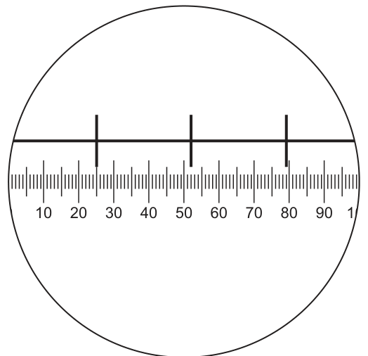
The diagram shows a view of an eyepiece graticule being calibrated using a stage micrometer.
Each division on the stage micrometer scale is .
Which row shows a correct calculation to calibrate each eyepiece graticule unit and shows the appropriate units?
Options
| calculation | units | |
|---|---|---|
| A | ||
| B | ||
| C | ||
| D |
Working
From Fig. 1.1, the three large vertical lines are major stage micrometer marks. They are spaced at 27 eyepiece graticule units apart, and each major interval = 1 stage micrometer division = 0.1 mm.
Taking the span between the leftmost and rightmost large lines:
Length of one eyepiece graticule unit:
Answer
C
C
Background Concept
An eyepiece graticule is a small glass disc carrying a scale (usually 100 equal divisions) that sits inside the eyepiece of a light microscope. Because the apparent size of the graticule changes with the objective lens in use, the actual length of one graticule division is NOT fixed — it must be found (calibrated) for each objective before the graticule can be used to measure specimens.
Calibration is done with a stage micrometer: a microscope slide on which a precise scale of known length has been etched. In this question, each division of the stage micrometer is 0.1 mm. By superimposing the stage micrometer scale on the eyepiece graticule scale (as in Fig. 1.1) you can read off how many graticule units span a known stage-micrometer distance.
The general formula is:
and the result in mm is then multiplied by 1000 to convert to µm (since 1 mm = 1000 µm).
Understanding the Question
Fig. 1.1 shows the two scales lined up. The bottom scale is the eyepiece graticule (labelled 10, 20, 30, … 100). The upper scale is the stage micrometer, on which three long vertical lines mark the major divisions. The 0 of the eyepiece graticule lines up with a major stage micrometer mark, and the 100 of the eyepiece graticule lines up with another major mark further to the right.
The question asks which row gives a correct calculation of the length of ONE eyepiece graticule unit, together with the appropriate unit.
Approach
- Read the positions of the three large vertical lines on the eyepiece graticule scale — they are at approximately 23, 50 and 77.
- Work out the eyepiece-unit gap between the leftmost and rightmost large lines (77 − 23 = 54 units).
- Count how many major stage micrometer divisions sit between those same two large lines (the middle line is one major mark in between, so there are 2 major divisions = 0.2 mm).
- Divide distance by eyepiece units, then convert mm → µm by multiplying by 1000.
Step-by-Step Reasoning
Reading Fig. 1.1:
- The three long vertical lines fall at roughly 23, 50 and 77 on the graticule scale.
- Spacing between successive lines: 50 − 23 = 27 units; 77 − 50 = 27 units. So each major interval on the stage micrometer spans 27 eyepiece graticule units.
- Each major interval = 1 stage micrometer division = 0.1 mm.
Using the full span between the outer two lines:
- Eyepiece units covered: 77 − 23 = 54
- Stage micrometer distance: 2 × 0.1 mm = 0.2 mm
Length of one eyepiece graticule unit:
Converting mm → µm:
This is exactly the calculation in row C: 0.2 / 54 × 1000 µm.
Why the other rows fail
- A — 0.1/27 ÷ 1000 mm. The numbers 0.1 and 27 are also consistent with the image (one major interval = 0.1 mm covering 27 eyepiece units), so the length comes out the same. The mark is lost because the unit conversion is the wrong way round: dividing by 1000 turns the answer into 3.7 × 10⁻⁶ mm = 3.7 nm, not 3.7 µm. To go from mm to µm you must multiply by 1000, not divide.
- B — 0.1/52 × 1000 µm. This would mean 52 eyepiece units correspond to 0.1 mm, which does not match the graticule in Fig. 1.1 (the large lines are 27 units apart, not 52).
- D — 0.2/79 × 1 000 000 nm. This implies 79 eyepiece units span 0.2 mm, which does not match the figure (the outer two large lines are 54 units apart). Even if the count were right, µm is the conventional unit for microscope measurements of cells; quoting the result in nm is inappropriate here.
Key Takeaways
- An eyepiece graticule must be calibrated against a stage micrometer of known dimensions before it can be used to measure specimens.
- The calibration formula is length of one eyepiece unit = (stage micrometer distance) / (eyepiece units in that distance).
- The standard unit for microscope measurements is the micrometre (µm): convert mm to µm by multiplying by 1000, never dividing.
- Always re-check both the count and the unit on every MCQ row — A had the right number but the wrong conversion direction.
Common Mistakes
- Writing ÷ 1000 instead of × 1000 when converting mm to µm. This makes the answer 1000× too small (nm instead of µm).
- Using nm when the question expects µm for a microscope measurement of cells.
- Miscounting the eyepiece graticule units (e.g. reading 52 or 79 instead of 54) by looking at the wrong pair of stage micrometer marks.
- Confusing the major stage micrometer marks (the long vertical lines) with the minor tick marks.
Things to Be Careful About
- The figure must be read carefully: the three large vertical lines are at 23, 50 and 77 on the graticule — NOT at 0 and 100 of the graticule. The 0 and 100 of the graticule sit at major stage micrometer marks at the edges of the field of view, but the calibration is most easily read off the three interior lines.
- The conversion 1 mm = 1000 µm is a multiplication by 1000, not a division.
- A correct numerical answer in the wrong unit is still 0 marks — units are part of the answer in calibration questions.
The diagram was drawn from an electron micrograph of an animal cell.
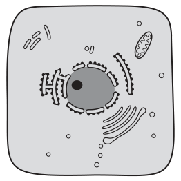
Which diagram would represent the same cell viewed with a simple light microscope, using daylight as the only light source?
Options
Working
An electron microscope has far higher resolution (≈0.5 nm) than a simple light microscope (≈200 nm), so it reveals small organelles such as ribosomes, rough ER, Golgi apparatus, and mitochondrial cristae.
A simple light microscope using only daylight (no stain) can only show structures large enough to be resolved AND that have sufficient natural contrast. The nucleus is the only organelle large enough and dense enough to be seen without staining.
Therefore the cell would appear as just the cell outline with a nucleus visible inside.
Answer
A
A
Background Concept
Microscopes are limited by their resolution — the minimum distance between two points at which they can still be distinguished as separate. A simple light microscope has a resolution of approximately , limited by the wavelength of visible light. An electron microscope uses electron beams (much shorter wavelength) and achieves a resolution of approximately , around 400 times better.
This means the electron microscope can reveal fine subcellular details that the light microscope simply cannot resolve, regardless of magnification. Resolution is the key property — not magnification.
Additionally, many cell structures are colourless and transparent. In a light microscope, these structures only become visible after staining (e.g., methylene blue, iodine, or specific dyes). With daylight as the only light source and no staining, only structures with natural colour/contrast differences can be seen. The nucleus is the largest organelle and appears denser/darker than the surrounding cytoplasm, giving it natural contrast.
Understanding the Question
Fig. 2.1 shows the cell as drawn from an electron micrograph. The drawing includes:
- Nucleus with nucleolus and nuclear envelope
- Rough endoplasmic reticulum (with ribosomes shown as dots)
- Mitochondrion (with cristae)
- Golgi apparatus (stacked cisternae)
- Small vesicles
The question asks which of the four options (A, B, C, D) would represent this same cell when viewed with a simple light microscope using daylight only (so no stains, no special illumination techniques like phase contrast or dark-field).
Approach
Two filters must be applied:
- Resolution filter — what is below in size cannot be resolved. This removes ribosomes, ER detail, Golgi cisternae, and cristae.
- Contrast filter — without stain, only naturally contrasting structures are visible. This further removes mitochondria, Golgi, ER (all nearly transparent in living/unstained cells).
Only the nucleus survives both filters, because it is large (several µm across) and naturally denser/more opaque than the cytoplasm.
Step-by-Step Reasoning
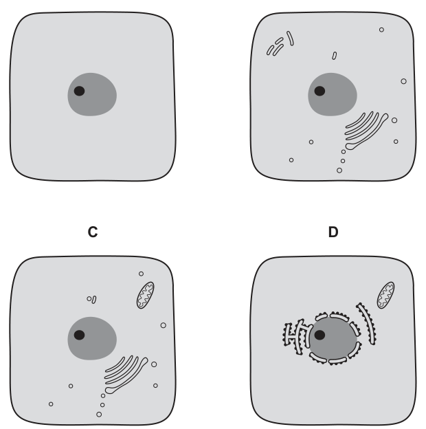
- Option A shows only the cell membrane outline and a single dark nucleus with a nucleolus. This matches what a simple light microscope with daylight (no stain) would reveal: just the outline of the cell and the prominent nucleus.
- Option B shows mitochondria, Golgi-like stacked structures, and small vesicles. These structures require the resolution of an electron microscope and/or staining to be visible. Incorrect.
- Option C shows a mitochondrion and Golgi apparatus. Same reasoning as B — these cannot be resolved or contrasted without staining. Incorrect.
- Option D shows rough endoplasmic reticulum with ribosomes (visible as the small dashes/dots along the membranes). Ribosomes are far below the resolution limit of a light microscope (~25 nm) and would never be visible as discrete structures. Incorrect.
The only option that respects both the resolution limit and the lack of staining is A.
Key Takeaways
- Resolution, not magnification, determines what can be seen. A light microscope cannot resolve structures smaller than ~.
- Common structures below the light microscope's resolution: ribosomes, endoplasmic reticulum detail, cristae of mitochondria, nuclear pores, most viruses.
- Without staining, light microscopy reveals only structures with natural contrast — typically the nucleus, chloroplasts (because of their green pigment), and cell walls.
- Electron microscopes produce monochrome (black and white) images because electrons have no colour; "colour" in EM images is added artificially/falsely.
Common Mistakes
- Confusing magnification with resolution — students sometimes think a more powerful light microscope could reveal the same detail as an electron microscope. It cannot, no matter how much it magnifies; the image would simply be a blur.
- Assuming all organelles in Fig. 2.1 would be visible with a light microscope. Only the largest, densest structures (the nucleus) survive both the resolution and contrast filters.
- Forgetting the staining requirement — many light-microscope structures are only visible because the specimen has been stained (e.g., methylene blue for nuclei).
Things to Be Careful About
- "Daylight as the only light source" is a deliberate clue: it excludes techniques like dark-ground illumination, phase contrast, or fluorescence, which can make unstained organelles visible.
- "Simple light microscope" is also deliberate: it rules out more sophisticated optical systems that might enhance contrast.
- The question tests the limits of the light microscope, not its typical classroom use where staining is standard practice. The correct answer (A) shows the minimum that would be visible under the stated conditions.
Norovirus has a diameter of .
Mimivirus has a diameter of .
Which viruses can be detected using a light microscope with a maximum resolution of ?
Options
| Norovirus | Mimivirus | |
|---|---|---|
| A | ✓ | ✓ |
| B | ✗ | ✗ |
| C | ✗ | ✓ |
| D | ✓ | ✗ |
key
✓ = can be detected
✗ = cannot be detected
Working
Convert the microscope resolution to the same units as the virus diameters:
An object can only be detected by a microscope if its size is at or above the resolution limit.
- Norovirus: → cannot be detected.
- Mimivirus: → can be detected.
Answer
C
C
Background Concept
A light microscope has a maximum resolution (the smallest distance between two points that can be distinguished as separate) of about (). In practice this is often quoted as (). For any object to be seen (resolved) by a microscope, it must be at or larger than this resolution limit. Smaller objects cannot be distinguished and so cannot be detected — this is why electron microscopes, with resolutions of around , are needed to visualise viruses and other very small structures.
Resolution is not the same as magnification. A light microscope can magnify an image thousands of times, but if the object is smaller than the resolution limit, the magnified image is just a blurred spot — no more detail is revealed. To see fine detail you need a microscope with a higher resolution (a shorter wavelength of illumination, hence the electron beam in an electron microscope).
Understanding the Question
Two viruses are given with their diameters:
- Norovirus — (a typical small virus, RNA capsid only)
- Mimivirus — (an unusually large virus, comparable in size to some small bacteria)
A light microscope with a maximum resolution of is provided. The question asks which of the two viruses can be detected by that microscope. Detection requires the virus to be equal to or larger than the resolution limit.
Approach
- Convert the microscope resolution into the same units (nanometres) as the virus diameters so the comparison is direct.
- Compare each virus's diameter with the converted resolution.
- If the virus is ≥ resolution → it can be detected (✓); if smaller → it cannot (✗).
Step-by-Step Reasoning
Step 1 — Unit conversion
Step 2 — Compare each virus
- Norovirus: . The virus is roughly eight times smaller than the resolution limit, so a light microscope cannot resolve it. → ✗
- Mimivirus: . The virus is larger than the resolution limit, so it can be resolved and detected. → ✓
Step 3 — Match to the options
The pattern (Norovirus ✗, Mimivirus ✓) is option C.
Key Takeaways
- The resolution of a light microscope (~0.2–0.25 µm) sets the lower size limit for what can be seen.
- An object must be at or larger than the resolution limit to be detected; magnification alone cannot compensate.
- Most viruses are far smaller than the light-microscope resolution limit and therefore require electron microscopy; only the very largest viruses (such as Mimivirus and Poxviruses) approach or exceed the light-microscope resolution and can in principle be seen with a light microscope.
- Remember — many exam questions hinge on this conversion.
Common Mistakes
- Confusing resolution with magnification — assuming that because a light microscope magnifies greatly, anything can be seen. The mark scheme rejects this reasoning.
- Unit slip — leaving the resolution in µm while the virus diameters are in nm, then comparing 0.25 with 30 and concluding wrongly that 30 is "bigger". Always convert to the same unit first.
- Reversing the inequality — saying the smaller virus (Norovirus) can be seen because it is "only 30" and the larger one cannot, when in fact size in nanometres is what matters against the 250 nm limit.
- Thinking all viruses are invisible to light microscopes — true for almost all viruses, but a few giant viruses (Mimivirus, Pandoravirus, Poxviruses) are exceptions and CAN be seen with a light microscope.
Things to Be Careful About
- Always quote the resolution in the same unit as the object size before comparing.
- The resolution figure given () is the maximum resolution; this is the smallest object that can be resolved. Objects at exactly are at the limit of visibility.
- Do not be distracted by the fact that both viruses are "viruses" — biological category is irrelevant; what matters is purely the physical size relative to the resolution.
- Option A is the trap for students who think "all viruses are too small for light microscopes, but Mimivirus is unusually large, so the question's wording suggests the answer should pick up on that". Read each comparison individually.
Which cell structures contain ribosomal RNA?
1 chloroplasts
2 mitochondria
3 nuclei
Options
A 1, 2 and 3
B 1 and 2 only
C 2 and 3 only
D 3 only
Working
Ribosomal RNA (rRNA) is a structural and catalytic component of ribosomes. Any organelle that contains ribosomes must therefore contain rRNA.
- Chloroplasts contain 70S ribosomes (in the stroma) and therefore contain rRNA.
- Mitochondria contain 70S ribosomes (in the matrix) and therefore contain rRNA.
- Nuclei contain the nucleolus, where rRNA is synthesised and where ribosomal subunits are assembled, so they contain rRNA.
All three statements are correct.
Answer
A
A
Background Concept
Ribosomes are the cellular machines responsible for protein synthesis. They are made up of ribosomal RNA (rRNA) molecules combined with ribosomal proteins. The rRNA is not just a scaffold — in the large subunit it is also the catalytic component that forms peptide bonds (it is a ribozyme). Every ribosome therefore contains rRNA, so asking "which structures contain rRNA?" is equivalent to asking "which structures contain ribosomes?".
Eukaryotic cells have two main sites of ribosome activity:
- Cytoplasmic ribosomes (80S), which sit free in the cytosol or are bound to the rough endoplasmic reticulum.
- Organellar ribosomes (70S), found inside chloroplasts and mitochondria. These 70S ribosomes are structurally and functionally similar to bacterial ribosomes, reflecting the endosymbiotic origin of these organelles.
The nucleus is also intimately involved with rRNA because rRNA genes are transcribed inside it, and the nucleolus is the dense region where rRNA is processed and combined with ribosomal proteins to form the large and small ribosomal subunits before they are exported to the cytoplasm.
Understanding the Question
This is a multiple choice question with three statements about cellular structures and four response options combining them. The candidate must decide which of chloroplasts, mitochondria and nuclei contain rRNA, then choose the option that lists all and only the correct statements. The mark scheme confirms the correct option is A (1, 2 and 3).
Approach
The cleanest reasoning is: "rRNA is found wherever ribosomes are found." Then check each named organelle for the presence of ribosomes (or, in the case of the nucleus, the site of rRNA synthesis).
Step-by-Step Reasoning
-
Chloroplasts (statement 1) — TRUE. Chloroplasts are semi-autonomous organelles derived from a cyanobacterial ancestor. Their stroma contains 70S ribosomes whose rRNA is encoded by the chloroplast's own genome. Chloroplasts therefore contain rRNA.
-
Mitochondria (statement 2) — TRUE. Mitochondria are also semi-autonomous, derived from an α-proteobacterial ancestor. Their matrix contains 70S ribosomes with mitochondrially-encoded rRNA. Mitochondria therefore contain rRNA.
-
Nuclei (statement 3) — TRUE. The nucleus contains the nucleolus, the site of rRNA transcription (by RNA polymerase I), rRNA processing, and assembly of the 60S and 40S ribosomal subunits. Even though the assembled ribosomes function in the cytoplasm, the rRNA itself is inside the nucleus during its synthesis and processing, so the nucleus contains rRNA.
All three statements are correct, so the answer is A: 1, 2 and 3.
Why the distractors fail:
- B (1 and 2 only) omits the nucleus. This is wrong because rRNA is actively made and processed inside the nucleus.
- C (2 and 3 only) omits the chloroplast. This is wrong because chloroplasts have their own ribosomes (and their own rRNA genes).
- D (3 only) is the most restrictive and is wrong because it ignores the rRNA inside both mitochondria and chloroplasts.
Key Takeaways
- rRNA is found in all ribosomes, so any organelle that contains ribosomes contains rRNA.
- The chloroplast (stroma) and the mitochondrion (matrix) each contain 70S ribosomes with their own rRNA, encoded by the organellar genome.
- The nucleus contains the nucleolus, where rRNA is transcribed, processed and assembled into ribosomal subunits.
- This question is a useful reminder that "where is rRNA found?" and "where are ribosomes found?" are not quite the same question — the nucleus hosts rRNA synthesis even though mature ribosomes function outside it.
Common Mistakes
- Forgetting that chloroplasts and mitochondria have their own ribosomes because of their endosymbiotic origin.
- Confusing "the nucleus contains ribosomes" (it does not, in the sense of assembled, functional ribosomes) with "the nucleus contains rRNA" (it does, during synthesis in the nucleolus).
- Picking B or C because of a vague memory that one of the organellar ribosomes "doesn't really count" — both count.
Things to Be Careful About
- 70S and 80S refer to sedimentation coefficients, not to the number of subunits or to absolute size in a simple way, but they are a quick way to distinguish cytoplasmic eukaryotic ribosomes (80S) from organellar/bacterial ribosomes (70S).
- The question asks about the presence of rRNA, not about where translation occurs. Translation occurs on assembled ribosomes, but rRNA is present in the nucleus during its synthesis and subunit assembly, so the nucleus still counts.
Immature red blood cells contain all the usual organelles and cell structures associated with animal cells. Mature red blood cells are specialised cells that have lost their organelles and cell structures.
Which statement correctly compares red blood cells with typical plant cells?
Options
A There are no mitochondria in mature red blood cells or plant cells.
B The only ribosomes found in immature red blood cells and plant cells are 80S ribosomes.
C There are no centrioles in mature red blood cells or plant cells.
D Immature red blood cells and plant cells contain large permanent vacuoles.
Working
Centrioles are absent from mature red blood cells because, during maturation, all organelles and cell structures are lost to maximise the space available for haemoglobin and to allow the cell to deform and squeeze through capillaries.
Centrioles are also absent from typical plant cells — they are a feature of animal cells, where they organise the spindle fibres during mitosis. Higher plant cells form the spindle without centrioles.
Therefore option C is the only statement that is correct for both cell types.
Answer
C
C
Background Concept
Eukaryotic cells share many common features (a nucleus, mitochondria, ribosomes, endoplasmic reticulum, Golgi apparatus, a cell surface membrane), but different cell types are specialised and may have lost or gained particular structures. Two important examples for this question are:
- Red blood cells (erythrocytes) begin life in the bone marrow as nucleated, organelle-containing immature cells. As they mature they extrude their nucleus and lose their other organelles (mitochondria, ribosomes, ER, Golgi, centrioles). This maximises the internal volume available for the oxygen-carrying pigment haemoglobin, and gives the mature biconcave disc its flexibility so it can squeeze through narrow capillaries.
- Typical plant cells differ from typical animal cells in several ways: they have a cellulose cell wall, a large permanent (tonoplast-bounded) central vacuole, and chloroplasts (in photosynthetic cells). They also lack centrioles; the mitotic spindle in higher plants is organised by acentriolar microtubule organising centres.
A further point relevant to one of the distractors is ribosome type. The cytoplasmic ribosomes of all eukaryotes are 80S, but mitochondria and chloroplasts contain their own 70S ribosomes — a relic of their prokaryotic evolutionary origin (endosymbiotic theory). So a eukaryotic cell that contains chloroplasts will have both 70S and 80S ribosomes.
Understanding the Question
The stem gives the key fact that must drive the comparison: mature red blood cells have lost their organelles, while immature red blood cells still have them. Each of the four statements makes a claim about both cell types (or about immature RBCs and plant cells), and the candidate must decide which claim is correct for both.
The command word "compares" signals that a side-by-side check of organelle presence in each named cell type is needed.
Approach
For each option, check whether the claim is true for (a) the red blood cell state specified and (b) typical plant cells. The answer is the option that is true in both cases.
Step-by-Step Reasoning
Option A — no mitochondria in mature red blood cells or plant cells.
- Mature red blood cells: true (mitochondria are lost during maturation; ATP is produced only by glycolysis).
- Plant cells: false. Plant cells contain mitochondria — for example, root cells and non-photosynthetic tissues rely on mitochondrial respiration.
- A is rejected.
Option B — the only ribosomes in immature red blood cells and plant cells are 80S.
- Immature red blood cells: true (eukaryotic cytoplasm contains 80S ribosomes).
- Plant cells: false. Plant cells also contain chloroplasts, and chloroplasts contain 70S ribosomes; mitochondria also have 70S ribosomes. So 80S is not the only type in a plant cell.
- B is rejected.
Option C — no centrioles in mature red blood cells or plant cells.
- Mature red blood cells: true (lost during maturation).
- Typical plant cells: true (centrioles are characteristic of animal cells; higher plant cells lack them and use other microtubule organising centres to form the spindle).
- C is the correct option.
Option D — immature red blood cells and plant cells contain large permanent vacuoles.
- Immature red blood cells: false. Immature RBCs have the usual animal cell organelles; they do not have a large permanent vacuole (this is a plant cell feature).
- Plant cells: true (the central vacuole is a defining plant cell feature).
- D is rejected.
Key Takeaways
- Mature mammalian red blood cells are unusual in being anuclear and lacking all organelles — an extreme example of cell specialisation for a single function (oxygen transport).
- Plant cells differ from animal cells in three headline ways: cellulose cell wall, large central vacuole, and chloroplasts (in green tissues) — and they notably lack centrioles.
- 80S ribosomes are the cytoplasmic ribosome of eukaryotes, but mitochondria and chloroplasts contain their own 70S ribosomes. A plant cell therefore contains both types.
Common Mistakes
- Assuming plant cells have no mitochondria because they photosynthesise. Photosynthesis and respiration occur in different organelles; non-photosynthetic plant cells and even photosynthetic plant cells in the dark rely on mitochondrial ATP.
- Forgetting that chloroplasts (and mitochondria) contain 70S ribosomes, which makes statement B false for any plant cell.
- Confusing immature with mature red blood cells — the stem specifically says mature cells have lost their organelles, so the question can ask different things of the two stages.
- Attributing centrioles to all eukaryotes. Centrioles are characteristic of animal cells; higher plants organise the spindle without them.
Things to Be Careful About
- Read the cell type specified in each option carefully (mature vs immature, plant vs animal) — a true statement about one of the two is not enough.
- "Typical plant cell" in the syllabus means a eukaryotic cell with cell wall, vacuole, and (in photosynthetic tissue) chloroplasts — it is not a prokaryote.
- The term "centriole" refers to the cylindrical structure in the centrosome; do not confuse it with the centromere, which is the region of a chromosome that attaches to the spindle.
A culture of human cells had its cell surface membranes removed, releasing the cell contents.
This material became contaminated by bacteria.
The material was then centrifuged, separating out the various cell structures according to size and mass.
Which cell structure would be separated out along with the bacteria?
Options
A endoplasmic reticulum
B mitochondria
C nuclei
D ribosomes
Working
Differential centrifugation separates cell structures according to size and mass: the largest and densest structures form a pellet first (at the lowest speed), while smaller structures remain in the supernatant until spun faster.
Approximate sizes of the options:
- Endoplasmic reticulum: small membrane fragments (~0.1 µm or less)
- Mitochondria: ~1–10 µm
- Nuclei: ~5–10 µm (largest organelle listed)
- Ribosomes: ~0.02 µm (much smaller than bacteria)
Bacteria are typically ~1–5 µm in length.
Answer
B
B
Background Concept
Differential centrifugation separates organelles by exploiting differences in their size and mass. When a homogenised cell sample is spun, the largest and densest structures sediment to the bottom of the tube first, forming a pellet. The supernatant is then spun at a higher speed to pellet the next-largest structures, and so on. Two structures of similar size and mass will pellet at the same speed and end up in the same fraction.
Bacteria are prokaryotic cells, typically 1–5 µm in length. Among the eukaryotic organelles, mitochondria are similar in size (about 1–10 µm long), and this size similarity is one piece of evidence supporting the endosymbiotic theory that mitochondria evolved from engulfed aerobic prokaryotes.
Understanding the Question
The question describes a homogenate of human (eukaryotic) cell contents that has been contaminated with bacteria, then centrifuged. The command word is implicit — you must identify which organelle has a size/mass similar enough to bacteria that it sediments in the same fraction.
The four options differ greatly in size:
- Nuclei: very large (5–10 µm or more)
- Mitochondria: medium (1–10 µm)
- Endoplasmic reticulum: when fragmented, very small pieces (~0.1 µm)
- Ribosomes: tiny (~0.02 µm = 20 nm), only visible by electron microscopy
Approach
Compare the size of each organelle to a typical bacterium and decide which one is closest. The structure closest in size and mass to a bacterium will be the one that pellets alongside it.
Step-by-Step Reasoning
- Bacteria are ~1–5 µm. Among the listed organelles, only mitochondria fall in this size range.
- Nuclei are much larger and would pellet at a much lower centrifuge speed, well separated from the bacterial fraction.
- Endoplasmic reticulum breaks into tiny membrane vesicles during homogenisation and would remain in the supernatant until very high speeds.
- Ribosomes (~20 nm) are far smaller than bacteria and require very high-speed (ultracentrifugation at >100,000 × g) to pellet, while bacteria pellet at relatively low speeds.
- Therefore mitochondria are the structure most similar in size/mass to bacteria, and they will sediment out together.
Key Takeaways
- Differential centrifugation separates structures by size and mass, not by function or identity.
- Bacteria and mitochondria are similar in size (1–5 µm), which is consistent with the endosymbiotic origin of mitochondria.
- Nuclei are the largest organelle and pellet first; ribosomes are the smallest and require the highest speed.
Common Mistakes
- Choosing C (nuclei): a tempting distractor because nuclei are the most prominent organelle, but they are far larger than bacteria and pellet at a much lower speed.
- Choosing D (ribosomes): a common error because ribosomes are described as 'small', but they are roughly 1000× smaller in linear dimension than bacteria, so they pellet only at ultracentrifuge speeds.
- Choosing A (endoplasmic reticulum): ER fragments are very small membrane vesicles, not comparable in mass to whole bacteria.
Things to Be Careful About
- Memorise approximate size ranges: bacteria/mitochondria (~1–10 µm), nuclei (~5–10 µm), ribosomes (~20 nm), and ER fragments (<0.1 µm).
- Read the wording — 'separated out along with' means in the same pellet, not just 'present in the tube'.
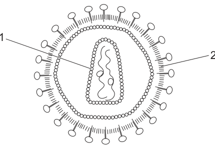
The diagram shows the structure of a virus.
Which row identifies the correct description of the structures labelled 1 and 2?
Options
| 1 | 2 | |
|---|---|---|
| A | protein coat called the capsid | virus envelope made from phospholipids |
| B | virus envelope made from phospholipids | protein coat called the capsid |
| C | virus envelope made from protein | phospholipid coat called the capsid |
| D | phospholipid coat called the capsid | virus envelope made from protein |
Working
Label 1 points to the inner protein coat surrounding the genetic material — this is the capsid.
Label 2 points to the outer membrane bearing the glycoprotein spikes — this is the envelope, derived from the host cell's phospholipid (and protein) membrane.
Answer
A
A
Background Concept
Viruses are non-cellular particles that lie on the boundary between living and non-living. A fully formed (mature) virus particle is called a virion and consists of genetic material (DNA or RNA) enclosed within a protein coat. The key structural components you need to know for CIE Biology are:
- Capsid — a protein coat made of repeating protein subunits called capsomeres. The capsid packages and protects the viral nucleic acid, and is responsible for recognising and binding to receptors on the host-cell surface.
- Envelope — an additional outer membrane found in some viruses (e.g. HIV, influenza, SARS-CoV-2). The envelope is derived from the host cell's plasma membrane as the virus buds out, so it is a phospholipid bilayer studded with glycoproteins (often drawn as "spikes"). These envelope glycoproteins attach to host-cell receptors during infection.
- Genetic material — either DNA or RNA (never both), located inside the capsid.
Not all viruses have an envelope. Non-enveloped (naked) viruses have only a capsid; enveloped viruses have both a capsid and an envelope.
Understanding the Question
The diagram shows a virus with two labelled structures. Label 1 is the inner box-shaped coat surrounding the wavy line (the nucleic acid), and label 2 is the outer dotted layer decorated with the "lollipop" spikes. The question asks you to name each structure correctly and to use the proper term for the material it is made from.
The command word is implicit ("Which row…"), so you simply need to choose the option that correctly pairs each label with its name and composition.
Approach
- Decide what label 1 is: it is inside the virus, immediately around the genetic material → it must be the protein coat / capsid.
- Decide what label 2 is: it is the outer membrane, with glycoprotein spikes projecting from it → it must be the envelope, which is made from phospholipids (it is host-cell membrane in origin).
- Match this to the row in the table that says exactly that.
Step-by-Step Reasoning
- Option A: "1 = protein coat called the capsid; 2 = virus envelope made from phospholipids." Both statements are correct. Label 1 is indeed the capsid (protein), and label 2 is indeed the envelope, which is a phospholipid bilayer. ✓
- Option B: This swaps the two labels — it claims label 1 is the envelope and label 2 is the capsid. Looking at the diagram, this is the wrong way round. ✗
- Option C: Calls label 1 a "virus envelope made from protein" (wrong — it is a capsid, not an envelope) and label 2 a "phospholipid coat called the capsid" (the term capsid is reserved for the protein coat, not a phospholipid layer). ✗
- Option D: Calls label 1 a "phospholipid coat called the capsid" (a capsid is by definition protein, not phospholipid) and label 2 a "virus envelope made from protein" (an envelope is phospholipid-based, not protein). ✗
So option A is the only one in which each label is given the correct name and the correct chemical composition.
Key Takeaways
- Capsid = protein coat, built from capsomeres, surrounds and protects the nucleic acid.
- Envelope = phospholipid (and protein) bilayer outside the capsid, derived from a host-cell membrane and bearing glycoprotein spikes.
- The terms capsid and envelope are not interchangeable: a capsid is always protein; an envelope is a (host-derived) lipid bilayer.
Common Mistakes
- Swapping capsid and envelope because of their relative positions — remember: the capsid is innermost (touching the nucleic acid), the envelope is outermost.
- Calling the capsid a "phospholipid coat" — the capsid is by definition made of protein.
- Calling the envelope a "protein coat" — the envelope is host-cell membrane, so it is mainly phospholipid.
- Confusing the glycoprotein spikes with the envelope itself — the spikes are glycoproteins embedded in the phospholipid envelope.
Things to Be Careful About
- On CIE diagrams the envelope is usually drawn as a dotted/double line studded with spikes, and the capsid as a box or geometric shape (often icosahedral) inside it. Use position, not shading, to identify them.
- "Capsid" and "capsule" are not the same word. A capsule is a polysaccharide layer found on some bacteria, not on viruses.
- Read both halves of the row before deciding — a row that gets one label right but the other wrong still scores zero on a "which row is correct" question.
Which statement about a macromolecule is correct?
Options
A Amylose is a branched polymer made of -glucose and -glucose monomers.
B DNA is an association of two polymers made of nucleotide monomers.
C Haemoglobin is an association of four polymers made up of monomers of haem groups and amino acids.
D Triglycerides are polymers made of fatty acids and glycerol.
Working
- A is wrong: amylose is unbranched and made only of α-glucose (not β-glucose); branching is a feature of amylopectin.
- B is correct: DNA consists of two polynucleotide strands, each a polymer of nucleotide monomers, held together by hydrogen bonds between complementary bases.
- C is wrong: haemoglobin has four polypeptide chains (polymers of amino acids) plus four haem prosthetic groups; the haem groups are not monomers.
- D is wrong: a triglyceride is a single molecule of one glycerol bonded to three fatty acids; it is not a polymer because the three fatty acids are not identical repeating units.
Answer
B
B
Background Concept
A macromolecule is a very large molecule built from smaller subunits. Many are true polymers — long chains of many identical or near-identical monomers joined by covalent bonds (e.g. polynucleotides from nucleotides, polypeptides from amino acids, polysaccharides from monosaccharides). Other large biological molecules are associations of polymers with non-polymeric components (e.g. haemoglobin is four polypeptide chains combined with four haem prosthetic groups), or are simply large molecules that are not polymers at all (e.g. a triglyceride). The four options test whether you can correctly identify monomers, polymers and non-polymeric associations in amylose, DNA, haemoglobin and triglycerides.
Understanding the Question
This is a single-best-answer MCQ. The candidate must pick the statement that is fully correct. Three of the four options contain a deliberate error relating to monomer identity, branching, or whether the molecule is genuinely a polymer. The mark scheme credits only option B.
Approach
Test each option in turn against two checkpoints: (1) is the molecule correctly described (correct identity, structure, branching)? (2) is the description of its monomers and polymer status correct? Reject any option that fails on either point.
Step-by-Step Reasoning
Option A — amylose. Amylose is one of the two components of starch (the other being amylopectin). It is an unbranched chain of α-glucose monomers linked by α-1,4-glycosidic bonds; the α-1,6 branch points are found only in amylopectin. β-glucose is the monomer of cellulose, not amylose. So both halves of statement A are wrong. Reject.
Option B — DNA. A DNA molecule is two polynucleotide strands running antiparallel and held together by hydrogen bonds between complementary base pairs (A–T, G–C). Each strand is a polymer built from nucleotide monomers (a deoxyribose sugar, a phosphate group, and one of four nitrogenous bases). DNA is therefore an association of two polymers of nucleotide monomers. Accept.
Option C — haemoglobin. Haemoglobin is a globular protein with quaternary structure: four polypeptide chains (2α and 2β), each carrying one haem prosthetic group that contains an iron ion. The polypeptide chains are polymers of amino acid monomers; the haem groups are not monomers — they are non-protein prosthetic groups. So although the four-polymers idea is right, describing haem groups as monomers is wrong. Reject.
Option D — triglycerides. A triglyceride consists of one glycerol molecule esterified to three (often different) fatty acids. Although it is large and formed by joining smaller units, the three fatty acids are not repeating identical monomers, so a triglyceride is not classified as a polymer in biology. Reject.
Only option B survives both checkpoints, so B is the correct answer.
Key Takeaways
- A polymer must be built from repeating monomer units; large molecules made from a few different joined units (like a triglyceride) are not polymers.
- Amylose is unbranched α-glucose only; amylopectin is the branched component of starch; cellulose is β-glucose.
- DNA is a double-stranded polymer of nucleotides joined by phosphodiester bonds within each strand and hydrogen bonds between strands.
- Haemoglobin has quaternary structure (4 polypeptide chains + 4 haem prosthetic groups); the haem groups are prosthetic, not monomeric.
Common Mistakes
- Saying "amylose is branched" — the branched component of starch is amylopectin, not amylose.
- Describing the components of a triglyceride as monomers — three different fatty acids plus glycerol do not constitute a polymer.
- Calling the haem group a monomer of haemoglobin — the polymers (monomers = amino acids) are the four polypeptide chains; haem is a prosthetic group.
- Confusing DNA with RNA, or forgetting that DNA is a polymer of nucleotides (not of bases, not of sugars on their own).
Things to Be Careful About
- "Macromolecule" is a broader term than "polymer" — every polymer is a macromolecule, but not every macromolecule is a polymer.
- Branching refers to α-1,6 glycosidic bonds found in amylopectin and glycogen, not in amylose or cellulose.
- When a statement combines two claims (e.g. "amylose is branched AND contains β-glucose"), both must be correct for the option to be the answer.
- In haemoglobin, distinguish carefully between polypeptide chain (the polymer) and haem group (a non-polymeric prosthetic group containing Fe²⁺).
What happens to molecules of sucrose when they are heated with acid?
Options
A Glycosidic bonds are broken by condensation using water, releasing only glucose.
B Glycosidic bonds are broken by condensation, releasing fructose, glucose and water.
C Glycosidic bonds are broken by hydrolysis using water, releasing fructose and glucose.
D Glycosidic bonds are broken by hydrolysis, releasing fructose, glucose and water.
Working
Sucrose is a disaccharide formed from one glucose and one fructose monomer joined by a glycosidic bond. Heating with dilute acid supplies water and the energy needed to break this bond.
The bond is broken by hydrolysis (using water), not condensation. Hydrolysis splits the glycosidic bond, inserting water across it and releasing the two separate monosaccharides: fructose and glucose (no free water is released — water is a reactant).
Answer
C
C
Background Concept
Disaccharides such as sucrose, maltose and lactose are formed when two monosaccharides join by a condensation reaction: a glycosidic bond forms between them and a molecule of water is released. The reverse reaction — hydrolysis — uses a water molecule to break that glycosidic bond, regenerating the two original monosaccharides.
Sucrose is a non-reducing sugar composed of one α-glucose linked to one fructose through a 1,2-glycosidic bond. Because the bond involves the anomeric carbons of both sugars, neither ring can open to expose a free reducing group, so sucrose does not reduce Benedict's (or Fehling's) reagent until it has been hydrolysed.
In the laboratory, hydrolysis of a non-reducing sugar is carried out by heating with dilute hydrochloric acid (typically at about 70 °C for a few minutes). The acid acts as a catalyst, lowering the activation energy of the reaction. After hydrolysis, the solution is cooled and neutralised (with sodium hydrogencarbonate) before performing the Benedict's test, since Benedict's reagent only works under alkaline conditions.
Understanding the Question
This is a multiple-choice question testing two pieces of knowledge:
- Whether the bond is broken by condensation or hydrolysis.
- The identity of the monosaccharide product(s) and whether water is a product or a reactant.
The command "What happens" requires the candidate to recognise the chemistry of the reaction between sucrose and hot acid.
Approach
Apply the definition of hydrolysis versus condensation, then match the products to those of sucrose hydrolysis (glucose + fructose). Eliminate any option that uses the wrong reaction type, the wrong product list, or incorrectly states that water is released.
Step-by-Step Reasoning
- Condensation vs hydrolysis: Hydrolysis uses water to break a bond; condensation forms a bond and releases water. Hot acid supplies water to break the glycosidic bond, so the reaction is hydrolysis. Options A and B (which say "condensation") are immediately wrong.
- Identity of products: Sucrose = glucose + fructose, not glucose alone. Option A is also wrong because it says "releasing only glucose".
- Water as product vs reactant: In hydrolysis, water is consumed — it is not released. Option D is wrong because it says water is a product.
- Option C correctly states that the glycosidic bond is broken by hydrolysis using water, releasing fructose and glucose (and no free water).
Key Takeaways
- Hydrolysis = bond broken by water; condensation = bond formed with release of water.
- Sucrose is composed of glucose + fructose, joined by an α-1,2-glycosidic bond.
- Heating with dilute acid hydrolyses glycosidic bonds; this is the basis of the non-reducing sugar test.
Common Mistakes
- Confusing hydrolysis and condensation — they are opposites.
- Thinking sucrose is made of two glucoses (it is actually glucose + fructose).
- Believing that water is a product of the reaction, when in fact water is a reactant in hydrolysis.
Things to Be Careful About
- The question specifies "heated with acid" — this is hydrolysis, not condensation.
- The correct products are the two monosaccharides only; water is not a product (it is a reactant in hydrolysis).
- Watch wording: options B and D mention water as a product, which is chemically inconsistent with hydrolysis.
The diagrams show how some polymers of glucose can be classified.
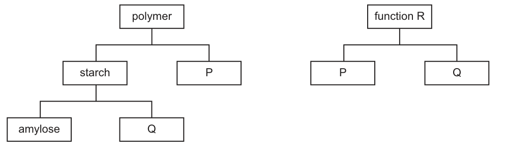
Which row correctly identifies P, Q and R?
Options
| polymer P | polymer Q | function R | |
|---|---|---|---|
| A | cellulose | amylopectin | structural support |
| B | glycogen | amylopectin | energy storage |
| C | cellulose | glycogen | energy storage |
| D | glycogen | cellulose | structural support |
Working
- The first flow chart shows 'polymer' splitting into 'starch' and 'P', and 'starch' splitting into 'amylose' and 'Q'.
- Starch is made of two polymers: amylose and amylopectin. Therefore Q = amylopectin.
- P is a polymer of glucose at the same level as starch. The two main categories of glucose polymers are storage polysaccharides (starch, glycogen) and structural polysaccharides (cellulose). To match the second diagram, P must be a storage polymer, so P = glycogen.
- The second flow chart shows 'function R' applying to both P and Q. Both glycogen and amylopectin are branched storage polysaccharides, so R = energy storage.
Answer
B
B
Background Concept
Glucose is a monomer that can be joined by glycosidic bonds to form several different polymers, each with very different properties and roles. The three polysaccharides of glucose that you must know for AS Biology are:
- Starch – the energy storage polysaccharide in plants. It is itself a mixture of two polymers:
- Amylose – long, unbranched chains of α-glucose joined by 1,4-glycosidic bonds; it coils into a helix.
- Amylopectin – chains of α-glucose with 1,4-glycosidic bonds and frequent 1,6-glycosidic branches, giving a bush-like shape.
- Glycogen – the energy storage polysaccharide in animals (and fungi). Like amylopectin, it is α-glucose with both 1,4 and 1,6 links, but it is more highly branched. It is stored in the liver and muscle.
- Cellulose – a structural polysaccharide in plant cell walls. It is built from β-glucose joined by 1,4-glycosidic bonds, producing straight, unbranched chains that hydrogen-bond together into strong microfibrils.
The key functional distinction is therefore storage (starch, glycogen, with amylose and amylopectin as the two starch components) vs structure (cellulose).
Understanding the Question
The question gives you two flow charts and asks you to fill in the labels P, Q and R.
- Chart 1: 'polymer' branches into 'starch' and 'P'; 'starch' branches into 'amylose' and 'Q'. So P is a polymer of glucose parallel to starch, and Q is the second component of starch alongside amylose.
- Chart 2: 'function R' applies to both P and Q. So whatever R is, it must be a function shared by P and Q.
The command word is implicit ("identify"); you simply need to recognise which glucose polymer fits each labelled box.
Approach
- Identify Q first: it is a sub-component of starch, so it must be amylopectin (the only other starch component alongside amylose).
- Identify P: it is a polymer of glucose on the same level as starch, and (from the second chart) it shares a function with amylopectin. Amylopectin is a storage polysaccharide, so P must also be a storage polysaccharide of glucose. That is glycogen (the other main α-glucose storage polymer).
- Identify R: the function shared by glycogen and amylopectin is energy storage.
- Match to the answer choices – only B lists glycogen, amylopectin and energy storage.
Step-by-Step Reasoning
- Starch has exactly two polymeric components: amylose (~20–30%) and amylopectin (~70–80%). So Q, the partner of amylose inside starch, is amylopectin. This rules out C (which calls Q glycogen — glycogen is a separate polymer, not a component of starch) and D (which calls Q cellulose — cellulose is not part of starch either).
- The remaining storage polymer of glucose, parallel to starch, is glycogen. So P = glycogen. This rules out A (which calls P cellulose — cellulose is structural, not storage, and the second chart shows that P shares a function with amylopectin, which is storage).
- The shared function of glycogen (animal storage) and amylopectin (plant storage) is energy storage. This makes R = energy storage, confirming B and ruling out A (which has R = structural support, but amylopectin is a storage molecule, not a structural one).
- Only option B (glycogen, amylopectin, energy storage) is fully consistent with both flow charts.
Key Takeaways
- Starch = amylose + amylopectin.
- Three glucose polymers to keep separate: starch (plant storage), glycogen (animal storage, more branched), cellulose (plant structure, β-glucose, straight chains).
- The functional split is storage (amylose, amylopectin, glycogen) versus structure (cellulose).
- Flow-chart / branching-diagram questions are simply classification tasks: identify the parent branch (starch → amylose + amylopectin; polymer of glucose → starch / glycogen / cellulose) and read off what fits.
Common Mistakes
- Confusing glycogen with amylopectin: both are branched α-glucose storage polymers with 1,4 and 1,6 links, but glycogen is the animal storage polysaccharide and amylopectin is a component of plant starch. They are not interchangeable as "components of starch".
- Putting cellulose as a component of starch — cellulose is built from β-glucose for cell-wall structure, while starch is built from α-glucose for storage; they are chemically and functionally different.
- Treating 'structural support' as the function of amylopectin — amylopectin's many 1,6 branches make it ideal for rapid mobilisation of glucose for energy, not for structural strength.
- Mixing up the two flow charts — P appears in both, so whatever P is, it must simultaneously (a) be a polymer of glucose parallel to starch and (b) share a function with Q (amylopectin). Only glycogen satisfies both.
Things to Be Careful About
- The question is testing classification, not detailed structure. A common trap is to overthink the 1,4 vs 1,6 bond patterns and end up selecting the wrong option because of one confused letter.
- Always read the entire flow chart before answering: P must satisfy both its position in chart 1 and its position in chart 2.
- Use the precise spelling/terminology: amylopectin (not amylose pectin, not amylopectrin) and glycogen (not glycogon).
Which molecules have products containing a carboxyl group when they are hydrolysed?
1 phospholipids
2 polysaccharides
3 proteins
Options
A 1 and 2
B 1 and 3
C 2 and 3
D 3 only
Working
- A carboxyl group is –COOH.
- Hydrolysis of phospholipids yields glycerol, phosphate and fatty acids. Fatty acids contain –COOH.
- Hydrolysis of polysaccharides yields monosaccharides, which do not contain a carboxyl group.
- Hydrolysis of proteins yields amino acids, which contain both an amino group (–NH₂) and a carboxyl group (–COOH).
Therefore 1 and 3 produce monomers with a carboxyl group.
Answer
B
B
Background Concept
Hydrolysis is the breaking of a bond by the addition of water. Each condensation polymer can be split back into its monomers by hydrolysis, and the functional groups on those monomers determine which category a molecule falls into.
A carboxyl group (–COOH) is a defining feature of two important classes of biomolecules in this question:
- Fatty acids – long hydrocarbon chains with a terminal –COOH; these are the building blocks (along with glycerol) of triglycerides and phospholipids.
- Amino acids – each has both an amino group (–NH₂) and a carboxyl group (–COOH) attached to the same central carbon, plus a variable R group.
Monosaccharides (e.g. glucose) carry hydroxyl (–OH) and carbonyl (C=O) groups, but no –COOH group.
Understanding the Question
This MCQ asks which of the three named macromolecules release monomers that carry a –COOH group when hydrolysed. You need to identify the monomers of each, then check for the carboxyl group.
The command word is implicit: identify the correct combination.
Approach
For each numbered option, write down the hydrolysis products and inspect their functional groups. The combinations that contain at least one monomer with –COOH are the answer.
Step-by-Step Reasoning
-
Phospholipids. A phospholipid is built from glycerol, a phosphate group, and two fatty acids, linked by ester bonds. Hydrolysis (enzymatic or acidic) breaks these ester bonds, releasing fatty acids. Each fatty acid has a –COOH head group — so phospholipid hydrolysis does produce a carboxyl-containing product.
-
Polysaccharides. Starch, glycogen and cellulose are polymers of α- or β-glucose joined by glycosidic bonds. Hydrolysis yields glucose (or other monosaccharides), whose functional groups are –OH and a ring oxygen/carbonyl — no –COOH is present. So polysaccharides do not qualify.
-
Proteins. Polypeptides are chains of amino acids joined by peptide bonds. Hydrolysis yields free amino acids, each of which carries an –NH₂ group and a –COOH group. So protein hydrolysis does produce a carboxyl-containing product.
Therefore the correct statement is 1 and 3, which corresponds to option B.
Key Takeaways
- Fatty acids (from lipid hydrolysis) and amino acids (from protein hydrolysis) both carry a carboxyl group (–COOH).
- Monosaccharides released from polysaccharide hydrolysis do not contain –COOH.
- Knowing the monomers of each biological molecule — and their functional groups — is essential for these questions.
Common Mistakes
- Confusing amino acids with sugars and answering that polysaccharides release carboxyl groups (incorrect — sugars lack –COOH).
- Forgetting that fatty acids are a hydrolysis product of phospholipids and choosing option A or D.
Things to Be Careful About
- The carboxyl group must be –COOH; the carbonyl (C=O) in a sugar ring is not a carboxyl group.
- Phospholipids have two fatty acid tails, so hydrolysis releases two fatty acid molecules per phospholipid.
Which bonds are involved in maintaining the secondary and tertiary levels of protein structure?
1 disulfide
2 hydrogen
3 ionic
Options
| secondary | tertiary | |
|---|---|---|
| A | 2 only | 1, 2 and 3 |
| B | 2 and 3 only | 2 and 3 only |
| C | 1 and 3 only | 1 and 3 only |
| D | 1, 2 and 3 | 1 only |
Working
Secondary structure (α-helix / β-pleated sheet) is held together by hydrogen bonds between the –NH and –C=O groups of the polypeptide backbone, so only bond 2 (hydrogen) is involved.
Tertiary structure is held together by all three of the bonds listed: hydrogen (2), ionic (3) and disulfide (1) bridges between R-groups, so 1, 2 and 3 are involved.
Answer
A
A
Background Concept
A protein's three-dimensional shape is described at four levels:
- Primary (1°) structure — the linear sequence of amino acids linked by peptide bonds.
- Secondary (2°) structure — local, regular folding patterns such as the α-helix and β-pleated sheet, produced by hydrogen bonding between the –N–H of one peptide bond and the C=O of another in the polypeptide backbone.
- Tertiary (3°) structure — the overall 3D folding of a single polypeptide chain, held by interactions between the side chains (R-groups).
- Quaternary (4°) structure — the arrangement of more than one polypeptide subunit in a multi-chain protein (e.g. haemoglobin).
The four R-group interactions that stabilise tertiary structure are:
- Hydrogen bonds – between polar/charged R-groups (e.g. –OH, –NH₂).
- Ionic (electrostatic) bonds – between oppositely charged R-groups (e.g. –COO⁻ and –NH₃⁺).
- Disulfide bridges – strong covalent S–S bonds between two cysteine residues.
- Hydrophobic interactions – non-polar R-groups clustering away from water.
Understanding the Question
The question asks which of three named bond types — disulfide (1), hydrogen (2), ionic (3) — contribute to maintaining the secondary structure and which contribute to the tertiary structure. The candidates must read the table correctly: each row pairs a list of bond types with a structural level, and the correct option is the one whose two lists match the biology.
Approach
Recall that secondary structure is held almost exclusively by hydrogen bonds in the backbone, whereas tertiary structure is held by a combination of R-group interactions including all three bond types listed. Match this to the options.
Step-by-Step Reasoning
- Secondary structure: in an α-helix, each C=O of one peptide bond hydrogen-bonds to the N–H of the peptide bond four residues along the chain; the same backbone hydrogen-bonding pattern gives the β-pleated sheet. These are hydrogen bonds (bond 2). Disulfide and ionic bonds do not stabilise secondary structure because they involve R-groups, not the repeating backbone, and R-group interactions are not part of the regular secondary pattern.
- Tertiary structure: the 3D folding of the chain is stabilised by R-group interactions, so any bond that can form between side chains may contribute. Disulfide (1) bridges, hydrogen bonds (2) and ionic bonds (3) all commonly occur. (Hydrophobic interactions, the fourth R-group interaction, are not in the question.)
- Therefore the correct row is: secondary — 2 only; tertiary — 1, 2 and 3, which is option A.
- Option B wrongly includes ionic bonds in secondary structure; option C wrongly excludes hydrogen bonds from both levels; option D wrongly includes all three in secondary and only disulfide in tertiary.
Key Takeaways
- Secondary structure = hydrogen bonds in the backbone only.
- Tertiary structure = R-group interactions: hydrogen, ionic, disulfide and hydrophobic (all four).
- A common error is to confuse the R-group interactions (tertiary) with the backbone interactions (secondary).
Common Mistakes
- Saying disulfide bonds contribute to secondary structure — they are covalent S–S links between R-groups and only appear in tertiary (or quaternary) folding.
- Saying ionic bonds stabilise the α-helix or β-sheet — these involve charged R-groups, not the regular backbone.
- Forgetting that hydrogen bonds contribute to tertiary structure as well as secondary — they form between polar R-groups throughout the folded chain.
Things to Be Careful About
- The question lists only three bond types; hydrophobic interactions are a real fourth stabiliser of tertiary structure but are not one of the numbered options, so do not be distracted by their absence.
- "Secondary structure held by hydrogen bonds" refers specifically to backbone H-bonds; do not credit a candidate who talks about R-group hydrogen bonds in the context of 2° structure.
- Distinguish peptide bonds (which hold primary structure together) from the other interactions — they are covalent but not one of the options here.
The diagrams show the structure of four amino acids in solution.
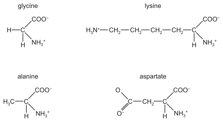
Which amino acids have no overall charge?
Options
A alanine and aspartate
B alanine and glycine
C aspartate and lysine
D glycine and lysine
Working
Examine each amino acid and count the charged groups:
- Glycine: one –NH3+ (+1) and one –COO− (−1) → net charge 0
- Lysine: one –NH3+ on the backbone (+1), one –COO− (−1), plus an extra –NH3+ on the R-group (+1) → net charge +1
- Alanine: one –NH3+ (+1) and one –COO− (−1) → net charge 0
- Aspartate: one –NH3+ (+1), one –COO− (−1), plus an extra –COO− on the R-group (−1) → net charge −1
Only glycine and alanine have no overall charge.
Answer
B
B
Background Concept
All amino acids share the same backbone: a central (α) carbon bonded to an amino group (–NH3+), a carboxyl group (–COO−), a hydrogen atom, and a variable side chain (R-group). At physiological pH (~7.4), the backbone –NH2 is protonated to –NH3+ (carrying a +1 charge) and the backbone –COOH is deprotonated to –COO− (carrying a −1 charge). These two charges cancel, so a "standard" amino acid has net charge 0.
What makes individual amino acids differ in charge is the R-group. Some R-groups contain additional ionisable groups:
- Basic R-groups (e.g. lysine, arginine, histidine) carry an extra positive charge at physiological pH, giving a net charge of +1.
- Acidic R-groups (e.g. aspartate, glutamate) carry an extra negative charge at physiological pH, giving a net charge of −1.
- R-groups without ionisable atoms (e.g. glycine, alanine, valine) leave the net charge at 0.
This property of having both positive and negative charges is called being zwitterionic, and amino acids can shift their overall charge as pH changes — the basis of techniques such as electrophoresis and isoelectric focusing.
Understanding the Question
Fig. 13.1 shows the structures of four amino acids in solution: glycine, lysine, alanine, and aspartate. The candidate must identify which two have a net charge of zero by inspecting the ionised groups drawn on each structure.
The command word is implicit but clear: identify which amino acids "have no overall charge" — i.e. whose positive and negative charges cancel exactly.
Approach
For each amino acid, list every group drawn and its charge, then add them up:
- –NH3+ contributes +1
- –COO− contributes −1
- An extra –NH3+ on the R-group contributes +1
- An extra –COO− on the R-group contributes −1
If the sum is zero, the amino acid has no overall charge.
Step-by-Step Reasoning
Glycine (R-group = H)
- Backbone: –NH3+ (+1) and –COO− (−1) → 0
- R-group is just a hydrogen — no additional charge
- Net charge = 0 ✓
Lysine (R-group = –CH2–CH2–CH2–CH2–NH3+)
- Backbone: –NH3+ (+1) and –COO− (−1) → 0
- R-group carries an extra –NH3+ → +1
- Net charge = +1 ✗
Alanine (R-group = –CH3)
- Backbone: –NH3+ (+1) and –COO− (−1) → 0
- R-group is a non-polar methyl group — no additional charge
- Net charge = 0 ✓
Aspartate (R-group = –CH2–COO−)
- Backbone: –NH3+ (+1) and –COO− (−1) → 0
- R-group carries an extra –COO− → −1
- Net charge = −1 ✗
Only glycine and alanine have no overall charge, so the correct option is B.
Key Takeaways
- At neutral pH, every amino acid has a zwitterionic backbone (+1 and −1 that cancel).
- The R-group determines whether the amino acid has an extra charge: basic R-groups (–NH3+) give +1, acidic R-groups (–COO−) give −1, and non-ionisable R-groups give 0.
- Memorising a few common R-groups (lysine = basic, aspartate/glutamate = acidic) lets you answer these questions quickly.
Common Mistakes
- Counting the charges on lysine or aspartate incorrectly: students often forget the extra charged group on the R-group and assume every amino acid is neutral, leading to an answer of "all of them".
- Confusing –NH2 with –NH3+ or –COOH with –COO−. At physiological pH in solution these are deprotonated/protonated, so the charged forms are the relevant ones. (Note: the question is about the forms drawn in Fig. 13.1, which already show the ionised versions.)
- Selecting "alanine and aspartate" (option A) by misreading aspartate's R-group charge as positive.
Things to Be Careful About
- Always inspect the R-group, not just the backbone — the R-group is what differs between amino acids.
- Lysine's R-group ends in –NH3+ (an extra +1), making it positively charged overall.
- Aspartate's R-group ends in –COO− (an extra −1), making it negatively charged overall.
- The question specifies "in solution", implying physiological pH where the backbone groups are ionised as drawn.
Which property of water means that there are relatively small changes in the temperature of the oceans and of the cytoplasm within cells?
Options
A the high latent heat of vaporisation of water
B the high specific heat capacity of water
C the low specific heat capacity of water
D the low latent heat of vaporisation of water
Working
A large body of water (an ocean) or a watery cytoplasm changing temperature only slowly is the result of a HIGH specific heat capacity: a lot of energy is needed to raise (or lower) the temperature of water by 1 °C. The latent heat of vaporisation relates to evaporative cooling (e.g. sweating), not to buffering bulk temperature changes. The "low" options are also wrong because water's specific heat capacity is unusually high compared with most other substances.
Answer
B
B
Background Concept
Water has several unusual physical properties that arise from its hydrogen bonding, and each property has a biological consequence. The two most commonly confused in exams are specific heat capacity and latent heat of vaporisation.
- Specific heat capacity is the energy required to raise the temperature of 1 g (or 1 kg, depending on the convention) of a substance by 1 °C. Water has a high specific heat capacity (≈ 4.2 J g⁻¹ °C⁻¹) because much of the energy added goes into breaking/restoring hydrogen bonds rather than increasing kinetic energy.
- Latent heat of vaporisation is the energy required to convert 1 g of liquid water into vapour, without any change in temperature. Water also has a high latent heat of vaporisation, which is why evaporative cooling (sweating, transpiration) is so effective.
The biological consequence of a high specific heat capacity is that large volumes of water resist temperature change. This buffers aquatic environments and the aqueous interior of cells against rapid heating or cooling.
Understanding the Question
The stem asks which property explains why oceans and the cytoplasm of cells show only small temperature changes. The command word is "which", so the candidate must pick the option that correctly names the property and describes its magnitude.
Approach
Link each option to its physical meaning, then ask which one produces a buffering effect (slow change in temperature of a bulk liquid):
- Latent heat of vaporisation → important for evaporative cooling at a surface, not for stabilising the bulk temperature of an ocean or cytoplasm.
- Specific heat capacity → directly determines how much energy is needed to change the temperature of a body of water; a high value means the temperature changes only slowly.
Step-by-Step Reasoning
- A and D refer to latent heat of vaporisation. This is the energy needed to change liquid water into water vapour, and it is relevant to cooling by evaporation (e.g. sweating, transpiration). It does not, however, explain why a large body of liquid water changes temperature slowly — that is the role of specific heat capacity. So A and D are wrong on two counts: the wrong property and, in D, the wrong direction (water's latent heat of vaporisation is high, not low).
- C states a low specific heat capacity. If water had a low specific heat capacity, it would heat up and cool down rapidly — the opposite of what the question describes. The other three options can therefore be eliminated.
- B states a high specific heat capacity. Because a large amount of energy is required to change water's temperature, oceans and the cytoplasm of cells experience only small temperature fluctuations. This is the correct answer.
Key Takeaways
- High specific heat capacity → buffers temperature in aquatic environments and inside cells (and in blood, helping to distribute heat around the body).
- High latent heat of vaporisation → enables cooling by evaporation (sweating, transpiration).
- The "low" options (C, D) are almost always wrong for water because water's anomalous properties are all in the direction of being large or strong.
Common Mistakes
- Confusing specific heat capacity with latent heat of vaporisation. The stem talks about bulk temperature change, which is specifically heat capacity, not vaporisation.
- Picking "low" options on the assumption that "less energy = less change". In fact, a low specific heat capacity would cause temperature to fluctuate easily — the very thing the question says does NOT happen.
- Choosing A on the grounds that sweating cools the body. Sweating is real and important, but it does not explain why oceans and cytoplasm have stable temperatures.
Things to Be Careful About
- Read the stem carefully: "small changes in temperature" → think buffering → specific heat capacity.
- "Latent heat of vaporisation" vs "specific heat capacity" is one of the most commonly tested distinctions in CIE 9700 AS Biology on the properties of water.
- Always check the magnitude word ("high" vs "low"); water's biologically useful properties are all HIGH or LARGE.
Histidine and proline are two amino acids commonly found in enzymes.
Histidine has a polar R-group and proline has a non-polar R-group.
Where would most histidines and prolines be positioned?
Options
| histidine | proline | |
|---|---|---|
| A | inside an enzyme | on the surface of an enzyme |
| B | on the surface of an enzyme | on the surface of an enzyme |
| C | inside an enzyme | inside an enzyme |
| D | on the surface of an enzyme | inside an enzyme |
Working
Amino acids with polar (hydrophilic) R-groups are positioned on the surface of the enzyme, where they can form hydrogen bonds with the surrounding water in the cytoplasm. Amino acids with non-polar (hydrophobic) R-groups are positioned inside the enzyme, away from water. Since histidine is polar, it is found on the surface; since proline is non-polar, it is found inside.
Answer
D
D
Background Concept
Amino acids differ in their R-groups (side chains), which may be polar/hydrophilic or non-polar/hydrophobic. When a polypeptide folds into its tertiary structure, the arrangement of R-groups is driven largely by interactions with the aqueous cellular environment:
- Polar/hydrophilic R-groups form hydrogen bonds with water and are therefore positioned on the outside (surface) of the protein, in contact with the cytoplasm or extracellular fluid.
- Non-polar/hydrophobic R-groups cannot interact favourably with water and are buried in the interior (hydrophobic core) of the protein, where they associate with one another via hydrophobic interactions.
This arrangement is stabilised further by hydrogen bonding, ionic interactions, and disulfide bridges between appropriately placed R-groups.
Understanding the Question
The question gives the R-group character of two amino acids:
- Histidine — polar R-group
- Proline — non-polar R-group
It then asks where each would most likely be located in an enzyme. The answer requires applying the hydrophilic-surface / hydrophobic-core rule to each amino acid independently.
Approach
Match each R-group's polarity to its expected position in the folded enzyme:
- Polar (histidine) → surface
- Non-polar (proline) → inside
Then select the option that pairs these two positions.
Step-by-Step Reasoning
- The cytoplasm and extracellular fluid are aqueous (water-based). Polar R-groups are attracted to water and so end up exposed to it on the protein surface.
- Non-polar R-groups are repelled by water; folding buries them in the protein interior to minimise contact with water, increasing the entropy of the surrounding water molecules (the hydrophobic effect).
- Applying this: histidine (polar) → on the surface; proline (non-polar) → inside.
- Checking the options: only option D gives histidine on the surface and proline inside.
Key Takeaways
- Polar/hydrophilic R-groups → protein surface
- Non-polar/hydrophobic R-groups → protein interior
- This principle underpins the stability of tertiary structure and the formation of the hydrophobic core.
Common Mistakes
- Assuming all amino acids are distributed randomly, regardless of R-group polarity.
- Confusing the two and placing non-polar residues on the outside (this would be energetically unfavourable in an aqueous environment).
- Selecting C (both inside) because the active site is internal; this ignores that most of the protein surface, not just the active site, is in contact with water.
Things to Be Careful About
- The active site is often a pocket containing both polar and non-polar residues to bind substrate; the question asks about the most common position, consistent with R-group polarity.
- "Polar" and "hydrophilic" are used interchangeably in this context, as are "non-polar" and "hydrophobic".
The activity of an enzyme can be affected by a competitive inhibitor.
Which row is correct for the effect of a competitive inhibitor on the value of and the reason for this?
Options
| effect of a competitive inhibitor on the value of | reason | |
|---|---|---|
| A | increased | Few substrate molecules will bind to the active site when the substrate concentration is low because the active site is blocked by the inhibitor. |
| B | increased | will increase because the substrate will only bind to the active site when the concentration of the substrate is high. |
| C | no effect | At high substrate concentrations, substrate molecules are still able to bind to the enzymes in the presence of a competitive inhibitor. |
| D | no effect | The inhibitor molecule does not bind to the active site; it binds to a site on another part of the enzyme. |
Working
A competitive inhibitor binds reversibly to the active site of the enzyme, so it directly competes with the substrate. To reach the same reaction rate, more substrate is required to out-compete the inhibitor, so the substrate concentration needed to reach is higher. This means increases, while is unchanged (high substrate can still saturate the active sites).
- A – Correct: increases because, at low substrate concentrations, the inhibitor blocks the active site, so more substrate is needed to bind.
- B – Wrong: is correct but does not increase with a competitive inhibitor (it is unchanged).
- C – Wrong: does increase; a competitive inhibitor does affect .
- D – Wrong: This describes a non-competitive inhibitor (binds to a site other than the active site), which is not the question.
Answer
A
A
Background Concept
Enzymes are biological catalysts that speed up reactions by binding substrate molecules at a specific region called the active site. The kinetics of enzyme-catalysed reactions are described by the Michaelis–Menten model, which introduces two important constants:
- : the maximum rate of reaction when all active sites are saturated with substrate.
- (the Michaelis constant): the substrate concentration at which the reaction rate is . A high means the enzyme has a low affinity for its substrate; a low means a high affinity.
Inhibitors reduce the rate of an enzyme-catalysed reaction:
- A competitive inhibitor has a shape similar to the substrate and binds reversibly to the active site, directly competing with the substrate. Because the inhibitor is in equilibrium with the enzyme, raising the substrate concentration can out-compete it, so can still be reached. However, more substrate is needed to achieve any given rate — so increases, while is unchanged.
- A non-competitive inhibitor binds to a site other than the active site (an allosteric site), changing the shape of the active site so the substrate cannot bind effectively. The maximum rate () decreases because some enzyme molecules are permanently inactivated, but the substrate concentration at which the (reduced) rate is half its new — and thus — is unchanged.
Understanding the Question
The question asks specifically about the effect of a competitive inhibitor on the value of , and the reason for that effect. The options each pair a stated effect on (increased or no effect) with a reason; only one pairing is biologically correct.
The command words are: which row is correct. The candidate must identify the option where both the effect and the mechanistic explanation are accurate for a competitive inhibitor.
Approach
- Recall the kinetic signature of a competitive inhibitor: increases, unchanged.
- Eliminate any option that conflicts with this signature.
- Confirm that the stated reason correctly describes competitive inhibition (binding at the active site, in competition with substrate).
Step-by-Step Reasoning
Step 1 — Apply the kinetic signature of competitive inhibition.
A competitive inhibitor binds to the active site. At low substrate concentrations the inhibitor occupies a large proportion of the active sites, so the reaction rate is reduced. To achieve the same rate as in the uninhibited reaction, a higher substrate concentration is needed. Therefore increases.
Step 2 — Eliminate distractors using the signature.
- Option B states that increases because increases. This is wrong on two counts: does increase (correct), but is unchanged with a competitive inhibitor (incorrect). Eliminated.
- Option C states that is unaffected. This is wrong — competitive inhibition does increase . Eliminated.
- Option D states that is unaffected because the inhibitor binds elsewhere. The mechanism described (binding to a site other than the active site) is that of a non-competitive inhibitor, and the kinetic effect ( unchanged, decreased) is also that of non-competitive inhibition. The question, however, asks about a competitive inhibitor. Eliminated.
Step 3 — Confirm option A.
Option A states that is increased, with the reason that at low substrate concentrations the inhibitor blocks the active site so fewer substrate molecules can bind. This correctly describes both the kinetic outcome and the molecular mechanism of a competitive inhibitor. A is the correct answer.
Key Takeaways
- Competitive inhibition → increases, unchanged.
- Non-competitive inhibition → unchanged, decreased.
- The mechanism behind the kinetic effect on is that a competitive inhibitor competes with the substrate for the active site, so more substrate is required to out-compete the inhibitor and reach any given rate.
- When reading a kinetics question, check both the kinetic effect and the mechanistic reason — MCQ distractors often mix a correct effect with an incorrect reason (as in option B).
Common Mistakes
- Confusing the inhibitor types: believing competitive and non-competitive inhibitors have the same kinetic profile, or mixing up which one changes and which changes .
- Assuming increases with a competitive inhibitor (option B) — this is the most seductive distractor; does not change, because high substrate concentrations can still saturate the active site by out-competing the inhibitor.
- Describing non-competitive inhibition when the question asks about competitive inhibition (option D) — a common slip when the candidate remembers "binds elsewhere" without checking which inhibitor is being described.
Things to Be Careful About
- is a concentration, not a rate — its value is read off the substrate-concentration axis at , not off the y-axis.
- " increases" means the enzyme has a lower apparent affinity for the substrate in the presence of the inhibitor, not that the enzyme has been chemically altered.
- The word "competitive" refers to the inhibitor competing with the substrate at the active site — any option that places the inhibitor elsewhere describes a non-competitive (or other) inhibitor and is wrong for this question.
The diagram shows part of a eukaryotic cell surface membrane.
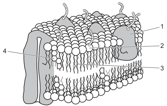
Which components act as antigens in cell surface membranes?
Options
A 1 and 2
B 1 and 3
C 2 and 3
D 2 and 4
Working
Antigens on a cell surface membrane are the short carbohydrate chains that project from the outer surface. These carbohydrate chains are attached either to a protein (forming a glycoprotein) or to a phospholipid head (forming a glycolipid). In the diagram, label 1 is a glycoprotein and label 2 is a glycolipid, so both act as antigens. The phospholipid (3) and the intrinsic protein (4) on their own do not carry these carbohydrate markers and are not antigens.
Answer
A
A
Background Concept
The cell surface membrane is described by the fluid mosaic model: a phospholipid bilayer in which proteins, cholesterol, and short carbohydrate chains are embedded or attached. Two of these components carry short, branched carbohydrate chains that project into the extracellular space:
- A glycoprotein = a membrane protein with a covalently attached oligosaccharide chain.
- A glycolipid = a phospholipid whose polar head has a covalently attached oligosaccharide chain.
These carbohydrate chains vary between individuals and between cell types. Because they are unique molecular "fingerprints" on the outer face of the membrane, they are recognised by the immune system as antigens — molecules capable of triggering an immune response (e.g. binding by antibodies or being presented to T-lymphocytes). They are essential for cell–cell recognition, including ABO blood-group antigens and the recognition of "self" versus "non-self".
Understanding the Question
The question shows a labelled fluid-mosaic diagram and asks which of the four labelled components act as antigens. We must (1) identify what each label represents and (2) decide which of those components expose carbohydrate groups to the outside of the cell.
The figure labels are:
- 1 — glycoprotein (a protein with a carbohydrate chain sticking out of the outer surface)
- 2 — glycolipid (a phospholipid with a carbohydrate chain sticking out of the outer surface)
- 3 — phospholipid (a plain phospholipid in the bilayer; no carbohydrate attached)
- 4 — intrinsic (integral) protein (a transmembrane protein; no carbohydrate attached in this diagram)
The command word is "act as antigens", so we are selecting the components whose structures actually present antigenic determinants.
Approach
The defining feature of an antigen on the cell surface membrane is the exposed carbohydrate chain. We therefore need to pick out the labels whose structures include such a chain: glycoprotein (1) and glycolipid (2). Phospholipids (3) and intrinsic proteins without attached carbohydrate (4) do not act as antigens on their own.
Step-by-Step Reasoning
- Recall the definition of a membrane antigen. Antigens on the cell surface membrane are the carbohydrate portions of glycoproteins and glycolipids; these short sugar chains are the actual molecular labels recognised by the immune system.
- Match each label to its structure. Label 1 is shown as a protein with a branching chain on the outer surface — a glycoprotein. Label 2 is shown as a phospholipid with a branching chain on the outer surface — a glycolipid. Label 3 is a plain phospholipid in the bilayer. Label 4 is a transmembrane protein without an attached chain.
- Decide which are antigens. Only 1 and 2 carry the carbohydrate chains that function as antigens. Labels 3 and 4 do not, so they are excluded.
- Select the option. "1 and 2" corresponds to option A.
Key Takeaways
- Antigens on the cell surface membrane are the carbohydrate chains of glycoproteins and glycolipids.
- These carbohydrate markers are responsible for cell–cell recognition and the immune system's ability to distinguish self from non-self (e.g. ABO blood groups, MHC molecules, transplant rejection).
- A plain phospholipid or a protein without an attached oligosaccharide is not an antigen on its own.
Common Mistakes
- Confusing the protein itself with the antigen. The whole protein is not the antigen; it is specifically the carbohydrate chain attached to the protein (or lipid) that acts as the antigen.
- Choosing a phospholipid (3) or a plain intrinsic protein (4). Neither carries the carbohydrate chain that makes a membrane component antigenic.
- Picking only one of the two carbohydrate-bearing components. Both glycoproteins and glycolipids contribute antigens, so the answer must include both 1 and 2.
Things to Be Careful About
- In a fluid-mosaic diagram the carbohydrate chains are typically drawn as small branching "beaded" lines on the outer surface only — their asymmetry is the clue that they project outwards and can be recognised by antibodies.
- An intrinsic protein (4) can also be a glycoprotein if a carbohydrate chain is attached; in this diagram no chain is shown on label 4, so it is treated as a plain protein here. Always look at what is actually drawn, not what could be drawn.
- "Antigen" in this context means cell-surface antigen, not any foreign molecule; the term here refers specifically to the membrane-borne carbohydrate markers.
Descriptions of three biological molecules found in cell surface membranes are given.
● X is a molecule comprised of carbon, hydrogen and oxygen, with a non-polar region and one region that extends out of the membrane capable of forming hydrogen bonds.
● Y is a molecule with a hydrophilic end that forms hydrogen bonds with the water outside the membrane, and a hydrophilic region that is located within the cell membrane.
● Z is a molecule that can position itself within the membrane due to a hydrophilic region at one end and two hydrophobic extensions that are positioned within the membrane.
Which row correctly identifies a carrier protein and a glycolipid?
Options
| carrier protein | glycolipid | |
|---|---|---|
| A | X | Z |
| B | X | Y |
| C | Z | Y |
| D | Y | X |
Working
- X contains only C, H and O, with a hydrophobic region within the membrane and a hydrophilic (hydrogen-bonding) region extending out of the membrane → glycolipid (carbohydrate chain extends out, lipid portion is embedded).
- Y has a hydrophilic end outside the membrane (hydrogen-bonding with water) and a hydrophilic region located within the membrane (lining a central aqueous channel) → carrier protein spanning the bilayer.
- Z has one hydrophilic region at one end and two hydrophobic extensions → phospholipid (phosphate head + two fatty acid tails).
Carrier protein = Y; glycolipid = X.
Answer
D
D
Background Concept
The cell surface membrane is described by the fluid mosaic model as a phospholipid bilayer in which various proteins are embedded or attached. The key molecular components are:
- Phospholipids form the bilayer. Each has a hydrophilic phosphate head (water-loving) and two hydrophobic fatty acid tails (water-fearing). They contain C, H, O, P (and sometimes N).
- Cholesterol molecules fit between the phospholipids; they have a small polar –OH group and a largely non-polar steroid ring system, and contain only C, H, O.
- Glycolipids and glycoproteins are lipids/proteins with short carbohydrate (sugar) chains attached. The carbohydrate portion is hydrophilic and extends from the outer surface of the membrane, where it is involved in cell recognition and signalling. Glycolipids contain C, H, O (the lipid part) and C, H, O (the sugar part, sometimes with N).
- Proteins in the membrane include channels, carriers, receptors and enzymes. A carrier protein binds a specific molecule, changes shape, and releases it on the other side. It spans the bilayer, so it has:
- hydrophilic R-groups on the parts exposed to water on either side,
- a hydrophilic central channel (lined with polar R-groups) that the transported substance passes through,
- hydrophobic R-groups on the outer surface where the protein contacts the fatty acid tails.
Understanding the Question
The question gives three descriptions (X, Y, Z) of molecules found in cell surface membranes. We must match each description to the correct type of membrane molecule, then identify which row in the table correctly labels a carrier protein and a glycolipid.
The command word is essentially "identify" — the descriptions must be decoded using knowledge of membrane structure and chemistry.
Approach
- Read each description carefully and extract the structural clues:
- Which chemical elements are present?
- Where are the hydrophilic/hydrophobic regions located?
- What is the overall shape or arrangement?
- Match each set of clues to a known membrane component.
- Read the options to find the row that puts a carrier protein and a glycolipid in the right cells.
Step-by-Step Reasoning
Molecule X — "comprised of carbon, hydrogen and oxygen, with a non-polar region and one region that extends out of the membrane capable of forming hydrogen bonds."
- The element list (C, H, O only) rules out phospholipids (they contain P) and proteins (they contain N and S).
- The non-polar region sits in the membrane; the polar region extends outwards and forms hydrogen bonds.
- This is the classic description of a glycolipid: the carbohydrate (sugar) chain is hydrophilic, extends from the outer surface, and forms hydrogen bonds with water; the lipid portion is hydrophobic and lies within the bilayer.
Molecule Y — "a hydrophilic end that forms hydrogen bonds with the water outside the membrane, and a hydrophilic region that is located within the cell membrane."
- The hydrophilic end on the outside is consistent with polar/charged amino acid R-groups of a protein exposed to the aqueous environment.
- A hydrophilic region within the membrane is the lining of an aqueous channel through a transport protein — polar R-groups that interact with the substance being carried across the membrane.
- The molecule therefore spans the membrane and transports polar substances; this is a carrier protein.
Molecule Z — "can position itself within the membrane due to a hydrophilic region at one end and two hydrophobic extensions that are positioned within the membrane."
- One hydrophilic head + two hydrophobic tails is the textbook structure of a phospholipid.
So:
- X = glycolipid
- Y = carrier protein
- Z = phospholipid
The row identifying Y as the carrier protein and X as the glycolipid is row D.
Key Takeaways
- Phospholipid = one hydrophilic head + two hydrophobic tails (C, H, O, P).
- Glycolipid = hydrophobic lipid in the bilayer + hydrophilic carbohydrate chain extending out (C, H, O).
- Carrier protein = spans the bilayer; hydrophilic regions on both surfaces and lining an internal channel; hydrophobic regions where the protein contacts the lipid tails (C, H, O, N, S).
- Chemical elements (especially the absence/presence of N and P) are powerful clues for distinguishing membrane molecules.
- The position of hydrophilic/hydrophobic regions (outside the membrane, within the membrane, spanning the membrane) reveals the type of molecule.
Common Mistakes
- Confusing glycolipid with phospholipid: both have a hydrophilic part, but phospholipids have two hydrophobic fatty acid tails and contain phosphorus, while glycolipids have a carbohydrate extending outwards and contain only C, H, O.
- Confusing carrier proteins with channel proteins: both are transport proteins with hydrophilic channels, but carriers change shape during transport while channels form a continuous open pore.
- Overlooking the "hydrophilic region within the membrane" clue in Y — this points to the aqueous channel of a transport protein, not the membrane interior itself.
- Misreading the element list: assuming X (C, H, O only) could be a phospholipid, but phospholipids also contain phosphorus.
Things to Be Careful About
- Always read the full description, including the location (outside / within / spanning) of each region.
- Pay close attention to the elements mentioned — C, H, O only is a deliberate clue.
- "Two hydrophobic extensions" is diagnostic of a phospholipid (two fatty acid tails).
- "Extends out of the membrane" + "hydrogen bonds" strongly suggests a carbohydrate (glycolipid/glycoprotein).
- "Hydrophilic region within the membrane" must mean the inside of a protein channel, not a freely water-filled space.
Antimycin is a chemical that inhibits the function of mitochondria.
Which methods of transport across the cell surface membrane could be directly affected by antimycin?
1 active transport
2 facilitated diffusion
3 endocytosis
Options
A 1, 2 and 3
B 1 and 2 only
C 1 and 3 only
D 2 and 3 only
Working
Antimycin inhibits mitochondrial function, so ATP synthesis via aerobic respiration stops.
- 1 (active transport): requires ATP, so it is directly affected. ✓
- 2 (facilitated diffusion): does not require ATP (passive, down the concentration gradient via channel/carrier proteins), so it is NOT affected. ✗
- 3 (endocytosis): requires ATP for vesicle formation and movement, so it is directly affected. ✓
Answer
C
C
Background Concept
The cell surface membrane is crossed by substances in several different ways:
- Diffusion and facilitated diffusion are passive — substances move down their concentration (or electrochemical) gradient. Facilitated diffusion uses channel or carrier proteins but consumes no metabolic energy.
- Active transport uses carrier proteins to move substances against their concentration gradient. It requires energy, supplied by ATP (or sometimes by an ion gradient that was itself set up using ATP).
- Endocytosis and exocytosis move large quantities of material in or out of the cell inside membrane-bound vesicles. Vesicle budding, movement through the cytoplasm, and fusion with the plasma membrane all require ATP.
The bulk of a cell's ATP is produced by aerobic respiration in the mitochondria, specifically by the electron transport chain on the inner mitochondrial membrane. Antimycin A blocks complex III of this electron transport chain, halting oxidative phosphorylation. Without oxidative phosphorylation, the cell's ATP supply collapses.
Understanding the Question
The question asks which of the listed transport methods could be directly affected by antimycin — i.e. which depend on ATP that would normally come from functional mitochondria. We have to decide, for each of the three named processes, whether it is ATP-dependent.
Approach
For each transport method, ask: "Does this process need ATP?" If yes, antimycin will affect it. If no, antimycin will not affect it directly (the method can still occur).
Step-by-Step Reasoning
-
Active transport (1): Carrier proteins (e.g. the Na⁺/K⁺ pump) hydrolyse ATP to change shape and move solutes against their gradient. Without mitochondrial ATP, active transport fails. → Directly affected.
-
Facilitated diffusion (2): Channel and carrier proteins allow substances to move down their concentration gradient. No ATP is hydrolysed. The process depends only on the existence of the gradient and a functional protein. → Not directly affected by a lack of ATP.
-
Endocytosis (3): Forming a vesicle from the plasma membrane, moving it into the cytoplasm, and processing it all require ATP (for cytoskeletal motor proteins, for membrane remodelling, for maintaining ion gradients that drive vesicle formation). → Directly affected.
Therefore, 1 and 3 are affected while 2 is not, giving option C (1 and 3 only).
Key Takeaways
- Mitochondria are the main source of cellular ATP.
- ATP-requiring membrane transport: active transport, endocytosis, exocytosis.
- ATP-independent membrane transport: simple diffusion, facilitated diffusion, osmosis.
- A useful test: if the process moves substances against a gradient or moves the membrane itself, it needs ATP.
Common Mistakes
- Assuming all membrane transport needs ATP — facilitated diffusion is the classic exception.
- Confusing endocytosis with passive transport because "the substance is moving down its gradient"; the energy cost is in moving the membrane and cytoskeleton, not in moving the dissolved substance.
- Forgetting that channel/carrier proteins in facilitated diffusion work without ATP; only active-transport carriers hydrolyse ATP.
Things to Be Careful About
- The question says "directly affected". Even facilitated diffusion could fail indirectly if the cell dies, but the mark is for the immediate, ATP-dependent step.
- "Inhibits the function of mitochondria" should be read as stopping ATP production by aerobic respiration, not as damaging the cell surface membrane itself.
- Note that anaerobic glycolysis can still make a small amount of ATP, so the effect is on ATP supply rather than its complete absence; this does not change which mechanisms are categorically ATP-dependent.
A student half filled a beaker with solution X. They placed a sealed Visking tubing bag containing solution Y into the beaker.
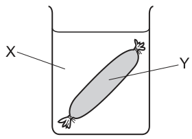
At 30 minutes, the solution in the beaker was orange and the solution inside the Visking tubing was blue-black.
What did solutions X and Y contain at the start to give these results?
Options
| solution X | solution Y | |
|---|---|---|
| A | starch | amylase and iodine |
| B | iodine | amylase |
| C | starch and amylase | iodine |
| D | iodine | starch |
Working
Visking tubing is partially permeable: small molecules (iodine, water, glucose) can pass through its pores, but large molecules (starch) cannot.
At 30 minutes:
- The solution in the beaker was still orange → iodine is in the beaker (no starch is present outside to turn it blue-black).
- The solution inside the Visking tubing was blue-black → starch is inside the tubing and iodine has diffused in from the beaker to react with it.
So solution X (in the beaker) = iodine and solution Y (in the Visking tubing) = starch.
Answer
D
D
Background Concept
Iodine solution (iodine dissolved in potassium iodide solution, I₂/KI) is orange-brown. When it meets starch, the iodine molecules slip inside the amylose helix of starch and form a starch–iodine complex, which appears blue-black. This is the standard chemical test for starch and is taught early in the Biological Molecules topic.
Visking tubing is an artificial partially permeable membrane. Its pores are large enough to let through small molecules and ions (water, glucose, iodine, mineral ions) but too small to let through large macromolecules (starch, proteins). Because of this, it is used as a simple model of a cell membrane in practical work, including experiments to test whether a substance is being digested into smaller products (e.g. amylase digesting starch into maltose, which can then escape through the tubing and be tested with Benedict's reagent outside).
The relative sizes of the two key molecules here matter:
- Iodine (I₂, often with I⁻) — small, passes through Visking tubing.
- Starch (a long polymer of α-glucose) — very large, does NOT pass through Visking tubing.
Understanding the Question
A student sets up a beaker containing solution X, with a sealed Visking tubing bag of solution Y suspended in it. After 30 minutes:
- Beaker solution = orange (so the beaker contains iodine but no starch at the end — if starch had been outside and iodine had got out, the beaker would be blue-black).
- Visking tubing solution = blue-black (so the tubing contains starch, and iodine must have entered from outside to react with it).
We are asked which pair of starting solutions gives this result.
Approach
Use two pieces of information together:
- The colour test: blue-black = starch present; orange = iodine present with no starch.
- The size selectivity of Visking tubing: starch is trapped; iodine can move freely.
Reason from the final colour backwards to the starting position of each substance.
Step-by-Step Reasoning
- If starch were in the beaker (option A and C), then any iodine that reached the beaker would react with it, turning the beaker blue-black. The beaker is orange, so starch cannot be in the beaker. This rules out A and C.
- If starch were in the Visking tubing and iodine in the beaker (option D), iodine would diffuse down its concentration gradient from beaker → tubing. Inside the tubing, iodine meets starch and forms the blue-black complex. The beaker, having only iodine and no starch, stays orange. This matches the observations exactly.
- If iodine were in the beaker and amylase in the tubing (option B), there is no starch in either compartment to give a blue-black colour. The iodine diffusing into the tubing would have nothing to react with, so neither compartment would turn blue-black. This rules out B.
The key insight is that the small iodine molecule can pass through the Visking tubing pores in both directions, so wherever starch is, the iodine will eventually find it. The starch, being far too large, can never leave its original compartment.
Key Takeaways
- Iodine = orange-brown; iodine + starch = blue-black.
- Visking tubing is partially permeable — small molecules pass, large molecules do not.
- Use both the colour change and the size of molecules to deduce the starting arrangement in any "Visking tubing + beaker" question.
Common Mistakes
- Putting starch in the beaker and iodine in the tubing. Students forget that iodine is small enough to pass outwards, so it would meet starch in the beaker and turn the beaker blue-black — opposite to the observed result.
- Confusing the roles of amylase. Amylase digests starch into maltose, but it does not itself give a colour with iodine; if starch is absent, no blue-black colour appears.
- Assuming a colour change in one direction is the only possible one — remember, the test can be done either way; it is the asymmetry of the membrane (small iodine moves, large starch does not) that fixes the result.
Things to Be Careful About
- "Blue-black" in the tubing, not the beaker, is the key clue: the starch must be inside the tubing.
- Amylase is irrelevant to the answer here because the question does not ask about digestion; it only tests the position of starch and iodine.
- Always check the size-selectivity of Visking tubing when interpreting these experiments — it is the same principle that explains dialysis, kidney filtration, and why starch is digested before absorption.
A cell is in mitosis. Each of the chromosomes in the cell consists of two chromatids. The chromosomes are not lined up at the equator.
Which stage of mitosis is described?
Options
A prophase
B metaphase
C anaphase
D telophase
Working
- Each chromosome has two chromatids → the centromeres have not yet split, so anaphase (and the later part of mitosis) is excluded.
- Chromosomes are NOT lined up at the equator → metaphase is excluded.
- In prophase, chromosomes condense and become visible as pairs of sister chromatids scattered within the cell, not yet attached to the equator.
Answer
A
A
Background Concept
Mitosis is divided into four named stages, each defined by a specific arrangement of the chromosomes:
- Prophase – Chromosomes condense (shorten and thicken) and become visible as discrete units. Each chromosome is made of two sister chromatids joined at a centromere. The nuclear envelope breaks down and a spindle forms. Chromosomes are not aligned at the equator.
- Metaphase – Chromosomes (still as two-chromatid units) line up along the cell's equator, attached to spindle fibres at their centromeres.
- Anaphase – The centromeres split and the sister chromatids are pulled apart to opposite poles. Each moving structure is now a single chromatid (called a chromosome).
- Telophase – Chromatids arrive at the poles, decondense, and new nuclear envelopes form; cytokinesis usually follows.
The two features in the question — "two chromatids per chromosome" and "not lined up at the equator" — together uniquely identify prophase.
Understanding the Question
The stem gives two pieces of information about a cell in mitosis:
- Each chromosome still consists of two chromatids.
- The chromosomes are not lined up at the equator.
The task is to pick which of the four mitotic stages fits both statements. The command word is implicit ("which stage…?") — the candidate only has to identify the stage.
Approach
Rule out each option systematically using the two given statements:
- Anaphase / Telophase (C, D): eliminated because the centromeres have split — chromosomes would be single chromatids, not pairs.
- Metaphase (B): eliminated because the defining feature of metaphase is alignment at the equator — the stem says they are NOT at the equator.
- Prophase (A): consistent with both statements — chromosomes are condensed two-chromatid structures that are still scattered through the cell, not yet on the spindle equator.
Step-by-Step Reasoning
- "Each chromosome consists of two chromatids" means the centromere of every chromosome is intact. This excludes anaphase (where centromeres split) and telophase (where single chromatids are arriving at the poles).
- "Chromosomes are not lined up at the equator" directly excludes metaphase, whose diagnostic event is equatorial alignment.
- The remaining stage, prophase, fits perfectly: chromosomes have condensed into visible two-chromatid units but have not yet been captured and aligned by the spindle at the cell's equator.
- Therefore the correct option is A.
Key Takeaways
- A "two-chromatid chromosome" is present in prophase and metaphase only; once centromeres split, the structures are single chromatids.
- Equatorial alignment = metaphase; chromatids at opposite poles = anaphase/telophase; scattered condensed chromosomes = prophase.
- When a question gives two defining features, use each feature to eliminate options until only one remains.
Common Mistakes
- Choosing B (metaphase) because the chromosomes are two-chromatid structures — students forget that the stem explicitly says they are NOT at the equator.
- Choosing C (anaphase) by confusing "two chromatids" (still joined) with "two chromatids moving apart" (already separated).
- Choosing D (telophase) by thinking "two chromatids" simply means "two groups of chromosomes" (one at each pole) — but each moving unit in anaphase/telophase is a single chromatid, not a pair.
Things to Be Careful About
- "Two chromatids per chromosome" describes the state of each chromosome as a duplicated unit; this is true only up to the start of anaphase.
- The exact wording "lined up at the equator" is the diagnostic feature of metaphase — never credit prophase if a description implies equatorial alignment.
- For Cambridge 9700, treat prophase and metaphase as the two stages in which a chromosome still has two chromatids; the position (scattered vs aligned) distinguishes them.
The diagrams show two stages of the cell cycle in different organisms.
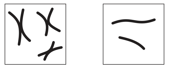
The number of specific structures in each diagram is counted.
Which row shows the correct combined total for the different structures?
Options
| chromosomes | centromeres | telomeres | |
|---|---|---|---|
| A | 5 | 3 | 8 |
| B | 5 | 5 | 16 |
| C | 8 | 3 | 16 |
| D | 8 | 5 | 8 |
Working
- Left diagram: 3 replicated chromosomes, each with 2 sister chromatids (6 chromatids total). 1 centromere per chromosome → 3 centromeres. 2 telomeres per chromatid → telomeres.
- Right diagram: 2 unreplicated chromosomes, each with 1 chromatid (2 chromatids total). 1 centromere per chromosome → 2 centromeres. 2 telomeres per chromatid → telomeres.
- Combined totals: chromosomes = ; centromeres = ; telomeres = .
Answer
B
B
Background Concept
A chromosome is a single, continuous molecule of DNA (with associated proteins) that carries genetic information. The number of chromosomes in a cell is defined by the number of centromeres present — each chromosome has exactly one centromere, regardless of how many chromatids it contains. This is why a cell in G₁ of the cell cycle (before DNA replication) and a cell in metaphase (after replication) have the same chromosome number, even though the latter has twice as much DNA.
A centromere is the constricted region of a chromosome where the two sister chromatids are joined (after replication) and where the spindle fibres attach during mitosis. A replicated chromosome has two chromatids joined at a single centromere.
Telomeres are specialised repetitive DNA sequences found at each end of every chromatid. Because each chromatid has two ends, every chromatid carries exactly two telomeres. Therefore:
- An unreplicated chromosome (1 chromatid) has 2 telomeres.
- A replicated chromosome (2 chromatids) has 4 telomeres.
Understanding the Question
We are shown two diagrams of chromosomes:
- Left diagram: three chromosomes, each with the classic X-shape — two sister chromatids joined at a centromere (a replicated chromosome, as seen in prophase or metaphase of mitosis).
- Right diagram: two chromosomes, each a single curved rod with no sister chromatid visible (an unreplicated chromosome, as seen in G₁ or telophase/G₁ of the next cycle).
The question asks for the combined total of three different structures across both diagrams: chromosomes, centromeres and telomeres. The command word is essentially "count correctly" — but the count depends entirely on applying the right definition of each structure.
Approach
Work through each structure in turn, using the rules:
- Count chromosomes = count centromeres.
- Count chromatids first, then multiply by 2 to get telomeres.
Step-by-Step Reasoning
Chromosomes:
- Left: 3 (the three X-shapes, each = 1 chromosome).
- Right: 2 (the two single rods, each = 1 chromosome).
- Total = .
Centromeres:
- Left: 3 (one per replicated chromosome, where the two chromatids meet).
- Right: 2 (one per unreplicated chromosome).
- Total = .
Telomeres:
- Left: each of the 3 chromosomes has 2 chromatids × 2 telomeres per chromatid = 4 telomeres per chromosome → telomeres.
- Right: each of the 2 chromosomes has 1 chromatid × 2 telomeres per chromatid = 2 telomeres per chromosome → telomeres.
- Total = .
So the combined totals are 5 chromosomes, 5 centromeres, 16 telomeres, which matches row B.
Key Takeaways
- The number of chromosomes = the number of centromeres — always, no matter the cell-cycle stage.
- A replicated chromosome (X-shape with 2 chromatids) is still one chromosome with one centromere and four telomeres.
- An unreplicated chromosome (rod with 1 chromatid) is one chromosome, one centromere, two telomeres.
- Telomeres = 2 × (number of chromatids) — never 2 × (number of chromosomes) unless every chromosome is unreplicated.
Common Mistakes
- Counting chromatids as chromosomes. Many students see 6 X-shaped arms on the left and call it 6 chromosomes — but those are chromatids, and the three X-shapes share centromeres, so they are 3 chromosomes.
- Thinking 2 telomeres per chromosome. This gives the right answer only for unreplicated chromosomes. It fails on the left diagram, which is why option D (5, 5, 8) is wrong.
- Forgetting that the right-hand diagram has any telomeres. Each rod has two free ends, so each unreplicated chromosome still has 2 telomeres.
- Assuming each X-shape has only 1 telomere per arm. Each tip of each chromatid is a telomere — that is 4 tips per X-shape, not 2.
Things to Be Careful About
- Distinguish between the number of chromosomes (defined by centromeres) and the amount of DNA (which doubles after S-phase and halves at cytokinesis).
- Apply the same rule to both diagrams — the centromere rule and the 2-telomeres-per-chromatid rule hold regardless of cell-cycle stage.
- Read the options carefully; the wrong answers (A, C, D) are designed to catch each common mistake: A confuses centromere with chromatid count, C doubles the chromosome count, D counts telomeres as 2 per chromosome instead of 2 per chromatid.
Which statements about tRNA are correct?
1 Hydrogen bonds between bases temporarily bond tRNA to mRNA.
2 The base sequences in the tRNA molecules are the same as the base sequences in the mRNA that is being translated.
3 Some codons in mRNA do not have corresponding tRNA anticodons.
Options
A 1, 2 and 3
B 1 and 2 only
C 1 and 3 only
D 2 and 3 only
Working
- Statement 1: tRNA anticodons pair with mRNA codons via hydrogen bonds between complementary bases. These bonds are temporary, breaking once the amino acid has been delivered. ✓ correct
- Statement 2: tRNA anticodons are complementary to mRNA codons, not identical to them (e.g. codon AUG pairs with anticodon UAC, not AUG). ✗ incorrect
- Statement 3: Stop codons (UAA, UAG, UGA) have no corresponding tRNA; instead, release factors bind them. ✓ correct
Statements 1 and 3 are correct.
Answer
C
C
Background Concept
Transfer RNA (tRNA) is a small RNA molecule (~75–95 nucleotides) folded into a cloverleaf secondary structure (and an L-shaped tertiary form). Each tRNA carries a specific amino acid at its 3′ end and contains a three-base anticodon loop. During translation, the anticodon base-pairs with a complementary three-base codon on the mRNA being read by the ribosome.
Key points to remember:
- Codon–anticodon pairing is complementary and antiparallel (e.g. mRNA codon 5′-AUG-3′ pairs with tRNA anticodon 3′-UAC-5′). Bases are joined by hydrogen bonds.
- The hydrogen bonds are temporary — they must break after the ribosome has checked the match, so the tRNA can move out of the A site and the ribosome can translocate along the mRNA.
- The genetic code is degenerate: more than one codon can code for the same amino acid, so multiple tRNAs (with different anticodons) carry the same amino acid. However, stop codons (UAA, UAG, UGA) do not code for any amino acid and have no corresponding tRNA — they are recognised instead by protein release factors.
Understanding the Question
This is a multiple-choice question asking you to decide which of three statements about tRNA are correct, then pick the option that lists exactly those statements. The command word "correct" means each statement must be evaluated independently against your knowledge of tRNA structure and translation.
Approach
Examine each statement one at a time, asking: "Is this biologically accurate?" A statement is correct only if every part of it is true — partial truths do not score.
Step-by-Step Reasoning
Statement 1: "Hydrogen bonds between bases temporarily bond tRNA to mRNA."
This is true. The anticodon of tRNA base-pairs with the codon of mRNA, and the two strands are held together by hydrogen bonds (A–U has 2; G–C has 3). The pairing is temporary because the ribosome must move on to the next codon, and the tRNA must then leave the ribosome. ✓
Statement 2: "The base sequences in the tRNA molecules are the same as the base sequences in the mRNA that is being translated."
This is false. The tRNA anticodon is complementary to the mRNA codon, not identical. For example, if the mRNA codon is 5′-AUG-3′ (which codes for methionine), the tRNA anticodon reads 3′-UAC-5′. The two sequences are not the same — they are mirror images following the A–U, G–C pairing rules. ✗
Statement 3: "Some codons in mRNA do not have corresponding tRNA anticodons."
This is true. The three stop codons (UAA, UAG, UGA) signal the end of translation and are not recognised by any tRNA. Instead, release factors (proteins) bind to the A site of the ribosome when a stop codon is reached, triggering the release of the completed polypeptide chain. ✓
Statements 1 and 3 are correct; statement 2 is incorrect. That matches option C.
Key Takeaways
- tRNA anticodons base-pair with mRNA codons using hydrogen bonds — the same type of bond that holds the two DNA strands together.
- The codon–anticodon relationship is complementary, not identical.
- The genetic code contains stop codons (UAA, UAG, UGA) that have no tRNA — they are recognised by protein release factors, not tRNA molecules.
- Codon–anticodon pairing is temporary by necessity, otherwise the ribosome could not translocate.
Common Mistakes
- Thinking the tRNA sequence "matches" the mRNA sequence (conflating "pairing" with "identical"). The tRNA carries the complementary anticodon, not a copy of the codon.
- Believing every codon has a tRNA. Stop codons are the obvious counter-examples — they terminate translation, so they must not be read by a tRNA carrying an amino acid.
- Confusing the anticodon (on tRNA) with the codon (on mRNA). They are on different molecules and have different (complementary) sequences.
Things to Be Careful About
- The hydrogen bonds are between complementary bases (A with U, G with C) — they are not covalent bonds. This is why the pairing is reversible and the tRNA can leave the ribosome after the amino acid has been transferred.
- When asked which statements are correct, treat each statement as a whole: if any part of a statement is wrong, the whole statement is wrong.
- Do not confuse stop codons with the start codon AUG — AUG is read by a tRNA carrying methionine (initiator tRNA), whereas UAA/UAG/UGA are not.
Why is the genetic code described as universal?
Options
A Each codon is made up of a sequence of three bases.
B More than one codon can represent one amino acid.
C Mutations can change the genetic code in all organisms.
D The genetic code is the same in all organisms.
Working
The genetic code is described as 'universal' because the same triplets of bases (codons) code for the same amino acids in virtually all organisms. For example, the codon AUG codes for methionine in bacteria, plants, fungi and animals alike.
- A — describes the triplet (non-overlapping) nature of the code, not universality.
- B — describes the degenerate (redundant) nature of the code, not universality.
- C — incorrect: mutations alter individual genes/sequences but do not change the code itself.
- D — correct: the genetic code is the same in all organisms, which is the meaning of universal.
Answer
D
D
Background Concept
The genetic code is the set of rules by which the sequence of bases in DNA (and the mRNA transcribed from it) is translated into the sequence of amino acids in a protein. Each amino acid is specified by a triplet of bases called a codon (e.g. AUG specifies methionine). The code has several notable properties:
- Triplet / non-overlapping — each codon is three bases long, read in a fixed frame.
- Degenerate (redundant) — more than one codon can specify the same amino acid (e.g. GCU, GCC, GCA and GCG all code for alanine).
- Contains start and stop codons — AUG (methionine) starts translation; UAA, UAG and UGA stop it.
- Universal — the same codons specify the same amino acids in essentially all organisms, from bacteria to humans.
It is this last property — universality — that is being asked about here.
Understanding the Question
The command word is essentially 'identify' (an MCQ asking the candidate to pick the option that best explains a key term). The stem defines the property to be explained: the genetic code is described as universal. The candidate must select the option that correctly defines what 'universal' means in this biological context.
Approach
The student needs to know that 'universal' in the context of the genetic code refers to the code being the same in all organisms — i.e. the same codon → amino acid mapping applies across the tree of life. The other options each describe a different property of the code, so they can be eliminated on that basis.
Step-by-Step Reasoning
- Option A ('Each codon is made up of a sequence of three bases') — this is true, but it describes the triplet nature of the code, not its universality. Eliminate.
- Option B ('More than one codon can represent one amino acid') — this is true, but it describes the degenerate / redundant nature of the code, not its universality. Eliminate.
- Option C ('Mutations can change the genetic code in all organisms') — this is biologically false. Mutations alter the base sequence of individual genes (and may therefore alter individual proteins), but they do not change the underlying codon → amino acid mapping (the 'code' itself) of an organism. Eliminate.
- Option D ('The genetic code is the same in all organisms') — this is precisely what 'universal' means: the codon-to-amino-acid assignments are shared by virtually every species on Earth. This is the defining evidence behind statements such as 'a human gene can be inserted into a bacterium and still be translated correctly'. Select D.
Key Takeaways
- The genetic code is described as universal because the same codons specify the same amino acids in (almost) all living organisms.
- Universality is one of several distinct properties of the code; the others (triplet, degenerate, non-overlapping, punctuated by start/stop codons) are separate features and must not be confused with it.
- Universality has a powerful practical consequence: a gene from one species can be cloned into another and still produce a functional protein — this is the basis of genetic engineering and recombinant DNA technology.
Common Mistakes
- Confusing universality with degeneracy (option B): both sound similar and both involve multiple codons, but degeneracy refers to several codons coding for the same amino acid, whereas universality refers to the code being the same across species.
- Choosing A because it is a true statement about the code: many true statements about the code are not answers to this question — only the statement that explains the word 'universal' earns the mark.
- Choosing C: students sometimes think that because mutations change DNA, they must change 'the code', but the code is the mapping from codons to amino acids, and that mapping itself is not altered by mutation.
Things to Be Careful About
- 'Universal' is a strong word — in reality a small number of minor variations exist (e.g. in mitochondrial genomes and in a few protists), so the code is almost universal rather than absolutely so. CIE accepts 'the same in all organisms' as a sufficient description at AS Level.
- The same term, universal, is used in different contexts in biology (e.g. 'the genetic code is universal' vs. 'a universal donor blood group') — always read the question stem carefully and interpret the word in the context of the topic being tested.
What correctly identifies the number and type of bonds between cytosine and guanine in a DNA molecule?
Options
A 2 hydrogen bonds
B 2 phosphodiester bonds
C 3 hydrogen bonds
D 3 phosphodiester bonds
Working
In a DNA double helix, the two purine–pyrimidine base pairs differ in the number of hydrogen bonds holding them together:
- Adenine–Thymine (A–T) is joined by 2 hydrogen bonds
- Cytosine–Guanine (C–G) is joined by 3 hydrogen bonds
Phosphodiester bonds join nucleotides along the same sugar–phosphate backbone, not between complementary bases across the helix.
Answer
C
C
Background Concept
DNA is a double helix made of two antiparallel polynucleotide strands. Each nucleotide has a deoxyribose sugar, a phosphate group, and one of four nitrogenous bases: adenine (A) and guanine (G), which are purines, and thymine (T) and cytosine (C), which are pyrimidines. The two strands are held together by hydrogen bonds between complementary base pairs. Because a purine always pairs with a pyrimidine, the helix has a uniform width.
Two distinct kinds of bond are involved in DNA structure, and they are easy to mix up:
- Hydrogen bonds — between complementary bases, holding the two strands together. They are individually weak, but many together give the molecule stability and allow separation during replication.
- Phosphodiester bonds — within a single strand, joining the 3′ carbon of one sugar to the 5′ phosphate of the next nucleotide. They form the strong covalent sugar–phosphate backbone.
The base-pairing pattern is specific: A pairs with T via 2 hydrogen bonds, and C pairs with G via 3 hydrogen bonds. This is often remembered as "A-T has 2, G-C has 3." The extra hydrogen bond in a C–G pair contributes to the higher melting temperature of GC-rich DNA, because more energy is needed to separate the strands.
Understanding the Question
This is a one-mark multiple-choice question asking you to identify both the number and the type of bond between cytosine and guanine. The four options test two ideas at once: the number (2 or 3) and the type of bond (hydrogen or phosphodiester). Only one combination is correct.
Approach
Recall the complementary base-pairing rules for DNA. Phosphodiester bonds do not form between bases; they only link adjacent nucleotides within one strand. Hydrogen bonds are the only bonds that connect bases across the two strands. Between C and G specifically, there are 3 hydrogen bonds.
Step-by-Step Reasoning
- Cytosine (a pyrimidine) pairs with guanine (a purine) across the DNA helix.
- Bonds that connect the two strands across the helix are hydrogen bonds, not phosphodiester bonds.
- The C–G pair is held together by 3 hydrogen bonds, while the A–T pair has only 2.
- Therefore the correct option is C: 3 hydrogen bonds.
The other options are wrong because:
- A (2 hydrogen bonds) is the A–T number, not the C–G number.
- B and D (phosphodiester bonds) misidentify the type of bond, regardless of number.
Key Takeaways
- A–T pairs: 2 hydrogen bonds. C–G pairs: 3 hydrogen bonds.
- Hydrogen bonds connect bases between strands; phosphodiester bonds link nucleotides within a strand.
- Higher GC content = more hydrogen bonds = higher DNA melting temperature.
Common Mistakes
- Confusing hydrogen and phosphodiester bonds: phosphodiester bonds form the sugar–phosphate backbone, never between complementary bases.
- Stating the A–T value (2 hydrogen bonds) for the C–G pair by mistake.
- Thinking the two strands are connected by phosphodiester bonds because the word "bond" appears in both strand and inter-strand contexts.
Things to Be Careful About
- Phosphodiester bonds are covalent and strong; they build the backbone, not the inter-strand links.
- Hydrogen bonds are non-covalent and individually weak; collectively they stabilise the double helix and are broken during replication.
- The pattern 2 (A–T) and 3 (C–G) is specific to DNA. In RNA, uracil replaces thymine but still pairs with adenine via 2 hydrogen bonds.
Three proteins that have a quaternary structure are listed.
● Type IX collagen is formed from three different polymers.
● The main form of haemoglobin contains two alpha globins and two beta globins.
● HIV protease consists of two identical polymers.
Which row shows the correct number of genes needed to code for each protein?
Options
| number of genes | |||
|---|---|---|---|
| type IX collagen | haemoglobin | HIV protease | |
| A | 1 | 2 | 2 |
| B | 1 | 4 | 1 |
| C | 3 | 2 | 1 |
| D | 3 | 4 | 2 |
Working
- Type IX collagen: three different polymers → each different polypeptide is coded by a separate gene → 3 genes.
- Haemoglobin (HbA): two alpha globins (identical) and two beta globins (identical). Only two types of polypeptide → 2 genes.
- HIV protease: two identical polymers → one gene is transcribed and translated to produce both copies → 1 gene.
| type IX collagen | haemoglobin | HIV protease | |
|---|---|---|---|
| number of genes | 3 | 2 | 1 |
Answer
C
C
Background Concept
A gene is a sequence of DNA that codes for a polypeptide. The central principle here is: one gene → one (type of) polypeptide chain. If a functional protein is made of several polypeptide chains (i.e. it has quaternary structure), the number of different chains determines the minimum number of genes required.
- Identical subunits (e.g. two copies of the same polypeptide) are products of the same gene, so only one gene is needed for that type of subunit.
- Non-identical subunits must be the products of different genes, because different amino acid sequences require different mRNAs and therefore different genes.
Understanding the Question
The question lists three proteins that all have quaternary structure (more than one polypeptide chain) and asks how many genes are needed to produce each. The key is to count the number of different polypeptide types, not the total number of chains.
The command word is implicit ("which row…"), so the answer is a single letter; the reasoning, however, requires analysing each protein in turn.
Approach
Apply the one-gene-one-polypeptide rule to each protein and count the types of polypeptide:
- Type IX collagen – three different polymers → three different genes.
- Haemoglobin (HbA, the main adult form) – two types of globin chain (α and β), each type present twice → two genes.
- HIV protease – a homodimer of two identical polypeptides → one gene.
Match these numbers to the row in the table that gives 3, 2, 1.
Step-by-Step Reasoning
-
Type IX collagen (3 genes): The question explicitly states it is formed from three different polymers. Different polymers have different amino acid sequences, so each must be encoded by a separate gene. Three different polymers therefore require three different genes.
-
Haemoglobin (2 genes): Adult haemoglobin (HbA) has the composition α₂β₂. There are two α chains and two β chains. The two α chains are identical to each other (same gene product, twice); the two β chains are identical to each other (a different gene product, twice). So only two distinct genes are required — one for α-globin (on chromosome 16) and one for β-globin (on chromosome 11).
-
HIV protease (1 gene): HIV protease is a homodimer — two copies of the same polypeptide. A single gene codes for that polypeptide, and the gene is expressed to produce two identical copies that assemble into the active enzyme. Only one gene is needed.
The row 3 – 2 – 1 corresponds to option C.
Key Takeaways
- Quaternary structure is built from two or more polypeptide chains, but the number of genes depends on the number of different chains, not the total chain count.
- Haemoglobin (α₂β₂) is a classic example of a quaternary-structure protein that needs only two genes despite having four chains.
- Dimers made of identical subunits (homodimers) need just one gene; heterodimers need two.
Common Mistakes
- Counting total chains instead of different chains. Saying haemoglobin needs 4 genes because it has 4 globin chains ignores the fact that the two α chains (and the two β chains) are identical.
- Assuming all quaternary-structure proteins need many genes. HIV protease has two subunits but only one gene.
- Misreading the table. Options A and D both list 3 for type IX collagen, but they differ in haemoglobin and HIV protease — picking the wrong pair is a common trap.
Things to Be Careful About
- "Different polymers" in the stem of the question is the cue that each is encoded by its own gene.
- Haemoglobin's two α and two β chains are the standard adult form (HbA) — the question is not asking about rarer variants such as HbA₂ (α₂δ₂) or foetal HbF (α₂γ₂), which would still need two genes but for a different β-family chain.
- The distractors in this question are constructed so that the only row consistent with the one-gene-one-polypeptide rule is C.
The photomicrograph shows a section of vascular tissue from the stem of a plant.
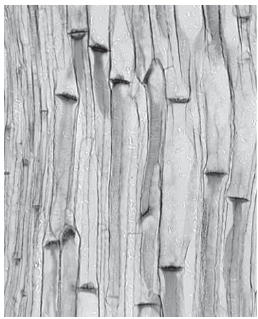
Which description of this photomicrograph is correct?
Options
A a transverse section showing xylem tissue
B a longitudinal section showing xylem tissue
C a transverse section showing phloem tissue
D a longitudinal section showing phloem tissue
Working
- The cells are long, narrow and tube-like, running vertically across the field of view. A transverse section would show circular or polygonal cell outlines, so this is a longitudinal section.
- The walls are relatively thin and there are perforated cross-walls (sieve plates) visible at intervals along some of the tubes, with smaller companion cells alongside. These are features of phloem (sieve tube elements), not xylem (which would show much thicker lignified walls with spiral, annular or reticulate thickening and no end-wall plates).
Answer
D
D
Background Concept
Vascular plants contain two conducting tissues arranged together in vascular bundles:
- Xylem transports water and dissolved mineral ions from roots to leaves. Its conducting cells (xylem vessel elements and tracheids) are dead at maturity, have thickened lignified walls, and are joined end-to-end into continuous tubes. The wall thickening is laid down in characteristic patterns — annular (rings), spiral, scalariform (ladder-like), reticulate (net-like) or pitted — which are clearly visible in a longitudinal section.
- Phloem transports assimilates (mainly sucrose) from sources to sinks. Its conducting cells (sieve tube elements) are living but lack a nucleus at maturity. They are joined end-to-end by sieve plates (perforated end walls) and are accompanied by companion cells, which carry out metabolic functions for the sieve tube element.
In a transverse section (TS) the conducting cells appear as roughly circular or polygonal profiles, with the wall thickening (in xylem) visible as a ring around the lumen. In a longitudinal section (LS) the cells appear as long tubes running along their length, which is when the spiral/scalariform thickenings of xylem, or the sieve plates and companion cells of phloem, become visible.
Understanding the Question
You are shown a photomicrograph of vascular tissue from a plant stem at ×80 magnification and asked to choose which description fits. The decision has two components:
- Section orientation — transverse (TS) or longitudinal (LS) — judged by cell shape.
- Tissue identity — xylem or phloem — judged by wall structure and any internal features.
The command word is "which description… is correct?" — a single best answer is required.
Approach
Read the image for the two pieces of evidence in turn:
- Cell shape tells you the plane of section. Long, narrow, parallel cells = LS; round/polygonal cells = TS.
- Wall features tell you the tissue. Thick lignified walls with obvious spiral/scalariform/reticulate patterning = xylem; thinner walls with transverse sieve plates and small companion cells alongside = phloem.
Both pieces of evidence must be combined to reach the correct option.
Step-by-Step Reasoning
- Orientation: Every cell in the field of view is elongated, with its long axis running vertically. There are no circular or polygonal profiles. This is unmistakably a longitudinal section. Options A and C (both transverse) are eliminated.
- Tissue identity: Look at the wall structure. The cell walls are not heavily thickened and there is no obvious spiral, annular, scalariform or reticulate lignification — the dominant feature of xylem in LS. Instead, several of the tubes show transverse sieve plates (perforated cross-walls) and there are smaller cells lying alongside the larger tubes, consistent with companion cells associated with sieve tube elements. These features identify the tissue as phloem.
- Combining the two conclusions: longitudinal section + phloem = option D.
A quick check — why is option B wrong? Option B also says longitudinal section, but claims the tissue is xylem. A longitudinal section of xylem would show conspicuous spiral, annular, scalariform or pitted wall thickening on long empty tubes; the micrograph shows neither thick lignified walls nor such patterning, only sieve plates.
Key Takeaways
- TS vs LS is decided by cell shape: round/polygonal profiles = TS, elongated tubes = LS.
- Xylem vs phloem in LS is decided by wall features:
- Xylem: thick lignified walls with spiral, annular, scalariform, reticulate or pitted thickening; no end walls (vessel elements are open at the ends).
- Phloem: thinner cellulose walls, transverse sieve plates joining sieve tube elements end-to-end, and small companion cells alongside.
- Magnification is irrelevant to the identification here — the structure of the cell walls is the diagnostic feature.
Common Mistakes
- Picking B (longitudinal xylem) because the cells look like long tubes, without noticing that the walls are thin and bear sieve plates rather than spiral/reticulate lignification.
- Picking A or C (transverse) on the mistaken belief that the dark stripes running across the image are cell outlines seen in cross-section. They are not — they are sieve plates seen in profile, and the tubes themselves are clearly running lengthwise.
- Confusing sieve plates with xylem wall thickenings. Sieve plates are transverse end walls crossing the whole lumen; xylem thickening is on the longitudinal side walls and does not cross the cell.
Things to Be Careful About
- At low magnification such as ×80, fine detail (e.g. individual pits in xylem walls) is hard to resolve, so rely on gross features — overall wall thickness, presence/absence of lignified patterning, presence of sieve plates and companion cells.
- Do not be misled by the dark horizontal bands in the image. They are inside elongated cells (sieve plates in LS), not circular cell boundaries (which would indicate a TS).
- The accompanying cells lying next to a sieve tube element are companion cells — their presence is a strong secondary clue that you are looking at phloem, not xylem.
The diagram shows a bond between two molecules transported in a xylem vessel.
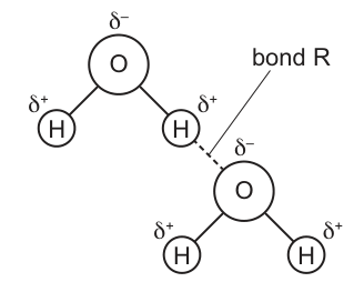
Which row correctly names bond R and states the role of the bond between these molecules for their transport in a xylem vessel?
Options
| name of bond R | role of the bond between these molecules for their transport in a xylem vessel | |
|---|---|---|
| A | hydrogen | adhesion of the molecules to the wall of the xylem vessel |
| B | hydrogen | cohesion of the molecules in the lumen of the xylem vessel |
| C | ionic | adhesion of the molecules to the wall of the xylem vessel |
| D | ionic | cohesion of the molecules in the lumen of the xylem vessel |
Working
The diagram shows two water molecules. Each H carries a partial positive charge () and the O carries a partial negative charge (). Bond R is a dashed line linking a hydrogen of one water molecule to the oxygen of the other — this is a hydrogen bond.
Water molecules linked to one another inside the xylem vessel stick together; this is cohesion, and it is what maintains an unbroken column of water in the lumen of the xylem under tension during the transpiration stream. Adhesion, by contrast, is the attraction of water to the xylem wall, not to another water molecule.
Answer
B
B
Background Concept
Water is a polar molecule. Because oxygen is much more electronegative than hydrogen, the shared electrons in each O–H bond spend more time near the oxygen, giving the oxygen a partial negative charge () and each hydrogen a partial positive charge (). A hydrogen bond forms when the hydrogen of one water molecule is electrostatically attracted to a lone pair on the oxygen of a neighbouring water molecule. Although individually weak, hydrogen bonds are collectively very strong and give water its unusual properties (high surface tension, high boiling point, cohesion, adhesion).
In the xylem, two consequences of hydrogen bonding matter for transport:
- Cohesion — water molecules hydrogen-bond to one another, so they are pulled along together as a continuous column in the lumen.
- Adhesion — water molecules hydrogen-bond to the hydroxyl groups of the cellulose in the xylem vessel wall, helping the water column resist gravity.
These two forces underpin the cohesion–tension theory of water movement: evaporation from the mesophyll cell walls (transpiration) generates a negative hydrostatic pressure (tension) that is transmitted down the continuous, hydrogen-bonded water column in the xylem, pulling water up from the roots.
Understanding the Question
The question shows a structural diagram of two water molecules, with partial charges marked and a dashed line labelled "bond R" running between a H of one molecule and the O of the other. You are asked to:
- Name bond R.
- State what the bonding between these molecules does for their transport in the xylem.
The command word is essentially identify and state; the difficulty is reading the partial-charge diagram correctly and then choosing between the two closely related consequences of hydrogen bonding in a xylem (cohesion vs adhesion, and lumen vs wall).
Approach
- Read the diagram: dashed line + – pattern + molecules labelled only with H and O → hydrogen bond between two water molecules.
- Decide what the bond is doing in this diagram: it links water molecule to water molecule, not water to the vessel wall → cohesion, and the location is the inside (lumen) of the xylem.
- Match these two conclusions to the four options.
Step-by-Step Reasoning
- The atoms in the diagram are only H and O, so the molecules are water.
- Bond R is dashed (the conventional way to represent a hydrogen bond) and joins a H of one molecule to a O of the other — the textbook definition of a hydrogen bond. This rules out C and D (ionic).
- The two molecules are joined to each other, so the bond holds water to water — this is cohesion, not adhesion (adhesion would join water to the vessel wall). This rules out A.
- The bonding shown is between water molecules in the column of water inside the vessel, i.e. in the lumen of the xylem.
- Therefore the correct row is: hydrogen bond; cohesion of the molecules in the lumen of the xylem vessel → option B.
Why the distractors fail:
- A (hydrogen; adhesion to wall): correctly names the bond but misidentifies the role — bond R is between two water molecules, not between water and the vessel wall.
- C (ionic; adhesion to wall): misnames the bond (no electron transfer / ions shown; the symbols indicate polarity, not full charges) and misidentifies the role.
- D (ionic; cohesion in lumen): misnames the bond; the role description is correct.
Key Takeaways
- Hydrogen bonds form between the H of one water molecule and a lone pair on the O of another — shown as a dashed line in diagrams.
- Cohesion = water-to-water attraction; adhesion = water-to-wall (or any non-water surface) attraction.
- In the xylem, cohesion maintains a continuous water column in the lumen; adhesion to the wall helps counteract gravity. Together they are the molecular basis of the cohesion–tension mechanism of water transport.
Common Mistakes
- Reading the symbols as full charges and naming the bond "ionic". CIE expects students to recognise that means partial and that the interaction is a hydrogen bond, not a true ionic bond.
- Confusing cohesion (water–water) with adhesion (water–vessel wall). The question specifically asks about the bond between these molecules, so cohesion is correct.
- Saying the hydrogen bond "helps water dissolve minerals" or "provides energy for transport". Hydrogen bonds do not provide energy; they hold the column together so that tension generated by transpiration can pull it up.
Things to Be Careful About
- Distinguish lumen (the hollow interior of the vessel, where cohesion matters) from wall (the cellulose/ lignin boundary, where adhesion matters).
- Note that the cohesion–tension theory depends on water being under tension (negative pressure); if the column breaks (cavitation/embolism), transport in that vessel stops. This is why xylem vessels are narrow and reinforced with lignin.
- "Hydrogen bond" is the CIE-accepted term. Markers will not credit "van der Waals" or "polar bond" here.
How can water and solutes move through a leaf?
Options
A through the lignified endodermis using the apoplast pathway
B through the cytoplasm of cells using the symplast pathway
C through the cell walls containing cellulose by osmosis
D through the cell walls containing suberin by diffusion
Working
Water and solutes can cross a leaf via either the apoplast (cell walls and intercellular spaces) or the symplast (cytoplasm connected by plasmodesmata). The symplast route is the one in which water and solutes pass through the cytoplasm of cells, linked from cell to cell by plasmodesmata. The apoplast is the network of cell walls, but movement here is not "by osmosis" (osmosis is the movement of water across a selectively permeable membrane) and suberin is found in the Casparian strip of the root endodermis, not in leaf cell walls. The endodermis is a root structure and is not lignified; the Casparian strip is suberised. Therefore, the only correct statement is B.
Answer
B
B
Background Concept
In a plant, water and dissolved solutes can move from cell to cell by two parallel routes:
- Apoplast pathway — through the continuous network of cell walls and intercellular spaces, without crossing any plasma membrane. Movement is driven by mass flow and diffusion down water-potential gradients. In the root, this pathway is blocked at the endodermis by the Casparian strip, a band of suberin in the radial and transverse walls of endodermal cells, which forces water and solutes to cross a plasma membrane and enter the symplast before they can reach the xylem.
- Symplast pathway — through the cytoplasm of living cells, connected from one cell to the next by plasmodesmata (cytoplasmic channels through cell walls). Both water and solutes can travel by this route, and once a solute has crossed a plasma membrane into one cell, it can continue through the symplast without crossing further membranes.
The endodermis is the innermost layer of the root cortex; leaves do not have an endodermis. Suberin is a waxy, waterproof substance found in the Casparian strip, in cork (suberised cells of the periderm), and in some seed coats — not as a general component of leaf cell walls. Lignin is found in xylem vessel walls and in some sclerenchyma; it is not characteristic of the endodermis. Osmosis is defined as the net movement of water molecules across a selectively permeable membrane down a water-potential gradient; it cannot be described as movement through cell walls.
Understanding the Question
The question asks how water and solutes can move through a leaf. The keyword is "and" — we need a pathway that can carry both. The four options each combine a structure, a substance in that structure, and a mechanism; we must check all three components.
Approach
For each option, decide:
- Is the named structure/tissue present in a leaf?
- Is the named substance (lignin, cellulose, suberin) in the right place?
- Is the named mechanism (osmosis, diffusion, symplast/apoplast) correctly matched to that structure?
Step-by-Step Reasoning
Option A — "through the lignified endodermis using the apoplast pathway"
- Leaves do not have an endodermis; the endodermis is a root tissue.
- The endodermis is characterised by the suberin-impregnated Casparian strip, not lignin.
- Two errors: wrong tissue and wrong substance. Reject.
Option B — "through the cytoplasm of cells using the symplast pathway"
- The symplast is, by definition, the continuous cytoplasm of cells connected by plasmodesmata.
- Water crosses the plasma membrane and moves through the cytoplasm; dissolved solutes (ions, sugars, amino acids) can also move by this route.
- Correctly describes a real, present-in-leaves pathway that carries both water and solutes. Accept.
Option C — "through the cell walls containing cellulose by osmosis"
- Cellulose is indeed the main component of plant cell walls, so the structure is right.
- However, osmosis is the movement of water only, across a selectively permeable membrane. It cannot carry solutes, and cell walls are not selectively permeable membranes.
- The apoplast pathway does not require "osmosis"; it is essentially mass flow / diffusion through the porous wall network. Reject.
Option D — "through the cell walls containing suberin by diffusion"
- Suberin is not a general constituent of leaf cell walls. It is found in the Casparian strip of root endodermal cells and in cork.
- Even if suberin were present, the description would be self-contradictory because suberin's role is to make walls waterproof, blocking apoplastic movement. Reject.
Key Takeaways
- The symplast = cytoplasm + plasmodesmata; both water and solutes can travel this way.
- The apoplast = cell walls and intercellular spaces; carries water (and some solutes) but is blocked by the suberised Casparian strip at the root endodermis.
- The endodermis and suberin belong to roots, not leaves.
- Osmosis refers specifically to water movement across a selectively permeable membrane; it is not a description of bulk flow through cell walls.
Common Mistakes
- Picking A because it mentions "apoplast pathway" — but the apoplast does not go through an endodermis in a leaf, and the endodermis is suberised, not lignified.
- Picking C because cellulose is in cell walls and "osmosis" sounds plant-like — but osmosis is water-only and requires a membrane.
- Picking D because suberin is associated with waterproofing — but suberin is not found in leaf cell walls, and the option combines a contradiction (diffusion through a waterproof wall).
- Confusing the apoplast (cell walls, no membrane crossing) with the symplast (cytoplasm, through plasmodesmata).
Things to Be Careful About
- Always check the location of a named tissue: endodermis = root, epidermis/cortex/mesophyll/xylem/phloem = leaf.
- Always check the mechanism matches the structure: osmosis needs a selectively permeable membrane, diffusion needs a concentration gradient across a permeable barrier, mass flow needs a pressure gradient.
- "Water and solutes" rules out any answer that only describes a water-only process (osmosis).
Atrial septal defect (ASD) is a heart defect found in some newborn babies. ASD is an opening between the right and left atria.
What are possible effects for babies born with ASD compared with babies born with no ASD?
Options
| blood in pulmonary artery | blood in aorta | |
|---|---|---|
| A | less oxygen | same level of oxygen |
| B | less oxygen | more oxygen |
| C | more oxygen | same level of oxygen |
| D | more oxygen | more oxygen |
Working
In a normal heart, the pulmonary artery carries deoxygenated blood (from right ventricle) and the aorta carries oxygenated blood (from left ventricle).
With an ASD, blood flows from the higher-pressure left atrium into the lower-pressure right atrium, so oxygenated blood from the left side mixes with deoxygenated blood in the right side.
This raises the oxygen content of blood reaching the right ventricle and pulmonary artery, while the left side of the heart and the aorta still receive normally oxygenated blood from the lungs.
Answer
C
C
Background Concept
The mammalian heart has four chambers: two atria (receiving chambers) and two ventricles (pumping chambers). The right side of the heart handles deoxygenated blood returning from the body and pumps it to the lungs via the pulmonary artery, where gas exchange occurs. The left side handles oxygenated blood returning from the lungs and pumps it to the body via the aorta.
Because the systemic circulation generates higher pressure than the pulmonary circulation, the left atrium normally has a slightly higher pressure than the right atrium. The interatrial septum normally keeps these two blood supplies completely separate, so deoxygenated and oxygenated blood do not mix.
Understanding the Question
The question describes an atrial septal defect (ASD), which is a hole in the wall (septum) separating the right and left atria. The command word "What are possible effects" asks us to compare the oxygen content of blood in the pulmonary artery and aorta between a baby with ASD and a baby with no ASD.
The answer table offers four combinations: less/same/more oxygen in the pulmonary artery, and same/more oxygen in the aorta. We must decide which combination is correct.
Approach
We need to:
- Determine the direction of blood flow through the ASD.
- Trace how that flow affects the oxygen content of blood leaving the right side of the heart (toward the pulmonary artery) and the left side of the heart (toward the aorta).
The key principle is that blood always flows down a pressure gradient, and the oxygen content of each vessel depends on how much oxygenated versus deoxygenated blood has mixed upstream of it.
Step-by-Step Reasoning
Step 1: Direction of the shunt.
The left atrium receives oxygenated blood returning from the lungs and is under slightly higher pressure (because it feeds the high-pressure systemic side of the circulation). The right atrium receives deoxygenated blood from the venae cavae and is under slightly lower pressure. Through the ASD, blood therefore flows from the left atrium into the right atrium — this is a left-to-right shunt.
Step 2: Effect on the pulmonary artery.
Oxygenated blood entering the right atrium mixes with the deoxygenated blood already there. The combined blood then enters the right ventricle and is pumped into the pulmonary artery. The blood in the pulmonary artery therefore contains more oxygen than it would in a normal baby. The pulmonary artery shows more oxygen.
Step 3: Effect on the aorta.
The left atrium still receives normally oxygenated blood from the pulmonary veins (via the lungs), so the blood in the left ventricle and the aorta has the same oxygen content as in a normal baby. The aorta shows the same level of oxygen.
Step 4: Match to the options.
- Pulmonary artery: more oxygen
- Aorta: same level of oxygen
This matches option C.
Why the other options are wrong:
- A: The pulmonary artery cannot have less oxygen; oxygenated blood is being added, not removed.
- B: The aorta cannot have more oxygen; no extra source of oxygen is added to the left side.
- D: The aorta's oxygen level is unchanged, not increased.
Key Takeaways
- A left-to-right shunt through an ASD adds oxygenated blood to the right side of the heart, raising the oxygen content of the pulmonary artery.
- The left side and aorta are unaffected, so systemic oxygen delivery is normal in simple ASD (at least initially).
- Direction of flow through a cardiac shunt is determined by the pressure gradient between the two connected chambers.
Common Mistakes
- Confusing the direction of the shunt: thinking blood flows from right to left (it does not, because left atrial pressure is higher).
- Assuming both vessels would have more oxygen, forgetting that the aorta only receives blood that has passed through the lungs.
- Thinking ASD would cause cyanosis (low systemic oxygen); in an isolated ASD this is not initially the case, although long-term volume overload can cause problems.
Things to Be Careful About
- Be precise about which vessel carries which type of blood normally: pulmonary artery = deoxygenated, aorta = oxygenated.
- Remember that a "left-to-right shunt" increases pulmonary blood flow and pulmonary artery oxygen content without immediately changing aortic oxygen content.
- Only consider what the mark scheme asks: changes in oxygen level, not pressure, volume or long-term complications.
Which statement is correct?
Options
A The contraction of heart muscle causes blood to enter arteries that pump the blood to organs, causing the formation of tissue fluid between cells before returning to the heart in veins.
B The heart connects two types of blood vessels so that oxygen from the lungs can be distributed by red blood cells and wastes can be collected from tissues directly into blood plasma for removal.
C The heart muscle contracts and relaxes which causes molecules such as carbon dioxide and antibodies to be transported in blood plasma through blood vessels that connect different parts of the body.
D The heart provides enough pressure to push blood through arteries to capillaries between cells, causing filtration of blood and the formation of tissue fluid which diffuses back into veins.
Working
The heart is a muscular pump whose contraction and relaxation generates the pressure that moves blood through the closed circulation. Blood plasma is the liquid component of blood and carries dissolved substances, including carbon dioxide (some CO₂ is carried dissolved in plasma, some as hydrogencarbonate ions) and antibodies (which are proteins dissolved in plasma). Blood vessels (arteries, capillaries and veins) connect the different parts of the body, allowing transport to and from tissues.
Checking each option:
- A is wrong: arteries do not pump blood — the heart pumps blood; also, tissue fluid forms by filtration at capillaries, not 'between cells' pumped by arteries.
- B is wrong: oxygen is carried inside red blood cells bound to haemoglobin, not distributed by them; wastes are not collected 'directly into blood plasma' — many wastes enter plasma at capillaries but the kidney, not direct collection, removes them.
- C is correct: the heart contracts and relaxes, carbon dioxide and antibodies are transported in blood plasma, and blood vessels link the different parts of the body.
- D is wrong: tissue fluid is reabsorbed back into capillaries (at the venous end), not into veins directly.
Answer
C
C
Background Concept
The mammalian circulatory system is a closed double circulation: blood is confined inside blood vessels and passes through the heart twice for each complete circuit — once in the pulmonary circuit (heart → lungs → heart) and once in the systemic circuit (heart → body tissues → heart). The heart is a muscular pump; the cardiac muscle in its walls contracts and relaxes rhythmically, generating the pressure that drives blood through the system.
The three main types of blood vessel are:
- Arteries — carry blood away from the heart at high pressure; thick, muscular, elastic walls.
- Capillaries — tiny, thin-walled vessels where exchange with tissues occurs; blood pressure here forces fluid and small solutes out to form tissue fluid.
- Veins — carry blood back to the heart at low pressure; thin walls with valves to prevent backflow.
Blood consists of plasma (the liquid matrix, mostly water with dissolved solutes) and cells (red blood cells, white blood cells, platelets). Different substances are carried in different ways:
- Oxygen is mostly bound to haemoglobin inside red blood cells.
- Carbon dioxide is transported in three forms: dissolved in plasma (~5%), as hydrogencarbonate ions HCO₃⁻ in plasma (~85%, formed via carbonic anhydrase in red cells), and bound to haemoglobin (~10%).
- Antibodies are proteins secreted by plasma cells; because they are soluble, they travel dissolved in plasma.
- Tissue fluid forms at the arterial end of a capillary where hydrostatic pressure exceeds oncotic pressure, forcing fluid out; at the venous end, oncotic pressure exceeds hydrostatic pressure, so most tissue fluid is reabsorbed back into the capillary. The remainder drains into the lymphatic system.
Understanding the Question
This is a multiple-choice item asking you to identify which single statement about the circulatory system is entirely correct. Each option contains several claims, and the mark is only awarded if every clause is biologically accurate. The command word is implicit — you must select the correct statement, not the 'most correct' one.
Approach
Read each option carefully and check every clause against what you know about the heart, blood vessels, plasma transport, and tissue fluid. The wrong options typically contain one correct-sounding idea mixed with one or two biological errors. Eliminate options with any error, regardless of how plausible the rest sounds.
Step-by-Step Reasoning
Option A — "The contraction of heart muscle causes blood to enter arteries that pump the blood to organs…":
- The first clause is correct: heart contraction does push blood into arteries.
- But arteries do not pump blood — the heart is the pump. Arteries are elastic conduits that stretch and recoil to maintain pressure.
- "causing the formation of tissue fluid between cells before returning to the heart in veins" misrepresents tissue fluid formation, which occurs by filtration at capillaries, not at the level of arteries or organs.
- Eliminate A.
Option B — "The heart connects two types of blood vessels so that oxygen from the lungs can be distributed by red blood cells and wastes can be collected from tissues directly into blood plasma for removal.":
- The heart connects all three vessel types (arteries, capillaries, veins), not just two.
- Oxygen is carried inside red blood cells bound to haemoglobin — the cells do not "distribute" oxygen in the sense implied.
- Wastes are not collected "directly into plasma for removal" — while some waste products do enter plasma at tissue capillaries, the wording implies direct collection by plasma, which is not how excretion works (the kidneys filter blood, for example).
- Eliminate B.
Option C — "The heart muscle contracts and relaxes which causes molecules such as carbon dioxide and antibodies to be transported in blood plasma through blood vessels that connect different parts of the body.":
- Heart muscle does contract and relax — true.
- Carbon dioxide is transported partly in plasma (dissolved and as HCO₃⁻) — true.
- Antibodies are proteins dissolved in plasma — true.
- Blood vessels connect different parts of the body — true (this is the essence of a closed circulation).
- Every clause is biologically correct. C is the answer.
Option D — "The heart provides enough pressure to push blood through arteries to capillaries between cells, causing filtration of blood and the formation of tissue fluid which diffuses back into veins.":
- The first part is reasonable — heart pressure drives blood through arteries to capillaries.
- But tissue fluid does not diffuse back into veins. It is reabsorbed into capillaries at their venous end, or drains into lymph vessels. Veins are downstream of capillaries and not in direct exchange with tissue fluid.
- Eliminate D.
Key Takeaways
- The heart (not arteries) is the pump that generates blood pressure.
- Plasma is the transport medium for dissolved substances, including some CO₂ and all antibodies.
- Oxygen is mostly carried inside red blood cells bound to haemoglobin, not in plasma.
- Tissue fluid forms by filtration at capillaries and is largely reabsorbed into the same capillary network — not directly into veins.
- For multi-clause MCQ options, check every clause: a single inaccuracy is enough to make the statement wrong.
Common Mistakes
- Believing that arteries "pump" blood — they recoil elastically, but the heart is the only pump.
- Thinking oxygen is carried in plasma — it is carried inside red blood cells bound to haemoglobin.
- Saying tissue fluid returns to veins — most of it re-enters the capillaries; the rest enters the lymph system.
- Conflating "between cells" with "in capillaries" — tissue fluid bathes cells but is formed by filtration across the capillary wall.
Things to Be Careful About
- When an option contains multiple claims, each one must be correct for the option to be correct.
- Use precise vocabulary: plasma (the liquid), red blood cells (the cells that carry O₂), and haemoglobin (the protein inside red cells that binds O₂) are not interchangeable.
- Remember the three CO₂ transport routes; an option that says only "in plasma" would be wrong, but saying "such as carbon dioxide" allows for the partial plasma transport to count as correct.
What describes the function of the atrioventricular node of the heart?
Options
A It causes the muscles of the atria to contract.
B It delays the transmission of a wave of electrical activity from the sinoatrial node.
C It initiates a new wave of electrical activity in the ventricles.
D It provides a non-conducting barrier between the atria and the ventricles.
Working
The atrioventricular node (AVN) lies at the base of the right atrium, at the junction between the atria and ventricles. Its role in the cardiac cycle is to receive the wave of electrical excitation that has spread from the sinoatrial node (SAN) across the atria and to pass it on to the Bundle of His, but only after a brief delay (~0.1–0.2 s).
This delay allows the atria to finish contracting and to empty their blood into the ventricles before the ventricles themselves are stimulated to contract.
- A — describes the SAN, which initiates the wave that makes the atrial muscle contract.
- B — correctly describes the AVN's function of delaying the wave of excitation.
- C — describes the Bundle of His / Purkyne fibres, which conduct the wave through the ventricles.
- D — describes the fibrous (non-conductive) tissue that forms the atrioventricular septum; this is not the AVN itself.
Answer
B
B
Background Concept
The mammalian heart is myogenic — it generates its own rhythm of electrical activity from within the cardiac muscle itself. The rhythm is set and coordinated by a specialised conducting system made of modified cardiac muscle cells:
- Sinoatrial node (SAN) — in the wall of the right atrium; acts as the heart's natural pacemaker, initiating each wave of excitation and setting the resting heart rate.
- Atrioventricular node (AVN) — at the base of the right atrium, between the atria and ventricles; receives the wave from the SAN and passes it on to the ventricles, but only after a short delay.
- Bundle of His and Purkyne fibres — conduct the wave rapidly down the interventricular septum and around the ventricle walls, triggering ventricular contraction from the apex upwards.
A fibrous, non-conducting band of tissue forms the atrioventricular septum and electrically isolates the atria from the ventricles, so the only route the excitation can take from atria to ventricles is via the AVN.
Understanding the Question
This is a multiple-choice question (MCQ) with four short statements, and we must pick the one that correctly describes the function of the atrioventricular node. The command word is implicit ("What describes..."), so we are looking for the precise function of one named cardiac structure, distinguishing it from the functions of the other components in the conduction system.
Approach
Recall the role of the AVN: it does not start the heartbeat (that is the SAN), it does not initiate ventricular activity from scratch (that is the Bundle of His / Purkyne fibres), and it is not itself the non-conducting barrier (that is the fibrous skeleton of the heart). Its defining feature is that it slows down conduction between atria and ventricles, allowing atrial contraction to complete before the ventricles contract.
Step-by-Step Reasoning
- Option A: the atria contract because of the wave of excitation that spreads outwards from the SAN. The AVN is not the cause of atrial contraction, so A is wrong.
- Option B: the AVN introduces a brief delay (~0.1 s) between atrial and ventricular contraction. This is its defining function, so B is correct.
- Option C: the wave that causes the ventricles to contract is initiated and conducted by the Bundle of His and Purkyne fibres, not the AVN. The AVN only hands the wave on; it does not start a new wave in the ventricles.
- Option D: the non-conducting barrier between the atria and ventricles is the fibrous tissue of the atrioventricular septum, not the AVN. The AVN sits within an opening in this barrier; the barrier itself is made of collagen-rich connective tissue.
Key Takeaways
- The SAN = pacemaker (initiates the wave).
- The AVN = delay (allows atria to empty before ventricular contraction).
- The Bundle of His and Purkyne fibres = rapid conduction that triggers ventricular contraction from the apex.
- The fibrous (non-conducting) ring of the atrioventricular septum = electrical insulator separating atria from ventricles.
Common Mistakes
- Confusing the AVN with the SAN and therefore choosing "initiates the heartbeat" (option A).
- Thinking the AVN itself generates the ventricular contraction rather than merely delaying and passing on the wave (option C).
- Confusing the AVN with the fibrous atrioventricular septum that surrounds it (option D).
Things to Be Careful About
Use the exact terms: "sinoatrial node (SAN)", "atrioventricular node (AVN)", and "Purkyne fibres". The delay at the AVN is a property of the nodal tissue, not of the fibrous barrier; the two are adjacent but functionally distinct. The delay is essential — without it the atria and ventricles would contract almost simultaneously and the heart could not pump blood effectively.
A sample of adult haemoglobin was tested to determine its saturation with oxygen. All other variables were standardised. At a partial pressure of , the saturation was . The saturation reached at a partial pressure of .
Which row shows the possible percentage saturation of haemoglobin at some intermediate partial pressures?
Options
| A | 5 | 10 | 15 | 40 |
| B | 10 | 25 | 40 | 65 |
| C | 15 | 56 | 80 | 93 |
| D | 60 | 80 | 90 | 95 |
Working
The adult haemoglobin oxygen dissociation curve is sigmoidal (S-shaped): saturation rises slowly at low , steeply through the middle, and plateaus at high . This reflects the cooperative binding of O₂ to the four haem groups.
Checking each row against this profile (0% at 0 kPa → 97% at 14 kPa):
- A (5, 10, 15, 40) — almost linear, no steep middle section. ✗
- B (10, 25, 40, 65) — almost linear, no plateau. ✗
- C (15, 56, 80, 93) — slow start, steep rise between 4 and 7 kPa, then plateaus near 14 kPa. Matches a sigmoidal curve. ✓
- D (60, 80, 90, 95) — already nearly saturated at very low ; lacks the initial slow phase. ✗
Answer
C
C
Background Concept
Adult haemoglobin (HbA) is a tetrameric globular protein made up of two α and two β polypeptide chains, each carrying one haem prosthetic group that binds one O₂ molecule. Because there are four binding sites that interact with one another, O₂ binding is cooperative: when the first O₂ binds to a haem group, it slightly shifts the shape of the whole molecule and makes the remaining three sites bind O₂ more readily. Once one or two O₂ molecules are bound, subsequent binding becomes progressively easier — until the molecule is almost fully loaded and the next O₂ is harder to add.
This cooperativity is what gives the oxygen dissociation curve its characteristic sigmoidal (S-shaped) form when % saturation is plotted against the partial pressure of oxygen ():
- A shallow lower portion (0–~2.7 kPa): it takes a relatively large increase in to start filling the molecule because the first O₂ is hard to bind.
- A steep middle portion (~2.7–~7 kPa): once a few O₂ are bound, the remaining sites fill rapidly — a small rise in produces a large rise in saturation.
- An upper plateau (~7–14 kPa): the last binding sites are harder to fill, so the curve levels off near 95–100% saturation.
In normal alveolar air ( ≈ 13–14 kPa) haemoglobin is about 95–98% saturated; in the tissues where falls to about 5 kPa, saturation drops to roughly 70%, allowing O₂ to be unloaded where it is needed.
Understanding the Question
You are given two anchor points on the curve:
- 0% saturation at kPa
- 97% saturation at kPa
You are then asked which row in the table gives plausible saturation values at intermediate partial pressures (2, 4, 7 and 12 kPa). The question is really testing whether you know the shape of the curve, not specific numerical values.
Approach
Mentally sketch the sigmoidal curve and check which row of data follows that shape: a slow rise, then a steep middle, then a plateau. Discard any row that is linear, or that climbs too steeply at the start and levels off too early (which would be characteristic of myoglobin, not haemoglobin).
Step-by-Step Reasoning
-
Eliminate linear patterns. Options A and B increase in roughly even steps (≈ +15 and ≈ +15–+25 per increase in kPa). A straight-line relationship would mean each O₂ binds independently with equal ease, which is not how haemoglobin works. Both are inconsistent with cooperative binding.
-
Eliminate the early-plateau pattern. Option D shows 60% saturation at just 2 kPa and 95% by 12 kPa. The curve here is steep at the start and flat at the end — the wrong way round. This pattern is in fact what myoglobin does, because myoglobin is a monomer with a single binding site and a very high O₂ affinity.
-
Confirm the sigmoidal pattern. Option C reads 15% (2 kPa) → 56% (4 kPa) → 80% (7 kPa) → 93% (12 kPa). The increments are:
- 0 → 2 kPa: +15%
- 2 → 4 kPa: +41%
- 4 → 7 kPa: +24%
- 7 → 12 kPa: +13%
This shows the slow-then-steep-then-plateau profile of cooperative binding, and the final value of 93% is consistent with reaching 97% at 14 kPa. This is the only row that matches a sigmoidal oxygen dissociation curve.
-
Conclude: Row C is the only plausible set of values.
Key Takeaways
- The adult haemoglobin O₂ dissociation curve is sigmoidal because of cooperative binding between the four haem groups.
- The curve has a slow start, a steep middle, and a plateau at high .
- A linear relationship implies independent binding (incorrect for Hb); an early-plateau pattern implies very high affinity (e.g. myoglobin), again incorrect for adult Hb.
Common Mistakes
- Picking a row simply because its final value (at 12 kPa) is close to 95–97%, without checking the shape at lower .
- Confusing the haemoglobin curve with the myoglobin curve, which is a simple hyperbola that is steep at low and plateaus early (this is essentially what option D shows).
- Assuming the curve must be linear because the two anchor points are roughly linear when joined — a line from (0, 0) to (14, 97) would actually pass through (7, ~48), not (7, 80) as in option C.
Things to Be Careful About
- "Saturation" is a percentage, not a partial pressure — the question's data is % saturation on the y-axis against on the x-axis.
- The sigmoidal shape arises specifically from cooperative binding in the tetrameric haemoglobin molecule; remember that fetal haemoglobin (HbF) has a similar but left-shifted sigmoidal curve, and myoglobin is a non-cooperative monomer with a hyperbolic curve.
- The Bohr shift, raised CO₂, raised temperature, raised 2,3-BPG and decreased pH all shift the sigmoidal curve to the right; the shape stays sigmoidal, only the position changes.
The photomicrograph shows a transverse section of part of the human gas exchange system.
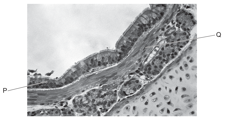
Which row shows the label and function of smooth muscle?
Options
| label | function | |
|---|---|---|
| A | P | regulates air flow towards the gas exchange surfaces |
| B | Q | pumps air towards the gas exchange surfaces |
| C | P | pumps air towards the gas exchange surfaces |
| D | Q | regulates air flow towards the gas exchange surfaces |
Working
Smooth muscle in an airway wall is the layer of spindle-shaped cells located beneath the ciliated epithelium and basement membrane. In Fig. 34.1:
- P points to the folded ciliated epithelium (pseudostratified columnar epithelium with cilia and goblet cells).
- Q points to the deeper layer of smooth muscle.
Smooth muscle contracts and relaxes to alter the diameter of the airway, thereby regulating the flow of air. It does not actively pump air — that is the role of the diaphragm and intercostal muscles during breathing.
Therefore: label = Q, function = regulates air flow towards the gas exchange surfaces.
Answer
D
D
Background Concept
The human gas exchange (respiratory) system is a branching network of tubes — trachea → bronchi → bronchioles → alveoli — whose walls are organised into recognisable tissue layers. From the lumen outward these are typically:
- Ciliated epithelium (often pseudostratified columnar with goblet cells) — warms, moistens and cleans the incoming air; cilia beat mucus and trapped particles upward.
- Basement membrane.
- Smooth muscle — a layer of spindle-shaped, uninucleate cells; contracts/relaxes to change airway diameter.
- Cartilage (in trachea and bronchi) or its absence (in bronchioles) — keeps larger airways open.
- Elastic fibres — allow the airway to stretch on inspiration and recoil on expiration.
Smooth muscle is not a pump. The actual driving force that moves air in and out of the lungs is the pressure change produced by contraction of the diaphragm and external intercostal muscles during breathing. Smooth muscle's role is to modulate airway diameter, and therefore regulate the rate of airflow, in response to nervous (autonomic) and chemical (e.g. histamine) signals. This is exactly why bronchoconstriction in asthma is a smooth-muscle problem and is treated with β₂-agonists (e.g. salbutamol) that relax airway smooth muscle.
Understanding the Question
The candidate is shown a transverse section through a bronchus or bronchiole and given two labels, P and Q, sitting on different tissue layers. The question is a two-part identification: pick the correct label for smooth muscle AND pick the correct description of its function. The four options permute these two independent choices.
The command word is implicit ("which row shows…"), so the answer must satisfy both halves of the row simultaneously.
Approach
- Look at Fig. 34.1 and identify which label sits on a layer of pale, elongated, spindle-shaped cells lying just deep to the epithelium — that is smooth muscle. The deeper, pale, fibrous-looking layer deeper still is cartilage or connective tissue.
- Decide whether smooth muscle "regulates" or "pumps" air. Regulation of airflow (constriction/dilation of the lumen) is the textbook function; pumping is the diaphragm's job.
- Match label + function to the correct row.
Step-by-Step Reasoning
- Identifying P: The label line P runs into a folded, darkly stained layer of tall cells on the luminal surface, with apical hair-like projections (cilia) and interspersed pale goblet cells. This is unmistakably ciliated (pseudostratified columnar) epithelium. P is therefore NOT smooth muscle.
- Identifying Q: The label Q points deeper into the wall, into a layer of more lightly stained, elongated/spindle-shaped cells with elongated nuclei — the smooth muscle layer. So Q = smooth muscle.
- Function of smooth muscle: Smooth muscle in the airway wall can contract (bronchoconstriction) or relax (bronchodilation), changing the lumen diameter and so changing the resistance to airflow. This regulates the rate at which air flows to the alveoli. It does not pump air — that would require rhythmic pressure changes produced by the diaphragm and intercostals.
- Matching to the rows:
- A: P + regulates — wrong label (P is epithelium).
- B: Q + pumps — right label, wrong function (no pump).
- C: P + pumps — wrong label AND wrong function.
- D: Q + regulates — correct label and correct function.
Key Takeaways
- In a TS of an airway, ciliated epithelium lines the lumen; smooth muscle sits just deep to it; cartilage (when present) lies further out.
- The function of airway smooth muscle is to regulate airflow by changing lumen diameter — not to drive ventilation.
- "Pumps air" is a classic distractor phrasing in CIE questions: ventilation is powered by the diaphragm/intercostals, not by airway smooth muscle.
Common Mistakes
- Confusing P (epithelium) with smooth muscle because it stains darkly and is highly visible; remember the epithelium is always the innermost lining.
- Writing "pumps air" because smooth muscle is muscular — the word "muscle" in the question misleads students into thinking of the heart. Smooth muscle here is doing a regulatory (tonic), not a pumping (phasic), job.
- Forgetting that bronchioles have smooth muscle but lack cartilage and (mostly) goblet cells, so a TS of a bronchiole still shows the same epithelium/muscle arrangement on a smaller scale.
Things to Be Careful About
- The epithelium in the photomicrograph looks folded because the airway was not fully inflated when fixed — this is normal and does not change the identity of the layer.
- "Regulates air flow" is a stronger mark-scheme phrase than vague alternatives like "controls breathing" or "moves air" — mirror the mark-scheme wording when revising.
- The mark scheme also credits (elsewhere) that elastic fibres in the wall recoil to help expel air, and that cartilage holds larger airways open; smooth muscle is specifically the active regulator of lumen diameter.
Which feature of the human gas exchange system helps to maintain a steep diffusion gradient?
Options
A A large number of alveoli are present in each lung.
B Alveoli walls contain elastic fibres allowing expansion.
C The air brought into the alveoli has a high concentration of oxygen.
D The endothelium of the capillary wall is made of flattened cells.
Working
A steep diffusion gradient is maintained by keeping the concentration difference across the gas exchange surface as large as possible. Bringing fresh, oxygen-rich air into the alveoli (and removing CO₂-rich air) keeps alveolar O₂ concentration high, while continuous blood flow keeps blood O₂ concentration low — maintaining a steep gradient.
- A refers to surface area (Fick's law)
- B refers to elastic recoil during breathing
- C directly addresses the concentration difference — fresh inspired air keeps alveolar O₂ concentration high
- D refers to a short diffusion distance (Fick's law)
Answer
C
C
Background Concept
Gas exchange in the lungs depends on the passive diffusion of oxygen and carbon dioxide between the alveolar air and the blood in pulmonary capillaries. The rate of this diffusion is described by Fick's law, which states that the rate of diffusion is proportional to:
- the surface area of the exchange surface,
- the concentration (or partial pressure) gradient across it, and
- the permeability of the membrane,
and inversely proportional to the thickness (diffusion distance) of the membrane.
A "steep diffusion gradient" specifically means a large concentration difference between the two sides of the exchange surface. To keep that gradient steep, the gas on the high-concentration side must be continuously replenished (so its concentration does not fall) and the gas on the low-concentration side must be continuously removed.
Understanding the Question
The question asks which feature of the human gas exchange system maintains a steep diffusion gradient for gases between alveolar air and blood. The command word is "helps to maintain a steep diffusion gradient" — this is asking specifically about the concentration gradient term in Fick's law, not the other terms. The four options each correspond to a different aspect of gas exchange, and only one directly concerns the concentration difference.
Approach
For each option, identify which factor of Fick's law (or which other physiological role) it describes, and decide whether it is the factor responsible for maintaining a concentration gradient.
Step-by-Step Reasoning
- A — Many alveoli per lung: This dramatically increases the surface area available for diffusion (Fick's law). It does not itself maintain a concentration gradient.
- B — Elastic fibres in alveolar walls: These allow the alveoli to stretch during inspiration and recoil during expiration, aiding ventilation mechanics. They do not affect the concentration gradient directly.
- C — Air brought into the alveoli has a high concentration of O₂: This is correct. Ventilation continuously brings fresh atmospheric air into the alveoli, keeping alveolar PO₂ high (~13.3 kPa). At the same time, the pulmonary circulation continuously brings deoxygenated blood with low PO₂ (~5.3 kPa) past the alveolar wall. This large and continually refreshed concentration difference is what maintains the steep diffusion gradient for O₂ (and similarly for CO₂ in reverse).
- D — Capillary endothelium made of flattened cells: This, together with the thin alveolar epithelium, gives a very short diffusion distance of about 0.5–1 µm between alveolar air and blood. It is a Fick's law distance factor, not a gradient factor.
So only option C is concerned with maintaining the concentration difference — i.e., the diffusion gradient itself.
Key Takeaways
- Fick's law identifies four factors that affect diffusion rate: surface area, concentration gradient, membrane permeability, and diffusion distance.
- A steep diffusion gradient is maintained by ventilation (replenishing alveolar O₂) and perfusion (removing O₂ from blood) — together keeping PO₂ high on the air side and low on the blood side.
- Many "features of the gas exchange system" (large surface area, thin walls, moist surface) speed up diffusion but do not themselves create or maintain the concentration gradient.
Common Mistakes
- Choosing A because "more alveoli = more gas exchange" — true, but a large surface area is a separate Fick's law factor and does not maintain a concentration difference.
- Choosing D because "thin walls = easy diffusion" — again a Fick's law factor (short distance), not the gradient.
- Confusing "maintaining a diffusion gradient" with "allowing diffusion to occur quickly."
Things to Be Careful About
- "Steep diffusion gradient" is precise language: it refers to the concentration difference (ΔC in Fick's law), not the rate of diffusion as a whole.
- The replenishment of alveolar air by breathing in (inspiration) and the removal of gas by blood flow (perfusion) are both required to keep the gradient steep — option C describes the ventilation half of this.
- The wording "high concentration of oxygen" in the air brought in is the key phrase: it is the continual renewal of this oxygen-rich air that keeps alveolar PO₂ high.
The diagram shows the structure of an antibody.
Which statements about the antibody are correct?
1 X has a similar shape to the antigen.
2 Y is the hinge region held by disulfide bonds.
3 Z is a constant region which binds to receptors on B-lymphocytes.
Options
A 1 and 2
B 1 and 3
C 2 and 3
D 2 only
Working
- Statement 1: X is the variable region. The variable region has a shape that is complementary to the antigen (so the two fit together like a lock and key), not a similar shape. A similar shape would not allow binding. → Incorrect
- Statement 2: Y is the hinge region, which is held together by disulfide bonds between the two heavy chains. This allows the two antigen-binding sites to move relative to each other. → Correct
- Statement 3: Z is the constant region. The constant region determines the class of antibody and binds to receptors on cells such as B-lymphocytes, macrophages and phagocytes. → Correct
Answer
C
C
Background Concept
An antibody (immunoglobulin) is a Y-shaped protein made of four polypeptide chains: two identical heavy chains and two identical light chains, linked together by disulfide bonds.
The molecule has distinct regions with different functions:
- Variable region (tips of the Y) — the amino acid sequence varies between different antibodies. This region forms the antigen-binding site, and its shape is complementary to a specific antigen (lock-and-key fit). It is not similar in shape to the antigen.
- Hinge region (the flexible middle of the Y) — held together by disulfide bonds between the two heavy chains. This gives the antibody flexibility so the two antigen-binding sites can move independently to attach to two antigens simultaneously.
- Constant region (the stem of the Y) — the amino acid sequence is the same within a class of antibody. It determines the antibody class (IgG, IgM, IgA, IgD, IgE) and binds to receptors on immune cells such as B-lymphocytes, macrophages and phagocytes, recruiting them to destroy the pathogen.
Understanding the Question
The question presents a diagram of an antibody labelled X, Y and Z, and asks which of three statements about these regions are correct. This is a multiple-choice question requiring the student to evaluate each statement independently before selecting the option that groups the correct ones.
Approach
For each labelled region, recall:
- What part of the antibody it points to (variable region, hinge region, or constant region).
- The defining structural or functional property of that region.
- Whether the statement accurately describes that property.
The trap in this question is statement 1, which tests whether the student can distinguish between complementary shape and similar shape — a common source of confusion in lock-and-key binding.
Step-by-Step Reasoning
Statement 1 — X has a similar shape to the antigen.
X points to the tips of the Y, i.e. the variable region containing the antigen-binding site. For an antibody to bind its antigen, the binding site must be complementary in shape to the antigen — the two fit together precisely, like a lock and key. A similar shape would not interlock. The statement is therefore incorrect.
Statement 2 — Y is the hinge region held by disulfide bonds.
Y points to the middle of the Y where the two heavy chains are joined. This is the hinge region, and the heavy chains are indeed linked here by disulfide bonds (S–S bridges between cysteine residues). The statement is correct.
Statement 3 — Z is a constant region which binds to receptors on B-lymphocytes.
Z indicates the stem of the Y, which is the constant region of the heavy chain. The constant region determines the antibody class and contains binding sites for Fc receptors on cells including B-lymphocytes, macrophages, neutrophils and mast cells. The statement is correct.
Since only statements 2 and 3 are correct, the answer is C.
Key Takeaways
- Antibodies are Y-shaped molecules with two heavy chains and two light chains held by disulfide bonds.
- Variable region = tip of the Y; has a shape complementary (not similar) to the antigen.
- Hinge region = flexible middle, joined by disulfide bonds.
- Constant region = stem of the Y; determines antibody class and binds to receptors on immune cells.
Common Mistakes
- Confusing "similar" with "complementary" shape in statement 1 — these are opposites in the context of binding. The variable region must be complementary, not similar, to the antigen.
- Thinking the constant region only determines the antibody class and forgetting that it also binds to receptors on immune cells such as B-lymphocytes.
- Confusing the location of disulfide bonds: they occur at the hinge (between heavy chains) and also between light and heavy chains, but are not found in the variable region itself.
Things to Be Careful About
- In an antibody diagram, X is typically drawn at the tips, Y in the middle, and Z on the stem — but always check the position of the label lines, not just the letter, because letters can be reused across different papers.
- "Complementary" and "similar" are not interchangeable in biology; complementary means the shapes fit together, similar means they look alike.
- The constant region binds to Fc receptors on many immune cell types, not only B-lymphocytes, so the statement is still credited as correct even though it is more specific than strictly required.
Which statement is a possible description of how resistance to penicillin may develop in bacteria?
Options
A A mutation of a bacterial gene prevents penicillin from binding to DNA in bacterial cells, which allows DNA replication to continue.
B A mutation of a bacterial gene causes penicillin to bind less readily to certain proteins, which allows bacteria to continue producing cell walls.
C A mutation of a bacterial gene allows the production of an enzyme that breaks down penicillin, which allows the ribosomes to continue functioning.
D A mutation of a bacterial gene changes the tertiary structure of an enzyme that penicillin inhibits, which allows bacteria to continue producing cell membranes.
Working
Penicillin inhibits the enzyme transpeptidase (a penicillin-binding protein) that cross-links peptidoglycan chains in the bacterial cell wall. Resistance by altered target arises when a mutation changes the binding protein so penicillin no longer binds effectively, allowing cell wall synthesis to continue.
- A – incorrect: penicillin does not bind to DNA.
- B – correct: mutation alters the target protein so penicillin binds less readily, and bacteria continue producing cell walls.
- C – incorrect: although some bacteria do produce penicillinase, the second clause is wrong because penicillin targets cell wall synthesis, not ribosomes.
- D – incorrect: penicillin inhibits cell wall synthesis, not cell membrane production.
Answer
B
B
Background Concept
Penicillin is a β-lactam antibiotic. Its mechanism of action depends on its structural similarity to the terminal D-alanyl-D-alanine dipeptide of the peptidoglycan precursor in bacterial cell walls. Penicillin binds irreversibly to transpeptidase (also known as a penicillin-binding protein, PBP), the enzyme that forms the peptide cross-links between adjacent peptidoglycan strands. When transpeptidase is inhibited, the cell wall cannot be cross-linked, the bacterium cannot withstand its internal turgor pressure, and it lyses. Penicillin is therefore specifically active against bacteria that are synthesising peptidoglycan cell walls — it has no equivalent target in animal cells, and it does not affect viruses at all (which lack a cell wall entirely).
Bacterial resistance to penicillin can arise by several mechanisms:
- Enzymatic destruction of the drug (e.g. β-lactamase / penicillinase hydrolysing the β-lactam ring).
- Altered target — mutation changes the transpeptidase (PBP) so penicillin no longer binds effectively. MRSA is the classic example, with PBP2a encoded by mecA.
- Reduced uptake or active efflux of the antibiotic.
- Bypassing the cross-linking step using an alternative enzyme.
The key fact for this question is that penicillin's target is the cell wall, not the DNA, ribosomes, or the cell membrane.
Understanding the Question
This is a multiple-choice item testing whether you know exactly what penicillin binds to and what its inhibition prevents. The command "possible description of how resistance to penicillin may develop" requires you to choose the option in which both the mutation effect AND the downstream biological consequence are correct.
Approach
For each option, identify:
- What molecule/cell component penicillin is said to bind to (or affect), and
- What biological process is rescued as a result.
A correct answer must identify the cell wall as the process that is preserved, and must give a biologically accurate target for penicillin (transpeptidase/PBP, not DNA, ribosomes, or membrane enzymes).
Step-by-Step Reasoning
Option A — Claims penicillin binds to DNA, with resistance allowing DNA replication to continue. This is wrong on two counts: (i) penicillin's target is the cell-wall-synthesis enzyme transpeptidase, not DNA; (ii) antibiotics that target DNA replication (e.g. quinolones such as ciprofloxacin) do so by inhibiting DNA gyrase, not by binding DNA directly. Eliminate.
Option B — States that a mutation causes penicillin to bind less readily to certain proteins, allowing bacteria to continue producing cell walls. This is the textbook description of target-modification resistance: the penicillin-binding proteins (PBPs) are altered so penicillin no longer binds effectively, and transpeptidation (cell wall cross-linking) continues. Correct.
Option C — Claims a mutation allows production of an enzyme (i.e. β-lactamase) that breaks down penicillin, then says this allows the ribosomes to continue functioning. The first half of the statement is a real resistance mechanism, but the second half is wrong — ribosomes are not penicillin's target; cell walls are. Eliminate.
Option D — Claims penicillin inhibits an enzyme involved in cell membrane production. Wrong — penicillin inhibits cell wall synthesis. The cell membrane is not its target. Eliminate.
Key Takeaways
- Penicillin inhibits transpeptidase (PBP), which cross-links peptidoglycan in the bacterial cell wall.
- Resistance by altered target = the binding protein is mutated so penicillin no longer binds effectively (e.g. MRSA's PBP2a).
- Resistance by enzymatic breakdown = β-lactamase destroys the drug (e.g. many Staphylococcus aureus strains).
- Penicillin is ineffective against viruses because viruses have no cell wall and no peptidoglycan.
Common Mistakes
- Confusing penicillin's target with that of other antibiotic classes (e.g. rifampicin → RNA polymerase, streptomycin/gentamicin → ribosomes, ciprofloxacin → DNA gyrase).
- Picking option C because it mentions penicillinase — a real resistance enzyme — without noticing that the consequence ("ribosomes to continue functioning") is wrong.
- Picking option D because "enzyme that penicillin inhibits" sounds plausible, forgetting that penicillin's enzyme target is in cell wall synthesis, not membrane synthesis.
Things to Be Careful About
- A statement can be "half right" and still score zero. Both clauses must be biologically correct for the option to work.
- Memorise the targets of the major antibiotic classes; the CIE mark scheme frequently exploits this kind of cross-class confusion.
- "Cell wall" and "cell membrane" are not interchangeable — they are different structures with different chemistry (peptidoglycan vs phospholipid bilayer).
Some events that happen during phagocytosis are listed.
1 Molecules on the surface of a pathogen bind to the cell surface membrane of a phagocyte.
2 Endocytosis takes place forming a specialised vesicle.
3 Enzymes catalyse hydrolysis reactions.
4 Lysosomes migrate through the cytoplasm and fuse with an organelle.
What is the correct order of these events during phagocytosis?
Options
A 1 → 2 → 4 → 3
B 1 → 4 → 2 → 3
C 3 → 2 → 4 → 1
D 3 → 4 → 2 → 1
Working
Phagocytosis proceeds in the following sequence:
- Recognition/binding — molecules on the pathogen surface bind to receptors on the phagocyte's cell surface membrane.
- Engulfment — the membrane invaginates and endocytosis forms a phagosome (the "specialised vesicle").
- Lysosome fusion — lysosomes migrate through the cytoplasm and fuse with the phagosome, forming a phagolysosome.
- Digestion — hydrolytic enzymes within the lysosome catalyse the breakdown of the pathogen.
Matching the listed events (1, 2, 4, 3) gives the order 1 → 2 → 4 → 3.
Answer
A
A
Background Concept
Phagocytosis is the engulfment and digestion of solid particles (such as invading pathogens) by specialised cells called phagocytes. Macrophages and neutrophils are the principal phagocytes in mammals. The process is a form of endocytosis, and it relies on the recognition of non-self molecules on the pathogen surface, the remodelling of the phagocyte's cell surface membrane to enclose the particle, and the action of hydrolytic (digestive) enzymes stored in lysosomes.
A lysosome is a membrane-bound organelle containing hydrolytic enzymes such as lysozyme, proteases, lipases and nucleases. These enzymes work best at an acidic pH (around pH 4.5–5.0), which is maintained inside the lysosome by proton pumps in its membrane.
Understanding the Question
The question gives four descriptive statements, each describing a single event that occurs during phagocytosis, and asks for the correct order in which they happen. The four events are:
- Pathogen surface molecules bind to the phagocyte's cell surface membrane.
- Endocytosis forms a specialised vesicle.
- Enzymes catalyse hydrolysis reactions.
- Lysosomes migrate through the cytoplasm and fuse with an organelle.
The candidate must sequence them correctly to identify the answer.
Approach
Recall the standard narrative of phagocytosis: (a) recognition, (b) engulfment (endocytosis, forming a phagosome), (c) fusion of the phagosome with a lysosome to form a phagolysosome, and (d) enzymatic hydrolysis of the pathogen inside this vesicle. Map each of the four statements onto these stages and read off the order.
Step-by-Step Reasoning
- Statement 1 describes the recognition step. Receptor proteins on the phagocyte's cell surface membrane bind to molecules (e.g. PAMPs such as lipopolysaccharide or mannose-containing structures) on the pathogen surface. This is a prerequisite for everything that follows, so 1 must come first.
- Statement 2 describes endocytosis. Once the pathogen is bound, the membrane flows around it and pinches off internally, forming a vesicle called a phagosome. This is the second step.
- Statement 4 describes lysosome fusion. Lysosomes carrying hydrolytic enzymes travel through the cytosol and fuse with the phagosome, converting it into a phagolysosome. This is the third step.
- Statement 3 describes the hydrolysis. Only after the lysosome has fused does its enzyme content act on the pathogen contents, catalysing the breakdown of proteins, carbohydrates, lipids and nucleic acids. This is the final step.
So the order is 1 → 2 → 4 → 3, which corresponds to option A.
- B (1 → 4 → 2 → 3) is wrong because lysosomes cannot fuse with anything until the phagosome (formed by endocytosis) exists.
- C (3 → 2 → 4 → 1) is wrong because hydrolysis cannot occur before the pathogen is even inside the cell, and binding must precede engulfment.
- D (3 → 4 → 2 → 1) is wrong because hydrolysis must come last, not first.
Key Takeaways
- Phagocytosis proceeds in this order: recognition → engulfment (phagosome formation) → lysosome fusion (phagolysosome) → enzymatic hydrolysis.
- Lysosomes contribute hydrolytic enzymes; they do not act on free cytoplasm.
- "Endocytosis forming a specialised vesicle" refers to phagosome formation and must precede lysosome fusion.
Common Mistakes
- Placing hydrolysis (3) first or second. Digestion requires that the enzymes are inside the phagolysosome, which does not yet exist at the start of phagocytosis.
- Putting lysosome fusion (4) before endocytosis (2). There is nothing for the lysosome to fuse with until the phagosome is formed.
- Treating "binding" as something that happens after digestion. Binding is the trigger for engulfment and must come first.
Things to Be Careful About
- Distinguish the phagosome (formed by endocytosis around the pathogen) from the phagolysosome (formed by lysosome fusion). Statement 2 specifically refers to the phagosome.
- "Migrate through the cytoplasm and fuse with an organelle" in statement 4 means fuse with the phagosome, not with another lysosome.
- "Hydrolysis reactions" (statement 3) refers to enzymatic digestion inside the phagolysosome, not to events occurring in the cytosol.
What are the functions of plasma cells during an immune response?
1 to destroy cancer cells
2 to differentiate into memory cells
3 to secrete antibodies
Options
A 1, 2 and 3
B 2 and 3 only
C 2 only
D 3 only
Working
- Statement 1 is wrong: destruction of cancer (and other infected/abnormal) body cells is carried out by cytotoxic T cells, not plasma cells.
- Statement 2 is wrong: when an activated B cell divides, its daughter cells differentiate into either plasma cells or memory cells. A plasma cell does not then go on to become a memory cell — the two cell types are separate end-products of B-cell differentiation.
- Statement 3 is correct: plasma cells are highly specialised antibody-secreting cells; they synthesise and release large quantities of antibody.
Only statement 3 is correct.
Answer
D
D
Background Concept
The specific (adaptive) immune response depends on several types of lymphocyte, each with a clearly defined role:
- B lymphocytes recognise antigen (via surface antibodies) and, when activated by a helper T cell, proliferate and differentiate into plasma cells and memory B cells.
- Plasma cells are essentially antibody factories: they have an extensive rough endoplasmic reticulum and Golgi apparatus to synthesise and secrete antibodies at very high rates.
- Memory B cells are long-lived cells that remain in the body and mount a rapid, large secondary response if the same antigen is encountered again.
- T lymphocytes come in several forms. Helper T cells coordinate the response by releasing cytokines that activate B cells and other T cells. Cytotoxic (killer) T cells directly destroy body cells that display non-self antigen on their surface — for example, virus-infected cells and cancerous cells — by releasing perforins that cause lysis.
Understanding the Question
The question lists three possible functions of plasma cells and asks which are correct. The command word is implicit but the task is evaluation: assess each numbered statement against what plasma cells actually do.
Approach
For each statement, ask: is this a known function of plasma cells, or is it the job of a different cell type? If any statement is wrong, the answer must exclude it.
Step-by-Step Reasoning
- Statement 1 — destroy cancer cells. This is the function of cytotoxic T cells, not plasma cells. Plasma cells do not engage in cell-mediated killing; their role is humoral (antibody-based). Reject statement 1.
- Statement 2 — differentiate into memory cells. An activated B cell divides and its daughters follow two separate differentiation paths: some become plasma cells, others become memory B cells. A plasma cell is a terminally differentiated antibody-secreting cell; it does not then convert into a memory cell. The cell that can become a memory cell is the activated B cell, before it has become a plasma cell. Reject statement 2.
- Statement 3 — secrete antibodies. This is the defining function of a plasma cell. Accept statement 3.
Only statement 3 is correct, so the answer is D.
Key Takeaways
- Plasma cells secrete antibodies; they do not kill cells directly and they do not become memory cells.
- Destruction of cancer/infected cells is the job of cytotoxic T cells.
- Memory cells and plasma cells are sibling cell types produced from the same activated B cell — neither arises from the other.
Common Mistakes
- Choosing A because antibodies "help fight cancer": antibodies flag abnormal cells for destruction by other components of the immune system, but the plasma cell itself does not destroy them. Cytotoxic T cells are the actual killers.
- Choosing B because students recall that B cells give rise to memory cells, forgetting that once a B cell has differentiated into a plasma cell, it is at the end of that lineage and cannot switch fate.
- Confusing plasma cells with macrophages (which engulf pathogens by phagocytosis) or with natural killer cells (which kill virus-infected and tumour cells).
Things to Be Careful About
- Read each numbered statement independently — a wrong statement does not invalidate a correct one, and only the combination listed in the answer choice matters.
- Use the precise CIE terminology: plasma cell (the antibody-secreting effector B cell), not just "B cell"; and cytotoxic T cell (the killer), not just "T cell".
- Remember that B cells and T cells arise from the same bone-marrow stem cell but mature in different places and perform fundamentally different functions — humoral versus cell-mediated immunity respectively.
Which disease is caused by a eukaryotic pathogen?
Options
A cholera
B HIV/AIDS
C malaria
D tuberculosis
Working
Malaria is caused by Plasmodium (e.g. Plasmodium falciparum), a protoctist. Protoctists are eukaryotic organisms, so malaria is the disease caused by a eukaryotic pathogen.
The other options are all caused by non-eukaryotic pathogens:
- Cholera — Vibrio cholerae (prokaryotic bacterium)
- HIV/AIDS — HIV (virus; non-cellular)
- Tuberculosis — Mycobacterium tuberculosis (prokaryotic bacterium)
Answer
C
C
Background Concept
Pathogens are disease-causing organisms (or particles) and fall into several distinct biological categories. To answer this question, you need to remember the type of pathogen responsible for each of the four diseases in the options, because the question hinges on the eukaryotic vs prokaryotic distinction.
- Prokaryotes are organisms whose cells lack a true membrane-bound nucleus and other membrane-bound organelles. All bacteria are prokaryotes.
- Eukaryotes have cells with a membrane-bound nucleus and membrane-bound organelles. This group includes animals, plants, fungi and protoctists (single-celled eukaryotes such as Plasmodium, Amoeba and Trypanosoma).
- Viruses are non-cellular — they consist of a nucleic acid core enclosed in a protein capsid (sometimes with a lipid envelope). They are not living cells and are neither prokaryotic nor eukaryotic.
The four diseases and their causative agents are:
- Cholera — Vibrio cholerae (a bacterium; prokaryote)
- HIV/AIDS — Human Immunodeficiency Virus (a virus; non-cellular)
- Malaria — Plasmodium species such as Plasmodium falciparum (a protoctist; eukaryote)
- Tuberculosis (TB) — Mycobacterium tuberculosis (a bacterium; prokaryote)
Understanding the Question
The question is a direct identification task: from the four diseases listed, pick the one whose pathogen belongs to the eukaryotes. The command word "caused by" makes it clear that you need to know the causative agent, not just the symptoms. The mark scheme confirms that the answer is C — malaria, because Plasmodium is a eukaryotic protoctist.
Approach
For each option, identify the causative agent and then classify that agent as prokaryotic, eukaryotic, or viral. The single eukaryotic pathogen among the list gives the answer.
Step-by-Step Reasoning
- Cholera (A) — caused by the bacterium Vibrio cholerae. Bacteria are prokaryotes, so this is not the answer.
- HIV/AIDS (B) — caused by the Human Immunodeficiency Virus. Viruses are non-cellular particles, not eukaryotes. Discard.
- Malaria (C) — caused by Plasmodium (a protoctist). Protoctists are eukaryotes — their cells have a true nucleus and organelles such as mitochondria and (in many species) an apicoplast. This is the only eukaryotic pathogen in the list.
- Tuberculosis (D) — caused by the bacterium Mycobacterium tuberculosis. Again a prokaryote, so this is not the answer.
- The unique eukaryotic option is C — malaria.
Key Takeaways
- Bacteria (prokaryotes) cause cholera and TB.
- A virus (non-cellular) causes HIV/AIDS.
- A protoctist (eukaryote) — Plasmodium — causes malaria.
- A useful memory hook: among the four major infectious diseases named in the AS syllabus, only malaria has a eukaryotic pathogen.
Common Mistakes
- Choosing B (HIV/AIDS) because HIV is often perceived as a "complex" pathogen. It is a virus, not a cell, so it cannot be eukaryotic.
- Confusing bacteria with protoctists. Both can be single-celled and microscopic, but only protoctists are eukaryotes.
- Assuming "pathogen" always means "bacterium". Fungi and protoctists are also eukaryotic pathogens (e.g. Plasmodium for malaria).
Things to Be Careful About
- "Eukaryotic" describes the cell type of the pathogen, not the disease severity or transmission method.
- Protoctists are formally classified within the domain Eukarya, so they count as eukaryotic pathogens even though they are single-celled.
- Viruses are deliberately excluded from both the prokaryote and eukaryote categories because they are acellular.
Your score so far
Answer a question to start scoring
Your marks add up here as you work through the paper.