Biology 9700/11 — May/June 2025
Cambridge AS Level · Multiple Choice (AS Level) · answer key with instant marking and worked solutions
Topics Biological Molecules · Cell Structure · The Mitotic Cell Cycle · Transport in Mammals · Transport in Plants · Enzymes · +5 more
Tap an option under each question to check it — your score builds as you go.
Which statements about light microscopes are correct?
1 To calculate the magnification of a light microscope, the eyepiece lens and objective lens magnifications are added together.
2 The resolution of a light microscope is limited by the wavelength of light.
3 The divisions on the scale on a stage micrometer are closer together than the divisions on the scale on an eyepiece graticule.
Options
A 1, 2 and 3
B 1 and 2 only
C 2 only
D 3 only
Working
Evaluate each statement:
-
Incorrect. The total magnification of a compound light microscope is obtained by multiplying the eyepiece lens magnification by the objective lens magnification, not by adding them. (e.g. eyepiece ×10 with objective ×40 gives a total magnification of ×400, not ×50.)
-
Correct. The resolution of a light microscope is limited by the wavelength of light; shorter wavelengths can resolve smaller structures, which is why electron microscopes (using electron beams of much shorter wavelength) have a much higher resolution than light microscopes.
-
Incorrect. The stage micrometer has divisions that are very small and precisely known (typically or apart), whereas the eyepiece graticule has arbitrary divisions whose apparent spacing depends on the objective lens in use; the eyepiece graticule divisions are generally further apart than the stage micrometer divisions, not closer.
Only statement 2 is correct.
Answer
C
C
Background Concept
A compound light microscope has two main lens systems that together produce the magnified image:
- The eyepiece lens (ocular), usually ×10 in school/college microscopes.
- One of several objective lenses on a revolving nosepiece, e.g. ×4, ×10, ×40 or ×100.
Two quantities describe how well a microscope performs:
- Magnification — how much larger the image is compared to the object. It is the ratio of image size to actual size, and for a compound microscope it is the product of the magnifications of the eyepiece and objective lenses.
- Resolution — the smallest distance between two points at which they can still be seen as separate. Resolution is governed by Abbe's equation, and is fundamentally limited by the wavelength of the radiation used to form the image. Light has a wavelength of roughly , so a light microscope can resolve structures down to about at best; electron beams have much shorter wavelengths and so electron microscopes can resolve much finer detail.
To measure the size of an object under a microscope, two scales are used together:
- The stage micrometer is a slide on which a precise scale of known length is engraved — typically divisions (or ) apart. It is an absolute, fixed reference.
- The eyepiece graticule is a small glass disc with arbitrary divisions (usually 100) that sits in the eyepiece. It has no fixed unit until it is calibrated against the stage micrometer for each objective lens in turn.
Understanding the Question
The question presents three statements about light microscopes and asks which combination is correct, with four answer options. The correct answer is the option whose set of statement numbers exactly matches the true statements.
- Statement 1 tests knowledge of how total magnification is calculated.
- Statement 2 tests what limits resolution in a light microscope.
- Statement 3 tests the relative size of divisions on the stage micrometer and the eyepiece graticule.
Approach
Judge each statement independently using the definitions above, then match the combination of correct statements to one of the four options.
Step-by-Step Reasoning
Statement 1 — "magnifications are added":
- The total magnification of a compound microscope = (eyepiece magnification) × (objective magnification).
- Example: ×10 eyepiece with ×40 objective → ×400, not ×50.
- Therefore statement 1 is false. (Any answer containing "1" can be ruled out: A and B are eliminated.)
Statement 2 — "resolution is limited by the wavelength of light":
- This is the standard formulation of the limit of resolution. Light's wavelength (~) sets a lower limit on how close two points can be and still be distinguished.
- Therefore statement 2 is true.
Statement 3 — "stage micrometer divisions are closer together than eyepiece graticule divisions":
- The stage micrometer has divisions of fixed, very small known length (typically or ).
- The eyepiece graticule has arbitrary divisions, and once calibrated, each division corresponds to a length that depends on the objective lens in use. With low-power objectives, each eyepiece division represents a relatively large real distance; with high-power objectives, it represents a much smaller distance — but the eyepiece graticule divisions are still typically further apart than the precisely engraved stage micrometer divisions (and are not fixed).
- Therefore statement 3 is false. (Any answer containing "3" can be ruled out: A and D are eliminated.)
Only statement 2 is correct, which matches option C.
Key Takeaways
- Total magnification of a compound microscope = eyepiece × objective (multiplied, not added).
- Resolution is limited by the wavelength of the radiation used; this is why electron microscopes (very short wavelength) outperform light microscopes.
- A stage micrometer is a precisely engraved scale of known length (mm divisions); an eyepiece graticule is an arbitrary scale that must be calibrated against the stage micrometer for each objective lens.
Common Mistakes
- Adding rather than multiplying the eyepiece and objective magnifications — a common error that immediately rules out answers A and B here.
- Confusing resolution with magnification: increasing magnification does not increase resolution; resolution is set by wavelength and the numerical aperture of the lens.
- Assuming the eyepiece graticule has fixed units — it does not; it must be calibrated against a stage micrometer for the objective being used, otherwise measurements are meaningless.
- Believing that the eyepiece graticule divisions are smaller than the stage micrometer divisions — in practice the stage micrometer has very fine, fixed divisions and the eyepiece graticule divisions are coarser before calibration.
Things to Be Careful About
- "Added together" vs "multiplied" is the single most-tested idea on microscope magnification.
- Resolution limit wording: it is the wavelength of light (not the brightness, intensity, or magnification) that limits resolution.
- The stage micrometer is a physical slide with a real, known scale; the eyepiece graticule is a reticle inside the eyepiece with no fixed units of its own.
- Always check each option: here, options A and B contain 1, option D contains 3, and only C contains statement 2 alone — consistent with the analysis above.
A student measured the width of a mitochondrion in an electron micrograph of an animal cell with a magnification of . The width was .
What was the actual width of the mitochondrion?
Options
A
B
C
D
Working
Convert to µm (×1000):
Answer
B
B
Background Concept
When a specimen is viewed through a microscope, the image produced is larger than the actual specimen. The relationship is given by:
Rearranged to find actual size:
Biological structures such as mitochondria, ribosomes and bacteria are typically measured in micrometres (µm) or nanometres (nm). , so dividing a millimetre value by 1000 converts it to micrometres.
Understanding the Question
The question gives:
- Image width of the mitochondrion =
- Magnification of the electron micrograph =
The student must find the actual (real) width of the mitochondrion. The image was measured in millimetres but mitochondria are sub-cellular organelles whose dimensions are usually expressed in micrometres, so a unit conversion is required at the end.
Approach
- Apply the rearranged magnification formula: actual size = image size ÷ magnification.
- Carry out the division in millimetres.
- Convert the result from mm to µm by multiplying by 1000.
- Match the answer to the closest option.
Step-by-Step Reasoning
Step 1 — Substitute the values into the formula:
Step 2 — Calculate:
Step 3 — Convert to micrometres. Since :
Step 4 — Round and select the option. rounds to , which is option B.
A real mitochondrion is typically – wide, so this value is biologically sensible — a useful sanity check.
Key Takeaways
- The triangle relationship: , , .
- Image size is usually measured in mm or cm on the print/screen, while actual biological size is in µm or nm — a conversion is almost always required.
- Conversions: .
- Always sanity-check against known biological dimensions: animal mitochondria ≈ – wide.
Common Mistakes
- Multiplying instead of dividing (treating the image size as the actual size and scaling up further) — gives , clearly wrong.
- Forgetting to convert units: looks like a tiny value, but is the right biological size.
- Choosing option C () by mis-converting (dividing by 10000 instead of 1000) or by misplacing the decimal.
Things to Be Careful About
- Make sure the formula is rearranged correctly — many students write and end up with a result thousands of times too large.
- Watch the decimal place: with values this small, one wrong zero changes the answer by an order of magnitude.
- Note the units in the options (all in µm) — if the final answer is left in mm it cannot be matched to an option.
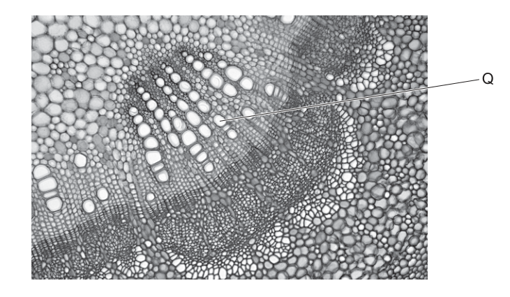
The photomicrograph shows a section of the stem of a plant.
Which statements could describe Q?
1 The cell wall contains pits.
2 The cell wall contains cellulose and also lignin.
3 It is connected to adjacent cells via plasmodesmata.
Options
A 1 and 2
B 1 and 3
C 1 only
D 2 and 3
Working
Q is a xylem vessel element (large, thick-walled, wide lumen, in the vascular tissue).
- The cell wall contains pits — TRUE. Xylem vessel walls have pits (gaps in the secondary wall) to allow lateral movement of water between vessels.
- The cell wall contains cellulose and also lignin — TRUE. Xylem vessel walls are lignified, providing waterproofing and mechanical support.
- It is connected to adjacent cells via plasmodesmata — FALSE. Xylem vessel elements are dead at maturity; they lack cytoplasm and so cannot have plasmodesmata (which are cytoplasmic channels between living cells).
Statements 1 and 2 are correct.
Answer
A
A
Background Concept
Xylem is the plant tissue responsible for transporting water and dissolved mineral ions from the roots to the rest of the plant. It is composed of several cell types, of which the most prominent in a transverse section are the xylem vessel elements. These are formed from files of cells (vessel elements) joined end-to-end; the end walls break down during development so the cells form continuous, hollow tubes. By the time they are functional, vessel elements are dead, with no cytoplasm, nucleus or organelles — only their lignified cell walls remain.
Because they must withstand the tension generated by the transpiration pull and resist collapse under negative pressure, vessel walls are heavily reinforced with lignin, a rigid, waterproof polymer laid down in the secondary cell wall (over the cellulose primary wall). However, lignin is not laid down uniformly — small regions called pits are left where only the primary wall (cellulose) is present, allowing water to pass laterally between adjacent vessels and tracheids. Because vessel elements are dead, there are no plasmodesmata (which are cytoplasmic channels connecting the living protoplasts of adjacent cells).
Understanding the Question
The photomicrograph shows a transverse section of a plant stem. The label Q points to a single, large, thick-walled cell with a wide lumen — characteristic features of a xylem vessel element. The question asks which of three statements correctly describe this cell.
Approach
Test each statement against the known properties of mature xylem vessel elements:
- Statement 1 → about wall structure (pits)
- Statement 2 → about wall composition (cellulose + lignin)
- Statement 3 → about cytoplasmic connections (plasmodesmata)
A key discriminator is the cell's maturity: xylem vessels are dead, so anything requiring a living protoplast (like plasmodesmata) must be rejected.
Step-by-Step Reasoning
- Identify Q. The large cell with a thick, darkly stained wall and a wide, empty lumen in the xylem region of the stem is a xylem vessel element.
- Evaluate statement 1. The walls of xylem vessels are not uniformly thickened; they contain pits that allow water to move sideways between adjacent vessels. → TRUE.
- Evaluate statement 2. The wall is composed of cellulose (in the primary wall) plus lignin (in the secondary wall), which gives waterproofing and mechanical strength. → TRUE.
- Evaluate statement 3. Plasmodesmata are cytoplasmic channels passing through the cell walls of living cells. Xylem vessel elements are dead at maturity, with no cytoplasm. → FALSE.
- Combine the true statements. Only 1 and 2 are correct, so the answer is A (1 and 2).
Key Takeaways
- Mature xylem vessel elements are dead, hollow tubes reinforced with lignin.
- Their walls have pits to allow lateral water movement.
- Because they lack cytoplasm, they have no plasmodesmata — a feature only of living cells.
Common Mistakes
- Confusing xylem vessels with phloem sieve tubes. Sieve tubes are living (although they lack a nucleus) and do have plasmodesmata-like connections via sieve pores. Confusing the two would lead to incorrectly accepting statement 3.
- Thinking pits are holes. Pits are thin regions of the wall (primary wall only), not perforations; this is why water can pass but the wall still provides strength.
- Assuming all plant cell walls contain lignin. Lignin is a feature of support and water-conducting cells (xylem, sclerenchyma); most parenchyma cells have only cellulose walls.
Things to Be Careful About
- Always link a feature to whether the cell is living or dead — this single fact eliminates statement 3 here.
- Read the image carefully: Q's large lumen and thickened wall are the diagnostic features for a xylem vessel, not a fibre (which has a very narrow lumen) or a parenchyma cell (thin wall).
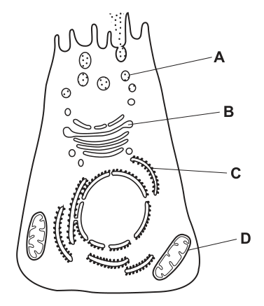
The diagram is taken from an electron micrograph of a cell that secretes enzymes.
Where are the polypeptides for these enzymes made?
Options
A A
B B
C C
D D
Working
C is the rough endoplasmic reticulum (RER) — note the ribosomes (small dots) studded on its cytoplasmic surface. Polypeptides for secreted enzymes are synthesised by these ribosomes. The newly formed polypeptide is threaded into the lumen of the RER, then transported via vesicles to the Golgi apparatus (where it is modified and packaged) and finally secreted.
- A — microvilli on the apical plasma membrane; site of absorption, not synthesis.
- B — secretory vesicle; carries the finished enzyme to the plasma membrane, but does not make the polypeptide.
- D — mitochondrion; site of aerobic respiration, not polypeptide synthesis.
Answer
C
C
Background Concept
Eukaryotic cells contain a system of membrane-bound organelles that work together to produce, modify and export proteins — known collectively as the secretory pathway (or endomembrane system). The key organelles involved are:
- Rough endoplasmic reticulum (RER) — flattened membrane sacs (cisternae) studded with ribosomes on the cytoplasmic face. Ribosomes on the RER translate mRNA into polypeptides that are destined for secretion, insertion into membranes, or delivery to lysosomes.
- Golgi apparatus — a stack of flattened cisternae that receives proteins from the RER, modifies them (e.g. by adding carbohydrate groups = glycosylation), sorts them, and packages them into vesicles.
- Secretory vesicles — small membrane-bound sacs that bud off the Golgi and travel to the plasma membrane, where they release their contents by exocytosis.
The actual synthesis of the polypeptide chain (translation) is carried out by ribosomes. In a cell that secretes enzymes, the ribosomes responsible are those bound to the RER, so the polypeptide is said to be "made" on the RER.
Understanding the Question
The electron micrograph shows a secretory cell. Four structures are labelled:
- A — finger-like microvilli on the apical plasma membrane (increase surface area for absorption/secretion).
- B — a secretory vesicle near the apical surface.
- C — a structure studded with ribosomes — the rough endoplasmic reticulum.
- D — a mitochondrion (bean-shaped, with cristae visible).
The question asks specifically where the polypeptides for these enzymes are made. The command word is are made, which points to the site of synthesis (i.e. translation), not modification, packaging or release.
Approach
- Identify each labelled organelle from its structure.
- Decide which organelle is the site of polypeptide synthesis.
- Match this to one of the labels (A, B, C, D).
Polypeptide synthesis = ribosomes carrying out translation. In a secretory cell, the relevant ribosomes are those attached to the RER.
Step-by-Step Reasoning
- Identify C. The structure labelled C is shown with small dots covering its cytoplasmic surface — these dots are ribosomes. The presence of ribosomes defines the rough endoplasmic reticulum.
- Recall the function of RER-bound ribosomes. When a ribosome on the RER translates an mRNA for a secreted protein, the growing polypeptide is fed directly into the RER lumen through a translocon. This is the moment the polypeptide is made.
- Check the other options to be sure:
- A (microvilli) — these are extensions of the plasma membrane that increase surface area; they do not synthesise polypeptides.
- B (secretory vesicle) — this carries an already-made enzyme from the Golgi to the plasma membrane; the polypeptide inside was made earlier, on the RER.
- D (mitochondrion) — produces ATP by aerobic respiration; it does not synthesise polypeptides (mitochondria have their own ribosomes but make only a handful of inner-membrane proteins, not secreted enzymes).
- Conclude. The polypeptides for the secreted enzymes are made on the RER, which is structure C.
Key Takeaways
- The RER is the site of synthesis for proteins (including enzymes) destined for secretion, because the ribosomes on its surface translate mRNAs whose products enter the secretory pathway.
- The secretory pathway flows: RER → transport vesicle → Golgi apparatus → secretory vesicle → plasma membrane (exocytosis).
- Each organelle in the pathway has a distinct role: RER (synthesis), Golgi (modification and sorting), secretory vesicles (storage and transport), plasma membrane (release).
- Do not confuse the site of synthesis (RER-bound ribosomes) with the site of modification/packaging (Golgi) or the site of release (plasma membrane via exocytosis).
Common Mistakes
- Choosing the Golgi apparatus (B or C, depending on labelling) because it is "where proteins are processed for secretion". Processing is not the same as synthesis; the Golgi modifies an already-existing polypeptide, it does not make it.
- Choosing a mitochondrion (D) because the cell is "active in secretion". Mitochondria supply ATP but do not synthesise secretory proteins.
- Choosing a secretory vesicle (B) because the question is about secreted enzymes. The vesicle contains the finished product, but the polypeptide was made earlier on the RER.
- Confusing rough and smooth ER. The "rough" appearance comes from ribosomes; smooth ER has no ribosomes and is involved in lipid synthesis and detoxification, not polypeptide synthesis.
Things to Be Careful About
- Read the question precisely: "Where are the polypeptides … made?" — this is asking for the site of synthesis, not the site of modification, packaging or release.
- Identify organelles by their structural features: dots on a membrane = ribosomes on RER; stacked curved cisternae = Golgi; double-membrane bean shape = mitochondrion; small spherical bodies near the membrane = secretory vesicles.
- A common distracter in this style of question is to offer a structure that is part of the secretory pathway but not the site of polypeptide synthesis (e.g. the Golgi or a secretory vesicle). Recognise that being "involved" in secretion is not the same as being the site of polypeptide synthesis.
Which comparisons between a typical bacterial cell and a typical plant cell are correct?
| bacterial cell | plant cell | |
|---|---|---|
| 1 | cytoplasmic DNA | nuclear DNA |
| 2 | no smooth ER present | smooth ER present |
| 3 | peptidoglycan cell walls found | cellulose cell walls found |
| 4 | 80S ribosomes | 70S ribosomes |
Options
A 1, 2 and 3
B 1, 2 and 4
C 1, 3 and 4
D 2, 3 and 4
Working
- Statement 1: Bacteria (prokaryotes) have DNA free in the cytoplasm (nucleoid region); plant cells (eukaryotes) enclose their DNA within a nucleus. → Correct.
- Statement 2: Bacteria lack membrane-bound organelles, so no smooth ER; plant cells have smooth ER. → Correct.
- Statement 3: Bacterial cell walls contain peptidoglycan; plant cell walls contain cellulose. → Correct.
- Statement 4: Bacterial ribosomes are 70S; plant (eukaryotic) ribosomes are 80S. The statement reverses these, so it is incorrect.
Therefore, statements 1, 2 and 3 are correct.
Answer
A
A
Background Concept
Living cells are divided into two fundamental types based on internal organisation: prokaryotes (bacteria and archaea) and eukaryotes (plants, animals, fungi, protists). The key distinction is the presence or absence of a true membrane-bound nucleus and other membrane-bound organelles.
Prokaryotic (bacterial) cells are smaller (typically 1–5 µm), lack a nucleus, and have:
- A single circular DNA molecule located free in the cytoplasm in a region called the nucleoid
- 70S ribosomes (smaller than eukaryotic ribosomes)
- No membrane-bound organelles such as endoplasmic reticulum, mitochondria or chloroplasts
- A cell wall made of peptidoglycan (also called murein)
Eukaryotic plant cells are larger (typically 10–100 µm) and have:
- DNA enclosed within a double-membrane nucleus
- 80S ribosomes in the cytoplasm (plus 70S ribosomes inside chloroplasts and mitochondria)
- Membrane-bound organelles including smooth and rough endoplasmic reticulum (ER), Golgi apparatus, mitochondria, and (in plants) chloroplasts and a large central vacuole
- A cell wall made primarily of cellulose
Understanding the Question
This is a multiple-choice question asking you to identify which of four pairwise comparisons between a typical bacterial cell and a typical plant cell are correct. You need to assess each numbered statement independently and then pick the option that lists only the correct ones. The question is testing your knowledge of the structural and molecular differences between prokaryotes and eukaryotes.
The command word is implicit ("which comparisons … are correct?") — there is no extended answer required, just selection of the right option letter after checking each statement.
Approach
Go through each of the four statements one at a time and decide whether it accurately describes the bacterial cell on the left AND the plant cell on the right. Statements that get the bacterial feature right AND the plant feature right are correct. A single reversal or error in either side of the comparison makes the whole statement wrong.
Step-by-Step Reasoning
Statement 1 — DNA location:
- Bacterial cell: ✓ has cytoplasmic DNA (the circular chromosome sits free in the cytoplasm in the nucleoid region; there is no nuclear envelope).
- Plant cell: ✓ has nuclear DNA (the chromosomes are enclosed within the double-membrane nucleus).
- → Statement 1 is correct.
Statement 2 — smooth ER:
- Bacterial cell: ✓ no smooth ER (prokaryotes have no endoplasmic reticulum of any kind, smooth or rough, because they lack internal membrane systems).
- Plant cell: ✓ smooth ER is present (smooth ER is involved in lipid synthesis, detoxification and calcium storage in eukaryotic cells).
- → Statement 2 is correct.
Statement 3 — cell wall composition:
- Bacterial cell: ✓ peptidoglycan (a polymer of NAG and NAM cross-linked by short peptides) makes up the bacterial cell wall.
- Plant cell: ✓ cellulose (a β-1,4-linked glucose polymer) is the main structural polysaccharide of the plant cell wall.
- → Statement 3 is correct.
Statement 4 — ribosome size:
- The table claims bacteria have 80S ribosomes and plant cells have 70S ribosomes.
- This is the reverse of the truth: bacteria have 70S ribosomes, and eukaryotic plant cells have 80S ribosomes in the cytoplasm.
- → Statement 4 is incorrect.
So the correct statements are 1, 2 and 3, which corresponds to option A.
Key Takeaways
- Prokaryotes vs eukaryotes: a quick mental checklist of contrasts is (i) nucleus/no nucleus, (ii) membrane-bound organelles/absent, (iii) 70S/80S ribosomes, (iv) peptidoglycan/cellulose or other cell-wall types, (v) circular/chromosomal DNA, (vi) size, (vii) division by binary fission/mitosis.
- When a question presents a table of comparisons, check both sides of every row independently — a statement is only as correct as its weaker half.
- The "S" value (Svedberg unit) reflects how ribosomes sediment in a centrifuge; eukaryotic cytoplasmic ribosomes are larger and more complex (80S, made of 60S + 40S subunits), while prokaryotic ribosomes are 70S (50S + 30S subunits). Note that 70S ribosomes also occur inside eukaryotic chloroplasts and mitochondria, reflecting their endosymbiotic origin.
Common Mistakes
- Reversing the ribosome sizes (writing 80S for bacteria and 70S for eukaryotes) — a very common slip, and exactly the trap set in statement 4.
- Forgetting that plant cells have other features beyond just "being eukaryotic" — they still have a cellulose cell wall, a large central vacuole, and chloroplasts, which are not present in animal cells.
- Assuming "bacteria have no DNA in the cytoplasm" — they do, but it is not enclosed in a membrane, which is the key difference.
- Confusing the cell wall chemistry: fungal cell walls are made of chitin (not peptidoglycan or cellulose), and archaeal cell walls contain pseudopeptidoglycan or are simply protein-based S-layers.
Things to Be Careful About
- A comparison is only "correct" when both sides are right; a single misattributed feature invalidates the whole row.
- Svedberg units (S) are not strictly additive across subunits, so 60S + 40S makes an 80S ribosome (not 100S), and 50S + 30S makes 70S.
- "Smooth ER present" in plant cells is true even though plant cells are often summarised by their more distinctive features (chloroplasts, vacuole, cellulose wall); do not assume "plant-specific" excludes common eukaryotic organelles.
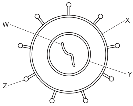
The diagram shows a virus.
Which row correctly identifies the labelled biological molecules?
Options
| W | X | Y | Z | |
|---|---|---|---|---|
| A | RNA | proteins | phospholipids | carbohydrates |
| B | DNA | phospholipids | proteins | proteins |
| C | DNA | proteins | proteins | carbohydrates |
| D | RNA | phospholipids | phospholipids | proteins |
Working
An enveloped virus consists of: a nucleic-acid core (DNA or RNA), a protein capsid surrounding the core, a phospholipid envelope derived from a host-cell membrane, and glycoprotein spikes projecting from the envelope.
Matching the labels on the diagram:
- W — the wavy strand inside the capsid is the genetic material. The diagram shows a single continuous strand with no uracil/T pairing implied, and the only option where the core is a nucleic acid that fits is DNA. → DNA
- X — points to the outer membrane surrounding the capsid. This bilayer is derived from the host cell's plasma membrane and is therefore a phospholipid bilayer.
- Y — points to the capsid shell enclosing the nucleic acid. The capsid is built from repeating protein subunits (capsomeres). → proteins
- Z — points to the spike projecting from the envelope. Spikes are glycoproteins; the principal structural component is the protein chain (with a small carbohydrate attached for receptor binding). → proteins
Only row B (DNA, phospholipids, proteins, proteins) matches all four labels.
Answer
B
B
Background Concept
Viruses are non-cellular particles that straddle the boundary between chemistry and biology: they have a genome but no metabolism, and they can only reproduce inside a host cell. Despite their simplicity, a typical enveloped virus has a clearly layered structure, and each layer is built from a specific biological molecule. Knowing which molecule forms which part is essential to the CIE syllabus topic on viruses.
The four molecular components to recognise are:
- Genome (nucleic-acid core). Every virus contains either DNA or RNA as its genetic material, never both. The nucleic acid may be single-stranded or double-stranded, and in RNA viruses it may be sense (+) or antisense (–). The core holds the information needed to make new viral particles.
- Capsid. A protein shell, built from repeating protein subunits called capsomeres, that surrounds and protects the nucleic acid. In some viruses (e.g. tobacco mosaic virus) the capsid alone is enough; in others it is enclosed by an envelope.
- Envelope. A phospholipid bilayer that wraps around the capsid in "enveloped" viruses (HIV, influenza, herpes, SARS-CoV-2). The envelope is stolen from a host-cell membrane as the new virion buds out, so it has the same phospholipid composition as a plasma membrane.
- Spikes. Projections that stick out from the envelope. They are glycoproteins — a protein with a short carbohydrate chain attached — and they are what recognises and binds to receptors on the next host cell. Without functional spikes, the virus cannot enter a new cell.
Understanding the Question
The question shows a labelled diagram of an enveloped virus (Fig. 6.1) with four pointers, W, X, Y and Z, and asks which row of a four-column table correctly names the biological molecule at each pointer. The command word is "identifies", so the candidate must match each labelled part of the drawing to the correct molecular class. There is one mark, awarded only for the row that is correct in every column.
Approach
- First, decide what each pointer is actually indicating on the drawing (nucleic acid core, capsid, envelope, spike).
- Then, recall the molecule that forms each of those viral structures.
- Finally, scan the four options and find the one whose four entries match.
Step-by-Step Reasoning
-
Identify W. W's pointer leads to the wavy strand inside the inner circle. That is the genome. The strand is drawn as one continuous line, and the only row in which W is a nucleic acid that fits a single-strand representation is one that names either DNA or RNA. Option B names W as DNA, which is the genome of a DNA virus (e.g. a herpesvirus or smallpox). This is consistent.
-
Identify X. X's pointer leads to the outer membrane layer around the capsid. Viral envelopes are derived from host-cell plasma membranes, and plasma membranes are phospholipid bilayers. Option B names X as phospholipids, which is correct.
-
Identify Y. Y's pointer leads to the inner shell enclosing the nucleic acid. That is the capsid, which is assembled from proteins (capsomeres). Option B names Y as proteins, which is correct.
-
Identify Z. Z's pointer leads to one of the projections on the surface of the envelope. Surface projections (spikes/peplomers) are glycoproteins, but their principal structural and functional mass is the protein chain, with a small carbohydrate responsible for receptor binding. At A-level, the spike is correctly classified under the proteins column. Option B names Z as proteins, which is correct.
-
Check the other rows:
- A: W is RNA (could fit an RNA virus, but the other three columns — X as proteins, Y as phospholipids, Z as carbohydrates — are wrong for the parts indicated).
- C: W is DNA (correct), X is proteins (the spike is a glycoprotein, but the pointer for X is the envelope, not the spike — so this is wrong), Y is proteins (could be the capsid, but in row C X has taken the protein assignment) and Z is carbohydrates (spikes are not pure carbohydrate).
- D: W is RNA (could be an RNA virus, but again the other columns don't match).
Only row B places DNA at the genome, phospholipids at the envelope, and proteins at both the capsid and the spike.
Key Takeaways
- An enveloped virus = nucleic-acid core (DNA or RNA) + protein capsid + phospholipid envelope + glycoprotein spikes.
- The envelope is host-cell plasma membrane, so it is a phospholipid bilayer.
- Spikes are glycoproteins — at A-level, classify them under proteins (the carbohydrate is small and the question is asking for the structural molecule).
- A virus has either DNA or RNA, never both.
Common Mistakes
- Calling the spikes "carbohydrates" because they are "glycoproteins". The spike is dominated by its protein component, and at this level it is credited as a protein.
- Labelling the envelope as protein. The envelope is a phospholipid bilayer taken from the host membrane; the proteins embedded in it are far less than the lipid mass.
- Assuming the genetic material must be RNA "because viruses are small". Both DNA and RNA viruses exist; the diagram alone decides which is shown.
- Confusing the capsid with the envelope. The capsid is the inner protein shell that directly surrounds the nucleic acid; the envelope is the outer phospholipid membrane.
Things to Be Careful About
- Read every column of the chosen row before selecting it; one mark is lost if any of the four is wrong.
- "Glycoprotein" is not the same as "carbohydrate". The spike is a glycoprotein; the molecule that builds the spike is protein.
- The envelope and the spike are both on the outside of the virus, but they are different molecules — don't swap them.
- A phospholipid bilayer is a specific structure (two layers of phospholipids with hydrophilic heads outward). Don't confuse it with a single layer of phospholipid or with a generic "lipid".
The iodine test and the Benedict’s test were carried out on samples of a starch solution. Amylase was added to another sample of the starch solution and incubated for 10 minutes. The Benedict’s test was then carried out on this sample.
What were the results of the tests?
Options
| iodine test before adding amylase | Benedict’s test before adding amylase | Benedict’s test after adding amylase | |
|---|---|---|---|
| A | ✗ | ✓ | ✓ |
| B | ✓ | ✓ | ✗ |
| C | ✓ | ✗ | ✓ |
| D | ✗ | ✗ | ✗ |
key
✓ = positive result
✗ = negative result
Working
- Iodine test on the starch solution: starch is present, so iodine turns blue–black → positive ().
- Benedict's test on the starch solution: starch is a non-reducing polysaccharide, so Benedict's remains blue → negative ().
- After adding amylase and incubating for 10 minutes: amylase hydrolyses starch into maltose, a reducing sugar, so Benedict's gives a brick-red precipitate → positive ().
- Pattern of results: , , .
Answer
C
C
Background Concept
Two qualitative biochemical tests sit at the centre of this question, together with one enzyme-catalysed reaction.
Iodine test for starch. Iodine (in potassium iodide solution, /) slots inside the helical coils of amylose, the unbranched component of starch. The triiodide–amylose complex absorbs visible light strongly and appears blue–black. A negative result is the yellow-brown colour of iodine solution itself. The test is therefore specific to starch (and related polymers such as glycogen, which gives a red-brown colour).
Benedict's test for reducing sugars. Benedict's reagent contains copper(II) sulfate in alkaline citrate. A reducing sugar has a free aldehyde (–CHO) or ketone group capable of reducing blue to brick-red on heating. Common reducing sugars are glucose, fructose, maltose and lactose. Starch is a polysaccharide in which every glucose unit is locked into a glycosidic bond (α-1,4 and α-1,6), so it has no free reducing end available to react — it is a non-reducing sugar and gives a negative Benedict's test.
Amylase action. Amylase is a hydrolytic enzyme that catalyses the breakdown of the α-1,4 glycosidic bonds in starch. The disaccharide product is maltose, which has a free anomeric carbon and so is a reducing sugar. The hydrolysis is fast at body temperature, so a 10-minute incubation is more than enough to give a clearly positive Benedict's test.
Understanding the Question
We are given three test results to predict for a single starting material (a starch solution):
- Iodine test on the starch solution itself.
- Benedict's test on the starch solution itself.
- Benedict's test on the same solution after amylase has been allowed to act for 10 minutes.
The command word is implicit — choose the row whose tick/cross pattern matches these three predictions. The reasoning is: think about what each test detects, then ask whether the substrate (or its breakdown product) contains the relevant chemical group.
Approach
For each of the three columns:
- Decide which functional group the test detects.
- Decide whether the substance present in the tube (starch, or starch + amylase products) contains that group.
- Translate the answer into a (positive) or (negative) and read off the matching row.
Step-by-Step Reasoning
- Column 1 — iodine test on the starch solution. Starch is the very substance the test is designed to detect. A blue–black colour will appear, so the result is positive ().
- Column 2 — Benedict's test on the starch solution. Starch has no free reducing group; every potential reducing carbon is engaged in a glycosidic bond. Heating with Benedict's reagent produces no brick-red precipitate; the solution stays blue. Result: negative ().
- Column 3 — Benedict's test after amylase, 10 min incubation. Amylase cleaves the α-1,4 glycosidic bonds in starch, producing maltose (and some limit dextrins). Maltose is a reducing sugar, so on heating with Benedict's reagent a brick-red precipitate forms. Result: positive ().
- The pattern , , is option C.
Why the distractors are wrong:
- A claims the iodine test is negative and Benedict's is positive on the starch solution — the opposite of correct (starch is present, and starch does not reduce Benedict's).
- B claims the Benedict's test is positive on starch (incorrect — starch is non-reducing) and negative after amylase (incorrect — amylase produces the reducing sugar maltose).
- D claims every test is negative, ignoring both the presence of starch and the amylase-catalysed hydrolysis.
Key Takeaways
- Iodine + starch → blue–black (positive); no starch → yellow-brown (negative).
- Benedict's + reducing sugar → brick-red precipitate; no reducing sugar (or a non-reducing sugar such as starch or sucrose) → stays blue.
- Starch is a non-reducing polysaccharide because its glycosidic bonds leave no free anomeric –OH on a reducing carbon.
- Amylase hydrolyses starch to maltose, a reducing disaccharide — so a starch + amylase mixture gives a positive Benedict's test after sufficient incubation.
Common Mistakes
- Confusing starch with a reducing sugar. Starch is a polysaccharide and is non-reducing; do not give it a positive Benedict's test.
- Forgetting that iodine must be added without heating, while Benedict's reagent must be heated in a water bath — a test carried out at room temperature will not register.
- Assuming amylase 'denatures' the starch. Amylase is not denaturing anything; it is hydrolysing glycosidic bonds.
- Confusing the colour of a positive iodine test (blue–black) with that of a positive Benedict's test (brick-red).
Things to Be Careful About
- The Benedict's test before amylase must be negative: a common slip is to award it a positive because 'starch is a sugar'. Starch is a sugar in the biochemical sense (a polysaccharide of glucose), but the test specifically requires a free reducing group.
- The 10-minute incubation is important: a very short incubation (a few seconds) might leave the starch largely unhydrolysed, but the question states 10 minutes, which is more than sufficient for amylase to act.
- The key in the question tells you explicitly what and mean — read it before matching rows.
Which row about -glucose and -glucose molecules is correct?
Options
| carbon atom on which the OH position is different | cellulose contains both molecules | |
|---|---|---|
| A | 1 | no |
| B | 1 | yes |
| C | 4 | no |
| D | 4 | yes |
Working
- α-glucose and β-glucose differ only in the position of the −OH group on carbon 1 (not carbon 4). This rules out C and D.
- Cellulose is a polymer of β-glucose only, joined by β-1,4-glycosidic bonds; it does not contain α-glucose. This rules out B (which says yes).
- Only row A (carbon 1, no) is consistent with both facts.
Answer
A
A
Background Concept
α-Glucose and β-glucose are two isomeric forms of the same six-carbon sugar. They have identical molecular formulae and the same functional groups, but they differ in the orientation of the hydroxyl (−OH) group attached to carbon 1 (the anomeric carbon). In α-glucose the −OH on C1 sits below the plane of the ring; in β-glucose it sits above the plane. This small stereochemical difference has major structural consequences:
- Starch and glycogen are polymers of α-glucose (α-1,4- and α-1,6-glycosidic bonds), giving a helical, energy-storage molecule.
- Cellulose is a polymer of β-glucose only, with β-1,4-glycosidic bonds. The alternating orientation of the glucose units produces long, straight, unbranched chains that hydrogen-bond together into strong microfibrils — ideal for plant cell walls.
Understanding the Question
The question presents a small table with two statements and four possible combinations. The candidate must decide:
- On which carbon atom does the −OH position differ between α- and β-glucose?
- Does cellulose contain both α- and β-glucose molecules?
A correct row must have both statements right.
Approach
Test each row against the two facts above. Eliminate any row that gets either fact wrong.
Step-by-Step Reasoning
- Carbon atom question: α- and β-glucose differ at C1 (the anomeric carbon), not C4. So rows claiming "4" (C and D) are eliminated immediately.
- Cellulose question: Cellulose is built from β-glucose monomers linked by β-1,4-glycosidic bonds; it contains no α-glucose. Therefore any row stating "yes — cellulose contains both" (B and D) is wrong.
- The only row that survives both filters is A (carbon 1, no).
Key Takeaways
- The α/β distinction is defined by the −OH on C1.
- Starch/glycogen = α-glucose; cellulose = β-glucose only.
- A 1-mark MCQ like this often disguises two independent facts behind a single letter — always check every column.
Common Mistakes
- Saying the difference is on C4: a very common error arising from confusing the α/β designation with the C4 epimer (e.g. galactose vs glucose), or with where the next glycosidic bond forms in cellulose.
- Believing cellulose "contains both" forms because it is a structural carbohydrate and structural carbohydrates are sometimes loosely associated with starch in memory.
Things to Be Careful About
- In cellulose the glycosidic bond is formed at C1, but it is a β-1,4 bond (the bond also involves C4 of the neighbouring glucose). The α/β label, however, refers to the configuration at C1, not the bond position. Don't confuse "1,4 bond" with "α vs β at C4".
- α-Amylose (in starch) and cellulose are both built on 1,4 linkages — what differs is the α or β configuration at C1, which determines whether the chain coils or straightens.
Which molecules are monosaccharides?
1 ribose
2 glucose
3 deoxyribose
4 sucrose
Options
A 1, 2 and 3
B 1, 2 and 4
C 1, 3 and 4
D 2, 3 and 4
Answer
A
A
Background Concept
Monosaccharides are the simplest carbohydrate monomers — single sugar units that cannot be hydrolysed into smaller carbohydrates. They share the general formula (for ) and are classified by the number of carbon atoms they contain:
- Trioses (): e.g. glyceraldehyde
- Pentoses (): e.g. ribose and deoxyribose
- Hexoses (): e.g. glucose, fructose, galactose
Two monosaccharide units joined by a glycosidic bond form a disaccharide (e.g. sucrose, maltose, lactose), while long chains of monosaccharides form polysaccharides (e.g. starch, glycogen, cellulose).
Ribose is a pentose found in RNA, NAD, FAD, and ATP. Deoxyribose is a pentose (it lacks one oxygen atom at C2) found in DNA. Glucose is the most common hexose — the main respiratory substrate and the monomer of starch, glycogen and cellulose.
Understanding the Question
This is a Paper 1 multiple choice question asking the candidate to identify which of the four listed molecules are monosaccharides. Three correct answers and one distractor must be chosen.
The command word is implicit ("Which molecules are monosaccharides?"), so the candidate simply has to recognise each sugar and classify it.
Approach
Test each numbered molecule individually:
- Ribose — pentose sugar (5 C) → monosaccharide ✓
- Glucose — hexose sugar (6 C) → monosaccharide ✓
- Deoxyribose — pentose sugar (5 C), found in DNA → monosaccharide ✓
- Sucrose — composed of glucose + fructose linked by a glycosidic bond → disaccharide ✗
Therefore, the monosaccharides are 1, 2 and 3.
Step-by-Step Reasoning
- Ribose () is a single sugar unit, hence a monosaccharide.
- Glucose () is a single sugar unit, hence a monosaccharide.
- Deoxyribose () is a single sugar unit, hence a monosaccharide.
- Sucrose is formed when a glucose molecule and a fructose molecule are joined by an α-1,β-2 glycosidic bond; with two monomer units it is a disaccharide, not a monosaccharide.
The combination 1, 2 and 3 corresponds to option A.
Key Takeaways
- All monosaccharides are single sugar units (cannot be hydrolysed into smaller sugars).
- The common monosaccharides in biology are the pentoses ribose and deoxyribose (nucleic acids, ATP) and the hexoses glucose, fructose and galactose.
- Sucrose, maltose and lactose are disaccharides — composed of two monosaccharide units joined by a glycosidic bond.
Common Mistakes
- Confusing sucrose with a monosaccharide because its name ends in "-ose". The "-ose" suffix indicates a sugar but not its size; sucrose is specifically a disaccharide.
- Forgetting that deoxyribose is still a monosaccharide even though it is "deoxy" — it is simply a pentose lacking one oxygen compared to ribose, not a smaller subunit.
Things to Be Careful About
- The classification depends on the number of monomer units, not on the number of carbon atoms or whether the name contains "-ose".
- "Reducing" vs "non-reducing" sugars is a separate property: glucose is a reducing sugar, sucrose is non-reducing, but both are classified here purely on whether they are mono- or disaccharides.
Many animals use triglycerides for long-term energy storage.
Which statements are correct additional functions of triglycerides?
1 They provide buoyancy in some marine animals.
2 They are the main components of cell membranes.
3 They are used as thermal insulation.
Options
A 1, 2 and 3
B 1 and 2 only
C 1 and 3 only
D 2 and 3 only
Working
- Statement 1: Triglycerides are less dense than water, so stored deposits of fat (e.g. blubber) help some marine mammals float. ✓
- Statement 2: Cell membranes are made of phospholipids (not triglycerides); the phosphate head and hydrophilic/hydrophobic regions of phospholipids form the bilayer. ✗
- Statement 3: Adipose tissue stores triglycerides and acts as a thermal insulator (e.g. blubber in whales and seals, subcutaneous fat in mammals). ✓
Answer
C
C
Background Concept
Triglycerides are a class of lipid formed from one glycerol molecule esterified to three fatty acid chains. They are hydrophobic, energy-dense, and stored in specialised cells of adipose tissue. Their key properties — low density relative to water, poor heat conduction, and high energy yield on oxidation — give them several biological roles beyond simple energy storage.
Phospholipids, by contrast, have only two fatty acid chains attached to a glycerol, with a phosphate group (often joined to another polar group) occupying the third position. This gives them an amphipathic character (hydrophilic head, hydrophobic tails) that allows them to spontaneously form bilayers — the basis of every cell membrane. Triglycerides have no such polar head and cannot form bilayers.
Understanding the Question
This is a multiple-choice question. The stem tells us triglycerides are used for long-term energy storage, and asks which of the three additional statements are also correct functions of triglycerides. The command word is implicit: select the option that contains only the correct statements. We need to evaluate each statement on its biological merits and identify which are true.
Approach
For each statement, recall the physical/chemical property of triglycerides that would support the claimed function, and rule out any function that properly belongs to a different lipid class.
Step-by-Step Reasoning
Statement 1 — buoyancy in marine animals: Triglycerides have a density of around 0.9 g cm⁻³, which is less than that of water (~1.0 g cm⁻³). Animals that store large quantities of triglyceride (e.g. seals, whales, some fish) are therefore more buoyant. The lipid-rich blubber of marine mammals aids floating as well as insulation. ✓ Correct.
Statement 2 — main components of cell membranes: This is a classic misconception trap. Membranes are made predominantly of phospholipids, not triglycerides. The phosphate head group is essential — it makes one end of the molecule hydrophilic while the fatty acid tails remain hydrophobic, allowing the bilayer structure. Triglycerides lack the phosphate group and would not form a stable bilayer. ✗ Incorrect.
Statement 3 — thermal insulation: Adipose tissue (the specialised connective tissue in which triglycerides are stored) is a poor conductor of heat. Layers of subcutaneous fat and blubber reduce heat loss from the body core to the environment, which is vital for animals in cold water (e.g. whales, seals) and for general thermoregulation in mammals. ✓ Correct.
Statements 1 and 3 are correct; statement 2 is wrong. This matches option C (1 and 3 only).
Key Takeaways
- Triglycerides = energy storage + buoyancy + thermal insulation (and, in some animals, waterproofing).
- Phospholipids, NOT triglycerides, form the structural framework of cell membranes.
- Distinguishing the two lipid classes by their structural features (phosphate head vs none) directly explains their different biological roles.
Common Mistakes
- Choosing D (2 and 3) because students remember "lipids are in membranes" without distinguishing triglycerides from phospholipids.
- Choosing A (all three) because each statement sounds plausible when read in isolation.
- Confusing cholesterol (a lipid but neither triglyceride nor phospholipid) with triglycerides.
Things to Be Careful About
- "Lipid" is a broad category — never use it as a synonym for triglyceride. The mark scheme rewards the specific molecule.
- Buoyancy and insulation both rely on the low density and low thermal conductivity of stored triglycerides, but these are two separate functions and should be credited as such.
- Phospholipids (not triglycerides) are amphipathic and form bilayers; this is the key reason statement 2 must be rejected.
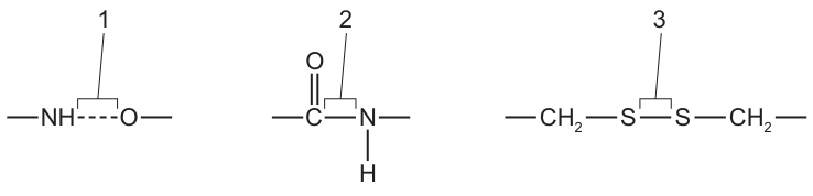
The diagrams show three different bonds.
Which bonds are found in proteins?
Options
A 1, 2 and 3
B 1 and 2 only
C 1 and 3 only
D 2 and 3 only
Working
- Bond 1 is a hydrogen bond (N–H···O). Hydrogen bonds form the secondary structure of proteins (α-helix, β-pleated sheet).
- Bond 2 is a peptide bond (–C(=O)–N(H)–) formed by condensation between two amino acids; peptide bonds form the primary structure of proteins.
- Bond 3 is a disulfide bridge (–S–S–) between two cysteine side chains; disulfide bridges stabilise the tertiary structure of proteins.
- All three bonds are found in proteins.
Answer
A
A
Background Concept
Proteins are polymers of amino acids joined by peptide bonds (primary structure). The polypeptide chain then folds into specific shapes held in place by a variety of weaker interactions and one covalent link:
- Primary structure — the linear sequence of amino acids linked by peptide (covalent) bonds.
- Secondary structure — α-helices and β-pleated sheets, stabilised by hydrogen bonds between the N–H of one peptide bond and the C=O of another.
- Tertiary structure — the 3-D folding held by hydrogen bonds, ionic bonds, hydrophobic interactions, and disulfide bridges (covalent S–S bonds) between two cysteine residues.
- Quaternary structure — assembly of polypeptide subunits (e.g. haemoglobin); uses the same bonding types as tertiary structure.
Understanding the Question
The diagram shows three different bonds labelled 1, 2 and 3. The question asks which of them occur in proteins. The three bonds shown are typical of the bonds listed above: a hydrogen bond (1), a peptide bond (2) and a disulfide bridge (3). All three appear in proteins — at different levels of structure — so the correct answer is "all of them".
Approach
For each labelled bond, recognise the bond type from its atoms and then recall the level of protein structure with which it is associated. Any bond that contributes to any level of protein structure counts as being "found in proteins".
Step-by-Step Reasoning
- Bond 1: –NH···O–. A dotted line between an N–H group and an oxygen represents a hydrogen bond. Hydrogen bonds are abundant in proteins: they hold α-helices and β-sheets together (secondary structure) and are also present in tertiary and quaternary structure. ✓
- Bond 2: –C(=O)–N(H)–. A carbon double-bonded to oxygen and single-bonded to a nitrogen is the peptide bond formed by a condensation reaction between two amino acids. Every protein contains many peptide bonds along its backbone. ✓
- Bond 3: –CH₂–S–S–CH₂–. Two sulfur atoms covalently bonded between two methylene (–CH₂–) groups is a disulfide bridge, formed by oxidation of two cysteine –SH side chains. Disulfide bridges are covalent cross-links that lock in the tertiary (and sometimes quaternary) structure of proteins such as insulin, immunoglobulins and many extracellular enzymes. ✓
All three bonds are present in proteins, so option A (1, 2 and 3) is correct.
Key Takeaways
- Peptide bonds (primary), hydrogen bonds (secondary and others) and disulfide bridges (tertiary) are all characteristic bonds of proteins.
- Being able to recognise each bond type from a structural diagram is a routinely tested skill.
- The "bonding in proteins" sub-topic also includes ionic bonds and hydrophobic interactions, but they are not shown here.
Common Mistakes
- Choosing C (1 and 3 only) by forgetting that peptide bonds are themselves bonds in proteins (they are sometimes thought of as "between" amino acids rather than "in" the protein).
- Choosing B (1 and 2 only) by not recalling that disulfide bridges form in proteins containing cysteine.
- Choosing D (2 and 3 only) by not recognising the dotted line as a hydrogen bond, or by thinking hydrogen bonds occur only in water/cytoplasm.
Things to Be Careful About
- A dotted line (····) is the standard way to draw a hydrogen bond; a single solid line is a covalent bond.
- The peptide bond is shown as a C(=O)–N(H) link, not a C–O–N (ester) link — check the atoms carefully.
- A disulfide bridge is two single-bonded sulfurs, not a double bond (C=S) and not a thioether (C–S–C).
What describes the quaternary structure of a collagen molecule?
Options
A polypeptides that form an -helix, which are held together by covalent bonds
B polypeptides that form a double helix, which are held together by hydrogen bonds
C four polypeptides that are held together by covalent bonds, which form a helix
D three polypeptides that form helix structures, which are held together by hydrogen and covalent bonds
Answer
Collagen is a fibrous protein whose quaternary structure consists of three polypeptide chains, each coiled into a left-handed helix, twisted together to form a right-handed triple helix. The three chains are held together by hydrogen bonds between adjacent chains, and are additionally stabilised by covalent cross-links between lysine and hydroxylysine residues on neighbouring chains.
D
D
Background Concept
Proteins have four levels of structure. Primary structure is the linear sequence of amino acids linked by peptide bonds. Secondary structure arises from hydrogen bonding between peptide bonds along the backbone, producing regular patterns such as the α-helix and β-pleated sheet. Tertiary structure is the overall 3-D folding of a single polypeptide chain, stabilised by interactions between R-groups (hydrogen bonds, ionic bonds, disulfide bridges and hydrophobic interactions). Quaternary structure exists only in proteins with two or more polypeptide chains (subunits) and describes how those subunits are arranged and held together.
Collagen is the most abundant fibrous protein in animals and the main component of connective tissue (tendons, bone, cartilage, skin, blood-vessel walls). It is synthesised as three polypeptide α-chains, each of which is a left-handed helix (its secondary structure). These three chains wrap around each other in a right-handed triple helix — this is collagen's quaternary structure, also called tropocollagen. The three α-chains are held together by hydrogen bonds between the chains, and the molecule is further stabilised by covalent cross-links that form between lysine (and hydroxylysine) side chains on adjacent α-chains. This cross-linking gives collagen fibres their great tensile strength.
Understanding the Question
The stem asks specifically about the quaternary structure of collagen, which means the question is testing what collagen's three subunits look like and how they are held to each other — not the secondary structure (the single-chain helix) and not primary structure (amino-acid sequence).
Approach
Match each option against the two key facts: (1) how many polypeptide chains make up collagen (three), and (2) what kinds of bond hold them together (hydrogen and covalent).
Step-by-Step Reasoning
- Option A is wrong on two counts. Collagen's α-chains do not form an α-helix — the regular α-helix is a different secondary structure seen in proteins such as keratin or myoglobin, but collagen has its own unique helical secondary structure. Also, "polypeptides held together by covalent bonds" alone omits the essential hydrogen bonding between chains.
- Option B describes DNA, not a protein. DNA's double helix has two polynucleotide strands held together by hydrogen bonds between complementary base pairs.
- Option C gives the wrong number — four polypeptide chains is the quaternary structure of haemoglobin (two α-globin and two β-globin chains), not collagen. Hemoglobin is also a globular protein, whereas collagen is fibrous.
- Option D is correct: three polypeptide chains, each forming a helix, held together by both hydrogen bonds and covalent bonds. This matches collagen's quaternary structure exactly.
Key Takeaways
- Collagen's quaternary structure = three α-chains wound into a right-handed triple helix.
- The triple helix is stabilised by hydrogen bonds AND covalent cross-links between lysine/hydroxylysine residues.
- Be careful not to confuse collagen's secondary structure (the single α-chain helix) with its quaternary structure (the triple helix of three chains).
- Don't confuse the quaternary structure of collagen (3 chains, fibrous) with that of haemoglobin (4 chains, globular).
Common Mistakes
- Picking A because of a vague recollection that "collagen is helical" — but α-helix is the wrong secondary structure and the bonding description is incomplete.
- Picking B because "helix with hydrogen bonds" sounds familiar — this is the description of DNA.
- Picking C because of confusion with haemoglobin, which does have four polypeptide chains.
Things to Be Careful About
- "Polypeptides" (plural) = quaternary structure; a single polypeptide's fold = tertiary structure; the helical pattern in one chain = secondary structure. Always match the level asked to the description given.
- "Hydrogen bonds" alone are insufficient to describe collagen's quaternary structure — the covalent cross-links are an essential part of the answer and must be mentioned to gain full credit on free-response versions of this fact.
The diagram shows the structure of the enzyme amylase.
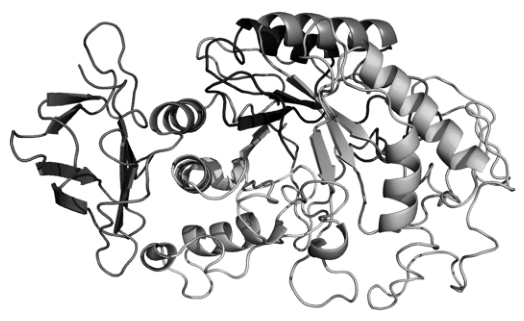
Which level of protein structure results in the folding of amylase into its globular shape?
Options
A primary
B secondary
C tertiary
D quaternary
Working
- Primary structure = linear sequence of amino acids linked by peptide bonds; it does not produce folding.
- Secondary structure = coiling/folding into α-helices and β-pleated sheets (held by H-bonds between backbone groups); still not the overall 3D globular shape.
- Tertiary structure = further folding of the polypeptide into a specific 3D shape, including a globular form, stabilised by interactions between R groups (e.g. disulfide bridges, ionic, hydrogen and hydrophobic interactions).
- Quaternary structure = association of more than one polypeptide subunit; amylase is a single folded chain, not a multi-subunit assembly in this context.
Answer
C
C
Background Concept
Proteins are built from amino acids joined by peptide bonds, and they fold into four recognised levels of structure:
- Primary structure — the linear sequence of amino acids in a polypeptide, held together by peptide bonds between the carboxyl group of one amino acid and the amino group of the next. This is simply the order of residues; it produces no 3D shape on its own.
- Secondary structure — the first stage of folding, where hydrogen bonds form between the –NH and C=O groups of the peptide backbone. This produces regular local motifs, mainly α-helices (a coil) and β-pleated sheets (zig-zag strands).
- Tertiary structure — the further folding of the whole polypeptide into its overall 3D shape. For many proteins this is a compact, roughly spherical globular form. It is stabilised by interactions between the side chains (R groups) of different amino acids, including hydrogen bonds, ionic bonds, disulfide bridges (covalent, between cysteine residues) and hydrophobic interactions that drive non-polar R groups into the interior.
- Quaternary structure — only present in proteins with more than one polypeptide subunit (e.g. haemoglobin, with four chains). It describes how those subunits pack together; a single-chain enzyme like amylase does not have quaternary structure.
Enzymes are typically globular proteins. The specific 3D shape of the enzyme creates the active site where substrate binds.
Understanding the Question
The figure shows a ribbon diagram of the enzyme amylase folded into a compact, roughly spherical 3D form. The question asks: at which level of protein structure does this globular folding arise? The four options correspond to primary, secondary, tertiary and quaternary structure. We must pick the level that produces the overall 3D globular shape of the molecule shown.
Approach
Match each option to what it actually represents in a protein:
- Primary → sequence only (no shape).
- Secondary → α-helices and β-sheets (local motifs, not the whole-globule fold).
- Tertiary → the full 3D folding of a single polypeptide into its functional shape, often globular.
- Quaternary → only relevant for multi-subunit proteins; amylase here is a single folded chain.
The only level that creates the overall globular 3D shape of a single-chain protein is the tertiary structure.
Step-by-Step Reasoning
- The ribbon diagram of amylase shows helices, sheets and loops packed together into a compact, ball-like shape. The helices and sheets themselves are features of secondary structure, but the question asks what produces the globular folding of the whole molecule — that is the next level up.
- Tertiary structure is the folding of the entire polypeptide chain into its 3D conformation, driven by R-group interactions. This is what gives amylase (and other globular enzymes) their roughly spherical shape and positions the active-site residues correctly.
- Quaternary structure would only be the answer if amylase were a multi-subunit protein and the question referred to how the subunits pack together. Although some amylases are multi-domain, the ribbon diagram here shows a single folded chain, and the question is about the globular fold itself.
- Therefore the answer is C — tertiary.
Key Takeaways
- Primary = amino acid sequence.
- Secondary = α-helices and β-sheets (H-bonding in the backbone).
- Tertiary = overall 3D globular folding of one polypeptide (R-group interactions).
- Quaternary = assembly of multiple polypeptide subunits.
- Enzymes are globular proteins; their tertiary structure creates the active site.
Common Mistakes
- Choosing B (secondary) because you can see helices and sheets in the diagram. The helices/sheets are secondary structure, but the overall globular fold is the next level up — tertiary.
- Choosing D (quaternary) by assuming any complex-looking 3D protein is multi-subunit. Quaternary structure only exists when two or more separate polypeptide chains are present.
- Choosing A (primary): primary structure is a sequence, not a shape, and so cannot by itself fold a protein.
Things to Be Careful About
- Distinguish what each level describes from what each level produces visually: secondary produces the helices/sheets; tertiary produces the whole 3D fold.
- The wording "folding of amylase into its globular shape" specifically points to tertiary, not secondary.
- If a question shows a protein with several chains packed together and asks about the overall assembly, then quaternary is the answer — read the question carefully.
Which statement explains why large volumes of water maintain a constant temperature when the air temperature increases?
Options
A The latent heat of vaporisation of water is high.
B The specific heat capacity of water is high.
C Each water molecule can form a hydrogen bond with a maximum of three water molecules.
D Water is able to evaporate rapidly from a surface with little loss of heat.
Working
The question asks why large volumes of water resist temperature change when the surrounding air warms up.
- A – Latent heat of vaporisation relates to evaporative cooling, not to a bulk volume resisting warming.
- B – Specific heat capacity is the energy required to raise the temperature of 1 kg of a substance by 1 °C. A high value means much energy must be absorbed for a small rise in temperature, so a large body of water heats up only slowly.
- C – A water molecule can actually form up to four hydrogen bonds (two H donors, two lone-pair acceptors), and this does not explain temperature stability.
- D – Evaporation removes substantial heat from the surface, and 'rapidly' is inconsistent with 'little loss of heat'.
Answer
B
B
Background Concept
Water has several unusual physical properties arising from its extensive hydrogen bonding. Two that matter most for thermal behaviour are:
- Specific heat capacity (SHC) – the energy needed to raise the temperature of 1 kg of a substance by 1 °C. For liquid water this is about , which is unusually high.
- Latent heat of vaporisation – the energy needed to convert 1 kg of liquid to gas without a change in temperature (≈ for water). This large value explains why evaporative cooling is so effective (e.g. sweating).
These two quantities describe different processes: SHC governs how temperature changes when heat is added or removed from the bulk; latent heat governs phase changes at constant temperature.
Understanding the Question
The stem asks why a large volume of water stays at a roughly constant temperature while the air around it warms up. This is a temperature-buffering situation: heat is being transferred into the water from the air, but the water's temperature barely rises.
Approach
We need the property that makes a large mass of water resist warming. That points directly to specific heat capacity — the larger the mass and the higher the SHC, the more heat energy must be absorbed to produce a given temperature change. A swimming pool, lake or ocean therefore changes temperature only slowly, which is also why aquatic environments are thermally stable habitats.
Step-by-Step Reasoning
- A – Latent heat of vaporisation matters when water is evaporating; the question is about a closed body of water being warmed, not losing mass. Even where evaporation occurs, it cools the water, it does not buffer it against warming.
- B – Because SHC is high, each °C rise in temperature demands a lot of energy. For a large volume, the absolute heat required to raise its temperature by even 1 °C is enormous, so fluctuations in air temperature produce only very small changes in water temperature. ✓
- C – The hydrogen-bonding claim is incorrect in number (water can form up to four H-bonds per molecule) and irrelevant in any case to thermal buffering.
- D – Rapid evaporation is associated with substantial heat loss (high latent heat of vaporisation), so the option is internally contradictory and again describes cooling rather than buffering.
Key Takeaways
- High specific heat capacity → water resists temperature change; useful for organisms because aquatic and internal environments stay thermally stable.
- High latent heat of vaporisation → evaporation removes large amounts of heat; the basis of cooling by sweating and transpiration.
- These are two distinct thermal properties; do not confuse them.
Common Mistakes
- Choosing A because 'latent heat is high' sounds relevant — it is a real property of water but it explains cooling by evaporation, not resistance to warming of a bulk volume.
- Choosing D because evaporation seems to 'stabilise' temperature — but this option is also factually muddled (rapid evaporation cannot involve 'little loss of heat').
- Choosing C if you remember that water is hydrogen-bonded but forget that bonding patterns are not what stabilises temperature.
Things to Be Careful About
- Read the wording carefully: 'constant temperature when the air temperature increases' describes resistance to warming, not cooling by evaporation.
- Distinguish SHC (sensible heat, temperature change) from latent heat (latent heat, phase change at constant temperature).
- The biological relevance: many organisms exploit water's high SHC (thermoregulation, aquatic habitats, evaporative cooling), but each effect has its own physical basis.
Which statements about the mode of action of an enzyme are correct?
1 Some enzymes have a region on their surface to which the substrate has a complementary shape.
2 Some enzymes and their substrates can change shape slightly as the substrate enters the active site.
3 Enzymes permanently change shape when they react with their substrates to form an enzyme–substrate complex.
Options
A 1, 2 and 3
B 1 and 2 only
C 1 and 3 only
D 2 and 3 only
Working
- Statement 1 describes the lock-and-key model: the active site has a fixed shape complementary to the substrate. ✓ correct
- Statement 2 describes the induced-fit model: the active site (and sometimes the substrate) moulds around the substrate as it binds. ✓ correct
- Statement 3 is wrong: any shape change in the active site is temporary; the enzyme returns to its original conformation after releasing the products, so the enzyme is not used up.
Therefore only statements 1 and 2 are correct.
Answer
B
B
Background Concept
Enzymes are biological catalysts — globular proteins with a specific region called the active site where the substrate binds. Two models describe how this binding occurs:
- Lock-and-key hypothesis (Fischer, 1894): the active site has a rigid, pre-formed shape that is exactly complementary to the substrate, just as a key fits a lock. The substrate slots in without altering the enzyme's structure.
- Induced-fit hypothesis (Koshland, 1958): the active site is not a perfect fit initially. As the substrate enters, the enzyme (and sometimes the substrate) changes shape slightly so that the active site moulds around the substrate, maximising contact and catalytic efficiency. When the products leave, the enzyme relaxes back to its original conformation.
The key point is that any conformational change is reversible and temporary — enzymes are not consumed or permanently altered by the reaction, which is why one enzyme molecule can catalyse many reactions in succession.
Understanding the Question
This is a multiple-choice question asking which of three statements about enzyme action are correct. Each statement maps onto one of the key ideas above:
- Statement 1 → lock-and-key
- Statement 2 → induced-fit
- Statement 3 → a (wrong) claim of a permanent shape change
The task is to identify the one statement that is biologically false.
Approach
Evaluate each statement independently against the two accepted models of enzyme action, then select the option that contains all and only the correct statements.
Step-by-Step Reasoning
Statement 1: "Some enzymes have a region on their surface to which the substrate has a complementary shape."
- This is the classic description of the lock-and-key model. The word "some" is important — it is accurate because the lock-and-key model is one valid description of enzyme–substrate interaction, even though induced-fit is often a more accurate picture for many enzymes. ✔ Correct.
Statement 2: "Some enzymes and their substrates can change shape slightly as the substrate enters the active site."
- This is the induced-fit model. Note that both the enzyme and the substrate may adjust — for example, hexokinase closes around glucose, and the substrate itself can be slightly distorted (a process called "substrate strain") to lower the activation energy. ✔ Correct.
Statement 3: "Enzymes permanently change shape when they react with their substrates to form an enzyme–substrate complex."
- This is the critical error. The induced-fit model says the active site changes shape, but only temporarily while the substrate is bound. After the products are released, the enzyme returns to its original tertiary structure. A permanent change would mean the enzyme could only catalyse one reaction, which contradicts the fundamental property of enzymes as reusable catalysts. ✘ Incorrect.
Only 1 and 2 are correct → option B.
Key Takeaways
- The lock-and-key model proposes a rigid, pre-shaped complementary active site.
- The induced-fit model proposes a flexible active site that moulds around the substrate (and the substrate may distort too).
- Any shape change during catalysis is temporary and reversible — enzymes are not permanently altered and can be reused many times.
Common Mistakes
- Choosing A (1, 2 and 3) by accepting the word "permanently" without thinking it through.
- Choosing C (1 and 3) by forgetting that induced-fit is the more widely accepted model and is therefore a correct statement.
- Choosing D (2 and 3) by rejecting the lock-and-key model outright — but the question deliberately says "some enzymes", which is true.
Things to Be Careful About
- The qualifier "some" in statements 1 and 2 is doing real work: it makes both statements defensible, because both models have evidence supporting them. Examiners often include "some" to make a statement harder to dismiss as absolute.
- The word "permanently" in statement 3 is the giveaway that it is wrong. If you see "always", "never", "permanently" or "irreversibly" in a biology statement, double-check whether the claim is actually universal.
The graph shows the effect of increasing substrate concentration on the rate of reaction of two enzymes that catalyse reactions with the same substrate.
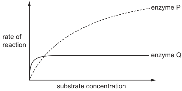
Which statement is correct?
Options
A Enzyme Q has a higher and a higher affinity for the substrate than enzyme P.
B Enzyme Q has a higher and a lower affinity for the substrate than enzyme P.
C Enzyme Q has a lower and a higher affinity for the substrate than enzyme P.
D Enzyme Q has a lower and a lower affinity for the substrate than enzyme P.
Working
- is the substrate concentration at which the rate of reaction is half of .
- Enzyme Q reaches its (lower) plateau at very low substrate concentration, so the substrate concentration giving half of Q's is very small → Q has a low .
- Enzyme P rises gradually and needs a much higher substrate concentration to approach its , so the substrate concentration giving half of P's is larger → P has a higher than Q.
- A low corresponds to high affinity (the enzyme needs little substrate to work efficiently), and a high corresponds to low affinity.
- Therefore, enzyme Q has a lower and a higher affinity for the substrate than enzyme P.
Answer
C
C
Background Concept
Enzymes are biological catalysts whose rate of reaction depends on substrate concentration. As substrate concentration rises, the rate increases until it plateaus at (the maximum rate), when the enzyme active sites are saturated. The shape of this curve is described by the Michaelis–Menten model.
The Michaelis constant, , is defined as the substrate concentration at which the rate of reaction is half of . It is a measure of the enzyme's affinity for its substrate:
- Low → the enzyme reaches half its maximum rate at a low substrate concentration → high affinity (binds substrate efficiently).
- High → the enzyme needs a lot of substrate to reach half its maximum rate → low affinity (binds substrate less efficiently).
So and affinity are inversely related.
Understanding the Question
The graph shows two enzymes, P and Q, both acting on the same substrate. They differ in two ways:
- Enzyme Q plateaus very quickly (at a low substrate concentration) but at a lower .
- Enzyme P rises more gradually and reaches a much higher .
The question asks which combination of and affinity correctly describes enzyme Q compared to enzyme P. The trap in this question is that students often confuse with , or assume that a higher plateau means a higher .
Approach
For each enzyme, identify (visually) the substrate concentration at which the rate is half of that enzyme's own . That point on the x-axis is the . Compare the two values, then translate the lower into a higher affinity for the substrate.
Step-by-Step Reasoning
-
Find the of enzyme Q. Enzyme Q's is the height of its plateau. Half of that height is reached at a very small substrate concentration (just where the curve begins to level off). So Q's is low.
-
Find the of enzyme P. Enzyme P's is much higher. Half of that height is reached at a substrate concentration well to the right on the x-axis (the curve is still rising steeply there). So P's is high — and clearly higher than Q's.
-
Relate to affinity. Because and affinity are inversely related:
- Q (low ) → high affinity.
- P (high ) → low affinity.
-
Match to the options. Enzyme Q has a lower and a higher affinity than enzyme P. This is statement C.
-
Why the other options are wrong:
- A and B both claim Q has a higher than P. This is wrong because the substrate concentration giving half of Q's is much smaller than the one giving half of P's .
- D correctly identifies Q's lower but wrongly pairs it with a lower affinity — these are contradictory, since a lower means a higher affinity.
Key Takeaways
- = [substrate] at , read off the x-axis at the half-maximum rate.
- Lower → higher enzyme–substrate affinity (and vice versa).
- On a Michaelis–Menten plot, a curve that saturates quickly at a low [S] indicates a low / high affinity; one that rises slowly and saturates only at high [S] indicates a high / low affinity.
- Do not confuse the height of the plateau () with — they are independent features of the curve.
Common Mistakes
- Equating "higher " with "higher " — these are unrelated. depends on how many enzyme molecules are present (or their turnover number); depends on binding affinity.
- Reading from the wrong point — some students use where the curve starts to level off instead of the half-maximum height. Always mark on the y-axis, halve it, then drop down to the curve and across to the x-axis.
- Forgetting the inverse relationship between and affinity, leading to the wrong pairing in D.
Things to Be Careful About
- When comparing between two enzymes, each enzyme's own is used as the reference for "half-maximum" — do not halve one enzyme's and use it to find the other's .
- A steeply rising, quickly saturating curve is the visual signature of high affinity / low , regardless of where the plateau sits on the y-axis.
- The question asks about Q relative to P, so state the comparison in those terms (lower than P, higher affinity than P).
The cell surface membrane structure is described as a ‘fluid mosaic’.
What correctly describes the ‘mosaic’ part of the cell surface membrane?
Options
A the different patterns that are obtained by the moving phospholipid molecules
B the random distribution of cholesterol molecules within the phospholipid bilayer
C the regular pattern produced by the phospholipid heads and membrane proteins
D the scattering of the different proteins within the phospholipid bilayer
Working
The fluid mosaic model has two key features in its name:
- 'Fluid' refers to the movement of phospholipids and proteins within the bilayer.
- 'Mosaic' refers to the appearance created by the different types of protein molecules scattered (irregularly distributed) among the phospholipid bilayer — like tiles in a mosaic.
A — describes movement, which is the 'fluid' part, not the 'mosaic' part.
B — cholesterol is not the defining feature of the mosaic; it is not even present in all membranes.
C — the arrangement is irregular, not regular.
D — correctly describes the mosaic as the scattering of different proteins within the phospholipid bilayer.
Answer
D
D
Background Concept
The fluid mosaic model (Singer and Nicolson, 1972) is the accepted model of cell surface membrane structure. It describes the membrane as a double layer of phospholipids in which various proteins are embedded. The name captures two distinct features:
- Fluid — the phospholipid molecules and many of the proteins are able to move laterally within the bilayer, giving the membrane a fluid (non-rigid) consistency rather like a light oil film.
- Mosaic — the membrane surface, when viewed from above, looks like a mosaic (a picture made from many small irregular tiles). This appearance is produced by the scattered, irregular arrangement of the many different types of membrane protein set into the phospholipid bilayer.
Membrane proteins are not all the same: there are integral (intrinsic) proteins spanning the bilayer, peripheral (extrinsic) proteins on the surface, and glycoproteins/glycolipids with carbohydrate chains. Their variety and scattered distribution create the 'mosaic' pattern.
Understanding the Question
The question asks specifically about the meaning of the word 'mosaic' in 'fluid mosaic'. It is a straightforward recall/discrimination question: you must identify which option correctly defines the mosaic feature and reject those that confuse it with the 'fluid' feature, or that misdescribe the arrangement (e.g. as 'regular').
The command word is implicit ('What correctly describes…') — you are selecting the one correct statement.
Approach
Split the two words in the model's name and match each to a separate feature of the membrane:
- 'Fluid' → lateral movement of phospholipids and proteins.
- 'Mosaic' → the irregular, scattered pattern of proteins within the phospholipid bilayer.
Eliminate options that:
- describe movement (A → 'fluid' not 'mosaic'),
- misidentify the molecule responsible (B → cholesterol),
- misdescribe the pattern (C → 'regular' is wrong; the distribution is irregular/scattered).
Step-by-Step Reasoning
- A mentions "moving phospholipid molecules" producing different patterns. Movement is what makes the membrane fluid, not mosaic. ❌
- B mentions cholesterol molecules. While cholesterol is present in many eukaryotic membranes, the mosaic appearance is not defined by cholesterol, and the option's reference to a 'random distribution of cholesterol' misattributes the effect. ❌
- C says "regular pattern produced by the phospholipid heads and membrane proteins". The distribution of proteins in the bilayer is irregular, not regular, so this contradicts the mosaic idea. ❌
- D says "the scattering of the different proteins within the phospholipid bilayer". This correctly captures the mosaic analogy: proteins of different types are scattered throughout the bilayer like tiles in a mosaic. ✅
Key Takeaways
- 'Fluid mosaic' = fluid (movement of lipids/proteins) + mosaic (irregular pattern of scattered proteins in the phospholipid bilayer).
- The pattern is produced by proteins, not cholesterol, and it is irregular, not regular.
- Distractors on this style of question typically swap the 'fluid' and 'mosaic' definitions, name the wrong molecule, or use the wrong adjective (regular vs irregular/scattered).
Common Mistakes
- Confusing 'fluid' with 'mosaic' — picking A because it mentions the membrane, but A describes movement (the 'fluid' aspect).
- Assuming the membrane has a regular, repeating structure — it does not; the protein distribution is irregular.
- Attributing the mosaic appearance to cholesterol rather than to the variety and scattered distribution of proteins.
Things to Be Careful About
- Read the two halves of the model name carefully — each is a separate concept and is tested separately.
- Do not be misled by options containing correct biology (e.g. cholesterol really is in the bilayer) if they do not answer the specific question asked (the mosaic part).
A student immersed a plant tissue in solution S.
For the next 30 minutes, the student used a microscope to observe the cells in the plant tissue.
During this time, the cytoplasm of the cells shrunk and no longer pushed up against the cell wall. In many cells, a space appeared between the cytoplasm and the cell wall.
Which statements are correct conclusions from these observations?
1 The water potential in solution S was more negative than the water potential in the cytoplasm of the cells at the start of the experiment.
2 Without a cell wall to provide support, it is likely that many of the cells would have burst.
3 The clear space between the cell wall and the cytoplasm of many of the cells was filled with air.
Options
A 1 and 3
B 1 only
C 2 and 3
D 2 only
Working
The cells underwent plasmolysis: cytoplasm pulled away from the cell wall because water left the cell by osmosis.
Statement 1 – For water to leave the cytoplasm, water must move down a water-potential gradient, i.e. from a less negative Ψ (cytoplasm) to a more negative Ψ (solution S). So solution S had a more negative water potential than the cytoplasm at the start. ✓
Statement 2 – The external solution is hypertonic (it drew water out of the cells), not hypotonic. An animal cell in a hypertonic solution would shrivel (crenate), not burst. The cell wall is irrelevant to bursting in this scenario because no water is entering under pressure. ✗
Statement 3 – The cell wall is freely permeable, so the space left between the shrunken cytoplasm and the wall is filled with solution S, not air. ✗
Answer
B
B
Background Concept
Water potential (Ψ) is the tendency of water to move from one place to another. Pure water has the highest (least negative) water potential, defined as 0 kPa. Dissolving solutes makes the water potential more negative. Water moves by osmosis from a region of higher (less negative) water potential to a region of lower (more negative) water potential across a partially permeable membrane, such as the plant cell surface membrane.
In a turgid plant cell the cytoplasm presses outward against the rigid cell wall; this is turgor pressure, supported by the wall. If the external solution has a more negative water potential than the cytoplasm, water leaves the cell, the protoplast shrinks, and the cytoplasm pulls away from the wall. This visible event is called plasmolysis, and the resulting gap between cytoplasm and wall is filled with the external solution (because the cell wall is freely permeable to water and small solutes).
Understanding the Question
The student placed plant tissue in solution S and watched for 30 minutes. The cytoplasm shrank and pulled away from the cell wall in many cells. We must decide which of three statements are valid conclusions from these observations.
The command word here is "correct conclusions" – each statement must be consistent with the evidence described. The information inherited from the stem is the observation of plasmolysis; nothing else (such as the identity of the solution or whether it was concentrated or dilute) is given, so we must reason purely from the visible result.
Approach
Identify the osmotic condition implied by plasmolysis (hypertonic external solution), then test each statement against that condition using precise water-potential terminology. Reject any statement that misrepresents what fills the gap or misapplies the role of the cell wall.
Step-by-Step Reasoning
Statement 1 – water potential of S vs cytoplasm.
Water moved out of the cytoplasm, so the water potential gradient ran from cytoplasm → solution S. Because water moves from less negative to more negative Ψ, solution S must have been more negative than the cytoplasm at the start. ✔ Correct.
Statement 2 – cells would have burst without the wall.
This describes what happens to animal cells in hypotonic solutions, where water enters under turgor pressure and the membrane ruptures. The experiment shows the opposite – water left the cells. In a hypertonic solution an animal cell would shrivel (crenate), not burst. The cell wall's protective role against bursting is not relevant here because there is no inward pressure to resist. ✗ Incorrect.
Statement 3 – the gap was filled with air.
The space between the retracted cytoplasm and the cell wall is occupied by whatever surrounds the cell. The cell wall is freely permeable to water and small solutes, so solution S (not air) flows in and fills the gap. ✗ Incorrect.
Only statement 1 is valid → answer B.
Key Takeaways
- Plasmolysis is the visible sign that the external solution is hypertonic (more negative Ψ) relative to the cytoplasm.
- Water potential uses negative values; "more negative" = lower Ψ = net water loss from the cell.
- The cell wall is freely permeable; the space between wall and shrunken cytoplasm is filled with the bathing solution, not air.
- The cell wall protects against bursting only in hypotonic conditions, not in the hypertonic conditions described here.
Common Mistakes
- Confusing plasmolysis (hypertonic, water loss) with haemolysis/bursting (hypotonic, water gain) and concluding that the cell wall must have prevented bursting.
- Thinking the gap must be "empty" or "full of air" – it is in fact filled with the external solution because the wall is freely permeable.
- Writing Ψ values inconsistently; remember the convention: pure water = 0 kPa, solutions = negative kPa.
Things to Be Careful About
- Use the precise phrase "more negative water potential" – CIE mark schemes reject vague equivalents such as "solution S had a lower water concentration" when the candidate should be using Ψ terminology.
- Direction of osmosis is always stated as "high Ψ → low Ψ", i.e. less negative → more negative.
- The cell wall provides mechanical support and prevents bursting under turgor; it is not a selectively permeable barrier and plays no direct role in osmosis.
A sample of 500 cells dividing by mitosis was examined to identify the stage of mitosis for each cell.
The table shows the results.
| stage of mitosis | percentage of cells in the stage |
|---|---|
| late prophase | 23 |
| end of metaphase | 45 |
| between the start of anaphase and the end of telophase | 24 |
Which row is correct?
Options
| number of cells with chromosomes at the equator of the cell | number of cells with separated chromatids | |
|---|---|---|
| A | 225 | 120 |
| B | 225 | 345 |
| C | 380 | 120 |
| D | 380 | 345 |
Working
- Cells with chromosomes at the equator: these are in metaphase. The table gives 45% at the end of metaphase, so 45% of 500 = 225 cells.
- Cells with separated chromatids: chromatids separate from the start of anaphase. The table gives 24% between the start of anaphase and the end of telophase, so 24% of 500 = 120 cells.
| chromosomes at equator | separated chromatids |
|---|---|
| 225 | 120 |
Answer
A
A
Background Concept
Mitosis is a continuous process traditionally divided into four stages, each defined by a key chromosome event:
- Prophase – chromosomes condense and become visible as two sister chromatids joined at the centromere. They are scattered, not yet at the equator.
- Metaphase – chromosomes (still as joined sister chromatids) are aligned at the equator of the cell, attached to spindle fibres at their centromeres.
- Anaphase – the centromeres split and sister chromatids are pulled apart to opposite poles. After this point, the cell contains separated chromatids (now individual chromosomes).
- Telophase – chromatids arrive at the poles, decondense, and nuclear envelopes reform. The cell still contains separated chromatids until cytokinesis divides them into two daughter cells.
So the defining feature of each stage is:
- equator alignment = metaphase only
- separated chromatids = anaphase + telophase (and any cell that has passed the metaphase → anaphase transition)
Understanding the Question
The question gives percentages of 500 cells found in three different stage ranges. It then asks which row correctly gives the number of cells with (a) chromosomes at the equator and (b) separated chromatids. This is a two-step task: identify the stage(s) corresponding to each feature, then convert the relevant percentage into an absolute number of cells (out of 500).
Approach
- Decide which stage in the table corresponds to "chromosomes at the equator".
- Decide which stage(s) correspond to "separated chromatids".
- Convert each percentage to a cell count using 500 cells as the base.
- Match the two numbers to the correct option row.
Step-by-Step Reasoning
Step 1 – Equator of the cell:
Only one stage in the table places chromosomes at the equator — end of metaphase (45%). Prophase cells have not aligned yet, and anaphase/telophase cells have already moved past the equator.
Step 2 – Separated chromatids:
Chromatids begin to separate at the start of anaphase, and they remain separated throughout anaphase and telophase. The table groups these together as "between the start of anaphase and the end of telophase" (24%).
Step 3 – Match to the options:
- Row A: 225, 120 ✓
- Row B: 225, 345 ✗ (this would be 500 − 155, the wrong complement)
- Row C: 380, 120 ✗ (380 is 76% — adding prophase and metaphase)
- Row D: 380, 345 ✗ (both figures wrong)
The correct row is A.
Key Takeaways
- The single diagnostic feature of metaphase is chromosome alignment at the equator.
- Separated chromatids are the diagnostic feature of anaphase + telophase (from the moment the centromeres split).
- Convert percentages to absolute numbers using the total sample size (here, 500 cells).
- A useful sanity check: percentages must sum to 100% (23 + 45 + 24 = 92% here — note the remaining 8% would be in interphase, which is why the absolute counts do not sum to 500).
Common Mistakes
- Equating "metaphase" with anaphase/telophase figures. A common error is to assume cells with separated chromatids are also "at the equator" — they are not; they have moved to opposite poles.
- Forgetting to multiply by 500 and leaving the answer as a percentage (e.g. writing 45 and 24 instead of 225 and 120). The question asks for number of cells.
- Adding prophase to metaphase when asked about equator cells, producing 23% + 45% = 68% → 340 (or 380 if 8% is mistakenly added). In prophase, chromosomes are condensed but not aligned at the equator.
- Thinking chromatids separate in metaphase. They only separate at the start of anaphase.
Things to Be Careful About
- Read the question wording carefully: "end of metaphase" and "between the start of anaphase and the end of telophase" are continuous ranges — the start of one overlaps with the end of the previous one only in wording, not in cell identity (a cell cannot be in both metaphase and anaphase).
- Percentages in the table sum to 92%, not 100%. The remaining 8% represents cells in interphase, which is the longest part of the cell cycle and is consistent with a real population of dividing cells.
- Always carry out the multiplication (45% × 500, 24% × 500) — examiners often include the trap options B, C and D precisely because candidates forget this step.
The diagram shows a chromosome.
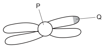
What are structures P and Q?
Options
| P | Q | |
|---|---|---|
| A | centriole | chromatid |
| B | centromere | telomere |
| C | centromere | centriole |
| D | centriole | telomere |
Working
The diagram shows a replicated chromosome with two sister chromatids joined at a central constriction.
- P points to the constricted central region where the two chromatids are held together — this is the centromere.
- Q points to the end (tip) of a chromatid arm — this is the telomere.
A centriole is a separate organelle involved in spindle formation during cell division and is not a part of a chromosome. A chromatid is one of the two arms of the replicated chromosome, not a structure at a point.
Answer
B
B
Background Concept
A replicated chromosome (as seen after DNA replication during S phase and before anaphase of mitosis) consists of two identical sister chromatids joined at a centromere. Each chromatid is a single DNA double helix packaged with histone proteins. The key structural features a biologist must recognise are:
- Centromere — the specialised, often constricted region of DNA/protein where the two sister chromatids are held together. The kinetochore assembles here, and spindle microtubules attach to it during mitosis to pull the chromatids apart.
- Telomere — the repetitive nucleotide sequence (TTAGGG in humans) at each end of a chromatid. Telomeres protect the ends of the chromosome from degradation and from being recognised as DNA damage, and they shorten with each round of replication.
- Chromatid — one of the two identical arms of a replicated chromosome. It is not a sub-structure at a particular point; it is the whole arm itself.
- Centriole — a cylindrical organelle made of microtubules, found in the centrosome. It organises the spindle fibres during cell division in animal cells, but it is not part of the chromosome.
Understanding the Question
The question shows a diagram of a single replicated chromosome (Fig. 20.1) with two arms (sister chromatids) and two labelled points, P and Q. P points to the central constriction, and Q points to the shaded tip of one chromatid arm. The task is to identify what these two regions are called.
Approach
For each label, recall the part of a chromosome that occupies that position and eliminate the distractors using the definitions above. Centriole and chromatid are the two decoys: a centriole is not part of a chromosome, and a chromatid is the whole arm, not a point on it.
Step-by-Step Reasoning
- Position of P — P is at the central constriction joining the two chromatids. By definition, this is the centromere. This rules out options A, C and D, all of which place "centriole" at P.
- Position of Q — Q is at the very end (tip) of a chromatid arm. By definition, this is the telomere.
- Confirm with the options — only option B lists centromere at P and telomere at Q.
- Check the decoys — A centriole would be a separate barrel-shaped structure outside the chromosome, not a point on it. A chromatid is the whole arm, not the shaded tip at the end. Both are wrong by definition, which is why they are the distractors.
Key Takeaways
- Centromere = the central point where sister chromatids are joined.
- Telomere = the end of a chromatid.
- Chromatid = one of the two arms of a replicated chromosome (not a point).
- Centriole = a microtubule-based organelle involved in spindle formation; it is not a part of the chromosome itself.
Common Mistakes
- Choosing A or D because of the word "centriole" — students sometimes confuse centriole with centromere, but centrioles are organelles and centromeres are regions of the chromosome.
- Choosing C (centromere + centriole) — same confusion at the Q end; a centriole is never found at the tip of a chromatid.
- Thinking a chromatid is "a structure on the chromosome" rather than one of its two arms.
Things to Be Careful About
- The diagram shows a replicated chromosome (two chromatids joined at the centromere). If it showed an unreplicated chromosome, the centromere would still be at the constriction, but only one chromatid would be present.
- Telomeres are present at both ends of each chromatid; Q simply points to one of the four telomeres on this chromosome.
- Distractors in chromosome-structure questions often mix the words centriole/centromere and chromatid/chromatin — keep the four terms (centromere, centriole, chromatid, chromatin) clearly distinguished.
Which processes are used by stem cells during tissue repair?
1 cytokinesis
2 DNA replication
3 transcription
Options
A 1, 2 and 3
B 1 and 2 only
C 1 and 3 only
D 2 and 3 only
Working
Stem cells repair tissue by dividing mitotically to produce new differentiated cells.
- DNA replication (2): required during S phase of interphase so that each daughter cell receives a full copy of the genome.
- Cytokinesis (1): divides the cytoplasm, producing two separate daughter cells from one parent cell.
- Transcription (3): required to produce mRNA encoding the proteins needed for cell division (e.g. cyclins, DNA polymerase, tubulin for spindle fibres) and the structural/functional proteins of the replacement tissue.
All three processes are used, so the answer is A.
Answer
A
A
Background Concept
Stem cells are unspecialised cells that retain the ability to divide and differentiate into a range of specialised cell types. They are essential for growth, replacement of worn-out cells, and repair of damaged tissue.
Tissue repair depends on mitotic cell division. A complete mitotic cell cycle consists of:
- Interphase — divided into G1 (cell growth and protein synthesis), S (DNA replication) and G2 (further growth and preparation for mitosis).
- Mitosis — division of the nucleus (prophase, metaphase, anaphase, telophase).
- Cytokinesis — division of the cytoplasm, producing two genetically identical daughter cells.
For a stem cell to divide successfully and produce functional replacement cells, it must first copy its DNA, then physically separate the genetic material and cytoplasm, and continuously synthesise new proteins to drive every stage of the process.
Understanding the Question
The question lists three candidate processes and asks which are used by stem cells during tissue repair:
- 1 — cytokinesis (cytoplasmic division)
- 2 — DNA replication (synthesis of a new DNA copy during S phase)
- 3 — transcription (synthesis of mRNA from a DNA template)
We need to determine whether each process is required, and pick the combination that is correct.
Approach
For each numbered process, decide whether a stem cell carrying out tissue repair would need to perform it, then match the resulting set to one of the four options.
Step-by-Step Reasoning
Process 1 — Cytokinesis: Yes. Cytokinesis is the final step of the cell cycle and is essential for producing two separate daughter cells from a single parent stem cell. Without it, the stem cell could not increase in number to provide new cells for repair.
Process 2 — DNA replication: Yes. Before mitosis, the entire genome must be copied during S phase of interphase. This ensures that each daughter cell receives a complete set of chromosomes. DNA replication is therefore a prerequisite for any mitotic division involved in tissue repair.
Process 3 — Transcription: Yes. Transcription occurs throughout interphase to produce mRNAs that are translated into the proteins required for the cell cycle itself — for example, DNA polymerase and histones for replication, cyclins that regulate the cycle, tubulin for the mitotic spindle, and the proteins that form the new cell membrane and organelles. Transcription is also required as daughter cells begin to differentiate into the specialised cell types that replace the damaged tissue.
Since all three processes (1, 2 and 3) are required, the correct option is A.
Key Takeaways
- Tissue repair by stem cells requires a complete mitotic cell cycle, not just mitosis itself.
- DNA replication (S phase) and transcription (G1, S, G2) are both active during interphase and supply the genetic material and proteins needed for division.
- Cytokinesis is the step that physically generates the new cells, completing the cycle.
Common Mistakes
- Choosing B (1 and 2 only): forgetting that transcription is required to make the proteins (enzymes, cyclins, structural proteins) that drive every stage of the cycle and that allow daughter cells to differentiate.
- Choosing C (1 and 3 only): forgetting that DNA must be replicated before mitosis so each daughter cell receives a full chromosome set.
- Choosing D (2 and 3 only): forgetting that without cytokinesis, no new cells are produced regardless of how much DNA and protein is made.
- Confusing transcription with translation; the question is about mRNA synthesis from DNA, which is required for any protein production.
Things to Be Careful About
- "Tissue repair" implies cell number must increase, so cell division (and therefore cytokinesis) is essential.
- All stages of the cell cycle require ongoing gene expression — transcription is not restricted to a particular phase.
- Read the option list carefully: a "both" or "all" option is the one to pick only when every numbered statement is true.
The enzyme telomerase prevents loss of telomeres after many mitotic cell cycles.
Which cells transcribe a high concentration of telomerase?
1 neutrophils
2 mature red blood cells
3 activated memory T-lymphocytes
Options
A 1 and 2
B 1 and 3
C 1 only
D 3 only
Working
Telomerase is needed in cells that undergo many mitotic divisions, so that telomeres are not progressively lost.
- Neutrophils (1): short-lived phagocytes produced in the bone marrow. They are terminally differentiated and do not divide further, so they do not need high telomerase activity.
- Mature red blood cells (2): lose their nucleus (and other organelles) during maturation, so they have no DNA and cannot transcribe any enzyme.
- Activated memory T-lymphocytes (3): long-lived cells that must be able to undergo rapid clonal proliferation on re-exposure to antigen. They require high telomerase activity to preserve their telomeres through many future divisions.
Only statement 3 is correct.
Answer
D
D
Background Concept
Telomeres are repetitive, non-coding DNA sequences (in humans, the TTAGGG repeat) found at the ends of linear chromosomes, together with associated protective proteins. They cap the chromosome ends and prevent them from being recognised as DNA breaks or from fusing with neighbouring chromosomes.
A problem arises during DNA replication: the enzymes that copy DNA (DNA polymerases) can only add nucleotides to an existing 3′-OH end, and the very last few nucleotides at the 5′ end of each new strand cannot be copied. This is the end-replication problem, and it means that a small amount of DNA — including telomeric DNA — is lost from each chromosome every S phase. After many cell cycles, telomeres become critically short and the cell enters replicative senescence (stops dividing) or undergoes apoptosis.
Telomerase is a ribonucleoprotein that counteracts this shortening. It carries its own short RNA template, which it uses to reverse-transcribe new telomeric DNA directly onto the 3′ end of the chromosome, lengthening (or maintaining) the telomere. Because the problem only becomes significant after many divisions, cells that divide repeatedly throughout life — germ cells, stem cells, and some activated lymphocytes — maintain high telomerase activity, while most somatic cells do not.
Understanding the Question
This is a multiple-choice question. The stem reminds us that telomerase prevents telomere loss after many mitotic cell cycles, and asks which of three blood-cell types transcribe a high concentration of it. The options combine the three statements in different ways, so the job is to decide which of statements 1, 2 and 3 are individually true, then pick the matching combination.
Approach
For each cell type, two questions need to be answered:
- Does the cell have the molecular machinery to transcribe telomerase? (i.e. does it have a nucleus and DNA?)
- Does the cell need to keep dividing for a long time? (i.e. is its division potential high?)
Only cells that answer yes to both questions will produce high telomerase concentrations.
Step-by-Step Reasoning
Statement 1 — Neutrophils:
Neutrophils are short-lived granulocytes (they circulate for only hours to a day or two) and are produced in huge numbers from bone-marrow precursors. Once released into the blood, they are terminally differentiated and do not divide. Therefore they do not need telomerase to maintain telomeres. Statement 1 is incorrect.
Statement 2 — Mature red blood cells (erythrocytes):
During erythropoiesis, the developing red cell expels its nucleus (and degrades its remaining organelles) before it enters the circulation. A mature mammalian red blood cell is essentially a bag of haemoglobin with no nucleus, no DNA and no RNA synthesis. It therefore cannot transcribe any protein, let alone telomerase. Statement 2 is incorrect.
Statement 3 — Activated memory T-lymphocytes:
Memory T cells are long-lived cells that can persist in the body for years. On re-exposure to their specific antigen, they must rapidly proliferate (clonal expansion) to mount a fast, strong secondary immune response. Because they can undergo many rounds of division each time they are activated, they must protect their telomeres. They therefore express high levels of telomerase, allowing them to divide many times over a lifetime. Statement 3 is correct.
Only statement 3 is true, so the correct combination is D (3 only).
Key Takeaways
- Telomerase is needed in cells that undergo many mitotic divisions; it prevents telomere shortening and replicative senescence.
- Mature mammalian red blood cells have no nucleus and so cannot transcribe any gene product — they should be excluded from any "transcribes X" question.
- Terminally differentiated cells (like neutrophils) that no longer divide do not need telomerase.
- Long-lived cells that retain proliferative capacity — memory lymphocytes, germ cells, stem cells and many cancer cells — maintain high telomerase activity.
Common Mistakes
- Selecting 1 (neutrophils) because neutrophils "fight infection" and so are assumed to be active cells. Activity in the immune-response sense is not the same as mitotic activity; neutrophils do their job by migrating, phagocytosing and degranulating, not by dividing.
- Selecting 2 (mature red blood cells) because they are "abundant in blood". Abundance is irrelevant — without a nucleus there is no transcription of any kind.
- Forgetting that mature RBCs are anuclear in mammals. This is a very common trap; if a question asks which cells transcribe something, RBCs are almost never the answer.
Things to Be Careful About
- The question specifies transcribe a high concentration — it is about gene expression, so the cell must have a nucleus and active transcription. Any anucleate cell (mature RBC) or fully differentiated, non-dividing cell can be ruled out.
- "High concentration of telomerase" is not the same as "any telomerase"; the question expects you to identify the cell type whose normal biology depends on sustained telomerase activity.
- Distinguish carefully between neutrophils (innate, short-lived, non-dividing) and memory lymphocytes (adaptive, long-lived, divide on reactivation) — they look similar in name but behave very differently with respect to telomere biology.
Which statements are correct?
1 All polypeptides are coded for by genes.
2 All genes are made up of a sequence of nucleotides.
3 All gene mutations result in a change to the nucleotide sequence.
4 All gene mutations will result in an altered polypeptide.
Options
A 1, 2 and 3 and 4
B 1, 2 and 3 only
C 1 and 4 only
D 2 and 3 only
Working
- All polypeptides are coded for by genes. True. Every polypeptide synthesised on ribosomes is translated from mRNA, which is transcribed from a gene.
- All genes are made up of a sequence of nucleotides. True. A gene is a sequence of DNA nucleotides (some definitions include regulatory regions).
- All gene mutations result in a change to the nucleotide sequence. True. By definition, a gene mutation is a change in the base sequence of a gene.
- All gene mutations will result in an altered polypeptide. False. A substitution mutation can be silent — because the genetic code is degenerate, a changed codon may still code for the same amino acid, so the polypeptide is unchanged. Mutations in introns or non-coding regions also leave the polypeptide unaltered.
Statements 1, 2 and 3 are correct; statement 4 is incorrect.
Answer
B
B
Background Concept
A gene is a sequence of DNA nucleotides that codes for a functional product, usually a polypeptide. Information flows from DNA → mRNA (transcription) → polypeptide (translation). The genetic code is read in triplets (codons), and because 64 codons specify only 20 amino acids, the code is degenerate — several different codons can code for the same amino acid.
A gene mutation is any change in the sequence of bases (nucleotides) in a gene. Common types are substitution, deletion and insertion. Whether the mutation alters the final polypeptide depends on where it occurs and what it does to the codon read-out.
Understanding the Question
This is a "which statements are correct" MCQ. The candidate must judge each of the four statements independently and then select the option whose list matches the correct combination. The key distinction the question probes is between a mutation at the DNA level (always a change in nucleotide sequence) and a mutation's effect at the polypeptide level (not always a change, because of code degeneracy and non-coding regions).
Approach
Test each statement against the definitions:
- Statement 1 → uses the central dogma (all polypeptides come from mRNA translated from a gene).
- Statement 2 → uses the definition of a gene as a nucleotide sequence.
- Statement 3 → uses the definition of a gene mutation (a change in base sequence).
- Statement 4 → tests whether every DNA-level change automatically changes the amino acid sequence. Consider silent substitutions and non-coding mutations.
Step-by-Step Reasoning
Statement 1 – All polypeptides are coded for by genes.
In a cell, polypeptides are assembled on ribosomes by reading an mRNA template, which was itself transcribed from a gene. There is no other source of polypeptide sequence information. → True.
Statement 2 – All genes are made up of a sequence of nucleotides.
Genes are stretches of DNA; DNA is a polymer of deoxyribonucleotides. → True.
Statement 3 – All gene mutations result in a change to the nucleotide sequence.
A gene mutation is, by definition, a change in the base sequence of a gene. The sequence must change for it to count as a mutation. → True.
Statement 4 – All gene mutations will result in an altered polypeptide.
This is where the genetic code matters. Because the code is degenerate, a substitution that changes a codon to another codon for the same amino acid (a synonymous/silent mutation) leaves the polypeptide unchanged. Mutations in introns, or in non-coding flanking regions, likewise have no effect on the polypeptide. Therefore it is NOT true that every gene mutation alters the polypeptide. → False.
Only statements 1, 2 and 3 are correct, so the answer is B.
Key Takeaways
- A gene mutation is defined at the DNA level (a change in base sequence), not at the protein level.
- The degeneracy of the genetic code means that DNA-level changes do not always change the amino acid sequence.
- Distinguish where a mutation occurs (exon vs intron, coding vs non-coding) and what type it is (silent substitution, missense, nonsense, frameshift).
Common Mistakes
- Assuming any change in DNA automatically changes the protein — forgetting silent mutations caused by a degenerate code.
- Confusing "gene mutation" with "chromosome mutation"; the question is specifically about gene (point) mutations.
- Thinking mutations in introns must still alter the polypeptide — splicing removes introns, so changes there often do not affect the mature mRNA.
Things to Be Careful About
- Read each statement precisely: "will result in an altered polypeptide" is a strong claim. One counter-example (silent substitution) is enough to make it false.
- Do not reject statement 1 just because some short peptides can be made non-ribosomally — at A-level the accepted position is that all cellular polypeptides are gene-coded.
- The question awards no partial credit for "near misses"; only the option that lists exactly the correct statements is credited.
A double-stranded DNA molecule was analysed and 29% of its nucleotide bases were found to be adenine.
Which percentage of its nucleotide bases will be cytosine?
Options
A 21%
B 29%
C 42%
D 58%
Working
In double-stranded DNA, A pairs with T and G pairs with C (Chargaff's rule), so %A = %T and %G = %C.
Given %A = 29%, then %T = 29%.
%G + %C = 100% − (29% + 29%) = 100% − 58% = 42%.
Since %G = %C, %C = 42% ÷ 2 = 21%.
Answer
A
A
Background Concept
In a double-stranded DNA molecule, the two polynucleotide strands are held together by hydrogen bonds between complementary nitrogenous bases. Adenine (A) always pairs with thymine (T) via two hydrogen bonds, and guanine (G) always pairs with cytosine (C) via three hydrogen bonds. Because of this strict complementary base pairing (Chargaff's rule), in any double-stranded DNA molecule:
The two strands are also antiparallel and the total of all four bases must equal 100%.
Understanding the Question
The question gives the percentage of adenine bases (29%) in a double-stranded DNA molecule and asks for the percentage of cytosine bases. This is a direct application of Chargaff's complementary base-pairing rule. The options include 21% (A), 29% (B), 42% (C), and 58% (D), so the candidate must pick the one consistent with complementary pairing.
Approach
Use the relationship %A = %T to find %T, then subtract A + T from 100% to get %G + %C, and finally halve that to get %C (since %G = %C).
Step-by-Step Reasoning
- %A = 29% (given). By complementary base pairing, %T = 29% as well.
- A + T together = 29% + 29% = 58%.
- The remaining bases, G and C, must therefore make up 100% − 58% = 42%.
- Since G and C pair with each other and are present in equal amounts, %C = 42% ÷ 2 = 21%.
So cytosine accounts for 21% of the nucleotide bases, corresponding to option A.
Key Takeaways
- Chargaff's rule: in double-stranded DNA, A = T and G = C in molar proportions.
- The total A + T + G + C always equals 100%.
- If one base is given, the other three can be deduced: A given → T same; the remainder split equally between G and C.
Common Mistakes
- Forgetting that A pairs with T, so if A = 29%, T is also 29% — the most common error is treating A and T as independent.
- Confusing single-stranded RNA (where Chargaff's rule does not strictly apply) with double-stranded DNA.
- Halving the wrong value: a candidate who halves 58% gets 29% (option B), which is the value of A and T individually, not G or C.
- Choosing 42% (option C), which is %G + %C combined, not cytosine alone.
- Choosing 58% (option D), which is the total of A + T.
Things to Be Careful About
- The question specifies a double-stranded DNA molecule, so Chargaff's rule applies directly.
- Note that this rule holds for double-stranded DNA but not for RNA, which is typically single-stranded.
- A quick sanity check: A + T + G + C must equal 100% (29 + 29 + 21 + 21 = 100 ✓).
In some cells, non-coding sequences of RNA are removed after transcription.
Which row correctly states the name of these non-coding sequences of RNA and the type of cell in which the non-coding sequences are removed?
Options
| name of non-coding sequences | type of cell in which non-coding sequences are removed | |
|---|---|---|
| A | exons | eukaryotic |
| B | exons | prokaryotic |
| C | introns | eukaryotic |
| D | introns | prokaryotic |
Working
Non-coding sequences removed from primary RNA transcripts after transcription are called introns. This removal (RNA splicing) takes place inside the nucleus, so it occurs in eukaryotic cells. Prokaryotic cells do not generally have introns to remove, because their mRNA is translated as it is being transcribed and is not processed in this way.
Answer
C
C
Background Concept
When a gene is transcribed, the RNA polymerase produces a primary RNA transcript. In eukaryotes this pre-mRNA contains both coding sequences (which will form the mature mRNA and be translated into protein) and non-coding sequences (which do not code for amino acids and must be removed before translation). The non-coding sequences are called introns (int-ervening sequences), while the coding sequences that are retained and joined together are called exons (ex-pressed sequences).
The removal of introns and joining of exons is called RNA splicing, and it is carried out by a ribonucleoprotein complex known as the spliceosome. Splicing happens in the nucleus, after transcription and before the mature mRNA leaves through nuclear pores to be translated at ribosomes.
Prokaryotic cells (bacteria and archaea) do not have a nucleus, and their DNA is not organised with the same intron–exon structure seen in most eukaryotic genes. Bacterial mRNA is typically translated co-transcriptionally (as it is being made), so there is no separate RNA-processing step in which introns are removed.
Understanding the Question
The question asks two things: (1) the name of the non-coding sequences removed after transcription, and (2) the type of cell in which this removal occurs. The table provides combinations and the correct row must satisfy both criteria.
Approach
Recall that:
- the non-coding sequences are called introns (not exons — exons are the parts kept);
- the removal occurs in eukaryotic cells, because splicing is a nuclear, post-transcriptional process.
Match these to the rows: only row C lists introns + eukaryotic.
Step-by-Step Reasoning
- Exons code for amino acids and are kept in the mature mRNA, so option A and B (which name exons as the removed sequences) are wrong.
- Prokaryotic cells do not carry out intron splicing — their mRNA is generally translated as it is transcribed, with no introns to remove. So option D (introns in prokaryotic) is wrong.
- Option C correctly identifies introns as the non-coding sequences removed and states that this happens in eukaryotic cells, which possess a nucleus where splicing occurs before translation.
Key Takeaways
- Introns = non-coding sequences removed from pre-mRNA; exons = coding sequences that are retained and joined.
- Splicing occurs in the nucleus of eukaryotic cells; it is a feature of eukaryotic gene expression.
- Prokaryotes generally lack introns and do not perform this processing step.
Common Mistakes
- Confusing introns (removed) with exons (kept) — a frequent slip, and it would lead to selecting A or B.
- Forgetting that splicing is a eukaryotic process and choosing D.
- Believing that because bacteria have operons and polycistronic mRNA they must also remove introns — incorrect, as bacterial genes are typically uninterrupted.
Things to Be Careful About
- Remember the mnemonic: exons are expressed, introns are intervening (and removed).
- The "non-coding" wording in the question is a deliberate clue — only introns fit this description in the standard splicing model.
Water potentials were measured inside a leaf cell, in the air spaces of the leaf and in the atmosphere.
Which values of water potential () would allow water to move from the leaf to the atmosphere?
Options
| inside the leaf cell / MPa | of air spaces / MPa | of atmosphere / MPa | |
|---|---|---|---|
| A | –1 | –7 | –100 |
| B | –100 | –7 | –1 |
| C | 1 | 7 | 100 |
| D | 100 | 7 | 1 |
Working
Water moves from a region of higher (less negative) water potential to a region of lower (more negative) water potential.
For water to move from the leaf cell → air spaces → atmosphere, the water potential must become progressively more negative (decrease) along that path:
- A –1, –7, –100 MPa: values become progressively more negative ✓
- B –100, –7, –1 MPa: values become less negative (water would move into the leaf) ✗
- C 1, 7, 100 MPa: positive values are not realistic water potentials ✗
- D 100, 7, 1 MPa: positive values and wrong direction ✗
Answer
A
A
Background Concept
Water potential () is a measure of the tendency of water to move from one place to another. By convention, pure water under standard conditions has a water potential of (megapascals), and the addition of any solute lowers the water potential, giving it a negative value. The more negative the water potential, the stronger the tendency of water to leave that region.
A core rule in biology: water moves from a region of higher (less negative) water potential to a region of lower (more negative) water potential by osmosis (across a membrane) or by evaporation (from a wet surface to drier air). The steeper the gradient (the bigger the difference in ), the faster the net movement.
In a transpiring leaf, water exists as a continuous column — cell → cell wall → air space inside the leaf → atmosphere. Evaporation from the moist cell walls lining the sub-stomatal air spaces creates a very negative water potential in those air spaces, and diffusion of water vapour out through stomata then continues down the gradient into the atmosphere. Typical values: leaf cell around –1 to –2 MPa, air spaces roughly –5 to –10 MPa (depending on humidity inside the leaf), and the atmosphere can be around –50 to –100 MPa when relative humidity is below about 99%.
Understanding the Question
The question presents a table of three water potentials: the leaf cell interior, the air spaces inside the leaf, and the atmosphere. It asks which set of values would allow water to move out of the leaf into the atmosphere, i.e. in the direction cell → air spaces → atmosphere.
The command word is implicit ("which values…") — the candidate must select the row that gives a valid water potential gradient in the required direction.
Approach
Check two things for each option:
- Are the values realistic? Water potentials in real biological systems are zero or negative (never positive in this context). This immediately eliminates C and D, where the values are all positive.
- For the remaining realistic options (A and B, both with negative values), is the gradient in the correct direction? Water must move from the highest (least negative) to the lowest (most negative) , in the order: leaf cell → air spaces → atmosphere.
Step-by-Step Reasoning
Option A: –1, –7, –100 MPa.
- The leaf cell is at –1 MPa (least negative = highest ).
- The air spaces are at –7 MPa (more negative = lower ).
- The atmosphere is at –100 MPa (most negative = lowest ).
- The gradient is: –1 > –7 > –100, so water moves correctly from the cell, into the air spaces, and out to the atmosphere. ✓
Option B: –100, –7, –1 MPa.
- Here the leaf cell is the most negative and the atmosphere is the least negative. Water would move in the opposite direction (from atmosphere → air spaces → cell), which is not transpiration. ✗
Option C: 1, 7, 100 MPa.
- Positive water potentials are not realistic for plant cells or the atmosphere (and these are not in the correct order for water movement). ✗
Option D: 100, 7, 1 MPa.
- Again positive and in the wrong order. ✗
Only A satisfies both the realistic (non-positive) values and the correct gradient direction.
Key Takeaways
- Water always moves down a water potential gradient, from less negative to more negative .
- Water potentials in real biological systems are ; positive values are unrealistic in this context.
- A transpiring leaf maintains a steep gradient from the mesophyll cells (least negative) through the air spaces (intermediate) to the atmosphere (most negative), driving water vapour out through the stomata.
Common Mistakes
- Choosing B because the numbers "look more dramatic" in the opposite order; the candidate has failed to identify which way the gradient runs.
- Choosing C or D because the candidate did not realise that water potentials in plant cells and air are never positive. (In soil, can be very slightly negative, approaching 0; in well-watered cells it is just below 0.)
- Forgetting that "atmosphere" with low humidity has a very negative water potential — often the most negative of the three, not the least.
Things to Be Careful About
- Always read the column order carefully: the question fixes the column order as cell, air spaces, atmosphere. Do not transpose it.
- "More negative" means a lower (more negative) number on the number line, e.g. –100 < –7 < –1.
- Don't confuse water potential (, measured in MPa) with solute potential or pressure potential; only the combined drives water movement.
Three samples of liquid were taken from different locations in a plant and tested to measure the concentration of four solutes. The results are shown.
| sample | concentration of solute / | |||
|---|---|---|---|---|
| sucrose | nitrate ions | amino acids | magnesium ions | |
| 1 | 652 | 0.14 | 41 | 3.4 |
| 2 | 0.12 | 7.1 | 7.2 | 1.1 |
| 3 | 433 | 0.6 | 68 | 3.7 |
Which samples have been taken from phloem sieve tube elements?
Options
A 1, 2 and 3
B 1 and 2 only
C 1 and 3 only
D 2 and 3 only
Working
Phloem sieve tube elements transport assimilates (mainly sucrose, plus amino acids) from sources to sinks, so phloem sap is characterised by very high sucrose and relatively high amino acid concentrations. Xylem carries water and dissolved mineral ions (e.g. nitrate, magnesium) but very little sucrose.
- Sample 1: sucrose 652 and amino acids 41 mol m⁻³ — both very high, consistent with phloem.
- Sample 2: sucrose 0.12 mol m⁻³ — far too low for phloem; the higher nitrate (7.1) and magnesium (1.1) match xylem sap.
- Sample 3: sucrose 433 and amino acids 68 mol m⁻³ — both high, consistent with phloem.
Answer
C
C
Background Concept
A vascular plant has two long-distance transport tissues. Xylem moves water and dissolved mineral ions (e.g. nitrate, , and magnesium, ) from the roots up to the rest of the plant; it carries essentially no organic solutes. Phloem, made up of sieve tube elements and companion cells, translocates the products of photosynthesis (assimilates) — chiefly sucrose, but also amino acids — from "sources" (e.g. mature photosynthesising leaves) to "sinks" (e.g. roots, developing fruits, young leaves). The companion cells load these organic solutes actively into the sieve tubes, and the resulting mass flow carries them through the plant. Because of this active loading, phloem sap is dominated by sucrose and contains appreciable amino acids, while xylem sap is dominated by mineral ions with very little sucrose.
Understanding the Question
A data table reports the concentrations of four solutes — sucrose, nitrate ions, amino acids and magnesium ions — in three liquid samples drawn from somewhere inside a plant. The task is to decide which sample(s) come from phloem sieve tube elements, by matching their solute profile to what we expect phloem sap to contain. The parent stem is the table itself, since the question text merely introduces it.
The command word is implicit: "Which samples have been taken from phloem sieve tube elements?" — i.e. identify the phloem samples from the chemistry of the fluid.
Approach
Look for the chemical signature of phloem: very high sucrose and elevated amino acids, with mineral ions in the background. A sample with negligible sucrose but substantial nitrate/magnesium is xylem, not phloem. So scan the sucrose column first to find phloem candidates, then confirm with the amino-acid column and check that the excluded sample(s) fit the xylem profile instead.
Step-by-Step Reasoning
- Sample 1: sucrose = 652 mol m⁻³, amino acids = 41 mol m⁻³. These are very high values for the organic solutes that phloem transports. Nitrate (0.14) and magnesium (3.4) are present in the background, as expected. This is the classic phloem-sap profile.
- Sample 2: sucrose = 0.12 mol m⁻³ — almost zero. Sucrose is the marker solute for phloem, so this cannot be phloem. Instead, the relatively high nitrate (7.1) and the low amino-acid figure fit xylem sap (water plus dissolved mineral ions from the root). Magnesium is also present, consistent with xylem transport of .
- Sample 3: sucrose = 433 mol m⁻³, amino acids = 68 mol m⁻³ — both high. This matches phloem. (The slightly different ratios from Sample 1 simply reflect that phloem sap composition varies with the source-sink status of the plant, e.g. a leaf exporting sucrose versus a storage organ loading it.)
Therefore samples 1 and 3 are from phloem sieve tube elements → option C.
Key Takeaways
- Phloem sap is rich in sucrose and amino acids; xylem sap is rich in mineral ions (nitrate, magnesium) with very little sucrose.
- The dominant organic solute of phloem is sucrose, so a near-zero sucrose reading effectively rules out a phloem origin.
- Phloem composition varies quantitatively (e.g. depending on what is being loaded at the source or unloaded at the sink), so use the pattern across solutes, not any single absolute value.
Common Mistakes
- Choosing A (1, 2 and 3) because all three samples are from "a plant" — but the question is about distinguishing tissues, not just locations.
- Choosing B (1 and 2) or D (2 and 3) by latching onto the magnesium or nitrate column and ignoring the decisive sucrose column.
- Reading the table in the wrong units or swapping columns (e.g. mistaking the very low sucrose figure for amino acids in Sample 2).
Things to Be Careful About
- The deciding evidence is the sucrose concentration: it is the marker of phloem sap because sucrose is the principal assimilate loaded into sieve tubes by companion cells.
- Always cross-check with the amino-acid column — phloem also translocates amino acids, so phloem samples should show higher amino-acid concentrations than xylem samples.
- Do not be misled by a high mineral-ion reading in a phloem sample; small amounts of nitrate and magnesium can be present, but sucrose is the discriminator.
A heated band was wrapped around the stem of a plant at a height of above the ground and left for 10 minutes.
How will heating a small section of the stem to affect the transport of phloem sap and xylem sap between the roots and leaves?
Options
| transport of phloem sap | transport of xylem sap | |
|---|---|---|
| A | ✓ | ✓ |
| B | ✓ | ✗ |
| C | ✗ | ✓ |
| D | ✗ | ✗ |
key
✓ = transport continues
✗ = transport stops
Working
- Phloem transport (translocation) depends on the active loading of sucrose into sieve tubes at the source. This requires ATP, proton pumps and enzymes in the companion cells.
- Heating to denatures the enzymes and damages the membranes of the companion cells, so active loading stops and the pressure gradient driving mass flow is lost → phloem transport stops (✗).
- Xylem transport depends on the cohesion-tension mechanism: transpiration pull, cohesion of water molecules by hydrogen bonding, and adhesion to the walls of the dead, lignified xylem vessels. None of these require living cells or enzymes.
- Heating does not break the hydrogen bonds that hold the water column together, so the transpiration stream can still be pulled past the heated section → xylem transport continues (✓).
Answer
C
C
Background Concept
Plants have two independent long-distance transport systems, each with a very different mechanism:
-
Xylem carries water and dissolved mineral ions from the roots up to the leaves and shoots. Its vessels are made of dead, hollow, lignified cells arranged end-to-end to form continuous tubes. The driving force is the cohesion-tension theory: water evaporating from the mesophyll cells of the leaf (transpiration) creates a negative pressure (tension) that pulls a continuous column of water up the xylem. The column is held together by hydrogen bonding between water molecules (cohesion) and to the vessel walls (adhesion). This is a purely physical, passive process — no living cells and no ATP are required inside the xylem itself.
-
Phloem carries assimilates (mainly sucrose, plus amino acids) from sources (e.g. photosynthesising leaves, storage roots) to sinks (e.g. growing shoots, roots, fruits, storage organs). It consists of living sieve tube elements (which lack a nucleus at maturity) closely associated with companion cells that do all the metabolic work. Translocation is explained by the mass-flow (pressure-flow) hypothesis: sucrose is actively loaded into the sieve tube at the source using a proton pump (H⁺/sucrose antiport), raising the solute potential, drawing in water by osmosis, and so generating a high hydrostatic pressure. At the sink, sucrose is actively unloaded, water leaves, and pressure falls. The resulting pressure gradient pushes the sap from source to sink. This is an active process that depends on ATP, enzymes and intact plasma membranes in the companion cells.
Because phloem transport relies on enzymatic, active loading while xylem transport relies on physical properties of water and dead vessels, anything that selectively destroys the active component — but leaves the physical component intact — will stop one but not the other.
Understanding the Question
A small section of a plant stem has been wrapped in a heated band at 60 °C for 10 minutes. We are asked what happens to the upward and downward flow of sap through both xylem and phloem at and beyond that heated zone. The four options test whether the candidate can predict, for each system, whether transport continues or stops.
The command word is implicit in an MCQ: select the option that correctly matches the biological prediction. The trap is to assume that all plant transport is biological, so heat damages everything — or alternatively to assume that physical = immune to anything. The reality is more subtle: the physical column of xylem water is unaffected, but the active loading mechanism of the phloem is destroyed.
Approach
- Ask: what does the heated section contain, and what does each transport system need to function?
- Xylem at the heated section = dead lignified vessels full of water. Need: continuous water column, evaporation at the leaf, hydrogen bonds. Heat does not break hydrogen bonds in bulk water (boiling would vaporise the column, but 60 °C is well below this).
- Phloem at the heated section = living sieve tubes + companion cells. Need: ATP, proton pumps, sucrose-loading enzymes, intact membranes. At 60 °C these proteins denature — their tertiary structure unfolds and they lose function.
- Apply: phloem transport stops (✗), xylem transport continues (✓) → option C.
Step-by-Step Reasoning
-
Xylem flow at the heated section. The water column inside the xylem is held together by cohesion (hydrogen bonds between water molecules) and adhesion to the lignin walls. Hydrogen bonds in liquid water are stable well above 60 °C; they only break en masse when water boils (100 °C at atmospheric pressure). Transpiration at the leaves still continues, so the negative pressure (tension) is still transmitted down the unbroken column through the heated zone. Therefore xylem sap continues to be pulled past the heated section (✓).
-
Phloem flow at the heated section. Mass flow requires a pressure gradient built up by active loading of sucrose at the source. Active loading is driven by a H⁺-ATPase proton pump on the companion cell plasma membrane and a sucrose–H⁺ symporter. Both are proteins. At about 60 °C, proteins denature: their specific 3-D shape is lost, so the binding sites no longer recognise their substrates and the pump/symporter stop working. With active loading halted, the high hydrostatic pressure at the source cannot be maintained and the pressure gradient driving mass flow collapses. The sieve tubes also become leaky as membrane proteins are damaged. Therefore phloem sap transport stops (✗).
-
Combining the two predictions. Phloem stops (✗) and xylem continues (✓), which corresponds to option C.
Key Takeaways
- Xylem transport is passive and physical — it is driven by transpiration pull and the cohesion of water. It does not require living cells or enzymes inside the stem, so heating does not stop it.
- Phloem transport is active — it requires ATP, proton pumps and enzymes in the companion cells to load and unload sucrose. Heating to 60 °C denatures these proteins and stops mass flow.
- A general principle: active, enzyme-dependent processes are heat-sensitive; passive, physically driven processes are not.
Common Mistakes
- Saying both stop (option D) — the misconception that all living processes in a plant are heat-sensitive. The xylem vessels are dead; only the physical water column matters, and that survives 60 °C.
- Saying both continue (option A) — the misconception that xylem and phloem are essentially the same kind of tube. They are not; the phloem's active loading is essential and is destroyed by 60 °C.
- Saying only phloem stops but xylem also stops (the wrong half of the trap above) — confusing cohesion-tension with active pumping and assuming the water column "evaporates" out of the heated section. 60 °C is far below boiling, so the column does not break.
- Forgetting that the companion cells are the metabolically active partner of the sieve tubes — answers that talk about "phloem being dead" are wrong (the sieve tube elements lose their nucleus but the companion cells are very much alive).
Things to Be Careful About
- The mark scheme is testing mechanism, not the word "denature". To justify a choice like C, the reasoning must say why each system is or is not affected — pointing at the active loading in phloem and the passive cohesion-tension mechanism in xylem, not just at "the heat damages proteins".
- Do not be distracted by the direction of transport: phloem carries assimilates both up and down, so this is not about source/sink position relative to the band — it is about whether the loading mechanism still works.
- 60 °C is the classic denaturation temperature for many plant proteins, which is why the mark scheme uses this specific value rather than, say, 40 °C (which would be uncomfortable but tolerable) or 100 °C (which would boil the water in the xylem and actually break the column).
- A very high-performing answer would also note that 60 °C can cause localised cavitation (air bubble formation) in xylem, which in real plants can interrupt flow in individual vessels — but the overall xylem system has many parallel vessels, and the question is about transport in general, so the safe answer is still that xylem transport continues.
The diagram shows the structure of an amphibian heart and the movement of blood through it.
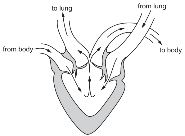
Which statements about differences between the amphibian heart and a human heart are correct?
1 The hearts have a different number of ventricles.
2 The hearts have a different number of atrioventricular valves.
3 A septum is absent in the amphibian heart and a septum is present in the human heart.
Options
A 1, 2 and 3
B 1 and 2 only
C 1 and 3 only
D 2 and 3 only
Working
Statement 1 – number of ventricles: the diagram shows a single ventricle in the amphibian heart; the human heart has two ventricles (left and right). ✔ Different number of ventricles.
Statement 2 – number of atrioventricular (AV) valves: in the amphibian heart, each of the two atria is separated from the single ventricle by its own AV valve, giving 2 AV valves. The human heart also has 2 AV valves (tricuspid and bicuspid/mitral). ✘ Same number of AV valves, not different.
Statement 3 – septum: the amphibian heart has no ventricular septum (the single ventricle is undivided), whereas the human heart has a complete interventricular septum. ✘ A septum is absent in the amphibian ventricle and present in the human ventricle.
Statements 1 and 3 are correct.
Answer
C
C
Background Concept
Mammals (including humans) and amphibians both have closed, double circulations, but the internal anatomy of their hearts differs in a key way. The human (mammalian) heart is a four-chambered pump: a right atrium and right ventricle receive deoxygenated blood from the body and pump it to the lungs, while a left atrium and left ventricle receive oxygenated blood from the lungs and pump it to the body. A muscular interventricular septum completely separates the two ventricles, keeping oxygenated and deoxygenated blood apart.
The amphibian heart is a three-chambered pump: two atria (right receiving deoxygenated blood from the body, left receiving oxygenated blood from the lungs/skin) but only one ventricle. Deoxygenated and oxygenated blood mix to some extent in the single ventricle, although trabeculae and the spiral valve help direct flows so that the body still receives somewhat better-oxygenated blood than would otherwise occur. An atrial septum separates the two atria, but no complete ventricular septum exists.
The atrioventricular (AV) valves sit between the atria and the ventricle(s) and prevent backflow. Each AV valve guards one atrioventricular opening, so:
- Human heart: 2 AV valves (tricuspid on the right, bicuspid/mitral on the left).
- Amphibian heart: 2 AV valves (one between the right atrium and the ventricle, one between the left atrium and the ventricle).
Understanding the Question
The diagram (Fig. 29.1) shows a typical amphibian heart: two atria at the top ("from body" enters the right atrium, "from lung" enters the left atrium), a single ventricle at the bottom, and outflow vessels leading "to lung" and "to body". The question asks which of the three statements correctly describe a difference between this amphibian heart and the human heart.
The command word is essentially "identify correct statements" — we must tick or cross each one and then choose the option that lists the correct combination.
Approach
For each statement, write down the structure in the amphibian heart (as shown in the figure) and the structure in the human heart, then judge whether the two differ in the way the statement claims.
Step-by-Step Reasoning
Statement 1 — "The hearts have a different number of ventricles."
- Amphibian: 1 ventricle.
- Human: 2 ventricles.
- These are different numbers. ✔ Statement 1 is correct.
Statement 2 — "The hearts have a different number of atrioventricular valves."
- Amphibian: 2 AV valves (one for each atrium–ventricle junction).
- Human: 2 AV valves (tricuspid and bicuspid/mitral).
- The number is the same, not different. ✘ Statement 2 is incorrect.
Statement 3 — "A septum is absent in the amphibian heart and a septum is present in the human heart."
- The key comparison here is the ventricular septum. The amphibian ventricle is undivided — there is no complete septum separating a left ventricle from a right ventricle. The human heart, in contrast, has a complete interventricular septum dividing the left and right ventricles.
- Statement 3 is correct in this comparative sense. ✔
Statements 1 and 3 are correct; statement 2 is not. The answer is therefore C (1 and 3 only).
Key Takeaways
- The amphibian heart has 2 atria + 1 ventricle (3 chambers); the human heart has 2 atria + 2 ventricles (4 chambers).
- The number of AV valves is the same in both (2) — one valve per AV junction.
- The presence of a complete ventricular septum is a key feature of the mammalian heart, allowing full separation of oxygenated and deoxygenated blood. Its absence in the amphibian ventricle allows some mixing, although anatomical features partially limit that mixing.
Common Mistakes
- Assuming that the number of AV valves must equal the number of ventricles, leading to the wrong answer that statement 2 is correct. The number of AV valves equals the number of atrial–ventricular connections, which is 2 in both hearts.
- Forgetting that "septum" here refers to the (inter)ventricular septum, which is what the statement is comparing. The amphibian heart does have an atrial septum, but the question is about the ventricular septum, which is the prominent feature absent in the amphibian.
- Confusing semilunar valves (e.g. aortic, pulmonary) with atrioventricular valves — only the AV valves are relevant to statement 2.
Things to Be Careful About
- When a comparative statement says "X is absent in A and present in B", take it as referring to the structure being compared, not every possible partition. The relevant partition here is the ventricular septum.
- "Different number" means strictly unequal; equal numbers make the statement false even if the valves themselves differ in other ways (shape, attachments, etc.).
- Read each option carefully: A says all three are correct, B says 1 and 2, C says 1 and 3, D says 2 and 3. Only C matches the analysis above.
A mutation in one of the genes that codes for haemoglobin changes the affinity of haemoglobin for oxygen.
This mutation lowers the partial pressure of oxygen at which haemoglobin is 50% saturated with oxygen.
Which row shows the effect of the mutation on the affinity of haemoglobin for oxygen and the change in the position of the oxygen dissociation curve for haemoglobin?
Options
| the effect of the mutation on the affinity of haemoglobin for oxygen | change in the position of the oxygen dissociation curve for haemoglobin | |
|---|---|---|
| A | higher | shift to the left |
| B | higher | shift to the right |
| C | lower | shift to the left |
| D | lower | shift to the right |
Working
The partial pressure of O₂ at which haemoglobin is 50% saturated is called the P₅₀.
A mutation that lowers the P₅₀ means haemoglobin is 50% saturated at a lower pO₂ than normal — i.e. it picks up oxygen more readily at any given pO₂. This corresponds to a higher affinity of haemoglobin for oxygen.
A higher affinity means the oxygen dissociation curve is displaced to the left (and slightly steeper at the top), because for any given pO₂, haemoglobin is more saturated than usual.
So: higher affinity and a leftward shift.
Answer
A
A
Background Concept
Haemoglobin (Hb) is a globular protein with four subunits, each carrying a haem group that reversibly binds one O₂ molecule. Its oxygen-binding behaviour is described by an oxygen dissociation curve — a sigmoid (S-shaped) graph of percentage saturation of haemoglobin against the partial pressure of oxygen (pO₂).
Two key concepts link to this curve:
- Affinity — how readily haemoglobin binds O₂. A high-affinity haemoglobin binds O₂ easily; a low-affinity haemoglobin binds O₂ less easily and releases it more readily.
- P₅₀ — the partial pressure of O₂ at which haemoglobin is 50% saturated. P₅₀ is the standard measure of affinity.
The relationship between them is inverse:
- A low P₅₀ → Hb is 50% saturated at a low pO₂ → Hb binds O₂ readily → high affinity.
- A high P₅₀ → Hb needs a higher pO₂ before it is 50% saturated → Hb binds O₂ less readily → low affinity.
On the graph, a change in affinity appears as a horizontal shift of the entire sigmoid curve:
- Higher affinity → curve shifts to the left (more saturated at any given pO₂).
- Lower affinity → curve shifts to the right (less saturated at any given pO₂).
Understanding the Question
The stem tells us that a mutation has changed one of the haemoglobin genes such that the partial pressure of O₂ at which Hb is 50% saturated has been lowered (i.e. P₅₀ has decreased). We must combine this with the two key relationships above to decide:
- What has happened to the affinity of haemoglobin for O₂?
- In which direction has the dissociation curve shifted?
The options present four combinations of (higher/lower affinity) × (left/right shift). The question is a single mark MCQ.
Approach
- Translate "lower pO₂ at 50% saturation" into a change in affinity using the inverse P₅₀–affinity relationship.
- Translate the change in affinity into a left/right shift using the affinity–curve position relationship.
- Match the resulting pair to the correct row.
Step-by-Step Reasoning
-
Step 1 — Effect on affinity.
P₅₀ is the pO₂ at which Hb is 50% saturated. If the mutation lowers this pO₂, then less oxygen pressure is required to half-saturate the haemoglobin. Less pO₂ needed = haemoglobin binds O₂ more readily. Therefore, the affinity of haemoglobin for oxygen is higher. -
Step 2 — Effect on curve position.
At any given pO₂, a higher-affinity haemoglobin carries a greater percentage saturation than normal. On a dissociation curve, this is represented by the whole sigmoid moving to lower pO₂ values — i.e. shifting to the left. -
Step 3 — Match to options.
- A: higher affinity, shift to the left ✓
- B: higher affinity, shift to the right ✗ (a rightward shift would mean lower affinity)
- C: lower affinity, shift to the left ✗ (these two are inconsistent with each other)
- D: lower affinity, shift to the right ✗ (this would be the result of a mutation that raised the P₅₀, not lowered it)
Hence the correct row is A.
Key Takeaways
- P₅₀ and affinity are inversely related: lower P₅₀ ↔ higher affinity.
- Higher affinity ↔ leftward shift of the oxygen dissociation curve; lower affinity ↔ rightward shift.
- A useful way to remember: "a left-shifted curve holds on to O₂ more tightly" — a higher-affinity Hb loads O₂ easily in the lungs but is also more reluctant to release it in respiring tissues.
- Real-world example: Hb Rainier and other high-affinity Hb mutants have a decreased P₅₀ and a left-shifted curve; they cause clinical problems because O₂ is not released efficiently in peripheral tissues.
Common Mistakes
- Mixing up the direction of the P₅₀–affinity link. Students often say "lower pO₂ at 50% saturation = lower affinity" because they associate "lower" with "worse". It is the opposite: a smaller pO₂ is needed because binding is easier.
- Confusing left and right shift with the direction of affinity change. A rightward shift means decreased affinity (e.g. due to the Bohr effect, ↑CO₂, ↑H⁺, ↑2,3-BPG), not increased.
- Choosing D because it sounds like the "default" answer when something is "lower". Always read carefully what is being lowered — here it is the pO₂ required, not the affinity.
Things to Be Careful About
- The curve position is judged at a fixed percentage saturation: a leftward shift means the same % saturation is reached at a lower pO₂.
- "Higher affinity" and "shift to the left" always go together, as do "lower affinity" and "shift to the right" — this pairing is internally consistent in the table and is your quick check.
- The sigmoid shape is preserved when P₅₀ changes; only the horizontal position moves. A change in cooperativity would change the steepness, not addressed here.
Which processes are responsible for the Bohr shift?
1 Carbon dioxide reacts with haemoglobin to form carbaminohaemoglobin.
2 Carbon dioxide reacts with water to form carbonic acid.
3 Haemoglobinic acid is formed from the dissociation of carbonic acid.
Options
A 1, 2 and 3
B 1 only
C 2 and 3 only
D 3 only
Working
The Bohr shift is the rightward shift of the oxygen dissociation curve caused by increased partial pressure of CO2 (and the resulting fall in pH), which lowers haemoglobin's affinity for O2.
- CO2 + Hb → carbaminohaemoglobin. This does occur in respiring tissues but is NOT the cause of the Bohr shift. → not responsible for the Bohr shift.
- CO2 + H2O → H2CO3 (carbonic acid), catalysed by carbonic anhydrase in red blood cells. This is the first step that produces the H+ responsible for the shift. → responsible.
- H2CO3 → H+ + HCO3−, and the H+ combines with haemoglobin to form haemoglobinic acid (HHb). The H+ binding to Hb stabilises the deoxy (T) state, reducing O2 affinity. → responsible.
Therefore, only statements 2 and 3 produce the Bohr shift.
Answer
C
C
Background Concept
Haemoglobin is an allosteric protein whose affinity for oxygen depends on the conditions in its environment. The oxygen dissociation curve shows the percentage saturation of haemoglobin at different partial pressures of oxygen. The Bohr shift is the rightward displacement of this curve that occurs when the partial pressure of CO2 rises (and pH falls), causing haemoglobin to release oxygen more readily. This is physiologically important because actively respiring tissues produce CO2 and H+, and the Bohr shift ensures that precisely these tissues receive more O2 from the blood.
Three things happen to CO2 once it diffuses from respiring tissues into a red blood cell:
- Reaction with water (catalysed by carbonic anhydrase):
- Dissociation of carbonic acid:
The H+ then binds to haemoglobin, forming haemoglobinic acid (HHb). The HCO3− diffuses out of the red cell (with the chloride shift) and is transported in the plasma.
- Direct combination with haemoglobin:
The Bohr shift is driven by the first two of these reactions, because it is the accumulation of H+ (and CO2 acting through H+) that lowers haemoglobin's affinity for O2. Carbaminohaemoglobin formation is a separate consequence of high CO2 but does not itself cause the Bohr shift.
Understanding the Question
The question lists three reactions and asks which of them are responsible for the Bohr shift. The key is to know precisely which reaction produces the H+ that drives the shift, and to recognise that direct combination of CO2 with haemoglobin (carbaminohaemoglobin) is a different effect.
The command word is "responsible for" — the test is mechanism, not merely whether the reaction occurs in respiring tissue.
Approach
- Recall the definition of the Bohr shift: a fall in O2 affinity caused by H+ (and CO2) accumulation.
- Trace where that H+ comes from: CO2 + H2O → H2CO3, then H2CO3 → H+ + HCO3−.
- Identify which of the three statements matches that pathway: statements 2 and 3.
- Rule out statement 1 because carbaminohaemoglobin formation, while real, is a separate mode of CO2 carriage and is not the cause of the Bohr shift.
Step-by-Step Reasoning
- Statement 1 – CO2 + Hb → carbaminohaemoglobin: This accounts for roughly 20–23% of CO2 transport in the blood and is favoured where pCO2 is high. However, it does not by itself generate the H+ that causes the Bohr shift. (The carbamino groups do stabilise the deoxy form, but in the strict definition used at A-level, this reaction is not credited as causing the Bohr shift.) → NOT responsible for the Bohr shift.
- Statement 2 – CO2 + H2O → H2CO3: This hydration, catalysed by carbonic anhydrase inside the red blood cell, is the first step that converts molecular CO2 into an acid, and is essential for the Bohr shift. → responsible.
- Statement 3 – formation of haemoglobinic acid from the dissociation of carbonic acid: The H+ released by H2CO3 dissociation binds to histidine residues on haemoglobin to form HHb. The binding of H+ stabilises the T (tense) state, lowering O2 affinity and shifting the dissociation curve to the right. → responsible.
Therefore only 2 and 3 are responsible for the Bohr shift, and the answer is C.
Key Takeaways
- The Bohr shift is caused by H+ (from the dissociation of carbonic acid) binding to haemoglobin, lowering its O2 affinity.
- The full pathway is: CO2 + H2O → H2CO3 → H+ + HCO3−; then H+ + Hb → HHb (haemoglobinic acid).
- Carbaminohaemoglobin formation is a distinct mode of CO2 transport and is not the cause of the Bohr shift.
Common Mistakes
- Choosing A (1, 2 and 3) by assuming "all three reactions happen, so all must be the Bohr shift". The Bohr shift is specifically the H+-mediated reduction in O2 affinity, not the direct binding of CO2 to haemoglobin.
- Choosing B (1 only) by confusing carbaminohaemoglobin with the Bohr shift.
- Choosing D (3 only) and forgetting that the H+ has to come from somewhere — the hydration of CO2 in step 2 supplies it.
Things to Be Careful About
- Keep the three fates of CO2 in red blood cells clearly separated: hydration/acid dissociation, carbamino formation, and dissolved CO2.
- Remember that the chloride shift accompanies the bicarbonate pathway and helps maintain electrochemical balance, but is not the cause of the Bohr shift either.
- The Bohr shift is a rightward shift (lower O2 affinity); the reverse (leftward) shift at the lungs is sometimes called the Haldane effect — don't mix the two up.
A student viewed a drop of human blood with a microscope and described a cell as having a very large diameter and a U-shaped nucleus that occupies half the volume of the cell.
Which type of cell was the student viewing?
Options
A monocyte
B neutrophil
C red blood cell
D lymphocyte
Working
A monocyte is the largest of the leucocytes, with a characteristic kidney- or U-shaped (horseshoe) nucleus that typically occupies a large part of the cell's volume. A neutrophil has a multi-lobed (3–5 lobes) nucleus, a lymphocyte has a large round nucleus filling most of the cell, and a red blood cell lacks a nucleus.
Answer
A
A
Background Concept
Human blood contains red blood cells (erythrocytes) and several types of white blood cell (leucocytes), each with a distinctive appearance under the light microscope. The features used to identify them are:
- Red blood cell (erythrocyte): biconcave disc, no nucleus (in mammals), diameter ~7–8 µm, pale centre.
- Lymphocyte: small leucocyte (~7–10 µm), with a large, round, densely-stained nucleus that occupies most of the cell, leaving only a thin rim of cytoplasm.
- Neutrophil: ~10–12 µm, with a multi-lobed nucleus (usually 3–5 lobes joined by thin chromatin strands) and finely granular cytoplasm.
- Monocyte: the largest leucocyte (~15–20 µm), with a characteristic kidney- or U-shaped (horseshoe) nucleus that occupies a substantial portion (often around half) of the cell volume, and abundant pale cytoplasm.
Understanding the Question
The question gives two diagnostic features of an unknown blood cell observed under the microscope:
- a very large diameter (compared with the other options), and
- a U-shaped nucleus that occupies about half of the cell's volume.
These features must be matched against the four cell types listed (A–D).
Approach
Compare each option's defining features with the two observations in the question, and reject those that do not match.
Step-by-Step Reasoning
- C – red blood cell: has no nucleus. Fails the "U-shaped nucleus" criterion. Rejected.
- D – lymphocyte: has a large, round (not U-shaped) nucleus that takes up most of the cell, and is small rather than very large. Fails both criteria. Rejected.
- B – neutrophil: has a multi-lobed (3–5 lobes) nucleus, not U-shaped, and is smaller than a monocyte. Fails both criteria. Rejected.
- A – monocyte: is the largest of the four, and its nucleus is classically described as kidney- or horseshoe-shaped, occupying roughly half the cell. Both criteria are met.
Key Takeaways
- Monocytes are the largest white blood cells and have a U-shaped (kidney/horseshoe) nucleus.
- Neutrophils are recognised by their multi-lobed nucleus, not a U-shape.
- Lymphocytes have a round nucleus filling most of the cell.
- Mature mammalian red blood cells are anuclear.
Common Mistakes
- Confusing a monocyte with a neutrophil because both have "indented" nuclei; remember the neutrophil nucleus is divided into distinct lobes, whereas the monocyte's is a single U/kidney shape.
- Choosing lymphocyte because its nucleus is large; the lymphocyte nucleus is round, not U-shaped, and the cell is much smaller.
Things to Be Careful About
- The word "very large diameter" is the first clue — only the monocyte is the largest of the listed options.
- A U-shaped (single curved indent) is different from a multi-lobed (segmented) nucleus — do not interchange the two.
How many times must an oxygen molecule pass through a cell surface membrane to get from the air in the alveolus to the haemoglobin in a red blood cell?
(Assume there are no pores between the cells the oxygen molecule must pass through.)
Options
A 2
B 3
C 4
D 5
Working
The oxygen molecule must cross:
- The apical membrane of the type I pneumocyte (alveolar epithelial cell) — 1
- The basal membrane of the type I pneumocyte — 1
- The basal membrane of the capillary endothelial cell — 1
- The apical (luminal) membrane of the capillary endothelial cell — 1
- The membrane of the red blood cell — 1
Total = cell surface membranes.
Answer
D
D
Background Concept
Gas exchange in the lungs occurs across the respiratory surface, which is built to be extremely thin to minimise the diffusion distance for oxygen and carbon dioxide. The key tissue layers an oxygen molecule crosses are:
- The alveolar wall: a single layer of flat type I pneumocytes (squamous epithelial cells). Because every cell is bounded by its own plasma (cell surface) membrane, an oxygen molecule entering the cell from the alveolar air space must cross the apical membrane, then exit through the basal membrane on the other side — i.e. 2 cell surface membranes.
- The capillary wall: a single layer of flat endothelial cells. Again, the oxygen must enter through the basal membrane and leave through the luminal membrane facing the blood — another 2 cell surface membranes.
- The red blood cell membrane: once inside the blood plasma, the oxygen diffuses into the erythrocyte and binds to haemoglobin. Crossing into the red blood cell requires crossing 1 cell surface membrane.
The question's instruction to assume there are no pores between cells is important: in reality, the fused basal laminae of the alveolar epithelium and capillary endothelium create a very thin barrier, but the oxygen still has to cross the cells themselves (and so all their membranes).
Understanding the Question
The stem sets up a specific scenario: an oxygen molecule starts in the alveolar air and ends bound to haemoglobin inside a red blood cell. The candidate must count every cell surface membrane the molecule crosses along the way. The phrase "no pores between the cells" removes any possibility of squeezing between adjacent cells and only counts direct crossings of plasma membranes.
This is a "trace and count" question — the answer is found by enumerating, in order, the membranes in the diffusion path.
Approach
- List the cell layers the oxygen crosses, in order.
- Remember that each cell contributes two membranes (apical and basal) because the cell is a discrete compartment bounded by its own plasma membrane on every side.
- The red blood cell is the final cell entered, and so adds only one more membrane (the cell's outer boundary) — there is no further cell beyond it for the oxygen to enter.
- Add the membranes up.
Step-by-Step Reasoning
- Membrane 1 — apical (air-facing) surface of the type I pneumocyte. Oxygen dissolves in the thin film of fluid lining the alveolus and diffuses into the epithelial cell through its apical plasma membrane.
- Membrane 2 — basal (blood-facing) surface of the type I pneumocyte. Having entered the cytoplasm of the pneumocyte, oxygen now exits across the basal membrane into the interstitial space (fused basal lamina).
- Membrane 3 — basal (tissue-facing) surface of the capillary endothelial cell. Oxygen enters the endothelial cell from outside.
- Membrane 4 — luminal (blood-facing) surface of the capillary endothelial cell. Oxygen exits the endothelial cell into the blood plasma.
- Membrane 5 — the red blood cell membrane. Oxygen diffuses across the erythrocyte plasma membrane and binds to haemoglobin inside the cell.
Total: cell surface membranes.
Key Takeaways
- The respiratory surface is a two-cell-thick barrier (alveolar epithelium + capillary endothelium) plus the red blood cell membrane.
- Every cell crossed adds two membranes to the count — entering and leaving — except the final cell, where the oxygen stops and so contributes only one.
- The very thin total barrier (about in healthy lungs) is what makes efficient gas exchange possible; the Fick's law requirement of a short diffusion distance is satisfied by each cell layer being a flattened squamous cell.
Common Mistakes
- Forgetting the basal membrane of the pneumocyte or endothelial cell and answering 3. Each cell has two faces, so each contributes two membranes.
- Counting only the epithelial cell + the red blood cell (answer 3), and forgetting the capillary endothelial cell entirely.
- Doubling the red blood cell membrane, treating it as if the oxygen were going to enter and leave it (it is not — oxygen binds haemoglobin inside and stops there).
- Confusing "no pores" with "fewer membranes": the absence of pores between cells means oxygen must pass through the cells (so all membranes count), not that it can bypass them.
Things to Be Careful About
- Always read the wording: "cell surface membrane" means the plasma membrane of a cell, not the alveolar wall or capillary wall as a whole.
- The endothelium and alveolar epithelium are each only one cell thick, so the count is straightforward — do not assume multiple cell layers.
- The red blood cell is not a flat squamous cell that the oxygen passes through; it is the destination, so it adds one membrane only.
Asthma can affect children and adults. The air passages become narrower, severely restricting the flow of air into the lungs.
Salbutamol is a drug that widens the air passages to relieve the symptoms of asthma.
How does salbutamol relieve the symptoms of asthma?
Options
A It decreases mucus secretions in the terminal bronchioles.
B It decreases the surface area of the alveoli.
C It causes recoil of elastic tissue in the bronchioles.
D It relaxes smooth muscle in the bronchi and bronchioles.
Working
Asthma causes bronchoconstriction: the smooth muscle in the walls of the bronchi and bronchioles contracts, narrowing the airways and restricting airflow. Salbutamol is a β₂-adrenergic receptor agonist; binding to receptors on airway smooth muscle triggers relaxation, which dilates (widens) the air passages and restores airflow.
- A — incorrect: salbutamol does not act on mucus-secreting cells.
- B — incorrect: reducing alveolar surface area would impair, not relieve, gas exchange.
- C — incorrect: elastic recoil narrows the airways; salbutamol does the opposite.
- D — correct: salbutamol relaxes smooth muscle in the bronchi and bronchioles, opening the airways.
Answer
D
D
Background Concept
The conducting division of the human gas exchange system (trachea → bronchi → bronchioles → terminal bronchioles) is supported in its walls by cartilage (in larger airways), smooth muscle, and elastic fibres, all lined by a ciliated, mucus-secreting epithelium. The smooth muscle is particularly important in the bronchi and bronchioles, where it encircles the lumen. Contraction of this smooth muscle (bronchoconstriction) narrows the airway lumen; relaxation (bronchodilation) widens it. Asthma is a condition in which the smooth muscle is hyper-responsive and contracts excessively in response to triggers (allergens, cold air, exercise, etc.), producing the characteristic wheeze and breathlessness.
Salbutamol is a short-acting β₂-adrenergic receptor agonist. It mimics the action of adrenaline at β₂ receptors located on the smooth muscle of the bronchi and bronchioles. Activation of these receptors triggers an intracellular signalling cascade (↑ cAMP) that lowers intracellular Ca²⁺ in the smooth muscle cells, causing them to relax. The relaxed muscle allows the airway to widen, reducing resistance to airflow and relieving the symptoms of asthma.
Understanding the Question
This is a multiple-choice question asking for the mechanism by which salbutamol relieves asthma. The stem gives the key facts: asthma narrows the air passages, salbutamol widens them. The options each propose a different mechanism, and we must select the one that correctly describes what salbutamol does at the tissue level.
The command word is implicit — we need to identify the correct statement about salbutamol's action.
Approach
Recall that salbutamol is a bronchodilator acting on smooth muscle, then rule out each distractor on biological grounds:
- Anything that would narrow the airway further is wrong (so not C).
- Anything that would impair gas exchange rather than restore airflow is wrong (so not B).
- Salbutamol's clinical use is for bronchodilation, not for drying up mucus (so not A).
- The correct mechanism is relaxation of airway smooth muscle — D.
Step-by-Step Reasoning
- Locate the target tissue. Salbutamol targets β₂ receptors, which are abundant on the smooth muscle of the bronchi and bronchioles (not on alveolar walls or on goblet cells).
- Identify the cellular effect. Receptor activation raises intracellular cAMP, reducing Ca²⁺ availability, so the smooth muscle cells relax.
- Identify the organ-level consequence. Relaxed smooth muscle allows the airway to widen (bronchodilation), so airflow resistance falls and the patient can breathe more easily.
- Check the options against this mechanism:
- A (decreases mucus): goblet cells/submucosal glands are the mucus source, not the β₂ target. Salbutamol does not meaningfully reduce mucus production.
- B (decreases alveolar surface area): alveoli have no smooth muscle and are not the site of asthma narrowing; reducing their surface area would worsen gas exchange, not relieve asthma.
- C (causes elastic recoil): elastic fibres in the airway walls recoil inward on expiration, which would tend to narrow the lumen — the opposite of the relief salbutamol provides.
- D (relaxes smooth muscle in bronchi and bronchioles): this matches the established mechanism exactly.
Key Takeaways
- Asthma = reversible bronchoconstriction due to contraction of smooth muscle in the bronchi and bronchioles.
- Salbutamol is a β₂-agonist; it relaxes airway smooth muscle, producing bronchodilation.
- Distinguishing the actions of the three structural components of airway walls (cartilage, smooth muscle, elastic fibres) is essential for explaining how asthma drugs and irritants change airway diameter.
Common Mistakes
- Choosing C because the word "recoil" sounds mechanically associated with airways — but recoil of elastic tissue narrows the lumen, so this would worsen, not relieve, asthma.
- Choosing A because mucus is part of the asthma picture; however, salbutamol is not an anticholinergic/mucolytic, and the immediate relief it provides is from smooth muscle relaxation, not from changing mucus.
- Choosing B because the question is "about the lungs"; the bronchioles, not the alveoli, are the site of obstruction in asthma, and salbutamol does not act on alveoli.
Things to Be Careful About
- The question asks how salbutamol relieves symptoms — focus on the dilating action, not on what causes asthma in the first place.
- Distinguish bronchodilation (relaxation of smooth muscle) from bronchoconstriction (contraction); the same smooth muscle can do either, and the direction of change matters.
- Note that the correct answer refers to both bronchi and bronchioles — both contain smooth muscle in their walls and both can constrict in asthma.
The photomicrograph shows the transverse section of part of the trachea.
Which letter represents a tissue that can contract and relax to adjust the diameter of the airways?
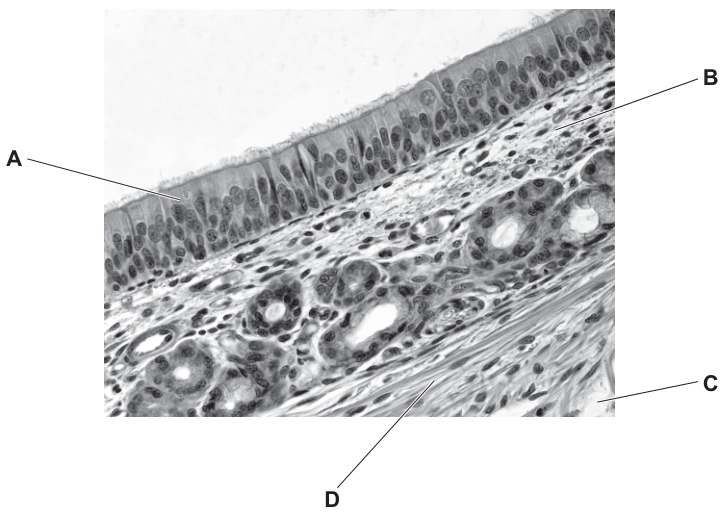
Options
A A
B B
C C
D D
Working
The tissue that can contract and relax to change the diameter of an airway is smooth muscle. In the photomicrograph, D points to the layer of densely packed, elongated cells with cigar-shaped nuclei running in parallel at the outer edge of the tracheal wall — the characteristic appearance of smooth muscle. (A is the ciliated epithelium, B is a goblet cell, and C is the loose connective tissue/lamina propria containing blood vessels and seromucous glands.)
Answer
D
D
Background Concept
The trachea is a tube whose wall, from the lumen outwards, is built up of:
- Pseudostratified ciliated columnar epithelium with goblet cells (mucosa) — traps dust and pathogens in mucus and sweeps them upwards.
- Lamina propria / submucosa — loose connective tissue with seromucous glands and blood vessels.
- Smooth muscle (the trachealis muscle at the posterior aspect of the C-shaped cartilage) — contracts and relaxes to change airway diameter (bronchoconstriction / bronchodilation).
- Hyaline cartilage (C-shaped rings) — keeps the airway permanently open so it does not collapse during inhalation.
- Adventitia — outer connective tissue.
Only smooth muscle is the contractile tissue. Cartilage is rigid and supportive; epithelium is a protective lining; goblet cells secrete mucus. So the question is really a histology-recognition test for smooth muscle.
Understanding the Question
The command is essentially "identify". The photomicrograph labels four structures (A, B, C, D) and the candidate must pick the one that is contractile. The biology is restricted to: which of these four tissues can shorten and lengthen actively? The answer is determined by knowing what each layer is.
Approach
- Recall the order of tissues in the tracheal wall.
- Match each label to a tissue using position and cell shape: ciliated columnar cells with surface cilia → epithelium; pale rounded cells in the epithelium → goblet cells; loose tissue with vessel profiles → connective tissue; dense, parallel elongated cells → smooth muscle.
- Select the smooth muscle layer.
Step-by-Step Reasoning
- A points to the tall columnar cells whose free surface carries a fringe of cilia (the wavy brush border at the top of the image). This is the ciliated epithelium — a protective/cleaning lining, not a contractile tissue.
- B points to a large, pale, rounded cell sitting within the epithelium. This is a goblet cell, which secretes mucus. Goblet cells have no contractile function.
- C points to a region of loose tissue containing the round, empty profiles of blood vessels (and possibly seromucous gland ducts). This is the lamina propria / submucosa (connective tissue). It is supportive, not contractile.
- D points to a thick band of densely packed, elongated cells with cigar-shaped nuclei running roughly in parallel. This is the smooth muscle layer. Smooth muscle cells contain actin and myosin filaments; on nervous or hormonal stimulation they shorten (bronchoconstriction → narrower airway) and lengthen (bronchodilation → wider airway). Hyaline cartilage, by contrast, would appear as large chondrocytes sitting in lacunae within a glassy, basophilic matrix — not as parallel elongated fibres — and it cannot contract.
Therefore D is the tissue that can contract and relax to adjust airway diameter.
Key Takeaways
- The tracheal wall contains ciliated epithelium, goblet cells, connective tissue, smooth muscle and hyaline cartilage, in that order from lumen outwards.
- Only smooth muscle is contractile. Bronchoconstriction (parasympathetic, e.g. via acetylcholine) and bronchodilation (sympathetic, e.g. via adrenaline) adjust airway diameter.
- Histological recognition cues: smooth muscle = parallel elongated cells with cigar-shaped nuclei; cartilage = chondrocytes in lacunae; ciliated epithelium = columnar cells with an apical brush of cilia; goblet cells = large pale rounded mucin-filled cells within the epithelium.
Common Mistakes
- Picking A (epithelium) because it is the most prominent layer — epithelium is a lining, not a muscle.
- Picking B (goblet cells) because they look distinctive — goblet cells secrete mucus, they do not contract.
- Picking D thinking it is cartilage and assuming cartilage is "hard" therefore "active" — cartilage is rigid and supportive; it cannot shorten. (If a candidate confuses the bottom layer with cartilage, they have misread the histology.)
- Confusing connective tissue (C) with smooth muscle — connective tissue has scattered cells in abundant extracellular matrix; smooth muscle is densely cellular with elongated, parallel cells.
Things to Be Careful About
- Read the cell shapes, not just the position: parallel elongated cells = smooth muscle; chondrocytes in lacunae = cartilage.
- The question is about contracting and relaxing — that single phrase rules out cartilage (rigid), epithelium (lining) and goblet cells (secretory).
- In a transverse section of the trachea the smooth muscle is not always a complete ring; it is concentrated posteriorly (the trachealis). Its histological appearance (parallel elongated cells) is the same wherever it occurs.
Which statements about an infectious disease may be correct?
1 It can be caused by a protoctist.
2 It can be transmitted by an insect vector.
3 It can be transmitted from mother to child.
Options
A 1, 2 and 3
B 1 and 2 only
C 1 and 3 only
D 2 and 3 only
Working
Malaria is an infectious disease that satisfies all three statements:
- Statement 1: Malaria is caused by Plasmodium, a protoctist. ✓
- Statement 2: Malaria is transmitted by the bite of a female Anopheles mosquito, an insect vector. ✓
- Statement 3: Malaria can be transmitted vertically from mother to fetus/child (congenital malaria). ✓
All three statements are correct, so the answer includes 1, 2 and 3.
Answer
A
A
Background Concept
Infectious diseases are caused by pathogens — micro-organisms that invade a host and cause harm. The main groups of pathogens are:
- Bacteria (e.g. Mycobacterium tuberculosis, Vibrio cholerae)
- Viruses (e.g. HIV, measles)
- Protoctists (protists) (e.g. Plasmodium species causing malaria)
- Fungi (e.g. Candida, causing thrush)
Transmission can occur by several routes, including:
- Direct contact (person-to-person, e.g. droplet spread in TB)
- Indirect contact (contaminated water in cholera, contaminated food)
- Vectors — typically insects that carry the pathogen from one host to another (e.g. mosquitoes in malaria, tsetse flies in sleeping sickness)
- Vertical (mother-to-child) transmission — across the placenta, during birth, or via breast milk (e.g. HIV, malaria, syphilis)
Understanding the Question
This is a "which statements may be correct" question, meaning we need to find at least one real infectious disease that fits each statement. The word may is key — the statement does not need to apply to every infectious disease, only to at least one. The mark scheme confirms that all three are achievable, so the candidate must identify a disease (or diseases) that collectively satisfy statements 1, 2 and 3.
Approach
The most efficient single example is malaria, which satisfies all three conditions simultaneously:
- Pathogen: Plasmodium (a protoctist) — statement 1
- Vector: female Anopheles mosquito (an insect) — statement 2
- Vertical transmission: malaria can cross the placenta — statement 3
If a candidate cannot recall congenital malaria, two other diseases — HIV (vertical transmission, viral) and malaria (protoctist, insect vector) — together also cover all three statements. Either route leads to the same answer.
Step-by-Step Reasoning
- Statement 1 — caused by a protoctist: Plasmodium falciparum (and other Plasmodium species) is a eukaryotic single-celled organism classified within the protoctista. Malaria is therefore an infectious disease caused by a protoctist. ✓
- Statement 2 — transmitted by an insect vector: The female Anopheles mosquito injects Plasmodium sporozoites into a human host when taking a blood meal. Mosquitoes are insects, so this is insect-vector transmission. ✓
- Statement 3 — transmitted from mother to child: Plasmodium can cross the placenta and infect the developing fetus, leading to congenital malaria. HIV is another classic example of vertical transmission (across the placenta, during birth, or via breast milk), and is itself caused by a virus — but the question only asks whether an infectious disease can be so transmitted, and malaria satisfies this on its own. ✓
Because all three statements are supported by a real infectious disease (malaria), the correct option must include 1, 2 and 3.
Key Takeaways
- "May be correct" questions require you to find at least one example, not a universal rule.
- Malaria is a versatile example that links three syllabus points: protoctist pathogen, insect vector, and vertical transmission.
- CIE AS Biology lists malaria as the canonical example for protoctist-caused disease and for vector transmission.
Common Mistakes
- Choosing B (1 and 2 only) — forgetting that congenital malaria and HIV both enable mother-to-child transmission.
- Choosing C (1 and 3 only) — forgetting that mosquitoes are insects (a common misclassification, but mosquitoes belong to class Insecta).
- Choosing D (2 and 3 only) — incorrectly assuming no protoctist causes human disease, when in fact Plasmodium is a protoctist.
- Confusing the classification: some students place Plasmodium with bacteria or viruses; it is a eukaryote and a protoctist.
Things to Be Careful About
- The question uses the word may, signalling that a single counter-example (or supporting example) is enough — not a universal statement.
- "Insect vector" is specific: mosquitoes (Diptera), tsetse flies, sandflies and triatomine bugs are all insects. Ticks and mites are arachnids and are not insects, so diseases transmitted by them (e.g. Lyme disease) would not satisfy statement 2.
- Vertical transmission of malaria is less commonly taught than mosquito transmission but is a well-established phenomenon, especially in endemic regions.
Which statements explain why cholera has not been eradicated by vaccination?
1 There is limited availability of an affordable vaccine.
2 Protection takes several weeks to develop after vaccination.
3 The under-reporting of cholera in the community.
4 Immunity to cholera decreases after two years.
Options
A 1, 2, 3 and 4
B 1 and 2 only
C 2 and 4 only
D 3 and 4 only
Working
All four statements are recognised reasons why cholera has not been eradicated by vaccination:
- Statement 1 is valid: an affordable vaccine has had limited availability in many endemic regions.
- Statement 2 is valid: protection (antibody-mediated immunity) takes several weeks to develop after vaccination, leaving recently vaccinated individuals temporarily unprotected.
- Statement 3 is valid: under-reporting of cholera cases in the community allows outbreaks to spread undetected, undermining vaccination and other control efforts.
- Statement 4 is valid: immunity to cholera decreases after about two years, so vaccinated individuals become susceptible again.
Answer
A
A
Background Concept
Cholera is an acute diarrhoeal disease caused by the bacterium Vibrio cholerae, transmitted primarily through contaminated drinking water. It has not been eradicated, unlike smallpox, for a combination of biological, logistical and socio-economic reasons. Eradication by vaccination depends on: (i) having an effective, affordable vaccine that is widely available; (ii) the vaccine inducing long-lasting immunity; (iii) the immunity developing quickly enough to be useful during outbreaks; and (iv) effective surveillance so that all cases can be identified and contained. Failure of any one of these conditions can prevent eradication.
Understanding the Question
This is a multiple-choice question asking the candidate to identify which of the four listed statements correctly explain why cholera has not been eradicated by vaccination. Each statement must be evaluated on its own merits — the correct option is the one that includes all and only the valid statements.
Approach
Work through each statement in turn, checking it against knowledge of cholera vaccination:
- Statement 1 (limited availability of an affordable vaccine): In many cholera-endemic regions (parts of Africa and South-East Asia), vaccine supply has been limited and cost has been a barrier to mass immunisation programmes. ✔ Valid.
- Statement 2 (protection takes several weeks to develop): Like all vaccines, cholera vaccines stimulate the production of antibodies by B-lymphocytes, a process that takes days to weeks. During an outbreak, this delay means recently vaccinated people are not yet protected. ✔ Valid.
- Statement 3 (under-reporting of cholera in the community): Cholera cases, especially mild ones, are often not reported because of poor surveillance, lack of access to healthcare, or stigma. This allows infected individuals to continue transmitting the disease. ✔ Valid.
- Statement 4 (immunity decreases after two years): Protection conferred by cholera vaccines wanes with time; antibody titres fall and booster doses are needed. ✔ Valid.
All four statements are valid reasons, so the correct option is A.
Step-by-Step Reasoning
- Read each statement carefully.
- Recognise that statements 1, 2, 3 and 4 each describe a real and distinct obstacle to eradicating cholera by vaccination.
- Eliminate options that omit any of these valid reasons: B omits 3 and 4; C omits 1 and 3; D omits 1 and 2.
- Conclude that A (1, 2, 3 and 4) is the only option that contains all four correct statements.
Key Takeaways
- Eradication by vaccination requires an effective, affordable vaccine that induces long-lasting, rapidly-developing immunity, combined with strong disease surveillance.
- Cholera fails on several of these criteria simultaneously: supply/cost issues, slow onset of immunity, waning immunity, and under-reporting all contribute to its persistence.
- Distinguish between reasons rooted in vaccine biology (statements 2 and 4) and those rooted in public health infrastructure (statements 1 and 3).
Common Mistakes
- Confusing the rate of onset of immunity (weeks — statement 2) with the duration of immunity (years — statement 4); these are two different reasons.
- Assuming under-reporting is irrelevant to vaccination: surveillance is essential to identify who needs vaccinating and to monitor outbreaks.
- Overlooking socio-economic barriers: even a good vaccine cannot eradicate a disease if it is unaffordable or unavailable where needed.
Things to Be Careful About
- Read each statement independently — do not assume any is automatically wrong because it seems similar to another.
- "Decreases after two years" is a mark-scheme-accepted simplification of waning cholera immunity; do not reject it as imprecise.
- The command word is "explain why", so the statements must give valid reasons, not merely describe features of cholera.
Antibiotic resistance is a serious threat to global health.
Which statement describes a step that can be taken to reduce the impact of antibiotic resistance?
Options
A Antibiotics prescribed by a doctor should be shared with family members to protect them.
B Antibiotics should only be used to treat infectious diseases caused by viruses.
C Farmers should give antibiotics to healthy animals to prevent infections occurring.
D Vaccination programmes should be used to reduce the spread of bacterial diseases.
Working
Antibiotic resistance arises when bacteria evolve (through natural selection) to survive exposure to antibiotics. Reducing its impact therefore depends on lowering unnecessary antibiotic use and on preventing bacterial infections in the first place so antibiotics are needed less often.
- A is wrong: sharing prescribed antibiotics is unsafe, encourages incomplete courses, and exposes more bacteria to the drug, selecting for resistance. It does not reduce the impact of resistance.
- B is wrong: antibiotics act on bacteria and have no effect on viruses; using them to treat viral infections is pointless and only adds selection pressure for resistance.
- C is wrong: routinely giving antibiotics to healthy livestock selects for resistant bacteria, which can transfer to humans via the food chain — a recognised driver of resistance.
- D is correct: vaccination prevents bacterial infections, so fewer people need antibiotics, which reduces the selection pressure driving resistance.
Answer
D
D
Background Concept
Antibiotic resistance occurs when a bacterial population evolves the ability to survive exposure to an antibiotic that would normally kill it or stop its growth. It arises by natural selection: within any large bacterial population, random mutations occasionally produce individuals that are less affected by the drug. When the antibiotic is present, susceptible bacteria are killed while resistant ones survive and reproduce, so the frequency of resistance alleles in the population rises over time. The more often bacteria are exposed to antibiotics (and the more incompletely they are exposed), the stronger this selection pressure becomes.
Because resistant infections are harder and more expensive to treat, and can spread between people, controlling antibiotic resistance is a major public-health priority. Strategies to reduce its impact focus on two complementary ideas:
- Reduce the need for antibiotics — by preventing bacterial infections in the first place (e.g. through vaccination, improved sanitation, infection control in hospitals).
- Use antibiotics more carefully — only when needed, at the correct dose, for the correct duration, and targeted at the infecting bacterium.
Understanding the Question
This is a multiple-choice question asking the candidate to identify which of four statements describes a valid step that reduces the impact of antibiotic resistance. The command word is implicit ("which statement describes…"), and the distractors are deliberately plausible — each one describes behaviour that is either irrelevant, harmful, or actively counter-productive in the fight against resistance. To score the mark, the candidate must reject three options on biological or public-health grounds and select the one that genuinely lowers selection pressure for resistance.
Approach
For each option, ask two questions: (i) does it reduce bacterial exposure to antibiotics, and (ii) does it prevent bacterial infection in the first place? A statement that does either of these — without introducing new selection pressure — is a valid answer.
Step-by-Step Reasoning
- Option A — sharing prescribed antibiotics with family members. A prescribed course is calibrated to one patient's infection. Sharing leads to under-dosing, incomplete courses, and the use of antibiotics by people who may not need them. All of these increase selection pressure for resistance and risk harming the family member (wrong drug, wrong dose, missed diagnosis). The mark scheme rejects this.
- Option B — using antibiotics to treat viral infections. Antibiotics target bacterial structures and processes (cell-wall synthesis, ribosomes, DNA replication); they have no effect on viruses. Using them against viral illnesses (e.g. common colds, most sore throats) exposes the body's normal bacterial flora to the drug for no therapeutic benefit, selecting for resistance. This is exactly the misuse the mark scheme warns against.
- Option C — giving antibiotics to healthy farm animals as a preventative. Routinely dosing healthy livestock is a major documented driver of resistance: resistant bacteria such as MRSA and ESBL-producing E. coli can spread to humans through food, water, and direct contact with animals. The mark scheme rejects this as a step that increases, not reduces, the impact of resistance.
- Option D — vaccination programmes to reduce the spread of bacterial diseases. Vaccines prevent people from being infected in the first place. Fewer infections means fewer courses of antibiotics prescribed, so less selection pressure on bacterial populations, and slower spread of resistant strains. This is widely recommended by bodies such as WHO as a key measure to combat resistance.
Key Takeaways
- Antibiotic resistance is driven by selection pressure: more antibiotic use → faster spread of resistance.
- Valid strategies either prevent infection (vaccination, hygiene, sanitation) or use antibiotics more carefully (correct indication, dose, duration; completing courses; not using them for viral disease).
- Routine antibiotic use in agriculture is a recognised contributor to resistance, not a solution to it.
Common Mistakes
- Choosing B because it sounds like a careful/controlled use of antibiotics — but antibiotics simply do not work against viruses, so any use against them is misuse.
- Choosing C because "prevention sounds sensible" — prevention of infection is good, but using antibiotics to do it is what creates the resistance problem; vaccination is the prevention method that does not create resistance.
- Choosing A under the impression that "the more people treated, the better" — sharing courses leads to under-dosing and spread of resistant strains.
Things to Be Careful About
- The phrase "reduce the impact of antibiotic resistance" can be misread as "reduce antibiotic use" only; in fact, both prevention of infection and smarter use of antibiotics count.
- Vaccination reduces the impact of resistance even though it is not itself an antibiotic — it lowers the demand for antibiotics, which is the underlying lever.
- The mark scheme awards the mark only for the letter, but the reasoning above is what justifies it; in a structured question, the same biological logic would need to be stated explicitly.
Tetanus is an infectious disease caused by a type of bacterium. This bacterium produces a protein that is a toxin which causes illness.
Scientists have produced a vaccine for tetanus which contains a harmless form of the toxin called a toxoid.
The toxoid is produced by mixing the toxin with the chemical formaldehyde. This chemical binds to the toxin making it harmless.
Which statement about the tetanus vaccine is correct?
Options
A After vaccination, the toxoid in the vaccine will be ingested by neutrophils and displayed in their cell surface membranes.
B The tetanus vaccine containing the toxoid does not contain antigens because it does not contain any cells of the pathogen.
C The toxoid protein in the vaccine stimulates the production of antibodies which remain in the blood to provide long-term immunity.
D When formaldehyde binds to the toxin, it causes the toxin to completely change shape.
Working
A is correct because phagocytes, including neutrophils, engulf the toxoid and then display antigen fragments on their cell surface membranes. This presentation is what allows T-lymphocytes to recognise the antigen and trigger the specific immune response.
B is incorrect because the toxoid itself is a protein and therefore acts as an antigen. An antigen does not have to be a whole pathogen cell — any molecule that is recognised as foreign and triggers an immune response is an antigen.
C is incorrect because long-term immunity is provided by memory cells (memory B- and T-lymphocytes), not by antibodies remaining in the blood. Antibodies are produced rapidly during the secondary response, but they do not persist long-term.
D is incorrect because formaldehyde does not cause a complete change in the toxoid's shape. It modifies the toxin so that it is no longer harmful, while still leaving enough of its structure intact for it to be recognised as an antigen.
Answer
A
A
Background Concept
A vaccine contains an antigenic preparation that primes the specific immune system to respond rapidly on future exposure to the actual pathogen. The antigen may be:
- a live, weakened (attenuated) pathogen,
- a killed (inactivated) pathogen,
- a fragment of the pathogen such as a surface protein, or
- a toxoid — a harmless derivative of a bacterial toxin that has been chemically treated so it can no longer cause disease but still retains its antigenic shape.
When the vaccine enters the body, the antigens are recognised as foreign. Phagocytes (neutrophils and macrophages) engulf the antigenic material by phagocytosis. Inside the phagocyte, the antigen is broken down and fragments are displayed on the cell-surface membrane — this is antigen presentation. Macrophages (and to some extent neutrophils) are the main antigen-presenting cells (APCs), and they pass the antigen to T-helper lymphocytes, which in turn stimulate B-lymphocytes to produce antibodies and form memory cells.
Long-term immunity is conferred by memory lymphocytes that persist in the body for many years. On re-exposure to the same antigen, these memory cells divide rapidly and produce a swift, large secondary response, neutralising the pathogen before symptoms develop. Antibodies themselves circulate only for weeks to months after an infection or vaccination; they are not the basis of long-term immunity.
Understanding the Question
The question is an MCQ testing several interconnected ideas about how the tetanus vaccine works. The stem tells us that:
- Tetanus is caused by a bacterium that produces a toxic protein.
- The vaccine contains a toxoid — the toxin treated with formaldehyde to make it harmless.
- The formaldehyde binds to the toxin, rendering it non-toxic.
You must select the one statement that is biologically correct from the four options. The distractor statements are designed to test common misconceptions about antigens, antibodies, memory and the effect of formaldehyde.
Approach
Evaluate each option against what you know about:
- What phagocytes do with antigens (engulf and present).
- The definition of an antigen (any foreign molecule recognised by the immune system, not just whole pathogens).
- The basis of long-term immunity (memory cells, not persistent antibodies).
- The action of formaldehyde on toxins (modifies rather than completely reshapes the molecule).
Then choose the statement that is the most accurate.
Step-by-Step Reasoning
Option A — ingested by neutrophils and displayed in their cell surface membranes.
Phagocytes, including neutrophils, engulf (phagocytose) foreign material at the site of vaccination. Once inside the phagocyte, antigens are processed and fragments are inserted into the cell-surface membrane, where they are "displayed" to passing lymphocytes. Although macrophages are the principal antigen-presenting cells, neutrophils also perform this role. Statement A is therefore the correct answer.
Option B — does not contain antigens because it does not contain any cells of the pathogen.
This is a common misconception. An antigen is any molecule capable of triggering an immune response — it does not have to be a whole pathogen. The toxoid is a foreign protein and is itself the antigen. Statement B is incorrect.
Option C — stimulates the production of antibodies which remain in the blood to provide long-term immunity.
The toxoid does stimulate antibody production, but the part about "remaining in the blood" is wrong. Antibody levels in the blood fall over weeks or months. Long-term immunity depends on memory B- and T-lymphocytes, which can rapidly proliferate and produce a large amount of antibody on a second exposure to the antigen. Statement C is incorrect.
Option D — formaldehyde causes the toxin to completely change shape.
Formaldehyde cross-links amine groups on the toxin, inactivating it without completely unfolding it. Crucially, the toxoid must retain enough of its original 3-D shape to be recognised as the same antigen by lymphocytes — otherwise vaccination would be useless. Statement D is incorrect.
Key Takeaways
- A vaccine works by exposing the immune system to an antigen in a safe form so that memory cells can form.
- Phagocytes (neutrophils and macrophages) engulf antigens and display them on their surface membranes for lymphocytes to recognise.
- An antigen is any foreign molecule that triggers an immune response — it need not be a whole cell or pathogen.
- Long-term immunity is due to memory lymphocytes, not to antibodies lingering in the blood.
- Toxoids are toxins that have been chemically inactivated (e.g. with formaldehyde) but retain enough of their shape to remain antigenic.
Common Mistakes
- Believing that "antigen" means a whole cell or pathogen — the toxoid alone is sufficient to act as an antigen.
- Confusing antibodies with memory cells: antibodies give short-term protection; memory cells give long-term protection.
- Thinking formaldehyde denatures the toxin completely — the toxoid must still be recognisable as an antigen for the vaccine to work.
- Assuming only macrophages can present antigens — phagocytes in general, including neutrophils, can present antigens on their surface.
Things to Be Careful About
- Read the wording of option C very carefully: the small clause "which remain in the blood" makes the statement false, even though the rest is true.
- For option D, the distinction between "modified/inactivated" and "completely changes shape" is critical — the toxoid must keep its antigenic shape.
- Do not be misled by the apparent specificity of "neutrophils" in option A; in CIE biology, phagocytes as a class engulf and present antigens, and the statement is the best of the four given.
What is an effect on the immune system of a reduced number of T-helper cells?
Options
A a decrease in the destruction of infected body cells by T-helper cells
B a decrease in the activation of B-lymphocytes
C an increase in the activation of T-killer cells
D an increase in the production of plasma cells
Working
T-helper cells coordinate the immune response by releasing cytokines that activate B-lymphocytes (so they proliferate and differentiate into plasma cells) and that activate T-killer cells. T-helper cells do not themselves destroy infected body cells — that is the role of T-killer (cytotoxic) cells.
If the number of T-helper cells is reduced:
- A — incorrect: T-helper cells do not destroy infected body cells.
- B — correct: fewer T-helper cells means less activation of B-lymphocytes.
- C — incorrect: T-helper cells activate T-killer cells, so a reduction would decrease (not increase) T-killer cell activation.
- D — incorrect: T-helper cells help B-lymphocytes become plasma cells, so a reduction would decrease plasma cell production.
Answer
B
B
Background Concept
The adaptive immune response depends on close collaboration between several types of lymphocyte. T-lymphocytes mature in the thymus and are subdivided into functional types:
- T-helper cells (CD4⁺) — release cytokines that activate and direct other immune cells. They do not directly kill infected cells. They stimulate B-lymphocytes to proliferate and differentiate into plasma cells, and they stimulate T-killer cells to mature and attack infected cells.
- T-killer cells (CD8⁺, cytotoxic T cells) — recognise and destroy body cells that display non-self antigen on their surface (e.g. virus-infected cells or tumour cells).
- B-lymphocytes — when activated (largely by T-helper cells), proliferate and differentiate into plasma cells, which secrete antibodies, and into memory B cells.
Because T-helper cells are the main coordinators of the cellular and humoral responses, a fall in their number cripples both arms of adaptive immunity. HIV provides the classic real-world example: it binds to the CD4 marker on T-helper cells and destroys them, producing the immunodeficiency of AIDS.
Understanding the Question
The stem asks for an effect of a reduced number of T-helper cells on the immune system. We need to identify a statement that correctly describes what happens as a downstream consequence of losing T-helper cell activity. Each option is phrased as a directional change (a decrease or an increase), so we must check both the cellular function implied and the direction of the change.
Approach
For each option, ask two questions:
- Does this describe a real function involving T-helper cells?
- Does the stated change in direction follow correctly from a reduction in T-helper cells?
The correct option must pass both tests.
Step-by-Step Reasoning
- Option A claims "a decrease in the destruction of infected body cells by T-helper cells." T-helper cells do not destroy infected body cells — that is the job of T-killer cells. The statement is biologically incoherent and is rejected.
- Option B claims "a decrease in the activation of B-lymphocytes." T-helper cells activate B-lymphocytes via cytokine signalling (and through antigen presentation on MHC class II). Fewer T-helper cells therefore means less activation of B-lymphocytes. The function and the direction are both correct. This is the answer.
- Option C claims "an increase in the activation of T-killer cells." T-helper cells help activate T-killer cells, so fewer T-helper cells would decrease (not increase) T-killer cell activation. Wrong direction.
- Option D claims "an increase in the production of plasma cells." Plasma cells are produced from activated B-lymphocytes. Because T-helper cells activate B cells, fewer T-helper cells would decrease plasma cell production. Wrong direction.
Key Takeaways
- T-helper cells are coordinators, not killers: they activate B-lymphocytes and T-killer cells via cytokines.
- T-killer (cytotoxic) cells, not T-helper cells, destroy infected body cells.
- Any reduction in T-helper cell number compromises both antibody production (humoral immunity) and cytotoxic killing (cell-mediated immunity) — this is the basis of the immunodeficiency seen in HIV/AIDS.
Common Mistakes
- Confusing T-helper cells with T-killer cells and assuming T-helpers directly destroy infected cells (Option A).
- Forgetting the direction of effect: stating an increase in some downstream activity when T-helper cells actually drive that activity, so a reduction would cause a decrease (Options C and D).
- Believing B-lymphocytes can be fully activated by antigen alone; in T-dependent responses, T-helper cell signals are required.
Things to Be Careful About
- Read each option's direction word (decrease vs increase) carefully — a plausible function stated with the wrong sign is still wrong.
- Distinguish between the three T-cell roles: helper (coordinate), killer (destroy), and the other T-cell subsets (e.g. memory, regulatory).
- Note that B-lymphocytes can respond to some antigens independently (T-independent antigens), but the question concerns the general T-helper-dependent pathway.
Your score so far
Answer a question to start scoring
Your marks add up here as you work through the paper.