Biology 9700/22 — February/March 2025
Cambridge AS Level · AS Level Structured Questions · worked solutions for every part, with the mark scheme
Topics Cell Structure · Cell Membranes and Transport · Transport in Plants · Biological Molecules · Transport in Mammals · Infectious Diseases · +4 more
Smilax china is a herbaceous plant.
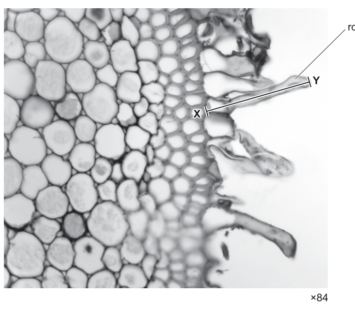
Fig. 1.1 shows part of a transverse section of a root of S. china with root hair cells visible.
Answer
Light (microscope).
Light microscope
Background Concept
A compound light microscope (also called an optical or bright-field microscope) uses a beam of visible light that passes up through a thin, often stained, specimen and a series of glass lenses to produce a magnified, coloured image. Its maximum useful magnification is around ×1500 and its resolution (the smallest distance between two points that can be distinguished as separate) is limited by the wavelength of visible light to about 200 nm. A transmission electron microscope, by contrast, fires a beam of electrons through an ultra-thin specimen; because electrons have a much shorter wavelength than visible light, resolution can reach ~0.2 nm and useful magnification exceeds ×500 000. Electron micrographs are always greyscale (black and white), cannot show living material, and cannot be in colour because the image is formed by electron scattering, not light absorption.
Understanding the Question
You are given Fig. 1.1 — a coloured image of plant tissue in which you can see individual cell walls, vacuoles and a root hair cell with the line X–Y drawn along it. The image is also presented at a modest magnification (×84). The question asks you to identify the type of microscope that produced this image.
Approach
Look at the visual characteristics of Fig. 1.1 and use them to decide whether a light or electron microscope was used.
Step-by-Step Reasoning
Fig. 1.1 is a coloured image (you can see different shades of pink/magenta where the tissue is stained). The cell walls, vacuoles and a whole root hair cell with surrounding cells are all visible at once. These features point to a light microscope:
- Colour is present — light microscopes can show stains; electron micrographs are greyscale.
- The magnification is only ×84, well within the working range of a light microscope.
- Whole cells with internal organisation are visible — the resolution is consistent with light microscopy, not the very high resolution of an electron microscope.
Key Takeaways
Light (optical) microscopes produce coloured images of whole cells using visible light; electron microscopes produce greyscale images at much higher magnification and resolution. Recognising which type of microscope was used is a matter of looking at whether the image is in colour, what magnification it carries, and what level of detail is visible.
Common Mistakes
- Writing "stereo microscope" or "dissecting microscope" — these are specific types of light microscope used for thicker specimens and lower powers; the question is asking for the broad class.
- Writing "scanning electron microscope" — this would give a 3-D-looking surface view in greyscale, not what is shown.
- Leaving the answer as just "microscope" without specifying the type.
Things to Be Careful About
- The mark scheme accepts either "light" or "optical" — both are correct.
- Do not credit "fluorescence" or other specific techniques unless that is clearly indicated.
- The image is a transverse section through a herbaceous root, so a typical bright-field light microscope is the obvious answer.
Calculate the actual length, in micrometres (), of the root hair cell labelled in Fig. 1.1. Use the image length of the root hair cell along line X–Y in your calculation.
actual length = ______
Working
Image length of X–Y measured on the figure ≈ 39 mm
Answer
464 µm
464 µm
Background Concept
A microscope produces a magnified image of a specimen. The relationship between the size of the image you see, the actual size of the specimen, and the magnification of the microscope is given by:
Rearranging:
Because the specimen here is very small, you will need to convert units: .
Understanding the Question
The question gives you the magnification of the photograph (×84) and shows a line X–Y drawn along the length of a root hair cell. You need to use the image length of X–Y (which you measure with a ruler from the printed figure) to calculate the actual length of the cell, in micrometres.
Approach
Measure the printed length of line X–Y with a ruler (in mm), then use the rearranged magnification formula to convert that image length into actual length, and finally convert the result from mm to µm.
Step-by-Step Reasoning
- Measure the printed line X–Y: in the original paper it is approximately 39 mm long. (Your own ruler reading should give an answer in the range 38–40 mm.)
- Substitute into the formula:
- Evaluate:
- Convert mm to µm by multiplying by 1000:
Key Takeaways
The magnification formula rearranged for actual size is: actual size = image size ÷ magnification. Unit conversion: , so to get µm you multiply the mm answer by 1000. Always quote the answer with the correct unit and to a sensible number of significant figures.
Common Mistakes
- Inverting the formula and multiplying instead of dividing (image × magnification) — this would give a length many times bigger than the real cell.
- Forgetting to convert mm to µm and leaving the answer in mm, or converting incorrectly.
- Using the magnification as a length in mm (e.g. dividing by "84 mm").
- Quoting an answer such as "0.46" with no unit.
Things to Be Careful About
- The mark scheme accepts values in the range 450–480 µm depending on individual ruler measurements; it explicitly accepts 460 and 464. Your measurement should produce a number in this range.
- The line X–Y is the image length, not the actual length — this is the crucial distinction.
- Do not include units in the answer box for "actual length"; the box is already labelled with µm.
Root hairs are important adaptations of root hair cells for the uptake of water.
Explain one way in which root hairs adapt root hair cells for the uptake of water.
Answer
Root hairs increase the surface area of the root for the uptake of water.
Increase the surface area for water uptake
Background Concept
The rate at which a cell or organ can exchange materials (such as water, ions or gases) with its environment is limited by the surface area available for exchange. This is captured by the surface-area-to-volume ratio (SA:V). A long, thin extension such as a root hair dramatically increases surface area without greatly adding to the volume of the cell, so the SA:V is much higher than for a roughly spherical cell. This increases the rate at which water (and dissolved mineral ions) can move into the cell by osmosis (and by diffusion / active transport).
Understanding the Question
The question asks you to explain ONE way that root hairs adapt root hair cells for the uptake of water. You must describe an adaptation (something about the root hair's structure or position) AND link it to how it helps water uptake. Only one mark is available, so one clear point is enough.
Approach
Pick a single, specific adaptation of root hairs and state how it improves water uptake. The most obvious and most commonly credited adaptation is the very large surface area that the hair provides.
Step-by-Step Reasoning
Root hairs are long, thin tubular extensions of the outer (epidermal) cells of the root. Each hair:
- Greatly increases the surface area of the root available for absorption of water and mineral ions.
- Because the surface area is increased without a large increase in cell volume, the SA:V ratio is very high.
- A higher SA:V gives a faster rate of osmosis / diffusion into the cell.
- (An alternative credited point: the hair reaches into a larger volume of soil so more water comes into contact with the cell's cell surface membrane.)
- (Another credited point: the cell surface membrane of a root hair cell contains many aquaporins, allowing more rapid entry of water by osmosis.)
Key Takeaways
Root hairs are an adaptation that increase the surface area to volume ratio of the root, allowing faster uptake of water (and ions) from the soil. Other acceptable adaptations include reaching a larger volume of soil and increasing the density of aquaporins in the cell surface membrane.
Common Mistakes
- Saying "they absorb more water" without explaining why — the question demands an explanation of HOW the adaptation helps.
- Stating incorrect reasons such as "root hairs have a thin cell wall so water passes through easily" — the cell wall is fully permeable to water in any cell, so this is not a special feature of root hairs.
- Confusing root hairs with root hair cells; the hair is the extension, not a separate cell type.
Things to Be Careful About
- One mark is for one explanation; do not pad with three points. Pick the strongest single point.
- The question asks specifically about uptake of WATER, not mineral ions. (Mineral-ion uptake is covered in a later part.)
Mineral ions are taken up by root hair cells.
Table 1.1 shows the concentrations of sodium ions () and potassium ions () inside the root hair cells of a plant root and in the soil solution surrounding the root.
Table 1.1
| concentration of | concentration of | |||
|---|---|---|---|---|
| root hair cell | soil solution | root hair cell | soil solution | |
| 0.35 | 3.34 | 5.46 | 0.16 |
With reference to Table 1.1:
- suggest the mechanisms involved in the transport of and from the soil solution into the root hair cells
- state the reasons for your suggestions.
Answer
- Na⁺ enters the root hair cell by facilitated diffusion because the concentration of Na⁺ is higher in the soil solution (3.34 g dm⁻³) than inside the root hair cell (0.35 g dm⁻³), i.e. down the concentration gradient.
- K⁺ enters by active transport because the concentration of K⁺ is higher inside the root hair cell (5.46 g dm⁻³) than in the soil solution (0.16 g dm⁻³), i.e. against the concentration gradient.
- Ions cannot diffuse through the phospholipid bilayer of the cell surface membrane.
- Facilitated diffusion therefore uses channel / carrier proteins, and active transport uses carrier proteins (and energy from ATP).
See working
Background Concept
Cells exchange substances with their surroundings across the cell surface membrane. Small non-polar molecules (e.g. O₂, CO₂) and water cross the phospholipid bilayer directly by simple diffusion or by osmosis. Ions, however, are charged and are repelled by the hydrophobic core of the bilayer, so they cannot cross by simple diffusion. They need help from membrane proteins — either:
- Channel proteins (form a hydrophilic pore) for facilitated diffusion down an electrochemical gradient. No ATP is needed.
- Carrier proteins (bind the ion and change shape) for either facilitated diffusion or active transport. Active transport moves the ion against its electrochemical gradient using energy from ATP, and is usually coupled to the movement of another ion down its gradient (e.g. H⁺).
The direction of net movement is dictated by the concentration gradient:
- Down the gradient → passive (facilitated) diffusion.
- Against the gradient → active transport.
Understanding the Question
Table 1.1 gives you the concentrations of Na⁺ and K⁺ inside the root hair cell and in the soil solution. You have to:
- Suggest a transport mechanism for each ion.
- State the reason (the direction of the gradient).
You also have to explain why transport proteins are needed in both cases.
Approach
For each ion, compare the cell and soil concentrations, decide whether the ion is moving down or against its concentration gradient, and pick the matching mechanism. Then add the general point that ions cannot cross the phospholipid bilayer unaided.
Step-by-Step Reasoning
Na⁺: Soil = 3.34 g dm⁻³; cell = 0.35 g dm⁻³. The concentration outside is much higher than inside, so Na⁺ can move down its concentration gradient without using ATP. The mechanism is facilitated diffusion, via channel or carrier proteins. (Mark scheme point 1.)
K⁺: Soil = 0.16 g dm⁻³; cell = 5.46 g dm⁻³. The concentration is much higher inside the cell than in the soil, so K⁺ must be moved against its concentration gradient into the cell. This requires active transport, which uses carrier proteins and ATP. (Mark scheme point 2.)
Why proteins are needed for both: Na⁺ and K⁺ are ions — they are charged. They cannot diffuse through the hydrophobic phospholipid bilayer of the cell surface membrane. Both facilitated diffusion (channel / carrier) and active transport (carrier) therefore use membrane proteins. (Mark scheme points 3 and 4.)
Key Takeaways
- Down a concentration gradient + ions → facilitated diffusion (channel or carrier proteins, no ATP).
- Against a concentration gradient + ions → active transport (carrier proteins, ATP).
- The same idea applies to many other ions (Mg²⁺, Ca²⁺, NO₃⁻, etc.) in plant and animal cells.
- The mark scheme caps the answer at 2 marks if you only discuss one ion — both must be addressed.
Common Mistakes
- Saying Na⁺ is moved by active transport and K⁺ by diffusion because the numbers "look like" the ions are at higher concentration outside (confusing which ion is which).
- Saying "diffusion" without specifying facilitated diffusion (simple diffusion of ions is impossible).
- Giving a reason such as "the cell needs Na⁺ and K⁺" — that is a biological need, not a mechanistic reason.
- Not mentioning that ions cannot pass through the phospholipid bilayer — this is what makes proteins essential.
Things to Be Careful About
- You must mention both ions and give a direction of gradient for each.
- Quote the actual numbers from the table to make your answer specific; generic "higher outside" / "higher inside" statements are weaker.
- "Carrier proteins" is required for active transport; "channel proteins" alone is not enough.
The region between the outer layer of a root and the endodermis is known as the cortex.
Table 1.2 shows the water potential of two adjacent cells, A and B, in the cortex of a root.
Table 1.2
| water potential / kPa | |
|---|---|
| cortex cell A | |
| cortex cell B |
With reference to Table 1.2, explain the direction of water movement between cell A and cell B.
Answer
Water moves from cell A to cell B by osmosis, from higher (less negative) water potential to lower (more negative) water potential, i.e. down the water potential gradient.
From cell A to cell B, by osmosis, down the water potential gradient.
Background Concept
Water potential () is a measure of the tendency of water to move from one place to another. Pure water has a water potential of 0 kPa. Adding solutes lowers the water potential, so all living cells have negative water potentials (e.g. −120 kPa, −350 kPa). The more negative the value, the lower the water potential and the stronger the tendency of that cell to draw water in. Water moves by osmosis across a selectively permeable membrane from a region of higher (less negative) water potential to a region of lower (more negative) water potential — i.e. down the water potential gradient.
Understanding the Question
Table 1.2 gives you the water potentials of two adjacent cortex cells, A and B. You must state the direction of water movement between them and justify it using the water-potential gradient.
Approach
Compare the two values, identify which cell has the higher water potential (less negative) and which has the lower (more negative), then state that water moves from the higher to the lower by osmosis.
Step-by-Step Reasoning
- Cell A: = −120 kPa. Cell B: = −350 kPa.
- −120 kPa is a higher (less negative) water potential than −350 kPa.
- So water moves from A to B.
- The movement is by osmosis (water only, across the partially permeable cell surface membrane between the two adjacent cells).
- The driving force is the water potential gradient: water moves from higher (less negative) to lower (more negative) , i.e. down the gradient.
Key Takeaways
- "More negative " means "lower " — do not confuse the sign.
- Water always moves down the water potential gradient by osmosis.
- In a root, water enters at the root hair, then moves from cortex cell to cortex cell toward the xylem because each successive cell has a more negative water potential (driven by the accumulation of solutes in the xylem and by transpiration pull from the leaves).
Common Mistakes
- Saying "water moves from high concentration of water to low concentration of water" — this loses the mark. You must use the term water potential ().
- Saying "water moves from B to A" — students sometimes reverse the direction because −350 is "bigger" than −120. The rule is the more negative number = lower water potential.
- Not naming osmosis as the process.
Things to Be Careful About
- Quote the units (kPa) when comparing values.
- The direction is from −120 kPa to −350 kPa, i.e. A → B.
- The question is worth 2 marks — one for naming the direction (A → B by osmosis), one for explaining it (down the water potential gradient).
Some types of soil are made of small negatively charged clay particles that attract and bind to positive ions such as iron ions ().
that is bound to clay particles cannot be absorbed by root hair cells. This reduces the concentration of free that is available in soil solution for absorption.
Some plants that grow in soils containing a high proportion of clay particles are able to increase the concentration of free in the soil solution for absorption. In these plants, the carbon dioxide released by the respiration of root hair cells reacts with water in the soil solution, which changes the pH of the soil solution. This affects the binding of positively charged ions, such as , to clay particles.
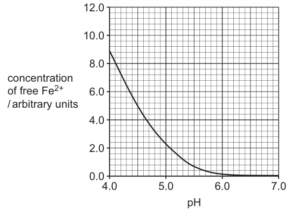
Fig. 1.2 shows the results of one investigation into the effect of pH on the concentration of free that is available in soil solution for absorption by root hair cells. The soil sample analysed in this investigation was from a soil that contained a high proportion of clay particles.
With reference to Fig. 1.2, suggest and explain how the carbon dioxide released by the respiration of root hair cells can increase the concentration of free for absorption by root hair cells.
Answer
- CO₂ released by respiration of root hair cells reacts with H₂O in the soil solution to form carbonic acid (H₂CO₃).
- Carbonic acid dissociates into hydrogencarbonate ions (HCO₃⁻) and protons (H⁺).
- The concentration of H⁺ rises, so the pH of the soil solution falls (the soil becomes more acidic).
- Fig. 1.2 shows that as pH falls below about 5–6, the concentration of free Fe²⁺ in the soil solution rises steeply (e.g. ~8.8 arbitrary units at pH 4, but ~0 at pH 7).
- At this lower pH, the H⁺ ions displace Fe²⁺ from the negatively charged clay particles (which had been binding the positively charged Fe²⁺), releasing Fe²⁺ into the soil solution.
- The hydrogencarbonate ions (HCO₃⁻) attract the displaced Fe²⁺ and hold it in solution, raising the concentration of free Fe²⁺ available for absorption by the root hair cells.
See working
Background Concept
Respiring cells release CO₂. In water, CO₂ reacts reversibly with water to form carbonic acid (H₂CO₃), which then dissociates into a hydrogencarbonate ion (HCO₃⁻) and a proton (H⁺):
The release of H⁺ lowers the pH of the surrounding solution, making it more acidic.
In soils rich in clay, the tiny clay particles carry a net negative charge on their surface and so bind positively charged ions such as Fe²⁺, Mg²⁺ and Ca²⁺. These bound ions are not free in solution and so cannot be taken up by roots. Adding H⁺ (lowering the pH) competes with Fe²⁺ for the negatively charged binding sites on the clay. When enough H⁺ is present, Fe²⁺ is displaced from the clay into the soil solution, and the bicarbonate ion (HCO₃⁻) can pair with the Fe²⁺ and keep it soluble.
Fig. 1.2 shows the empirical result: as pH falls from 7.0 to 4.0, the concentration of free Fe²⁺ in the soil solution rises sharply (from ~0 to ~8.8 arbitrary units).
Understanding the Question
The parent stem tells you that:
- CO₂ released by root hair cell respiration reacts with water in the soil solution and changes the pH.
- This change in pH affects the binding of positively charged ions such as Fe²⁺ to clay particles.
- Fig. 1.2 shows how the free Fe²⁺ concentration in a clay-rich soil depends on pH.
You must use these clues to suggest AND explain how CO₂ from respiration can increase the free Fe²⁺ available for absorption.
Approach
Build a logical chain: respiration → CO₂ → carbonic acid in soil water → H⁺ released → pH falls → H⁺ displaces Fe²⁺ from clay → free Fe²⁺ rises. Each link must be supported by either the chemistry given in the stem or a reading from Fig. 1.2.
Step-by-Step Reasoning
- Root hair cells respire aerobically, releasing CO₂ as a waste product. The CO₂ diffuses out into the soil solution surrounding the root.
- In water, CO₂ reacts to form carbonic acid (H₂CO₃).
- Carbonic acid is a weak acid that dissociates into HCO₃⁻ and H⁺. (The bicarbonate / hydrogencarbonate ion is the conjugate base.)
- The H⁺ released makes the soil solution more acidic — i.e. the pH of the soil solution falls. (Compare, for example, pH 7 with pH 5.)
- Reading Fig. 1.2: at pH 5 the free Fe²⁺ concentration is ~3 arbitrary units, at pH 4 it is ~8.8 — i.e. as pH falls, free Fe²⁺ rises steeply.
- Mechanistically, the H⁺ ions compete with Fe²⁺ for the negative binding sites on the clay particles. At lower pH (higher [H⁺]) the H⁺ displaces the Fe²⁺ from the clay.
- The Fe²⁺ released from the clay into the soil solution is held in solution by HCO₃⁻ (which forms a soluble salt with Fe²⁺).
- The result is a higher concentration of free Fe²⁺ in the soil solution, available to be absorbed by root hair cells.
Key Takeaways
- CO₂ + H₂O ⇌ H₂CO₃ ⇌ H⁺ + HCO₃⁻ is the carbonic-acid equilibrium.
- Adding CO₂ to a watery environment lowers the pH (more H⁺).
- Lower pH releases cations such as Fe²⁺ from negatively charged binding sites on clay.
- Reading a pH–concentration graph correctly: as pH decreases (x-axis to the left), the free Fe²⁺ increases.
Common Mistakes
- Saying that "CO₂ makes the soil more acidic" without mentioning carbonic acid or H⁺.
- Saying that CO₂ "directly releases" Fe²⁺ from clay — there must be the intermediate pH step.
- Reading Fig. 1.2 the wrong way round — "as pH increases, Fe²⁺ increases" is the OPPOSITE of what the graph shows.
- Forgetting the role of HCO₃⁻ in keeping the Fe²⁺ in solution once it has been displaced.
Things to Be Careful About
- Quote paired data from Fig. 1.2 where possible, e.g. "at pH 4 the free Fe²⁺ concentration is ~8.8, but at pH 7 it is ~0". This earns the mark for "correct paired data quotes" if given instead of stating that pH falls.
- The reaction CO₂ + H₂O ⇌ H₂CO₃ is uncatalysed in the soil solution (no carbonic anhydrase), so the rate is slow — this is a valid AVP point.
- The question is worth 4 marks; aim for at least 4 of the 6 points above.
Dissolved is transported across the tissues of the root to the xylem.
When the amount of dissolved absorbed by root hair cells is greater than the amount that is needed by the plant, not all of the that is absorbed is transported to the xylem.
Suggest how the endodermis can reduce the amount of reaching the xylem.
Answer
- To reach the xylem, Fe²⁺ must cross the cell surface membrane of endodermal cells and pass through their cytoplasm (the symplastic route).
- The apoplastic route is blocked by the Casparian strip — a band of suberin in the cell walls of the endodermis that is impermeable to water and dissolved ions.
- Because Fe²⁺ must enter the cell, the endodermal cell can regulate the number / activity of transport proteins in its cell surface membrane, controlling how much Fe²⁺ crosses.
- Excess Fe²⁺ can be converted into insoluble iron compounds (and/or transported into the vacuole) and stored inside the endodermal cell, so less Fe²⁺ reaches the xylem.
See working
Background Concept
The endodermis is a single layer of cells surrounding the vascular tissue (xylem and phloem) of the root. A key feature of these cells is the Casparian strip — a ring of suberin (a waxy, hydrophobic material) deposited in the radial and transverse cell walls. The Casparian strip is impermeable to water and to dissolved ions, so the apoplastic pathway (movement through cell walls and intercellular spaces without crossing any membrane) is blocked at the endodermis. To continue towards the xylem, ions and water must cross the cell surface membrane of an endodermal cell and pass through its cytoplasm (the symplastic pathway).
Because they have to cross a membrane, ions can be regulated by the cell. The cell can:
- Adjust the number / activity of membrane transport proteins.
- Pump excess ions into the vacuole for storage.
- Convert the ion into an insoluble form that precipitates inside the cell.
Understanding the Question
The parent stem tells you that when more Fe²⁺ is absorbed by root hair cells than the plant needs, not all of it reaches the xylem. You have to suggest how the endodermis can reduce the amount of Fe²⁺ that gets through to the xylem.
Approach
The endodermis is the bottleneck for solute movement into the xylem. Two complementary mechanisms can be invoked:
- Restrict entry by controlling membrane transport.
- Remove what does enter by storing or precipitating it.
Step-by-Step Reasoning
- The Casparian strip blocks the apoplastic route through the endodermal cell walls, so any Fe²⁺ moving into the stele (and on into the xylem) must first cross the cell surface membrane of an endodermal cell, into the cytoplasm.
- Because Fe²⁺ has to cross a membrane, the endodermal cell can regulate the quantity / activity of transport proteins in that membrane — if fewer transporters are inserted, less Fe²⁺ crosses per unit time.
- Alternatively, Fe²⁺ that has entered the endodermal cell can be actively transported into the vacuole for storage, removing it from the symplastic stream that leads to the xylem.
- Fe²⁺ can also be converted (e.g. by binding to organic acids or to ferritin) into insoluble iron compounds that precipitate inside the cell, again preventing onward movement to the xylem.
- The combined effect is that only the Fe²⁺ the plant actually needs is loaded into the xylem for transport up to the shoot.
Key Takeaways
- The Casparian strip is the structural reason why the endodermis is the control point for ion entry into the xylem.
- Once an ion has to cross a cell surface membrane, the cell can regulate it (transporters), store it (vacuole), or immobilise it (insoluble compounds).
- This is one of the key selective-barrier functions of the endodermis and is essential for plant ion homeostasis.
Common Mistakes
- Saying "the endodermis blocks Fe²⁺" without naming the Casparian strip — vague answers are not credited.
- Saying "the endodermis stores Fe²⁺" without giving a mechanism (vacuole, insoluble compounds).
- Confusing the endodermis with the epidermis or pericycle.
- Saying "active transport prevents Fe²⁺ entering the xylem" — active transport loads Fe²⁺ INTO the xylem; the regulation here is by reducing transport into the cell, or by storing it.
Things to Be Careful About
- Worth 3 marks — aim for at least 3 of the 4 points.
- The terms "symplastic route" and "Casparian strip / suberin" are the technical terms the mark scheme wants.
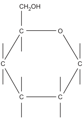
Fig. 2.1 is an incomplete diagram of the structure of an -glucose molecule.
Complete Fig. 2.1 to show the structure of an -glucose molecule.
Answer
Add the missing H and OH groups to each ring carbon:
- C1 (right of the ring O): H above, OH below — the OH below on C1 is the diagnostic feature of the α-anomer.
- C2 (bottom right): H above, OH below.
- C3 (bottom left): OH above, H below.
- C4 (left): H above, OH below.
- C5 (top left): H below; the CH2OH group is already shown above.
See diagram.
Background Concept
Glucose is a hexose monosaccharide with molecular formula C6H12O6. In aqueous solution it exists predominantly as a six-membered ring (pyranose), formed when the C1 aldehyde reacts with the C5 hydroxyl to give a ring of five carbons and one oxygen. In this ring form, each of the four non-CH2OH-bearing carbons (C1–C4) carries one H and one OH; C5 carries the CH2OH group. The position of the OH on C1 distinguishes the two anomers: in α-glucose the OH on C1 lies on the opposite side of the ring from the CH2OH (drawn below the ring in the standard Haworth projection), whereas in β-glucose it lies on the same side (drawn above the ring).
Understanding the Question
The question shows a partially drawn α-glucose molecule: the carbon skeleton, the ring oxygen, and the CH2OH attached to C5 are present, but the H and OH groups on every ring carbon are missing. The candidate must complete the structure, including correctly placing the OH on C1 below the ring (the α-anomer feature).
Approach
Work around the ring one carbon at a time, placing each H and OH above or below the ring plane according to the standard α-D-glucose stereochemistry. Use the conventional Haworth projection as a reference.
Step-by-Step Reasoning
- C1 (right of the ring oxygen): In α-glucose the OH is on the opposite side of the ring from the CH2OH. Since the CH2OH is drawn above the ring (attached to C5), the OH on C1 must be drawn below the ring; the H is above. Getting C1 right is awarded one mark on its own (the mark scheme gives a separate mark for H and OH correct on C1).
- C2 (bottom right): H above, OH below.
- C3 (bottom left): OH above, H below — the orientation flips at C3.
- C4 (left): H above, OH below.
- C5 (top left): H below; the CH2OH group is already drawn above.
All remaining H and OH placements (on C2, C3, C4 and the H on C5) must be complete and correct for the second mark.
Key Takeaways
- α-glucose and β-glucose differ only in the orientation of H and OH on C1.
- The ring form of glucose has 5 carbons + 1 oxygen, with each ring carbon carrying either an H, an OH, or a CH2OH.
- Drawing glucose correctly requires memorising the alternating pattern of H/OH positions around the ring.
Common Mistakes
- Drawing the OH on C1 above the ring — this is β-glucose, not α-glucose.
- Drawing the OH on C3 in the wrong orientation (the OH flips above the ring at C3, not below).
- Forgetting to add the H on C5.
- Putting H above OH on C3 instead of OH above and H below.
Things to Be Careful About
- The mark scheme awards one mark specifically for H/OH correct on C1 — this is the diagnostic feature of the α-anomer.
- The other H/OH positions must all be correct for the second mark; a single error in placement can lose the mark.
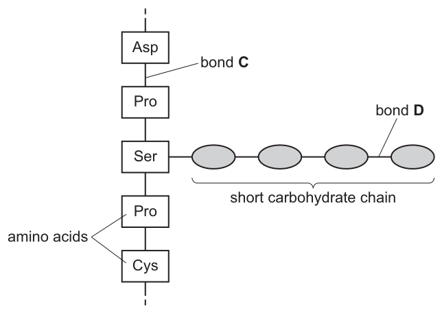
Fig. 2.2 shows part of a glycoprotein molecule.
Answer
C — peptide (bond)
D — glycosidic (bond)
C: peptide bond; D: glycosidic bond
Background Concept
Biological molecules are built by linking monomers into polymers using covalent bonds formed by condensation reactions. The specific bond formed depends on which monomers are joining:
- Two amino acids join via a peptide bond, formed by condensation between the α-COOH of one amino acid and the α-NH2 of the next, releasing a molecule of water.
- Two monosaccharides join via a glycosidic bond, formed by condensation between two hydroxyl (–OH) groups, again releasing water.
In a glycoprotein the polypeptide chain has one or more short carbohydrate side chains attached, usually to the R-group of an amino acid such as serine, threonine or asparagine. The protein part is held together by peptide bonds; the carbohydrate part is held together by glycosidic bonds.
Understanding the Question
Fig. 2.2 shows part of a glycoprotein: a vertical chain of amino acids (Asp-Pro-Ser-Pro-Cys) with a short carbohydrate chain of four sugar units attached to the serine. Two bonds are labelled: bond C lies in the amino-acid chain, bond D lies in the carbohydrate side chain. The candidate must name each bond type.
Approach
Look at what each labelled bond is joining: C joins two amino acids → peptide; D joins two sugar units → glycosidic.
Step-by-Step Reasoning
- Bond C is in the vertical chain between the Asp and Pro amino acids. The covalent bond linking two amino acids in a polypeptide is a peptide bond.
- Bond D is in the horizontal short carbohydrate chain, between two of the sugar (oval) units. The covalent bond linking two monosaccharides is a glycosidic bond.
Key Takeaways
- Peptide bond = amino acid to amino acid.
- Glycosidic bond = monosaccharide to monosaccharide.
- Both are covalent, both formed by condensation, but they join different types of monomer.
Common Mistakes
- Calling bond C a disulfide bond — disulfide bonds form between two cysteine R-groups, not between adjacent amino acids in the backbone.
- Calling bond D a peptide bond.
- Spelling "glycosidic" as "glucosidic" or "glycoside".
Things to Be Careful About
- Bond D is between sugars within the carbohydrate side chain, not between the protein and the carbohydrate (the protein-to-carbohydrate attachment is a different linkage, sometimes called an O- or N-glycosidic linkage, but it is not what the question is asking about).
Many glycoproteins in the cell surface membrane are involved in cell signalling.
State the role in cell signalling of glycoproteins in cell surface membranes.
Answer
Act as receptors for signalling molecules (e.g. hormones) at the cell surface, binding the ligand and triggering a response inside the cell.
Act as receptors / bind to ligands (qualified).
Background Concept
The cell surface membrane is the cell's outer boundary and the first point of contact with the extracellular environment. Many integral membrane proteins have short carbohydrate chains attached to extracellular domains, forming glycoproteins. The carbohydrate portion projects into the extracellular space.
In cell signalling, a chemical messenger (a ligand) such as a hormone, neurotransmitter or growth factor binds to a specific receptor on the cell surface. Many cell-surface receptors are glycoproteins, with their extracellular carbohydrate chains and amino-acid R-groups together forming a binding site complementary in shape to the ligand. Ligand binding triggers a response inside the cell — for example, opening an ion channel, activating a second-messenger cascade, or altering gene expression.
Understanding the Question
The question asks specifically for the role of glycoproteins in cell signalling (not all their roles, which also include cell recognition and cell adhesion). The mark scheme accepts: act as receptors / bind to ligands, qualified — meaning the candidate should specify what kind of ligand binds.
Approach
Recall that cell-surface receptors for hydrophilic signalling molecules are usually glycoproteins, and that the carbohydrate chain and the extracellular R-groups together form the ligand-binding site.
Step-by-Step Reasoning
- Many signalling molecules (e.g. peptide hormones, neurotransmitters) are hydrophilic and cannot cross the phospholipid bilayer directly.
- They must therefore bind to a receptor on the outer face of the cell surface membrane.
- Many of these cell-surface receptors are glycoproteins; the extracellular carbohydrate chain and the surrounding amino-acid R-groups form the binding site for the ligand.
- Binding of the ligand to the receptor triggers a conformational change or downstream cascade that produces a response inside the cell.
Key Takeaways
- Membrane glycoproteins act as receptors for cell-signalling molecules.
- The carbohydrate portion contributes to ligand binding on the extracellular side of the membrane.
- This is the mechanism by which cells respond to hormones, neurotransmitters and other hydrophilic signals that cannot cross the membrane directly.
Common Mistakes
- Saying glycoproteins are antigens — this is a recognition role, not a signalling role, and is not what the question asks.
- Saying glycoproteins transport substances — that is the role of channel/carrier proteins, not signalling.
- Failing to qualify the answer with the type of ligand (e.g. hormones, neurotransmitters) — the mark scheme explicitly requires qualification.
Things to Be Careful About
- "Act as receptors" alone is not sufficient — the answer must specify what binds (a hormone, a neurotransmitter, a ligand, a signalling molecule).
- The question asks about the role in cell signalling specifically, not all roles of glycoproteins.
One of the proteins found in milk is -casein.
A molecule of -casein consists of a single polypeptide of approximately 200 amino acids.
Molecules of -casein have a high proportion of the amino acids proline and leucine. These amino acids have hydrophobic R-groups.
Fig. 2.3 compares the structure of proline (Pro) with the general structure of an amino acid. This shows that proline is an unusual amino acid because it has a cyclic R-group and the nitrogen atom is attached to only one hydrogen atom.
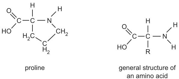
When a molecule of proline becomes part of a polypeptide, the hydrogen atom is lost from the nitrogen atom and is therefore not available for the formation of hydrogen bonds.
Molecules of -casein have a relatively low proportion of the amino acids cysteine and serine. Table 2.1 shows the R-groups of cysteine and serine.
Table 2.1
| amino acid | R-group | feature of R-group |
|---|---|---|
| cysteine (Cys) | can form a disulfide bond | |
| serine (Ser) | hydroxyl group present |
Compared to other protein molecules with a similar number of amino acids, -casein molecules have:
- a much less organised secondary structure
- relatively little tertiary structure.
With reference to the four named amino acids, proline, leucine, serine and cysteine, suggest possible explanations for these two observations.
less organised secondary structure ______
relatively little tertiary structure ______
Answer
Less organised secondary structure
- High proportion of proline: its nitrogen has no H available to donate a hydrogen bond, so less H-bonding occurs between the backbone –NH of one amino acid and the –C=O of another.
- This prevents regular α-helix / β-pleated sheet formation, leaving the chain as a more random coil.
Relatively little tertiary structure
- Low proportion of cysteine: few disulfide bonds can form between R-groups to lock the folded shape in place.
- Low proportion of serine: few side-chain hydrogen bonds form between R-groups in the tertiary fold.
- High proportion of proline and leucine (hydrophobic R-groups): few ionic / polar R-group interactions are available to stabilise a defined 3D shape.
See working above.
Background Concept
Proteins have several levels of structure:
- Primary — the linear sequence of amino acids joined by peptide bonds.
- Secondary — local regular folding (α-helix, β-pleated sheet) stabilised by hydrogen bonds between the backbone –NH of one amino acid and the –C=O of another amino acid a few residues along.
- Tertiary — the overall 3D folding of a single polypeptide, stabilised by interactions between R-groups: hydrogen bonds, ionic bonds (between charged R-groups), covalent disulfide bonds (between two cysteine –SH groups, forming –S–S–), and hydrophobic interactions (between non-polar R-groups in the interior of the protein).
- Quaternary — assembly of multiple polypeptide subunits.
The R-group of each amino acid determines what interactions it can participate in:
- Proline — its R-group is cyclic and loops back to bond the nitrogen, leaving only one H on the nitrogen instead of the usual two. When proline joins a polypeptide, this H is lost, so proline cannot donate a hydrogen bond through its backbone nitrogen.
- Leucine — non-polar, hydrophobic R-group; contributes to hydrophobic interactions in the protein interior.
- Serine — R-group has a hydroxyl (–OH) that can act as both donor and acceptor in hydrogen bonding with other polar R-groups.
- Cysteine — R-group has a sulfhydryl (–SH) that can oxidise with another cysteine to form a covalent disulfide (–S–S–) bond.
Understanding the Question
β-casein is a single polypeptide of ~200 amino acids with a high proportion of proline and leucine and a low proportion of cysteine and serine. The question states that, compared with other proteins of similar size, β-casein has a much less organised secondary structure and relatively little tertiary structure. The candidate must use the chemistry of the four named amino acids to explain these two observations.
Approach
For each observation, identify which of the four named amino acids is responsible, and link its R-group chemistry to the missing structural feature:
- Less secondary structure → blame the high proline content (disrupts backbone H-bonding).
- Less tertiary structure → blame the low cysteine (no disulfide bonds), low serine (few side-chain H-bonds), and the dominance of hydrophobic R-groups (fewer polar/ionic interactions).
Step-by-Step Reasoning
Less organised secondary structure:
- Secondary structure (α-helix and β-pleated sheet) requires regular hydrogen bonding between the backbone –NH of one amino acid and the –C=O of another amino acid a few residues away.
- Proline is unusual: its R-group is a ring that loops back and bonds to the nitrogen, so the nitrogen carries only one H (not two). When proline joins the polypeptide chain, this single H is lost (the question states this directly), so proline cannot act as a hydrogen-bond donor in the backbone.
- Regions of the polypeptide containing proline therefore cannot form the regular α-helix or β-pleated sheet, and instead adopt a random coil conformation.
- Because β-casein has a high proportion of proline, much of its polypeptide chain lacks the regular H-bonding pattern needed for organised secondary structure — hence the much less organised secondary structure.
Relatively little tertiary structure:
- Tertiary structure is stabilised by interactions between R-groups: hydrogen bonds, ionic bonds, disulfide bonds, and hydrophobic interactions.
- Cysteine residues form covalent disulfide (–S–S–) bonds, which are particularly strong and help lock the folded 3D shape in place. β-casein has few cysteine residues, so very few disulfide bonds can form — losing one of the main anchors of tertiary structure.
- Serine has a hydroxyl (–OH) R-group that can hydrogen-bond with other polar/charged R-groups, contributing to the network of side-chain interactions that stabilises tertiary folding. β-casein has few serine residues, so fewer side-chain hydrogen bonds can form.
- The very high proportion of proline and leucine (both with hydrophobic, non-polar R-groups) means there are few amino acids available to form ionic bonds between charged R-groups, and fewer polar R-groups available to form side-chain H-bonds. The R-group composition simply does not support a highly organised, compact 3D fold with a well-defined tertiary structure; instead, hydrophobic interactions between proline and leucine R-groups dominate, but these do not impose the same kind of precise 3D architecture as a balanced mix of polar, charged and non-polar R-groups would.
Key Takeaways
- Proline's cyclic R-group prevents backbone H-bonding and therefore disrupts regular secondary structure.
- Tertiary structure depends on R-group composition: few cysteine → few disulfide bonds; few serine → few side-chain H-bonds; high hydrophobic content → fewer ionic / polar interactions.
- β-casein's unusual amino acid composition directly explains its unusual structural properties.
- Protein structure levels (1°, 2°, 3°, 4°) all depend on the chemistry of the amino acids in the sequence.
Common Mistakes
- Saying proline is hydrophobic and therefore disrupts hydrophobic interactions — this is irrelevant; proline's key feature is that it cannot donate a backbone H-bond.
- Attributing the loss of tertiary structure mainly to proline — proline's effect is primarily on secondary structure; the tertiary structure loss is due to the low cysteine, low serine, and the dominance of hydrophobic R-groups.
- Saying "β-casein has no tertiary structure" — the question states "relatively little", not "none".
- Failing to name the specific bond type that is missing (e.g. saying "no cysteine" without explaining that this means no disulfide bonds).
- Forgetting to mention that the question requires reference to the four named amino acids — generic statements about H-bonding or disulfide bonding without naming the specific amino acid(s) will not earn full marks.
Things to Be Careful About
- The two answers (less secondary, less tertiary) must be clearly separated and each linked to specific amino acids.
- The mark scheme awards up to 3 marks from each section, with 4 marks in total — a balanced answer (2 + 2 or 3 + 1) is usually best.
- The "AVP" mark in the mark scheme allows credit for an additional valid point, e.g. noting that hydrophobic interactions are the dominant tertiary interaction in β-casein but do not give the same defined 3D shape as a balanced mix of interactions would.
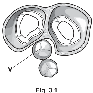
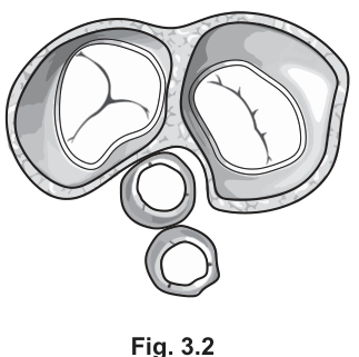
Fig. 3.1 and Fig. 3.2 are diagrams of transverse sections of the human heart during different stages of the cardiac cycle. The sections pass through the heart at a level that is just above the valves.
Answer
Semilunar (aortic) valve.
Semilunar (aortic) valve
Background Concept
The human heart contains four valves that ensure one-way blood flow. The two atrioventricular (AV) valves lie between the atria and the ventricles — the tricuspid valve on the right and the bicuspid (mitral) valve on the left. The two semilunar valves sit at the exits of the ventricles into the great arteries — the pulmonary valve (right ventricle → pulmonary artery) and the aortic valve (left ventricle → aorta). On a transverse section taken just above the valve level, the AV valves appear as the larger, two prominent openings, while the semilunar valves appear smaller and more central/anterior.
Understanding the Question
The diagram Fig. 3.1 shows a transverse section of the heart with the two larger AV valves open and the two smaller semilunar valves closed. The label V points to one of the closed semilunar valves. The question asks only for the name of that valve.
Approach
Locate the labelled valve on the diagram and decide which type it is from its appearance. The valve labelled V is one of the smaller, closed valves, so it is a semilunar valve. Because the diagram does not distinguish left from right, either name is acceptable.
Step-by-Step Reasoning
- Fig. 3.1 shows two large open valves (the AV valves — tricuspid and bicuspid) and two small closed valves (the semilunar valves).
- Label V points to one of the small, closed valves, so it is a semilunar valve.
- The semilunar valves are the aortic valve and the pulmonary valve. Either name is credited; "semilunar valve" is the general answer.
Key Takeaways
- AV valves (tricuspid, bicuspid) are larger and lie between atria and ventricles.
- Semilunar valves (aortic, pulmonary) are smaller and guard the exits of the ventricles.
- A transverse section just above the valves shows the AV valves as the larger openings.
Common Mistakes
- Calling V the "mitral" or "bicuspid" valve — these are AV valves and they are open in Fig. 3.1, not closed as V is shown.
- Calling V the "tricuspid" valve for the same reason.
Things to Be Careful About
The mark scheme accepts "aortic valve" or "pulmonary valve" as alternative correct answers because the diagram is a mirror view and the two semilunar valves are not distinguished. The safest single answer is the generic "semilunar valve".
With reference to the chambers of the heart and the main blood vessels, describe the flow of blood through the heart that occurs when the transverse section of the heart appears as shown in Fig. 3.1.
Answer
- Blood flows from the (left and right) atria into the ventricles.
- Blood enters the atria from the pulmonary vein (left atrium) and the vena cava (right atrium).
Blood flows from atria to ventricles, having entered the atria from the pulmonary vein and the vena cava.
Background Concept
The heart is a double pump. Deoxygenated blood returns from the body via the superior and inferior venae cavae into the right atrium; oxygenated blood returns from the lungs via the pulmonary veins into the left atrium. From the atria, blood passes through the AV valves into the ventricles. The semilunar valves (aortic and pulmonary) are closed whenever the ventricles are not actively ejecting blood, preventing backflow from the arteries.
Understanding the Question
In Fig. 3.1 the AV valves are open and the semilunar valves are closed. Open AV valves mean blood is moving from atria to ventricles; closed semilunar valves mean blood is not being ejected into the arteries. The question therefore asks for the route of blood flow during this filling phase.
Approach
Work backwards from the open AV valves: blood must be entering the ventricles from the atria. To complete the description, say where the blood in the atria has just come from — the great veins (pulmonary veins on the left, vena cava on the right).
Step-by-Step Reasoning
- Open AV valves = blood passes from atria into ventricles.
- Blood does not leave the ventricles at this stage (semilunar valves are closed), so the only motion is atria → ventricles.
- The atria are being filled simultaneously from the great veins: pulmonary vein (oxygenated blood, left side) and vena cava (deoxygenated blood, right side).
- Combining these gives the two mark-scheme points: atria → ventricles, and atria filled from pulmonary vein and vena cava.
Key Takeaways
- Valve position reveals the direction of blood flow.
- Open AV valves + closed semilunar valves = ventricular filling (diastole).
- The great veins (pulmonary veins and venae cavae) deliver blood to the atria.
Common Mistakes
- Saying blood flows from ventricles to atria — wrong direction given the open AV valves.
- Omitting one of the great veins (e.g. only mentioning the vena cava).
- Calling the great veins "arteries" — they are veins in this context (they carry blood towards the heart).
Things to Be Careful About
The mark scheme accepts "venae cavae" or "superior and inferior vena cava" for the right-side input. The plural "pulmonary veins" is implied by the left atrium receiving oxygenated blood from the lungs.
Identify the stage of the cardiac cycle shown in Fig. 3.2.
Give a reason for your answer.
stage of cardiac cycle ______
reason ______
Answer
- Stage of cardiac cycle: ventricular systole.
- Reason: the atrioventricular (bicuspid and tricuspid) valves are closed, while the semilunar (aortic and pulmonary) valves are open.
Stage: ventricular systole. Reason: AV valves closed and semilunar valves open.
Background Concept
The cardiac cycle has two main phases per chamber. During ventricular systole the ventricles contract, the pressure inside them rises above that in the great arteries, the semilunar valves open so blood is ejected into the pulmonary artery and aorta, and the AV valves are forced shut to prevent backflow into the atria. During ventricular diastole the reverse holds: the AV valves are open and the semilunar valves are closed, allowing the ventricles to fill from the atria.
Understanding the Question
Fig. 3.2 shows the same transverse section of the heart as Fig. 3.1, but now the two large AV valves are closed and the two smaller semilunar valves are open. The candidate must name the phase this represents and justify it with a feature of the diagram.
Approach
Identify which valves are open and which are closed, then match that pattern to a phase of the cardiac cycle. Closed AV + open semilunar = ventricular systole.
Step-by-Step Reasoning
- In Fig. 3.2 the larger valves (bicuspid and tricuspid) are shut, while the smaller valves (aortic and pulmonary) are open.
- This is the configuration in which the ventricles are emptying into the arteries, i.e. ventricular systole.
- The reason given must reference the valve positions shown in the figure — either the closed AV valves or the open semilunar valves (or both).
Key Takeaways
- Valve position is the visual signature of each phase of the cardiac cycle.
- Ventricular systole: AV valves closed, semilunar valves open.
- Ventricular diastole: AV valves open, semilunar valves closed.
Common Mistakes
- Naming "atrial systole" — atrial contraction alone does not force the AV valves shut; the AV valves are shut only when ventricular pressure exceeds atrial pressure, i.e. during ventricular systole.
- Stating only "ventricles are contracting" without reference to the valves shown — the question specifically asks for a reason that links to the figure.
Things to Be Careful About
The mark scheme accepts either half of the reason (closed AV valves OR open semilunar valves). The strongest single statement names both types of valve and their state.
The rate and rhythm of the heartbeat are controlled by an area of specialised muscle tissue in the wall of the right atrium, called the sinoatrial node.
Describe the sequence of events that control contraction of the ventricles during the cardiac cycle.
Answer
- Impulses from the sinoatrial node (SAN) spread across the atrial walls and reach the atrioventricular node (AVN).
- There is a short delay (about ) at the AVN, allowing the atria to finish contracting and the ventricles to fill with blood.
- The AVN then sends impulses down through the Bundle of His / Purkyne fibres in the septum, which carry the impulses to the base (apex) of the ventricles.
- The impulses travel upwards through the ventricular walls, causing the ventricles to contract from the base upwards, so blood is forced up into the pulmonary artery and aorta; both ventricles contract at the same time.
SAN → AVN (with a 0.1 s delay) → Purkyne fibres / Bundle of His → apex of ventricles → ventricles contract from base upwards; both ventricles contract simultaneously.
Background Concept
The heart is myogenic — it generates its own electrical impulses, without needing nervous stimulation. The impulse originates in the sinoatrial node (SAN) in the wall of the right atrium. The SAN sets the resting heart rate, so it is called the pacemaker. From the SAN, the impulse spreads across the atrial walls, causing the atria to contract simultaneously. The impulse then reaches the atrioventricular node (AVN), which lies between the atria and ventricles. The AVN delays the impulse by about to allow the atria to complete their contraction and empty into the ventricles. After the delay, the AVN passes the impulse down the Bundle of His and into the Purkyne (Purkinje) fibres, which run through the interventricular septum and spread the impulse rapidly up the ventricular walls. The ventricles then contract from the base (apex) upwards, squeezing blood up into the pulmonary artery and aorta.
Understanding the Question
The question gives the start of the story (SAN) and asks the candidate to describe the sequence of events that controls ventricular contraction. Three marks are available, so up to three points are required from the mark scheme's list of seven.
Approach
Lay out the conduction pathway in order — SAN → atrial walls → AVN → Bundle of His / Purkyne fibres → ventricular walls — and add the two important functional points: the AVN delay (so the atria empty first) and the upward direction of ventricular contraction (so blood is efficiently ejected).
Step-by-Step Reasoning
- Step 1 — AVN receives impulse. Impulses from the SAN spread across the atria and arrive at the AVN.
- Step 2 — Delay at the AVN. A short delay (≈ ) occurs, allowing the atria to complete contraction and the ventricles to fill with blood. This delay also means the atria and ventricles do not contract at the same time.
- Step 3 — Impulse travels down the septum. The AVN sends the impulse through the Bundle of His and Purkyne fibres, which carry it down the interventricular septum.
- Step 4 — Impulse reaches the apex. The Purkyne fibres distribute the impulse to the base (apex) of the ventricles.
- Step 5 — Upward contraction. The impulse then travels upwards through the ventricular muscle, so the ventricles contract from the base upwards, forcing blood out through the semilunar valves into the pulmonary artery and aorta.
- Step 6 — Simultaneous contraction. Because the impulse reaches both ventricles via the same system at the same time, the left and right ventricles contract together.
The mark scheme credits any three of these seven points; in practice the strongest three are the AVN delay, the Purkyne/Bundle of His conduction, and the upward contraction of the ventricles.
Key Takeaways
- The heart is myogenic; the SAN is the pacemaker.
- The AVN introduces a ~ delay that lets the atria empty before the ventricles contract.
- Purkyne fibres / Bundle of His conduct the impulse rapidly through the ventricles.
- Ventricles contract from the base (apex) upwards to eject blood efficiently into the great arteries.
- Both ventricles contract simultaneously.
Common Mistakes
- Stating that the SAN "sends" impulses to the AVN — the impulse actually spreads across the atrial muscle and the AVN is the next node it reaches; this is acceptable but the wording matters.
- Saying "the AVN delays the impulse so the heart can rest" — the delay is specifically to allow the atria to finish contracting and the ventricles to fill.
- Saying the ventricles contract from the top down — they contract from the base/apex upwards.
- Omitting the Bundle of His / Purkyne fibres — these are the named conducting tissue and earn a separate mark.
Things to Be Careful About
The mark scheme accepts either "Purkyne" or "Purkinje" spelling, and the Bundle of His is also called the AV bundle. Use the precise terms but do not lose a mark for an accepted alternative. The numerical value of the delay () is a specific marking point and should be quoted exactly.
Vaccination programmes are widely used to help control the spread of infectious diseases.
Answer
An infectious disease is caused by a pathogen and can be transmitted from one person (or host) to another.
Caused by a pathogen and transmissible from one host to another.
Background Concept
An infectious disease is one that is caused by a living organism — a pathogen — that can invade a host, multiply, and be passed on. Pathogens include bacteria, viruses, fungi and protoctists. The defining feature is that the disease can be transmitted between individuals (or from an animal/environmental reservoir to a person), for example by droplets, body fluids, contaminated food/water, or a vector.
This distinguishes infectious diseases from non-infectious conditions such as genetic disorders (e.g. sickle cell anaemia), degenerative diseases, nutritional deficiencies, or auto-immune conditions, which are not caused by an external organism and cannot be caught.
Understanding the Question
The command word is state — a brief, factual point is required. The examiner wants the candidate to articulate both halves of the definition: the cause (a pathogen) AND the fact that it can spread between hosts. Only one of these on its own would not earn the mark.
Approach
Read the two required components off the syllabus definition of an infectious disease: (1) a causative pathogen and (2) transmissibility between hosts. State both clearly.
Step-by-Step Reasoning
- A disease is infectious if it is caused by a living pathogen (bacterium, virus, fungus or protoctist). Without an invading organism, the disease is not infectious — it is non-communicable.
- The disease must also be transmissible: it can spread from one host to another by some route (airborne, contact, vector, etc.). Pathogens have mechanisms that allow this spread (e.g. Mycobacterium tuberculosis in droplets, Plasmodium in mosquito saliva).
- Both conditions must be met: a disease caused by a toxin (e.g. food poisoning from a pre-formed toxin) is technically a non-infectious condition, even though the toxin came from a bacterium.
Key Takeaways
- An infectious disease = caused by a pathogen and transmissible.
- Both criteria are required; the mark scheme will not credit one alone.
Common Mistakes
- Saying only "caused by pathogens" or only "transmissible". Both parts are needed for the single mark awarded.
- Confusing infectious with non-infectious (e.g. CVD, sickle cell, scurvy) — these are caused by lifestyle, genetic or dietary factors and are not transmissible.
Things to Be Careful About
- Avoid saying "caused by germs" — the precise term is pathogen.
- "Communicable" is an acceptable alternative to "transmissible".
A person was given an injection to give protection against infectious disease E. The person had not previously been infected with disease E. 26 days later, a second injection to give protection against disease E was given.
The concentration in the blood of the antibody specific to disease E was measured over a period of 60 days from the time of the first injection.
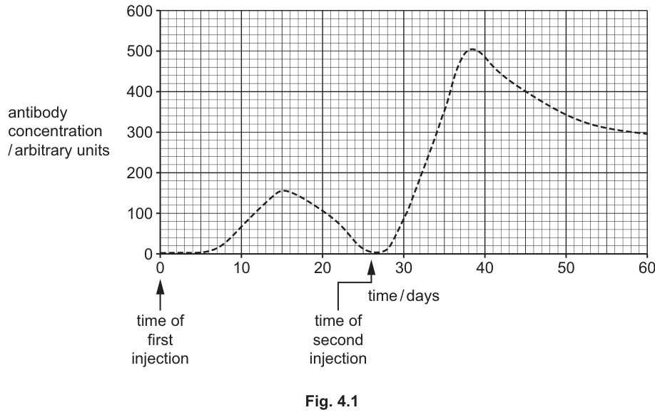
Fig. 4.1 shows the concentration of the antibody in the blood over the period of 60 days.
Explain why the concentration of the antibody in the blood was higher after the second injection than after the first injection.
Answer
- The first injection stimulated a primary immune response, producing (B and T) memory cells specific to the antigens of disease E.
- On second exposure, these memory cells rapidly recognise the antigens, so there is a much greater chance of the antigen encountering a lymphocyte with a complementary receptor.
- Memory cells divide and differentiate into many more plasma cells than in the primary response, producing a faster and greater output of antibody — hence the higher peak in Fig. 4.1.
Memory cells from the primary response are present; they recognise the antigen rapidly, divide into many more plasma cells, producing a faster and larger amount of antibody.
Background Concept
When a new antigen enters the body for the first time, a primary immune response is mounted. Activated B-lymphocytes proliferate (clonal expansion) and differentiate into plasma cells, which secrete antibody, and into memory cells (B- and T-memory cells), which persist in the body for months, years or decades.
If the same antigen is encountered a second time, the secondary (anamnestic) response occurs. The pre-existing memory cells recognise the antigen almost immediately, undergo rapid clonal selection and expansion, and differentiate into antibody-secreting plasma cells much faster than naive B-cells. The result is:
- a shorter lag phase,
- a faster rise in antibody concentration,
- a higher peak concentration,
- antibodies of higher affinity (because memory cells have already undergone affinity maturation).
Understanding the Question
Fig. 4.1 shows the antibody concentration in response to two injections of the same vaccine (disease E antigens). The first peak is small (≈ 150 arbitrary units, peaking at day 15). The second peak, after the booster at day 26, is much larger (≈ 500 arbitrary units) and rises faster (peaking at day 38). The question asks for an explanation of the difference, requiring the candidate to link the graph features to the cellular events of the primary and secondary response.
Approach
- Identify what the first injection produced (primary response + memory cells).
- Identify what changes at the second injection (memory cells are already present).
- Explain how memory cells accelerate and amplify the response.
Step-by-Step Reasoning
- First injection — a primary response. The antigen stimulates naive B-lymphocytes, which proliferate and form some plasma cells (small antibody output, slow rise to ~150 AU) and — crucially — memory cells.
- Second injection — these memory cells are still present. Because there are many more lymphocytes bearing receptors complementary to the antigen, the antigen has a far higher probability of being bound (i.e. increased chance of encountering the right cell).
- Memory B-cells divide rapidly and differentiate into large numbers of plasma cells, which secrete antibody in greater quantities than in the primary response.
- Therefore the rate of antibody production is greater, the peak concentration is much higher (≈ 500 vs ≈ 150 AU), and this is achieved in a similar or shorter time window. This is the hallmark of the secondary response.
Any three of these creditable points earn the three marks available.
Key Takeaways
- The secondary response is faster, larger and longer-lasting because of memory cells.
- The second peak in a vaccination curve is direct evidence of the cellular basis of immunological memory.
- Booster injections work because they re-expose the immune system to the same antigen, allowing memory cells to mount the secondary response.
Common Mistakes
- Saying the second response is larger "because the body has more time to make antibodies" — this is wrong; the graph shows the response begins almost immediately after the booster.
- Saying the second peak is bigger because there are "more antibodies left over from the first response" — antibodies are proteins that are degraded over weeks (cf. Fig. 4.2) and are not stockpiled indefinitely.
- Omitting the explicit term memory cell.
Things to Be Careful About
- The question says the person had not previously been infected with disease E, so memory cells could only have come from the first injection, not from natural infection. State this if relevant.
- Use precise phrasing: "B-memory cells" or simply "memory cells" is acceptable; "T-cells" alone is too vague for the antibody (humoral) response.
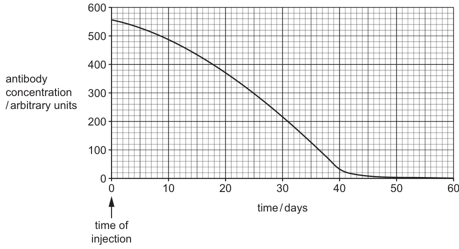
A second person was given a different injection to give protection against another infectious disease, infectious disease F. The person had not previously been infected with disease F.
The concentration in the blood of the antibody specific to disease F was measured over a period of 60 days from the time of the injection.
Fig. 4.2 shows the concentration of the antibody in the blood over the period of 60 days.
State the type of immunity that results from the injection given as protection against disease F.
Answer
Passive and artificial immunity.
Passive and artificial immunity.
Background Concept
Immunity can be classified along two perpendicular axes:
- Active vs passive: in active immunity, the host's own immune system makes its own antibodies (in response to infection or vaccination). In passive immunity, antibodies are provided directly.
- Natural vs artificial: natural immunity is acquired through everyday exposure (e.g. catching the disease, or maternal antibodies crossing the placenta or in breast milk). Artificial immunity is acquired through a deliberate medical intervention (e.g. vaccination, antibody injection).
Understanding the Question
Fig. 4.2 shows antibody concentration already at its maximum at day 0 (the moment of injection), then declining to zero by ~day 45. There is no lag phase and no rising titre — antibody is present the moment it is administered and is then cleared. This is the signature of passive immunity by injection of ready-made antibodies.
Because the antibodies were given by injection (not acquired from infection or from the mother), the artificial part of the classification is also correct.
Approach
Two diagnostic questions for a graph of antibody concentration over time:
- Is the host producing the antibody (active) or just receiving it (passive)?
- Is the source natural (maternal/infection) or artificial (injection/vaccination)?
A single injection that gives an immediate, transient antibody peak indicates passive + artificial.
Step-by-Step Reasoning
- The curve starts at its maximum — no time has been needed for clonal selection, expansion, differentiation and antibody secretion. So the immune system itself is not the source.
- The person had not previously been infected, so this is not a natural primary response.
- An injection delivering pre-formed antibodies fits both criteria: passive (antibodies are supplied) and artificial (delivered by a medical procedure, not by placenta or breast milk).
Key Takeaways
- Passive immunity = ready-made antibodies supplied; immediate but short-lived.
- Artificial immunity = induced by deliberate medical intervention.
- The two axes combine: a single answer requires both descriptors, e.g. "passive artificial" or "active natural".
Common Mistakes
- Writing only "passive" or only "artificial". The mark scheme expects both.
- Writing "active" because the immune system is involved — but it is not; the host is not making its own antibodies here.
- Confusing this curve with that of a primary response (Fig. 4.1), which has a lag phase.
Things to Be Careful About
- Both terms are required; the mark scheme treats them as one combined mark.
Describe features of the type of immunity resulting from the injection given as protection against disease F.
Answer
- The immunity is short-term / temporary; antibody concentration falls as the antibodies are broken down and removed.
- The effect is immediate; protection is provided as soon as the antibodies are injected.
- It involves the injection of (ready-made) antibodies / immunoglobulin, and does not stimulate the host's own immune system (no primary response and no memory cells are produced).
Short-term/temporary; immediate; antibodies are injected and the host's immune system is not stimulated, so no memory cells are produced.
Background Concept
Passive artificial immunity is conferred by injecting pre-formed antibodies (e.g. anti-tetanus immunoglobulin, anti-rabies immunoglobulin, monoclonal antibodies). Because the host's own immune system is bypassed:
- No primary response is mounted;
- No memory cells are generated;
- Protection starts immediately (no lag phase);
- Protection is short-lived — the foreign antibodies are themselves proteins that the host will eventually catabolise and clear.
Understanding the Question
The question asks for features (plural) of this type of immunity. Two marks means two distinct creditable points must be made. The mark scheme accepts any two of: temporary, immediate, injection of antibodies, no stimulation of the immune system, no memory cells, no new antibody production.
Approach
Look for the distinguishing features of passive artificial immunity compared to active immunity (typically vaccination) and natural passive immunity (maternal transfer):
- Time-scale — short-term (antibodies have a half-life of days to weeks).
- Onset — immediate (no clonal expansion needed).
- Mechanism — antibodies supplied, immune system not stimulated.
- Consequence — no memory cells, so no lasting protection and no secondary response on re-exposure.
Step-by-Step Reasoning
- The Fig. 4.2 curve reaches its peak at day 0 and decays to zero by ~day 45. This decay is the catabolism of the injected antibodies — the response is therefore temporary/short-term.
- There is no lag phase, so the effect is immediate.
- Because no antigen is presented to the host's lymphocytes in a way that triggers clonal selection, the immune system is not stimulated and no memory cells are produced. A subsequent infection with disease F would therefore not produce a secondary response.
Key Takeaways
- Passive artificial immunity is fast but short-lived.
- It provides antibodies but no immunological memory.
- It is used when rapid protection is needed (e.g. post-exposure prophylaxis) and the patient has not had time to mount their own active response.
Common Mistakes
- Saying it is "permanent" or "long-lasting" — the mark scheme requires temporary/short term.
- Saying it produces memory cells — the defining feature is that it does not.
- Confusing the immunoglobulin injection with a vaccine — a vaccine contains antigens and triggers an active response; this injection contains antibodies and bypasses the active response.
Things to Be Careful About
- "AVP" (any valid point) is allowed by the mark scheme, so credit may be given for any other defensible feature (e.g. antibodies may be monoclonal, may come from a non-human source and carry a small risk of allergy).
In 2021, the number of new cases of tuberculosis (TB) was estimated to be 10.6 million.
Answer
Mycobacterium tuberculosis (or Mycobacterium bovis).
Mycobacterium tuberculosis (accept Mycobacterium bovis).
Background Concept
Tuberculosis (TB) is caused by a small, slow-growing Gram-positive, acid-fast bacterium. The species that almost always causes human TB is Mycobacterium tuberculosis. A related species, Mycobacterium bovis, primarily infects cattle but can be transmitted to humans via unpasteurised milk and causes a similar disease.
Both are written in binomial form: italicised, with a capital genus and lower-case specific epithet.
Understanding the Question
This is a one-mark recall question — a name, with correct spelling and italicisation. The mark scheme accepts either species.
Approach
- Recall the species.
- Write it in the correct binomial form.
Step-by-Step Reasoning
- The genus is Mycobacterium (note the capital M).
- The species epithet is tuberculosis (lower-case t).
- The whole name should be italicised, e.g. Mycobacterium tuberculosis.
- M. bovis is also accepted.
Key Takeaways
- Correct binomial form must be used: italics, capital genus.
- The two acceptable answers are M. tuberculosis and M. bovis.
Common Mistakes
- Writing "Mycobacterium TB" or "the TB bacteria" — these are not the species name.
- Failing to italicise, or capitalising the species epithet ("Mycobacterium Tuberculosis").
- Confusing TB with other respiratory diseases (e.g. attributing TB to a virus).
Things to Be Careful About
- Always underline or italicise the binomial.
- The abbreviation M. is acceptable only after the name has been written in full at least once in a question.
TB is often treated with several different drugs at the same time. This is necessary to kill multiple drug resistant (MDR) strains of bacteria. This treatment is usually lengthy, taking 6 months or more to make sure all bacteria are killed.
Researchers investigated how the bacteria that cause TB react to the presence of rifampicin. Rifampicin is an antibiotic often used in the successful treatment of TB.
- Researchers combined rifampicin with a coloured marker dye and added the coloured rifampicin to a culture of living bacteria.
- When initially viewed under the microscope, the researchers could see that the coloured rifampicin was present inside the cytoplasm of the bacterial cells.
- Hours later, the coloured rifampicin was not visible inside the bacterial cells but was visible in the medium surrounding the bacterial cells.
- The researchers concluded that rifampicin was being pumped out of the bacterial cells through the cell surface membrane.
- Further research showed that when some drugs commonly used to treat indigestion were added to bacterial cultures containing coloured rifampicin, the coloured rifampicin stayed inside the bacterial cytoplasm.
Suggest and explain how knowledge gained from this research could improve TB treatment and reduce the chance of rifampicin-resistant bacteria developing.
Answer
- Rifampicin is normally pumped out of TB cells by an efflux pump in the cell surface membrane, so the antibiotic is less effective. The indigestion drugs appear to inhibit this pump, keeping rifampicin inside the bacterial cytoplasm where it can kill the bacteria more effectively.
- Combining rifampicin with such pump-inhibiting drugs should therefore kill bacteria more effectively and more quickly, allowing the length of treatment to be reduced from around six months to a shorter course.
- A shorter course is more likely to be completed by the patient, which reduces the chance that a small number of resistant mutants survive and proliferate.
- A shorter course also means less time for new resistance-conferring mutations to arise in the bacterial genome, lowering the probability that rifampicin-resistant strains of TB emerge.
Co-administer rifampicin with drugs that block the bacterial efflux pump; rifampicin stays inside and kills bacteria faster, allowing a shorter course of treatment, which is more likely to be completed and leaves less time for resistance mutations to develop.
Background Concept
Bacteria can become resistant to antibiotics in several ways:
- Enzymatic inactivation of the drug (e.g. β-lactamase).
- Modification of the target so the drug no longer binds.
- Reduced uptake or active efflux of the drug via pumps in the cell surface membrane.
- By-pass of the inhibited metabolic step.
Active efflux pumps are membrane proteins that export antibiotics (and other toxic compounds) out of the cytoplasm, lowering the intracellular concentration below the level needed to kill or inhibit the bacterium. Some drugs (such as certain proton-pump inhibitors used for indigestion) can interfere with these pumps, keeping the antibiotic inside the cell.
In TB, multi-drug resistant (MDR) strains are defined as resistant to at least the two most powerful first-line antibiotics — isoniazid and rifampicin. Long treatment courses are required because:
- M. tuberculosis is slow-growing (divides roughly once a day) and can enter dormant states;
- the bacterium hides inside macrophages, where antibiotics penetrate poorly.
Long courses lead to poor patient compliance: incomplete treatment allows partially resistant bacteria to survive and multiply, increasing the chance of full resistance emerging by spontaneous mutation during replication.
Understanding the Question
This is a 4-mark suggest-and-explain question, requiring the candidate to (1) describe the treatment improvement suggested by the research and (2) explain how that improvement reduces the development of rifampicin resistance. The mark scheme splits the marks into "treatment" (max 3) and "reduce chance of resistance" (the remaining marks), with up to 4 marks overall from a bank of 8 possible points.
The research findings are:
- Rifampicin enters the bacterial cell but is then pumped out via the cell-surface membrane (efflux).
- Indigestion drugs (likely proton-pump inhibitors) block this efflux, so rifampicin stays inside.
Approach
Translate the experimental observation into a clinical application, and then connect the application to the evolutionary mechanism of resistance:
- Step 1 — What is the new treatment strategy? Co-administer rifampicin with pump-inhibiting drugs.
- Step 2 — Why is it more effective? Rifampicin stays inside, killing more bacteria and faster.
- Step 3 — What does that mean for the patient? Treatment can be shorter; shorter courses are easier to complete.
- Step 4 — Why does that reduce resistance? Less time for new mutations; better completion leaves fewer survivors that could evolve resistance.
Step-by-Step Reasoning
- Mechanistic point (treatment, mark 1): The research shows rifampicin is removed from the cell by an efflux pump. Drugs already used for indigestion can block this pump, so combining rifampicin with an indigestion drug keeps rifampicin inside the cytoplasm where it can act on its target (RNA polymerase).
- Efficacy (treatment, mark 2): With rifampicin retained, the antibiotic kills bacteria more effectively / more quickly.
- Course length (treatment, mark 3): A more effective antibiotic means the duration of treatment can be reduced from the standard 6+ months.
- Compliance and resistance (resistance, mark 4): A shorter course is more likely to be completed by the patient. Incomplete courses are the main driver of acquired resistance, because the few partially-resistant bacteria that survive sub-lethal doses are the ones that proliferate.
- Alternative resistance point (could substitute for mark 4): Less treatment time means less time for spontaneous mutations in the bacterial genome to occur, reducing the chance of a rifampicin-resistant mutant arising in the first place.
A valid alternative resistance point is that the indigestion drugs are already cheap, mass-produced and have known safety profiles, so the intervention could be deployed at scale without major new cost.
Key Takeaways
- Efflux pumps are a real mechanism of antibiotic resistance; blocking them with existing drugs is a promising strategy.
- The link between treatment duration and resistance is evolutionary: every additional day of sub-lethal antibiotic exposure increases the probability that a resistant mutant will be selected.
- Drug repurposing (using an indigestion drug alongside an antibiotic) is a way to extend the useful life of existing antibiotics.
Common Mistakes
- Suggesting that the indigestion drug itself kills the bacteria — it does not; it only helps the rifampicin stay inside.
- Not linking the shorter course to the mechanism of resistance evolution (mutation + selection).
- Only describing the experiment without applying it to clinical practice.
- Confusing the role of DOTS (Directly Observed Treatment, Short-course) with the new intervention — the new strategy is adjuvant therapy, not direct observation.
Things to Be Careful About
- The question asks to suggest and explain. Both verbs must be present: state the suggestion, then give the biological/mechanistic reason.
- Marks are split: at least one mark must address resistance reduction. Even a perfect "treatment" answer (3 marks) leaves one mark unclaimed unless the resistance link is also made.
Eukaryotic cells and prokaryotic cells contain DNA.
Complete the passage about DNA in eukaryotic cells and prokaryotic cells, using the most appropriate terms.
In eukaryotic cells, the DNA is located mainly in the chromosomes of the nucleus. Chromosomal DNA is associated with proteins called ______ . Two other eukaryotic cell structures that contain DNA are mitochondria and ______ .
In prokaryotic cells, for example ______ , the DNA is found in the ______ and is usually circular.
Answer
- histones
- chloroplasts
- bacteria (accept cyanobacteria / Archaea / a named prokaryote)
- cytoplasm
histones; chloroplasts; bacteria; cytoplasm
Background Concept
DNA is the genetic material in all living cells, but its location and organisation differ between eukaryotes and prokaryotes. In eukaryotic cells, the vast majority of DNA is enclosed within the nucleus, where it is wrapped around proteins called histones to form chromatin. The DNA–histone complex coils and supercoils, condensing further into visible chromosomes during cell division. Small additional quantities of DNA are also found in two membrane-bound organelles: mitochondria (the sites of aerobic respiration) and chloroplasts (the sites of photosynthesis in plant cells). This organellar DNA supports the endosymbiotic theory, which proposes that these organelles evolved from once free-living prokaryotes.
In prokaryotic cells (cells that lack a nucleus, e.g. bacteria), there is no nuclear membrane. The DNA therefore lies free in the cytoplasm, typically in a region called the nucleoid. Unlike eukaryotic chromosomal DNA, prokaryotic DNA is usually a single, circular molecule and is not associated with histones.
Understanding the Question
The question asks for four specific biological terms that complete a passage comparing DNA location in eukaryotic and prokaryotic cells. Each blank requires the most precise, technically correct term.
Approach
Work through the passage blank by blank, drawing on:
- knowledge of chromatin structure (DNA + histones);
- the DNA-containing organelles in eukaryotes;
- examples and DNA location in prokaryotes.
Step-by-Step Reasoning
Blank 1 — proteins associated with chromosomal DNA
DNA in eukaryotic nuclei is wrapped around histone proteins, forming nucleosomes. The required term is histones; the vague word "proteins" is rejected by the mark scheme.
Blank 2 — second organelle containing DNA (in addition to mitochondria)
Both chloroplasts and mitochondria contain their own DNA, a feature consistent with their endosymbiotic origin.
Blank 3 — example of a prokaryotic cell
Any prokaryote is credited. The most general term is bacteria; specific accepted alternatives include cyanobacteria, Archaea, or any named prokaryote.
Blank 4 — location of DNA in prokaryotes
Prokaryotes lack a nucleus, so their DNA sits in the cytoplasm. (The technical name for this region is the nucleoid, but "cytoplasm" is the mark-scheme-credited term.)
Key Takeaways
- Histones are the proteins around which eukaryotic nuclear DNA is wrapped to form chromatin.
- Mitochondria and chloroplasts are the two eukaryotic organelles that contain DNA in addition to the nucleus.
- Prokaryotic DNA is found in the cytoplasm, is usually circular, and is not associated with histones.
Common Mistakes
- Writing "proteins" instead of "histones" — too vague to score.
- Forgetting that chloroplasts (not just mitochondria) contain DNA.
- Naming the wrong example of a prokaryote, e.g. a eukaryote such as yeast or amoeba.
- Confusing the nucleoid with the nucleus.
Things to Be Careful About
- Use the precise term "histones" — it is the only credited answer for Blank 1.
- "Bacteria" is the safest answer for the prokaryote example, but cyanobacteria and Archaea are also accepted.
- "Cytoplasm" is the credited term for prokaryotic DNA location, not "nucleoid".
Fig. 5.1 shows transcription of the first six nucleotides of a gene by the enzyme RNA polymerase. The bases of the first six nucleotides on DNA strand X are shown, but the bases on the template DNA strand are not shown.
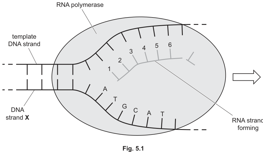
Answer
Non-transcribed strand
Non-transcribed strand
Background Concept
During transcription, RNA polymerase reads only one of the two DNA strands — the template (antisense) strand — to synthesise a complementary RNA molecule. The other DNA strand is not used as a template and has several names. The CIE syllabus uses the term non-transcribed strand; other textbooks call it the sense strand or coding strand, because its sequence matches the RNA product (with T replaced by U).
Understanding the Question
Fig. 5.1 labels one DNA strand as X and explicitly identifies it as the strand whose bases are shown but which is NOT the template strand. You must state the term for this non-template strand.
Approach
Recognise that transcription uses only one strand (the template). The other strand is named using a specific term — use the CIE-preferred wording.
Step-by-Step Reasoning
In Fig. 5.1, strand X is shown alongside the template strand, with the RNA being built against the template. Because strand X is not read by RNA polymerase, it is the non-transcribed strand.
Key Takeaways
- Only one DNA strand (the template/antisense strand) is transcribed at a time.
- The other strand is the non-transcribed strand (also called sense or coding strand).
Common Mistakes
- Writing "sense strand" or "coding strand" — these are correct biologically, but CIE marks this question with the wording "non-transcribed strand".
- Confusing X with the template strand — X is explicitly the non-template strand.
Things to Be Careful About
- The mark scheme specifically accepts "non-transcribed strand"; use this wording to be safe.
Answer
Primary transcript
Primary transcript
Background Concept
Eukaryotic genes contain coding regions (exons) interrupted by non-coding regions (introns). When RNA polymerase transcribes such a gene, it first produces a long RNA molecule containing BOTH introns and exons. This initial RNA product is called the primary transcript (also called pre-mRNA). It is then processed — introns are removed by splicing, a 5' cap is added, and a poly-A tail is added at the 3' end — to produce the mature mRNA that can be translated.
Understanding the Question
The question asks for the name of the RNA molecule produced during transcription of a eukaryotic gene.
Approach
Recognise that in eukaryotes, the immediate transcription product is unprocessed (still contains introns).
Step-by-Step Reasoning
The RNA molecule formed during transcription (before splicing) is the primary transcript. "Mature mRNA" is incorrect here because mature mRNA has already had its introns removed — it is the processed form, not the immediate transcription product.
Key Takeaways
- Eukaryotic transcription produces a primary transcript (pre-mRNA).
- Splicing and other modifications convert the primary transcript into mature mRNA.
Common Mistakes
- Writing "mRNA" — this is the processed, translatable form, not the immediate product of transcription.
- Writing "tRNA" or "rRNA" — these have different roles and are not the typical gene product described.
Things to Be Careful About
- "Primary transcript" (or pre-mRNA) is the credited term for the immediate transcription product in eukaryotes.
Complete Table 5.1 to show the letters of the six bases indicated on Fig. 5.1 by the numbers 1 to 6.
Table 5.1
| 1 | 2 | 3 | 4 | 5 | 6 |
|---|---|---|---|---|---|
Working
DNA strand X (non-template): A — T — G — C — A — T
Template strand: T — A — C — G — T — A (complementary base pairing)
mRNA synthesised: A — U — G — C — A — U (complementary to template; U replaces T)
Answer
| 1 | 2 | 3 | 4 | 5 | 6 |
|---|---|---|---|---|---|
| A | U | G | C | A | U |
A U G C A U
Background Concept
Base pairing rules are universal in nucleic acids: adenine (A) always pairs with thymine (T) in DNA (or uracil, U, in RNA), and guanine (G) always pairs with cytosine (C). During transcription, RNA polymerase builds an RNA strand complementary to the DNA template strand, substituting U for T.
Understanding the Question
Given the six bases on DNA strand X (the non-template strand), determine the six bases on the mRNA strand being synthesised at positions 1–6.
Approach
Because strand X is the non-template strand, the mRNA will have the same sequence as strand X, but with every T replaced by U.
Step-by-Step Reasoning
Strand X: A T G C A T (positions 1–6)
Template strand: T A C G T A (complementary to X)
mRNA: A U G C A U (complementary to template; U replaces T)
Key Takeaways
- mRNA is complementary to the template DNA strand.
- mRNA contains U (uracil) instead of T (thymine).
- The mRNA sequence matches the non-template (sense/coding) strand, with U replacing T.
Common Mistakes
- Writing T instead of U in the RNA positions — easy to do when copying directly from the DNA strand.
- Getting the complementary base wrong (e.g. pairing A with C or G with A).
- Reversing the direction of the sequence.
Things to Be Careful About
- mRNA contains uracil (U), not thymine (T).
- The bases 1–6 are read in the order they appear in the diagram (5' to 3' along strand X).
RNA polymerase is composed of several polypeptides that move together and change shape as the enzyme performs its functions.
The death cap mushroom, Amanita phalloides, produces a toxin called alpha-amanitin, which binds to RNA polymerase at a site other than the active site. Alpha-amanitin reduces the activity of RNA polymerase.
Use the information provided to suggest how alpha-amanitin reduces the activity of RNA polymerase.
Answer
- Alpha-amanitin acts as a non-competitive inhibitor (it binds at a site other than the active site) ;
- Binding changes the shape of the active site, so it is no longer complementary to the DNA substrate ;
- Fewer enzyme–substrate complexes form, so RNA polymerase cannot bind to / unwind the DNA, and transcription is slowed down.
Non-competitive inhibition distorts the active site, so RNA polymerase can no longer bind / unwind DNA.
Background Concept
Enzyme inhibitors fall into two main categories. Competitive inhibitors bind to the active site and directly block substrate binding — their effect can be overcome by raising substrate concentration. Non-competitive inhibitors bind to a different site (an allosteric site), causing a conformational change in the enzyme that distorts the active site and reduces its activity. Because the inhibitor and substrate do not compete for the same site, the inhibition cannot be overcome by adding more substrate.
RNA polymerase is the enzyme that catalyses transcription. To function it must bind to DNA, unwind the double helix, move along the template strand, and add complementary RNA nucleotides. RNA polymerase is composed of several polypeptide subunits (as stated in the question) and changes shape as it moves.
Understanding the Question
The question describes alpha-amanitin, which binds RNA polymerase at a site other than the active site and reduces its activity. You are asked to suggest HOW this binding reduces RNA polymerase activity.
Approach
First, recognise the type of inhibition from the description (binding at a non-active site = non-competitive). Then explain the consequence of this binding — a shape change in the active site — and link that to the specific functions of RNA polymerase.
Step-by-Step Reasoning
-
Type of inhibition. Alpha-amanitin binds at a site other than the active site. This is the defining feature of non-competitive inhibition.
-
Shape change. Binding to the allosteric site changes the shape of the active site, so it is no longer complementary to the DNA substrate.
-
Consequence. Fewer enzyme–substrate complexes form. RNA polymerase cannot bind to / unwind the DNA helix, cannot change shape (induced fit), and cannot move along the template to add nucleotides. Transcription is therefore slowed or halted.
Any three of these points (or the equivalent mark-scheme points about blocked movement, prevented induced fit, or prevented nucleotide addition) scores 3 marks.
Key Takeaways
- Non-competitive inhibitors bind at an allosteric site, not the active site.
- They change the shape of the active site, reducing its complementarity to the substrate.
- The inhibition cannot be overcome by increasing substrate concentration.
- The same mechanism applies to any enzyme, including multi-subunit enzymes like RNA polymerase.
Common Mistakes
- Describing alpha-amanitin as a competitive inhibitor — it binds at a different site, so this is wrong.
- Saying the active site "no longer works" without specifying why — the key mechanism is the conformational change.
- Stating only that transcription "is reduced" without giving a biological reason — this scores zero.
- Confusing non-competitive with competitive inhibition generally.
Things to Be Careful About
- The mark scheme explicitly looks for the phrase "non-competitive inhibition".
- State BOTH where alpha-amanitin binds AND the consequence of that binding — a bare "it reduces activity" scores zero.
- The mechanism applies even though RNA polymerase is multi-subunit; the inhibition principle is the same.
Fig. 6.1 shows photomicrographs of individual cells from the root tip of an onion, Allium sp., at different times in the mitotic cell cycle.
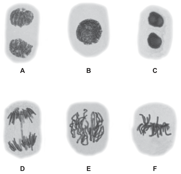
Place the letters representing the individual cells in the correct sequence of the mitotic cell cycle. The first letter has already been filled in.
Answer
- Box 1: B (already given) — interphase (nucleus intact, chromatin not condensed)
- Box 2: E — prophase (chromosomes condensed, scattered in the cell)
- Box 3: F — metaphase (chromosomes aligned at the equator)
- Box 4: D — anaphase (chromosomes being pulled to opposite poles)
- Box 5: A — telophase (two groups of chromosomes at opposite poles, nuclear envelopes re-forming)
- Box 6: C — late telophase / end of mitosis (two daughter nuclei clearly separated)
B, E, F, D, A, C
Background Concept
The mitotic cell cycle is the sequence of events by which a cell replicates its chromosomes and divides to produce two genetically identical daughter nuclei. After interphase (G₁, S, G₂), mitosis proceeds through four recognisable stages — prophase, metaphase, anaphase and telophase — followed by cytokinesis. Each stage has a distinctive appearance under the light microscope because of the changing state of the chromosomes and the spindle.
Understanding the Question
You are given six photomicrographs (A–F) of onion root tip cells fixed at different points in the cycle. Box 1 is already filled with B, and you must place the remaining five letters in the correct order. The mark scheme accepts only the exact sequence.
Approach
Use the position and morphology of the chromosomes (and the presence/absence of a nuclear envelope and spindle) to identify each stage:
- Interphase — intact nuclear envelope, decondensed chromatin, single nucleus.
- Prophase — chromosomes condensed but not yet aligned; nuclear envelope breaking down; spindle forming.
- Metaphase — chromosomes lined up along the cell's equator.
- Anaphase — sister chromatids separated, moving towards opposite poles.
- Telophase — two groups of chromosomes at opposite poles; nuclear envelopes reforming.
- Late telophase / end of mitosis — two fully separated daughter nuclei.
Step-by-Step Reasoning
- B shows a single round nucleus with diffuse chromatin and no visible chromosomes — this is interphase (and is already in box 1).
- E shows chromosomes that are condensed and visible but not aligned — this is prophase.
- F shows chromosomes lined up in a single row across the middle of the cell — this is metaphase.
- D shows two clear groups of chromosomes being pulled towards opposite poles of the cell — this is anaphase.
- A shows chromosomes already grouped at the two poles, with the cytoplasm beginning to be partitioned between them — this is telophase.
- C shows two small, dense, completely separated daughter nuclei — this is late telophase, the end of mitosis.
Key Takeaways
- Each stage of mitosis has a diagnostic appearance under the microscope: condensation (prophase), alignment (metaphase), separation (anaphase), regrouping and envelope re-formation (telophase).
- Onion root tips are a classic source of dividing cells because the meristem contains many cells in mitosis at any one time.
Common Mistakes
- Confusing prophase and metaphase — chromosomes in prophase are scattered, not aligned.
- Confusing anaphase and telophase — in anaphase the chromatids are still moving; in telophase they have reached the poles and are starting to decondense.
Things to Be Careful About
- Cell C can look similar to cell A; the deciding feature is whether the two chromosome groups have been enclosed in reforming nuclear envelopes (C) or are still relatively loose (A).
Cell A in Fig. 6.1 is in one of the main stages of mitosis.
Describe the events that occur during this main stage of mitosis.
Answer
Cell A is in telophase. Any two of the following events occur:
- The nuclear envelope re-forms around each group of chromosomes at opposite poles.
- The nucleolus (nucleoli) re-form(s) within each new nucleus.
- The (daughter) chromosomes uncoil / lengthen to become chromatin.
- The spindle breaks down.
Telophase — nuclear envelope re-forms around each chromosome group, nucleolus re-forms, chromosomes uncoil and spindle breaks down (any two).
Background Concept
Mitosis is divided into four named stages — prophase, metaphase, anaphase and telophase — followed by cytokinesis. Telophase is the final stage of nuclear division: the chromosomes have already reached the poles, and the events reverse those of prophase, restoring the interphase appearance of two nuclei in preparation for cytokinesis.
Understanding the Question
Cell A in Fig. 6.1 shows two distinct groups of chromosomes sitting at opposite poles of the cell. You are asked to name and describe what happens during this stage of mitosis. The mark scheme credits any two events from a defined list, so you do not need all four.
Approach
Identify the stage first, then list the recognisable events of that stage. The mark scheme lists four creditable events for telophase — pick two and state each clearly with the correct terminology.
Step-by-Step Reasoning
- Cell A has two groups of chromosomes at opposite poles, with the cytoplasm being partitioned between them but no nuclear envelopes yet fully formed. This is telophase.
- The defining events of telophase, in any order, are:
- The nuclear envelope (nuclear membrane) re-forms around each set of chromosomes, producing two new nuclei.
- Nucleoli re-form inside each new nucleus.
- The chromosomes uncoil / decondense back into chromatin (the reverse of prophase condensation).
- The spindle breaks down; the microtubules depolymerise.
- Two marks are available, so two clear points are enough. Use the precise words from the mark scheme where you can.
Key Takeaways
- Telophase is essentially prophase in reverse: the events that dispersed the nuclear envelope and condensed the chromosomes are now undone.
- Cytokinesis (cytoplasmic division) overlaps with telophase but is technically a separate process.
Common Mistakes
- Confusing telophase with anaphase — in anaphase the chromatids are still moving; in telophase they have reached the poles.
- Giving prophase events instead — chromosomes condensing, spindle forming, nuclear envelope breaking down. These are the wrong stage.
Things to Be Careful About
- The mark scheme uses the exact word "re-form" for nuclear envelope and nucleoli — using "appears" or "is made" is less precise and risks losing the mark.
- "Chromosomes uncoil" is the wording the examiner rewards; "chromosomes disappear" is rejected because the chromosomes do not vanish, they decondense.
Complete Table 6.1 by stating the term that matches each of the descriptions.
Table 6.1
| term | description |
|---|---|
| region of DNA with repeated nucleotide sequences located at the ends of chromosomes | |
| organises microtubules to form the spindle in animal cells | |
| point of attachment between two sister chromatids |
Answer
| Term | Description |
|---|---|
| Telomere | region of DNA with repeated nucleotide sequences located at the ends of chromosomes |
| Centriole (or centrosome) | organises microtubules to form the spindle in animal cells |
| Centromere | point of attachment between two sister chromatids |
Each term earns one mark.
- Telomere; 2. Centriole (or centrosome); 3. Centromere.
Background Concept
A chromosome is more than just a strand of DNA. Before mitosis each chromosome consists of two identical sister chromatids held together at a specialised constricted region called the centromere. The ends of the chromatids are capped by telomeres — repetitive non-coding DNA sequences that protect the chromosome ends from degradation and from being recognised as DNA damage. In animal cells, the mitotic spindle is nucleated by a pair of centrioles sitting in a centrosome at each pole.
Understanding the Question
You are given a table with three descriptions of chromosome/cell structures and must write the matching term in the left-hand column. Each correct term is worth one mark (three marks total). The mark scheme accepts either centriole(s) or centrosome for the spindle-organising structure in animal cells.
Approach
Read each description carefully and match it to the standard term. The clues are:
- "repeated nucleotide sequences at the ends of chromosomes" → telomere.
- "organises microtubules to form the spindle in animal cells" → centriole / centrosome.
- "point of attachment between two sister chromatids" → centromere.
Step-by-Step Reasoning
- Telomere — the repetitive DNA cap at each end of a chromosome. It prevents loss of coding DNA during replication and stops chromosome ends from fusing with one another.
- Centriole (or centrosome) — in animal cells, the centrosome (containing a pair of centrioles) is the microtubule-organising centre that nucleates the spindle fibres. Plant cells lack centrioles but still form a spindle from dispersed microtubule-organising centres.
- Centromere — the constricted region where the two sister chromatids of a duplicated chromosome are joined. Spindle microtubules attach here via kinetochores during mitosis.
Key Takeaways
- Three essential chromosome terms: telomere (end), centromere (middle), centriole/centrosome (spindle organiser in animal cells).
- Telomeres shorten with each round of replication and are associated with cell ageing; centromeres are the mechanical attachment point for the spindle.
Common Mistakes
- Confusing telomere and centromere — they sound similar but are at opposite ends of the chromosome in terms of function.
- Writing "centrosome" alone when the description specifies the spindle-organising role in animal cells — both "centriole" and "centrosome" are accepted by the mark scheme here, but "spindle" alone is not.
- Spelling "centromere" as "centrometre" — this is a structure, not a unit of length.
Things to Be Careful About
- Use the singular spelling the mark scheme uses: centromere (one m), not "centromere-centromere" or "centromeres" when singular is implied.
- The mark scheme states the terms must be written in the left column; spelling counts, so avoid phonetic misspellings.