Biology 9700/12 — February/March 2025
Cambridge AS Level · Multiple Choice (AS Level) · answer key with instant marking and worked solutions
Topics Cell Structure · Biological Molecules · The Mitotic Cell Cycle · Infectious Diseases · Cell Membranes and Transport · Transport in Mammals · +5 more
Tap an option under each question to check it — your score builds as you go.
In an electron micrograph, the length of a mitochondrion is measured as . The magnification of the electron micrograph is .
What is the actual length of the mitochondrion?
Options
A
B
C
D
Working
Convert to micrometres ():
Answer
D
D
Background Concept
The magnification of a micrograph tells you how many times larger the printed/displayed image is compared with the real specimen. The relationship between the three quantities is:
so, rearranged to find the real size of the object:
Two facts make these calculations work smoothly:
- Units must be consistent — both the image size and the actual size must end up in the same unit for the ratio to be valid. The safe way is to convert the image size to the unit you want the answer in before dividing.
- Unit conversions in the metric system are powers of ten. The biologically useful units for cell organelles are the millimetre (), micrometre () and nanometre (): . Because of this, dividing by a large magnification and converting units is really just arithmetic with powers of ten.
Understanding the Question
You are told that a mitochondrion appears long in an electron micrograph printed at a magnification of . You need to find the mitochondrion's real size. The answer choices are all in micrometres, so the target unit is .
The command word is implicit ("what is the actual length…?") — this is a pure calculation, so the marks go on getting the right number and the right unit with a sensible number of significant figures (here, three, matching the data).
Approach
- Use the rearranged magnification formula: actual size = image size ÷ magnification.
- Convert the image size from centimetres to micrometres first (or divide then convert — both give the same result, but converting first is easier to keep track of).
- Pick the matching answer from the options.
Step-by-Step Reasoning
Step 1 — substitute into the formula.
Step 2 — perform the division.
Step 3 — convert centimetres to micrometres.
There are in (since ):
Step 4 — match to the options.
The actual length is , which is option D.
A common sanity check: a real mitochondrion is typically – long, so an answer in that range is plausible; the much smaller and options would be the size of a bacterium, and is on the small side for a typical mitochondrion — but is right in the heart of the expected range.
Key Takeaways
- The magnification formula is one of the most heavily tested relationships in Paper 1 / Paper 3: , rearranged to either side as needed.
- Always state the working — both the formula and the substituted values — and quote the unit with the final number.
- Learn the metric ladder: . The exponent of ten you add or subtract for each step up or down is the easy part to slip on.
- Realistic biological check: knowing roughly how big common organelles are (– for a mitochondrion; – for an animal cell diameter) lets you eliminate absurd options instantly.
Common Mistakes
- Inverting the formula — dividing the magnification by the image size gives , which, mistakenly treated as "", would look like option C (). This is a classic wrong answer caused by getting the rearrangement backwards.
- Forgetting the unit conversion — leaving the answer as without converting to loses the mark because it does not match any option.
- Dropping a power of ten — a slip of in the unit conversion produces (option B), so always write out the conversion factor explicitly.
- Reading 17.1 as 171 or 38 000 as 3800 — careless transcription of a leading digit shifts the answer by an order of magnitude.
Things to Be Careful About
- Match significant figures to the data: the image size is given to three significant figures, so the final answer should also be to three significant figures (, not or ).
- On an MCQ, the "trap" distractors are precisely the answers that students get when they invert the formula, drop a power of ten, or fail to convert units. Slow down and check the unit before selecting.
- If you ever doubt, double-check by multiplying back: ✓.
There is a theory that mitochondria and chloroplasts were originally free-living prokaryotes. It is thought that millions of years ago these free-living prokaryotes were taken into larger cells by endocytosis where, instead of being digested, they became functional organelles.
Which features of mitochondria and chloroplasts support this theory?
1 Mitochondria and chloroplasts are surrounded by double membranes.
2 Mitochondria and chloroplasts have small, circular DNA.
3 Mitochondria and chloroplasts synthesise proteins.
Options
A 1, 2 and 3
B 1 and 2 only
C 1 and 3 only
D 2 and 3 only
Working
Statement 1 — Double membrane: correct. The inner membrane is the original prokaryote's plasma membrane, and the outer membrane derives from the host cell's endocytic vesicle.
Statement 2 — Small, circular DNA: correct. Prokaryotes carry a single circular DNA molecule in a nucleoid region; mitochondria and chloroplasts contain similar circular DNA, distinct from the linear nuclear DNA.
Statement 3 — Synthesise proteins: correct. Mitochondria and chloroplasts have 70S ribosomes (like prokaryotes) and can synthesise some of their own proteins.
All three statements are evidence for the endosymbiotic theory.
Answer
A
A
Background Concept
The endosymbiotic theory proposes that mitochondria (in eukaryotes) and chloroplasts (in photosynthetic eukaryotes) originated from free-living prokaryotes that were engulfed by larger ancestral cells. Instead of being digested, they established a mutually beneficial relationship — the host cell gained an efficient means of aerobic respiration (and, in photosynthetic lineages, photosynthesis), while the endosymbiont gained a stable, protected environment and a supply of nutrients. Over millions of years the endosymbionts became integrated and dependent, evolving into the organelles we see today.
Three lines of structural and molecular evidence are typically cited to support this theory:
- Double membrane — Mitochondria have an outer and inner membrane separated by an inter-membrane space. The inner membrane is biochemically similar to a prokaryotic plasma membrane (e.g. cardiolipin content, protein composition), while the outer membrane resembles a eukaryotic endocytic/ER membrane. This is exactly what would be expected if the organelle had been engulfed by invagination of the host's plasma membrane.
- Circular DNA — Prokaryotes carry their genome as a single, circular DNA molecule in a nucleoid region (not enclosed by a nuclear envelope). Mitochondria and chloroplasts each contain their own small, circular DNA (mtDNA and cpDNA) that is physically separate from the linear, histone-bound nuclear DNA.
- 70S ribosomes and protein synthesis — Bacterial ribosomes are 70S (made of 50S and 30S subunits) and are sensitive to antibiotics such as streptomycin and chloramphenicol. Mitochondria and chloroplasts contain 70S ribosomes and can synthesise a small subset of their own proteins using their own mRNA, again showing prokaryotic affinity.
Other supporting evidence includes binary fission as the mode of organelle division (prokaryote-like), and the sensitivity of organelle ribosomes to antibacterial antibiotics.
Understanding the Question
This is a multiple-choice question (Paper 1 style) that asks which of three given statements are valid pieces of evidence for the endosymbiotic theory. The candidate must decide whether each statement is correct AND whether it actually supports the theory, then select the combination of correct statements that matches one of the four options.
The command word is implicit ("Which features … support this theory?"), so the test is whether each feature is a recognised piece of endosymbiotic evidence.
Approach
Go through each statement in turn and ask two questions:
- Is the statement biologically true?
- Does it constitute evidence for the endosymbiotic (not just any) theory?
If both are yes, count the statement. Then read the options to see which combination matches.
Step-by-Step Reasoning
Statement 1: "Mitochondria and chloroplasts are surrounded by double membranes."
- True. Both organelles have two membranes.
- Supports the theory: yes — the outer membrane is interpreted as a remnant of the host's endocytic vesicle, and the inner membrane as the original plasma membrane of the engulfed prokaryote.
- COUNT IT.
Statement 2: "Mitochondria and chloroplasts have small, circular DNA."
- True. Each mitochondrion and chloroplast has multiple copies of a small, circular DNA molecule.
- Supports the theory: yes — circular DNA is characteristic of prokaryotes; eukaryotes otherwise have linear, histone-associated nuclear DNA.
- COUNT IT.
Statement 3: "Mitochondria and chloroplasts synthesise proteins."
- True. They contain their own 70S ribosomes and translate some of their own proteins from organelle-encoded mRNA.
- Supports the theory: yes — prokaryote-like 70S ribosomes carrying out prokaryote-like protein synthesis is a key molecular clue to their bacterial ancestry.
- COUNT IT.
All three statements are valid evidence, so the correct option is the one that includes 1, 2 and 3 → A.
Key Takeaways
- The endosymbiotic theory is supported by structural evidence (double membrane), genetic evidence (circular DNA) and molecular evidence (70S ribosomes and protein synthesis).
- Mitochondria and chloroplasts are sometimes described as "semi-autonomous" because they retain some of their own machinery but depend on nuclear-encoded proteins for most functions.
- This idea, formalised by Lynn Margulis in the 1960s–70s, is now widely accepted and is one of the few major evolutionary events that can be traced by direct comparison with modern prokaryotes (mitochondria resemble alpha-proteobacteria; chloroplasts resemble cyanobacteria).
Common Mistakes
- Rejecting statement 1 because "both membranes look similar" — they don't, and the fact that there are two of them is the point.
- Rejecting statement 3 because the nucleus controls most organelle proteins — even though the organelle can only synthesise a small number of proteins, the ability to do so at all is prokaryote-like.
- Confusing circular DNA with prokaryote DNA in general — nuclear DNA is linear and wrapped around histones; the organelle DNA is naked (or nearly so) and circular, like a bacterial chromosome.
- Choosing B (1 and 2 only) by forgetting that 70S ribosomes allow organelle protein synthesis.
Things to Be Careful About
- The question is specifically about mitochondria AND chloroplasts; a feature only true of one would not be accepted unless stated for both.
- "Supports the theory" means the feature is prokaryote-like, not just organelle-like. Any feature shared with all eukaryotic organelles (e.g. having membranes) would not be evidence — but the double membrane and the circular DNA and the 70S ribosomes are specifically prokaryote-like.
- Do not credit a feature simply because it is true; it must also be relevant evidence.
Which row correctly describes a function of each cell structure?
Options
| lysosome | mitochondrion | smooth endoplasmic reticulum | |
|---|---|---|---|
| A | digestion of unwanted structures | abundant in sites of active transport | processing of proteins |
| B | digestion of unwanted structures | ATP synthesis | lipid production |
| C | spherical sacs containing hydrolytic enzymes | abundant in sites of active transport | lipid production |
| D | spherical sacs containing hydrolytic enzymes | ATP synthesis | processing of proteins |
Working
The question asks for a function of each organelle.
- A — lysosome function correct, but mitochondria are not the energy source for active transport (that is ATP from respiration); smooth ER does not process proteins (rough ER does). ✗
- B — lysosome: digestion of unwanted structures ✓; mitochondrion: ATP synthesis ✓; smooth ER: lipid production ✓. All three are correct functions. ✓
- C — lysosome description is a structure, not a function; mitochondrion function is wrong. ✗
- D — lysosome description is a structure, not a function; smooth ER function is wrong (rough ER processes proteins). ✗
Answer
B
B
Background Concept
The question tests recall of three eukaryotic organelles and, crucially, the distinction between a function (what the organelle does) and a structure (what it is made of / how it looks).
- Lysosome: a small spherical organelle surrounded by a single membrane, containing hydrolytic (digestive) enzymes that work at low pH. Its function is to digest unwanted material — worn-out organelles, engulfed bacteria, or macromolecules delivered to it.
- Mitochondrion: a double-membraned organelle whose inner membrane is folded into cristae. It is the site of aerobic respiration, where the link reaction, Krebs cycle and oxidative phosphorylation generate ATP from the oxidation of glucose and other substrates.
- Smooth endoplasmic reticulum (smooth ER): a network of flattened membrane-bound sacs without ribosomes. Its functions include synthesis of lipids and steroids (e.g. cholesterol, steroid hormones, phospholipids) and detoxification. Protein processing is carried out by the rough ER, which has ribosomes on its surface.
Understanding the Question
The command word is implicit but important: the stem says "function of each cell structure". Each entry in the chosen row must therefore describe what the organelle does, not what it is.
The three organelles to match to functions are: lysosome, mitochondrion, smooth endoplasmic reticulum.
Approach
- Eliminate any row where an entry describes structure rather than function (this rules out C and D, where the lysosome is described as "spherical sacs containing hydrolytic enzymes").
- Among the remaining rows (A and B), check the function given for the mitochondrion. Mitochondria are abundant where ATP is consumed rapidly (e.g. in muscle cells, in cells with many active transport pumps), but their function is ATP synthesis, not active transport. So the entry "abundant in sites of active transport" is an odd phrasing — it is neither a function nor quite right as a description of where mitochondria are most concentrated (they are concentrated at sites of high ATP demand, of which active transport is one example but not the defining function). This rules out A.
- Row B gives ATP synthesis for the mitochondrion and lipid production for the smooth ER — both correct functions.
Step-by-Step Reasoning
- Lysosome function = digestion of unwanted structures. ✓ (rows A and B)
- Rows C and D instead describe the lysosome as "spherical sacs containing hydrolytic enzymes" — that is a structural description, not a function, so they fail the stem's requirement.
- Mitochondrion function = ATP synthesis. ✓ (rows B and D)
- Row A says "abundant in sites of active transport". This is not the function of a mitochondrion; it is a (slightly inaccurate) comment on where you might find lots of mitochondria. Functionally, a mitochondrion synthesises ATP, which is then used for active transport among other things.
- Row C repeats this same error.
- Smooth ER function = lipid production. ✓ (rows B and C)
- Row A says "processing of proteins" — that is the function of the rough ER.
- Row D makes the same error.
Only row B has three correct functions.
Key Takeaways
- When a question asks for a function, a description of structure scores zero even if the structure is correct.
- Mitochondria perform ATP synthesis (aerobic respiration); they do not themselves carry out active transport.
- Smooth ER is the site of lipid/steroid synthesis; rough ER (with ribosomes) is the site of protein synthesis and processing.
- Lysosomes are functional in intracellular digestion.
Common Mistakes
- Confusing structure with function: picking C or D because "spherical sacs containing hydrolytic enzymes" is a true description of a lysosome — but the question asked for a function.
- Attributing protein processing to smooth ER instead of rough ER (leading to A or D).
- Confusing "where mitochondria are abundant" with "what mitochondria do" (a trap that loses A and C).
Things to Be Careful About
- Read the command word carefully: "function" rules out purely structural descriptions.
- Do not assume every reasonable-sounding statement is correct; "abundant in sites of active transport" is partially true but is not the function of a mitochondrion.
- Smooth vs rough ER: ribosomes = rough ER = proteins; no ribosomes = smooth ER = lipids and detoxification.
Which cell components are present in typical prokaryotic cells?
Options
| cell wall | 80S ribosomes | |
|---|---|---|
| A | ✓ | ✓ |
| B | ✓ | ✗ |
| C | ✗ | ✗ |
| D | ✗ | ✓ |
key
✓ = present
✗ = not present
Working
Prokaryotic cells (e.g. bacteria) possess a cell wall (made of peptidoglycan/murein), but their ribosomes are 70S, smaller than the 80S ribosomes of eukaryotic cells.
Answer
B
B
Background Concept
Prokaryotic cells (bacteria and archaea) are simpler and smaller than eukaryotic cells. Two structural features are particularly diagnostic:
- Cell wall: present in virtually all prokaryotes. In bacteria it is made of a unique polymer called peptidoglycan (murein), which is the target of antibiotics such as penicillin. Note that plant cells and fungi also have cell walls, but the chemistry is different (cellulose in plants; chitin in fungi).
- Ribosomes: prokaryotes have 70S ribosomes (S = Svedberg unit, a measure of how fast a particle sediments in a centrifuge; it reflects size/shape, not mass directly). Eukaryotic cells have larger 80S ribosomes in the cytoplasm, and 70S ribosomes inside mitochondria and chloroplasts. Antibiotics such as streptomycin exploit this difference to attack bacterial ribosomes without harming the host's cytoplasmic ribosomes.
Eukaryotic cells also differ from prokaryotes in having a true nucleus bounded by a nuclear envelope, membrane-bound organelles (mitochondria, ER, Golgi), and (in plants) chloroplasts and a large central vacuole — none of which prokaryotes possess.
Understanding the Question
The table lists two features — a cell wall and 80S ribosomes — and asks which combination is present in a typical prokaryotic cell. The ticks indicate presence. You must decide independently for each row whether a prokaryote has that feature, then match the combination to A–D.
Approach
Recall the two structural markers for prokaryotes:
- Cell wall → present (✓)
- 80S ribosomes → absent (✗); prokaryotes have 70S ribosomes
The combination ✓/✗ matches option B.
Step-by-Step Reasoning
- Identify the cell type: the question specifies prokaryotes.
- For each feature, decide present/absent:
- Cell wall: present in typical prokaryotes ✓
- 80S ribosomes: 80S ribosomes are a feature of eukaryotic cytoplasm; prokaryotes have 70S ribosomes ✗
- Match ✓/✗ to the table: option B.
Why the distractors are wrong:
- A (✓/✓) — implies prokaryotes have 80S ribosomes; they do not.
- C (✗/✗) — implies prokaryotes lack a cell wall; they have one.
- D (✗/✓) — implies both absence of a cell wall and presence of 80S ribosomes; neither is correct.
Key Takeaways
- Prokaryotes = cell wall (peptidoglycan) + 70S ribosomes, no membrane-bound nucleus.
- Eukaryotes = 80S cytoplasmic ribosomes, plus membrane-bound organelles.
- The S values are easily confused; remember "pro = small = 70S" and "eu = larger = 80S".
Common Mistakes
- Confusing 70S and 80S ribosomes — the most common error here. Some students remember only that "ribosomes are in all cells" and pick the ✓/✓ option A.
- Forgetting that many prokaryotes have a cell wall (e.g. thinking only plants have walls).
Things to Be Careful About
- The Svedberg unit is not additive: a 70S plus a 30S subunit gives a 70S ribosome, not 100S, because sedimentation depends on shape as well as mass.
- Archaea also lack peptidoglycan, but for typical (bacterial) prokaryotes — the kind examined at AS — the cell wall is treated as present.
- Mitochondria and chloroplasts contain 70S ribosomes (an evolutionary clue to their prokaryotic origin), so the 70S vs 80S distinction is about cellular location as well as cell type.
X-ray analysis of fossilised cells found in rocks in central India dating back 1.6 billion years has revealed several features.
1 The cells are up to long.
2 The cells are joined end to end to form filaments.
3 The cells contain some internal cell structures.
4 The cells are surrounded by a cell wall.
Which two features, when taken together, provide most support for the conclusion that the cells are plant cells?
Options
A 1 and 3
B 1 and 4
C 2 and 3
D 2 and 4
Working
Evaluate each feature against what is unique (or most characteristic) of plant cells:
- 1. Size up to 145 µm: This is far larger than typical prokaryotic cells (1–10 µm) and consistent with a eukaryotic plant cell.
- 2. Filamentous arrangement: Many cyanobacteria and filamentous algae also form filaments, so this does not specifically indicate a plant cell.
- 3. Internal cell structures: Vague; all cells possess some internal structures, so this is not diagnostic.
- 4. Cell wall: Plants, fungi and most bacteria have cell walls, so a wall alone is not diagnostic.
Combining the large size (1) — pointing to a eukaryote — with the cell wall (4) — narrowing the eukaryotic candidates to plants/fungi — gives the strongest support for a plant cell.
Answer
B
B
Background Concept
A plant cell is a eukaryotic cell bounded by a cellulose cell wall and containing membrane-bound organelles such as a nucleus, mitochondria, endoplasmic reticulum and (in photosynthetic cells) chloroplasts and a large central vacuole. Distinguishing plant cells from other cells requires evidence that is uniquely (or at least most characteristically) plant.
Two key considerations when comparing cell types:
- Size — typical prokaryotic (bacterial) cells measure 1–10 µm, whereas eukaryotic cells are typically 10–100 µm or more. A cell of 145 µm is therefore far too large to be a typical bacterium and points clearly to a eukaryote.
- Cell wall — cell walls are present in plants, fungi, and most bacteria (and some protists), so the wall alone cannot distinguish a plant cell from a fungal or bacterial cell. However, within the eukaryotes, the presence of a cell wall eliminates animal cells and most protists, leaving plants and fungi as the most likely candidates. Combined with other features (e.g. very large size, filaments), the cell wall narrows the identification toward plants.
Understanding the Question
The question supplies four observations about 1.6-billion-year-old fossilised cells and asks which pair of observations, taken together, gives the strongest evidence that the cells are plant cells. The command word is implicit: it is an evaluate-and-select task — the candidate must judge which features are diagnostic of plant cells and which are also found in other groups. There is no calculation; the answer is a single letter (B).
Approach
- List the four features.
- For each, ask: "Do only plant cells have this?" If yes, it is diagnostic alone. If not, note which other groups also share it.
- Look for the pair where, combined, the features rule out the most non-plant possibilities and leave plant cells as the best-supported conclusion.
Step-by-Step Reasoning
- Feature 1 (size 145 µm): This is large. Bacteria are typically 1–10 µm, so a 145 µm cell is unlikely to be a prokaryote. Large size is consistent with a eukaryotic plant cell, but fungi and some protists are also eukaryotic and could be large. On its own, size supports "eukaryote" rather than "plant" specifically.
- Feature 2 (filaments end-to-end): Many cyanobacteria (prokaryotes), filamentous green algae, and some fungi form filaments. This is not diagnostic of plant cells.
- Feature 3 (internal cell structures): All cells have some internal structures. The statement is too vague to be informative, and the mark scheme does not credit it as evidence. Not diagnostic.
- Feature 4 (cell wall): Plants, fungi, and most bacteria have cell walls. A wall alone does not prove the cell is a plant cell.
The strongest combination must eliminate the most non-plant groups:
- Size (1) rules out prokaryotes (bacteria, including cyanobacteria).
- Cell wall (4) rules out animal cells and most protists, leaving plants (or fungi) as the most likely eukaryotes.
Together, 1 + 4 is the most diagnostic pair for plant cells.
Other pairs:
- 1 + 3: size + vague "internal structures" — no elimination of fungi or protists.
- 2 + 3: filaments + vague structures — filamentous bacteria and algae also fit; not diagnostic.
- 2 + 4: filaments + cell wall — cyanobacteria also have cell walls and form filaments, so this does not rule out bacteria.
Therefore the correct answer is B (1 and 4).
Key Takeaways
- A single feature rarely identifies a cell uniquely; combinations of features are needed.
- Cell wall is a plant feature only when combined with other plant-specific evidence (e.g. chloroplasts) or, as here, with a feature that rules out the other wall-bearing groups (large size rules out bacteria).
- Filamentous arrangement is shared by many non-plant organisms (cyanobacteria, filamentous fungi, some algae) and so is not, on its own, evidence of a plant cell.
- "Internal cell structures" is too vague to be informative in this context.
Common Mistakes
- Choosing D (2 and 4): thinking that filaments + cell wall proves a plant cell. This ignores cyanobacteria, which also form filaments and have cell walls.
- Choosing A (1 and 3): size is consistent with a eukaryote, but "some internal structures" is too vague to add useful information.
- Choosing C (2 and 3): neither feature is specific to plants.
- Treating a cell wall as exclusive to plant cells. Cell walls are present in plants, fungi and most bacteria.
Things to Be Careful About
- The cell wall being made of cellulose (a plant feature) is not stated in the question; the candidate must not assume it.
- The size threshold between prokaryote and eukaryote is approximate — anything well above ~10 µm is essentially always eukaryotic in this context.
- "Joined end to end" must be distinguished from multicellular tissues; even a one-dimensional filament can be a colony or a simple multicellular body.
The electron micrograph shows an organelle found in some cells of many multicellular organisms.
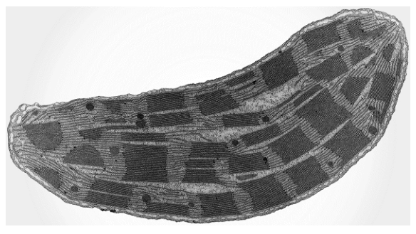
Which row shows structures that are expected to be present in cells that contain this organelle?
Options
| cell wall | centrioles | plasmodesmata | |
|---|---|---|---|
| A | ✗ | ✓ | ✗ |
| B | ✓ | ✗ | ✗ |
| C | ✗ | ✓ | ✓ |
| D | ✓ | ✗ | ✓ |
key
✓ = expected to be present
✗ = not expected to be present
Working
The electron micrograph shows a chloroplast — identifiable by the double outer membrane, stacks of thylakoids forming grana, lamellae linking the grana, and the surrounding stroma (with dark starch grains).
Chloroplasts are found in plant cells (and some algae). Structures expected in plant cells:
- Cell wall — present ✓ (made of cellulose)
- Centrioles — absent ✗ (centrioles are characteristic of animal cells; they are not found in higher plant cells)
- Plasmodesmata — present ✓ (cytoplasmic connections through cell walls linking adjacent plant cells)
Answer
D
D
Background Concept
The micrograph shows a chloroplast, the site of photosynthesis in eukaryotic cells. Its diagnostic features visible under the electron microscope are:
- a double (envelope) membrane,
- stacks of disc-shaped thylakoids forming grana,
- lamellae (intergranal membranes) connecting the grana,
- the fluid stroma surrounding the thylakoids,
- and often starch grains (the dark, dense regions) where glucose from photosynthesis is stored as starch.
Because chloroplasts are organelles of photosynthesis, they are confined to plant cells and algal cells (the group collectively called the Viridiplantae plus related lineages). They are absent from animal cells and from fungi.
Plant cells differ from animal cells in several characteristic features:
- They have a rigid cell wall of cellulose outside the plasma membrane.
- Adjacent plant cells are connected by plasmodesmata — fine cytoplasmic channels that traverse the cell walls and allow transport and communication between cells.
- They generally lack centrioles. (Centrioles, which organise the spindle during cell division in many eukaryotes, are present in animal cells but absent in the cells of higher plants; plant cells form their spindle microtubules without centrioles.)
Understanding the Question
The question presents an EM image of an organelle and asks you to identify which combination of structures (cell wall, centrioles, plasmodesmata) would be expected in a cell that contains this organelle. Because chloroplasts are plant-cell organelles, the answer hinges on knowing the typical contents of a plant cell.
Approach
- Identify the organelle from the EM (chloroplast).
- Decide which cell type possesses this organelle (plant cell).
- For each of the three listed structures, decide whether it is present in a plant cell.
- Match your conclusion to the option that gives the correct ✓/✗ pattern.
Step-by-Step Reasoning
- Organelle identification. The combination of double membrane + grana + lamellae + stroma is unique to the chloroplast. Mitochondria have cristae but no grana; the Golgi apparatus consists of flattened cisternae; the ER is a network of membranes with ribosomes. → It is a chloroplast.
- Cell type. A cell containing a chloroplast is a plant (or algal) cell.
- Cell wall: A cellulose cell wall is a defining feature of plant cells. → Present (✓).
- Centrioles: These are microtubule-based structures found in animal cells (and most animal-like protists) that help organise the mitotic spindle. Higher plant cells do not contain centrioles and still form spindles at the cell poles. → Absent (✗).
- Plasmodesmata: These are channels through plant cell walls that connect the cytoplasm of neighbouring cells, allowing symplastic transport. They are characteristic of plant tissues. → Present (✓).
So the pattern is cell wall ✓, centrioles ✗, plasmodesmata ✓, which matches row D.
Key Takeaways
- Chloroplasts are restricted to plant (and algal) cells; identifying one tells you the cell type.
- Plant cells have cell walls and plasmodesmata, and they lack centrioles.
- Animal cells have centrioles but lack both cell walls and plasmodesmata.
- EMs can be diagnostic of an organelle: look at membrane arrangement, internal structure, and inclusions.
Common Mistakes
- Confusing chloroplasts with mitochondria (both have a double membrane, but mitochondria have cristae, not grana and lamellae).
- Assuming all eukaryotes have centrioles — they are absent in higher plant cells.
- Thinking plasmodesmata are a feature of animal cells — they are exclusively plant.
Things to Be Careful About
- "Expected to be present" is the key phrase: only structures typical of plant cells should be ticked.
- The dark dense regions in the chloroplast are starch grains — useful confirmatory evidence, not a separate organelle to consider.
- Some lower plants (e.g. mosses, ferns) and many algae also lack centrioles, so the absence of centrioles is consistent with any plant-like cell containing chloroplasts.
What could take place during a hydrolysis reaction?
1 A glycosidic bond is broken.
2 A molecule of water is produced.
3 A sucrose molecule is split into fructose and glucose.
Options
A 1, 2 and 3
B 1 and 2 only
C 1 and 3 only
D 2 and 3 only
Working
Hydrolysis breaks a bond by adding a molecule of water (water is a reactant, not a product).
- Statement 1: A glycosidic bond is broken — true (water is added across a glycosidic bond, breaking it).
- Statement 2: A molecule of water is produced — false (water is consumed, not produced; water is produced in a condensation reaction, the reverse process).
- Statement 3: A sucrose molecule is split into fructose and glucose — true (sucrose = glucose + fructose joined by a glycosidic bond; hydrolysis yields these two monosaccharides).
Statements 1 and 3 only are correct.
Answer
C
C
Background Concept
Hydrolysis is a chemical reaction in which a bond in a larger molecule is broken by the addition of a molecule of water. One fragment of the broken molecule receives the H from water and the other receives the OH. It is the reverse of a condensation reaction, in which two small molecules join together and a molecule of water is released.
In biology, the most important hydrolysis reactions involve:
- Glycosidic bonds in disaccharides and polysaccharides (e.g. sucrose, maltose, starch, glycogen)
- Peptide bonds in polypeptides
- Phosphodiester bonds in nucleic acids
- Ester bonds in triglycerides and phospholipids
Hydrolysis is catalysed by specific hydrolase enzymes (e.g. amylase, sucrase, proteases, lipases).
Sucrose is a disaccharide formed by a condensation reaction between α-glucose and fructose, linked by an α-1,2 glycosidic bond. Adding water across this bond (hydrolysis) splits sucrose back into glucose and fructose.
Understanding the Question
This is a multiple-choice question asking which of three statements could correctly describe a hydrolysis reaction. Each statement must be evaluated against the strict definition of hydrolysis. The test of statement 2 hinges on a precise understanding: hydrolysis uses water; it does not make it.
The command word is implicit ("What could take place…"), so the candidate must judge truth rather than write a sentence.
Approach
- State the definition of hydrolysis and contrast it with condensation.
- Test each statement against that definition.
- Select the option whose statements are all true.
Step-by-Step Reasoning
-
Statement 1: "A glycosidic bond is broken." — Hydrolysis of any disaccharide (or polysaccharide) breaks the glycosidic bond linking the monomers. This is a textbook feature of hydrolysis. ✓ True.
-
Statement 2: "A molecule of water is produced." — This describes a condensation (anabolic) reaction, not hydrolysis. In hydrolysis, water is a reactant that is split and its H and OH are added to the two fragments; it is not generated. ✗ False.
-
Statement 3: "A sucrose molecule is split into fructose and glucose." — Sucrose is a non-reducing disaccharide composed of α-glucose and fructose joined by a 1,2-glycosidic bond. The enzyme sucrase (or acid hydrolysis) cleaves this bond, releasing one glucose and one fructose molecule. ✓ True.
Only statements 1 and 3 are correct, matching option C.
Key Takeaways
- Hydrolysis = bond broken by adding water; condensation = bond formed with the loss of water.
- Glycosidic, peptide and ester bonds are all cleaved by hydrolysis (and formed by condensation).
- Sucrose + water → glucose + fructose is the classic hydrolysis of a disaccharide.
Common Mistakes
- Confusing hydrolysis with condensation, leading to the belief that water is a product (this is the only statement 2 trap here).
- Assuming hydrolysis only applies to carbohydrates — in fact peptide and ester bonds are also hydrolysed.
- Writing "breaking a bond with water" imprecisely — say "by the addition of a water molecule" for clarity.
Things to Be Careful About
- Read each statement independently; a single false statement (here, statement 2) eliminates any option that includes it.
- "Could take place" wording means any one correct example is sufficient — do not look for an exhaustive list.
- Watch the option key: options B and D both include statement 2, so the test effectively rewards spotting that one error.
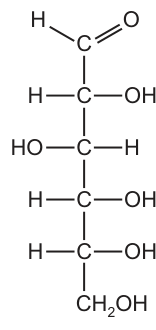
Sugars with a ring structure can also have a linear structure.
Which sugar molecules could be represented by the linear structure shown in the diagram?
Options
A glucose, deoxyribose and ribose
B glucose only
C deoxyribose and ribose only
D deoxyribose only
Working
The structure shown has six carbon atoms in a vertical chain, so it is a hexose.
The C1 group is an aldehyde (), so the sugar is an aldose — specifically an aldohexose.
- Glucose is an aldohexose (6C, aldehyde at C1) → matches the structure.
- Ribose is an aldopentose (only 5 carbons) → does not match.
- Deoxyribose is an aldopentose (5 carbons, with one H replacing an OH on C2) → does not match.
Answer
B
B
Background Concept
Monosaccharides (simple sugars) can be classified on two independent criteria:
-
Number of carbon atoms in the chain
- Triose — 3 carbons (e.g. glyceraldehyde)
- Pentose — 5 carbons (e.g. ribose, deoxyribose)
- Hexose — 6 carbons (e.g. glucose, fructose, galactose)
-
Functional group on C1 (or C2 for ketoses)
- Aldose — the C1 carbon carries an aldehyde group (, written )
- Ketose — the C2 carbon carries a ketone group ()
The two classifications are combined to name a sugar: glucose is an aldohexose, ribose is an aldopentose, fructose is a ketohexose, and so on. In solution, sugars cyclise to form ring structures, but in their open-chain (linear) form they show the carbonyl group and the carbon chain clearly.
Understanding the Question
The question provides a linear (open-chain) structural formula in Fig. 8.1 and asks which of the three sugars — glucose, ribose, deoxyribose — could be represented by that structure. To answer it, the candidate must read the structure and check which features match each sugar.
Approach
Read two features from the diagram:
- Count the carbon atoms in the vertical chain (1, 2, 3, 4, 5, 6 …).
- Identify the group on the topmost carbon — is it an aldehyde () or something else?
Then compare these two features against the known identity of each sugar.
Step-by-Step Reasoning
- The figure shows six carbon atoms (C1 at the top, C6 at the bottom as ). This makes the molecule a hexose.
- The C1 group is , which is the aldehyde group. The sugar is therefore an aldose, and combined with the 6-carbon count, it is an aldohexose.
- Glucose is the textbook aldohexose: a 6-carbon chain with an aldehyde on C1 and a hydroxyl on every other carbon. The arrangement of and on C2, C3, C4 and C5 in the figure matches glucose exactly → glucose fits.
- Ribose is an aldopentose — it has only 5 carbons in its chain. Because the figure clearly shows 6 carbons, ribose cannot be the sugar shown.
- Deoxyribose is also an aldopentose (5 carbons; it is "deoxy" because the on C2 is replaced by an ). Again, the 5-carbon backbone rules it out.
- Only glucose is consistent with both the 6-carbon chain and the aldehyde at C1. The correct option is B.
Key Takeaways
- A sugar's identity is fixed by (i) the number of carbons and (ii) the carbonyl position (aldose vs ketose).
- Glucose, ribose and deoxyribose are all aldoses, but glucose has 6 carbons while ribose and deoxyribose have only 5.
- When given a linear structural formula, count the carbons first — it is the quickest way to rule pentoses in or out.
Common Mistakes
- Picking A because all three sugars are reducing sugars / aldoses. This ignores the carbon-number difference.
- Picking C because ribose and deoxyribose "look like glucose". The two pentoses are the building blocks of RNA and DNA respectively, so they are commonly tested, but they have only 5 carbons.
- Picking D because deoxyribose is "simpler" (missing one ). It is still a 5-carbon sugar, so the chain length alone excludes it.
- Forgetting that a linear sugar formula can be drawn in different orientations (the aldehyde can appear at the top or the bottom depending on how the sugar is rotated); the criterion is the chain length and functional group, not the orientation.
Things to Be Careful About
- "Hexose" means 6 carbons, not "a sugar" in general — always count.
- An aldehyde group is on a terminal carbon (C1); a ketone is on an internal carbon (C2 in sugars).
- "Deoxy" refers to the loss of an oxygen (an replaced by ), not a loss of a carbon — deoxyribose is still a pentose, not a tetrose.
- The question asks what could be represented; the figure must match every carbon and functional group, not just the most prominent one.
Which statement about triglycerides is correct?
Options
A Each triglyceride molecule is formed by combining three fatty acid molecules with a glycogen molecule.
B A triglyceride molecule contains four ester bonds, each formed in a condensation reaction.
C Triglyceride molecules form a bilayer in the cell surface membranes of cells due to hydrophobic and hydrophilic interactions.
D The ratio of oxygen atoms to carbon atoms is lower for triglyceride molecules than for carbohydrate molecules.
Working
A triglyceride consists of one glycerol molecule joined to three fatty acid molecules by three (not four) ester bonds — A and B are wrong. Triglycerides are entirely hydrophobic (no polar/charged head) so they do not form bilayers; it is phospholipids that do — C is wrong. Fatty acid chains are long hydrocarbon (-CH₂-) backbones with very few oxygen atoms, while carbohydrates have roughly equal numbers of C and O atoms (e.g. glucose C₆H₁₂O₆ gives an O:C ratio of 1:1). Hence the O:C ratio is lower for triglycerides than for carbohydrates.
Answer
D
D
Background Concept
A triglyceride is a lipid formed by the condensation of one glycerol molecule (a 3-carbon triol) with three fatty acid molecules. Each carboxyl (-COOH) group of a fatty acid reacts with one of glycerol's three hydroxyl (-OH) groups, releasing a molecule of water and forming an ester bond. Because three fatty acids are joined, a triglyceride contains three ester bonds in total.
Fatty acids have long hydrocarbon chains — typically a -COOH head followed by a chain of -CH₂- units (e.g. 15–17 CH₂ groups in a C16 or C18 fatty acid). This means the molecular formula is dominated by C and H, with O only at the ester linkages and the original glycerol backbone. By contrast, a carbohydrate such as glucose (C₆H₁₂O₆) contains an oxygen atom on every carbon, giving an O:C ratio of 1:1. This large difference in O:C ratio underlies why lipids are far more reduced (and therefore release more energy per gram on oxidation) than carbohydrates.
Triglycerides are entirely hydrophobic because none of their constituent groups is charged or strongly polar. They do not form bilayers — that property belongs to phospholipids, which have a charged/polar phosphate head attached to glycerol and two (not three) fatty acid tails. This is why cell surface membranes are built from phospholipids, not triglycerides.
Understanding the Question
This is a multiple-choice question asking which single statement about triglycerides is correct. Each option probes a different feature:
- A: composition (glycerol vs glycogen, number of components)
- B: number of bonds (ester bond count)
- C: behaviour in cell membranes (bilayer formation)
- D: elemental composition (O:C ratio compared with carbohydrates)
The command word implied is identify the correct statement — exactly one must be right, and the others contain a specific, identifiable error.
Approach
Eliminate each wrong option by pinpointing the precise error, then check the surviving statement against the structural chemistry of a triglyceride. The fastest route is:
- Recall the components of a triglyceride → test A and B
- Recall whether a triglyceride can form a bilayer → test C
- Compare the O:C ratio of a typical fatty acid with a typical monosaccharide → test D
Step-by-Step Reasoning
Option A — wrong. The 'glyc' prefix is misleading. A triglyceride is built from glycerol (a small 3-carbon alcohol), not glycogen (a large branched polymer of glucose used for energy storage in animals). Confusion between these two similarly-named molecules is a common trap.
Option B — wrong. Three fatty acids × one ester bond each = three ester bonds, not four. The figure 'four' would be plausible if a diglyceride (two fatty acids + phosphate + a fourth group) were confused with a triglyceride.
Option C — wrong. Bilayer formation requires an amphipathic molecule — a polar/charged head plus non-polar tails. Triglycerides have no polar head (all three glycerol -OH groups are esterified to fatty acids), so they are completely hydrophobic and form droplets (e.g. in adipocytes) rather than bilayers. It is phospholipids (with a polar phosphate head) that form the bilayer of cell surface membranes.
Option D — correct. Consider a representative triglyceride such as tripalmitin, C₅₁H₉₈O₆. The O:C ratio is 6/51 ≈ 0.12. Compare this with glucose C₆H₁₂O₆, where the O:C ratio is 6/6 = 1.0. The triglyceride's O:C ratio is therefore very much lower than that of a carbohydrate — the statement is correct. The biological consequence is that triglycerides, being more reduced, yield more energy per gram on complete oxidation than carbohydrates do.
Key Takeaways
- A triglyceride = glycerol + 3 fatty acids, joined by 3 ester bonds (formed in condensation reactions).
- Triglycerides are fully hydrophobic and do not form bilayers; phospholipids do.
- Triglycerides have a much lower O:C ratio than carbohydrates because their hydrocarbon chains carry few oxygen atoms.
- This high reduction state is why lipids store more than twice the energy per gram of carbohydrates.
Common Mistakes
- Confusing glycerol (a 3-carbon alcohol, part of a triglyceride) with glycogen (a glucose polymer used for storage). The similar names are a deliberate distractor.
- Counting four ester bonds in a triglyceride. Always count: one fatty acid → one ester bond × three fatty acids = three ester bonds.
- Attributing bilayer formation to triglycerides rather than phospholipids. Without a polar/charged head group, a triglyceride cannot orient with hydrophilic heads outward.
- Reversing the comparison and saying that carbohydrates have a lower O:C ratio than triglycerides.
Things to Be Careful About
- 'Ester bond' is the precise term for the linkage between a fatty acid -COOH and glycerol -OH; do not call it a 'glycosidic bond' (that term applies to sugars).
- The RQ of a triglyceride is ~0.7, reflecting its low oxygen content, while the RQ of a carbohydrate is 1.0 — a useful cross-check on the O:C ratio argument.
- When asked about cell membrane structure, always associate bilayer formation with phospholipids, never with triglycerides.
Hydroxyproline is synthesised by addition of an –OH group to the R-group of the amino acid proline.
Hydroxyproline is a major component of collagen and has an important role in increasing the stability of its structure.
Which statement explains why the addition of an –OH group to proline could increase the stability of collagen?
Options
A It strengthens hydrogen bonding between the R-groups of adjacent polypeptide chains, resulting in a tertiary structure that is more resistant to heat denaturation.
B It increases the number of sites available for the formation of hydrogen bonds within the secondary structure of collagen, resulting in more stable alpha helices.
C It increases the formation of hydrogen bonds between R-groups and water molecules, which help to hold the chains of the collagen triple helix together by strengthening hydrophilic interactions.
D It strengthens the quaternary structure of collagen by providing more sites for hydrogen bonding between the R-groups of distantly separated amino acids within the same polypeptide chain.
Answer
Collagen has a quaternary structure formed from three polypeptide chains wound together in a triple helix. The –OH group added to proline to form hydroxyproline is polar, so it forms hydrogen bonds with surrounding water molecules. These hydrogen bonds help hold the three chains of the triple helix together, increasing the stability of collagen.
Answer
C
C
Background Concept
Collagen is the most abundant fibrous protein in animals. Its mature functional form is a triple helix (sometimes called tropocollagen): three left-handed polypeptide α-chains wound around each other to form a right-handed superhelix. The triple helix is held together by hydrogen bonds and is the quaternary structure of collagen. Note that collagen does not contain α-helices — its regular secondary structure is a left-handed helix, but the three chains together constitute its quaternary structure.
A key feature of collagen is the abundance of two unusual amino acids:
- Glycine — every third residue, because its single H R-group is small enough to fit where the three chains meet in the centre of the triple helix.
- Hydroxyproline — formed by the post-translational hydroxylation of proline (catalysed by the enzyme prolyl hydroxylase, which requires vitamin C as a cofactor; deficiency causes scurvy).
Hydroxyproline's –OH group is polar and can form hydrogen bonds. It is these hydrogen bonds — between hydroxyproline/water and between hydroxyproline residues on adjacent chains — that lock the three chains together and stabilise the triple helix.
Understanding the Question
The question tests whether you understand why adding an –OH group to proline stabilises collagen. The four options differ in two ways:
- Which level of protein structure the stabilisation occurs at (secondary / tertiary / quaternary).
- What the –OH group hydrogen-bonds to (adjacent R-groups, water, etc.).
You must pick the statement that correctly identifies the level of structure involved and gives the right bonding partner.
Approach
First, identify collagen's structural level: its stability comes from the quaternary triple helix (three chains together). Second, identify the bonding role of the –OH: hydroxyproline is on the outside of the triple helix, where it forms hydrogen bonds with surrounding water molecules; some of these also bridge to nearby R-groups on the neighbouring chain, helping to cement the three strands together.
Step-by-Step Reasoning
- Option A — claims the effect is on the tertiary structure and the –OH strengthens H-bonds between R-groups of adjacent polypeptide chains to resist heat denaturation. Tertiary structure refers to the folding of a single polypeptide chain, not multiple chains. The effect described is actually on quaternary structure. Also, heat denaturation is not what hydroxyproline primarily protects against. Incorrect.
- Option B — claims the effect is on the secondary structure and produces more stable α-helices. Collagen does not contain α-helices; its regular structure is the triple helix (a quaternary feature). Incorrect.
- Option C — states the –OH groups increase hydrogen bonding between R-groups and water, which helps hold the chains of the triple helix together by hydrophilic interactions. This matches the established role of hydroxyproline: its polar –OH forms hydrogen bonds with water on the surface of the triple helix, stabilising the assembly of the three chains. Correct.
- Option D — claims the effect strengthens the quaternary structure but says the –OH provides H-bonding sites between distantly separated amino acids within the same polypeptide chain. That description is the textbook definition of tertiary structure (long-range folding within one chain), not quaternary. The statement is internally contradictory and biologically wrong. Incorrect.
Key Takeaways
- Collagen's stabilising feature is its quaternary triple helix, not an α-helix.
- Hydroxyproline is made by post-translational modification of proline; its –OH group hydrogen-bonds with water on the outside of the triple helix, cementing the three chains.
- Be precise about protein structural levels:
- Primary — sequence of amino acids.
- Secondary — local regular folding (α-helix, β-pleated sheet) stabilised by H-bonds between backbone groups.
- Tertiary — overall 3D folding of one polypeptide chain, stabilised by R-group interactions (H-bonds, ionic, disulfide, hydrophobic).
- Quaternary — assembly of two or more polypeptide chains into a functional protein (e.g. haemoglobin, collagen).
Common Mistakes
- Saying collagen contains α-helices — it does not; it has a unique triple-helix quaternary structure.
- Confusing tertiary and quaternary structure; remembering that "more than one chain" is the hallmark of quaternary.
- Believing hydroxyproline makes covalent (e.g. disulfide) bonds — the stabilising bonds are hydrogen bonds, not covalent.
- Forgetting that vitamin C is needed to hydroxylate proline; deficiency impairs collagen stability and causes scurvy.
Things to Be Careful About
- Read the structural level named in the option and check it matches the description that follows (e.g. Option D is internally inconsistent — it says "quaternary" but describes a tertiary-style interaction).
- "Stability" of fibrous structural proteins like collagen is not principally about resisting heat denaturation; it is about maintaining the triple-helix assembly under physiological conditions.
- Hydrogen bonding with water (hydrophilic interaction) is the key; bonding directly between R-groups is the minor contribution.
Plant cell walls are strengthened by cellulose molecules that are arranged in several layers. Within each layer, the cellulose molecules are arranged in the same direction (parallel).
Which row shows the bonds that hold adjacent cellulose molecules together within each layer and the arrangement of cellulose molecules in different layers?
Options
| bonds that hold adjacent cellulose molecules together | arrangement of cellulose molecules in different layers | |
|---|---|---|
| A | glycosidic | in different directions |
| B | glycosidic | parallel |
| C | hydrogen | in different directions |
| D | hydrogen | parallel |
Working
Cellulose is a polysaccharide of β-glucose monomers joined by glycosidic bonds. The glycosidic bonds join monomers within a single cellulose chain; they do not join adjacent chains.
Adjacent cellulose chains are held together by hydrogen bonds that form between the –OH (hydroxyl) groups on neighbouring β-glucose units. This hydrogen bonding is what gives plant cell walls their tensile strength.
Between layers, the cellulose molecules run in different directions (the layers are offset at an angle to each other), producing a cross-ply arrangement that adds further strength to the cell wall.
Answer
C
C
Background Concept
Cellulose is the main structural polysaccharide of plant cell walls. It is a polymer of β-glucose, in which successive monomers are rotated 180° relative to each other and joined by 1,4-glycosidic bonds. Because the glycosidic bond is between C1 of one β-glucose and C4 of the next, the chain has a straight, unbranched shape — ideal for laying down as fibres.
Individual cellulose chains line up side by side to form microfibrils. Many microfibrils are bundled into macrofibrils and then woven into the layers of the cell wall. The cell wall is built up of many such layers, and the orientation of the cellulose molecules differs from one layer to the next (a cross-ply or plywood-like arrangement). This is the structural basis of the cell wall's great tensile strength.
Two distinct types of bond are involved in cellulose:
- Glycosidic bonds — strong, covalent bonds that link glucose monomers within a single cellulose chain. These give the chain its continuity.
- Hydrogen bonds — weaker, non-covalent bonds that form between the hydroxyl (–OH) groups of glucose units on neighbouring cellulose chains. Many hydrogen bonds together hold the parallel chains firmly side by side.
Understanding the Question
This is a multiple-choice question testing two structural features of cellulose in plant cell walls:
- The type of bond between adjacent cellulose molecules within a single layer.
- The orientation of cellulose molecules between different layers.
The stem explicitly tells us that within each layer, cellulose molecules are parallel. The options then offer different combinations of bond type (glycosidic vs hydrogen) and between-layer arrangement (in different directions vs parallel).
Approach
Recall the two-tier structure of cellulose:
- Within one layer: chains are parallel, held together by hydrogen bonds (not glycosidic — those are the covalent bonds along the chain).
- Between layers: chains are arranged in different directions (not parallel — that would defeat the cross-ply strengthening).
Match these two facts to the option that combines them.
Step-by-Step Reasoning
- Eliminate options A and B: the bond within a layer, between adjacent molecules, is hydrogen, not glycosidic. Glycosidic bonds run along the chain, joining glucose to glucose within a single cellulose molecule, never between two separate cellulose chains.
- Eliminate option D: in different layers, the cellulose molecules are not all parallel. The cell wall achieves strength precisely because successive layers have their cellulose microfibrils running in different directions — like plywood or woven fabric.
- Option C states: hydrogen bonds hold adjacent molecules together within a layer, and the molecules in different layers run in different directions. This is the correct description of cellulose structure in plant cell walls.
Key Takeaways
- Glycosidic bonds = covalent, run along the cellulose chain (between monomers in the same chain).
- Hydrogen bonds = non-covalent, run between adjacent cellulose chains in the same layer.
- The wall's layers are cross-plied — cellulose microfibrils in one layer lie at an angle to those in the next, giving the wall multi-directional strength.
Common Mistakes
- Confusing glycosidic bonds with hydrogen bonds. Glycosidic bonds hold monomers together within a single chain; hydrogen bonds hold separate chains together.
- Thinking all layers are parallel. If all cellulose molecules ran in the same direction, the wall would be strong in one direction but weak in others; the cross-ply arrangement gives uniform strength.
Things to Be Careful About
- The question asks about bonds between adjacent cellulose molecules (i.e. between chains), not bonds within a cellulose molecule (i.e. between monomers). This wording distinguishes hydrogen bonds from glycosidic bonds here.
- The term "different layers" refers to the layered architecture of the cell wall, not the layered appearance of a single chain.
Galactogen is a storage polysaccharide in some animal species. It is a branched polymer that is formed from -galactose monomers.
Which comparison of galactogen with another polysaccharide correctly summarises one similarity and one difference?
Options
A Glycogen and galactogen are both branched, but glycogen is not a storage polysaccharide in animals.
B Glycogen and galactogen are both storage polysaccharides in animals, but glycogen is unbranched.
C Cellulose and galactogen are both branched, but cellulose is a structural polysaccharide found in plants.
D Amylopectin and galactogen are both storage polysaccharides, but amylopectin is formed from -glucose monomers.
Working
Galactogen is a branched storage polysaccharide made from β-galactose monomers.
- A is wrong: glycogen IS a storage polysaccharide in animals (stored in liver and muscle).
- B is wrong: glycogen IS branched (like amylopectin, with α-1,4 and α-1,6 glycosidic bonds).
- C is wrong: cellulose is UNBRANCHED — straight chains of β-glucose linked by β-1,4 glycosidic bonds.
- D is correct: amylopectin (a component of starch) is a storage polysaccharide, and it is built from α-glucose monomers, whereas galactogen is built from β-galactose.
Answer
D
D
Background Concept
Polysaccharides are long-chain carbohydrates formed when many monosaccharide monomers join by glycosidic bonds (formed in a condensation reaction, releasing water). Three polysaccharides are core to A-Level Biology:
- Starch — the main storage polysaccharide in plants. It is a mixture of two polymers: amylose (long, unbranched chains of α-glucose linked by α-1,4 glycosidic bonds, which coil into a helix) and amylopectin (branched chains of α-glucose with α-1,4 links along the chain and α-1,6 links at the branch points, roughly every 24–30 glucose units).
- Glycogen — the main storage polysaccharide in animals (mainly liver and muscle). It has the same α-1,4 / α-1,6 linkages as amylopectin, but is more highly branched (branches every 8–12 glucose units), allowing more rapid mobilisation of glucose when needed.
- Cellulose — a structural polysaccharide in plant cell walls. It is made of β-glucose monomers linked by β-1,4 glycosidic bonds. Because every other glucose is flipped 180°, the chains are straight, and many chains hydrogen-bond together into strong microfibrils.
The type of glycosidic bond (α vs β) and the degree of branching determine whether the polymer is a compact storage molecule (helical, branched — good for packing many glucose units in a small space) or a tough structural molecule (straight, hydrogen-bonded — good for cell-wall strength).
Galactogen (mentioned in the question) is a less-commonly taught analogue of glycogen found in some invertebrates (e.g. snails). Like glycogen it is branched, but unlike glycogen it is built from β-galactose rather than α-glucose monomers.
Understanding the Question
This is a "Which comparison…" question: the candidate must identify the option that contains one correct similarity AND one correct difference between galactogen and another named polysaccharide. Only one of the four options does both correctly.
Key facts about galactogen given in the stem:
- it is a storage polysaccharide in some animals
- it is branched
- it is made of β-galactose monomers
The candidate must check each option's similarity (true?) and difference (true?).
Approach
For each option, verify (1) the claimed similarity and (2) the claimed difference, in either order. Eliminate any option in which either side is false.
Step-by-Step Reasoning
Option A — "Glycogen and galactogen are both branched, but glycogen is not a storage polysaccharide in animals."
- Similarity: both branched — TRUE.
- Difference: glycogen is not a storage polysaccharide — FALSE. Glycogen is the principal animal storage polysaccharide.
- Eliminate A.
Option B — "Glycogen and galactogen are both storage polysaccharides in animals, but glycogen is unbranched."
- Similarity: both storage polysaccharides in animals — TRUE.
- Difference: glycogen is unbranched — FALSE. Glycogen is one of the most highly branched polysaccharides known (branch points every 8–12 glucose units).
- Eliminate B.
Option C — "Cellulose and galactogen are both branched, but cellulose is a structural polysaccharide found in plants."
- Similarity: both branched — FALSE. Cellulose is unbranched; its β-1,4-linked β-glucose chains are straight and align in parallel to form microfibrils.
- Eliminate C.
Option D — "Amylopectin and galactogen are both storage polysaccharides, but amylopectin is formed from α-glucose monomers."
- Similarity: both storage polysaccharides — TRUE (amylopectin is the branched component of plant starch, a storage polysaccharide).
- Difference: amylopectin is formed from α-glucose monomers — TRUE (amylopectin is built from α-glucose with α-1,4 and α-1,6 glycosidic bonds), whereas galactogen is built from β-galactose.
- Option D is correct.
Key Takeaways
- Storage polysaccharides in animals = glycogen (highly branched, α-glucose).
- Storage polysaccharides in plants = starch = amylose (unbranched, α-glucose) + amylopectin (branched, α-glucose).
- Structural polysaccharide in plants = cellulose (unbranched, β-glucose, β-1,4 links).
- The α/β orientation of the monomer's OH group on C1 determines which type of glycosidic bond can form, which in turn determines whether the chain is helical/branched (storage) or straight/hydrogen-bonded (structural).
- When a question gives a polymer's properties in the stem, check each claimed fact in the options against the stem — and check the comparative claim against your prior knowledge of the other polymer.
Common Mistakes
- Assuming all storage polysaccharides are made of α-glucose. The stem explicitly says galactogen uses β-galactose — a useful reminder that "β" does not automatically mean "structural".
- Thinking cellulose is branched — it is not; its straight β-1,4-linked chains are precisely what allow it to form strong fibres.
- Confusing amylose (unbranched) with amylopectin (branched) when comparing starch components.
- Forgetting that glycogen, like amylopectin, is a branched α-glucose polymer — glycogen is just more highly branched.
Things to Be Careful About
- A "similarity AND difference" MCQ requires both halves of the option to be true; one correct and one incorrect is enough to eliminate it.
- Watch the monomer identity (α-glucose vs β-glucose vs β-galactose) — galactose is a different sugar from glucose, with the OH on C4 in the opposite orientation, so it is not interchangeable.
- Glycogen's defining feature, beyond being a storage polymer, is its high degree of branching (1,6 branch points roughly every 8–12 residues) — this is what allows rapid release of glucose by glycogen phosphorylase.
- "Animal storage" is essentially synonymous with glycogen; if an option claims something else is the animal storage polysaccharide, it is wrong.
Which levels of protein structure are always involved in forming the active site of an enzyme?
Options
| primary | tertiary | quaternary | |
|---|---|---|---|
| A | ✓ | ✓ | ✓ |
| B | ✓ | ✓ | ✗ |
| C | ✗ | ✓ | ✓ |
| D | ✗ | ✗ | ✓ |
key
✓ = always involved
✗ = not always involved
Working
The active site of an enzyme is a 3-D pocket formed by the folding of the polypeptide chain.
- Primary structure is always involved: the specific sequence of amino acids determines how the polypeptide folds, ultimately producing the 3-D shape of the active site.
- Tertiary structure is always involved: the overall 3-D folding of the polypeptide brings particular amino acid R-groups into the correct spatial positions to form the active site.
- Quaternary structure is not always involved: many enzymes (e.g. lysozyme, amylase) are single polypeptide chains and have no quaternary structure.
Answer
B
B
Background Concept
Proteins have up to four levels of structure:
- Primary structure – the linear sequence of amino acids joined by peptide bonds.
- Secondary structure – regular folding patterns (α-helices and β-pleated sheets) stabilised by hydrogen bonds between atoms of the polypeptide backbone.
- Tertiary structure – the overall 3-D folding of a single polypeptide chain, held by interactions between the R-groups of the amino acids (hydrogen bonds, ionic bonds, disulfide bridges, hydrophobic interactions).
- Quaternary structure – the association of two or more polypeptide subunits in a multi-subunit protein.
Enzymes are globular proteins, and their active site is a specifically shaped 3-D pocket (or groove) where the substrate binds. The shape and chemical properties of the active site are determined by the R-groups of the amino acids positioned at that site.
Understanding the Question
The question asks which levels of protein structure are always involved in forming an enzyme's active site. The keyword is always – the answer must be true for every enzyme, not just some. The options present combinations of primary, tertiary and quaternary structure (secondary structure is not tested here).
Approach
To answer, work through each level of structure and decide whether it is essential to the active site in every enzyme:
- Could the active site exist without the primary structure? No – without a defined amino acid sequence, there is no specific 3-D shape, and therefore no specific active site. Primary is always involved.
- Could the active site exist without tertiary structure? No – the active site is a 3-D feature produced by folding. Tertiary is always involved.
- Could the active site exist without quaternary structure? Yes – many enzymes consist of a single polypeptide chain and have no quaternary structure at all. Quaternary is not always involved.
The combination that fits is primary ✓, tertiary ✓, quaternary ✗, which is option B.
Step-by-Step Reasoning
- Primary structure is always involved. The amino acid sequence determines which R-groups are present and, in turn, the bonding interactions that drive the polypeptide to fold into its characteristic 3-D shape. Without the specific primary sequence, the active site would not form. (Codon-level changes in primary structure – e.g. the sickle-cell mutation in haemoglobin – can abolish or alter function, illustrating this point.)
- Tertiary structure is always involved. The active site is a 3-D pocket created by the folding of the polypeptide. R-groups from quite distant parts of the primary sequence are brought close together in 3-D space to form the binding site for the substrate. This is the tertiary structure.
- Quaternary structure is NOT always involved. Many enzymes (e.g. lysozyme, ribonuclease, many digestive enzymes such as amylase and trypsin) are single-chain globular proteins with no quaternary structure. Only multi-subunit enzymes (e.g. lactate dehydrogenase, which is tetrameric) possess quaternary structure, and even then the active site typically lies within a single subunit. Hence quaternary structure cannot be marked as "always involved".
- Options C and D are wrong because they include quaternary structure as always required, or exclude primary structure. Option A is wrong because quaternary is shown as always required.
Key Takeaways
- The active site of an enzyme is a 3-D feature produced by the tertiary structure of a polypeptide.
- Primary structure underpins tertiary structure – it is therefore indirectly but always required.
- Quaternary structure is present only in multi-subunit enzymes; it is not a universal feature of enzymes and is not always required for an active site to form.
- Secondary structure (α-helices, β-sheets) is not directly listed here, but it is part of the tertiary-level folding and contributes to overall 3-D shape.
Common Mistakes
- Saying that the active site is formed by the secondary structure (α-helix/β-sheet): these local motifs are stabilised by backbone H-bonds and contribute to folding, but the active-site residues are brought together by tertiary folding.
- Assuming all enzymes have quaternary structure because familiar examples such as haemoglobin do. Many textbook enzymes (lysozyme, trypsin, amylase) are monomeric.
- Confusing the role of primary structure: it does not form the active site directly, but without it there is no defined sequence, and therefore no defined tertiary shape or active site.
Things to Be Careful About
- The word always is decisive. A level of structure that is only sometimes present (e.g. quaternary) cannot be the correct answer for a question requiring universality.
- Some mark schemes also credit the idea that the active site is formed by the R-groups of amino acids brought together in 3-D space – this emphasises tertiary, not secondary, structure.
- Enzymes are globular proteins, not fibrous; this is what permits the compact, specific 3-D active site to form.
Catalase is an enzyme that breaks down hydrogen peroxide into water and oxygen.
Catalase was added to a solution of hydrogen peroxide and the oxygen produced was collected in a gas syringe.
The total volume of oxygen produced from the start of the reaction was recorded every 10 seconds for 1 minute.
The results are shown in the table.
| time / s | total volume of oxygen produced / |
|---|---|
| 0 | 0 |
| 10 | 22 |
| 20 | 40 |
| 30 | 50 |
| 40 | 55 |
| 50 | 57 |
| 60 | 58 |
What can be concluded from these results?
Options
A The reaction stopped after 60 seconds and no more oxygen was produced.
B The highest rate of oxygen production occurred 10 seconds after the start of the reaction.
C It took more than 20 seconds from the start of the reaction for half of the substrate to be converted to water and oxygen.
D The mean rate of reaction between 20 and 30 seconds was twice the mean rate of reaction between 30 and 40 seconds.
Working
Calculate the mean rate of oxygen production for each 10-second interval by dividing the increase in volume by 10 s:
- 0–10 s:
- 10–20 s:
- 20–30 s:
- 30–40 s:
- 40–50 s:
- 50–60 s:
Check option D: the rate between 20 and 30 s () is exactly twice the rate between 30 and 40 s ().
Answer
D
D
Background Concept
Catalase is an intracellular enzyme that catalyses the breakdown of hydrogen peroxide () into water and oxygen:
When the reaction is followed by collecting the oxygen in a gas syringe, the total volume of oxygen increases over time. A plot of total volume against time gives a typical enzyme progress curve: the curve rises steeply at first (high rate) and then levels off (rate approaching zero) as substrate is used up. The rate of reaction at any moment is the gradient of the curve, which can be estimated as the mean rate over a short interval:
Understanding the Question
The data table gives the total volume of oxygen collected at 10-second intervals. Each option is a different conclusion that can (or cannot) be drawn from these numbers. The question tests whether you can read the table, calculate rates, and judge whether a stated claim is supported.
Approach
For each option, decide what calculation or check is needed:
- A – claims the reaction has stopped. The data only extend to 60 s; the volume is still increasing, so the reaction has not stopped and we cannot predict beyond 60 s.
- B – claims the highest rate occurs 10 s after the start. Compare the rate in the 0–10 s interval with the 10–20 s interval.
- C – claims it took more than 20 s to convert half the substrate. Without knowing the total possible oxygen yield, we cannot say when half the substrate was used. Even if we assume the yield at 60 s (58 cm³) is total, half (29 cm³) was reached between 10 and 20 s, not after 20 s.
- D – claims the mean rate in 20–30 s is twice the mean rate in 30–40 s. Calculate both rates and compare.
Step-by-Step Reasoning
Option A: The volume increases from 57 cm³ at 50 s to 58 cm³ at 60 s — the reaction is still proceeding. We have no data after 60 s, so we cannot say it has stopped. Reject.
Option B: Mean rate 0–10 s = . Mean rate 10–20 s = . The highest rate was during the first 10 seconds, not the second 10 seconds. Reject.
Option C: We are not given the total possible oxygen yield. Even if we use 58 cm³ as a proxy for the total, half (29 cm³) was reached between 10 s (22 cm³) and 20 s (40 cm³), so it took less than 20 s, not more. Reject.
Option D:
- Mean rate 20–30 s =
- Mean rate 30–40 s =
The ratio is , so the rate in 20–30 s is indeed twice the rate in 30–40 s. Accept.
Key Takeaways
- The rate of an enzyme-catalysed reaction decreases over time as substrate is consumed; this is shown by the curve flattening.
- The mean rate over an interval is found from between two readings.
- Always check that a conclusion is supported by the data: a statement like "the reaction has stopped" is only valid if the data show a flat (zero-change) section AND you are sure no further reaction can occur.
- For "half the substrate converted" questions, you need either the maximum possible yield or another reference point — never assume without evidence.
Common Mistakes
- Assuming the reaction has stopped just because the rate is small (option A) — a small positive gradient is still a positive gradient.
- Picking the interval with the largest single reading rather than the largest rate (confusing B-style options).
- Inferring when half the substrate is used without knowing the maximum yield (option C).
- Forgetting to subtract successive values before dividing by 10 s, which would make every rate look the same.
Things to Be Careful About
- Mean rate = (change in volume) ÷ (time interval), not just the volume at a time point.
- The unit of rate here is ; keep the time unit consistent.
- An MCQ conclusion must be fully supported by the data — if a comparison is required, the numbers must give exactly the relationship claimed.
Succinic dehydrogenase is an enzyme that catalyses the conversion of succinate to fumarate in aerobic respiration.
Malonate is a reversible inhibitor of succinic dehydrogenase. Malonate reduces the enzyme’s activity by binding to its active site. Malonate and succinate cannot bind to the active site at the same time.
Which statement describes the effect of malonate on the activity of succinic dehydrogenase?
Options
A In the presence of malonate, can still be reached if the concentration of succinate is increased.
B Malonate has no effect on the .
C In the presence of malonate, can still be reached if the concentration of fumarate is increased.
D Malonate decreases the .
Working
The stem states that malonate binds to the active site of succinic dehydrogenase and that malonate and succinate cannot bind at the same time. This is the definition of competitive inhibition.
For a competitive inhibitor:
- is unchanged — at sufficiently high [substrate], substrate molecules out-compete the inhibitor for the active site, so the maximum rate is still attainable.
- increases — more substrate is required to reach half because some active sites are occupied by the inhibitor.
Evaluating each option:
- A: True. Increasing [succinate] overcomes competition for the active site, so can still be reached.
- B: False. Competitive inhibition increases the apparent .
- C: False. Fumarate is the product; raising [product] does not overcome competitive inhibition of the substrate.
- D: False. Competitive inhibition increases (not decreases) .
Answer
A
A
Background Concept
Enzyme inhibitors reduce the rate of an enzyme-catalysed reaction. Two reversible types are tested at A-Level:
- Competitive inhibitor: structurally similar to the substrate and binds to the same active site. Substrate and inhibitor cannot occupy the active site simultaneously. The effect depends on the relative concentrations: raising [substrate] displaces the inhibitor.
- Non-competitive inhibitor: binds to a site other than the active site (an allosteric site), changing the shape of the active site so the substrate can no longer bind effectively. Raising [substrate] does not overcome the inhibition because the inhibitor is not competing for the active site.
The two key kinetic parameters from a Michaelis–Menten graph (reaction rate vs substrate concentration) are:
- — the maximum rate, reached when all active sites are saturated with substrate.
- — the substrate concentration at which the rate is . A low indicates high enzyme affinity for the substrate.
Effects on these parameters:
| Inhibitor | ||
|---|---|---|
| Competitive | unchanged | increased |
| Non-competitive | decreased | unchanged |
Understanding the Question
The stem gives three clues about malonate:
- It binds to the active site of succinic dehydrogenase.
- It is reversible.
- Malonate and succinate cannot bind to the active site at the same time.
Together these unambiguously define competitive inhibition. The question then asks which statement correctly describes the effect of malonate on the enzyme's activity, framed in terms of and .
Approach
Recognise the inhibition type from the stem, recall its effect on and , then test each option against that prediction. Eliminate options that contradict the kinetic behaviour of competitive inhibition.
Step-by-Step Reasoning
Identifying the inhibition type:
Because malonate binds to the active site and is mutually exclusive with succinate, it must be a competitive inhibitor. (A non-competitive inhibitor would bind elsewhere and would not be excluded by substrate.)
Applying the kinetic consequences:
- — at very high [succinate], every active site is occupied by succinate rather than malonate. So is unchanged from the uninhibited reaction. This is the diagnostic feature of competitive inhibition.
- — because some active sites are blocked at any given [substrate], the enzyme needs a higher [substrate] to reach half its maximum rate. So the apparent rises.
Testing the options:
- A claims is still reachable if [succinate] is increased — this matches the behaviour of competitive inhibition. ✔
- B claims is unchanged — wrong; competitive inhibition increases . ✘
- C claims raising [fumarate] (the product) restores — wrong on two counts: fumarate is the product, not the substrate, and adding more product would actually slow the forward reaction (product inhibition / equilibrium shift), not overcome the inhibitor. ✘
- D claims decreases — wrong; increases in competitive inhibition. ✘
Key Takeaways
- An inhibitor that binds to the active site and is mutually exclusive with the substrate is, by definition, competitive.
- Competitive inhibition leaves unchanged but increases .
- Non-competitive inhibition reduces but leaves unchanged.
- Manipulating the product cannot rescue an enzyme from competitive inhibition of its substrate.
Common Mistakes
- Confusing competitive and non-competitive inhibition: students often state that competitive inhibition lowers , which is the behaviour of non-competitive inhibition.
- Assuming all inhibitors affect in the same way — only competitive inhibitors raise .
- Choosing an option that confuses substrate with product (option C). Fumarate is the product; only the substrate (succinate) competes with malonate.
- Forgetting that reversible competitive inhibition can always be overcome by raising [substrate] — this is what distinguishes it from non-competitive inhibition.
Things to Be Careful About
- " has no effect" vs " is unchanged" — option B uses the exact phrasing in the question, but the correct biology is that does change (increases), so the statement is false.
- Read the stem precisely: the phrase "cannot bind to the active site at the same time" is the key mutual-exclusivity that defines competitive inhibition. Without that clause, binding to the active site could still describe an irreversible or non-competitive-style inhibitor.
- being "still reachable" is the hallmark of competitive inhibition. If a question states that is reduced, the inhibitor must be non-competitive (or irreversible).
The diagram shows the dimensions of two blocks of agar. The diagram has been drawn to scale.
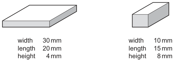
The blocks of agar were stained pink with a pH indicator. In acidic conditions, the pink pH indicator becomes colourless.
The two blocks of agar were placed in a beaker of acid at the same time. As the acid diffused into the blocks, the blocks became colourless.
What is the surface area to volume ratio of the block that became completely colourless first?
Options
A
B
C
D
Working
Acid diffuses in from the surface, so the block whose centre is reached first will be the one with the higher surface area to volume ratio. Calculate SA:Vol for each block.
Block 1 (30 mm × 20 mm × 4 mm):
Block 2 (10 mm × 15 mm × 8 mm):
Block 1 has the higher SA:Vol ratio, so it becomes completely colourless first.
Answer
B
B
Background Concept
Diffusion is the passive net movement of particles (here, H⁺ ions from the acid) from a region of higher concentration to a region of lower concentration. The rate at which a substance diffuses through a block of material depends on:
- the concentration gradient (steeper = faster),
- the surface area available for entry (more surface = faster),
- the distance the substance has to travel to reach the centre (shorter = faster).
For a block of fixed shape, the key idea is the surface area to volume (SA:Vol) ratio. A small, thin block has a much larger proportion of its volume close to a surface than a large, thick block does. This is the same reason why cells must be small — they need a high SA:Vol ratio so that oxygen, nutrients and waste can diffuse across the membrane fast enough to service the entire cell volume.
In this experiment, the agar is stained pink with a pH indicator. When acid (H⁺) reaches the indicator, the indicator turns colourless. The block whose centre turns colourless first is the one into which H⁺ ions have diffused to the middle fastest — i.e. the block with the higher SA:Vol ratio.
Understanding the Question
We are given two rectangular blocks of agar:
- Block 1: 30 mm × 20 mm × 4 mm (a wide, flat slab)
- Block 2: 10 mm × 15 mm × 8 mm (a smaller, chunkier block)
Both are placed in acid at the same time. The question asks for the SA:Vol ratio of the block that becomes completely colourless first.
The command word is "What is…", so we need a numerical answer (one of the four options). The four options are pairs of SA:Vol ratios, and we must identify the correct one.
Approach
- Calculate surface area and volume of each block (rectangular cuboid formula).
- Form the SA:Vol ratio for each and compare.
- The block with the higher ratio turns colourless first.
- Select the option matching that ratio.
Step-by-Step Reasoning
Block 1 (30 mm × 20 mm × 4 mm):
- Volume:
- Surface area of a cuboid:
- SA:Vol =
Block 2 (10 mm × 15 mm × 8 mm):
- Volume:
- Surface area:
- SA:Vol =
Block 1 has the higher ratio (0.67 : 1 vs 0.58 : 1), so it becomes colourless first. The answer is therefore B (0.67 : 1).
Note that although Block 2 is smaller in volume, it is more "cube-like" (relatively chunky in all three dimensions), so its volume-to-surface-area relationship is less favourable. Block 1 is a thin slab — the 4 mm height means the centre is only 2 mm from the nearest surface, so acid reaches the middle very quickly.
Key Takeaways
- Diffusion into a block is fastest when SA:Vol is high.
- The thinnest dimension matters most, because diffusion distance to the centre is half that dimension.
- A higher SA:Vol ratio is the reason cells are small and why organisms have specialised exchange surfaces (alveoli, villi, gill lamellae).
Common Mistakes
- Mixing up which block changes first. Students sometimes calculate both ratios correctly but then pick the block with the lower ratio. Remember: faster diffusion = higher SA:Vol.
- Using the wrong formula for surface area (e.g. just adding the three face areas once instead of doubling them — a cuboid has 6 faces, in 3 pairs).
- Forgetting units — the ratio is dimensionless, but the intermediate SA and Vol should carry mm² and mm³.
- Cancelling down incorrectly — simplifies to , which as a decimal is , not for Block 2.
Things to Be Careful About
- Always check the smallest dimension (here, 4 mm) — it controls how far acid has to diffuse to the centre (only 2 mm in Block 1, but 4 mm in Block 2 from the shortest side, and 5 mm or 7.5 mm from the others).
- "Surface area to volume ratio" is sometimes written as a fraction (e.g. 0.67) and sometimes as a ratio (0.67 : 1); both mean the same thing.
- The diagram is "drawn to scale" — a useful sanity check: Block 1 in the picture looks much larger overall, but it is flatter; Block 2 looks more compact. The numbers will confirm what the picture suggests.
The graph shows how the rate of facilitated diffusion of substance X across a cell surface membrane changed as the concentration of substance X increased. All conditions, except for the concentration of substance X, were kept constant. Temperature was maintained at .
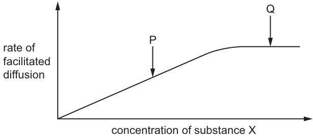
Which statement about the rate of facilitated diffusion is correct?
Options
A The rate of facilitated diffusion of substance X at Q will increase if the temperature is increased to .
B The rate of facilitated diffusion of substance X at P will increase if the concentration of ATP is increased.
C The rate of facilitated diffusion of substance X at Q will increase if the concentration of substance X is increased.
D The rate of facilitated diffusion of substance X at P will increase if the length of time over which the rate is measured is increased.
Working
At point Q, the curve has plateaued because all carrier/channel proteins are saturated — every transport protein is occupied and working at maximum rate, so adding more X cannot increase the rate (this rules out C).
Increasing temperature from to increases the kinetic energy of substance X molecules (more successful collisions with transport proteins) and increases the fluidity of the phospholipid bilayer, so even saturated proteins transport X faster. This raises the rate at Q.
- B is wrong: facilitated diffusion is passive and does not require ATP.
- D is wrong: rate is measured per unit time, so changing the time interval does not change the rate itself.
Answer
A
A
Background Concept
Facilitated diffusion is a passive process that moves substances across a cell surface membrane down their concentration gradient with the help of carrier proteins and channel proteins. Because it is passive:
- It does not require ATP (energy comes from the concentration gradient itself).
- It shows saturation kinetics — as the concentration of the substance increases, the rate rises steeply at first and then levels off to a plateau. The plateau occurs because at high concentrations every available transport protein is already in use; there are no spare proteins to handle more molecules per unit time.
Temperature affects the rate of facilitated diffusion in two ways:
- Higher temperature gives molecules more kinetic energy, so they collide with transport proteins more often and with greater force, increasing the rate of successful transport events.
- Higher temperature increases the fluidity of the phospholipid bilayer and the kinetic energy of the transport proteins themselves, allowing them to change shape (in the case of carriers) or open/close (in the case of channels) more rapidly.
These two effects mean that even when the carriers are saturated (at the plateau), raising the temperature still raises the maximum rate.
Understanding the Question
The graph shows the familiar saturation curve for facilitated diffusion:
- Point P lies on the steep rising portion — here rate is limited mainly by the supply of substance X.
- Point Q lies on the plateau — here rate is limited by the number of transport proteins; all are saturated.
Temperature is held at , which is below the optimum for most membrane proteins, so raising the temperature should increase the rate at all points on the curve, including Q.
The question asks which statement is correct. The command word is implicit ("which is correct") — we must identify the single true statement.
Approach
Work through each option and decide whether the proposed change would, in principle, alter the rate of facilitated diffusion:
- Will the change increase the rate?
- Does the change act on a variable that actually limits the rate at the named point (P or Q)?
Eliminate options whose mechanism either does not apply to facilitated diffusion or does not affect a true limiting factor.
Step-by-Step Reasoning
Option A — increase temperature to at point Q:
At Q the carriers are saturated. More X cannot help (no spare carriers). But raising temperature increases kinetic energy of X molecules and the fluidity/activity of the carrier proteins. The maximum rate (the height of the plateau) rises. Correct.
Option B — increase ATP at point P:
Facilitated diffusion does not hydrolyse ATP; ATP powers active transport, not facilitated diffusion. Changing ATP has no effect on the rate of facilitated diffusion. Incorrect.
Option C — increase concentration of X at point Q:
At Q the curve has plateaued. Adding more X makes no difference because the rate is limited by the number of transport proteins, not the supply of X. The curve is flat for exactly this reason. Incorrect.
Option D — lengthen the time of measurement at point P:
Rate is an intensive quantity — amount per unit time. The graph already plots rate on the y-axis. Measuring over a longer interval would give a larger total quantity transported, but the rate (quantity / time) is unchanged. Incorrect.
Only A is true.
Key Takeaways
- Facilitated diffusion is passive (no ATP) and shows saturation because transport proteins are limited in number.
- On the rising part of the curve the rate is limited by substrate concentration; on the plateau it is limited by the number of transport proteins.
- Temperature is the one variable that can raise the rate even at saturation, by acting on kinetic energy and membrane/protein dynamics.
- A rate is a quantity per unit time; changing the duration of measurement does not change the rate itself.
Common Mistakes
- Confusing facilitated diffusion with active transport and assuming ATP would help (choosing B).
- Assuming the plateau means the system has "reached equilibrium" and therefore cannot be sped up — forgetting that temperature acts on the carriers themselves.
- Forgetting the distinction between rate and total quantity, leading to the trap in D.
- Picking C because "more substrate should increase rate" — true on the rising part (P), but not on the plateau (Q).
Things to Be Careful About
- "Rate" must be read as a quantity per unit time, not a raw amount.
- "Facilitated diffusion" is specifically the passive carrier/channel-mediated process — the answer would be different if the question said active transport.
- The plateau height is set by the number and turnover rate of transport proteins, not by the substrate concentration. Only factors that act on the proteins (temperature, pH, inhibitors) can move the plateau up or down.
Which molecule forms a bilayer in the cell surface membrane of a bacterial cell?
Options
A fatty acid
B peptidoglycan
C phospholipid
D cholesterol
Working
The cell surface membrane of all cells (prokaryotic and eukaryotic) is a phospholipid bilayer. A phospholipid has a hydrophilic phosphate head and two hydrophobic fatty acid tails; in water, they arrange tail-to-tail into a bilayer with heads facing outward.
- A: fatty acid — a single molecule, not a bilayer-forming unit.
- B: peptidoglycan — forms the bacterial cell wall, not the membrane.
- C: phospholipid — correct; forms the bilayer of the cell surface membrane.
- D: cholesterol — found in eukaryotic membranes only and does not form a bilayer.
Answer
C
C
Background Concept
Every cell, whether prokaryotic or eukaryotic, is surrounded by a cell surface (plasma) membrane built as a phospholipid bilayer. A phospholipid molecule is amphipathic: it has a hydrophilic (water-loving) phosphate head and two hydrophobic (water-fearing) fatty acid tails. In an aqueous environment, phospholipids spontaneously arrange into a bilayer so that the heads face the water on either side and the tails are tucked together in the middle, away from water. This bilayer provides the basic structural framework of the membrane; proteins, cholesterol (in eukaryotes) and other components are embedded in or attached to it.
Bacteria are prokaryotes. They have a cell surface membrane made of a phospholipid bilayer, but unlike eukaryotic cells they generally lack cholesterol in that membrane. The bacterial cell envelope also includes a peptidoglycan cell wall outside the membrane, but peptidoglycan is a structural polymer of sugars and amino acids — it does not form a bilayer.
Understanding the Question
This is a multiple-choice question asking which molecule forms the bilayer of the cell surface membrane in a bacterial cell. The wording specifies "bacterial" to test whether students realise that the same fundamental membrane structure (phospholipid bilayer) is found in prokaryotes as well as in eukaryotic cells.
Approach
Recall the basic composition of a biological membrane and check each option against it:
- Which molecule has the amphipathic property that drives bilayer formation?
- Which options are not part of the cell surface membrane at all?
Step-by-Step Reasoning
- Phospholipid (C) is correct. Its amphipathic nature (hydrophilic heads, hydrophobic tails) drives the formation of a bilayer in water, and this is the structural basis of every cell surface membrane, including those of bacteria.
- Fatty acid (A) is a single component, not a bilayer-forming molecule. Fatty acids do have a hydrophilic head and hydrophobic tail, but they typically form micelles (small spheres) rather than bilayers, and free fatty acids are not the structural unit of membranes.
- Peptidoglycan (B) is the structural polymer of the bacterial cell wall, located outside the cell surface membrane. It is a mesh of sugar chains cross-linked by short peptides and does not form bilayers.
- Cholesterol (D) is found in eukaryotic plasma membranes (and in some bacteria in very small amounts), but it is interspersed within the phospholipid bilayer rather than forming it, and the question is asking for the bilayer-forming molecule.
Key Takeaways
- The cell surface membrane of all cells — including bacteria — is a phospholipid bilayer.
- Phospholipids are amphipathic: hydrophilic head, hydrophobic tails, so they self-assemble into bilayers in water.
- Peptidoglycan is a bacterial cell wall component, not a membrane component.
- Cholesterol modulates eukaryotic membrane fluidity; it does not form the bilayer.
Common Mistakes
- Choosing B (peptidoglycan) because it is associated with bacteria — confusing the cell wall with the cell surface membrane.
- Choosing D (cholesterol) because it is widely known as a membrane lipid, forgetting it is a modifier of eukaryotic membranes rather than the bilayer itself.
- Choosing A (fatty acid) by associating lipids with membranes without recalling the specific amphipathic structure of phospholipids.
Things to Be Careful About
- "Cell surface membrane" and "cell wall" are different structures; do not confuse them.
- The question emphasises "bacterial" to test understanding of prokaryotic structure; do not assume bacteria have a fundamentally different membrane composition from eukaryotes — both use a phospholipid bilayer.
A nucleus in a body cell of a species of fruit fly has 8 chromosomes.
How many strands of DNA are present in the nucleus at the end of interphase?
Options
A 8
B 16
C 32
D 64
Working
- At the end of interphase, DNA has been replicated during S phase, so each chromosome now consists of 2 sister chromatids.
- 8 chromosomes × 2 chromatids per chromosome = 16 DNA molecules.
- Each DNA molecule is a double helix made of 2 polynucleotide strands.
- 16 DNA molecules × 2 strands per molecule = 32 strands of DNA.
Answer
C
C
Background Concept
A chromosome in a non-dividing cell is a single, long DNA molecule wrapped around histone proteins, packaged tightly enough to fit inside the nucleus. Each DNA molecule is a double helix consisting of two antiparallel polynucleotide strands held together by hydrogen bonds between complementary base pairs (A–T and G–C).
The cell cycle consists of interphase (G₁, S, G₂) followed by mitosis and cytokinesis. The crucial event for this question happens during the S (synthesis) phase of interphase: the entire genome is replicated by semi-conservative replication. After S phase, every chromosome is made of two identical sister chromatids, each containing one complete DNA double helix, joined at the centromere.
So at the end of interphase, for a cell with n chromosomes:
- chromosomes = n
- chromatids (and therefore DNA molecules) = 2n
- polynucleotide strands of DNA = 2 × 2n = 4n
Understanding the Question
We are told a fruit-fly body cell has 8 chromosomes. We are asked how many strands of DNA are in the nucleus at the end of interphase (i.e. after S phase, just before mitosis begins). The trap is that "strands" here means individual polynucleotide strands of the double helix, not DNA molecules and not chromatids.
Approach
Work in three steps:
- From chromosome number to chromatid/DNA-molecule number using the S-phase replication fact.
- From DNA molecules to polynucleotide strands using the fact that each DNA molecule is double-stranded.
- Multiply the two scaling factors by the starting chromosome number.
Step-by-Step Reasoning
- Chromatids after replication: At the end of interphase, each of the 8 chromosomes has been replicated and now has 2 sister chromatids.
- DNA molecules = chromatids: Each chromatid contains one DNA double helix, so there are 16 DNA molecules.
- Strands per DNA molecule: Each DNA molecule is a double helix of 2 polynucleotide strands.
The correct answer is therefore C — 32.
Key Takeaways
- During S phase of interphase, every chromosome is replicated, doubling the number of chromatids (and DNA molecules) without changing the chromosome count.
- The terms chromosome, chromatid, DNA molecule and DNA strand are not interchangeable:
- chromosome count = number of centromere-bearing structures
- chromatid count = 2 × chromosome count (after S phase)
- DNA molecule count = chromatid count
- DNA strand count = 2 × DNA molecule count (because DNA is double-stranded)
- The scaling chain is therefore: chromosomes → ×2 → chromatids (DNA molecules) → ×2 → strands.
Common Mistakes
- Choosing A (8): Forgetting that S phase has happened; the cell still has 8 chromosomes but each is duplicated.
- Choosing B (16): Correctly doubling for replication, but forgetting that each DNA molecule is itself a double helix of two strands. The 16 refers to DNA molecules (chromatids), not to strands.
- Choosing D (64): Treating the original 8 chromosomes as already being 2 strands and then quadrupling, double-counting.
- Confusing strands of DNA with strands of a chromatid or with strands of mRNA — the question is about the nuclear DNA double helices.
Things to Be Careful About
- "At the end of interphase" is the cue that S phase has already occurred — replication is complete, so every chromosome is in its duplicated (two-chromatid) form.
- "Body cell" means a somatic, diploid cell — the 8 is the diploid number; you do not halve it for a gamete.
- "Strands of DNA" in CIE mark schemes means the polynucleotide strands of the double helix, not whole DNA molecules. Read the wording of any similar question very carefully.
- The fruit fly Drosophila melanogaster actually has 8 chromosomes (2n = 8), so the number given is biologically realistic — the question is testing the doubling logic, not the species.
The photomicrograph shows plant cells in different stages of the mitotic cell cycle. Four of the cells are labelled with a number to identify them.
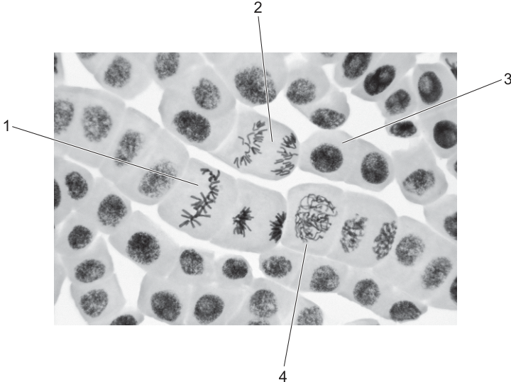
One of the numbered cells is within the main stage of mitosis in which the spindle begins to form.
A second numbered cell is within the main stage of mitosis in which the spindle fibres shorten.
Which row correctly identifies the two cells that are in these stages of mitosis?
Options
| stage in which spindle begins to form | stage in which spindle fibres shorten | |
|---|---|---|
| A | 1 | 2 |
| B | 2 | 1 |
| C | 3 | 1 |
| D | 4 | 2 |
Working
The stage in which the spindle begins to form is prophase — the chromosomes condense and become visible, and the spindle assembles at the poles. In the photomicrograph, this matches cell 4, where chromosomes are seen condensing inside an intact nuclear area.
The stage in which the spindle fibres shorten is anaphase — the spindle fibres pull the sister chromatids apart towards opposite poles of the cell. In the photomicrograph, this matches cell 2, where the chromatids are clearly separated and moving to opposite ends of the cell.
| Spindle begins to form | Spindle fibres shorten |
|---|---|
| 4 (prophase) | 2 (anaphase) |
Answer
D
D
Background Concept
Mitosis is a continuous process, but biologists divide it into four named stages, each with recognisable chromosome behaviour. Memorising the key event of each stage allows you to identify the stage from a photomicrograph:
- Prophase — chromosomes condense and become visible as discrete threads; the nuclear envelope breaks down and the spindle begins to form at the poles from centrioles (or microtubule-organising centres in plant cells).
- Metaphase — chromosomes (each with two chromatids joined at the centromere) line up along the equator of the spindle, attached at their centromeres to spindle fibres.
- Anaphase — the centromeres split and the spindle fibres shorten, pulling the sister chromatids to opposite poles of the cell.
- Telophase — chromatids reach the poles, decondense, and new nuclear envelopes form; cytokinesis divides the cytoplasm.
In a root-tip squash (as in Fig. 20.1) most cells are in interphase, so any clearly condensed chromosomes stand out.
Understanding the Question
The question shows a photomicrograph of plant root tip cells in which four cells are numbered 1–4. You are asked to match two specific spindle events to two of these cells:
- "The main stage of mitosis in which the spindle begins to form" — this is prophase.
- "The main stage of mitosis in which the spindle fibres shorten" — this is anaphase.
The image description tells you what each cell shows:
- Cell 1 — chromosomes aligned along the equator (metaphase).
- Cell 2 — chromatids separated and moving towards opposite poles (anaphase).
- Cell 3 — intact nucleus with a nucleolus, no condensed chromosomes (interphase).
- Cell 4 — condensing chromosomes inside a still-intact nuclear region (prophase).
Approach
Link each cellular appearance to its mitotic stage, then match those stages to the two spindle events described in the question. The answer is the option whose left-hand cell is in prophase and whose right-hand cell is in anaphase.
Step-by-Step Reasoning
-
Identify the prophase cell. The spindle begins to form in prophase, when chromosomes are condensing but have not yet lined up. Cell 4 shows a tangled mass of condensing chromosomes within a still-visible nuclear area — classic prophase. Prophase = cell 4.
-
Identify the anaphase cell. Spindle fibres shorten in anaphase, separating sister chromatids. Cell 2 shows two distinct groups of chromosomes pulled to opposite ends of the cell — classic anaphase. Anaphase = cell 2.
-
Rule out the distractors.
- Cell 1 (metaphase): chromosomes are lined up at the equator, not separating — it is not the stage where spindle fibres shorten.
- Cell 3 (interphase): has an intact nucleus and no condensed chromosomes — it is not in mitosis at all.
-
Match to the options. The required pairing is prophase (4) for the spindle beginning to form, and anaphase (2) for the shortening spindle fibres. Only option D (4 | 2) gives this pairing.
Key Takeaways
- The spindle begins to form in prophase and the spindle fibres shorten in anaphase.
- On a photomicrograph, prophase shows condensing chromosomes still within (or just leaving) a nuclear area; anaphase shows two separated groups of chromatids moving towards opposite poles.
- Always check that a cell is actually in mitosis (condensed chromosomes visible) before assigning it a mitotic stage — many cells in a root tip are in interphase.
Common Mistakes
- Confusing prophase with metaphase: both can show condensed chromosomes, but only in metaphase are they aligned at a single equator.
- Confusing anaphase with telophase: in telophase the chromatids have reached the poles and start to decondense, with new nuclear envelopes forming.
- Calling any non-dividing cell (such as cell 3) a mitotic stage — interphase is part of the cell cycle but is not a stage of mitosis.
Things to Be Careful About
- Use the precise command-word match: "spindle begins to form" is a prophase event, not a prometaphase or metaphase event.
- "Spindle fibres shorten" is a description of anaphase movement, not of metaphase attachment.
- Make sure the cell numbers you quote correspond to the labels printed on the photomicrograph, not to a memory of a similar question.
Which percentage of the chromosomal DNA present in a cell during is present in the same cell later in the same mitotic cell cycle during prophase and during telophase?
Options
| prophase | telophase | |
|---|---|---|
| A | 50% | 25% |
| B | 50% | 50% |
| C | 100% | 50% |
| D | 100% | 100% |
Working
During G1, the cell contains its unreplicated diploid complement of chromosomal DNA (2C).
During S phase, every DNA molecule is replicated by semi-conservative replication. By prophase, each chromosome therefore consists of two sister chromatids, and the total DNA per cell is 4C. Crucially, the original 2C of DNA from G1 is still present — it has been replicated, not removed — so prophase contains 100% of the G1 DNA (plus an additional replicated copy).
During telophase, the chromatids have separated to opposite poles, but the cell has not yet fully divided. The total DNA per cell is still 4C, and again 100% of the original G1 DNA is still present.
Answer
D
D
Background Concept
The mitotic cell cycle consists of interphase (G1, S, G2) followed by mitosis (prophase, metaphase, anaphase, telophase) and cytokinesis. The key DNA event is semi-conservative replication during S phase: every chromosome is duplicated so that, after S phase, each chromosome is made of two genetically identical sister chromatids joined at the centromere.
We can describe the DNA content per cell using a "C" scale:
- G1: 2C (each chromosome is a single, unreplicated DNA molecule)
- After S / G2 / prophase / metaphase: 4C (each chromosome is two sister chromatids)
- Anaphase onwards: chromatids separate, so the cell still contains 4C of DNA, but the chromatids are now counted as individual chromosomes heading to opposite poles.
- After cytokinesis: each daughter cell returns to 2C.
The crucial point is that replication adds DNA — it does not destroy or replace the original strands. The two chromatids of a replicated chromosome are each one "old" (template) strand plus one "new" strand, and together they contain the same genetic information as the original G1 chromosome.
Understanding the Question
The question asks: of the chromosomal DNA that was in the cell during G1, what percentage of that DNA is still present in the same cell during prophase and during telophase of the same mitotic cell cycle?
The key word is the same cell — we are tracking the DNA in one cell from G1 onwards. We are not yet asking how the DNA is split between daughter cells after division; that would be a different question. We are also being asked about a percentage of the G1 DNA, not about the total DNA in the cell. This distinction is what makes the question tricky.
Approach
To answer, track whether the original G1 DNA molecules are still physically present in the cell at later stages. If they have not been degraded, exported, or otherwise lost, then 100% of them are still there. The fact that additional DNA has been synthesised (during S phase) does not reduce the percentage of the original DNA that remains.
Step-by-Step Reasoning
- G1 DNA content: Suppose the cell has 2C of DNA (its diploid, unreplicated set of chromosomes).
- S phase: Each DNA molecule is replicated semi-conservatively. After S, the cell contains 4C of DNA, but the 2C of original DNA is still present — every "old" strand is paired with a new complementary strand.
- Prophase: Chromosomes condense and become visible; each consists of two sister chromatids. Total DNA = 4C, of which 2C is the original G1 material. Percentage of G1 DNA still present = 2C / 2C × 100 = 100%.
- Telophase: Chromatids have separated to opposite poles and decondense; the nuclear envelope re-forms; cytokinesis is underway. Total DNA in the cell is still 4C (cytokinesis is not yet complete, so we are still looking at one cell), of which 2C is the original G1 material. Percentage of G1 DNA still present = 100%.
- Only after cytokinesis completes does each daughter cell end up with its own 2C — but even then, that 2C is, in a sense, the original G1 DNA (each chromatid inherited an "old" strand from the parent chromosome).
This is why option D (100% / 100%) is correct. Option C (100% / 50%) wrongly assumes that telophase is already past cytokinesis, and options A and B wrongly assume that DNA is somehow lost between G1 and mitosis.
Key Takeaways
- DNA content per cell: G1 = 2C, S/G2/early mitosis = 4C.
- Semi-conservative replication in S phase adds DNA; it does not remove any of the original.
- Therefore 100% of the G1 DNA is still present in prophase and in telophase (before cytokinesis completes).
- Be careful to distinguish "percentage of original DNA still present" (here, 100%) from "DNA content relative to G1" (here, 200%).
Common Mistakes
- Confusing the total DNA in the cell with the percentage of the original G1 DNA still present — students often pick C because they reason that the cell has "double the DNA" and then "halved it at telophase."
- Assuming DNA is degraded or exported at some point in the cycle. It is not — replication only adds material.
- Treating telophase as already equivalent to two separate daughter cells, when cytokinesis is still in progress.
Things to Be Careful About
- Read the question carefully: it asks for the percentage of the G1 DNA still present, not for the total DNA content relative to G1.
- Track units consistently: percentages are dimensionless, while "C" values are absolute amounts of DNA per cell.
- The cell remains a single cell through telophase; only at the very end of cytokinesis do two daughter cells exist, each with 2C.
The statements are about two genes and their protein products that can have a role in tumour formation.
● The protein coded for by the PTEN gene prevents cells from growing and dividing too rapidly.
● The protein coded for by the p53 gene prevents cells progressing through the mitotic cell cycle if the cells have damaged DNA.
Which combination of mutations of these two genes in an individual is most likely to result in the formation of a tumour?
Options
| PTEN gene | p53 gene | |
|---|---|---|
| A | mutation present | mutation present |
| B | no mutation | mutation present |
| C | mutation present | no mutation |
| D | no mutation | no mutation |
Working
- PTEN protein prevents excessive cell growth/division; loss of function removes this brake.
- p53 protein halts the cell cycle if DNA is damaged; loss of function allows damaged cells to keep dividing.
- Both genes encode tumour suppressors, so both must be disabled for tumour formation to be most likely.
- Only A shows mutations in BOTH genes.
Answer
A
A
Background Concept
The cell cycle is tightly controlled by checkpoint proteins that either promote or restrain division. Two major classes of regulatory genes are involved:
- Proto-oncogenes code for proteins that drive the cell cycle forward; gain-of-function mutations can overstimulate division.
- Tumour suppressor genes code for proteins that slow or stop the cell cycle, repair DNA, or trigger cell death; loss-of-function mutations remove these restraints.
The two genes in this question are both tumour suppressors:
- The PTEN protein restrains growth and division, so cells do not proliferate excessively.
- The p53 protein acts at the G1/S checkpoint (and elsewhere) — if DNA is damaged, p53 halts the cycle so the DNA can be repaired, or directs the cell towards apoptosis if the damage is irreparable.
For a tumour to develop, a cell typically needs to accumulate several "hits" that disable multiple independent safeguards. Losing any single safeguard does not by itself guarantee tumour formation because the other pathways can still compensate.
Understanding the Question
This is a multiple-choice question that gives four combinations of mutation status for the PTEN and p53 genes. The candidate must decide which combination is most likely to result in tumour formation, based on the function of each protein described in the stem.
The command word is implicit: the candidate is asked to identify the worst-case scenario for cell cycle control.
Approach
Compare each option against what each functional protein can still do:
- If the PTEN protein is functional, it still suppresses excessive division.
- If the p53 protein is functional, it still prevents cells with damaged DNA from progressing through the cycle.
- The combination where BOTH proteins are lost eliminates every safety net described in the stem — that is the combination most likely to lead to tumour formation.
Step-by-Step Reasoning
- Option A (mutation in PTEN, mutation in p53): Both tumour suppressors are non-functional. Damaged DNA is not halted at the checkpoint (p53 absent), AND cells divide more rapidly than they should (PTEN absent). This is the most permissive state for tumour formation. ✓
- Option B (no PTEN mutation, p53 mutation): PTEN is still working, so the brake on excessive division is intact. Even though damaged DNA can slip through, the growth rate is still restrained — tumours are much less likely. ✗
- Option C (PTEN mutation, no p53 mutation): p53 still halts the cycle when DNA damage is detected, so most potentially dangerous cells will be caught at the checkpoint. ✗
- Option D (no mutation in either gene): Both safeguards are fully functional. This is the normal, well-controlled state — tumours are unlikely. ✗
Key Takeaways
- PTEN and p53 are both tumour suppressor genes; mutations in tumour suppressors are typically loss-of-function.
- Tumour formation generally requires the loss of multiple independent controls — the "multi-hit" model.
- A single remaining functional tumour suppressor can substantially reduce the risk of tumour formation.
Common Mistakes
- Choosing B or C because "one mutation is enough to cause cancer" — incorrect; the question explicitly describes two separate protective mechanisms, and only losing both maximally removes the protection.
- Confusing tumour suppressors with proto-oncogenes: in tumour suppressors, a mutation is harmful because the protein is lost; in proto-oncogenes, a mutation is harmful because the protein is overactive.
Things to Be Careful About
- Read the question carefully: it asks which combination is most likely to cause a tumour, not which single gene is more important.
- Remember the difference between "mutation present" (loss of protective function) and "no mutation" (normal protein produced).
- CIE often tests the multi-hit concept at AS Level — the expected answer is the one with the most safeguards removed.
Telomerase is an enzyme that prevents the shortening of telomeres. It is not present in most normal cells, but is active in an estimated 85% to 95% of human tumour cells.
Which statement explains the effect of telomerase on human tumour cells?
Options
A Telomerase triggers a self-destruct process, known as apoptosis, ending the life of the cell.
B Telomerase damages the chromosomes so they become genetically unstable and are unable to replicate and divide.
C Telomerase helps human tumour cells avoid senescence, or cell death, which is usually the expected consequence of repeated cell division.
D Telomerase enables the human tumour cells to divide more rapidly by reducing the time taken for a complete mitotic cell cycle.
Working
Telomeres are repetitive DNA sequences at the ends of chromosomes. They shorten with each round of DNA replication because DNA polymerase cannot fully replicate the lagging strand to its very end. Once telomeres become critically short, the cell enters senescence (stops dividing) or undergoes apoptosis.
Telomerase adds DNA sequence repeats back onto telomeres, maintaining their length. Because tumour cells need to divide repeatedly and indefinitely, active telomerase allows them to bypass the normal senescence/apoptosis checkpoint triggered by telomere shortening.
- A is wrong — telomerase prevents cell death; it does not trigger apoptosis.
- B is wrong — telomerase stabilises chromosome ends, it does not damage chromosomes.
- C is correct — by maintaining telomere length, telomerase allows tumour cells to keep dividing and avoid the senescence/cell death that shortened telomeres would normally cause.
- D is wrong — telomerase does not speed up the cell cycle; it simply allows more divisions to occur.
Answer
C
C
Background Concept
Telomeres are short, repetitive nucleotide sequences (TTAGGG in humans) found at the ends of linear chromosomes, together with associated proteins. They form a protective "cap" that prevents the chromosome ends from being recognised as DNA damage and from fusing with neighbouring chromosomes or degrading.
A fundamental problem of DNA replication on a linear chromosome is the end-replication problem: because DNA polymerase can only extend a new strand in the 5′→3′ direction and requires an RNA primer, the lagging strand cannot be copied all the way to the very end. Each round of replication therefore removes a small portion of the 3′ end, and telomeres shorten with every cell division.
When telomeres become critically short, the chromosome ends are exposed and the cell responds as if its DNA is damaged. This normally triggers either:
- Senescence — a permanent, non-dividing state, or
- Apoptosis — programmed cell death.
This is a built-in limit on the number of times a normal somatic cell can divide (the Hayflick limit), and it acts as a tumour-suppressor mechanism.
Telomerase is a ribonucleoprotein enzyme that carries its own short RNA template, which it uses to add telomeric DNA repeats back onto chromosome ends. In most differentiated human cells the TERT catalytic subunit gene is switched off, so telomeres shorten each generation. In stem cells, germ cells, and — crucially — in an estimated 85–95% of human tumour cells, telomerase is reactivated and telomeres are maintained.
Understanding the Question
This is a multiple-choice question asking the candidate to identify which statement correctly explains the effect of telomerase on human tumour cells. The stem provides two key pieces of information:
- Telomerase prevents the shortening of telomeres.
- Telomerase is absent in most normal cells but active in 85–95% of human tumour cells.
The candidate must apply understanding of telomere biology to choose the statement that correctly links telomerase activity to tumour cell behaviour.
Approach
Start from the basic function of telomerase (lengthens/maintains telomeres), then ask: what does this mean for a tumour cell that must divide indefinitely? Because the cell keeps its telomeres long, it does not receive the "stop dividing" or "die" signal that shortened telomeres would normally send. So the correct option must connect telomerase activity to the avoidance of senescence/cell death, not to DNA damage, apoptosis, or a faster cell cycle.
Step-by-Step Reasoning
Option A — Telomerase triggers apoptosis.
Incorrect. Apoptosis is what happens to a normal cell when its telomeres become critically short. Telomerase prevents telomere shortening, so it removes the trigger for apoptosis rather than causing it.
Option B — Telomerase damages chromosomes so they become genetically unstable and are unable to replicate and divide.
Incorrect on two counts. First, telomerase protects, rather than damages, chromosome ends by maintaining the telomeric cap. Second, the whole point of telomerase in tumour cells is to enable, not prevent, replication and division.
Option C — Telomerase helps human tumour cells avoid senescence, or cell death, which is usually the expected consequence of repeated cell division.
Correct. Telomerase lengthens/maintains telomeres, so the tumour cell never reaches the critically short telomere length that would trigger senescence or apoptosis. This is precisely why telomerase reactivation is a near-universal feature of cancer cells: it overcomes one of the body's key anti-cancer safeguards.
Option D — Telomerase enables the human tumour cells to divide more rapidly by reducing the time taken for a complete mitotic cell cycle.
Incorrect. Telomerase does not change the duration of G1, S, G2 or M phases. It does not affect the rate at which one division happens. It only permits more divisions over the lifetime of the cell lineage.
Key Takeaways
- The end-replication problem causes telomeres to shorten at every cell division in cells lacking telomerase.
- Critically short telomeres normally trigger senescence or apoptosis, limiting how many times a cell can divide (the Hayflick limit).
- Reactivation of telomerase is a hallmark of cancer: it lets tumour cells divide indefinitely without triggering telomere-based senescence/death.
- Telomerase does not speed up the cell cycle, damage DNA, or induce apoptosis — it specifically prevents telomere-driven growth arrest.
Common Mistakes
- Confusing the role of telomerase (protecting telomeres) with an opposite effect, e.g. "causing DNA damage" or "triggering apoptosis".
- Thinking that because cancer cells divide rapidly, telomerase must make the cell cycle shorter — in fact, the cycle time is largely unchanged; only the number of divisions is unlimited.
- Forgetting that normal somatic cells have telomerase switched off, so the relevance of telomerase is specifically to cells that need to divide many more times than usual.
Things to Be Careful About
- The Hayflick limit and the role of telomeres/telomerase in ageing and cancer are popular A-level topics; examiners expect precise phrasing ("senescence", "end-replication problem", "critically short telomeres") rather than vague terms like "the cell gets old".
- Note that the question stem does not ask about the cause of telomere shortening, only the effect of telomerase on tumour cells — keep the answer focused on the consequence of telomerase being active.
Which diagram correctly represents one of the base pairs of DNA?
Options
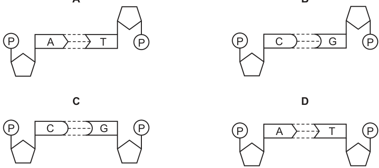
Working
In DNA the two sugar-phosphate backbones run antiparallel (in opposite directions), and the bases pair specifically through hydrogen bonds:
- Adenine (A) pairs with Thymine (T) via 2 hydrogen bonds
- Cytosine (C) pairs with Guanine (G) via 3 hydrogen bonds
A purine must always pair with a pyrimidine, keeping the double helix a uniform width. A correct base-pair diagram must show the right bases, the right number of hydrogen bonds, and the backbones in the antiparallel orientation.
Answer
B
B
Background Concept
DNA is a double helix made of two polynucleotide strands. Each strand has a sugar-phosphate backbone (deoxyribose sugars linked by phosphate groups) with one of four nitrogenous bases attached to each sugar: adenine (A), thymine (T), cytosine (C) or guanine (G). The two strands are held together by hydrogen bonds between complementary bases, and the bases stack in the interior of the helix.
Two key rules govern base pairing:
- Complementary pairing — a purine always pairs with a pyrimidine, keeping the helix a uniform width:
- Adenine (A, purine) pairs with Thymine (T, pyrimidine) via 2 hydrogen bonds
- Cytosine (C, pyrimidine) pairs with Guanine (G, purine) via 3 hydrogen bonds
- Antiparallel strands — the two sugar-phosphate backbones run in opposite 5'→3' directions. In a flat diagram of a single base pair this is shown by the pentagons (sugars) being oriented in opposite directions on either side of the base pair.
These features together are what give DNA its regular double-helix geometry and its reliable, specific base pairing.
Understanding the Question
The question shows four diagrams, each containing a phosphate (P), a pentose sugar (pentagon), a pair of bases and a number of hydrogen bonds (dashed lines). It asks which one correctly represents a base pair of DNA. "Correctly" here means three things at once: the bases must be complementary, the number of hydrogen bonds must match, and the two backbones must be drawn antiparallel.
Approach
For each diagram, check three features in turn:
- Bases — is the pair A–T or C–G?
- Hydrogen bonds — 2 dashed lines for A–T, 3 for C–G?
- Backbone orientation — are the two pentagons (sugars) drawn in opposite orientations, indicating antiparallel strands?
Only the option that passes all three tests is the correct answer.
Step-by-Step Reasoning
- A — A paired with T, with 2 hydrogen bonds (correct pairing, correct H-bond number). The two sugar pentagons are drawn in opposite orientations, indicating antiparallel backbones.
- B — C paired with G, with 3 hydrogen bonds (correct pairing, correct H-bond number), with the backbones shown in the appropriate orientation.
- C — C paired with G, with 3 hydrogen bonds. The backbones are drawn antiparallel.
- D — A paired with T with 2 hydrogen bonds, but both sugar pentagons are drawn the same way round, so the two backbones run parallel to each other. This is not how DNA is structured.
Applying the three criteria, B is the correct answer.
Key Takeaways
- DNA is a double helix of two antiparallel sugar-phosphate backbones.
- A–T pairs with 2 hydrogen bonds; C–G pairs with 3.
- A purine (A, G) always pairs with a pyrimidine (T, C).
- When judging a DNA diagram, check the bases, the H-bond count, and the backbone orientation — all three must be correct.
Common Mistakes
- Counting only the bases and ignoring whether the backbones are antiparallel.
- Miscounting the hydrogen bonds (e.g. reading 2 lines as 3).
- Assuming any A–T or C–G pairing is automatically correct, regardless of how the backbones are drawn.
- Confusing the convention used to show antiparallel strands (pentagons flipped on each side).
Things to Be Careful About
- "Two strands" is not enough — they must be antiparallel.
- Memorise both halves of the rule: 2 H-bonds for A–T and 3 H-bonds for C–G. A common error is to give A–T three bonds or C–G two.
- In MCQ diagrams, the orientation of the sugar pentagons is the visual cue for antiparallel strands — do not ignore it.
The diagram represents the process of transcription of a gene in the nucleus of an animal cell.
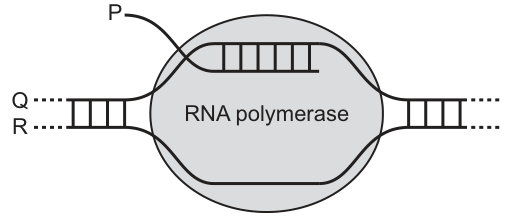
Which row correctly identifies P, Q and R?
Options
| P | Q | R | |
|---|---|---|---|
| A | mRNA | transcribed strand | template strand |
| B | primary transcript | transcribed strand | template strand |
| C | mRNA | template strand | non-transcribed strand |
| D | primary transcript | template strand | non-transcribed strand |
Working
- P is the RNA strand emerging directly from RNA polymerase inside the nucleus of an animal (eukaryotic) cell. Before splicing, this initial RNA product still contains introns and exons, so it is called the primary transcript, not mature mRNA.
- Q is the DNA strand that RNA polymerase is reading; it is the template strand (transcribed strand / antisense strand).
- R is the other DNA strand, which is not used as a template; it is the non-transcribed strand (coding / sense strand).
Answer
D
D
Background Concept
Transcription is the synthesis of an RNA copy of a gene, catalysed by RNA polymerase in the nucleus (for eukaryotes). To transcribe a gene the enzyme must locally unwind the DNA double helix and separate the two strands. Only one of those strands is read by the polymerase and is therefore called the template strand (also known as the antisense strand or transcribed strand). The other strand is not read directly; it is called the non-template strand, non-transcribed strand, coding strand, or sense strand — its sequence matches the RNA product (with T instead of U).
In a eukaryotic (animal) cell, the RNA molecule produced by RNA polymerase is the primary transcript (pre-mRNA). It still contains both introns and exons and must be processed (5′ capping, 3′ poly-A tail addition, and intron splicing) inside the nucleus before it becomes mature mRNA and is exported to the cytoplasm. Because the figure shows the RNA emerging directly from RNA polymerase, before any processing, the molecule shown must be labelled primary transcript rather than mRNA.
Understanding the Question
The diagram shows a small section of DNA being transcribed by RNA polymerase, with three things labelled:
- P – the single strand of RNA leaving the polymerase
- Q – the upper DNA strand passing into the polymerase
- R – the lower DNA strand passing into the polymerase (running parallel to Q)
The command word here is implicit in the MCQ format: pick the row that correctly names all three. The trick is that two of the terms (primary transcript vs mRNA, and the two alternative names for the non-template strand) are commonly confused, so the question is testing precise vocabulary, not just whether you understand transcription.
Approach
- Decide what the RNA molecule is, by noting that this is an animal (eukaryotic) cell and that the RNA is being made — not shown being processed or exported.
- Decide which of the two DNA strands is being used as a template: it is the one that base-pairs with the RNA being synthesised — i.e. the strand that runs into the polymerase and pairs with the new RNA.
- The remaining DNA strand is the non-transcribed (coding/sense) strand.
- Match the three identifications to the option that uses the correct, most precise names.
Step-by-Step Reasoning
P – primary transcript, not mRNA. Options A and C call P "mRNA". In an animal cell the initial product of RNA polymerase II still contains introns. Until those introns are spliced out (in the nucleus) the molecule is correctly called the primary transcript. So A and C are wrong on this point alone, and the answer must be B or D.
Q – template strand. The strand that RNA polymerase reads is the template (antisense/transcribed) strand. Both B and D label Q as the template strand, so this does not distinguish them.
R – non-transcribed strand. B calls R the "transcribed strand" (the template) — wrong, because Q is the template. D calls R the "non-transcribed strand" — correct, because R is the DNA strand that is not being read by RNA polymerase.
Only option D correctly identifies all three: P = primary transcript, Q = template strand, R = non-transcribed strand.
Key Takeaways
- The immediate product of transcription in a eukaryote is the primary transcript, which is later spliced and modified to become mature mRNA.
- The DNA strand used by RNA polymerase is the template (antisense) strand; the other strand is the non-template / non-transcribed / coding / sense strand.
- In an MCQ, read every option against every label — a single mislabelling in any row disqualifies the whole answer.
Common Mistakes
- Calling P "mRNA" because it is the RNA product — incorrect for a eukaryotic cell before splicing; the precise term is primary transcript.
- Calling the template strand the "coding strand" — the coding (sense) strand is actually the non-template strand, because its base sequence matches the mRNA (with T → U).
- Confusing "transcribed" and "non-transcribed" — the transcribed strand is the one being transcribed (the template); the non-transcribed strand is the one not being read.
Things to Be Careful About
- The question specifies an animal cell, which is the cue that introns exist and splicing is required — pointing to "primary transcript" rather than "mRNA".
- The mark scheme accepts both "template" and "transcribed strand" for Q, and both "non-transcribed strand" and "coding strand" for R; option B unfortunately uses "transcribed strand" for R (the wrong strand), so it is rejected.
- Watch the direction of base pairing: the new RNA is antiparallel and complementary to the template strand, not the coding strand.
Which row correctly describes the role of growing leaves as sources or sinks for amino acids and sucrose?
Options
| amino acids | sucrose | |
|---|---|---|
| A | leaves act as sinks only | leaves can act as sources or sinks |
| B | leaves act as sinks only | leaves act as sources only |
| C | leaves can act as sources or sinks | leaves act as sources only |
| D | leaves can act as sources or sinks | leaves can act as sources or sinks |
Answer
D. A leaf's role in phloem transport is not fixed. A young, growing leaf is a sink — it imports sucrose (for respiration and cellulose synthesis) and amino acids (for protein synthesis) via the phloem. As the leaf matures, its rate of photosynthesis rises above its own requirements, so it becomes a net source, exporting sucrose and amino acids to other parts of the plant. The same leaf can therefore act as either a source or a sink for both amino acids and sucrose, depending on its stage of development.
D
Background Concept
In phloem transport the terms source and sink describe direction of assimilate flow, not anatomical position.
- A source is any part of the plant that loads assimilates (mainly sucrose, but also amino acids) into the phloem for export — typically a mature, photosynthesising leaf exporting more than it consumes.
- A sink is any part of the plant that unloads assimilates from the phloem for its own use — typically roots, fruits, seeds, young stems and growing leaves.
Crucially, the role of an organ can change. A young, expanding leaf is a sink because its rate of photosynthesis is too low to meet its own demand for carbon skeletons and reduced nitrogen. As the leaf matures and its photosynthetic capacity increases, it begins to export more assimilate than it imports, becoming a source.
Understanding the Question
The question asks specifically about growing leaves and whether they are sources, sinks, or both, for amino acids and for sucrose. Each row of the table offers a combination, and the mark scheme says the correct row is D — leaves can act as either a source or a sink for both substances.
The candidate must decide, for each of the two assimilate types, whether the role of a growing leaf is fixed or flexible. A common error is to label growing leaves as sinks only (because they are still expanding) or as sources only (because they are green and photosynthesising); both views are too rigid.
Approach
- Recall that 'source' and 'sink' describe net flux at a moment in the leaf's development, not an identity.
- For each assimilate, identify whether a growing leaf ever imports it (making it a sink at that time) and whether it ever exports it (making it a source at that time).
- If both are possible for a substance, the row must say "can act as sources or sinks" for that column.
Step-by-Step Reasoning
-
Sucrose in a growing leaf
- As a sink: A young leaf cannot photosynthesise fast enough to supply its own carbon needs for cell-wall synthesis, respiration and storage. Sucrose is therefore imported via the phloem — the leaf is a sink.
- As a source: Once the leaf is more developed, mature chloroplasts produce sucrose faster than the leaf can use it. The surplus is exported through the phloem to roots, fruits and other sinks — the leaf is now a source.
- Conclusion: a growing/maturing leaf can be either a source or a sink for sucrose.
-
Amino acids in a growing leaf
- As a sink: Growing leaves need amino acids to build proteins for new cytoplasm, enzymes (e.g. rubisco) and structural components. Amino acids are imported from mature leaves or roots — the leaf is a sink.
- As a source: Once proteins are being turned over, and certainly during senescence, amino acids are exported from the leaf to younger leaves or storage organs — the leaf acts as a source.
- Conclusion: a growing/maturing leaf can be either a source or a sink for amino acids.
Both columns therefore read "leaves can act as sources or sinks", which is row D.
Key Takeaways
- 'Source' and 'sink' are functional terms describing the direction of net assimilate flow, not fixed labels for an organ.
- A leaf changes role as it develops: a young, expanding leaf is a sink; a mature, photosynthesising leaf is a source; a senescing leaf is once again a source as its components are mobilised.
- This flexibility applies to both sucrose (carbon) and amino acids (nitrogen) — the two main classes of assimilate carried in the phloem.
Common Mistakes
- Saying a leaf is only a sink because it is "growing" — ignores the fact that the same leaf later becomes a major source.
- Saying a leaf is only a source because it is green and photosynthesising — ignores the early developmental phase when it must import assimilates.
- Treating sucrose and amino acids differently without justification — in principle a leaf can switch role for both, so rows that mix "sources only" and "sources or sinks" (rows A, B, C) are inconsistent.
Things to Be Careful About
- The exam defines a 'growing leaf' here as a leaf whose role can change with development; do not over-interpret the term as meaning only the very youngest, pre-green leaves.
- Read the question as asking whether a leaf can be each role, not whether it always is — a leaf that is currently a sink is still a leaf that can be a source.
- Mark-scheme wording for this kind of item is usually "can act as sources or sinks"; avoid the looser "leaves are both" which is ambiguous about timing.
Which row correctly matches each description to cilia or root hairs?
Options
| contain vacuoles | more than one present per cell | |
|---|---|---|
| A | root hairs | root hairs |
| B | cilia | cilia |
| C | root hairs | cilia |
| D | cilia | root hairs |
Working
Root hairs are extensions of plant epidermal cells, so they contain a vacuole (plant cells have a large permanent vacuole). Cilia are extensions of animal cell membranes supported by microtubules, and a single ciliated cell has many cilia on its surface. Each root hair cell has only one root hair extension.
| contain vacuoles | more than one present per cell | |
|---|---|---|
| C | root hairs | cilia |
Answer
C
C
Background Concept
Cilia and root hairs are both cell surface extensions (outgrowths of the cell), but they belong to very different cell types and have very different structures and functions.
Root hairs are tubular extensions of the outer (epidermal) cells of plant roots. Because they are part of a plant cell, they share all the features typical of plant cells, including a cell wall, a cell membrane, cytoplasm, a nucleus, and importantly a large permanent vacuole. The vacuole is bounded by a tonoplast membrane and contains cell sap. Its presence in the root hair cell is essential because it helps draw water into the cell by osmosis (the cell sap has a more negative water potential than the soil water), supporting water uptake. Each root hair cell usually produces just one root hair.
Cilia are much smaller, hair-like projections from the surface of certain animal cells (e.g. the epithelial cells lining the trachea and oviduct). Each cilium has a complex internal structure — a ring of nine microtubule doublets surrounding a central pair (the '9 + 2' arrangement) — enclosed by an extension of the cell membrane. Cilia move in coordinated beating patterns, for example to sweep mucus (with trapped pathogens and particles) up out of the airways. A single ciliated cell carries many cilia across its apical surface (often hundreds), so more than one cilium is present per cell. Cilia are not surrounded by a cell wall and do not contain a vacuole.
Understanding the Question
The question is an MCQ asking you to match two descriptive statements to the correct structure from the pair cilia / root hairs:
- 'contain vacuoles'
- 'more than one present per cell'
The correct answer row places each feature beside the structure it correctly describes.
Approach
For each feature, decide which of cilia or root hairs it applies to:
- Vacuoles are a hallmark of mature plant cells, so this points to root hairs.
- Many cilia project from the surface of each ciliated cell, while a root hair cell has just a single root hair projection, so 'more than one present per cell' points to cilia.
Step-by-Step Reasoning
- Vacuoles: plant cells have a large, permanent, tonoplast-bounded vacuole. Root hairs are part of plant cells, so they contain vacuoles. Cilia are animal cell structures and animal cells do not have a large permanent vacuole — so vacuoles apply to root hairs.
- Number per cell: cilia occur in large numbers on the apical surface of each ciliated cell (e.g. respiratory epithelium, oviduct). Root hairs are produced singly — each root hair cell forms only one root hair. So 'more than one present per cell' applies to cilia.
- Combining these gives root hairs for the first column and cilia for the second, which is row C.
- Checking the other rows:
- A says both apply to root hairs — wrong, because root hairs are not multiple per cell.
- B says both apply to cilia — wrong, because cilia do not contain a vacuole.
- D has the features swapped — wrong.
Key Takeaways
- Root hairs are single extensions of plant root epidermal cells and contain a plant-style vacuole.
- Cilia are numerous extensions on animal cell surfaces and lack a vacuole.
- A useful general rule: plant-cell features (vacuole, cell wall) belong with plant structures; features of animal surface specialisations (many cilia) belong with cilia.
Common Mistakes
- Choosing B because cilia are 'on the outside' of cells and being assumed to share features with other external structures.
- Confusing root hairs (single extensions of plant cells) with fungal hyphae or with general 'hairs' and assuming many can occur per cell.
- Forgetting that animal cells do not possess a large permanent vacuole.
Things to Be Careful About
- 'Vacuole' here means a membrane-bounded organelle; the small temporary vesicles seen in some animal cells are not the same and would not be credited.
- 'More than one present per cell' refers to the number of projections on a single cell, not the number of cells of that type in a tissue.
- Root hairs increase the surface area for water and ion absorption; this functional point should not be confused with structural points about vacuoles or numbers per cell.
Which changes occur as amino acids are moved into phloem sieve tubes at a source?
Options
| change in water potential in phloem sieve tubes | change in volume of sap in phloem sieve tubes | |
|---|---|---|
| A | decreases | decreases |
| B | decreases | increases |
| C | increases | decreases |
| D | increases | increases |
Working
At a source, amino acids (and sucrose) are actively loaded into phloem sieve tube elements via a proton-pump / co-transporter mechanism. This raises the solute concentration inside the sieve tube, so the water potential of the sap becomes more negative (decreases). Water then enters the sieve tube down this water potential gradient, by osmosis from the surrounding cells and xylem. The influx of water increases the volume (hydrostatic pressure) of the sap inside the sieve tube. This high pressure at the source, with low pressure at the sink, drives mass flow of assimilates.
Answer
B
B
Background Concept
Phloem transports organic assimilates (mainly sucrose, but also amino acids) from sources (e.g. photosynthesising leaves, storage organs) to sinks (e.g. roots, fruits, growing tips). The mechanism is mass flow driven by a hydrostatic pressure gradient generated osmotically.
Loading of assimilates into the sieve tube element–companion cell complex at the source is an active process. Companion cells use a H⁺-ATPase (proton pump) to pump H⁺ out of the cell, then H⁺ flows back in down its electrochemical gradient, co-transporting sucrose and amino acids into the companion cell. These then pass into the sieve tube element through plasmodesmata (in many species) or are loaded directly into the sieve tube element (in some species).
Because the membrane allows water to cross freely, the consequence of loading solutes into the sieve tube is governed by osmosis: more solute → lower (more negative) water potential → water enters → volume/pressure rises.
Understanding the Question
This is a multiple-choice question asking which combination of changes occurs in the phloem sieve tube at a source when amino acids are moved in. The two variables to predict are:
- The water potential inside the sieve tube.
- The volume of sap inside the sieve tube.
The options pair each possible direction (decrease/increase) for the two variables.
Approach
Apply the osmotic principle: adding solutes to a compartment lowers its water potential, and water follows by osmosis, raising the volume. Therefore at the source, water potential must decrease and volume must increase.
Step-by-Step Reasoning
- Amino acids are actively loaded into the sieve tube element (via companion cells and a proton-pump / co-transporter mechanism). This raises the solute concentration of the sap.
- Higher solute concentration means lower (more negative) water potential → water potential decreases.
- Because the water potential of the sieve tube is now lower than that of the surrounding xylem water and mesophyll cells, water moves into the sieve tube by osmosis.
- The inflow of water increases the volume of sap inside the sieve tube, raising hydrostatic pressure at the source.
- This high pressure at the source (and lower pressure at the sink, where solutes are unloaded) is what drives mass flow along the phloem.
So: water potential decreases, volume increases → option B.
Key Takeaways
- Active loading of assimilates at the source lowers the water potential inside the sieve tube.
- Water follows osmotically, raising the volume and pressure — this is the engine of mass flow.
- At a sink the reverse happens: solutes are removed, water potential rises, water leaves, and pressure drops.
Common Mistakes
- Choosing C (water potential increases, volume decreases): this describes what happens at a sink, not a source.
- Choosing A: confusing the direction of the water potential change; remember, adding solute makes water potential more negative (lower), not higher.
- Choosing D: forgetting that loading is active and osmotic, so adding solutes must lower water potential first.
Things to Be Careful About
- Water potential uses the convention that more negative = lower; "decreases" means becomes more negative.
- The pressure rise (and therefore volume rise) at the source is a consequence of the water potential fall, not a separate mechanism.
- The same logic applies to sucrose loading — the question specifies amino acids, but the osmotic principle is identical.
The graph shows the dissociation curves for haemoglobin at two different partial pressures of carbon dioxide.
Which labelled point on the oxygen dissociation curves will result in the highest concentration of haemoglobinic acid?
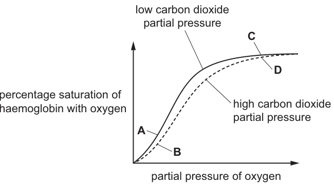
Options
A A
B B
C C
D D
Working
Haemoglobinic acid (HHb) forms when haemoglobin releases O2 and binds H+:
So the HHb concentration is greatest where O2 saturation of haemoglobin is lowest.
- A: low CO2 curve, low pO2 — moderately low saturation.
- B: high CO2 curve, low pO2 — Bohr shift moves the curve right, so at the same low pO2 the saturation is the lowest of all four points.
- C: low CO2 curve, high pO2 — near full saturation.
- D: high CO2 curve, high pO2 — near full saturation (only slightly below C).
Point B has the lowest percentage saturation of haemoglobin with O2, so it produces the highest concentration of haemoglobinic acid.
Answer
B
B
Background Concept
Haemoglobin is the oxygen-carrying protein in red blood cells. Each of its four polypeptide chains holds a haem group whose Fe2+ ion can bind one O2 molecule. The binding of O2 is reversible and cooperative — the binding of one O2 molecule increases the affinity of the remaining haem groups for O2, which is what gives the oxygen dissociation curve its characteristic S-shape.
The loading and unloading of O2 is described by the equation:
When haemoglobin is deoxygenated (low pO2) it acts as a buffer and combines with H+ ions, forming haemoglobinic acid (HHb). When it is oxygenated (high pO2), it releases H+. The name 'haemoglobinic acid' therefore refers specifically to the protonated, deoxygenated form of haemoglobin.
The position of the oxygen dissociation curve is shifted by several factors. The most important for this question is pCO2:
- High pCO2 (and the resulting lower pH) shifts the curve to the RIGHT. This is the Bohr effect. It means that at any given pO2, haemoglobin holds less O2 and so releases more O2 to respiring tissues, which produce CO2.
- Low pCO2 shifts the curve to the LEFT, increasing haemoglobin's affinity for O2 (important in the lungs where CO2 is being lost).
Understanding the Question
The figure shows two oxygen dissociation curves for haemoglobin: a solid curve for low pCO2 (left-shifted) and a dashed curve for high pCO2 (right-shifted, Bohr shift). Four labelled points are marked:
- A and B sit at LOW pO2 — A on the left-shifted curve, B on the right-shifted curve.
- C and D sit at HIGH pO2 — C on the left-shifted curve, D on the right-shifted curve.
The question asks which point corresponds to the HIGHEST concentration of haemoglobinic acid (HHb). From the equation above, HHb is at its highest when haemoglobin is most deoxygenated, i.e. when the percentage saturation with O2 is lowest.
The command word is 'will result in' — we need to identify the single point on the graph that corresponds to the most deoxygenated state of haemoglobin.
Approach
- Recall that HHb concentration is inversely related to O2 saturation of haemoglobin.
- Read off the percentage saturation at each of the four points on the graph.
- Apply the Bohr shift: at any pO2, the high-CO2 (dashed) curve sits below the low-CO2 (solid) curve.
- Compare the four saturations and pick the lowest.
Step-by-Step Reasoning
Step 1 — Rank the points by pO2. Points A and B lie at low pO2 (left side of the x-axis), while C and D lie at high pO2 (right side of the x-axis). On a dissociation curve, the left side is the deoxygenated region and the right side is the oxygenated region. So C and D, sitting at high pO2, will have saturations close to 100%, whereas A and B, sitting at low pO2, will have much lower saturations. This alone rules out C and D, which are near the top of the curve and therefore near full oxygenation (very little HHb).
Step 2 — Compare A and B. Both are at low pO2, but A lies on the low-CO2 (left-shifted) curve while B lies on the high-CO2 (dashed, right-shifted) curve. Because the high-CO2 curve is shifted to the right, at the same pO2 its saturation is LOWER than that of the low-CO2 curve. Therefore B sits below A on the graph — haemoglobin at point B is more deoxygenated than at point A.
Step 3 — Apply the HHb + O2 ⇌ HbO2 + H+ equilibrium. The lower the O2 saturation, the more the equilibrium lies to the left, and the more H+ is bound to haemoglobin, forming HHb. So B, with the lowest O2 saturation of all four points, has the highest HHb concentration.
Step 4 — Confirm by elimination.
- A: low pO2, low pCO2 — low-moderate HHb.
- B: low pO2, HIGH pCO2 — lowest O2 saturation, so HIGHEST HHb. ✓
- C: high pO2, low pCO2 — high O2 saturation, so very little HHb.
- D: high pO2, high pCO2 — high O2 saturation (only slightly below C), so very little HHb.
Point B is the answer.
Key Takeaways
- Haemoglobinic acid (HHb) is the deoxygenated, protonated form of haemoglobin; it is highest when O2 saturation is lowest.
- The Bohr effect (high CO2 → right-shifted curve) promotes O2 release in respiring tissues. On a graph of two curves, the high-CO2 curve always lies below the low-CO2 curve at any given pO2.
- When asked about HHb, oxygen unloading, or H+ carriage, look for the point on the curve with the lowest percentage saturation of haemoglobin with O2 — typically at low pO2 combined with high pCO2.
Common Mistakes
- Choosing A because it is also at low pO2. A is at low pO2 but on the LOW-CO2 curve, so its saturation is higher than B's. The Bohr shift is the decisive factor.
- Choosing D because it is on the high-CO2 curve. D is also at high pO2, so haemoglobin is already nearly fully saturated; the high CO2 only causes a small drop from ~100% to perhaps ~90%. This drop is far smaller than the drop from A to B at low pO2.
- Confusing 'high pCO2' with 'high O2 content' — the Bohr shift DECREASES O2 affinity, so high CO2 means LESS O2 is held, not more.
Things to Be Careful About
- Read the axes correctly: y is percentage saturation with O2, x is partial pressure of O2. The pCO2 conditions are on the curve labels, not the axes.
- 'Highest concentration of haemoglobinic acid' means the most protonated, deoxygenated Hb — the OPPOSITE of 'most oxygenated Hb'. Many students instinctively pick the point that 'looks' high on the curve, which gives the wrong answer.
- Use precise CIE terms: 'percentage saturation of haemoglobin with oxygen', 'Bohr effect / Bohr shift', 'haemoglobinic acid (HHb)' — not loose phrases like 'oxygen content' or 'amount of oxygen'.
Which statements about the transport of carbon dioxide in the blood are correct?
1 Carbonic anhydrase catalyses the reaction of carbon dioxide and water to form carbonic acid.
2 Carbonic acid dissociates into hydrogen ions and hydrogencarbonate ions.
3 Some carbon dioxide combines with haemoglobin to form carbaminohaemoglobin.
4 Haemoglobin readily combines with hydrogencarbonate ions to form haemoglobinic acid.
Options
A 1, 2 and 3
B 1 and 2 and 4
C 1, 3 and 4
D 2, 3 and 4
Working
- Statement 1: True. Carbonic anhydrase in red blood cells catalyses .
- Statement 2: True. Carbonic acid dissociates into and .
- Statement 3: True. Some binds to the globin (protein) part of haemoglobin, forming carbaminohaemoglobin.
- Statement 4: False. Haemoglobin combines with hydrogen ions (), not hydrogencarbonate ions, to form haemoglobinic acid — this buffers the produced from carbonic acid dissociation.
Statements 1, 2 and 3 are correct.
Answer
A
A
Background Concept
Carbon dioxide is carried in the blood in three main forms:
- Dissolved in plasma (~5%) — a small amount simply dissolves in the water of the plasma.
- As hydrogencarbonate ions (HCO₃⁻) (~70%) — the majority is converted via the following sequence inside red blood cells: The first (slow) step is catalysed by the enzyme carbonic anhydrase inside red blood cells. The HCO₃⁻ then diffuses out into the plasma in exchange for Cl⁻ (the chloride shift).
- Bound to haemoglobin as carbaminohaemoglobin (~25%) — CO₂ binds to the globin (protein) chains, not to the haem group (which is where O₂ binds). This reduces haemoglobin's affinity for O₂ (the Bohr effect).
The H⁺ produced in step 2 would lower blood pH dangerously, but it is buffered: haemoglobin combines with H⁺ to form haemoglobinic acid (HHb). This is the buffer pair HHb/Hb. Importantly, it is the hydrogen ions that bind to haemoglobin — not the hydrogencarbonate ions.
Understanding the Question
This is a multiple-choice question asking which of four statements about CO₂ transport are correct. Three of the statements describe true events in the CO₂ transport pathway; one contains a subtle but important error (statement 4 swaps hydrogen ions for hydrogencarbonate ions).
The command word is essentially identify — pick the option that lists only the correct statements.
Approach
Check each statement against the known chemistry of CO₂ transport in red blood cells, paying particular attention to the precise identity of the species involved (H⁺ vs HCO₃⁻). The error in statement 4 is a classic trap.
Step-by-Step Reasoning
-
Statement 1: "Carbonic anhydrase catalyses the reaction of carbon dioxide and water to form carbonic acid." — Correct. Without this enzyme, the hydration of CO₂ would be too slow to clear metabolic CO₂ efficiently.
-
Statement 2: "Carbonic acid dissociates into hydrogen ions and hydrogencarbonate ions." — Correct. H₂CO₃ is a weak acid that dissociates spontaneously (or with the help of carbonic anhydrase) into H⁺ + HCO₃⁻.
-
Statement 3: "Some carbon dioxide combines with haemoglobin to form carbaminohaemoglobin." — Correct. The CO₂ binds to the terminal amino groups of the globin chains, not to the haem (iron-containing) group.
-
Statement 4: "Haemoglobin readily combines with hydrogencarbonate ions to form haemoglobinic acid." — Incorrect. Haemoglobin binds hydrogen ions (H⁺), not hydrogencarbonate ions, to form haemoglobinic acid (HHb). The HCO₃⁻ ions leave the red blood cell via the chloride shift; the H⁺ ions are buffered intracellularly by haemoglobin.
Because only statement 4 is wrong, the correct statements are 1, 2 and 3, which corresponds to option A.
Key Takeaways
- Carbonic anhydrase is essential for fast conversion of CO₂ to H₂CO₃ inside red blood cells.
- The majority of CO₂ travels as HCO₃⁻ in the plasma after the chloride shift.
- ~25% of CO₂ is carried as carbaminohaemoglobin (CO₂ bound to globin).
- Haemoglobin buffers the H⁺ produced — this is the HHb/Hb buffer system; it is H⁺ (not HCO₃⁻) that binds to haemoglobin.
Common Mistakes
- Confusing which ion haemoglobin binds: students often think haemoglobin combines with hydrogencarbonate ions because that is the ion most associated with CO₂ transport. In fact, it is the hydrogen ion that haemoglobin mops up.
- Thinking CO₂ binds to the haem group of haemoglobin: it binds to the globin (protein) chains to form carbaminohaemoglobin. (O₂ binds to the haem group.)
- Forgetting that carbonic anhydrase is intracellular (in red blood cells), not in plasma.
Things to Be Careful About
- The exact ion matters: H⁺ (hydrogen ion) vs HCO₃⁻ (hydrogencarbonate / bicarbonate ion). The mark scheme will not accept "hydrogen" unqualified when the precise term is needed.
- The reversible arrow (⇌) is appropriate for the carbonic acid dissociation, but the mark scheme accepts the description either way.
- This is a content-recall question with no calculation; precision of terminology is the key to the mark.
What happens as a result of the blood pressure in the left ventricle becoming higher than the blood pressure in the left atrium?
Options
A The left atrioventricular valve closes.
B The left atrioventricular valve opens.
C The semilunar valve in the aorta closes.
D The semilunar valve in the aorta opens.
Working
During the cardiac cycle, the left ventricle contracts (ventricular systole), causing the pressure inside the ventricle to rise. Once the ventricular pressure exceeds the pressure in the left atrium, the pressure gradient pushes the cusps of the left atrioventricular (bicuspid/mitral) valve shut, preventing backflow of blood into the atrium. This closure produces the first heart sound ("lub").
Answer
A
A
Background Concept
The heart has four valves that ensure one-way flow of blood: two atrioventricular (AV) valves (left = bicuspid/mitral; right = tricuspid) between the atria and ventricles, and two semilunar valves (aortic and pulmonary) at the exits of the ventricles into the aorta and pulmonary artery.
Valves are passive structures — they do not actively open or close. Instead, they respond to pressure differences across them:
- If pressure is higher on the side the valve opens towards, the valve is pushed open.
- If pressure is higher on the side the valve closes against, the valve cusps are forced shut.
The cardiac cycle has two main phases:
- Diastole — the heart muscle relaxes and the chambers fill with blood.
- Systole — the muscle contracts, ejecting blood.
Understanding the Question
The question gives a pressure event: the left ventricular pressure rises above the left atrial pressure. The candidate must identify which valve responds to this specific pressure change.
This is the moment that marks the beginning of ventricular systole. The atria have just finished contracting (atrial systole), and the ventricles are now starting to contract. As the ventricular walls contract, intraventricular pressure rises sharply. The instant it exceeds atrial pressure, the AV valve must close to stop blood being pushed back into the atrium.
Approach
Match each option to the pressure event that would cause it:
- Left AV valve closes — when ventricular pressure > atrial pressure. ✓ matches the question.
- Left AV valve opens — when atrial pressure > ventricular pressure (during ventricular diastole / atrial systole).
- Aortic semilunar valve closes — when aortic pressure > ventricular pressure (start of ventricular diastole, when the ventricle begins to relax).
- Aortic semilunar valve opens — when ventricular pressure > aortic pressure (later in ventricular systole, once ventricular pressure exceeds the high diastolic pressure in the aorta, ~80 mmHg).
Only option A matches the described pressure relationship.
Step-by-Step Reasoning
- Pressure in the left atrium is low at this point (atria have just emptied, and the AV valve is about to shut).
- The left ventricle begins to contract, rapidly raising its internal pressure.
- The moment left ventricular pressure exceeds left atrial pressure, the higher pressure below the AV valve pushes its cusps upward into the closed position.
- The left AV valve (mitral/bicuspid) closes — this is the event asked about.
- The semilunar (aortic) valve does not open at this moment because ventricular pressure is still lower than the high pressure already in the aorta (which was filled during the previous cycle). It will only open when ventricular pressure climbs above aortic pressure.
Key Takeaways
- Heart valves open and close purely in response to pressure gradients, not by nervous or muscular action.
- The sequence in the left side of the heart during one cycle is: AV valve open (filling) → AV valve closes (start of systole) → semilunar valve opens (ejection) → semilunar valve closes (start of diastole) → AV valve opens (refilling).
- The closure of the left AV valve produces the first heart sound, "lub".
Common Mistakes
- Choosing B (AV valve opens) — this would occur during ventricular diastole when atrial pressure exceeds ventricular pressure, the opposite of the described event.
- Choosing D (aortic valve opens) — this requires ventricular pressure to exceed aortic pressure, which happens later in systole (ventricular pressure must rise to ~120 mmHg, well above atrial pressure which is only a few mmHg).
- Confusing the direction of pressure-driven valve movement. Remember: valves open when pressure is higher behind them, and close when pressure is higher in front of them.
Things to Be Careful About
- The aortic semilunar valve is closed when ventricular pressure is below aortic pressure — this is true for the brief moment when ventricular pressure first exceeds atrial pressure but has not yet reached aortic pressure. The AV valve closes first, the ventricle continues to contract (isovolumetric contraction), and only then does the aortic valve open.
The scanning electron micrograph shows the inner surface of a human trachea.
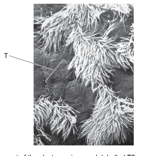
What is the identity of the part of the electron micrograph labelled T?
Options
A ciliated epithelial cell
B goblet cell
C mucus
D squamous epithelial cell
Working
The tracheal lining is a pseudostratified ciliated epithelium with two main cell types visible on the surface:
- Ciliated cells — the numerous hair-like projections covering most of the surface, which beat to move mucus upwards.
- Goblet cells — modified epithelial cells that have no cilia and appear as smooth, rounded, dome-shaped bulges among the cilia; they secrete mucus.
Structure T is a smooth, rounded, non-ciliated bulge between the ciliated regions, so it must be a goblet cell.
Answer
B
B
Background Concept
The wall of the trachea is lined by a pseudostratified ciliated columnar epithelium with mucus-secreting cells. The two most important cell types on the inner surface are:
- Ciliated epithelial cells — columnar cells whose free (apical) surface bears many cilia. The cilia beat in a synchronised, wave-like manner (the mucociliary escalator) to move a layer of mucus (and any trapped particles, e.g. dust or pathogens) upwards towards the larynx, where it is swallowed.
- Goblet cells — modified epithelial cells that are also columnar but have no cilia. They contain secretory vesicles loaded with mucin glycoproteins. At the apical surface they appear as smooth, rounded, dome-shaped bulges between the tufts of cilia. When they release their contents, the mucins hydrate to form the sticky mucus layer that traps inhaled particles and pathogens.
Other relevant features of the gas-exchange system include cartilage rings (keeping the airway open), smooth muscle, elastic fibres, and goblet/seromucous glands in the submucosa.
Understanding the Question
The micrograph (Fig. 32.1) shows the inner lining of the trachea, dominated by clumps of hair-like cilia. The label T points to a structure that is clearly not a tuft of cilia — it is a smooth, rounded bulge sitting among them. The question asks you to identify what that bulge is, from four options: ciliated cell, goblet cell, mucus, or squamous epithelial cell.
Approach
You do not need to know anything about the image other than what is visible: the rest of the surface is cilia, and T is a smooth, dome-shaped, non-ciliated area. Match each option to its expected appearance:
- Ciliated cell — should have a tuft of cilia on top. T does not. ✗
- Goblet cell — smooth, rounded apical bulge among the cilia. ✓
- Mucus — a film or droplet of secretion, not a discrete, well-defined cellular bulge. ✗
- Squamous epithelial cell — flat, thin cell; not found as the surface cell of the trachea (it lines alveoli and blood vessels). ✗
The only structure consistent with a smooth, cellular-looking dome between cilia is a goblet cell.
Step-by-Step Reasoning
- The micrograph shows the surface of the trachea. The hair-like projections are cilia belonging to the ciliated epithelial cells.
- Label T points to a structure that is not a tuft of cilia but a rounded, smooth, raised area between the ciliated tufts.
- In the tracheal epithelium, the only cell that appears this way at the surface is the goblet cell — its apical surface bulges out as a mucus-filled dome, with no cilia on it.
- The function of a goblet cell is to secrete mucus, which traps inhaled particles and is then moved up the trachea by the cilia.
- The remaining options can be excluded:
- It cannot be a ciliated cell because T has no cilia.
- It is not free mucus because T is a discrete, bounded cellular profile, not a surface film or droplet.
- It is not a squamous epithelial cell because the trachea is not lined by squamous epithelium (squamous cells are flat, e.g. in alveoli or the endothelium of blood vessels).
- Therefore, T is a goblet cell, option B.
Key Takeaways
- The trachea is lined by pseudostratified ciliated epithelium with goblet cells.
- Ciliated cells carry cilia on their apical surface; goblet cells do not, and instead appear as smooth, dome-shaped bulges.
- Together they form the mucociliary escalator: goblet cells secrete mucus, and cilia propel it (with trapped particles) upward to the throat.
- On a micrograph, a smooth, non-ciliated bulge between tufts of cilia is the diagnostic feature of a goblet cell.
Common Mistakes
- Choosing A (ciliated cell) — assuming that anything on the tracheal surface is ciliated. Remember: only some of the cells are ciliated; the goblet cells are interspersed among them and are easily seen as bare bulges.
- Choosing C (mucus) — confusing the cell that makes mucus (the goblet cell) with the mucus secretion itself. Mucus is a sticky layer or droplet lying on the surface, not a discrete rounded cellular structure.
- Choosing D (squamous epithelial cell) — squamous means "flat"; these cells are not found on the tracheal surface (they line alveoli, blood vessels, and Bowman's capsule).
Things to Be Careful About
- Read the image carefully: T is a rounded structure with a defined boundary, not a film of liquid or a tuft of projections.
- Make sure the terminology is exact: "ciliated epithelial cell" and "goblet cell" are the standard CIE terms — avoid the vague phrase "ciliated cell" on its own if the mark requires the full name.
- Distinguish goblet cells (single epithelial cells that secrete mucus) from mucous / seromucous glands (compound glands in the submucosa that also secrete mucus) — the micrograph shows a surface cell, so the correct answer is goblet cell.
Which rows correctly summarise the typical distribution of cartilage and smooth muscle within different parts of the gas exchange system of humans?
1 bronchi
2 bronchioles
3 trachea
Options
| cartilage | smooth muscle | |
|---|---|---|
| A | 1, 2 and 3 | |
| B | 1 and 2 only | |
| C | 1 and 3 only | |
| D | 2 and 3 only |
key
✓ = present
✗ = not present
(Note: The table in the PDF shows ticks and crosses for each row: 1 has ✓ for both, 2 has ✗ for both, 3 has ✓ for both. The options A-D refer to which rows are correct.)
Working
- Trachea: C-shaped rings of cartilage in the wall + a band of smooth muscle (trachealis) posteriorly. Row 3 (✓ cartilage, ✓ smooth muscle) is correct.
- Bronchi: irregular plates of cartilage in the wall + smooth muscle in the wall. Row 1 (✓ cartilage, ✓ smooth muscle) is correct.
- Bronchioles: have NO cartilage in their walls, but DO have smooth muscle. Row 2 (✗ cartilage, ✗ smooth muscle) is wrong because it omits the smooth muscle.
- Therefore rows 1 and 3 are correct, but row 2 is not.
Answer
C
C
Background Concept
The human gas exchange system is a branching tree of airways that becomes progressively smaller, simpler in wall structure, and more dominated by smooth muscle as it approaches the gas-exchange surface. The key tissues in the wall of each airway are:
- Cartilage — a rigid supporting tissue that holds the larger airways open so they do not collapse during inhalation. It is present in the trachea (as C-shaped rings, open posteriorly where the oesophagus lies) and in the bronchi (as irregular plates that completely surround the lumen). It is absent from the bronchioles, whose walls are too narrow to accommodate cartilage plates without obstructing the lumen.
- Smooth muscle — involuntary muscle that can constrict the airway (bronchoconstriction) or relax it (bronchodilation), thereby controlling airflow and matching ventilation to demand. It is present in the trachea, the bronchi, and the bronchioles. In the smaller bronchioles smooth muscle becomes a relatively more important component of the wall because the airway diameter is regulated largely by its tone.
- Other wall components (ciliated epithelium, goblet cells, elastic fibres) are also distributed in characteristic patterns along the airway, but the question focuses only on cartilage and smooth muscle.
Understanding the Question
The candidate is shown a table with three rows (bronchi, bronchioles, trachea) and two columns (cartilage, smooth muscle), each cell marked with a tick (✓ present) or cross (✗ absent). The question asks which of those rows are correct summaries of the true distribution of these two tissues. We therefore have to compare the candidate's tick/cross pattern in each row against the actual anatomy and identify the mismatches.
The command word is implicit but the question type is a "which rows are correct" MCQ. The answer depends on the precise biological pattern, not on a generic statement that "bronchioles are smaller".
Approach
Apply the standard CIE AS-Level distribution:
| Structure | Cartilage | Smooth muscle |
|---|---|---|
| Trachea | ✓ (C-rings) | ✓ |
| Bronchi | ✓ (plates) | ✓ |
| Bronchioles | ✗ | ✓ |
Compare this with the candidate's table:
- Row 1 (bronchi): ✓ / ✓ — matches the truth.
- Row 2 (bronchioles): ✗ / ✗ — wrong: bronchioles lack cartilage but they DO have smooth muscle, so the second cross is incorrect.
- Row 3 (trachea): ✓ / ✓ — matches the truth.
Rows 1 and 3 are correct, row 2 is not. The option that selects rows 1 and 3 only is C.
Step-by-Step Reasoning
- Trachea (row 3). The trachea is kept patent by 15–20 C-shaped rings of hyaline cartilage with the posterior gap bridged by the trachealis smooth muscle. So both cartilage and smooth muscle are present — the candidate's row 3 (✓ / ✓) is correct.
- Bronchi (row 1). Each bronchus is supported by irregular cartilage plates that fully encircle the lumen, and its wall also contains smooth muscle. The candidate's row 1 (✓ / ✓) is therefore correct.
- Bronchioles (row 2). Bronchioles are defined as the airways that lack cartilage and have a diameter of ≤1 mm. They do, however, retain a layer of smooth muscle in their walls (this is what allows bronchoconstriction in asthma, for example). The candidate's row 2 (✗ cartilage, ✗ smooth muscle) is wrong on the second cross — smooth muscle is present.
- Because rows 1 and 3 are correct and row 2 is not, the right option is the one that selects only rows 1 and 3, which is C.
Key Takeaways
- Cartilage is present in trachea and bronchi only; bronchioles have no cartilage.
- Smooth muscle is present throughout the airway tree — trachea, bronchi AND bronchioles.
- A common exam trap is to assume bronchioles contain neither tissue because they are "small and simple"; in fact, smooth muscle is one of their defining wall components and is the tissue responsible for bronchoconstriction/bronchodilation.
- When asked to evaluate candidate-drawn rows, scan each row's cell-by-cell claim and flag any cell that disagrees with the accepted distribution.
Common Mistakes
- Ticking or crossing both columns for bronchioles in the same direction. Because cartilage is absent but smooth muscle is present, the two columns must be filled differently.
- Assuming that because the trachealis muscle is at the back of the trachea it "isn't really in the wall" — the trachea still counts as having smooth muscle.
- Confusing cartilage with bone or confusing hyaline cartilage with elastic cartilage — the cartilage of the trachea and bronchi is hyaline.
- Choosing D (2 and 3) on the false belief that bronchioles are the only place with smooth muscle, or choosing A (1, 2 and 3) on the false belief that bronchioles are too small to have any muscle at all.
Things to Be Careful About
- The CIE AS syllabus explicitly teaches that smooth muscle is present in bronchioles; do not write it off as "absent" even in simplified answers.
- A "✗ for both" in row 2 is the specific error being tested — the question rewards noticing the second cross, not just the first.
- Read each option's wording precisely. "1 and 3 only" is not the same as "1 and 2 only" — getting the rows mixed up by one number changes the answer.
A molecule of oxygen diffuses from the air in an alveolus to haemoglobin in a red blood cell.
Assuming that the molecule crosses cellular layers by passing through cells, rather than between cells, what is the minimum number of phospholipid layers that the molecule of oxygen must pass through?
Options
A 5
B 6
C 8
D 10
Working
From the alveolus to haemoglobin inside a red blood cell, the O₂ molecule must cross three cells, each bounded by a phospholipid bilayer (i.e. 2 phospholipid layers):
- Alveolar epithelial cell — O₂ enters and exits → 2 + 2 = 4 phospholipid layers
- Capillary endothelial cell — O₂ enters and exits → 2 + 2 = 4 phospholipid layers
- Red blood cell — O₂ enters only (it binds to haemoglobin inside, so does not exit) → 2 phospholipid layers
Total = 4 + 4 + 2 = 10 phospholipid layers.
Answer
D
D
Background Concept
Every cell is surrounded by a plasma membrane built as a phospholipid bilayer: two sheets (leaflets) of phospholipid molecules arranged tail-to-tail. Crossing a plasma membrane therefore means crossing two phospholipid layers (the outer leaflet and the inner leaflet), not one.
In the lungs, the blood–gas barrier is extremely thin to allow rapid diffusion of O₂ and CO₂. The cells it comprises are:
- the alveolar epithelial cell (a type I pneumocyte), which lines the alveolus,
- the capillary endothelial cell, which forms the wall of the pulmonary capillary, and
- the red blood cell (erythrocyte), whose cytoplasm contains haemoglobin.
A thin fused basement membrane lies between the alveolar and endothelial cells, but it is not a cell and is therefore not counted when the question specifies "passing through cells".
Understanding the Question
The question asks for the minimum number of phospholipid layers that one O₂ molecule must cross on its journey from the alveolar air to a haemoglobin molecule inside an RBC. The key word is minimum — we use the smallest possible number of cells in the barrier. The key instruction is "passing through cells, rather than between them" — this rules out paracellular routes through gaps/junctions and forces the molecule to cross every cell membrane in the path.
The other crucial interpretation: "phospholipid layer" means one leaflet of a phospholipid bilayer, not the bilayer as a single structure.
Approach
- List the cells the O₂ molecule must cross, in order.
- For each cell, decide whether the molecule must enter only, or enter and exit.
- Multiply the number of membrane transits by 2 (one bilayer = two phospholipid layers).
- Sum the totals.
Step-by-Step Reasoning
Step 1 — Cells crossed:
The O₂ molecule must traverse three cells:
- alveolar epithelial cell,
- capillary endothelial cell,
- red blood cell.
Step 2 — Transits per cell:
- For the alveolar epithelial cell, O₂ must enter from the alveolar side and exit on the tissue-fluid side → 2 membrane transits.
- For the capillary endothelial cell, O₂ must enter from the tissue-fluid side and exit into the plasma of the blood → 2 membrane transits.
- For the red blood cell, O₂ must enter from the plasma, but does not need to exit because it binds to haemoglobin inside the cytoplasm → 1 membrane transit.
Total transits = 2 + 2 + 1 = 5.
Step 3 — Convert transits to phospholipid layers:
Each transit through a plasma membrane crosses one phospholipid bilayer, which is 2 phospholipid layers.
Step 4 — Verification by cell:
- Alveolar cell: 4
- Capillary endothelial cell: 4
- Red blood cell: 2
- Sum: 4 + 4 + 2 = 10 ✓
Key Takeaways
- A plasma membrane = 1 phospholipid bilayer = 2 phospholipid layers.
- The blood–gas barrier consists of 3 cells; O₂ transits 5 membranes in total (the last membrane only once, because it binds haemoglobin inside the RBC).
- Whenever a question asks "how many phospholipid layers", the answer is always 2× the number of cell-membrane crossings.
Common Mistakes
- Treating each cell membrane as 1 phospholipid layer → gives 5 (option A). This is wrong because the bilayer has two leaflets.
- Forgetting that the O₂ must exit the alveolar and capillary cells (counting only entry) → gives 6 (option B).
- Assuming the O₂ must also exit the red blood cell → gives 12, which is not even an option but is a common error in counting.
- Adding an extra cell such as an extra pneumocyte or a "basement-membrane cell" → not required because the question says minimum and the basement membrane is acellular.
Things to Be Careful About
- Read "phospholipid layer" carefully — in Cambridge mark schemes it refers to one leaflet of the bilayer, not the bilayer itself.
- "Minimum" means we assume the thinnest possible version of the blood–gas barrier: one alveolar cell, one endothelial cell, one RBC.
- The oxygen's destination is haemoglobin inside the RBC, so it only needs to enter the red blood cell, not leave it.
Which row shows the type of pathogen that causes cholera and its mode of transmission?
Options
| pathogen | mode of transmission | |
|---|---|---|
| A | protoctist | contaminated food or water |
| B | protoctist | airborne droplets |
| C | bacterium | contaminated food or water |
| D | bacterium | airborne droplets |
Working
Cholera is caused by Vibrio cholerae, which is a bacterium (prokaryotic pathogen), not a protoctist. Transmission occurs via the faecal–oral route, when water or food is contaminated with faeces containing the bacterium.
Answer
C
C
Background Concept
Infectious diseases are caused by pathogens, which are biological agents capable of causing disease in a host. The major categories of pathogen are:
- Bacteria — prokaryotic cells (e.g. Vibrio cholerae, Mycobacterium tuberculosis).
- Viruses — non-cellular particles requiring a host cell to replicate (e.g. HIV, influenza).
- Protoctists (protists) — eukaryotic single-celled organisms, often with complex life cycles (e.g. Plasmodium causing malaria).
- Fungi — eukaryotic organisms, some of which are pathogenic (e.g. Candida species).
Each pathogen is transmitted by a characteristic route that reflects where it lives, how it leaves the host, and how it enters a new host. Common routes include airborne droplets, contaminated food or water, bodily fluids, vectors (e.g. mosquitoes), and direct contact.
Understanding the Question
This is a multiple-choice question (Paper 1) asking for the correct pairing of:
- The type of pathogen that causes cholera.
- The mode of transmission of cholera.
The command word is essentially "identify" — the candidate must select the row that correctly matches both columns.
Approach
Recall the basic microbiology of cholera: identify the pathogen and its transmission route, then locate the option that pairs both correctly. The distractors mix up the pathogen type (protoctist vs bacterium) and the transmission route (airborne vs food/water), so a candidate must know both facts independently.
Step-by-Step Reasoning
- Pathogen causing cholera: Cholera is caused by Vibrio cholerae, which is a Gram-negative bacterium. This immediately eliminates options A and B, which list "protoctist" (the protoctist pathogen is Plasmodium, the cause of malaria).
- Mode of transmission: Vibrio cholerae is transmitted via the faecal–oral route, typically when drinking water or eating food contaminated with faecal matter from an infected person. The bacterium can survive in fresh water and brackish water, and outbreaks are strongly associated with poor sanitation and contaminated water supplies. This is a "contaminated food or water" route, not an airborne one — eliminating option D.
- Conclusion: The only row with both a correct pathogen type and a correct transmission route is C — bacterium, contaminated food or water.
Key Takeaways
- Cholera is caused by the bacterium Vibrio cholerae.
- Cholera is transmitted by contaminated water or food (faecal–oral route).
- Distinguishing between the four diseases commonly tested (cholera, malaria, TB, HIV/AIDS) on both pathogen type and transmission route is essential Paper 1 content.
Common Mistakes
- Confusing cholera (bacterium) with malaria (protoctist) and selecting option A or B.
- Confusing cholera's water-borne transmission with TB's airborne transmission and selecting option D.
Things to Be Careful About
- Vibrio cholerae is sometimes described as a "comma-shaped bacterium" — make sure not to confuse it with viral or protoctist pathogens.
- "Contaminated food or water" is the key transmission phrase; airborne transmission is associated with TB and many viral diseases, not cholera.
Which statement about tuberculosis (TB) is not correct?
Options
A TB can be controlled by vaccination.
B TB is caused by a virus spread by droplet infection.
C HIV/AIDS increases the risk of developing TB.
D TB may be transmitted by eating contaminated meat.
Working
TB is caused by the bacterium Mycobacterium tuberculosis, not a virus. It is spread by droplet infection (inhaled respiratory droplets from an infected person). Vaccination (BCG) can help control it, HIV/AIDS greatly increases susceptibility to TB, and while TB primarily affects the lungs, some forms (e.g. bovine TB from M. bovis) can be transmitted via contaminated meat/milk. Option B is therefore the incorrect statement.
Answer
B
B
Background Concept
Tuberculosis (TB) is a serious infectious disease caused by the bacterium Mycobacterium tuberculosis. It is one of the four major infectious diseases highlighted in the Cambridge A-Level Biology syllabus (alongside cholera, malaria, and HIV/AIDS). TB primarily infects the lungs (pulmonary TB) but can also affect other parts of the body such as the lymph nodes, bones, and central nervous system.
The bacterium is spread when an infected person coughs, sneezes, or speaks, releasing tiny respiratory droplets containing the bacteria into the air. A susceptible person nearby inhales these droplets and can become infected. This mode of spread is called droplet infection or airborne transmission. The bacteria then multiply inside the lung tissue, and the immune system walls them off in structures called tubercles.
There is also a form of TB called bovine tuberculosis, caused by a closely related species, Mycobacterium bovis, which can be transmitted to humans through unpasteurised milk or undercooked meat from infected cattle. This is why pasteurisation of milk and meat inspection are important public health measures.
Understanding the Question
This is a multiple-choice question asking which statement about TB is not correct. The candidate must evaluate all four options and identify the one that contains a factual error. The correct answer is B because TB is caused by a bacterium, not a virus.
Approach
Test each option against established facts about TB:
- A: TB can be controlled by vaccination. The BCG (Bacillus Calmette–Guérin) vaccine is used in many countries as part of TB control programmes, so this is correct.
- B: TB is caused by a virus spread by droplet infection. The pathogen is a bacterium (Mycobacterium tuberculosis), so the word "virus" makes this statement incorrect.
- C: HIV/AIDS increases the risk of developing TB. People with HIV have weakened immune systems, making them far more susceptible to TB infection and progression to active disease. This is correct.
- D: TB may be transmitted by eating contaminated meat. Bovine TB (M. bovis) can be transmitted via unpasteurised dairy or undercooked meat, so this is correct.
Step-by-Step Reasoning
-
Option A — Vaccination as a control measure: The BCG vaccine, made from a weakened strain of Mycobacterium bovis, is widely used to provide partial protection against TB, especially in children. It is not fully effective in all populations, but it is a legitimate control tool. Statement A is correct.
-
Option B — The causative agent: The exam's most important testable fact is the type of pathogen. Mycobacterium tuberculosis is a bacterium (a prokaryote with a cell wall rich in mycolic acids, which gives it its characteristic acid-fast staining property). The statement that TB is caused by a virus is wrong; viruses such as HIV, influenza, or measles have entirely different structures and are not responsible for TB. Statement B is therefore the incorrect one.
-
Option C — HIV/AIDS as a risk factor: HIV destroys CD4+ T-lymphocytes, which are central to the cell-mediated immune response that contains TB bacteria. HIV-positive individuals are therefore at much higher risk of both new TB infection and reactivation of latent TB. Statement C is correct.
-
Option D — Food-borne transmission: While the most common route is airborne, bovine TB (M. bovis) can indeed be transmitted to humans through contaminated, unpasteurised milk or undercooked meat. This is recognised in the syllabus and in public health practice (e.g. pasteurisation of milk). Statement D is correct.
Since only option B contains the error (calling the pathogen a virus), the answer is B.
Key Takeaways
- TB is caused by the bacterium Mycobacterium tuberculosis (and M. bovis for the bovine form).
- TB is spread primarily by droplet (airborne) infection from the coughs/sneezes of infected individuals.
- The BCG vaccine is a tool for TB control.
- HIV/AIDS dramatically increases TB risk because it weakens cell-mediated immunity.
- Bovine TB can be transmitted via unpasteurised milk or undercooked meat — a less common but recognised route.
Common Mistakes
- Confusing the pathogen type: TB is a bacterial disease, not a viral one. This is a high-frequency error because the question lists many distractors about TB and students may rush past the key word.
- Assuming all TB is airborne: forgetting that bovine TB has a food-borne route can cause confusion with option D.
- Confusing TB with other respiratory diseases: TB should not be lumped together with viral infections like influenza or COVID-19, which have very different pathogens, treatments, and prevention strategies.
Things to Be Careful About
- Pay close attention to the command word "not correct" — the question asks for the false statement, not a true one.
- The pathogen identity is often worth a full mark on its own in TB-related questions; always state Mycobacterium tuberculosis (bacterium) rather than just "a microbe" or "a germ".
- Distinguish between M. tuberculosis (human TB, airborne) and M. bovis (bovine TB, can be food-borne) when discussing transmission routes.
More cases of malaria are being reported in Europe. Other diseases, formerly confined to tropical countries and transmitted in the same way as malaria, have also spread to parts of Europe. Tropical countries have higher mean temperatures and humidity than Europe.
What could explain the increase in the number of cases of these diseases in Europe?
1 rising temperatures and humidity in Europe as a result of climate change
2 increased travel between Europe and tropical countries
3 creation of wetland areas such as marshes to increase biodiversity
Options
A 1, 2 and 3
B 1 and 2 only
C 1 and 3 only
D 2 and 3 only
Working
Malaria is transmitted by Anopheles mosquitoes, so any factor that allows mosquitoes to thrive in Europe, or that brings the parasite (or infected people) into Europe, will increase cases.
- Statement 1 — True. Warmer, more humid conditions in Europe allow Anopheles mosquitoes to survive and breed, and the Plasmodium parasite needs a minimum temperature to complete its development inside the mosquito.
- Statement 2 — True. Increased travel can introduce infected humans (and accidentally transported mosquitoes) from tropical countries into Europe.
- Statement 3 — True. Marshes and other wetlands are breeding sites for mosquitoes; creating them increases the local vector population.
All three statements are correct.
Answer
A
A
Background Concept
Malaria is caused by the protoctist Plasmodium, transmitted between humans by the bite of an infected female Anopheles mosquito. The mosquito is therefore the vector, and the disease only spreads where (a) the mosquito can survive and breed and (b) the parasite can complete its development inside the mosquito, which requires sufficiently warm temperatures. Tropical regions provide both conditions; temperate Europe historically has not. Anything that shifts European conditions closer to those of the tropics, or that brings the parasite or infected people into Europe, will increase the number of cases reported there.
Understanding the Question
The question asks which of three statements could explain the rise in malaria (and similar vector-borne diseases) in Europe. We must judge each statement on its biological merit — does it genuinely create the conditions for more transmission? — not on whether it sounds plausible in general.
Approach
Go through each statement and ask: does this change the European environment so that the Anopheles mosquito (and the Plasmodium parasite it carries) can now survive, breed or be introduced there?
Step-by-Step Reasoning
Statement 1 — rising temperatures and humidity in Europe as a result of climate change ✓
Climate change is producing warmer, wetter summers in parts of southern and central Europe. Anopheles mosquitoes need warm, humid conditions to breed — larvae develop in standing water and the adults are active in warm temperatures. Crucially, Plasmodium requires a minimum ambient temperature (around 16–18 °C as a lower threshold) to complete its sporogony cycle inside the mosquito. Warmer European temperatures therefore extend both the geographical range and the transmission season of malaria.
Statement 2 — increased travel between Europe and tropical countries ✓
Every year millions of people travel between Europe and malaria-endemic tropical regions. Travellers who are infected (and may not yet show symptoms) can be bitten by a European mosquito, which then transmits Plasmodium locally. Aircraft and ships can also accidentally transport adult mosquitoes. Importation of the parasite and vector together is a well-documented route of re-introduction into non-endemic countries.
Statement 3 — creation of wetland areas such as marshes to increase biodiversity ✓
Although biodiversity schemes are environmentally valuable, the standing water in marshes and similar wetlands is precisely the habitat in which Anopheles lays its eggs and in which the larvae develop. Creating more wetlands therefore directly increases the local breeding population of the vector, raising the potential for transmission if Plasmodium is introduced.
All three statements are valid contributors, so the correct option is the one that includes all of them: A — 1, 2 and 3.
Key Takeaways
- The spread of a vector-borne disease depends on the vector's ecology, the pathogen's environmental requirements, and the movement of infected hosts/vectors.
- Climate change, international travel and habitat creation each affect one of these three components for malaria.
- A "biodiversity-positive" action (wetland creation) is not cost-free in terms of human health and must be balanced against vector-borne disease risk.
Common Mistakes
- Choosing B because "wetlands increase biodiversity so they must be good" — students forget that the same standing water that supports amphibians and insects also supports mosquito larvae.
- Choosing D because they reject climate change as a driver, even though there is strong epidemiological evidence for it.
- Confusing malaria (a vector-borne protoctist disease) with air-borne or directly-transmitted diseases, which would not respond to these same factors.
Things to Be Careful About
- The question says diseases "transmitted in the same way as malaria" — i.e. by mosquitoes — so any factor that helps mosquitoes in Europe will apply to all of them, not just malaria.
- "Could explain" means a scientifically credible mechanism, not necessarily the largest current cause; all three statements are credited as plausible contributors.
- The mark scheme accepts climate change, travel and habitat change together; do not try to rank them or pick a single "best" answer.
The diagrams show three different bonds.
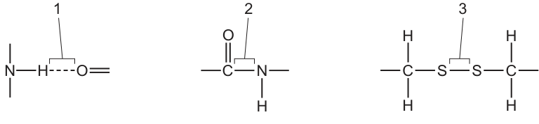
Which bonds are found in an antibody molecule?
Options
A 1, 2 and 3
B 1 and 2 only
C 1 and 3 only
D 2 and 3 only
Working
Bond 1 is a hydrogen bond (dashed line between an N–H and a C=O group).
Bond 2 is a peptide bond (C(=O)–N–H linkage between two amino acid residues).
Bond 3 is a disulfide bond (C–S–S–C between two cysteine side chains).
An antibody (immunoglobulin) is a globular protein with all four levels of structure:
- Primary structure: amino acids joined by peptide bonds (bond 2).
- Secondary structure: α-helices and β-pleated sheets stabilised by hydrogen bonds (bond 1) between N–H and C=O groups.
- Tertiary and quaternary structure: folding held together by hydrogen bonds, ionic interactions, hydrophobic interactions and disulfide bridges (bond 3) between cysteine residues — antibodies are especially rich in disulfide bonds, which link the two heavy chains together and each light chain to a heavy chain.
All three bonds are therefore present.
Answer
A
A
Background Concept
Antibodies (immunoglobulins) are Y-shaped globular proteins made of four polypeptide chains: two identical heavy chains and two identical light chains, linked by disulfide bridges. Like every protein, an antibody is built up at four levels of organisation:
- Primary structure — the linear sequence of amino acids, held together by peptide bonds (C–N covalent linkages formed by condensation between the carboxyl group of one amino acid and the amino group of the next).
- Secondary structure — regular folding into α-helices and β-pleated sheets, stabilised by hydrogen bonds between the N–H of one peptide bond and the C=O of another.
- Tertiary structure — the 3-D folding of a single polypeptide, maintained by hydrogen bonds, ionic (salt) bridges, hydrophobic interactions and disulfide bonds (covalent S–S linkages between the –SH side chains of two cysteine residues).
- Quaternary structure — the assembly of two or more polypeptide chains; in antibodies, the two heavy chains are joined to each other and each light chain is joined to a heavy chain by additional disulfide bonds.
The three bonds shown in Fig. 38.1 are therefore all characteristic features of a typical antibody.
Understanding the Question
The question shows three labelled bond diagrams and asks which of them occur inside an antibody molecule. We must identify each bond from its drawing and then decide whether it is present in an immunoglobulin.
- Bond 1: dashed line between an N–H and a C=O → a hydrogen bond.
- Bond 2: a covalent C(=O)–N–H linkage → a peptide bond.
- Bond 3: two sulfur atoms covalently joined, each bonded to a carbon that also carries an H → a disulfide bond between two cysteine side chains.
Because antibodies are proteins, all three must be considered, and we must remember that disulfide bonds are a particularly prominent feature of immunoglobulins.
Approach
The strategy is:
- Identify the three bonds from the diagrams.
- Check each one against the four levels of protein structure an antibody possesses.
- Any bond found at primary, secondary, tertiary or quaternary level is credited.
Step-by-Step Reasoning
- Bond 2 (peptide bond): Every protein, including every antibody, has a primary structure. By definition, the primary structure consists of amino acids joined by peptide bonds, so bond 2 must be present.
- Bond 1 (hydrogen bond): The secondary structure of antibodies is dominated by β-pleated sheets (antibodies have very little α-helix). These sheets are held together by hydrogen bonds between the N–H of one peptide bond and the C=O of another. Hydrogen bonds also occur throughout the tertiary structure, stabilising the 3-D shape of the variable and constant domains. So bond 1 is present.
- Bond 3 (disulfide bond): Antibodies are unusually rich in disulfide bridges. They are found within each domain (intradomain, stabilising tertiary structure) and between chains (interchain, stabilising quaternary structure) — for example, disulfide bonds link each light chain to a heavy chain, and link the two heavy chains together near the hinge region. So bond 3 is present.
Because bonds 1, 2 and 3 are all found in an antibody, the correct option is A (1, 2 and 3).
Distractor reasoning:
- B (1 and 2 only) — omits disulfide bonds. This is the most tempting distractor, because students often forget that disulfide bridges are a defining structural feature of immunoglobulins.
- C (1 and 3 only) — omits peptide bonds. This is impossible: an antibody is a polypeptide and must contain peptide bonds in its primary structure.
- D (2 and 3 only) — omits hydrogen bonds. This ignores the role of hydrogen bonding in maintaining secondary and tertiary structure.
Key Takeaways
- Antibodies contain all three bonds shown: peptide bonds (primary), hydrogen bonds (secondary/tertiary) and disulfide bonds (tertiary/quaternary).
- Disulfide bonds are especially characteristic of antibodies — they are the covalent "staples" that hold the four-chain structure together.
- When asked which bonds a protein contains, always think level-by-level (1° → 4°) rather than recalling a single bond type.
Common Mistakes
- Forgetting that disulfide bonds exist in proteins, and so choosing B (1 and 2 only).
- Confusing the hydrogen-bond diagram with another interaction and overlooking that the secondary structure of antibodies (β-sheets) is built on hydrogen bonds.
- Selecting C or D because peptide bonds or hydrogen bonds were not consciously linked to a specific structural level.
Things to Be Careful About
- A dashed line between an electronegative atom and an H attached to another electronegative atom is the standard way to draw a hydrogen bond — do not confuse it with a covalent bond.
- The C(=O)–N–H linkage is a peptide bond, the backbone of any protein.
- A C–S–S–C linkage (with each carbon also bearing an H) is a disulfide bond; the two sulfur atoms are covalently bonded, and the bond is formed by oxidation of two cysteine –SH groups.
- The number of disulfide bonds in an antibody is a deliberately tested feature — expect a question to single it out.
Specific monoclonal antibodies can be used to treat some forms of cancer. One example is trastuzumab, which can be used in the treatment of tumours caused by cancer.
Which statements could help to explain why monoclonal antibodies such as trastuzumab are suitable for this role?
1 The variable regions of monoclonal antibodies such as trastuzumab can change, allowing the antibodies to bind to cancer cells even if their antigens mutate.
2 Monoclonal antibodies such as trastuzumab bind to specific cell surface receptors on cancer cells, which prevents these receptors from receiving signals that are needed for cancer cells to grow and divide.
3 The antigen-binding sites of monoclonal antibodies such as trastuzumab are complementary to antigens found on some cancer cells, and have stable tertiary structures that do not change.
Options
A 1, 2 and 3
B 1 and 2 only
C 1 and 3 only
D 2 and 3 only
Working
Monoclonal antibodies are produced by a single clone of B cells, so all antibody molecules are identical with the same variable region and the same antigen-binding site.
Statement 1 is incorrect: the variable regions of a monoclonal antibody do NOT change. They are fixed by the clone, so if the antigen mutates the antibody can no longer bind effectively.
Statement 2 is correct: trastuzumab binds specifically to HER2 receptors on certain breast cancer cells, blocking growth signals and slowing tumour growth.
Statement 3 is correct: the antigen-binding site of a monoclonal antibody is complementary in shape to a specific antigen, and the tertiary structure that creates this site is stable.
Therefore, statements 2 and 3 only are correct.
Answer
D
D
Background Concept
Monoclonal antibodies are antibodies produced by a single clone of hybridoma cells (a B lymphocyte fused with a myeloma tumour cell). Because they all originate from one parent B cell, every antibody molecule produced is identical — they have the same variable regions and the same antigen-binding sites, and therefore bind to one specific epitope on one specific antigen.
The antigen-binding site sits at the tip of the variable region of the heavy and light chains. Its three-dimensional shape is determined by the variable region's tertiary structure, which is held in place by hydrogen bonds, ionic bonds, disulfide bridges and hydrophobic interactions. This tertiary structure is stable — it does not alter to fit different antigens. This is the principle of antibody–antigen specificity (the lock-and-key / induced-fit idea): a given antibody binds only to its complementary antigen.
Therapeutic monoclonal antibodies such as trastuzumab (trade name Herceptin) are designed to bind to a specific cell-surface receptor — in trastuzumab's case, HER2, which is over-expressed on some breast cancer cells. Binding blocks the receptor and prevents it from receiving growth signals, so the cancer cells stop dividing as rapidly.
Understanding the Question
This is a Paper 1 multiple-choice item. The stem describes trastuzumab, a monoclonal antibody used to treat certain cancers, and asks which of three statements correctly explain why monoclonal antibodies are suitable for this role. The candidate must judge each statement true or false and then choose the option listing only the true statements.
The command word is implicit ("which statements could help to explain") — the candidate is being asked to evaluate biological accuracy, not to recall a single fact.
Approach
Work through each statement one at a time, judging whether it is consistent with the known structure and function of monoclonal antibodies:
- Statement 1 concerns variability of the variable region — does it change?
- Statement 2 concerns the mechanism of action of trastuzumab — does binding block growth signals?
- Statement 3 concerns complementarity and stability of the binding site — is the tertiary structure fixed?
Then select the option that includes only the correct statements.
Step-by-Step Reasoning
Statement 1 — FALSE. A monoclonal antibody is, by definition, derived from a single clone of B cells, so every antibody molecule has the same variable region and the same antigen-binding site. The variable region does not change, and the antibody cannot adapt to bind a mutated antigen. If the cancer cell's surface antigen mutates so that the antibody's binding site is no longer complementary, the antibody will lose its effectiveness. The correct feature of monoclonal antibodies is specificity, not adaptability — this is the opposite of what statement 1 claims.
Statement 2 — TRUE. Trastuzumab binds to the HER2 cell-surface receptor on some breast cancer cells. HER2 normally receives a growth-factor signal that stimulates the cell to divide. When trastuzumab occupies the receptor, the growth signal cannot be received, so the cancer cells' growth and division are slowed. This is exactly the mechanism of action described.
Statement 3 — TRUE. The antigen-binding sites of monoclonal antibodies are complementary in shape to specific antigens — this is what allows them to recognise and bind only to their target. The tertiary structure that creates this binding site is held in place by the bonding interactions within the protein and is stable; it does not change shape to accommodate different antigens. (Note: the related induced-fit model describes a small conformational adjustment on binding, but the overall tertiary structure of the binding site itself does not undergo wholesale change to recognise a new antigen.)
Combining the judgements: statements 2 and 3 are correct; statement 1 is incorrect. The only option that lists exactly 2 and 3 is D.
Key Takeaways
- Monoclonal antibodies have identical, unchanging variable regions and binding sites — this gives them specificity but means a mutation in the target antigen can reduce their effectiveness.
- The antigen-binding site is complementary to a specific antigen, and its tertiary structure is stable.
- Some therapeutic monoclonal antibodies (e.g. trastuzumab) work by binding to a cell-surface receptor and blocking the signal the cell needs to grow and divide.
Common Mistakes
- Confusing monoclonal with polyclonal antibodies. Polyclonal antibodies are a mixture from many B-cell clones, with different variable regions that recognise different epitopes on the same antigen. Monoclonal antibodies are from a single clone and have one specific binding site. Statement 1's claim that the variable region "can change" would describe a polyclonal mixture, not a monoclonal antibody.
- Thinking the binding site reshapes to fit any antigen. The tertiary structure of the binding site is stable; the antibody is specific to one antigen, so it cannot simply adapt to whatever antigen is present.
- Ignoring the clinical mechanism. Many candidates forget that trastuzumab's clinical effect comes from blocking HER2 signalling, not from directly killing the cell (although some monoclonal antibodies do recruit immune effectors — that is a different mechanism).
Things to Be Careful About
- "Variable region" refers to the part of the antibody that varies between different antibodies (i.e. it is the source of antibody diversity), not something that varies within a single antibody over time. The variable region of any one monoclonal antibody is fixed.
- In the CIE mark scheme, monoclonal antibodies must be described as having a single specificity (one type of antibody binding one epitope) — be precise with this terminology.
- "Tertiary structure is stable" does not mean absolutely rigid; a small induced-fit adjustment on antigen binding is acceptable, but the structure does not change to recognise a different antigen.
The HIV virus causes illness by infecting and destroying T-helper cells. This leads to AIDS and an inability to produce an effective immune response.
Which components of the immune system are produced less effectively when AIDS develops?
1 memory cells
2 plasma cells
3 antibodies
Options
A 1, 2 and 3
B 1 and 2 only
C 1 and 3 only
D 2 and 3 only
Working
HIV infects and destroys T-helper cells (CD4⁺ lymphocytes). T-helper cells are required to activate B-lymphocytes during the primary immune response. Without this activation:
- B cells do not proliferate and differentiate into plasma cells (so 2 is reduced).
- B cells do not differentiate into memory cells (so 1 is reduced).
- With fewer plasma cells, fewer antibodies are produced (so 3 is reduced).
All three components are produced less effectively, so statements 1, 2 and 3 are correct.
Answer
A
A
Background Concept
The adaptive immune response relies on coordination between several types of lymphocyte. When a pathogen enters the body, antigen-presenting cells (such as macrophages) display antigens on their surface and present them to T-helper cells (CD4⁺). Once activated, T-helper cells release cytokines that:
- stimulate B-lymphocytes to proliferate and differentiate into plasma cells (antibody factories) and memory B cells;
- stimulate cytotoxic T cells to divide and attack infected host cells.
HIV specifically binds to the CD4 receptor on T-helper cells (along with a co-receptor such as CCR5), entering and replicating inside them. Over time, large numbers of T-helper cells are destroyed, and the body can no longer coordinate an effective adaptive response — this is the defining feature of AIDS (Acquired Immune Deficiency Syndrome).
Understanding the Question
The question gives the essential premise: HIV destroys T-helper cells → AIDS develops → immune response fails. It then asks which of three immune components (memory cells, plasma cells, antibodies) are produced less effectively as a result. The candidate must decide which of the three statements are true.
Approach
The strategy is to follow the logical chain:
- Recall the role of T-helper cells in the primary response.
- Identify which of the three listed components depend on T-helper cell help.
- Conclude that anything downstream of T-helper cell activation will be reduced.
Step-by-Step Reasoning
- Statement 2 — plasma cells: Plasma cells are formed when activated T-helper cells stimulate B cells to differentiate. With T-helper cells depleted, this differentiation is greatly reduced. ✓ Reduced.
- Statement 1 — memory cells: Memory B cells are produced alongside plasma cells during the same T-helper-cell-dependent activation. Without T-helper cell help, the pool of memory cells is also not replenished effectively. ✓ Reduced.
- Statement 3 — antibodies: Antibodies are secreted by plasma cells. Fewer plasma cells means less antibody production. ✓ Reduced.
All three are produced less effectively, so the correct combination is 1, 2 and 3.
Key Takeaways
- T-helper cells are the "conductors" of the adaptive immune response — their loss disrupts B-cell activation, cytotoxic T-cell activation, and antibody production.
- AIDS is defined by this collapse of immune coordination, not by the absence of any single component.
- Any question listing memory cells, plasma cells or antibodies as separate items should be answered by asking: "Does this require T-helper cell help?" If yes, it will be reduced in AIDS.
Common Mistakes
- Choosing C (1 and 3 only) or D (2 and 3 only): forgetting that memory B cells arise from the same T-helper-cell-dependent activation as plasma cells, so memory cell production is also impaired.
- Choosing B (1 and 2 only): forgetting that plasma cells are the only source of antibody secretion, so reduced plasma cell numbers automatically means reduced antibody levels.
- Confusing T-helper cells with cytotoxic T cells — HIV targets helper T cells, not the cytotoxic population directly.
Things to Be Careful About
- "Produced less effectively" applies to the generation of these cells/molecules after HIV has destroyed T-helper cells, not to whether they exist at all in the body.
- The three statements are not independent: plasma cells make antibodies, and both arise together with memory cells from activated B cells — they are reduced together.
- In MCQ combination questions, only one option is fully correct; treat partial combinations as distractors unless the question explicitly allows more than one answer.
Your score so far
Answer a question to start scoring
Your marks add up here as you work through the paper.