Biology 9700/23 — October/November 2024
Cambridge AS Level · AS Level Structured Questions · worked solutions for every part, with the mark scheme
Topics Cell Structure · Biological Molecules · Gas Exchange · Transport in Mammals · The Mitotic Cell Cycle · Nucleic Acids and Protein Synthesis · +4 more
1 Fig. 1.1 shows the structure of the human gas exchange system.
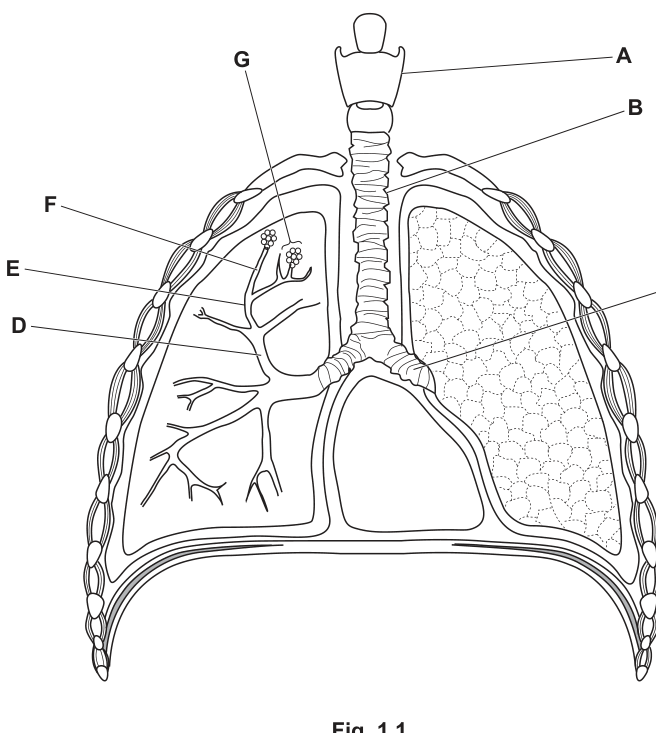
Use the letters in Fig. 1.1 to identify the parts of the gas exchange system that contain cartilage.
Answer
B and C (trachea and bronchus).
B and C
Background Concept
The human gas exchange system is a branching tree of airways that conducts air from the outside into the lungs. The larger airways (trachea, bronchi) must remain permanently open so that air can flow freely during breathing, while smaller airways (bronchioles) and the gas exchange surfaces (alveoli) do not need this rigid support. Cartilage provides that rigid support, and its distribution reflects this functional split: it is present in the trachea and bronchi but absent from the bronchioles and alveoli.
Understanding the Question
The candidate is given a labelled diagram (Fig. 1.1) of the gas exchange system with letters A to G pointing to different structures. The task is to identify which of these structures contain cartilage. The mark scheme accepts B and C, or A, B and C if all are correct; any inclusion of other letters voids the mark.
In Fig. 1.1:
- A = larynx (top of trachea)
- B = trachea
- C = bronchus
- D = bronchiole
- E = rib/intercostal muscle
- F = alveolus
- G = cluster of alveoli
Approach
Recall that cartilage is found in the walls of the trachea, bronchi and (sometimes accepted) the larynx, but not in bronchioles or alveoli. Then match these structures to the letters on the diagram.
Step-by-Step Reasoning
- Cartilage in the gas exchange system forms incomplete C-shaped rings in the trachea and similar plates in the bronchi. The mark scheme credits B and C.
- Letter A (the larynx) is also accepted; the larynx contains cartilages such as the thyroid and cricoid cartilage. If A, B and C are all written, the mark is still awarded as long as no incorrect letters appear.
- Letters D (bronchiole), E (rib/intercostal muscle), F and G (alveoli) do not contain cartilage in the gas exchange system. Including any of these voids the mark because the mark scheme states 'if any other letters do not award the mark, ignore names'.
Key Takeaways
- Cartilage is a feature of the larger conducting airways only.
- Always match anatomical knowledge to the specific labels in the figure supplied, not to general textbook labels.
Common Mistakes
- Including D, E, F or G, which immediately voids the mark regardless of how many correct letters are also given.
- Writing the structure names instead of letters; the mark scheme states 'ignore names', so letter answers must be the primary response.
Things to Be Careful About
The mark scheme explicitly says 'if any other letters [are given] do not award the mark'. This means even one incorrect letter loses the mark, so it is safer to list only the structures you are certain about.
Answer
- Cartilage keeps the trachea and bronchi open / prevents the airways from collapsing, so that air can flow freely into and out of the lungs.
- Cartilage allows the airways to be flexible, for example during bending of the neck or swallowing food.
Keeps airways open / prevents collapse; and provides flexibility.
Background Concept
Cartilage is a firm but slightly flexible connective tissue. In the larger airways of the gas exchange system it forms C-shaped (incomplete) rings in the trachea and irregular plates in the bronchi. These rings are open posteriorly, where the trachealis muscle (smooth muscle) sits. The combination of rigid cartilage and flexible smooth muscle gives the airways two key properties: they stay open to permit air flow, and they can change shape to accommodate movement of nearby structures.
Understanding the Question
The candidate must describe the role of cartilage in the gas exchange system, picking two marking points from a small list. Each point must stand on its own and use precise wording such as 'keeps the airway open' or 'prevents collapse'. Vague phrases such as 'provides strength' are explicitly ignored by the mark scheme.
Approach
Think about what would happen to a soft tube if no cartilage were present: it would collapse during inhalation when the pressure inside dropped. Then consider what happens around the trachea when you swallow or move your neck. Cartilage must perform two functions — mechanical support and flexibility.
Step-by-Step Reasoning
- Support / keeps airway open. Cartilage rings and plates in B (trachea) and C (bronchi) hold the lumen open during the pressure changes of breathing, so air can flow freely into and out of the lungs. The mark scheme phrases this as 'keeps the (named) airway(s) open / prevents collapse of airways' and explicitly rejects applying this idea to bronchioles or alveoli.
- Flexibility. Although cartilage is rigid, it has enough give (and is arranged in separate rings and plates) that the trachea can move when the neck bends or when food passes down the oesophagus behind it. The mark scheme accepts 'allows flexibility' or a description such as 'bending neck, swallowing food'.
- AVP — changes during breathing. The C-shape is incomplete posteriorly, and the trachea lengthens and widens slightly during inspiration. This is credited only as an additional valid point.
Key Takeaways
- Cartilage has two complementary roles: it stops the airway collapsing and it allows controlled movement.
- Always name the airway(s) where the function operates; do not generalise to the whole gas exchange system.
Common Mistakes
- Writing 'provides strength' alone — the mark scheme explicitly ignores this.
- Stating that cartilage is found in bronchioles or alveoli — these are rejected (R).
- Giving only one function when the question is worth two marks.
Things to Be Careful About
Use the term 'airways' or 'trachea and bronchi' rather than 'lungs', because the cartilage is in the conducting tubes, not in the lung tissue itself. The mark scheme credits naming the specific airway(s) where appropriate.
Microscope slides were prepared from two regions of the gas exchange system.
Fig. 1.2 and Fig. 1.3 are photomicrographs of the two slides.
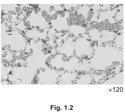
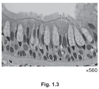
Complete Table 1.1.
• Use the letters from Fig. 1.1 to identify the regions of the gas exchange system from which the two slides were prepared.
• Identify one feature visible in Fig. 1.2 and one feature visible in Fig. 1.3.
• State one way in which each feature relates to its function.
Table 1.1
| Fig. | region of the gas exchange system (A, B, C, D, E, F or G) | one visible feature | one way in which the feature relates to its function |
|---|---|---|---|
| 1.2 | |||
| 1.3 |
Answer
| Fig. | region (letter) | one visible feature | feature relates to function |
|---|---|---|---|
| 1.2 | G | walls are thin / one cell thick | short distance for diffusion / gas exchange between air and blood |
| 1.3 | B | (many) cilia on the epithelial surface | cilia move mucus (towards the back of the throat) |
Alternative acceptable entries:
- Fig. 1.2 feature: numerous air spaces → provides a large surface area for diffusion / gas exchange.
- Fig. 1.2 feature: capillaries / good blood supply → maintains a steep concentration gradient (or transports oxygen away).
- Fig. 1.3 region: B (accept C or A).
- Fig. 1.3 feature: goblet cells → secrete mucus that traps pathogens / dust particles.
Fig. 1.2 = G, thin walls → short diffusion distance; Fig. 1.3 = B, cilia → move mucus.
Background Concept
Two distinct tissues are visible in the photomicrographs:
- Fig. 1.2 shows lung parenchyma: numerous thin-walled air spaces (alveoli) separated by delicate walls containing capillaries. The gas exchange surface must be thin and richly supplied with blood.
- Fig. 1.3 shows a ciliated epithelium: tall columnar cells with cilia on the apical (outer) surface and interspersed goblet cells. This tissue lines the larger conducting airways where it warms, moistens, and cleans incoming air.
Understanding the Question
The candidate must complete a four-row table:
- Identify, by letter from Fig. 1.1, where each photomicrograph came from.
- Name one visible feature from each micrograph.
- Explain how that feature relates to its function.
Each correct letter scores one mark; each correct feature-function pair scores one mark (four marks total). The mark scheme is strict: features must be visible in the image, and the function statement must be matched to the chosen feature.
Approach
- Look at Fig. 1.2: lots of air spaces, very thin walls, capillaries. This is the alveoli region, which in Fig. 1.1 is labelled G.
- Look at Fig. 1.3: tall columnar cells with hair-like projections (cilia) at the top, plus pale goblet cells. This is ciliated epithelium of the trachea/bronchus/larynx, which in Fig. 1.1 is labelled B (with C or A also accepted).
- Pick the most obvious feature in each image and write a single sentence linking it to its function.
Step-by-Step Reasoning
Fig. 1.2 (region = G):
- Visible feature options:
- Numerous air spaces → large surface area → maximises diffusion / gas exchange.
- Walls are thin / one cell thick → short diffusion distance → faster gas exchange between alveolar air and blood.
- Capillaries / good blood supply visible → maintains a steep concentration gradient (carries oxygenated blood away and brings deoxygenated blood) → efficient gas exchange.
- Choose one; mark scheme gives one mark for the correct feature matched with its correct function.
Fig. 1.3 (region = B, accept C or A):
- Visible feature options:
- (Many) cilia on the apical surface → cilia beat to move mucus (and trapped particles) up towards the throat.
- Goblet cells (pale, rounded cells among the columnar ones) → secrete mucus that traps dust and pathogens.
- Ciliated epithelium overall → provides a barrier to pathogens (when combined with mucus).
- Choose one; the function must match the chosen feature.
Key Takeaways
- Read micrographs for the most obvious structural feature, then make a single direct link to function.
- Always use the letters provided in the original diagram; if you are unsure, list only the letters you are confident about (extra incorrect letters void the mark).
- Recognise that alveoli look like bunches of grapes under the microscope, while conducting airways show tall columnar epithelium with cilia.
Common Mistakes
- Calling Fig. 1.3 'bronchiole' (D): bronchioles lack cartilage but they also lack the prominent ciliated columnar epithelium seen here; the mark scheme rejects mixing letters.
- Writing 'gas exchange' as the function when the visible feature is cilia or goblet cells — those features do not perform gas exchange.
- Writing 'cell wall' instead of 'cell wall of alveolus' or similar — the mark scheme rejects 'cell wall' unqualified.
- Giving an artery/vein instead of arteriole/venule in Fig. 1.2 — these are rejected because they are not the vessels found at this level.
Things to Be Careful About
- Features must be VISIBLE in the micrograph, not features you remember are present.
- For Fig. 1.3 the mark scheme accepts two or three letters (e.g. B and C) provided all are correct — but if any letter is wrong the mark is lost.
- Functions must relate to the feature you have named; mismatched pairs do not earn the mark even if both halves are individually correct.
There is a regular and efficient supply of blood from the heart to the lungs.
Describe the sequence of events that occurs in the heart to make sure that there is a regular and efficient supply of blood to the lungs.
Answer
- The sinoatrial node (SAN) in the wall of the right atrium generates impulses (waves of excitation / depolarisation).
- These impulses spread across the muscle walls of the right atrium, causing the right atrium to contract (atrial systole) and push blood into the right ventricle.
- The atrioventricular node (AVN) receives the impulses and introduces a short time delay.
- The AVN then sends impulses down the septum via the Purkyne (Purkinje) fibres / Bundle of His, causing the right ventricle to contract (ventricular systole).
- The tricuspid (right atrioventricular) valve opens to allow blood from the right atrium into the right ventricle, and closes to prevent backflow when the ventricle contracts.
- The semi-lunar (pulmonary) valve opens, allowing blood to flow from the right ventricle into the pulmonary artery, which carries blood to the lungs.
SAN → atrial contraction → AVN delay → Purkyne fibres → ventricular contraction → tricuspid valve action → pulmonary valve opens → blood into pulmonary artery.
Background Concept
The heart is a dual pump. Its rhythm is generated intrinsically by a specialised conducting system, not by nerve impulses from the brain (although the brain can modulate the rate). The right side of the heart receives deoxygenated blood from the body and pumps it to the lungs via the pulmonary artery.
The conducting system works as a wave:
- Sinoatrial node (SAN) in the right atrial wall is the pacemaker. It spontaneously depolarises faster than any other cardiac tissue, setting the resting heart rate.
- Atrioventricular node (AVN) sits between the atria and ventricles. It delays the impulse briefly so that the atria finish contracting before the ventricles begin.
- Bundle of His / Purkyne fibres carry the impulse down the interventricular septum and into the ventricular walls, triggering ventricular contraction from the apex upwards.
The valves ensure one-way flow: the tricuspid (right atrioventricular) valve between right atrium and right ventricle, and the semi-lunar (pulmonary) valve at the entrance to the pulmonary artery.
Understanding the Question
The stem explicitly links the question to the lungs: blood flows from the heart to the lungs through the pulmonary artery. The candidate must describe, in the correct sequence, the events inside the heart that deliver a regular and efficient supply of blood to the lungs. The question is worth five marks, so the answer must contain five creditable points. Because the question specifies the right side of the circulation (deoxygenated blood to the lungs), the candidate should focus on the right atrium, right ventricle, tricuspid valve and pulmonary valve — not the left side.
Approach
- Start with the origin of the heartbeat: the SAN.
- Follow the impulse through the atrial wall and describe atrial contraction.
- Identify the AVN and the delay it introduces — this is the 'efficient' part of the rhythm.
- Follow the impulse down the septum via the Bundle of His / Purkyne fibres.
- Describe ventricular contraction.
- Mention the valves in the correct order, including the pulmonary (semi-lunar) valve to direct blood into the pulmonary artery.
Step-by-Step Reasoning
- SAN releases impulses. The SAN in the right atrial wall is the pacemaker. The mark scheme rejects the term 'signals' and accepts 'impulses', 'waves of excitation' or 'waves of depolarisation'.
- Impulses spread across the atrial wall, causing the right atrium to contract. 'Pumps' is accepted as equivalent to 'contracts'.
- Right atrium contracts. This is atrial systole; it pushes blood through the open tricuspid valve into the right ventricle.
- Tricuspid valve opens to allow blood from the right atrium into the right ventricle. The mark scheme rejects 'bicuspid' or 'left atrioventricular' valve here.
- AVN sends impulses down the septum / Purkyne tissue. The AVN is at the base of the right atrium; from it, the impulse travels down the Bundle of His and the Purkyne fibres in the septum.
- AVN causes a time delay. This delay ensures that the ventricles contract after the atria have emptied, making the pumping efficient.
- Right ventricle contracts. This is ventricular systole, beginning at the apex and sweeping upwards.
- Semi-lunar valve opens and blood flows into the pulmonary artery. The pulmonary valve prevents backflow into the ventricle when the ventricle relaxes.
- AVP — tricuspid valve closes to stop backflow into the right atrium as the ventricle contracts. The mark scheme credits this only in the correct context, after the ventricle has filled.
Key Takeaways
- The heart's rhythm originates in the SAN, not in nerves.
- The AVN delay is the key to efficient pumping — without it, atria and ventricles would contract simultaneously and blood would not flow properly.
- Always name the side of the heart and the specific valve being described; the tricuspid and semi-lunar valves belong to the right side and the pulmonary circulation.
Common Mistakes
- Confusing the right and left sides: writing 'bicuspid valve' or 'aorta' instead of 'tricuspid valve' or 'pulmonary artery' is rejected.
- Saying 'SAN sends impulses down the septum' — the SAN only initiates atrial contraction; the AVN is the relay to the ventricles.
- Describing nerve impulses: the mark scheme explicitly rejects 'nervous impulses'.
- Skipping the AVN delay; without it the cycle is not 'efficient'.
- Naming 'signals' instead of 'impulses / waves of excitation'.
Things to Be Careful About
- The question is about the supply of blood to the LUNGS, so the answer must concentrate on the right side of the heart and the pulmonary artery.
- Use precise anatomical terms: 'right atrium', 'right ventricle', 'tricuspid valve', 'pulmonary valve', 'pulmonary artery', 'SAN', 'AVN', 'Bundle of His / Purkyne fibres'.
- The mark scheme accepts any five of the nine listed points, so include as many as possible and present them in the correct order.
Stem cells are found throughout the human body. Lgr5+ stem cells are found in the lining of the small intestine.
Fig. 2.1 is a flow chart showing stages in the development of one of the daughter cells produced by the mitotic division of an Lgr5+ stem cell.
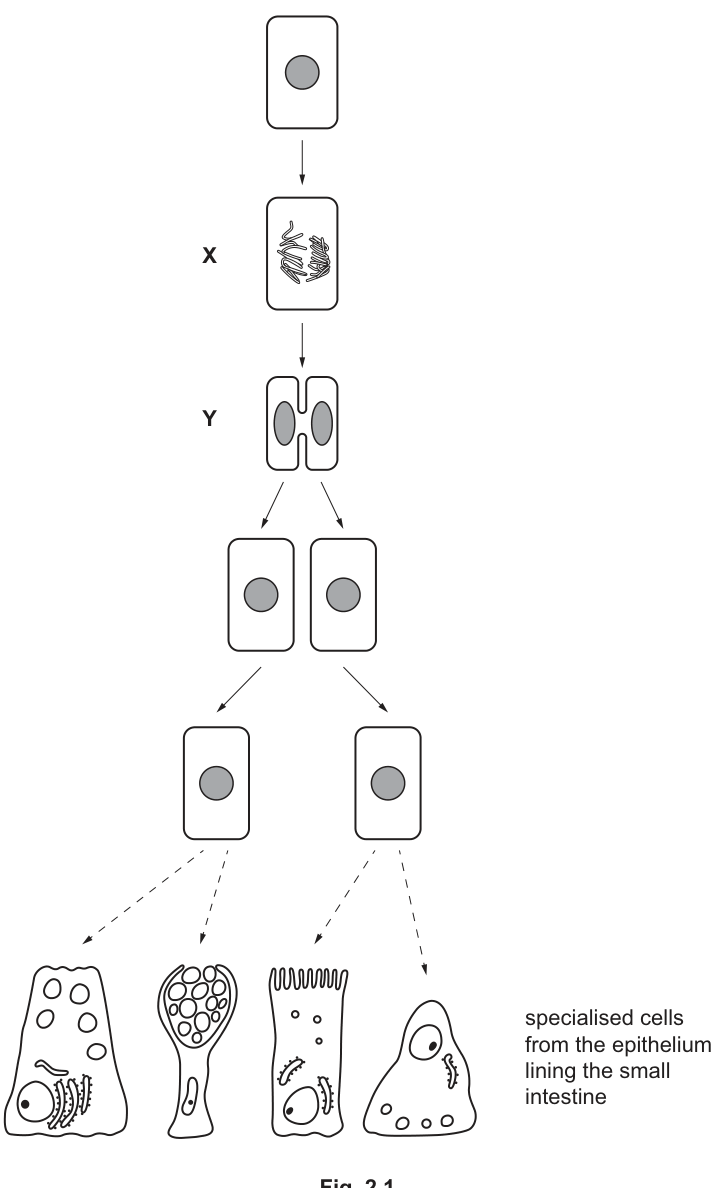
Explain why stem cells are required in places such as the lining of the small intestine.
Answer
- The epithelial cells lining the small intestine are short-lived and are constantly lost / worn away / damaged (e.g. by friction of food passing through and by pathogens), so they need to be replaced / repaired.
- Stem cells divide and differentiate / become specialised to form the different types of cell found in the epithelium.
Stem cells are required to replace lost/damaged epithelial cells of the small intestine, and they divide and differentiate to form the different specialised cell types.
Background Concept
Stem cells are unspecialised cells that can both self-renew (make more stem cells) and differentiate into one or more specialised cell types. Tissues that experience a high rate of cell loss or damage — such as the epithelium lining the small intestine, the skin, and bone marrow — depend on a resident stem-cell population to keep the tissue functional. The cells of the intestinal villus epithelium are continually abraded by passing food and exposed to digestive enzymes and microbes, so they are shed and must be replaced every few days.
Understanding the Question
The question asks why stem cells are needed in a site such as the small intestine lining. This is an "explain" question worth 2 marks, so the examiner expects two distinct, biological reasons — not just a general statement that "stem cells divide". The mark scheme requires either (a) that cells must be replaced/repaired, (b) that stem cells divide and differentiate, or (c) the idea of self-renewal.
Approach
Pick the two strongest, most distinct points from the mark scheme: replacement/repair and differentiation. The self-renewal idea is a valid third point but is worth less because the intestine is described as a site where stem cells exist, so the focus is on what they produce.
Step-by-Step Reasoning
- The small-intestine epithelium is a high-turnover tissue: cells are short-lived (only a few days), are abraded by food and damaged by acid/enzymes/pathogens, so they must be continuously replaced. → Marking point 1.
- Lgr5+ stem cells are multipotent: when they divide, one daughter remains a stem cell (self-renewal) while the other differentiates into the various specialised epithelial cells (enterocytes, goblet cells, etc.). → Marking point 2.
- Combining the two: stem cells supply a continuous supply of new, functional, specialised cells, which is exactly what the gut lining needs.
Key Takeaways
- Stem cells combine two properties: self-renewal and differentiation.
- They are essential in tissues with rapid cell turnover (gut, skin, blood).
- "Explain" questions need both the phenomenon and the reason — here, what is replaced and how it is replaced.
Common Mistakes
- Writing only "to make new cells" — too vague; you must say replace old/damaged cells or differentiate into specialised cells.
- Saying "to increase the number of stem cells" — this is the opposite of the self-renewal idea, which is about maintaining the stem-cell pool.
- Confusing stem cells with cancer cells (uncontrolled division) — stem cells divide in a regulated way.
Things to Be Careful About
- Use the precise phrase "divide and differentiate" rather than "multiply and change".
- Tie the answer to the small intestine (e.g. abrasion, enzymes, pathogens) rather than giving a generic stem-cell answer.
- "Replace dead cells" alone is only one mark — pair it with a differentiation point for the second mark.
Answer
Anaphase
Anaphase
Background Concept
Mitosis is divided into four named stages, each defined by a recognisable chromosome arrangement:
- Prophase — chromosomes condense, nuclear envelope breaks down.
- Metaphase — chromosomes line up on the equator (metaphase plate); each is held by spindle fibres attached to its centromere.
- Anaphase — centromeres split and sister chromatids are pulled to opposite poles by shortening spindle fibres.
- Telophase — chromatids arrive at opposite poles, nuclear envelopes re-form.
Cytokinesis (division of the cytoplasm) usually overlaps with telophase.
Understanding the Question
The question simply asks the candidate to name the stage of mitosis shown by cell X in Fig. 2.1. This is a "state" command word worth 1 mark.
Approach
Look at where the chromosomes are positioned and whether they are still joined as single chromosomes or have separated into chromatid groups.
Step-by-Step Reasoning
- In cell X, the chromosomes have separated into two groups that are being pulled toward opposite poles of the cell.
- This chromosome behaviour — separation of sister chromatids toward opposite poles — defines anaphase.
- (If the chromosomes were still aligned in a single line across the middle, the answer would be metaphase; if condensed but scattered, prophase.)
Key Takeaways
- Anaphase is identified by chromatids being pulled to opposite poles.
- A reliable cue is two separated clusters of chromatids, often V-shaped because the centromere leads.
Common Mistakes
- Confusing anaphase with metaphase (chromosomes still aligned) or telophase (chromatids at poles, nuclear envelopes re-forming).
- Spelling the answer "anaphase" with a capital or extra letter — examiners are usually tolerant but the spelling must be recognisable.
Things to Be Careful About
- The image is small; look carefully at whether the chromosomes are in two groups or one.
- The mark scheme is the authoritative answer; here it is anaphase.
Answer
Cytokinesis
Cytokinesis
Background Concept
The cell cycle has two major phases: interphase (G1, S, G2 — growth and DNA replication) and the M (mitotic) phase. The M phase itself contains mitosis (division of the nucleus: prophase → metaphase → anaphase → telophase) followed by cytokinesis (division of the cytoplasm to produce two separate daughter cells).
Understanding the Question
Y shows the cell physically splitting into two daughter cells. The question asks which part of the cell cycle this represents — a one-mark "state" question.
Approach
Recognise that the picture shows the cytoplasm being divided, not the chromosomes being rearranged. That distinction immediately points to cytokinesis rather than any of the four mitotic stages.
Step-by-Step Reasoning
- At Y, the two daughter nuclei are already formed and the cell membrane is pinching inward between them.
- This physical cleavage of the cytoplasm is, by definition, cytokinesis.
- Cytokinesis in animal cells proceeds by a cleavage furrow (actin–myosin contractile ring); in plant cells by a cell plate.
Key Takeaways
- Cytokinesis = division of the cytoplasm, producing two daughter cells.
- It overlaps with telophase and is the final step of the M phase.
Common Mistakes
- Writing "telophase" because the cell is splitting — but telophase refers only to the nuclear events.
- Writing "mitosis" — too vague; cytokinesis is the specific name.
Things to Be Careful About
- "State" questions need the single most precise term; "cell division" alone is too general and would not earn the mark.
Answer
- The centromere holds the two sister chromatids together (after DNA replication in S phase), so that each chromosome consists of two identical chromatids joined at the centromere.
- Spindle fibres attach to the centromere (at the kinetochore); when the centromeres divide at anaphase, the sister chromatids are pulled to opposite poles of the cell, allowing each daughter cell to receive an identical set of chromosomes.
Centromeres hold sister chromatids together and serve as the attachment point for spindle fibres; they divide in anaphase so that the chromatids can be pulled to opposite poles.
Background Concept
A chromosome is made of one DNA molecule packaged with histone proteins. After S phase the DNA has been replicated, so each chromosome consists of two identical sister chromatids joined at a constricted region called the centromere. The centromere is therefore the structural and functional hub of the chromosome during cell division.
Understanding the Question
This is a 2-mark "explain" question. The mark scheme accepts any two of: (1) holding sister chromatids together, (2) attachment of chromosomes/chromatids to spindle fibres, (3) centromere division in anaphase (with a dependent mark for moving chromatids to opposite poles).
Approach
Pick the two clearest, biologically independent points. The strongest pair is: (a) centromere holds sister chromatids together AND (b) spindle fibres attach at the centromere. Alternatively, (c) centromere divides in anaphase AND (d) allows chromatids to move to opposite poles — but (d) is only credited if (c) is given.
Step-by-Step Reasoning
- Point 1 — cohesion: After S phase, each chromosome comprises two sister chromatids held together at the centromere. This keeps the duplicated DNA as a single functional unit until it is time to separate.
- Point 2 — spindle attachment: Spindle microtubules (spindle fibres) bind to a protein structure on the centromere called the kinetochore. Each sister chromatid's centromere attaches to fibres from one pole of the spindle, so that the two chromatids of a chromosome are attached to opposite poles.
- Anaphase trigger: At the metaphase–anaphase transition, the cohesion between sister chromatids is broken and the centromere effectively "divides", allowing the spindle to shorten and pull the chromatids apart.
- Outcome: Because each chromatid is now an independent chromosome and is pulled to its respective pole, each daughter cell ends up with a complete, identical copy of the genome.
Key Takeaways
- The centromere is the joining point of two sister chromatids.
- It is the site of spindle attachment (via the kinetochore).
- Its division at anaphase is what allows sister chromatids to separate and move to opposite poles — the defining event that gives each daughter cell an identical chromosome set.
Common Mistakes
- Writing "sister chromosomes" instead of "sister chromatids" — this is explicitly rejected (R) by the mark scheme.
- Saying centromeres "hold chromosomes together" rather than "hold sister chromatids together" — too vague.
- Attributing movement of chromatids to the centromere alone — it is the spindle fibres that pull, with the centromere being the attachment site.
- Stating any of these events in interphase (e.g. centromeres attaching to spindle) — the mark scheme rejects this.
Things to Be Careful About
- "Sister chromatids" is the correct phrase — not "sister chromosomes".
- Use the term "spindle fibres" (or "spindle microtubules" / "kinetochore microtubules") rather than the vague "the spindle".
- If you choose the anaphase route, remember the dependent-mark rule: MP4 (chromatids move to opposite poles) only counts if MP3 (centromere divides) has already been given.
Fig. 2.2 shows three types of specialised cell that develop from Lgr5+ stem cells in the small intestine.
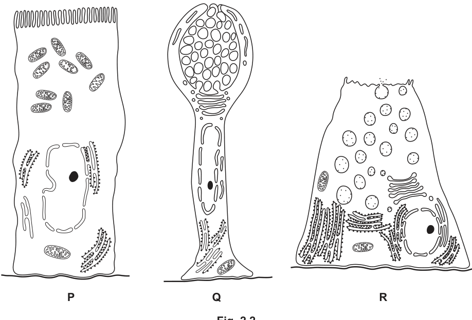
The structural features of a cell indicate its likely function.
Suggest a function of each of the cells shown in Fig. 2.2, and explain how the structure of each cell supports your suggestion.
cell P function ______
explanation ______
cell Q function ______
explanation ______
cell R function ______
explanation ______
Answer
Cell P
- Function: absorption / uptake of digested nutrients (e.g. glucose, amino acids) from the lumen of the small intestine.
- Explanation: microvilli on the apical surface greatly increase the surface area of the membrane (and therefore the number of transport proteins) for absorption; many mitochondria provide ATP for active transport of nutrients against a concentration gradient.
Cell Q
- Function: secretion of mucin (a glycoprotein) to form mucus, which lubricates and protects the epithelium.
- Explanation: the many secretory vesicles store mucin ready for release by exocytosis; rough endoplasmic reticulum synthesises the protein component of mucin and the Golgi body glycosylates / packages / modifies it.
Cell R
- Function: secretion / release of proteins (e.g. enzymes / hormones / peptides) by exocytosis.
- Explanation: abundant rough endoplasmic reticulum synthesises the proteins; a well-developed Golgi body modifies and packages them; secretory vesicles transport the proteins to the cell-surface membrane for release by exocytosis.
P – absorption of nutrients (microvilli increase surface area; mitochondria supply ATP for active transport). Q – secretion of mucin/mucus (RER makes the protein, Golgi glycosylates/packages it, vesicles store and release it). R – secretion of proteins (RER synthesises proteins, Golgi modifies/packages them, vesicles release them by exocytosis).
Background Concept
Cells in a single tissue can look very different because they are specialised to carry out particular jobs. Their structure is shaped by that job:
- Absorptive cells have lots of membrane (often folded into microvilli) and many mitochondria to power active uptake.
- Secretory cells have abundant rough endoplasmic reticulum (RER) for synthesising proteins, a prominent Golgi body for modifying and packaging those proteins, and secretory vesicles / vacuoles for storing and releasing the finished product by exocytosis.
- Mucin-secreting (goblet) cells are a special case: their apical end is packed with mucin-containing vesicles, giving a characteristic "goblet" shape; the protein backbone of mucin is made on RER and glycosylated in the Golgi.
Understanding the Question
The question supplies three drawings (P, Q, R) of epithelial cells from the small intestine and asks for a one-line function plus a structure-function explanation for each. The mark scheme awards one mark per cell for naming the function and a second mark for a structure that supports it (different valid structures can earn the second mark).
Approach
For each cell, identify its distinguishing structures, decide what they imply about its role, then write the function and the explanation as two short, paired sentences.
Step-by-Step Reasoning
Cell P — columnar cell with microvilli on the apical surface and many mitochondria.
- Microvilli = absorption surface (huge area of membrane).
- Many mitochondria = energy demand, so the cell is doing active transport of nutrients.
- Function → absorption / uptake of digested nutrients (e.g. glucose, amino acids) from the intestinal lumen.
- Explanation → microvilli increase the surface area (and the number of carrier/channel proteins) for absorption; mitochondria release ATP used to actively transport nutrients against their concentration gradient.
Cell Q — narrow-based cell with a wide apical portion filled with secretory vesicles/vacuoles (classic goblet-cell shape).
- Vesicles at the apex = stores mucin ready for release by exocytosis.
- Function → secretion of mucin (a glycoprotein that becomes mucus) to lubricate and protect the gut lining.
- Explanation → vesicles store mucin ready for exocytosis; (alternatively credited) RER makes the protein component of mucin; Golgi glycosylates/packages the mucin.
Cell R — cell with many secretory granules and extensive rough endoplasmic reticulum plus a Golgi body.
- Lots of RER + Golgi + vesicles = a protein-secreting cell.
- Function → secretion of (named) proteins — for example, hormones, peptides or digestive enzymes.
- Explanation → RER synthesises the proteins (translation of secreted proteins); Golgi modifies and packages them; vesicles transport the proteins to the cell-surface membrane and release them by exocytosis.
Key Takeaways
- Structure–function principle: organelle abundance tells you what a cell does.
- Microvilli → absorption; RER + Golgi + vesicles → secretion; many mitochondria → high ATP demand (often active transport).
- Goblet cells are specialised mucin-secreting cells with apical vesicles — a recognisable "goblet" outline.
Common Mistakes
- Saying microvilli "produce energy" or "are for digestion" — they are a surface-area adaptation, not digestive enzymes.
- Stating "produces energy" for mitochondria — examiners reject this; mitochondria release ATP, they do not make energy.
- For cell Q, saying "makes mucus" without naming mucin as the substance released, or saying "produces and provides" (also rejected).
- For cell R, saying "produces and provides" protein rather than "secretes/releases" — the question rewards the secretion verb.
- Failing to link at least one organelle directly to the named function.
Things to Be Careful About
- Name the substance secreted, not just "secretes". Mucin for Q; a named protein for R.
- Tie each organelle to a specific job (RER → synthesis; Golgi → modification/packaging; vesicles → storage/transport; microvilli → surface area for transport proteins).
- The mark scheme accepts any one valid explanation per cell, so you only need one structure-function link — but writing two makes the answer robust.
- Spelling: "mucin" (the glycoprotein) vs "mucus" (the slimy substance) — both are accepted.
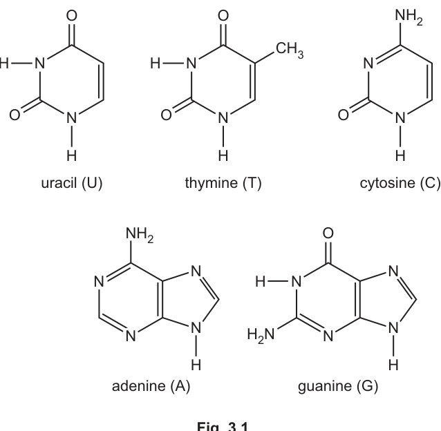
The five bases found in nucleic acids are described as nitrogenous organic compounds. There are two types of base.
Fig. 3.1 shows the structure of the five bases.
Answer
Pyrimidine(s).
Pyrimidine(s)
Background Concept
The five nitrogenous bases found in nucleic acids are split into two structural families:
- Pyrimidines — single-ring (six-membered) bases. They are smaller. The three pyrimidines are cytosine (C), thymine (T) and uracil (U).
- Purines — double-ring (fused five- and six-membered) bases. They are larger. The two purines are adenine (A) and guanine (G).
In DNA the four bases are A, T, C and G; in RNA T is replaced by U, so the four are A, U, C and G. The size difference matters because in the double helix a purine always pairs with a pyrimidine, giving a uniform width of ~2 nm.
Understanding the Question
The question shows Fig. 3.1, which gives the chemical structure of all five bases. U, T and C are drawn as single six-membered rings containing two nitrogen atoms; A and G are drawn as fused double rings containing more nitrogen atoms. The task is simply to name the family to which U, T and C belong.
Approach
Look at the ring structure in the figure and recall the two base families. A single ring = pyrimidine; a double ring = purine.
Step-by-Step Reasoning
- U, T and C each consist of a single six-membered ring.
- A and G each consist of a fused double ring (purine).
- Therefore U, T and C belong to the pyrimidine family.
Key Takeaways
- Pyrimidines = C, T, U (single ring).
- Purines = A, G (double ring).
- A purine must pair with a pyrimidine in the double helix so that the two backbones stay equally spaced.
Common Mistakes
- Writing "pyramid" or "purine" — the spelling pyrimidine is required (the mark scheme accepts phonetic attempts but rejects the wrong family).
- Confusing the family with a property such as "single-stranded" — pyrimidine is a structural classification, not a function.
Things to Be Careful About
Only the three bases named in the question (U, T, C) should be classified. Do not be distracted by A and G, which belong to the other family.
Answer
Each nucleic acid (DNA and RNA) only contains four (different) bases. In RNA, uracil (U) replaces thymine (T); DNA has A, T, C and G while RNA has A, U, C and G.
Each nucleic acid only contains four (different) bases; in RNA, U replaces T.
Background Concept
Although there are five nitrogenous bases in total across all nucleic acids, no single nucleic acid molecule contains all five at once:
- DNA contains adenine, thymine, cytosine and guanine (A, T, C, G).
- RNA contains adenine, uracil, cytosine and guanine (A, U, C, G).
The two nucleic acids differ in only one base: thymine (T) in DNA is replaced by uracil (U) in RNA. So any given nucleic acid has four bases, not five.
Understanding the Question
The stem says "all nucleic acids have five bases" and asks the candidate to give a reason why that statement is wrong. The mark scheme accepts any of three equivalent ideas.
Approach
Compare the base composition of DNA and RNA. Spot that they share A, C and G but differ in the fourth base, so each nucleic acid only carries four.
Step-by-Step Reasoning
- DNA and RNA are both nucleic acids but they do not have the same base set.
- DNA uses A, T, C and G; RNA uses A, U, C and G.
- Therefore each individual nucleic acid has only four bases, and the total of five only exists across the two types combined.
- A second valid point is the specific substitution: in RNA, uracil replaces thymine.
Key Takeaways
- Each nucleic acid (DNA or RNA) contains four bases.
- The fifth base (U in RNA or T in DNA) is absent from the other type.
- A common exam trap is to assume that because there are five bases overall, every nucleic acid carries all five.
Common Mistakes
- Stating "because some bases are in DNA and others are in RNA" without naming them — the mark scheme wants a concrete reference to which base is in which nucleic acid, or a clear statement that each nucleic acid has only four bases.
- Saying that DNA has five bases and RNA has four (or vice versa).
Things to Be Careful About
The mark scheme requires an explicit comparison (e.g. DNA has A, T, C and G; RNA has A, U, C and G) or the explicit "four bases per nucleic acid" idea, not a vague answer.
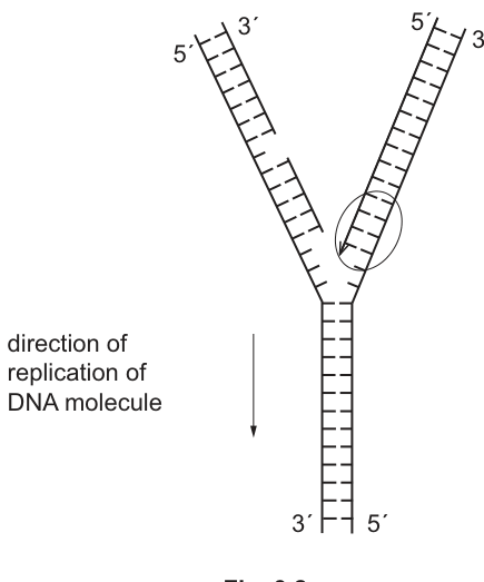
Fig. 3.2 shows a stage in the replication of DNA. The circled part is enlarged in Fig. 3.3 to show the elongation of the DNA strand that is being synthesised.
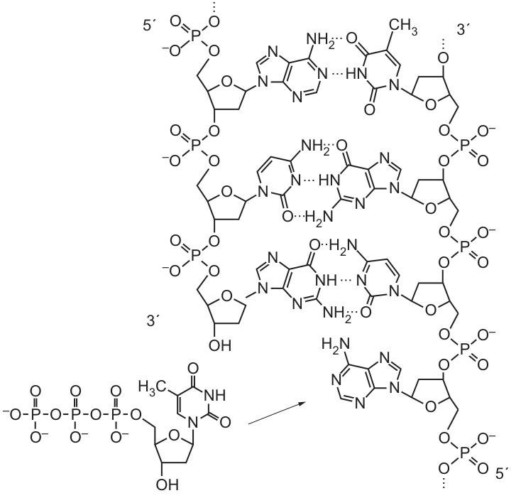
Describe the sequence of events that occurs at the stage shown in Fig. 3.3 to extend the synthesised strand.
Answer
- A free, activated (triphosphate) nucleotide carrying thymine (T) lines up alongside the exposed adenine (A) on the template strand.
- Complementary base pairing occurs: T pairs with A via two hydrogen bonds.
- DNA polymerase catalyses the pairing and bond formation.
- A phosphodiester bond forms between the 3′-OH of the deoxyribose on the existing strand and the phosphate of the incoming nucleotide.
- Pyrophosphate (two phosphates, PPᵢ) is released (hydrolysed), providing the energy for the reaction.
- The new nucleotide is therefore added to the 3′ end, so the new strand is elongated in the 5′ to 3′ direction.
An activated (triphosphate) nucleotide carrying T hydrogen-bonds (2 H-bonds) to A on the template strand; DNA polymerase catalyses formation of a phosphodiester bond between the 3′-OH of the existing strand and the phosphate of the incoming nucleotide, releasing pyrophosphate; the new strand is therefore extended at the 3′ end, i.e. in the 5′ to 3′ direction.
Background Concept
DNA replication is semi-conservative: each of the two original (parental) strands acts as a template for a new complementary strand, producing two daughter molecules each containing one old and one new strand. Replication occurs at a replication fork where helicase has unwound the double helix, exposing the template bases. New DNA is built from activated deoxyribonucleoside triphosphates (dATP, dTTP, dCTP, dGTP) which carry enough energy in their high-energy phosphate bonds to drive polymerisation.
The enzyme that builds the new strand is DNA polymerase. It can only add nucleotides to a free 3′-OH group, so the new strand is always elongated in the 5′ → 3′ direction. Each addition forms a phosphodiester bond between the 3′-OH of the last nucleotide already in place and the α-phosphate of the incoming nucleotide, and releases pyrophosphate (PPᵢ), which is then hydrolysed to two inorganic phosphates, making the reaction essentially irreversible.
Base-pairing rules are strict: A pairs with T (via two hydrogen bonds) and C pairs with G (via three hydrogen bonds). In Fig. 3.3, an incoming dTTP is shown pairing with an exposed A on the template strand.
Understanding the Question
The question asks the candidate to describe the sequence of events at the molecular scale that elongates a new DNA strand at the point circled in Fig. 3.2 and enlarged in Fig. 3.3. The mark scheme provides eight possible points and awards any four of them. The candidate must connect the molecular picture (an incoming triphosphate nucleotide opposite a template A) to the chemistry (hydrogen bonding, phosphodiester bond formation, pyrophosphate release) and to directionality (3′ end, 5′ to 3′).
Approach
- Identify the molecular event: a free nucleotide is being added to the growing strand.
- State what is happening to the incoming nucleotide (triphosphate, carries T, complementary to A on the template).
- State how it joins (H-bonds to A, then a phosphodiester bond formed by DNA polymerase to the 3′-OH of the previous nucleotide).
- State the by-product (pyrophosphate) and the consequence (the strand grows at the 3′ end, in the 5′ → 3′ direction).
Step-by-Step Reasoning
- An activated, free (triphosphate) nucleotide carrying thymine (T) is the substrate shown arriving at the bottom of Fig. 3.3.
- The template strand (already part of the original double helix) presents an adenine (A) base. T and A are held together by two hydrogen bonds — this is the complementary base-pairing step.
- The enzyme DNA polymerase catalyses the next step: it brings the incoming nucleotide into position and joins it to the existing strand.
- A phosphodiester bond is formed between the 3′-OH of the deoxyribose of the last nucleotide already on the new strand and the α-phosphate of the incoming nucleotide.
- Pyrophosphate (PPᵢ, two phosphates) is released and hydrolysed, providing the energy that makes the polymerisation reaction proceed in the forward direction.
- Because DNA polymerase can only add to a free 3′-OH, the new strand is elongated at the 3′ end, i.e. in the 5′ → 3′ direction.
Key Takeaways
- A new nucleotide is added to the 3′ end of the growing strand, so synthesis proceeds 5′ → 3′.
- The incoming nucleotide is a deoxyribonucleoside triphosphate; the energy comes from cleaving the high-energy phosphates (release of pyrophosphate).
- A pairs with T via two H-bonds; C pairs with G via three H-bonds.
- The enzyme is DNA polymerase (not DNA ligase — ligase joins pre-existing Okazaki fragments, it does not build the strand nucleotide-by-nucleotide).
Common Mistakes
- Mentioning DNA ligase instead of (or as well as) DNA polymerase — the mark scheme explicitly rejects ligase for this step.
- Writing that the new strand is extended at the 5′ end or in the 3′ → 5′ direction — this is the single most common error in this type of question.
- Saying the new nucleotide has "phosphate" attached rather than three phosphates (triphosphate), and omitting the release of pyrophosphate.
- Describing transcription / mRNA / ribosomes / tRNA — the mark scheme caps the answer at 2 marks if it strays into transcription rather than DNA replication.
- Stating the number of H-bonds for C–G instead of A–T.
Things to Be Careful About
- Use the term phosphodiester bond specifically, and locate it correctly (3′-OH to phosphate of incoming nucleotide).
- Use the term pyrophosphate (or PPᵢ / two phosphates) — not just "phosphate".
- Use the term DNA polymerase, not "polymerase" alone, and not "DNA ligase".
- Directionality: the mark scheme explicitly accepts "5′ to 3′" as an alternative to "nucleotide added to the 3′ end"; either wording scores the point.
The two strands in a molecule of DNA are described as antiparallel.
With reference to Fig. 3.2 and Fig. 3.3, state what is meant by antiparallel, and explain how the antiparallel arrangement of the strands determines how new strands are synthesised.
Answer
- The two strands of DNA are antiparallel: they run in opposite directions — one strand runs 5′ → 3′ and the other runs 3′ → 5′ (visible in Fig. 3.2).
- Because DNA polymerase can only add nucleotides to the 3′ end of the growing strand, each new strand must be elongated in the 5′ → 3′ direction.
- The strand being synthesised towards the replication fork is built continuously and is the leading strand; the other strand (whose template runs the opposite way relative to fork movement) is built in short Okazaki fragments, which are later joined together by DNA ligase — this is the lagging strand.
Antiparallel: the two DNA strands run in opposite directions (one 5'→3', the other 3'→5'). Because DNA polymerase only adds nucleotides to the 3' end, each new strand is built 5'→3', giving a continuously synthesised leading strand and a discontinuously synthesised lagging strand (Okazaki fragments joined by DNA ligase).
Background Concept
The two sugar-phosphate backbones of a DNA double helix are described as antiparallel: they run in opposite chemical directions. Each strand has a 5′ end (with a free phosphate on carbon 5 of the terminal deoxyribose) and a 3′ end (with a free -OH on carbon 3 of the terminal deoxyribose). In the double helix, the 5′ end of one strand lies next to the 3′ end of the other.
DNA polymerase is directionally restricted: it can only add a new nucleotide to a free 3′-OH group. This single constraint dictates how the two new strands are made at a replication fork:
- On one template the new strand runs continuously towards the fork — the leading strand (synthesised 5′ → 3′).
- On the other template the new strand must be built away from the fork in short pieces called Okazaki fragments — the lagging strand (still synthesised 5′ → 3′, but discontinuously). The fragments are later stitched together by DNA ligase.
Understanding the Question
The question has two linked parts:
- State what "antiparallel" means (referring to Fig. 3.2 which shows the 5′ and 3′ ends at the fork).
- Explain how the antiparallel arrangement determines how the new strands are synthesised (referring to Fig. 3.3 which shows the 3′-OH and the 5′ → 3′ direction of growth).
The mark scheme awards 1 mark for the definition and 2 further marks for any two of the mechanistic points.
Approach
- Definition: opposite directions, one 5′→3′ and one 3′→5′.
- Mechanistic consequence: DNA polymerase only adds to 3′-OH, so synthesis is 5′→3′, giving a leading strand (continuous) and a lagging strand (Okazaki fragments, joined by DNA ligase).
Step-by-Step Reasoning
- Fig. 3.2 shows the 5′ and 3′ ends labelled on both strands at the replication fork. Reading down the left strand: 5′ at the top, 3′ at the bottom. Reading down the right strand: 3′ at the top, 5′ at the bottom. They point in opposite directions — this is antiparallel.
- Because the backbones point in opposite directions, the chemical environment at the two growing points is different: one growing strand has a free 3′-OH pointing towards the fork, the other has a free 3′-OH pointing away from the fork.
- DNA polymerase only catalyses addition to a free 3′-OH, so synthesis always proceeds 5′ → 3′.
- On the template whose 3′-OH points towards the fork, the new strand can be made continuously — this is the leading strand.
- On the template whose 3′-OH points away from the fork, the new strand must be made in short Okazaki fragments moving away from the fork, each synthesised 5′ → 3′; the fragments are later sealed by DNA ligase — this is the lagging strand.
Key Takeaways
- Antiparallel = strands run in opposite chemical directions, one 5′→3′ and the other 3′→5′.
- 5′ → 3′ synthesis is the only direction DNA polymerase can work.
- This single constraint produces the leading strand (continuous) and the lagging strand (Okazaki fragments joined by DNA ligase).
Common Mistakes
- Defining antiparallel as "the strands are not parallel" without mentioning directions or 5′/3′ labels — the mark scheme requires the explicit opposite-direction idea, ideally with reference to 5′ and 3′.
- Stating that synthesis proceeds 3′ → 5′ — this is the most common misconception and is explicitly rejected by the mark scheme.
- Saying that one strand is "shorter" than the other or that the bases face each other — these are not the meaning of antiparallel.
- Confusing the leading and lagging strands (the leading strand is the one made continuously towards the fork, not the one nearer the top of Fig. 3.2).
Things to Be Careful About
- The mark scheme requires the direction of each strand (5′→3′ vs 3′→5′) in the definition, not just "opposite".
- The mark scheme accepts any two of the linked mechanistic points: (i) synthesis is 5′→3′, (ii) DNA polymerase moves 5′→3′ / adds to 3′ end, (iii) phosphate joins C3 of the last nucleotide, (iv) leading strand is continuous, (v) lagging strand is Okazaki fragments, (vi) fragments joined by DNA ligase.
- Reference back to Fig. 3.2 and Fig. 3.3 to ground the answer in the 5′/3′ labels that are actually drawn on the figures.
Biofuels contain alcohols that are produced by the fermentation of sugars derived from crop waste. This waste contains cellulose and other organic compounds in cell walls.
Scientists investigated the production of sugars from crop waste for biofuel production. The scientists discovered that a strain of the fungus Penicillium citrinum, isolated from soil, was a good source of three different extracellular enzymes, M, N and O. These enzymes break down polysaccharides in cell walls.
The scientists cultured P. citrinum in a liquid medium containing cell wall material. Samples of the liquid were taken, and the three enzymes were separated from the medium. Each enzyme was placed in a reaction mixture with an appropriate substrate. The activity of each enzyme was determined to give a measurement of the quantity of enzyme produced by P. citrinum.
The results are shown in Table 4.1.
Table 4.1
| enzyme | maximum activity / arbitrary units |
|---|---|
| M | 292.83 |
| N | 111.72 |
| O | 6.54 |
Optimum conditions for each enzyme were used to obtain the results in Table 4.1. The conditions were different for each enzyme.
The scientists carried out further research so that a solution containing the three enzymes (enzyme mixture) could be used for the most efficient production of sugars from crop waste.
Suggest what the scientists needed to find out in their research.
Answer
-
Find the optimum pH and temperature for the enzyme mixture (a compromise that allows the three enzymes to work together efficiently, since each enzyme has different optimum conditions).
-
Find the appropriate concentrations of each enzyme in the mixture to give the most efficient breakdown of the crop waste.
-
Find a suitable crop waste with a high concentration of substrate for enzyme M (which has the highest maximum activity), or find a crop waste that is a good substrate for all three enzymes.
-
Identify the sugars produced so that the most useful sugars for fermentation into biofuels can be obtained.
-
Find the rate of sugar production (how long the hydrolysis takes).
-
Find whether any products of the reaction are toxic to the enzymes.
-
Find whether any inhibitors or cofactors are present in the solution that could affect the enzymes.
-
Find the best pre-treatment of the crop waste (e.g., grinding to increase surface area).
-
Find the best ratio of enzyme mixture to crop waste.
-
Consider whether enzyme immobilisation could be used to allow reuse of the enzymes and increase efficiency.
Optimum pH and temperature for the enzyme mixture; appropriate concentrations of each enzyme; suitable crop waste; identify the sugars produced; rate of sugar production; check for inhibitors/cofactors or toxic products; pre-treatment of crop waste; ratio of enzyme mixture to substrate; enzyme immobilisation.
Background Concept
Enzymes are biological catalysts that speed up biochemical reactions without being consumed. Each enzyme has its own optimum conditions (pH and temperature) at which it works most efficiently. At these optimum conditions, the enzyme's active site has the correct shape to bind to the substrate, and the enzyme's tertiary structure is maintained.
The activity of an enzyme is affected by:
- Enzyme concentration - more enzyme means more active sites and a faster reaction
- Substrate concentration - more substrate means more enzyme-substrate complexes can form
- Temperature - higher temperatures increase kinetic energy but denature the enzyme above the optimum
- pH - affects the ionisation of amino acid side chains in the active site
- Inhibitors - competitive inhibitors block the active site, non-competitive inhibitors change the shape of the active site
- Cofactors - some enzymes require non-protein helpers (e.g., metal ions) to function
In industrial applications, multiple enzymes are often used together to break down complex substrates. The efficiency of the enzyme mixture depends on many factors, including the compatibility of the enzymes' optimum conditions, the ratio of enzymes to substrate, the pre-treatment of the substrate, and whether the products inhibit the enzymes.
Understanding the Question
The context is that scientists have discovered a fungus, Penicillium citrinum, that produces three extracellular enzymes (M, N, and O) which break down polysaccharides in cell walls. The enzymes have been studied individually, and their maximum activities are known (with M being the most active at 292.83 arbitrary units, N at 111.72, and O at 6.54). The scientists now want to use the three enzymes together as a mixture for the most efficient production of sugars from crop waste for biofuel production.
The question asks what the scientists needed to find out in their FURTHER research. This is a 'suggest' question, which means candidates need to use their biological knowledge to make reasoned suggestions about what experiments or investigations are needed. The command word 'suggest' indicates that there is no single correct answer - any well-reasoned suggestion is acceptable, and the mark scheme offers 11 alternative creditable points.
Approach
Since the enzymes have already been studied individually, the focus should be on what is needed to use them together as a mixture and to apply them to crop waste. Think about:
- What conditions are needed for the mixture to work efficiently (since each enzyme has different optimum conditions, finding a compromise is important)
- What substrate is needed (crop waste composition varies, and enzyme M has the highest activity)
- Whether the products are useful for the intended application (biofuel production)
- Whether anything could interfere with the enzymes (inhibitors, cofactors, toxic products)
- Industrial considerations (enzyme immobilisation, pre-treatment of substrate)
Step-by-Step Reasoning
-
Optimum pH and temperature for the mixture: Since each of the three enzymes has different optimum conditions (as stated in the question), the scientists need to find a pH and temperature at which the enzyme mixture as a whole works most efficiently. This is a compromise, as the conditions cannot be optimum for all three enzymes at the same time.
-
Concentrations of each enzyme in the mixture: The relative amounts of M, N, and O in the mixture will affect the rate at which the polysaccharides are broken down. The mixture should be optimised so that all three enzymes work together efficiently.
-
Suitable crop waste: Different types of crop waste have different compositions. The scientists need to find a crop waste that is a good substrate for the three enzymes, particularly for enzyme M (which has the highest maximum activity, suggesting it acts on the most abundant polysaccharide).
-
Identify the sugars produced: Different enzymes break down different polysaccharides, producing different sugars (e.g., glucose, xylose, arabinose). The scientists need to identify the sugars produced so they can be used for fermentation into biofuels.
-
Rate of sugar production: The scientists need to know how long the hydrolysis takes, so the process can be optimised for industrial production.
-
Toxic products: Some hydrolysis products may be toxic to the enzymes, slowing down or stopping the reaction (product inhibition). The scientists need to check for this.
-
Inhibitors or cofactors: The crop waste may contain substances that inhibit or enhance the activity of the enzymes. The scientists need to check for this.
-
Pre-treatment of crop waste: The crop waste may need to be pre-treated (e.g., ground, washed, heated) to make the polysaccharides more accessible to the enzymes and to remove any inhibitors.
-
Ratio of enzyme mixture to crop waste: This affects the rate and efficiency of the hydrolysis. Too little enzyme and the reaction is slow; too much and the enzymes are wasted.
-
Enzyme immobilisation: This is an industrial technique where enzymes are attached to a solid support, allowing them to be reused and making the product easier to separate. The scientists may want to consider this for the enzyme mixture.
Key Takeaways
- When using multiple enzymes together, the optimum conditions for the mixture may differ from the individual enzymes' optima.
- Many factors affect the efficiency of enzyme-catalysed reactions in industry, including substrate concentration, enzyme concentration, pH, temperature, and the presence of inhibitors or cofactors.
- The substrate must be appropriate for the enzyme, and the products must be useful for the intended application.
- 'Suggest' questions require the use of biological knowledge to make reasoned suggestions; there is no single correct answer, and the mark scheme typically offers a list of alternative creditable points.
Common Mistakes
- Stating 'find the optimum pH of each enzyme' - this has already been done and is what Table 4.1 shows (explicitly rejected by mark scheme with 'I' = ignore).
- Saying 'amount' or 'proportion' of enzymes instead of 'concentration' (explicitly rejected by mark scheme with 'I' = ignore for 'proportions').
- Vague answers like 'find the right conditions' - this is too vague and does not specify what conditions.
- Suggesting things that have already been done (e.g., finding the maximum activity of each enzyme, which is in Table 4.1).
- Not relating the suggestions to the specific context of using the enzymes together as a mixture on crop waste.
- Forgetting that the three enzymes have different optimum conditions, so a compromise is needed for the mixture.
Things to Be Careful About
- The question says 'for the enzyme mixture' - so conditions are for the mixture as a whole, not for individual enzymes.
- 'Concentrations' not 'proportions' or 'amounts' (this is specifically mentioned in the mark scheme as rejected).
- The substrate is crop waste, not pure polysaccharides - the scientists need to consider the variable composition of crop waste.
- The mark scheme gives 11 possible points - candidates need to choose any four well-reasoned suggestions.
- The question is worth 4 marks, so candidates need to give four distinct points.
Students investigated the composition of the cell wall of leaf cells of thale cress, Arabidopsis thaliana. The students began by isolating the cell wall components from the rest of the cell material.
The students used enzymes extracted from a fungal pathogen of A. thaliana to hydrolyse the cell wall components to smaller molecules.
The students prepared a reaction mixture containing the cell wall components and the enzymes.
After 24 hours, they separated and identified the smaller molecules found in the reaction mixture.
Four types of molecule were identified:
• short chains of -glucose
• -glucose
• peptides
• amino acids.
Explain the presence of these molecules in the reaction mixture after 24 hours of hydrolysis.
Answer
- The cell wall of A. thaliana contains cellulose, a polysaccharide of β-glucose monomers joined by β-1,4 glycosidic bonds. The enzyme mixture contained cellulase enzymes, which hydrolysed the glycosidic bonds.
- The presence of β-glucose (monomers) shows that some cellulose was completely hydrolysed to monomers.
- The presence of short chains of β-glucose shows that some cellulose was only partially hydrolysed (hydrolysis was incomplete after 24 hours, so not all glycosidic bonds were broken).
- The cell wall also contains proteins/polypeptides (e.g., expansins). The enzyme mixture contained protease enzymes, which hydrolysed the peptide bonds.
- The presence of amino acids shows that some proteins were completely hydrolysed.
- The presence of peptides shows that some proteins were only partially hydrolysed (not all peptide bonds were broken).
Cellulose was hydrolysed by cellulase (breaking β-1,4 glycosidic bonds) to β-glucose monomers and short β-glucose chains (partial hydrolysis). Proteins were hydrolysed by protease (breaking peptide bonds) to amino acids and peptides (partial hydrolysis). The cell wall contains both cellulose and proteins.
Background Concept
Plant cell walls are complex structures that provide support and protection to the cell. The main components of plant cell walls are:
- Cellulose - a polysaccharide made of β-glucose monomers joined by β-1,4 glycosidic bonds. Cellulose forms microfibrils that give the cell wall its tensile strength.
- Hemicellulose - a branched polysaccharide that cross-links the cellulose microfibrils
- Pectin - a polysaccharide that forms a gel-like matrix, making the cell wall flexible and hydrated
- Proteins - including structural proteins like expansins, which are involved in cell wall loosening and growth, and enzymes involved in cell wall metabolism
Enzymes can break down biological molecules by hydrolysis (the addition of water to break a covalent bond). Different enzymes break down different molecules:
- Cellulase breaks down cellulose by hydrolysing the β-1,4 glycosidic bonds between β-glucose monomers
- Protease breaks down proteins by hydrolysing the peptide bonds between amino acids
Hydrolysis can be complete (yielding monomers) or partial (yielding shorter polymers). The extent of hydrolysis depends on the time, the enzyme concentration, the temperature, and the pH. After a fixed time, a mixture often contains both monomers and short polymers because hydrolysis is incomplete.
Fungal pathogens of plants often secrete cell wall-degrading enzymes (cellulase, protease, pectinase) to break down the plant cell wall and gain access to the cell contents.
Understanding the Question
The context is that students investigated the composition of the cell wall of thale cress (Arabidopsis thaliana). They:
- Isolated the cell wall components from the rest of the cell material
- Used enzymes extracted from a fungal pathogen of A. thaliana to hydrolyse the cell wall components to smaller molecules
- Prepared a reaction mixture containing the cell wall components and the enzymes
- After 24 hours, separated and identified the smaller molecules in the reaction mixture
Four types of molecule were found:
- Short chains of β-glucose
- β-glucose
- Peptides
- Amino acids
The question asks to explain the presence of these molecules. This is an 'explain' question, which means candidates need to give reasons for the observations, not just describe them. The candidates need to link the products to the substrates (cell wall components) and the enzymes used, and explain why both monomers and short polymers are present.
Approach
To explain the presence of these four types of molecule, consider:
- What was the original substrate? (cell wall components of A. thaliana, i.e. cellulose and proteins)
- What enzymes were used? (fungal enzymes, including cellulase and protease)
- What bonds are broken? (β-1,4 glycosidic bonds in cellulose, peptide bonds in proteins)
- Why are there both monomers and short polymers? (partial hydrolysis - 24 hours was not enough for complete hydrolysis)
The presence of β-glucose AND short chains of β-glucose indicates that cellulose was broken down. The presence of amino acids AND peptides indicates that proteins were broken down. The presence of both monomers and short polymers indicates that hydrolysis was incomplete.
Step-by-Step Reasoning
-
Cellulose was present in the cell wall: Plant cell walls contain cellulose, which is a polymer of β-glucose monomers joined by β-1,4 glycosidic bonds. Cellulose is a major component of plant cell walls.
-
Cellulase hydrolysed the glycosidic bonds: The fungal pathogen of A. thaliana produces cellulase enzymes, which hydrolyse the β-1,4 glycosidic bonds in cellulose, breaking it down to β-glucose monomers.
-
β-glucose is present: Some of the cellulose was completely hydrolysed, breaking all the glycosidic bonds and releasing β-glucose monomers.
-
Short chains of β-glucose are present: Some of the cellulose was only partially hydrolysed - not all the glycosidic bonds were broken, so short chains of β-glucose remained. This indicates that 24 hours was not enough time for complete hydrolysis, or that some bonds are more resistant to hydrolysis than others (e.g., bonds in crystalline regions of cellulose).
-
Proteins were present in the cell wall: Plant cell walls contain proteins (e.g., expansins), which are polymers of amino acids joined by peptide bonds. This is a key point because some students forget that plant cell walls contain protein as well as polysaccharide.
-
Protease hydrolysed the peptide bonds: The fungal pathogen produces protease enzymes, which hydrolyse the peptide bonds in proteins, breaking them down to amino acids.
-
Amino acids are present: Some of the proteins were completely hydrolysed, breaking all the peptide bonds and releasing amino acids.
-
Peptides are present: Some of the proteins were only partially hydrolysed - not all the peptide bonds were broken, so short peptides remained. This indicates that 24 hours was not enough time for complete hydrolysis, or that some peptide bonds are more resistant to hydrolysis than others (e.g., bonds involving bulky or charged amino acid side chains).
Key Takeaways
- Plant cell walls contain both polysaccharides (cellulose) and proteins (e.g., expansins).
- Fungal pathogens produce hydrolytic enzymes (cellulase, protease) to break down plant cell walls.
- Hydrolysis can be complete (yielding monomers) or partial (yielding shorter polymers).
- The products of an enzyme reaction can give information about both the substrate and the enzyme.
- β-glucose is the specific monomer of cellulose; it is not the same as α-glucose (the monomer of starch and glycogen).
- An 'explain' question requires giving reasons, not just describing observations - the presence of both monomers and short polymers is the key observation that needs to be explained.
Common Mistakes
- Saying 'glucose' instead of 'β-glucose' - the question specifically says β-glucose, and the mark scheme credits this specificity. α-glucose is the monomer of starch and glycogen, not cellulose.
- Saying 'collagen' - collagen is an animal protein, not a plant cell wall protein (explicitly rejected by mark scheme with 'R' = reject). Plant cell wall proteins include expansins.
- Not mentioning the partial hydrolysis - the presence of both monomers AND short polymers is a key observation that needs to be explained.
- Saying 'polysaccharides' without specifying cellulose - the question is about cellulose specifically.
- Saying 'enzymes break down the cell wall' without specifying the bonds broken (glycosidic and peptide bonds).
- Confusing hydrolysis with condensation - hydrolysis breaks bonds by adding water; condensation forms bonds by removing water.
- Forgetting that plant cell walls contain protein (e.g., expansins) as well as polysaccharide.
Things to Be Careful About
- Use 'β-glucose' not 'glucose' - this is the specific monomer of cellulose.
- Use 'peptide bonds' for the bonds in proteins/polypeptides.
- Use 'glycosidic bonds' (or 'β-1,4 glycosidic bonds' for more specificity) for the bonds in cellulose.
- The 'short chains' and 'peptides' indicate partial hydrolysis - this is a key point and worth a marking point on its own.
- Cell wall proteins include expansins - mentioning specific examples (e.g., expansins) can be credited as AVP (additional valid point).
- The question is about hydrolysis, not digestion - the enzymes break down the polymers by adding water across the bonds.
- The question is worth 4 marks, so candidates need to give four distinct points covering both the polysaccharide and protein components and the partial vs complete hydrolysis.
5 T-lymphocytes are produced in bone marrow and mature in the thymus gland.
When mature, T-lymphocytes leave the thymus gland to travel throughout the body. They remain inactive inside organs, such as the spleen and lymph nodes, until activated by the presence of antigens.
Fig. 5.1 shows what happens to two inactive T-lymphocytes, U1 and V1, in the presence of an antigen from a virus.
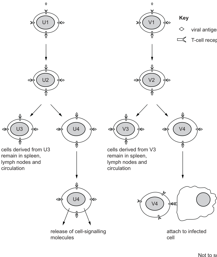
U4 and V4 are types of active T-lymphocyte. State the names given to these types of T-lymphocyte.
U4 ______
V4 ______
Answer
U4 — T-helper (lymphocyte / cell)
V4 — T-killer / T-cytotoxic (lymphocyte / cell)
U4: T-helper (lymphocyte/cell); V4: T-killer/T-cytotoxic (lymphocyte/cell)
Background Concept
T-lymphocytes (T cells) originate in the bone marrow but migrate to the thymus gland to mature. Once mature, each T cell carries a single, unique T-cell receptor on its surface that recognises one specific antigen. When an antigen is encountered, the matching T cell is selected, activated and divides by mitosis (clonal selection and clonal expansion), giving rise to a population of effector cells and long-lived memory cells. From one activated T cell, the body produces:
- T-helper cells — so named because they 'help' orchestrate the rest of the immune response by releasing signalling molecules.
- T-killer cells (also called T-cytotoxic cells) — so named because they directly destroy infected cells.
- Memory cells of each type, which remain dormant until the same antigen is met again.
The diagram in Fig. 5.1 shows this pattern for two independent T cells (U1 and V1), each selected by the same viral antigen. U1 gives rise to U3 (memory cells) and U4 (an effector cell releasing cell-signalling molecules), while V1 gives rise to V3 (memory cells) and V4 (an effector cell attaching to an infected cell).
Understanding the Question
The question asks for the standard names of the two effector T-lymphocyte sub-types shown in Fig. 5.1. Both U4 and V4 are active T-lymphocytes derived from activated, antigen-specific precursors. The clue is in what they do: U4 releases cell-signalling molecules (a defining feature of helper cells), and V4 attaches to and destroys an infected cell (the defining feature of killer/cytotoxic cells).
Approach
Match the visible behaviour in the diagram to the standard terminology:
- Cell that releases signalling molecules → T-helper (lymphocyte).
- Cell that attaches to and kills an infected cell → T-killer / T-cytotoxic (lymphocyte).
Step-by-Step Reasoning
- U4 is shown releasing 'cell-signalling molecules' that influence other immune cells. In immunology these signalling molecules are cytokines (interleukins), and the cell that produces them to coordinate B cells and macrophages is the T-helper lymphocyte.
- V4 is shown binding to an infected (host) cell and destroying it. The T-lymphocyte sub-type that recognises antigen presented on the surface of an infected cell and then kills that cell is the T-killer (or T-cytotoxic) lymphocyte.
- Both names must be supplied for the mark. Common synonyms are accepted, but the mark scheme explicitly ignores vague forms such as 'helping' or 'killing' cells — these are verbs, not cell names.
Key Takeaways
- All mature T lymphocytes arise in bone marrow and mature in the thymus.
- The two principal effector sub-types are T-helper and T-killer (cytotoxic) lymphocytes; each is paired with its own memory-cell population.
- Behaviour shown in a diagram (releasing cytokines vs killing an infected cell) identifies which sub-type is which.
Common Mistakes
- Writing 'helper cell' and 'killer cell' without the T-prefix — biology mark schemes usually want the full 'T-helper' / 'T-killer' label.
- Writing only the verb ('helping', 'killing') — these describe actions, not cell types, and are rejected.
- Confusing T-killer cells with NK (natural killer) cells, which are part of the innate immune system and not T-lymphocytes.
Things to Be Careful About
- Both U4 and V4 names must be given; one correct and one incorrect scores 0.
- 'T-cytotoxic' and 'T-killer' are interchangeable acceptable forms. 'Suppressor T cell' is a different sub-type and would be wrong here.
Answer
U4 (T-helper lymphocyte):
- secretes / releases cytokines (interleukins)
- to stimulate B-lymphocytes to divide and develop into plasma cells (that secrete antibodies)
- and to stimulate macrophages to carry out phagocytosis more actively
V4 (T-killer / T-cytotoxic lymphocyte):
- attaches to an infected cell and releases chemicals (e.g. perforin) that kill / break down the infected cell (e.g. by forming pores in its membrane, leading to lysis / apoptosis)
T-helper cells release cytokines to stimulate B cells (to become plasma cells) and macrophages; T-killer cells release chemicals such as perforin to destroy infected cells.
Background Concept
In a primary immune response, an antigen encountered for the first time activates a small number of antigen-specific lymphocytes that then proliferate. The two effector T-cell sub-types produced have complementary jobs:
- T-helper lymphocytes are the 'coordinators'. They do not themselves kill pathogens. Instead, they release soluble signalling proteins called cytokines (especially interleukins) that stimulate other immune cells — B-lymphocytes to multiply and mature into antibody-secreting plasma cells, and macrophages to phagocytose more aggressively.
- T-killer (T-cytotoxic) lymphocytes are the 'assassins'. They recognise viral (or other foreign) antigen presented on the surface of an infected host cell, bind to that cell, and release toxic chemicals that destroy it. The classic chemical is perforin, which punches holes in the infected cell's plasma membrane so that other enzymes (e.g. granzymes) can enter and trigger apoptosis. Hydrogen peroxide, released by some killer cells, oxidises cell components and contributes to the killing.
Both helper and killer responses also produce memory cells (U3 and V3 in the diagram), which remain after the infection has been cleared and respond rapidly if the same antigen reappears — the basis of the secondary response.
Understanding the Question
The question (worth 4 marks) requires describing — not just naming — the roles of U4 and V4 in the primary immune response. Marks are awarded as discrete bullet points, so the candidate should give distinct, separate statements about helper functions and killer functions. Each statement should use correct terminology rather than vague phrasing like 'helps fight infection' or 'kills the virus'.
Approach
Split the answer cleanly into two halves:
- U4 (T-helper): state the signalling molecules, the targets they act on (B cells, macrophages), and the effect on each target.
- V4 (T-killer): state that it secretes chemicals, name one (e.g. perforin), describe briefly how it kills infected cells.
Use only points that the mark scheme accepts. Avoid the words 'helping' and 'killing' on their own — they describe actions, not biology.
Step-by-Step Reasoning
For U4 (T-helper):
- Point 1: The T-helper cell secretes / releases cytokines (interleukins). Note that 'cell signalling molecules' shown on the diagram is too vague — the mark scheme requires the term 'cytokines' or 'interleukins'.
- Point 2: These cytokines stimulate B-lymphocytes to divide and develop into plasma cells (which then secrete antibodies). Acceptable alternatives include 'stimulates the humoral response' or 'clonal expansion'.
- Point 3: The cytokines also stimulate macrophages to become more actively phagocytic.
For V4 (T-killer):
- Point 4: The T-killer cell produces / secretes chemicals that kill the infected cell (or cause its lysis / apoptosis).
- Point 5: Name at least one such chemical — perforin (most common), hydrogen peroxide, or granzymes/proteases.
- Point 6 (optional extra): A short detail of how the named chemical works — e.g. 'perforin makes pores in the cell surface membrane so granzymes can enter and break down proteins inside the cell'.
Any four of these points earn full marks. A strong answer typically covers at least one helper point and one killer point with a named chemical.
Key Takeaways
- T-helper cells do not kill pathogens directly; they orchestrate the response via cytokines (interleukins) acting on B cells and macrophages.
- T-killer cells destroy infected body cells, principally using perforin to punch holes that allow granzymes to enter and trigger apoptosis.
- Both populations generate memory cells for a faster secondary response on re-exposure.
Common Mistakes
- Writing 'T-helper cells kill the virus' — helpers do not kill directly; they signal to other cells.
- Saying T-killer cells 'kill the virus' instead of 'kill the infected cell' — the virus is intracellular, so it is the host cell that must be destroyed.
- Using the vague term 'cell signalling molecules' from the diagram instead of the correct term 'cytokines' / 'interleukins'. The mark scheme explicitly ignores 'cell signalling molecules'.
- Naming 'antibiotics' as the killer substance — T cells do not produce antibiotics; this is a confused link with B cells and medicines.
Things to Be Careful About
- The mark scheme ignores 'endocytosis' as a description of macrophage activity and 'cytokinins' (a plant hormone) as a confusion with 'cytokines'.
- 'Lysis' and 'apoptosis' are both acceptable descriptions of how an infected cell is destroyed.
- 'Hydrolytic enzymes' is an acceptable alternative wording for granzymes / proteases.
- The question says 'primary' immune response; descriptions of the secondary response (memory cells, faster response) will not score marks here.
Polio is a highly infectious viral disease. The virus infects the nervous system of humans. The disease can cause total paralysis within hours and can be fatal.
The Global Polio Eradication Initiative (GPEI) was started in 1988 by the World Health Organization. In 2022, polio had been successfully eradicated from most of the world. However, cases of the disease have been recorded in some countries.
Discuss the steps that must be taken by health authorities during a vaccination programme if an infectious disease, such as polio, is to be eradicated from the whole world.
Answer
Any four from:
- Make the vaccine free and available globally, including to remote / rural populations (not just urban centres), so that no group is excluded.
- Achieve herd immunity by vaccinating a very high proportion of the population, so that the pathogen cannot spread to the small number of unvaccinated individuals.
- Vaccinate children as early as possible, ideally as part of a routine childhood immunisation schedule.
- Use an effective vaccine (e.g. a live / attenuated preparation) so that boosters are not needed, and provide boosters where required.
- Keep accurate records of who has been vaccinated and actively trace / identify those who have not, so that no one is missed.
- Maintain surveillance (and contact tracing) to detect any new cases quickly.
- On finding a case, immediately vaccinate everyone in the surrounding area (ring immunity / ring vaccination).
- Educate the public about the disease, how it is transmitted and the benefits of vaccination, and counter misinformation / antivax views.
- Ensure a sufficient supply of vaccine, with funding for production and distribution.
- Provide trained personnel to administer the vaccine.
- Maintain the cold chain (or use a thermostable vaccine) so the vaccine remains effective during transport and storage.
- Quarantine individuals known to have the disease to prevent further transmission.
- Monitor and evaluate the success of the programme throughout.
Make vaccine free and globally available; achieve herd immunity; vaccinate children early; use an effective vaccine (boosters if needed); accurate records; surveillance/contact tracing; ring vaccination on finding cases; public education; sufficient supply; trained personnel; cold chain; quarantine cases; monitor the programme.
Background Concept
Eradicating an infectious disease globally (as was achieved for smallpox in 1980) is far harder than controlling it locally. To succeed, the pathogen must have no animal reservoir, an effective vaccine must exist, and a coordinated international programme must reach essentially every susceptible person. Herd immunity is the key concept: when a sufficiently high proportion of a population is immune, the pathogen can no longer find new hosts and dies out, indirectly protecting the few who are unvaccinated (e.g. very young infants or immunocompromised individuals).
Vaccination programmes must therefore address four broad challenges:
- Access — getting the vaccine to every community, including remote and conflict-affected areas, at no cost to the recipient.
- Coverage — vaccinating enough people to reach the herd-immunity threshold, with boosters if immunity wanes.
- Surveillance and response — detecting any remaining cases and acting fast (contact tracing, ring vaccination, quarantine) to prevent re-establishment.
- Public engagement and logistics — education to counter vaccine hesitancy, a maintained cold chain, trained personnel, and funding.
Understanding the Question
The question gives polio as the worked example: a viral disease targeted by the Global Polio Eradication Initiative (GPEI), already eliminated from most of the world but still circulating in a few countries. The mark scheme rewards four distinct, well-articulated points (chosen from about a dozen creditable ideas) — these are discussion points, not a single correct answer.
The command word is 'discuss': the candidate should present reasoned, developed points that address what health authorities must do, not merely list single words.
Approach
- Aim for breadth, not depth on a single point — four different creditable ideas are worth more than one repeated four times.
- Use precise terms from the mark scheme where possible: 'herd immunity', 'ring vaccination', 'cold chain', 'contact tracing'.
- Avoid generic statements like 'give people vaccines' or 'educate people' — these are too vague to score.
- Tie each point to why it matters (e.g. 'free vaccine → removes economic barrier → higher coverage').
Step-by-Step Reasoning
-
Access and equity: Vaccination programmes fail when individuals cannot reach a clinic or cannot afford the vaccine. Making the vaccine free and ensuring global availability — including rural, remote and conflict zones — removes the economic and geographic barriers to uptake.
-
Herd immunity: This is the central concept of eradication. When a sufficiently high proportion of the population is immune, transmission chains break and the pathogen cannot sustain itself. The candidate must mention herd immunity (or describe it by its effect) for this mark.
-
Early childhood vaccination: Because children are highly susceptible and are major routes of transmission, vaccinating children as early as possible — ideally as part of routine childhood immunisation — gives the highest impact per dose.
-
Effective vaccines and boosters: Some vaccines (e.g. live attenuated vaccines such as the oral polio vaccine) give lifelong immunity in one or two doses. Others (e.g. subunit or killed vaccines) require boosters. Health authorities must use an effective vaccine and ensure boosters are provided if needed.
-
Records and identification of unvaccinated individuals: Without accurate records, missed individuals remain unprotected and can re-seed outbreaks. Record-keeping and actively tracing people who missed doses ensure full coverage.
-
Surveillance and contact tracing: Even after apparent eradication, surveillance is essential to detect any new case. Contact tracing then identifies everyone exposed.
-
Ring vaccination: When a case is found, vaccinating everyone in the immediate vicinity (the 'ring' around the case) creates a buffer of immunity that prevents spread — the strategy that finally eradicated smallpox.
-
Public education: Educating communities about the disease, its transmission, and the benefits of vaccination builds trust and uptake. Countering misinformation (including antivax movements) is part of this.
-
Supply and funding: A successful programme needs enough doses and money for production, distribution and delivery.
-
Trained personnel: Nurses, vaccinators and supervisors are needed in every region.
-
Cold chain / thermostable vaccines: Many vaccines (e.g. some polio vaccines) must be kept refrigerated. Maintaining the cold chain during transport and storage keeps the vaccine effective; alternatively, using a thermostable vaccine removes the cold-chain requirement in remote areas.
-
Quarantine and monitoring: Isolating known cases prevents further spread, and monitoring the programme's success (case counts, coverage rates) lets authorities respond to problems.
Any four distinct, clearly articulated points from this list earn the 4 marks.
Key Takeaways
- Eradication requires both a biological tool (an effective vaccine) and a coordinated public-health programme reaching every individual.
- Herd immunity is the underlying population-level principle that allows vaccination of a majority to protect a minority.
- A successful programme must combine access, coverage, surveillance and education, supported by funding, trained staff and a maintained cold chain.
- Discussion-style questions reward breadth and the use of precise public-health terminology.
Common Mistakes
- Writing vague points like 'vaccinate everyone' or 'tell people about vaccines' — these are too generic and do not earn marks.
- Confusing 'herd immunity' with individual immunity.
- Confusing 'ring vaccination' (around a case) with 'ring immunity' (immunity within a community).
- Saying 'make the vaccine cheap' — the mark scheme ignores 'cheap' and wants 'free' specifically, because cost is a known barrier in low-income settings.
- Saying 'develop new vaccines against new variants' — the mark scheme explicitly rejects this point.
Things to Be Careful About
- Stick to public-health / logistical steps, not the molecular biology of how vaccines work.
- The mark scheme rewards 'free' over 'cheap', 'available globally' over 'available', and 'trained personnel' over 'doctors'.
- 'Counter misinformation' and 'monitor the programme' are explicitly acceptable as discussion points.
- Be careful to discuss eradication (no cases anywhere) rather than just control (reduced cases in some areas) — the question asks specifically about eradicating polio from the whole world.
A student constructed a table to compare the structural features of a plant cell, a prokaryotic cell and a virus.
Complete Table 6.1.
Table 6.1
| feature | plant cell | prokaryotic cell | virus |
|---|---|---|---|
| external structure | cell wall composed of cellulose | cell wall composed of ______ | capsid composed of ______ |
| size of ribosomes | 80S and 70S | ______ | no ribosomes |
| nucleic acids | DNA and RNA | DNA and RNA | ______ |
Answer
| feature | plant cell | prokaryotic cell | virus |
|---|---|---|---|
| external structure | cell wall composed of cellulose | cell wall composed of peptidoglycan (murein) | capsid composed of protein (polypeptides) |
| size of ribosomes | 80S and 70S | 70S | no ribosomes |
| nucleic acids | DNA and RNA | DNA and RNA | DNA or RNA (not both) |
See table: prokaryotic cell wall = peptidoglycan/murein; capsid = protein; prokaryotic ribosomes = 70S; virus nucleic acids = DNA or RNA.
Background Concept
This question contrasts the three major categories of biological entity taught at AS: a eukaryotic plant cell, a prokaryotic cell (bacterium), and a virus (a non-cellular biological particle).
- Plant cells (eukaryotic) are enclosed by a cell wall made of cellulose, a polysaccharide of β-glucose. Plant cells contain membrane-bound organelles (nucleus, mitochondria, ER, Golgi), and their cytoplasmic ribosomes are 80S (with smaller 70S ribosomes inside mitochondria and chloroplasts — endosymbiotic origin).
- Prokaryotic cells (bacteria) have a cell wall made of peptidoglycan (also called murein), a polymer of sugars and short peptides unique to bacteria. Prokaryotes have no membrane-bound organelles, and their ribosomes are 70S (smaller than eukaryotic 80S ribosomes — this is the basis for many antibiotics that selectively inhibit bacterial protein synthesis).
- Viruses are non-cellular. They consist of a nucleic acid core (either DNA or RNA, never both in the same virus) enclosed in a protein coat called a capsid (built from repeating protein subunits called capsomeres). Some have an additional lipid envelope. Viruses lack ribosomes, a cytoplasm and any metabolism of their own — they only replicate inside a host cell.
Understanding the Question
A comparison table has been started with three columns (plant cell, prokaryotic cell, virus) and three rows of features (external structure, ribosome size, nucleic acids). Six cells are filled in already; four cells are blank and must be completed. The expected answers are short, factual statements — no explanations, just the missing terms.
Approach
For each blank, recall the single fact that the mark scheme expects. Watch out for the two traps:
- The virus nucleic acid blank — mark scheme explicitly rejects "DNA and RNA" because viruses contain only one type.
- The capsid — accept either "protein" or "polypeptides"; "capsomeres" (the subunits) is also accepted.
Step-by-Step Reasoning
- Prokaryotic cell wall → peptidoglycan/murein. Plants have cellulose; bacteria have peptidoglycan. Peptidoglycan is the target of penicillin-type antibiotics, which is why such drugs kill bacteria but not (host) eukaryotic cells.
- Capsid composition → protein / polypeptides. The viral capsid is built from repeating protein subunits. "Capsomeres" is also accepted because capsomeres are made of protein.
- Prokaryotic ribosome size → 70S. Both pro- and eukaryotes have ribosomes, but prokaryote ribosomes are smaller (70S) than the eukaryotic 80S cytoplasmic ribosome. Note that the plant cell column lists both 80S and 70S — the 70S ones are inside chloroplasts (and mitochondria).
- Viral nucleic acids → DNA or RNA. A virus particle contains only one kind of nucleic acid. Examples: HIV is a retrovirus with RNA; herpesviruses and smallpox have DNA.
Key Takeaways
- Plant cell walls = cellulose; bacterial cell walls = peptidoglycan (murein); viruses have a protein capsid (no cell wall).
- Prokaryotic ribosomes are always 70S; eukaryotic cytoplasmic ribosomes are 80S (eukaryotes additionally have 70S ribosomes inside chloroplasts and mitochondria).
- A virus contains either DNA or RNA, never both, and it has no ribosomes.
Common Mistakes
- Writing "chitin" or "cellulose" for the bacterial wall — common confusion, but chitin is fungal and cellulose is plant.
- Saying "80S" for prokaryotic ribosomes — mixing up the two cell types.
- Writing "DNA and RNA" for the virus — explicitly rejected; a virus carries one type.
- Writing "DNA, RNA and proteins" for the virus — wrong on every front.
Things to Be Careful About
Use the exact term peptidoglycan (or the synonym murein). Mark scheme accepts either but "polysaccharide" alone is too vague and is not credited. For the capsid, "protein" is the simplest answer that earns the mark.
The cholera bacterium releases a protein toxin called choleragen. The toxin causes the loss of chloride ions and water from epithelial cells into the lumen of the intestine.
Fig. 6.1 shows the events that occur in cells lining the intestine when choleragen binds to the membrane of one of these cells.
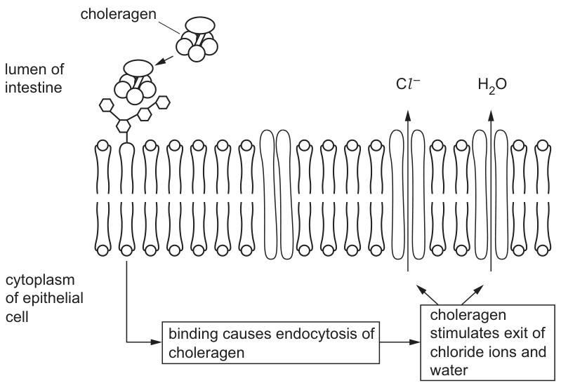
Answer
Contaminated drinking water (or untreated sewage / food contaminated with sewage).
Contaminated drinking water (untreated sewage or food contaminated with sewage).
Background Concept
Cholera is caused by the bacterium Vibrio cholerae. It is transmitted by the faecal–oral route: a susceptible person ingests water or food contaminated with faeces from an infected individual. The bacterium survives well in clean-looking fresh water and rapidly proliferates in sewage. A single infected person can shed huge numbers of bacteria, so even small contamination events can seed an outbreak.
Typical sources of outbreaks therefore involve the contamination of water supplies or food by human sewage:
- untreated or poorly treated sewage;
- drinking water taken from a contaminated source;
- food crops irrigated or fertilised with raw sewage, or seafood harvested from sewage-polluted water;
- very occasionally an infected person newly arriving in a previously cholera-free area (e.g. UN peacekeepers in Haiti, 2010).
Understanding the Question
The stem explains that V. cholerae releases choleragen, which causes loss of chloride ions and water from intestinal epithelial cells. Question (b)(i) asks for one likely source of an outbreak — a single point that explains how the bacterium reaches a new host population.
Approach
Recall the faecal–oral transmission route and name the most common vehicle. One clean, specific example is enough.
Step-by-Step Reasoning
Any of the following single points earns the mark:
- untreated/poorly treated sewage;
- contaminated drinking water;
- contaminated food (or crops fertilised with raw sewage, or seafood contaminated by sewage);
- an infected individual arriving in a cholera-free area.
Key Takeaways
- Cholera is spread by the faecal–oral route via contaminated water/food, never by casual contact.
- Improving water quality and sewage treatment is the primary public-health intervention.
Common Mistakes
- Naming the bacterium (V. cholerae) instead of a source. The question asks where the bacteria come from, not what the pathogen is.
- Vague answers such as "poor hygiene" — too general to score; name the specific vehicle (water or food).
Things to Be Careful About
The command word is "state" — a single brief point earns the mark. Do not over-elaborate.
With reference to Fig. 6.1, state why choleragen molecules are described as having quaternary structure.
Answer
Choleragen is composed of more than one polypeptide chain (several polypeptide subunits are visible in Fig. 6.1).
It is made up of more than one polypeptide (multiple polypeptide chains).
Background Concept
Protein structure has four levels:
- Primary — the linear sequence of amino acids linked by peptide bonds.
- Secondary — regular coiling or folding (α-helix, β-pleated sheet) stabilised by hydrogen bonds between backbone atoms.
- Tertiary — the overall 3-D folding of a single polypeptide, stabilised by interactions between R-groups (hydrogen, ionic, disulfide, hydrophobic).
- Quaternary — two or more separate polypeptide chains (subunits) associating together in a specific arrangement; each chain is itself already folded into its tertiary structure.
Haemoglobin is the classic example: four globin polypeptide chains + a haem group. A protein with only one polypeptide chain can never have quaternary structure.
Understanding the Question
Fig. 6.1 depicts choleragen as a cluster of circles attached to the cell surface — this is a stylised representation of a multi-subunit protein. The question asks, with reference to the figure, why choleragen is said to have quaternary structure. The required answer is therefore visible in the figure: several subunits (polypeptide chains) are shown.
Approach
Recall the definition of quaternary structure and link it directly to what the figure shows.
Step-by-Step Reasoning
Quaternary structure requires more than one polypeptide chain. Fig. 6.1 depicts choleragen as several subunits (the cluster of circles), so the toxin has quaternary structure.
Mark scheme accepts: "composed of more than one polypeptide" or any wording equivalent to "multiple/several polypeptide chains." Numerical answers ("6 or 7") are also accepted, but the structural principle (more than one chain) is what earns the mark.
Key Takeaways
- Quaternary structure = ≥ 2 polypeptide chains joined into one functional protein.
- A protein with only one polypeptide chain has only primary, secondary and tertiary structure — no quaternary structure.
Common Mistakes
- Saying choleragen "has a 3-D shape" — that describes tertiary, not quaternary.
- Confusing quaternary structure with secondary structure (α-helix/β-sheet).
- Saying choleragen "is a polypeptide" (singular) — this contradicts the figure and the definition.
Things to Be Careful About
The answer must explicitly state that more than one polypeptide is involved. Avoid hand-waving terms like "complex structure" — they do not score.
Answer
Glycolipid / glycoprotein (a carbohydrate-bearing membrane component).
Glycoprotein (or glycolipid).
Background Concept
The fluid-mosaic model places several types of molecule in the plasma membrane:
- a phospholipid bilayer;
- proteins (integral and peripheral);
- glycoproteins and glycolipids — proteins or lipids with short carbohydrate chains attached on the outer surface only;
- cholesterol between phospholipid tails.
The carbohydrate chains on the outer leaflet are highly variable and act as cell-surface markers / receptors. Pathogens, hormones, toxins and antibodies bind specifically to these carbohydrate-bearing components. A membrane receptor is therefore a protein (or occasionally a lipid) with a carbohydrate chain exposed on the cell surface — i.e. a glycoprotein (or glycolipid).
Understanding the Question
Fig. 6.1 shows choleragen docking onto a small hexagonal component sticking out of the outer leaflet of the membrane — a stylised receptor. The question asks for the type of membrane component forming this receptor.
Approach
Recall the membrane components involved in recognition/signalling on the outer surface — they all carry short carbohydrate chains.
Step-by-Step Reasoning
Receptor on outer surface + cell-recognition function ⇒ glycoprotein or glycolipid. Either term is accepted by the mark scheme.
Key Takeaways
- Cell-surface receptors for signalling molecules and pathogens are usually glycoproteins, occasionally glycolipids.
- The carbohydrate chains project from the outer leaflet only and are the basis of cell recognition and the ABO blood-group antigens.
Common Mistakes
- Writing "protein" alone (no carbohydrate) — too vague; mark scheme specifies glycoprotein/glycolipid.
- Writing "phospholipid" — phospholipids are not recognition molecules.
Things to Be Careful About
The carbohydrate chain is essential to receptor function. "Protein" on its own is not credited; say "glycoprotein" (or "glycolipid").
The process by which chloride ions leave the epithelial cell requires energy.
Name the phosphorylated nucleotide that is needed for this process.
Answer
ATP (adenosine triphosphate).
ATP (adenosine triphosphate).
Background Concept
Adenosine triphosphate (ATP) is the universal energy currency of the cell. It is a phosphorylated nucleotide consisting of:
- the nitrogenous base adenine;
- the pentose sugar ribose;
- three phosphate groups attached to the 5′-carbon of ribose.
The bonds between the phosphate groups (especially the terminal phosphoanhydride bond) are high-energy. Hydrolysis of the terminal phosphate releases energy that powers energy-requiring processes — including active transport across membranes, muscle contraction, and biosynthetic reactions. The products are ADP + Pᵢ.
Understanding the Question
The stem tells us that the process by which chloride ions leave the epithelial cell requires energy. The question asks for the name of the phosphorylated nucleotide that supplies this energy.
Approach
Any energy-requiring membrane transport process uses ATP, so name it.
Step-by-Step Reasoning
The phosphorylated nucleotide used for active processes in cells is ATP (adenosine triphosphate). Mark scheme explicitly ignores "adenine triphosphate" — because adenine triphosphate is not a real compound; the correct name is adenosine triphosphate.
Key Takeaways
- ATP = adenosine + ribose + 3 phosphate groups.
- ATP hydrolysis (→ ADP + Pᵢ) releases energy for active transport and other energy-requiring processes.
Common Mistakes
- Writing "adenine triphosphate" — rejected. Adenine is just the base; the molecule is adenosine triphosphate.
- Writing "GTP" or another nucleotide triphosphate — these have specific roles (e.g. GTP in translation) but are not the energy currency for ion pumping.
- Spelling out "adenosine triphosphate" correctly but omitting the abbreviation — perfectly acceptable, but make sure the spelling is right.
Things to Be Careful About
A phosphorylated nucleotide = nucleotide with extra phosphate(s). The answer here is specifically adenosine triphosphate (ATP), not just "a nucleotide".
Explain why water also moves from epithelial cells into the lumen of the intestine when choleragen is present.
Answer
- Chloride ions leaving the cell decrease the water potential of the lumen of the intestine (making the outside of the cells more negative / lower Ψ).
- Water therefore leaves the epithelial cells and enters the lumen by osmosis, down the water-potential gradient (from higher Ψ inside the cells to lower Ψ in the lumen).
Loss of Cl⁻ lowers the water potential of the lumen; water leaves the cells by osmosis down the water-potential gradient.
Background Concept
Water potential (Ψ) is the tendency of a solution to take up or lose water by osmosis. Pure water has Ψ = 0 kPa; adding solutes makes Ψ more negative (lower). Water moves by osmosis from a region of higher (less negative) Ψ to a region of lower (more negative) Ψ across a partially permeable membrane — i.e. down the water-potential gradient.
Key points to remember:
- A higher solute concentration ⇒ more negative Ψ.
- "Higher water potential" = closer to zero (e.g. −200 kPa is higher than −800 kPa).
- Osmosis requires no energy from the cell — it is driven by the water-potential gradient.
- The cell membrane is partially permeable: water crosses freely (through aquaporins), but solutes such as Cl⁻ cannot freely cross.
Understanding the Question
Fig. 6.1 shows choleragen stimulating the exit of chloride ions and water from the epithelial cell into the lumen of the intestine. The question asks why water follows the chloride ions. We must connect solute movement to water potential and then to osmosis.
Approach
Treat chloride ions as a solute. Pumping Cl⁻ out raises the solute concentration in the lumen. Use this to construct a water-potential argument, then explain water movement by osmosis.
Step-by-Step Reasoning
Marking point 1 — effect on water potential.
- Cl⁻ ions are a solute. When they leave the cytoplasm and accumulate in the lumen, the solute concentration of the lumen rises and its water potential falls (becomes more negative). Conversely, the cytoplasm loses solute so its Ψ becomes less negative / higher.
- The lumen now has a lower (more negative) Ψ than the cytoplasm of the epithelial cell.
Marking point 2 — water movement by osmosis.
- The partially permeable cell membrane allows water to pass more freely than Cl⁻.
- Water therefore moves down the water-potential gradient: from the cytoplasm (higher Ψ) into the lumen (lower Ψ). This is osmosis — a passive process, which is why no separate energy source is needed for water movement itself.
The result is a net loss of water from the epithelial cells into the intestinal lumen — producing the profuse, watery diarrhoea that characterises cholera.
Key Takeaways
- Osmosis is driven by water-potential differences, not directly by ion pumps.
- Pumping solutes (here Cl⁻) into one compartment changes the Ψ of that compartment and pulls water along by osmosis.
- "Higher" water potential = closer to zero (less negative). Always state direction by Ψ values, not by "concentration".
Common Mistakes
- Saying "water moves from a high concentration to a low concentration of water" — wrong wording; water moves from high water potential to low water potential.
- Saying water is "pumped" out of the cell — osmosis is passive, not active.
- Failing to mention the water-potential gradient at all — mark scheme requires the link between Cl⁻ loss and Ψ change.
Things to Be Careful About
Use the precise term water potential and the phrase down the water-potential gradient or from high to low water potential. Do not say "from low solute concentration to high solute concentration" — although the logic is right, the mark scheme credits the water-potential formulation.