Biology 9700/22 — October/November 2024
Cambridge AS Level · AS Level Structured Questions · worked solutions for every part, with the mark scheme
Topics Transport in Plants · Infectious Diseases · Cell Membranes and Transport · Transport in Mammals · Biological Molecules · Cell Structure · +5 more
The olive plant, Olea europaea, is grown in many parts of the world. The fruits of the plant (olives) and the oil that can be obtained from the fruits (olive oil), provide food for humans.
Triglycerides are the main type of lipid in olive oil. They are synthesised in the olive plant from glycerol and fatty acids.
Scientists can analyse samples of different olive oils to identify:
• the fatty acids used to synthesise triglycerides
• the composition of the different triglycerides present.
Table 1.1 shows some details of the five most common fatty acids found in samples of olive oil produced by olive plants grown in different regions in Portugal.
Table 1.1
Key
C:D = number of carbon atoms : number of double bonds in the hydrocarbon chain
X = missing detail
| fatty acid | percentage of total fatty acid content | C : D | chemical structure |
|---|---|---|---|
| oleic acid | 55.0–83.0 | 18 : 1 | |
| palmitic acid | 7.5–20.0 | 16 : ..... | |
| linoleic acid | 3.5–21.0 | 18 : ..... | |
| stearic acid | 0.5–5.0 | 18 : ..... | |
| palmitoleic acid | 0.3–3.5 | 16 : ..... |
Table 1.1 shows the C : D values for oleic acid.
In Table 1.1, write the values for D for each of the four other fatty acids listed.
Answer
The number of double bonds (D) is read directly from each chemical structure:
- Palmitic acid — D = 0 (no CH=CH in the chain; 16:0)
- Linoleic acid — D = 2 (two CH=CH units; 18:2)
- Stearic acid — D = 0 (no CH=CH; 18:0)
- Palmitoleic acid — D = 1 (one CH=CH; 16:1)
Saturated fatty acids have no C=C bonds in the hydrocarbon chain; unsaturated fatty acids have one (monounsaturated) or more (polyunsaturated).
palmitic acid: 0; linoleic acid: 2; stearic acid: 0; palmitoleic acid: 1
Background Concept
Fatty acids are long-chain carboxylic acids. The shorthand C:D gives two pieces of structural information: C = total number of carbon atoms in the chain, and D = number of carbon–carbon double bonds (C=C) within the chain. D is the structural marker of unsaturation: D = 0 means saturated (no C=C, every carbon is bonded to the maximum number of hydrogens); D ≥ 1 means unsaturated (one or more C=C). The higher the unsaturation, the lower the melting point — which is why olive oil (rich in the monounsaturated oleic acid) is liquid at room temperature, whereas fully saturated animal fats are solid.
Understanding the Question
Table 1.1 lists five fatty acids with their percentage range in olive oil, the C:D ratio (with the D value left blank for four of them), and the full chemical structure. The question asks the candidate to fill in D for palmitic, linoleic, stearic and palmitoleic acid. The C value is given, so the only job is to count the C=C bonds in each displayed structure.
Approach
Scan each structure for CH=CH (or C=C) units within the hydrocarbon chain and count them. The C=O of the carboxyl group at the end is not a C=C and does not contribute to D.
Step-by-Step Reasoning
- Oleic acid (18:1, given) — the structure shows one CH=CH, confirming the convention.
- Palmitic acid — structure contains no
=signs in the chain → D = 0 → 16:0. - Linoleic acid — structure contains two CH=CH units → D = 2 → 18:2.
- Stearic acid — structure has no double bonds → D = 0 → 18:0.
- Palmitoleic acid — structure contains one CH=CH → D = 1 → 16:1.
A useful cross-check: counting all carbons (CH₃ contributes 1, each (CH₂) block contributes 1 per unit, each CH=CH contributes 2, and the terminal COOH contributes 1) should give the C value stated in C:D. For example, oleic: 1 + 7 + 2 + 7 + 1 = 18 C ✓.
Key Takeaways
- The D value in C:D = number of C=C double bonds within the hydrocarbon chain.
- Saturated fatty acids always have D = 0.
- Recognising
CH=CHin a condensed formula is the skill being tested.
Common Mistakes
- Counting carbon atoms (C) instead of double bonds (D), or vice versa.
- Treating the C=O of the carboxyl group as a C=C — it is not, and is excluded from D.
- Reading
(CH₂)as(CH)and inventing extra bonds.
Things to Be Careful About
- Only C=C inside the chain counts. The C=O at the end is part of the carboxyl group, not the hydrocarbon chain.
- The total-carbon cross-check is a reliable way to verify the structure has been read correctly.
In the first column of Table 1.1, draw a circle around each of the fatty acids that can be described as saturated.
Answer
Circle drawn around palmitic acid and around stearic acid.
These are the two fatty acids with no double bonds (D = 0) in their hydrocarbon chain — palmitic acid (16:0) and stearic acid (18:0) — and are therefore described as saturated.
palmitic acid and stearic acid
Background Concept
A saturated fatty acid is one in which every carbon–carbon bond in the hydrocarbon chain is a single bond; every carbon holds the maximum number of hydrogens it can. In the C:D notation this corresponds to D = 0. A fatty acid with D ≥ 1 has at least one C=C double bond and is unsaturated (monounsaturated if D = 1, polyunsaturated if D ≥ 2).
Understanding the Question
Once part (i) is complete, every entry in Table 1.1 has its C:D value, and the candidate is asked to circle the fatty acids that are saturated.
Approach
Apply the definition: saturated = D = 0. Find the rows in Table 1.1 with D = 0 and circle them.
Step-by-Step Reasoning
- Palmitic acid 16:0 — D = 0 → saturated ✓ (circle)
- Stearic acid 18:0 — D = 0 → saturated ✓ (circle)
- Oleic acid 18:1 — D = 1 → unsaturated
- Linoleic acid 18:2 — D = 2 → unsaturated
- Palmitoleic acid 16:1 — D = 1 → unsaturated
So the saturated fatty acids are palmitic acid and stearic acid.
Key Takeaways
- Saturated fatty acid ⇔ D = 0 in C:D.
- The D value is a quicker test of saturation than visual inspection of the structure.
Common Mistakes
- Circling only one of the two saturated fatty acids and missing the other.
- Confusing the most abundant fatty acid (oleic) with the saturated one — oleic is unsaturated.
Things to Be Careful About
- "Saturated" describes the chemistry of the chain (no C=C), not its abundance in the oil.
State the detail of chemical structure, represented by X, which is missing from Table 1.1.
Answer
= COOH, the carboxyl group (also accepted: carboxylic acid group).
Every fatty acid has a hydrocarbon chain (R) attached to a carboxyl group (–COOH), and it is the –COOH that reacts with glycerol's –OH groups to form the ester bonds of a triglyceride.
COOH (carboxyl / carboxylic acid group)
Background Concept
All fatty acids — saturated or unsaturated, short or long — share the same molecular framework: a long hydrocarbon chain (the R group) attached at one end to a carboxyl group (–COOH) and at the other to a methyl group (–CH₃). The carboxyl group is what gives fatty acids their acidic character and is the site at which the fatty acid forms an ester bond with one of glycerol's three hydroxyl groups during triglyceride synthesis.
Understanding the Question
Every structure in Table 1.1 is drawn with the methyl end visible (CH₃…) but with the terminal functional group replaced by the symbol . The candidate must state what represents.
Approach
Recall the general formula of a fatty acid (R–COOH) and identify which end of the structure has been left as . The methyl end is shown explicitly; must therefore be the other end — the carboxyl group.
Step-by-Step Reasoning
- The generic structure of a fatty acid is .
- In Table 1.1, R (the hydrocarbon chain) is drawn in full for each fatty acid, so the missing at the other end is the COOH group.
- = COOH (carboxyl group / carboxylic acid group).
Key Takeaways
- Every fatty acid ends in a carboxyl group (–COOH); this is its defining functional group.
- The carboxyl group reacts with glycerol's –OH groups to form ester linkages in triglycerides.
Common Mistakes
- Writing just "OH" or "CO" — incomplete; both components of –COOH are needed.
- Writing "carbonyl and hydroxyl" as two separate items — the mark scheme accepts this, but a single term ("carboxyl") is cleaner.
- Confusing –COOH with –CHO (aldehyde) or –OH (alcohol).
Things to Be Careful About
- The C=O of the carboxyl group is not a C=C double bond and is not counted in the D value of C:D.
The analysis of the triglycerides present in the different samples of olive oil showed that:
• there are many different triglycerides present in olive oil
• each olive oil is different in its composition, but the same few triglycerides are present in all olive oils.
With reference to Table 1.1 and to the structure of triglycerides, suggest explanations for these observations.
Answer
-
Each triglyceride is built from three fatty acids. The fatty acids available in olive oil (Table 1.1) vary in chain length (16 or 18 carbons) and in the number of double bonds (D = 0, 1 or 2), and any one of these fatty acids can occupy any one of the three positions on the glycerol backbone. The number of distinct triglycerides that can be assembled from five different fatty acids at three positions is therefore very large — giving the many different triglycerides observed.
-
The same few triglycerides appear in every sample because they are built from the fatty acids present in the highest proportions — oleic (55.0–83.0%), palmitic (7.5–20.0%) and linoleic (3.5–21.0%). Any triglyceride made by combining oleic, palmitic and linoleic acids in different positions will be abundant in every olive oil, since those three fatty acids are abundant in every olive oil. Only the proportions of these fatty acids (and therefore the proportions of the dominant triglycerides) change with environmental conditions such as climate.
Many triglycerides form because each has three fatty acids that vary in chain length and saturation, allowing many combinations and positions; the same few dominate because they are made from the most abundant fatty acids (oleic, palmitic, linoleic).
Background Concept
A triglyceride consists of one glycerol molecule esterified to three fatty acids, one at each of glycerol's three –OH groups. Because the three fatty acids can be different, and because any one of them can occupy any of the three positions (sn-1, sn-2, sn-3), the combinatorial possibilities from even a small set of fatty acids are very large. Plants typically assemble triglycerides from a pool of fatty acids made in the plastid and endoplasmic reticulum, and the composition of that pool varies with the plant species and with environmental conditions (especially temperature).
Understanding the Question
Analysis of olive oils gives two observations:
- Observation 1: there are many different triglycerides in olive oil.
- Observation 2: every olive oil contains the same few triglycerides (only their proportions differ).
The candidate must explain both observations, drawing on Table 1.1 (the available fatty acids and their percentages) and on knowledge of triglyceride structure.
Approach
For Observation 1, count the combinations possible from the fatty acids in Table 1.1 (with varying chain lengths, saturation and positions). For Observation 2, recognise that the dominant fatty acids in olive oil — oleic, palmitic and linoleic — will always produce the same dominant triglyceride combinations, because they are always the dominant fatty acids.
Step-by-Step Reasoning
Why so many different triglycerides?
- Each triglyceride has three fatty acid "slots".
- The five fatty acids of Table 1.1 vary along two structural axes:
- Chain length: 16 C (palmitic, palmitoleic) vs 18 C (oleic, linoleic, stearic).
- Degree of unsaturation: D = 0 (palmitic, stearic), D = 1 (oleic, palmitoleic), D = 2 (linoleic).
- With three slots filled by five different fatty acids, and with positional isomerism (the same fatty acid can sit at position 1, 2 or 3 of the glycerol backbone), the number of distinct triglycerides is large.
- So chemical diversity in olive oil is structurally inevitable.
Why the same few in every sample?
- Oleic acid (55.0–83.0%) is by far the dominant fatty acid; palmitic (7.5–20.0%) and linoleic (3.5–21.0%) are next; stearic and palmitoleic together are usually below 8%.
- The triglycerides built from combinations of oleic, palmitic and linoleic acids in different positions are present in every olive oil, because those three fatty acids are present in every olive oil in the highest amounts.
- Environmental conditions (climate, soil, sunlight, water availability) affect the relative proportions of these fatty acids — e.g. plants grown in cooler climates tend to make more unsaturated fatty acids to maintain membrane fluidity — but they do not change which fatty acids dominate.
- So the same dominant triglycerides appear in all samples; only their proportions shift.
Key Takeaways
- Triglyceride diversity arises from (i) the three fatty acids chosen, (ii) their chain length and saturation, and (iii) the position each occupies on the glycerol backbone.
- The dominant triglycerides in any oil are dictated by the dominant fatty acids; if the same fatty acids dominate in every sample, the same triglycerides will dominate too.
- Environmental conditions alter proportions but not identity of the dominant species.
Common Mistakes
- Saying "different triglycerides come from different plants" without explaining the molecular mechanism.
- Claiming that saturated fatty acids release more energy than unsaturated ones — true biochemically, but not the reason the same triglycerides dominate.
- Forgetting positional isomerism (sn-1, sn-2, sn-3) — the same combination of three fatty acids can produce several distinct triglycerides.
- Confusing "many triglycerides" with "many fatty acids" — the fatty acids are few (5), but the triglycerides they form are many.
Things to Be Careful About
- Both observations must be explained; the question is a combined "suggest" prompt.
- Use specific examples from Table 1.1 (oleic, palmitic, linoleic) rather than vague references to "the fatty acids".
Glycerol is soluble in water. Triglycerides are insoluble in water.
Explain why water is a good solvent for some substances such as glycerol, but is a poor solvent for substances such as triglycerides.
Answer
-
Water is a polar (dipolar) molecule — the oxygen atom carries a slight negative charge () and the two hydrogen atoms carry slight positive charges ().
-
Glycerol is polar — it carries three hydroxyl (–OH) groups that can form hydrogen bonds with water. Water molecules therefore surround and disperse glycerol molecules, so glycerol dissolves (water is a good solvent for polar substances).
-
Triglycerides are non-polar — their long hydrocarbon chains contain only C–C and C–H bonds, with no partial charges. Water cannot form hydrogen bonds with them, and inserting them into water disrupts water's hydrogen-bond network. Triglycerides therefore remain insoluble in water (water is a poor solvent for non-polar substances).
Water is polar and forms hydrogen bonds with the polar hydroxyl groups of glycerol, dissolving it; triglycerides are non-polar and cannot form hydrogen bonds with water, so they remain insoluble.
Background Concept
Water's exceptional solvent properties come from its polarity. The oxygen atom is much more electronegative than hydrogen, so the shared electrons in each O–H bond spend more time near the oxygen. Combined with water's bent geometry, this gives the molecule a permanent dipole: a partial negative charge () on the oxygen and partial positive charges () on each hydrogen. The resulting partial charges allow water to form hydrogen bonds with other polar molecules and ions, surrounding them and pulling them into solution. Non-polar molecules (e.g. the long hydrocarbon chains of triglycerides) lack partial charges; placing them in water would disrupt the water–water hydrogen-bond network without any compensating water–solute interaction — energetically unfavourable. This is the basis of the hydrophobic effect.
Understanding the Question
The question sets up a contrast: glycerol (a small alcohol with three –OH groups) dissolves in water; triglycerides (esters of glycerol with three long fatty acids) do not. The candidate must explain this contrast at the molecular level.
Approach
- State water's polarity.
- Explain why polarity lets water dissolve polar molecules (glycerol).
- Explain why the same polarity leaves non-polar molecules (triglycerides) insoluble.
Step-by-Step Reasoning
- Water is polar / dipolar — partial charges on O () and H () mean water can attract other polar or charged species.
- Glycerol is polar — it has three hydroxyl (–OH) groups that can form hydrogen bonds with water. Water molecules cluster around glycerol molecules via hydrogen bonding, surrounding and dispersing them — i.e. dissolving them.
- Triglycerides are non-polar — the long hydrocarbon chains have only C–C and C–H bonds, which are essentially non-polar. There are no partial charges for water to interact with, and inserting triglyceride molecules into water would force water to reorient around them, breaking water–water hydrogen bonds without forming water–triglyceride hydrogen bonds. The triglycerides therefore remain as a separate phase — insoluble.
Key Takeaways
- Water dissolves polar / ionic substances because it can form hydrogen bonds or ion–dipole interactions with them.
- Water does not dissolve non-polar substances — this is the hydrophobic effect.
- The polarity of the solute determines whether water can interact with it.
Common Mistakes
- Saying "water dissolves everything" or "oil and water don't mix because oil is thick" — neither engages with polarity.
- Referring to "the fatty acid tails" without explaining why they are non-polar.
- Confusing "insoluble" with "denser than water" — triglycerides do float, but the underlying reason is non-polarity, not density.
- Citing only one of the two contrast points (e.g. only why glycerol dissolves, not why triglycerides don't).
Things to Be Careful About
- Use the precise terms polar / dipolar and hydrogen bond — these are the mark-bearing words.
- The "ora" (or reverse argument) is acceptable: "water cannot hydrogen-bond with non-polar substances" also scores.
Phloem is the plant tissue responsible for the transport of organic substances, such as fatty acids, from one area of a plant to another. The tissue is composed of more than one type of cell.
Name the type of cell that forms the transport vessels of phloem tissue.
Answer
Sieve tube element (also accepted: sieve element).
Sieve tube element
Background Concept
Phloem is the plant vascular tissue responsible for translocation — the transport of organic solutes (mainly sucrose, but also amino acids, fatty acids and other assimilates) from sources (e.g. photosynthesising leaves, storage tissues) to sinks (e.g. roots, fruits, growing tips). Unlike xylem, phloem is a living tissue at functional maturity. It is composed mainly of two cell types:
- Sieve tube elements — the conducting cells. They are joined end-to-end to form long sieve tubes, with perforated end walls called sieve plates between adjacent elements. At maturity a sieve tube element has lost its nucleus, ribosomes, vacuole and most other organelles — leaving a clear cytoplasmic pathway for the flow of sap.
- Companion cells — small cells closely associated with each sieve tube element (derived from the same parent cell by division). They retain their nucleus and normal organelles and provide metabolic support to the enucleate sieve tube element.
(The tissue also contains phloem fibres for support and phloem parenchyma for storage.)
Understanding the Question
The question asks for the cell type that forms the transport vessels of phloem tissue. "Transport vessels" points directly to the sieve tubes; the individual cells forming them are sieve tube elements.
Approach
Recall the cellular composition of phloem and identify the cell type whose job is to form the conducting tube.
Step-by-Step Reasoning
- The conducting tubes of phloem are sieve tubes.
- Each sieve tube is made up of many sieve tube elements joined end-to-end.
- "Sieve element" is an accepted alternative term.
Key Takeaways
- Phloem transport is performed by sieve tube elements (not companion cells, which support them).
- Sieve tube elements are unusual cells: alive at functional maturity but lacking a nucleus and most organelles, depending on their companion cell for metabolism.
Common Mistakes
- Writing "xylem vessel" — wrong tissue (xylem transports water and mineral ions).
- Writing "phloem cell" — too vague; the specific term "sieve tube element" is required.
- Writing "companion cell" — companion cells support the sieve tube elements; they do not form the conducting tube themselves.
Things to Be Careful About
- "Sieve tube element" (the individual cell) and "sieve tube" (the conducting tube made of many elements) are not the same thing. The question asks for the cell type — sieve tube element.
People who become infected with human immunodeficiency virus (HIV) are at risk of developing HIV/AIDs, particularly if antiretroviral therapy (ART) is not available.
In people infected with HIV, the use of ART also helps to reduce transmission of the virus to uninfected people.
Outline two control methods, other than ART, that can be used to reduce the transmission of HIV.
Answer
Any two from:
- Practising safe sex (e.g. using barrier contraceptives / condoms)
- Screening donated blood before transfusions / not accepting blood from high-risk individuals
- Treating donated blood to inactivate viruses (e.g. heat / chemical / solvent-detergent treatment)
- Not sharing needles / syringes (in context of drug users); using new / sterile needles or needle-exchange schemes
- Avoiding breastfeeding (or using formula milk instead)
- Using pre-exposure prophylaxis (PrEP) or post-exposure prophylaxis (PEP)
- Using sterile equipment for tattooing, surgery, dental procedures or body piercing
Two valid control methods (see working) — e.g. practising safe sex with condoms and screening donated blood before transfusion.
Background Concept
HIV is transmitted through the exchange of certain body fluids — blood, semen, vaginal secretions, rectal secretions and breast milk. The main transmission routes are unprotected sexual contact, blood-to-blood contact (contaminated needles in injecting-drug use, unscreened transfusions, unsafe medical or cosmetic procedures) and mother-to-child transmission (during pregnancy, birth or breastfeeding). Effective prevention therefore targets one or more of these routes with a specific intervention.
Understanding the Question
The stem tells you ART can both treat infected individuals and reduce onward transmission, but the question asks for two OTHER control methods. 'Control' here means population-level measures that reduce transmission in a community; ART alone is excluded.
Approach
For each transmission route, recall the specific public-health or behavioural intervention that interrupts it, then choose any two that together cover distinct routes.
Step-by-Step Reasoning
- Sexual transmission → practising safe sex with barrier contraceptives (condoms); reducing the number of partners.
- Blood-borne (transfusion) → screening donated blood, treating it to inactivate any virus (heat / chemical / solvent-detergent), refusing donations from high-risk donors.
- Blood-borne (drugs) → not sharing needles / syringes; needle-exchange and safe-injection schemes; using only new / sterile equipment.
- Mother-to-child → mothers with HIV avoiding breastfeeding (using formula milk); antiretroviral prophylaxis during pregnancy (this overlaps with ART but is sometimes credited under specific contexts).
- Iatrogenic (surgery, dental, tattooing, piercing) → using sterile equipment.
- Pre- / post-exposure prophylaxis (PrEP / PEP) for individuals at high risk of exposure.
The mark scheme rejects vague answers such as 'use contraceptives' without specifying barrier, and explicitly does NOT credit 'education', 'contact tracing', 'protective gear', 'early diagnosis' or 'testing' as standalone answers for this item.
Key Takeaways
Control of a sexually and blood-borne pathogen = identify each transmission route and apply the intervention that breaks it.
Common Mistakes
- Writing 'use contraceptives' without specifying barrier / condom — too vague to earn the mark.
- Writing 'screen blood' without saying 'donated' or 'before transfusion' — incomplete.
- Offering 'education' or 'contact tracing' alone — these are explicitly ignored by the mark scheme here.
- Confusing PrEP / PEP with ART — ART is excluded by the question; PrEP / PEP for uninfected individuals is acceptable.
Things to Be Careful About
Mention the specific element that earns the mark: barrier, screening OF DONATED blood, needle-exchange, PrEP / PEP, sterile equipment, etc. Vague answers are penalised.
In people with HIV/AIDs, a serious lung disease known as pneumocystis pneumonia can result from infection by an opportunistic pathogen known as Pneumocystis jirovecii.
Fig. 2.1 shows P. jirovecii cells in one stage of their life cycle, as seen using a light microscope at a magnification of .
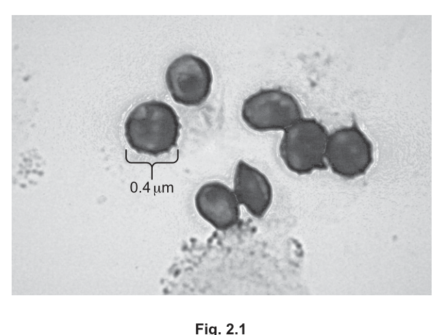
Define magnification.
Answer
The number of times larger the image is than the actual (specimen / object).
The number of times larger the image is than the actual specimen / object.
Background Concept
Magnification describes how much larger (or smaller) an image is compared with the actual object. It is a ratio — a pure number with no units. The formula is:
A magnification greater than 1 means the image is enlarged; less than 1 means it is reduced.
Understanding the Question
This is a recall-of-definition item. You must give the verbal definition that matches the formula, not the formula itself (although the formula is accepted as a supplement).
Approach
State the definition in words, anchoring it to a comparison between image size and actual size.
Step-by-Step Reasoning
A correct one-line answer is 'the number of times larger the image is than the actual (object/specimen)'. The mark scheme accepts a formula as an alternative or addition, but a verbal definition is more reliable because it is unambiguous and free of mathematical ambiguity.
Key Takeaways
Magnification is a dimensionless ratio — it depends on BOTH the image size and the specimen size, not on the microscope alone.
Common Mistakes
- Saying simply 'makes it bigger' — too vague; doesn't anchor to a comparison with the actual object.
- Writing only a formula without any words.
- Confusing magnification with resolution (the ability to distinguish two close points as separate).
Things to Be Careful About
Anchor the definition to 'image' vs 'actual (specimen / object)'. Acceptable phrasings include 'the number of times larger the image is than the actual' or 'image size ÷ actual size'.
Fig. 2.1 shows that P. jirovecii is a unicellular organism. Although the cells of many species of bacteria are the same size as those of P. jirovecii, research concluded that the organism is a eukaryote and is not a bacterium.
In 1988, analysis of ribosomal RNA (rRNA) resulted in P. jirovecii being classified as a fungus.
Studies of the structure of P. jirovecii have identified that the cell wall is made of polysaccharides such as chitin and .
Explain why this feature helped scientists to confirm that P. jirovecii is not a bacterium.
Answer
Bacteria have cell walls made of murein / peptidoglycan, and chitin is not a component of bacterial cell walls; the presence of chitin (and 1,3-β-D-glucan) in P. jirovecii therefore shows it is not a bacterium.
Bacterial walls are murein / peptidoglycan (chitin is absent); chitin + 1,3-β-D-glucan therefore rule out a bacterium.
Background Concept
Bacteria are prokaryotes whose cell walls are built from peptidoglycan (also called murein) — a polymer of sugars cross-linked by short peptide chains. Fungi, by contrast, are eukaryotes whose cell walls are built mainly from chitin (a polymer of N-acetylglucosamine) and β-glucans such as 1,3-β-D-glucan. Cell-wall composition is one of the classical biochemical features used to assign a microbe to its correct kingdom.
Understanding the Question
You are told P. jirovecii has chitin and 1,3-β-D-glucan in its wall. You must use this fact to explain why it cannot be a bacterium.
Approach
State what bacteria have in their walls and contrast this with what P. jirovecii has. The mark scheme accepts either: (a) bacteria have walls of murein / peptidoglycan, or (b) chitin is not found in bacterial walls.
Step-by-Step Reasoning
Bacterial walls are made of peptidoglycan / murein, not chitin or β-glucans. Because P. jirovecii contains chitin and β-D-glucans as a major wall component, its wall chemistry is incompatible with a bacterial identity. This was one of the lines of evidence that led to P. jirovecii being reclassified as a fungus (alongside the rRNA analysis mentioned in the stem).
Key Takeaways
Cell-wall chemistry (peptidoglycan vs chitin / β-glucan) is a quick and reliable way to distinguish bacteria from fungi.
Common Mistakes
- Saying 'bacteria don't have chitin' without mentioning what they DO have — the mark scheme wants either the positive fact (murein / peptidoglycan) or the negative fact (chitin not in bacterial walls); a vague 'different' will not earn the mark.
- Confusing cellulose with chitin — cellulose is the plant structural polysaccharide; chitin is the fungal / arthropod-exoskeleton one.
- Confusing fungi with plants — plant cell walls are cellulose; only fungal walls contain chitin.
Things to Be Careful About
Name the bacterial wall component (peptidoglycan / murein) for a clean mark, and remember that chitin absence is the strongest single contrast.
Scientists have identified other features of the cell structure of P. jirovecii. Some of these are listed in Table 2.1.
Complete each row of Table 2.1 so that the table shows:
• four structural features identified in P. jirovecii
• one function for each structural feature
• whether the structural feature is present (✓) or absent (✗) in bacterial cells.
Table 2.1
| structural feature of P. jirovecii | function | present (✓) or absent (✗) in bacterial cells |
|---|---|---|
| ribosomes | protein synthesis | |
| smooth endoplasmic reticulum | ||
| Golgi body | modification of proteins and lipids | |
| aerobic respiration |
Answer
| structural feature of P. jirovecii | function | present (✓) or absent (✗) in bacterial cells |
|---|---|---|
| ribosomes | protein synthesis | ✓ |
| smooth endoplasmic reticulum | synthesis of lipids / cholesterol / steroids | ✗ |
| Golgi body | modification of proteins and lipids | ✗ |
| mitochondrion | aerobic respiration | ✗ |
SER function: lipid / cholesterol / steroid synthesis (✗ in bacteria); missing feature: mitochondrion (✗ in bacteria); ribosomes ✓, Golgi ✗.
Background Concept
Eukaryotic cells have membrane-bound organelles (nucleus, mitochondria, endoplasmic reticulum, Golgi apparatus, lysosomes) plus ribosomes. Prokaryotic (bacterial) cells have no nucleus and no membrane-bound organelles — only ribosomes (smaller, 70S) and the nucleoid region. Each organelle has a characteristic function.
Understanding the Question
Three structural features of P. jirovecii are listed (ribosomes, smooth ER, Golgi body) plus a fourth function (aerobic respiration) without a named organelle. You must:
- name the missing organelle (the one whose function is aerobic respiration → mitochondrion),
- supply the missing function for smooth ER,
- tick / cross whether each feature is present in bacterial cells.
Approach
Recall each organelle's main role, then decide whether bacteria possess it.
Step-by-Step Reasoning
- Ribosomes: protein synthesis. Present (✓) in bacteria — bacterial ribosomes are smaller (70S) but ribosomes nonetheless.
- Smooth endoplasmic reticulum: synthesis of lipids / cholesterol / steroids (and detoxification of certain compounds). Absent (✗) in bacteria — there is no ER.
- Golgi body: modification of proteins and lipids (and packaging for secretion). Absent (✗) in bacteria — no Golgi.
- Mitochondrion: aerobic respiration. Absent (✗) in bacteria — aerobic bacteria respire across the plasma membrane (and mesosomes), not within membrane-bound mitochondria.
The marks are awarded one per correct column, so a fully correct tick column earns one mark, a fully correct function column earns one mark, and the mitochondrion (named) earns the third mark.
Key Takeaways
Bacteria have ONLY ribosomes as an organelle-like structure (plus cell wall, plasma membrane, nucleoid, plasmid, sometimes flagellum) — every other item in this list is a eukaryote-only feature.
Common Mistakes
- Writing RER (rough ER) for the smooth ER row.
- Saying SER 'produces proteins' — that is RER; SER is for lipids and detoxification.
- Ticking ribosomes with '✗' because they are smaller — wrong; ribosomes exist in both kingdoms.
- Naming 'cytoplasm' or 'nucleus' for the missing aerobic-respiration feature — the standard answer is mitochondrion.
Things to Be Careful About
Use the exact organelle name ('mitochondrion' or 'mitochondria'). The smooth ER's accepted functions are lipid / cholesterol / steroid synthesis or metabolism, or detoxification; ignore anything about carbohydrate metabolism.
P. jirovecii can adhere (attach) to squamous epithelial cells of the alveoli and to the network of fibrous proteins that support the alveolar wall, known as the extracellular matrix (ECM). Examples of proteins in the ECM are elastin and collagen.
Adhesion (attachment) of P. jirovecii to alveolar epithelial cells and the ECM stimulates the growth of its population.
Cell surface glycoproteins known as gpA glycoproteins are essential in allowing P. jirovecii cells to adhere to alveolar epithelial cells and ECM proteins.
Suggest how a gpA glycoprotein is able to adhere to alveolar epithelial cells and ECM proteins.
Answer
- gpA has a binding site that is complementary in shape to a receptor / protein / glycoprotein on the surface of the alveolar epithelial cell (and to proteins of the ECM).
- Once the shapes fit, attractive forces — hydrogen bonds, ionic interactions or other weak chemical bonds — hold gpA to the receptor, so gpA adheres to the alveolar cell surface and to ECM proteins.
Complementary shape of gpA's binding site to a receptor on the alveolar cell / ECM protein; attractive forces (hydrogen / ionic bonds) hold them together.
Background Concept
Cells communicate and attach to one another via cell-surface molecules. Glycoproteins and glycolipids on the plasma membrane can act as ligands — they bind specifically to receptor molecules on neighbouring cells or to extracellular-matrix components. The binding is specific because the ligand and receptor have complementary shapes (lock-and-key / induced-fit) and the interaction can be stabilised by hydrogen bonds, ionic interactions or other weak chemical bonds.
Understanding the Question
The question gives you a specific molecule (gpA, a glycoprotein on P. jirovecii) and two targets (alveolar epithelial cells and ECM proteins). You need to suggest HOW it adheres to both.
Approach
Apply the standard ligand-receptor model with two ingredients: a specific binding site AND a chemical interaction that holds the two together.
Step-by-Step Reasoning
- gpA has a binding site whose shape is complementary to a receptor / surface protein / glycoprotein on the alveolar epithelial cell, and complementary to ECM proteins (elastin, collagen).
- When the shapes fit, attractive forces — hydrogen bonds, ionic interactions or other weak chemical bonds — hold gpA to the target molecule.
- Because the binding is shape-specific, gpA can 'recognise' both alveolar-cell surface proteins and ECM proteins (which are themselves surface-exposed or associated with the cell membrane).
The mark scheme allows three routes to two marks: receptor / protein binding + complementary shape, OR receptor / protein + bond types, OR complementary shape + bond types.
Key Takeaways
Adhesion = complementary shape + attractive forces. A single vague 'matches the cell' without specifying which part or which interaction will not earn full marks.
Common Mistakes
- Saying gpA 'sticks to' the cell without explaining the mechanism.
- Using the word 'receptor' without saying what binds to it.
- Describing only one of the two targets (alveolar cell OR ECM) when both should be addressed.
Things to Be Careful About
Use the precise language: 'complementary shape', 'binding site', 'receptor', and name a bond type (hydrogen bond / ionic / electrostatic interaction) if you want the third mark.
One consequence of the pneumonia that results from P. jirovecii infection is a decrease in the quantity of oxygen that is delivered to body tissues.
Explain why a severe P. jirovecii infection results in a decrease in the quantity of oxygen that is delivered to body tissues.
Answer
- P. jirovecii cells cover / surround the alveolar epithelial cells and ECM, so the alveolar wall becomes thicker.
- The diffusion distance for O2 between alveolar air and capillary blood is increased (and the surface area for diffusion is decreased), so diffusion of O2 is impaired.
- Less O2 reaches the blood and binds to haemoglobin, so less oxyhaemoglobin is formed and less O2 is delivered to body tissues.
P. jirovecii thickens the alveolar wall → diffusion of O2 impaired (longer distance, smaller area) → less oxyhaemoglobin → less O2 delivered to tissues.
Background Concept
Efficient gas exchange in the alveoli requires: a large surface area, a thin wall (one squamous epithelial cell plus the capillary endothelium), a moist surface (so gases dissolve before crossing), and a steep diffusion gradient maintained by continuous ventilation (air movement) and perfusion (blood flow). Anything that thickens the alveolar wall, reduces surface area, flattens the diffusion gradient, or damages capillaries will reduce the rate at which O2 enters the blood — and therefore the amount of oxyhaemoglobin formed and O2 delivered to tissues.
Understanding the Question
You are told a severe P. jirovecii infection decreases O2 delivery to body tissues. You must explain the causal chain linking the infection to that final outcome.
Approach
Trace the chain from the pathogen to the tissues: pathogen covers alveoli → diffusion impaired → less O2 binds Hb → less O2 to tissues. Layer on additional effects (elastic-fibre impairment, ventilation, capillary damage, O2 consumed by the pathogen) for the higher marks.
Step-by-Step Reasoning
- P. jirovecii cells cover the alveolar epithelial cells and the ECM, making the alveolar wall thicker.
- The diffusion distance for O2 between alveolar air and capillary blood is increased; the surface area available for diffusion is decreased. O2 diffusion is therefore impaired.
- Less O2 reaches the blood and binds to haemoglobin in red blood cells → less oxyhaemoglobin is formed.
- (Optional / AVP supporting effects): the infection may impair recoil of the elastic fibres in the alveolar wall, so alveolar air is not refreshed and the diffusion gradient falls; P. jirovecii itself uses some O2 for its own aerobic respiration; the infection may damage alveolar capillaries, reducing perfusion; macrophages in the alveoli may physically hinder diffusion.
The chain ends with the question's stated outcome: a decrease in the quantity of oxygen delivered to body tissues.
Key Takeaways
Pneumonia reduces oxygen delivery not by one mechanism but by several overlapping ones; the Fick's-law-style ideas (surface area, diffusion distance, concentration gradient) link directly to the gas-exchange chapter.
Common Mistakes
- Stating 'the alveoli are damaged' without saying what that does to diffusion distance or surface area.
- Stopping at 'less O2 reaches the blood' without explaining the consequence for haemoglobin.
- Forgetting to link back to the body's tissues — the question explicitly asks about delivery to tissues.
Things to Be Careful About
Use the language of Fick's law: 'diffusion distance', 'surface area', 'diffusion gradient'. Mention oxyhaemoglobin explicitly to reach the question's endpoint.
P. jirovecii produces an enzyme known as synthase. The enzyme catalyses the synthesis of .
The therapeutic drug caspofungin is a non-competitive inhibitor of synthase.
With reference to the mechanism of action of caspofungin, explain how the drug may be useful to treat cases of pneumonia caused by P. jirovecii.
Answer
- Caspofungin binds to a site on 1,3-β-D-glucan synthase OTHER than the active site (an allosteric site).
- This changes the shape of the active site (the tertiary structure of the enzyme is altered).
- Substrates can no longer bind to the active site, so enzyme–substrate complexes do not form (or form in much smaller numbers).
- Less / no 1,3-β-D-glucan is produced, so fungal cell-wall synthesis is hindered and the cell wall is weakened.
- P. jirovecii cells cannot resist the osmotic influx of water (water enters down the water-potential gradient) and undergo osmotic lysis, so the population growth is reduced / fewer cells colonise the alveoli.
Caspofungin binds to an allosteric site → active-site shape changes → substrate cannot bind → no glucan made → weakened cell wall → osmotic lysis → reduced pathogen load.
Background Concept
Enzyme inhibitors fall into two broad classes. Competitive inhibitors bind to the active site and block substrate binding reversibly; their effect can be overcome by raising substrate concentration. Non-competitive inhibitors bind to a different site (an allosteric site) and change the shape of the enzyme so the active site can no longer accommodate the substrate; raising substrate concentration does NOT overcome the inhibition. The drug caspofungin is the latter type.
Fungal cell walls are built from β-glucans (especially 1,3-β-D-glucan) and chitin. If you stop β-glucan synthesis, the wall cannot form properly. A cell without an intact wall is vulnerable to osmotic lysis — water rushes in down the water-potential gradient, the membrane cannot withstand the pressure, and the cell bursts.
Understanding the Question
You are told caspofungin is a non-competitive inhibitor of 1,3-β-D-glucan synthase, the enzyme that makes a key wall component. You must explain, with reference to the mechanism, how this drug could treat P. jirovecii pneumonia.
Approach
Walk through the standard non-competitive-inhibition chain, then chain it on to fungal cell-wall synthesis, osmotic lysis, and the reduction in pathogen load. That's exactly five linked points — one chain per mark.
Step-by-Step Reasoning
- Caspofungin binds to a site on 1,3-β-D-glucan synthase OTHER than the active site (the allosteric site).
- This changes the shape of the active site (the tertiary structure of the enzyme changes).
- Substrate can no longer bind to the active site, so enzyme–substrate complexes do not form (or form in much smaller numbers).
- The rate of 1,3-β-D-glucan synthesis falls; the fungal cell wall is weakened / not formed properly.
- The cell cannot resist osmotic influx of water (water enters down the water-potential gradient) and undergoes osmotic lysis.
- The P. jirovecii population no longer grows / falls; fewer cells are available to colonise the alveoli, easing the infection and giving the body's immune system a better chance of clearing the remaining fungus.
Key Takeaways
Non-competitive inhibitors work by altering enzyme shape at a remote site; the chain drug → enzyme inhibition → no wall → lysis → fewer pathogens illustrates how a single molecular event can have a population-level therapeutic effect. This is the same logic that underpins many antimicrobial drugs that target cell-wall synthesis (β-lactam antibiotics, azole antifungals, etc.).
Common Mistakes
- Saying caspofungin 'binds to the active site' — that would make it competitive, not non-competitive.
- Saying only 'substrate cannot bind' without explaining WHY (shape change at the allosteric site).
- Stating 'the fungus dies' without naming the mechanism (osmotic lysis) or the reason (cell-wall weakening).
- Stopping at 'fewer cells' without linking to the immune system or to the symptoms easing.
Things to Be Careful About
Use the term 'allosteric site' (or 'site other than the active site'). State explicitly that the active site changes SHAPE so that substrate can no longer bind. Mention osmotic lysis for the final link, and ideally that water enters DOWN the water-potential gradient.
During transcription, base pairing occurs between nucleotides.
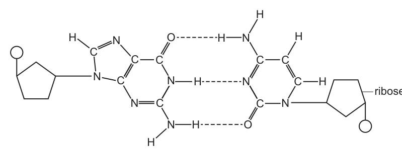
Fig. 3.1 is a diagram to show complementary base pairing between a DNA nucleotide and an RNA nucleotide.
Only the base pair is shown in molecular detail.
Answer
Phosphodiester bonds form between adjacent nucleotides on the same strand (linking the phosphate of one nucleotide to the sugar of the next) to build the sugar–phosphate backbone. Fig. 3.1 shows only one base pair — two nucleotides, one on each strand — so each phosphate is bonded to only one sugar; there is no second, adjacent nucleotide on either side for a phosphodiester bond to link to.
Phosphodiester bonds form between adjacent nucleotides on the same strand; Fig. 3.1 shows only one nucleotide on each strand, so each phosphate is bonded to only one sugar.
Background Concept
A nucleotide is the monomer of a nucleic acid. It has three parts: a phosphate group, a pentose sugar (deoxyribose in DNA, ribose in RNA), and a nitrogenous base. Bases are of two structural kinds: purines (double ring — adenine, guanine) and pyrimidines (single ring — cytosine, thymine, uracil).
Nucleotides are joined into a polynucleotide by phosphodiester bonds. A phosphodiester bond forms between the 3′ carbon of the sugar of one nucleotide and the 5′ phosphate of the next nucleotide on the same strand. This is the bond that builds the sugar–phosphate backbone of DNA or RNA. The backbone runs 5′ → 3′.
Across the two strands, bases pair by hydrogen bonds (two between A and T/U, three between G and C). Hydrogen bonds are weak and reversible (they must be broken to open the strands for replication or transcription); phosphodiester bonds are strong covalent bonds and are not broken during these processes.
Understanding the Question
The question gives a molecular diagram of a single base pair between a DNA nucleotide and an RNA nucleotide, with three hydrogen bonds shown as dashed lines. Part (a) asks you to reason about a bond that is absent from the figure: the phosphodiester bond. The instruction is to explain, so you must give a reason — not just say "there is no phosphodiester bond".
Approach
Think about what a phosphodiester bond needs in order to exist: two adjacent nucleotides, on the same strand, with a phosphate linking the 3′ of one sugar to the 5′ of the next. Then look at the figure and count: how many nucleotides are on each strand? Is the phosphate attached to one sugar or to two? Use that count to give a clear reason.
Step-by-Step Reasoning
- A phosphodiester bond is formed during polynucleotide synthesis, between the phosphate of one nucleotide and the 3′ carbon of the pentose sugar of the adjacent nucleotide on the same strand.
- In Fig. 3.1 only one nucleotide is shown on each strand (one DNA nucleotide, one RNA nucleotide). There is no second nucleotide alongside either of them.
- The phosphate group (the circle on the left of the DNA nucleotide) is therefore bonded to only one pentose sugar. A phosphodiester bond requires each phosphate to be bonded to two sugars.
- The same logic applies to the right-hand nucleotide. Hence, the structural pre-conditions for a phosphodiester bond are not met in this figure.
Either of these ideas, clearly expressed, earns the mark. The strongest answers combine the two.
Key Takeaways
- Phosphodiester bonds join adjacent nucleotides on the same strand; hydrogen bonds join bases across the two strands.
- A phosphodiester bond cannot exist unless a phosphate is shared between two sugars, i.e. between two adjacent nucleotides.
- A figure showing a single base pair therefore has hydrogen bonds but no phosphodiester bonds.
Common Mistakes
- Saying "there is no phosphodiester bond because it is a single base pair" without explaining what is required for a phosphodiester bond to form.
- Confusing phosphodiester bonds with hydrogen bonds (hydrogen bonds are present in the figure; phosphodiester bonds are not).
- Saying "only one nucleotide is shown" but failing to add that a phosphodiester bond needs an adjacent nucleotide on the same strand.
Things to Be Careful About
- The mark scheme rewards the idea of two adjacent nucleotides on the same strand. Wording such as "the phosphodiester bond joins nucleotides in a chain" or "joins the sugar of one nucleotide to the phosphate of the next" is acceptable.
- An alternative acceptable point is that the phosphate shown is attached to only one sugar, when a phosphodiester linkage requires attachment to two.
- Use the term nucleotide, not base, when referring to the building blocks of a polynucleotide.
Identify and describe the DNA-RNA nucleotide pair shown in Fig. 3.1.
You may add labels and annotations to Fig. 3.1 if you wish.
Answer
- The DNA nucleotide (left) contains the base guanine, and the RNA nucleotide (right) contains the base cytosine.
- The three dashed lines between the bases represent three hydrogen bonds, confirming a G–C pair.
- The circle attached to the deoxyribose sugar on the left is a phosphate group.
- The pentose sugar (pentagon) on the left is deoxyribose; the pentose on the right is ribose (already labelled).
- The double-ringed base on the left is a purine (guanine) and the single-ringed base on the right is a pyrimidine (cytosine).
DNA nucleotide (left) = guanine attached to deoxyribose with a phosphate; RNA nucleotide (right) = cytosine attached to ribose. Three hydrogen bonds (dashed lines) between the bases confirm a G–C pair; the left base is a purine (double ring) and the right base is a pyrimidine (single ring).
Background Concept
Every nucleotide has three components: a phosphate, a pentose sugar (deoxyribose in DNA, ribose in RNA), and a nitrogenous base. The five bases fall into two structural groups:
- Purines — double ring of carbon and nitrogen atoms: adenine (A) and guanine (G).
- Pyrimidines — single ring: cytosine (C), thymine (T) (DNA only) and uracil (U) (RNA only).
In a double-stranded nucleic acid, a purine on one strand always pairs with a pyrimidine on the other, so the helix has a constant width. The base-pairing rules are:
- A pairs with T (DNA) or U (RNA) via two hydrogen bonds.
- G pairs with C via three hydrogen bonds.
During transcription, the DNA double helix is unwound and one DNA strand acts as the template for synthesising a complementary mRNA strand. The pairing is therefore DNA–RNA: A (DNA) with U (RNA), T (DNA) with A (RNA), G (DNA) with C (RNA), and C (DNA) with G (RNA). Hydrogen bonds are shown in molecular diagrams as dashed lines.
Understanding the Question
Part (b) asks you to identify the specific DNA–RNA pair in Fig. 3.1 and to describe the features that allow you to recognise it. The figure shows a single base pair in molecular detail, with the RNA sugar already labelled. You are expected to use the visual evidence (number of rings, number of hydrogen bonds, the labelled sugar, the presence of a phosphate) together with the base-pairing rules to name the bases correctly and to label the diagram.
Approach
Use the visual clues systematically:
- Count the rings on each base — purine (double) vs pyrimidine (single).
- Count the hydrogen bonds (dashed lines) — 2 or 3 — to fix which base pair this is.
- Use the labelled sugar on the right (ribose) to assign that nucleotide to RNA, and therefore the other to DNA.
- Use the chemical groups drawn on the rings (e.g. C=O vs NH₂ positions) to distinguish guanine from adenine, and cytosine from uracil/thymine, if needed.
- Identify the other labelled/visibly distinct features: the circle is a phosphate, the left pentagon is deoxyribose, the dashed lines are hydrogen bonds.
Step-by-Step Reasoning
Identifying the bases
- The right-hand base is on a ribose sugar (labelled), so this is the RNA nucleotide.
- The right-hand base has a single ring, so it is a pyrimidine — either C, T or U. Uracil is RNA-specific; cytosine is found in both DNA and RNA; thymine is DNA-specific and so cannot appear in an RNA nucleotide.
- The left-hand base has a double ring, so it is a purine — either A or G.
- There are three dashed lines between the two bases. Three hydrogen bonds = G–C. (Two hydrogen bonds = A–T or A–U.)
- Therefore the left base is guanine (the DNA base) and the right base is cytosine (the RNA base). The cross-strand base pair is G(DNA) · C(RNA).
Confirming from the chemical groups drawn
- The left base has a C=O at the top of the ring and an NH₂ on the side, which is the signature of guanine. (Adenine would have an NH₂ at the top and no C=O on the six-membered ring.)
- The right base has an NH₂ group and a C=O on its single ring, which matches cytosine. (Uracil has two C=O groups and no NH₂; thymine has two C=O groups and a methyl group.)
Describing the other features
- The three dashed lines are hydrogen bonds — weak, non-covalent interactions that hold the two strands together and can be broken to separate the strands for transcription.
- The circle bonded to the deoxyribose on the left is the phosphate group, the third component of a nucleotide. (The RNA nucleotide on the right also has a phosphate — it is the small circle drawn below the ribose pentagon.)
- The pentagon on the left is the pentose sugar; because the figure is a DNA nucleotide this is deoxyribose. (Ribose, on the right, has one more oxygen than deoxyribose — the 2′ OH — but at A-level this distinction is usually shown only schematically, as in Fig. 3.1, where the right sugar is explicitly labelled.)
- The left base is a purine (double ring) and the right base is a pyrimidine (single ring); this 1-purine-with-1-pyrimidine pairing is what keeps the double helix a constant width.
Optional annotation (AVP)
Other annotations that would earn credit include: labelling the solid lines as covalent bonds, drawing a box around one complete nucleotide, or indicating the 5′ and 3′ ends to show that the strands run antiparallel.
Key Takeaways
- Three hydrogen bonds = G–C pair; two hydrogen bonds = A–T or A–U pair.
- A purine (double ring) always pairs with a pyrimidine (single ring) — visible directly from the figure.
- The sugar identifies the nucleic acid: deoxyribose → DNA, ribose → RNA. (The question tells you this is a DNA–RNA pair, so the ribose label fixes which nucleotide is which.)
- A complete nucleotide has three parts: phosphate + pentose + base. The diagram shows all three on the left; on the right the phosphate (small circle below the ribose) and the ribose (pentagon, labelled) are both present.
- During transcription the DNA strand acts as the template; the RNA strand is built complementary and antiparallel to it.
Common Mistakes
- Writing "cytosine and guanine" without making clear which is on the DNA strand and which is on the RNA strand. The mark scheme ignores reversed orderings unless further qualified.
- Calling the bases "DNA" and "RNA" (a base is not a nucleic acid) instead of naming the specific base.
- Saying "there are hydrogen bonds" without specifying that there are three of them — the count is what identifies the G–C pair.
- Confusing the sugar and the base — calling the pentagon a "base" or the ring a "sugar".
- Saying "thymine pairs with adenine" — this is a DNA–DNA rule; the question is about a DNA–RNA pair during transcription, so the RNA pyrimidine here is cytosine, not thymine or uracil.
- Saying "the strands are parallel". The two strands of a nucleic acid (and of the DNA–RNA hybrid formed during transcription) are antiparallel.
Things to Be Careful About
- The mark scheme accepts "pentose" for deoxyribose if the DNA context is clear, but "deoxyribose" is the precise term and is safer.
- "Purine" / "pyrimidine" earns the mark even if the base name is not given, but the base identification mark is separate — both are needed to be safe.
- Hydrogen bonds between the bases are weak and allow the strands to be separated during transcription; phosphodiester bonds in the backbone are covalent and are not broken.
- Where the figure shows a small circle attached to a sugar, that is the phosphate group — not a hydroxyl or some other functional group.
Adult stem cells are undifferentiated cells that are found in most animal tissues.
Adult stem cells can divide by mitosis throughout their lifespan to form identical stem cells (self-renewal) or to form cells that can differentiate into the functioning cells of that tissue.
Mitosis is important for the repair of tissues.
Explain what is meant by repair of tissues.
Answer
Repair of tissues is the replacement of cells that have been damaged, destroyed, worn out or become old, with new cells produced by mitosis.
Replacing cells that are damaged / destroyed / worn out / old.
Background Concept
Mitosis is a type of nuclear division that produces two genetically identical daughter nuclei, and is followed by cytokinesis to form two daughter cells. In a multicellular animal, mitosis is used for three main purposes: growth (increasing cell number during development), asexual reproduction (in some species), and repair (replacing cells lost through damage or wear).
Repair is a continuous process in many tissues because cells have a finite lifespan. Skin cells, intestinal epithelial cells, and red blood cell precursors, for example, are constantly being replaced. Adult stem cells supply the new cells by dividing mitotically; the daughter cells then either remain as stem cells (self-renewal) or go on to differentiate into the functional cells of that tissue.
Understanding the Question
Part (a) gives a short cue ('Mitosis is important for the repair of tissues') and asks the candidate to explain what 'repair of tissues' actually means. The command word is 'explain', but only one mark is awarded, so a single concise point is sufficient. The stem context (adult stem cells, identical daughter cells, differentiation) tells us that 'repair' is being used in the cellular sense.
Approach
The mark scheme rewards the idea that repair involves replacing cells that have been lost through damage, destruction, wear, or age. Pair this with the verb 'replacing' to make the link to mitosis explicit.
Step-by-Step Reasoning
- The candidate should identify the key action: replacing cells.
- The reason replacement is needed: because the original cells have become damaged, destroyed, worn out or old.
- The new cells come from mitosis (the parent stem makes this link).
Key Takeaways
'Repair' in a cellular context means restoring cell numbers by mitosis after loss. This is distinct from 'regeneration' of whole structures.
Common Mistakes
- Saying 'to heal wounds' — too vague and does not mention cells.
- Saying 'to make new cells' — partially right but misses the WHY (replacing damaged/old ones).
- Confusing repair with growth.
Things to Be Careful About
Use precise phrasing: 'replacing cells that are damaged / destroyed / worn out / old' matches the mark scheme wording. Any one of those four descriptors is sufficient on its own.
Uncontrolled cell division is a characteristic feature of tumour formation from a differentiated cell.
Describe other features of tumour formation from a fully differentiated cell.
Answer
Any two of:
- Result of a mutation (e.g. activation of a proto-oncogene to an oncogene, or inactivation of a tumour suppressor gene).
- Short(er) / fast(er) / continuous cell cycles, so cells do not stop dividing.
- No contact inhibition / cells continue to divide beyond the space available / may spread to nearby areas.
- Normal cell-cycle checkpoints no longer operate.
- Loss of the original (normal) function of the differentiated cell.
- Fault in cell signalling so the cell cycle continues.
- Cells do not undergo apoptosis / no programmed cell death.
- Increased telomerase activity, so telomeres do not shorten.
- Formation of new blood vessels (angiogenesis) into the tumour.
- Lack of adhesion between cells.
- Metastasis — cells travel in blood or lymph to form secondary tumours elsewhere.
Any two features, e.g. result of mutation; loss of contact inhibition / continuous cell cycle.
Background Concept
A tumour is a mass of cells that has escaped from the normal controls on cell division. Normal differentiated cells only divide when they receive the correct growth signals and stop when they contact neighbouring cells (contact inhibition), when DNA damage is detected (G1, G2 and M checkpoints), or when they are programmed to die (apoptosis). A cancer cell has typically accumulated mutations that override several of these controls.
Two key classes of gene are commonly mutated: proto-oncogenes (which when mutated become oncogenes and drive uncontrolled division) and tumour suppressor genes (which normally restrain division; when both copies are inactivated the brake is lost). Telomerase is also re-activated in many cancer cells, allowing them to keep dividing beyond the Hayflick limit.
Understanding the Question
The stem already tells us one feature — 'uncontrolled cell division' — and asks for OTHER features. The command word is 'describe', so concise factual statements are enough; two marks are available, so two clear points should be made.
Approach
Read the question carefully: do not repeat 'uncontrolled division' as one of the answers. Pick two distinct, well-known features from the mark scheme list. Aim for breadth: one cause and one consequence, or one feature of division and one of behaviour.
Step-by-Step Reasoning
Possible strong combinations:
- Cause: it is the result of a mutation (e.g. proto-oncogene → oncogene).
- Division behaviour: short / fast / continuous cell cycles, with no contact inhibition.
- Consequence: loss of the original differentiated function.
- Survival: no apoptosis.
- Telomeres: telomerase reactivated so telomeres do not shorten as fast.
- Spread: angiogenesis, metastasis.
The marking scheme accepts many alternatives, so any two well-articulated features earn full marks.
Key Takeaways
Tumours are defined by multiple features, not just rapid division: mutation origin, loss of checkpoints, loss of apoptosis, ability to form new blood vessels, ability to invade and spread.
Common Mistakes
- Restating 'uncontrolled cell division' as one of the two answers (this is already given).
- Saying simply 'mutation' without context — better to link to oncogenes or tumour suppressors.
- 'Cells grow larger' — this is hypertrophy, not tumour formation.
- Vague references to 'cancer spreading' without naming metastasis or blood/lymph.
Things to Be Careful About
The mark scheme explicitly IGNORES 'mutation' as a stand-alone term and REQUIRES either the gene context (proto-oncogene / tumour suppressor) or 'result of a mutation'. Avoid the bare word 'mutation'.
Telomeres prevent loss of genes.
Adult stem cells have chromosomes with long telomeres.
Explain why long telomeres are an advantage to cells that carry out many cell cycles.
Answer
Any two of:
- After each cell cycle (DNA replication), telomeres shorten; long telomeres therefore allow many more replications to occur before essential DNA is lost.
- Long telomeres do not contain genes / are non-coding, so when they shorten no genetic information is lost from the chromosome.
- The ends of chromosomes are protected from damage or from joining to other chromosomes.
- Detail, e.g. on the lagging strand DNA polymerase cannot replicate right up to the end, so nucleotides at the chromosome ends are lost each round of replication; long telomeres provide a buffer.
- Long telomeres therefore allow a long life span / many mitoses before cell death.
Telomeres shorten each division; long ones let many divisions occur without losing coding DNA.
Background Concept
Telomeres are repetitive, non-coding nucleotide sequences (TTAGGG in humans) at the ends of linear chromosomes. They protect chromosome ends from deterioration and from being recognised as DNA double-strand breaks (which would otherwise be joined together by repair enzymes).
Because DNA polymerase can only add nucleotides to an existing 3′-OH group, the very end of the lagging strand cannot be fully replicated — this is the 'end-replication problem'. Each round of DNA replication therefore removes a small stretch of nucleotides from the 5′ end, and telomeres get a little shorter every cell cycle. Eventually telomeres become critically short, the chromosome ends become unstable, and the cell enters senescence or dies. The enzyme telomerase adds sequence back onto telomeres and is active in stem cells and germ cells but not in most somatic cells.
Understanding the Question
The stem establishes that telomeres 'prevent loss of genes' and that adult stem cells have long telomeres. The candidate must explain why a long telomere is an ADVANTAGE to a cell that undergoes many cell cycles.
Approach
Connect two ideas: (1) telomeres shorten every division; (2) what would happen if they ran out (genes lost / chromosome ends damaged). Long telomeres delay or prevent this.
Step-by-Step Reasoning
- Each cell cycle shortens telomeres because of the end-replication problem on the lagging strand.
- Long starting telomeres therefore allow many more rounds of replication before telomere length becomes critically short.
- Telomeres are non-coding / do not contain genes, so when they shorten no genetic information is lost — they act as a disposable buffer.
- While telomeres are long enough, they continue to protect chromosome ends from being damaged or fused.
- The net effect: the cell can keep dividing (long life span, many mitoses) while its coding DNA stays intact.
Key Takeaways
Telomeres are disposable, protective caps that shorten every division. Long telomeres in stem cells are the molecular reason these cells can divide many times without losing genes.
Common Mistakes
- Saying 'long telomeres prevent ageing' — too general and not a marking point.
- Confusing telomeres with centromeres.
- Saying long telomeres 'replicate faster' — telomeres are the part lost during replication, not the part replicated faster.
- Saying 'genes' are lost when telomeres shorten — telomeres are non-coding, so this is the opposite of the truth.
Things to Be Careful About
The mark scheme is generous (any two of many options) but rewards PRECISION: name the end-replication problem, name non-coding status, or link to protection of chromosome ends. Avoid vague 'they stop DNA being lost' — say WHAT is lost and WHY (because of how DNA polymerase works on the lagging strand).
Haematopoietic stem cells (HSCs) are adult stem cells that are located in the bone marrow of bones. HSCs have a role in the formation of blood cells.
Fig. 4.1 is an outline summary showing the formation of some of the different types of blood cell that can be formed from HSCs. The first stage is the division of HSCs to produce progenitor cells. These cells are also able to divide by mitosis, but are not stem cells.
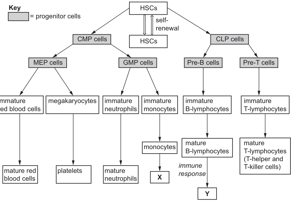
With reference to Fig 4.1, explain why GMP cells, which are progenitor cells, cannot be described as haematopoietic stem cells (HSCs).
Answer
Any three of:
- GMP cells are no longer undifferentiated: differentiation has already begun / they are partially specialised.
- GMP cells cannot self-renew — they do not produce more GMP (or HSC) cells when they divide; they only give rise to more differentiated cells.
- GMP cells cannot form all blood cell types — they only give rise to (immature) neutrophils and monocytes.
- (HSCs, by contrast, can self-renew AND form all the blood-cell types shown in Fig. 4.1.)
Differentiation has begun; no self-renewal; cannot form all blood cell types (only neutrophils and monocytes).
Background Concept
A stem cell is defined by two cardinal properties:
- Self-renewal — it can divide to produce at least one daughter cell that remains an identical, undifferentiated stem cell.
- Potency — it can give rise to multiple differentiated cell types.
A haematopoietic stem cell (HSC) is a multipotent adult stem cell in the bone marrow that can produce every type of blood cell shown in Fig. 4.1: red blood cells, platelets, neutrophils, monocytes, B-lymphocytes and T-lymphocytes.
A progenitor cell (also called a precursor or transit-amplifying cell) sits one step downstream. It has lost some potency (it can only give rise to a limited range of cell types) and it cannot self-renew — when it divides, both daughters differentiate further. Progenitor cells are shaded grey in Fig. 4.1.
Understanding the Question
The stem explains that the first stage of HSC differentiation is the production of progenitor cells, and that these CAN divide by mitosis but are NOT stem cells. Part (d) asks the candidate to justify why GMP cells — a specific type of progenitor cell shown in Fig. 4.1 — fail the definition of a stem cell.
Approach
Use Fig. 4.1 to compare GMP cells with HSCs. Check the three stem-cell criteria in turn:
- Are they undifferentiated? No — they are already along the differentiation pathway (shaded progenitor box).
- Can they self-renew? No — Fig. 4.1 shows GMP cells dividing only into more differentiated daughters; there is no loop back to HSCs.
- Are they multipotent? No — they give rise only to neutrophils and monocytes.
Step-by-Step Reasoning
- GMP cells lie on the CMP → GMP branch in Fig. 4.1; they are shaded as progenitor cells, meaning differentiation has already started.
- The only self-renewal arrow in the whole diagram is the loop on HSCs. GMP cells have no such loop; each division simply produces more differentiated daughters, so there is no self-renewal.
- Tracing GMP cells in the figure shows they lead only to immature neutrophils and immature monocytes (and then macrophages, platelets etc. are NOT on this branch). So GMP cells are committed to the granulocyte/monocyte lineage, not multipotent.
- A stem cell, by definition, must have all three properties — potency AND self-renewal AND undifferentiated status. GMP cells satisfy none of these fully.
Key Takeaways
Three criteria define a stem cell: undifferentiated, self-renewing, multipotent. Loss of ANY ONE of them means the cell is a progenitor, not a stem cell.
Common Mistakes
- Saying 'they are not found in the bone marrow' — irrelevant; GMP cells ARE in the bone marrow.
- Saying 'they cannot divide' — wrong; the stem tells us progenitor cells CAN divide by mitosis.
- Repeating just one criterion (e.g. 'no self-renewal') without linking it to the figure.
- Confusing GMP with CLP — the figure shows GMPs give neutrophils and monocytes; CLPs give lymphocytes.
Things to Be Careful About
Refer EXPLICITLY to Fig. 4.1 — the mark scheme's 'AW' allows alternative wording but the answer must be grounded in the figure: trace which boxes GMP connects to and notice that the only self-renewal loop is on HSCs.
Fig. 4.1 shows that monocytes differentiate into cell type X, which has a similar function to neutrophils.
Name cell type X.
Answer
Cell type X is a macrophage (macrophages).
Macrophage(s).
Background Concept
Monocytes circulate in the blood. When they migrate into tissues they mature and enlarge into macrophages. Macrophages are large phagocytic cells that engulf and digest pathogens, dead cells and cell debris by phagocytosis — the same broad function as neutrophils.
The monocyte → macrophage transition is the classical way in which the body converts short-lived blood phagocytes into long-lived tissue-resident phagocytes. Macrophages are also the cells that present pathogen antigens on their surface to T-helper cells, helping to activate the specific immune response.
Understanding the Question
The stem of part (e) tells us X is the cell a monocyte differentiates into, and that it has a similar function to neutrophils (i.e. it is phagocytic). The mark to be earned is naming X.
Approach
Recall the standard monocyte maturation pathway: blood monocyte → tissue macrophage. The 'similar function to neutrophils' cue (phagocytosis) confirms this.
Step-by-Step Reasoning
- Fig. 4.1 shows: immature monocyte → monocyte → X.
- Mature blood monocytes leave the bloodstream and mature into macrophages in the tissues.
- Macrophages, like neutrophils, are phagocytes — they engulf and digest pathogens and cellular debris.
- Therefore X = macrophage.
Key Takeaways
Monocytes are blood-borne phagocyte precursors; macrophages are their tissue-resident mature form.
Common Mistakes
- 'Mature monocytes' — the question already names monocytes; X must be the NEXT differentiated stage.
- 'Phagocyte' — too generic; the mark scheme explicitly rejects this.
- 'Granulocyte' — wrong lineage (granulocytes are neutrophils, eosinophils and basophils).
Things to Be Careful About
Do not write 'mature monocyte' — that is the box immediately above X in Fig. 4.1. The answer must be the differentiated cell type downstream of the monocyte.
Cell type Y shown in Fig. 4.1 releases molecules with antigen binding sites.
Name the molecules released by cell type Y.
Answer
Antibodies (immunoglobulins).
Antibody / immunoglobulin.
Background Concept
B-lymphocytes mature in the bone marrow. On first exposure to a specific antigen (with help from T-helper cells and antigen-presenting cells), a B-lymphocyte is activated, proliferates and differentiates into:
- plasma cells, which secrete antibodies (also called immunoglobulins);
- memory B-cells, which remain dormant until re-exposure.
Antibodies are Y-shaped glycoproteins with two antigen-binding sites at the tips of the Y. They neutralise pathogens by binding to antigens, marking them for destruction (opsonisation), agglutinating them, or activating complement.
Understanding the Question
Part (f) tells us cell type Y releases molecules with antigen-binding sites. Fig. 4.1 shows Y at the bottom of the mature B-lymphocyte branch.
Approach
Cells that secrete molecules with antigen-binding sites are plasma cells, and the molecules they secrete are antibodies.
Step-by-Step Reasoning
- Fig. 4.1 shows the B-lymphocyte branch ending at cell type Y, with 'immune response' labelled next to the mature B-lymphocyte box.
- The molecules released by (differentiated) B-lineage cells that have antigen-binding sites are antibodies / immunoglobulins.
- Therefore Y releases antibodies.
Key Takeaways
Plasma cells (differentiated B-lymphocytes) secrete antibodies; these antibodies bind specifically to antigens via their antigen-binding sites.
Common Mistakes
- 'Antigens' — antigens are what antibodies bind TO, not what B-cells release.
- 'Memory cells' — these are cells, not molecules.
- 'Cytokines' — released by T-helper cells, not B-cells.
Things to Be Careful About
'Antibody' and 'immunoglobulin' are both accepted and are interchangeable terms.
The differentiation of T-lymphocytes begins in the bone marrow and continues in an organ known as the thymus to produce fully differentiated T-helper and T-killer cells.
In the thymus, T-lymphocytes that bind to self antigens are destroyed.
Explain why T-lymphocytes that bind to self antigens need to be destroyed in the thymus.
Answer
Any three of:
- After the thymus, T-lymphocytes are released into the general circulation / bloodstream, where they encounter body cells.
- Self-antigens are present on the surface of the body's own cells; if a T-lymphocyte binds self antigens, an immune response would be triggered against the body's own cells.
- This would damage or destroy body cells (or, with T-killer cells, actively kill them).
- It would also lead to the formation of memory T-cells against self antigens, so the body would attack itself on every subsequent exposure (autoimmune response).
- By destroying self-reactive T-cells in the thymus, only T-lymphocytes that respond to foreign / non-self antigens are released, preventing autoimmune disease.
Self-reactive T-cells released into circulation would bind self antigens on body cells, triggering an immune response that destroys body cells and creates self-reactive memory cells (autoimmune disease).
Background Concept
T-lymphocyte precursors migrate from the bone marrow to the thymus, where they mature. During maturation, each developing T-cell randomly generates a T-cell receptor (TCR) with a specific binding site. Because this generation is random, some TCRs will, by chance, bind to 'self' antigens — molecules normally displayed on the surface of the body's own cells (presented on MHC molecules).
The thymus tests each T-cell for self-reactivity. T-cells that bind self antigens too strongly are destroyed by apoptosis — this is positive and negative selection, and it establishes self-tolerance. T-cells that bind self antigens weakly (or not at all) are released into the circulation; only these become mature T-helper and T-killer cells that respond to foreign antigens.
Understanding the Question
Part (g) gives two pieces of context:
- T-lymphocyte differentiation starts in the bone marrow and is completed in the thymus.
- T-lymphocytes that bind self antigens are destroyed IN the thymus.
The candidate must explain WHY this destruction is necessary.
Approach
Reason about what would happen if self-reactive T-cells were NOT destroyed:
- They would leave the thymus and enter the general circulation.
- There they would meet cells of the body, which display self antigens on MHC molecules.
- They would bind these self antigens and trigger an immune response.
- The consequences are: damage/destruction of the body's own cells; creation of memory T-cells against self antigens (so the attack persists on re-exposure) — i.e. autoimmune disease.
Step-by-Step Reasoning
- The thymus is the last checkpoint before T-cells enter the body. Once they leave, they are exposed to all body tissues.
- Self antigens are normal surface markers on the body's own cells. A T-lymphocyte whose receptor binds a self antigen would, if released, attack that body cell.
- T-killer cells would directly destroy the cell; T-helper cells would release cytokines, amplifying the response and recruiting phagocytes and B-cells against the body's own tissues.
- Some of these self-reactive lymphocytes would become memory cells, so the attack would be repeated and amplified on every subsequent exposure to those self antigens — this is autoimmunity.
- Destroying self-reactive T-cells in the thymus prevents all of the above, ensuring the immune system only attacks foreign material.
Key Takeaways
Self-tolerance is essential. Negative selection in the thymus removes T-cells that would attack the body's own tissues; without it, autoimmune disease results. This is one of the central mechanisms preventing self-destruction by the immune system.
Common Mistakes
- Saying 'so the T-cells don't attack themselves' — T-cells don't attack themselves; they attack OTHER cells displaying self antigens.
- Saying 'so the T-cells don't get attacked' — confusion of direction.
- Forgetting to mention memory cells and autoimmunity — these give the long-term consequence and are explicit mark-scheme points.
- Saying 'because self antigens are bad' — self antigens are not bad; the problem is that an immune response AGAINST them is bad.
Things to Be Careful About
The mark scheme specifically notes that points 2 and 3 require the candidate to make clear that self antigens are on the body's OWN cells. A bare statement like 'they would cause an immune response' does not earn these marks; add 'against body cells'.
Malaria is an infectious disease caused by the protoctist, Plasmodium.
As part of its lifecycle, Plasmodium infects human red blood cells. Researchers can compare haemoglobin from the red blood cells of a healthy person with haemoglobin from a person with malaria.
Throughout the world, most deaths from malaria are caused by P. vivax and P. falciparum.
Name one other species of Plasmodium that causes malaria.
Plasmodium ______
Answer
Plasmodium ovale
(accept Plasmodium malariae or Plasmodium knowlesi)
Plasmodium ovale
Background Concept
Malaria in humans is caused by protoctist parasites belonging to the genus Plasmodium. These are single-celled eukaryotes (protoctists) transmitted to humans through the bite of an infected female Anopheles mosquito. Five species of Plasmodium are known to cause malaria in humans, and they differ in their geographical distribution, severity and the clinical picture they produce.
The five species that infect humans are:
- Plasmodium falciparum — responsible for the majority of deaths worldwide, especially in sub-Saharan Africa; causes the most severe form (cerebral malaria).
- Plasmodium vivax — the most widespread geographically; causes relapsing malaria because it can lie dormant in the liver as hypnozoites.
- Plasmodium ovale — similar to P. vivax in causing relapses; found mainly in West Africa and the western Pacific.
- Plasmodium malariae — causes a milder, chronic form with a characteristic 72-hour fever cycle; widespread but less common.
- Plasmodium knowlesi — originally a parasite of macaques; now recognised as a significant cause of zoonotic malaria in South-East Asia.
Understanding the Question
This is a simple recall question. The stem has already told you that P. vivax and P. falciparum are the two species responsible for most malaria deaths. You simply have to name one of the remaining three species. The answer should be the binomial name written in the same italicised form, e.g. Plasmodium ovale.
Approach
Read the question carefully — it says "name one other species". Any one of the three remaining species will earn the mark. Spell the species name correctly; CIE mark schemes reject misspelt binomials.
Step-by-Step Reasoning
- Plasmodium ovale — the most common alternative answer, especially for CIE questions which list ovale and malariae as the standard "other two".
- Plasmodium malariae — also credited; the same spelling rules apply.
- Plasmodium knowlesi — credited, but the mark scheme warns that it "must be spelled correctly".
If you give two answers, both must be correct and correctly spelled, or the mark is lost.
Key Takeaways
- Five species of Plasmodium cause malaria in humans: falciparum, vivax, ovale, malariae, and (more recently recognised) knowlesi.
- P. falciparum and P. vivax are responsible for the great majority of malaria deaths and cases worldwide.
- Binomial names must be italicised with the genus capitalised and species in lower case.
Common Mistakes
- Writing just "ovale" or "malariae" without the genus Plasmodium — the binomial must be complete.
- Misspelling "knowlesi" — the mark scheme rejects incorrect spellings.
- Giving P. falciparum or P. vivax — these are explicitly excluded by the stem.
Things to Be Careful About
- Write the full binomial: Plasmodium in italics followed by the species epithet in lower case.
- If you write more than one answer, ensure each is correctly spelled — partial credit is not given.
In the laboratory, oxygen at different partial pressures can be bubbled through a solution of haemoglobin to determine the percentage saturation of haemoglobin at each partial pressure. A graph constructed from the results is known as an oxygen dissociation curve.
Fig. 5.1 is an oxygen dissociation curve for normal adult haemoglobin in humans.
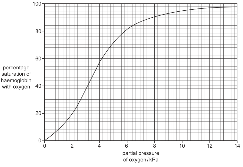
In the experiment used to obtain the results shown in Fig. 5.1, the temperature and pH were standardised.
Explain what the researchers would consider when deciding which temperature and pH to use in the experiment.
Answer
- The conditions used should be those that occur in the body — pH ≈ 7.4 (plasma/blood pH) and 37 °C (body temperature) — so the results reflect what happens in vivo.
- Oxygen uptake by haemoglobin is affected by changes in temperature and pH, so to obtain valid, comparable results these variables must be controlled.
Standardise at body conditions (pH ≈ 7.4, 37 °C) so that the in vitro results reflect in vivo haemoglobin behaviour, because oxygen uptake is affected by temperature and pH.
Background Concept
An oxygen dissociation curve plots the percentage saturation of haemoglobin with oxygen against the partial pressure of oxygen (pO₂). The position and shape of the curve depend on several physiological variables, including temperature, pH (and the partial pressure of CO₂ via the Bohr effect), the concentration of 2,3-BPG, and whether the haemoglobin is fetal or adult.
Because the curve is sensitive to these conditions, any laboratory measurement of haemoglobin saturation is only meaningful if those variables are fixed at known, reproducible values. This is a general principle of controlling variables in an experiment: change one variable at a time, hold all the others constant.
Understanding the Question
The stem tells you that the experiment uses a haemoglobin solution, and that temperature and pH are standardised. It then asks you to explain what the researchers would consider when choosing the values of those two variables. The command word is "explain" — you must give reasons, not just state values.
Approach
Two ideas are needed for the two marks:
- The values chosen should match the conditions that the haemoglobin actually experiences in the body, so the curve reflects normal physiology. This gives the appropriate values: pH ≈ 7.4 and 37 °C.
- Because haemoglobin's behaviour depends on temperature and pH, those variables would distort the results if not held constant; therefore the researchers must pick specific values and control them.
Step-by-Step Reasoning
- In the human body, plasma pH is tightly regulated at approximately 7.35–7.45 (≈ 7.4), and core body temperature is approximately 37 °C. Choosing these values means the in vitro curve corresponds to what the haemoglobin is doing inside a person.
- The mark scheme also accepts "neutral pH" and "36 °C / 38 °C" — values that approximate physiological conditions.
- If the experimenters used, say, 25 °C or pH 5, the curve would not be a meaningful representation of how the patient's haemoglobin behaves in life. The experiment is only valid if it mimics the in vivo environment.
- A second, separate point is that pH and temperature genuinely change haemoglobin's affinity for oxygen. Higher temperatures or lower (more acidic) pH shift the curve to the right (Bohr effect). So the experimenters must keep them constant — otherwise they cannot tell whether any change in the curve is due to the haemoglobin itself or to the experimental conditions.
Key Takeaways
- In any controlled experiment, the standardised variables should be set to the values that occur in the biological situation being modelled.
- pH ≈ 7.4 and 37 °C are the standard physiological values used for human blood experiments.
- Oxygen uptake by haemoglobin is sensitive to temperature and pH, so these variables must be controlled for the results to be valid.
Common Mistakes
- Stating values without explaining why those values were chosen (e.g. just writing "37 °C"). The question asks for an explanation.
- Saying the values should be the ones that "give the best results" or "work well" — this is too vague.
- Confusing standardisation with control of the independent variable — the experimenters are not testing temperature or pH; they are simply holding them at body values.
Things to Be Careful About
- Two distinct ideas are needed for two marks: (a) physiological conditions, and (b) the fact that these variables affect haemoglobin and must therefore be controlled.
- A single sentence that conflates the two (e.g. "use 37 °C because temperature affects haemoglobin") may only earn one mark.
Using a different, more rapid technique, researchers compared the haemoglobin contained in red blood cells of a healthy person with the haemoglobin of a person with malaria who had been infected with P. vivax.
By analysing the results, the researchers concluded that the oxygen dissociation curve of a person with malaria would be shifted to the right.
With reference to Fig. 5.1, explain how a shift to the right of the oxygen dissociation curve would affect oxygen loading in the lungs, and unloading in respiring tissues, in a person with malaria.
Answer
- The affinity of haemoglobin for oxygen is decreased, so at any given partial pressure of oxygen the percentage saturation is lower than normal.
- In the lungs: a higher pO₂ is needed to load the same amount of oxygen, so loading is more difficult and less oxygen is loaded.
- In respiring tissues: because the curve is shifted to the right, more oxygen is released (unloaded) at any given tissue pO₂, so oxygen is released more readily to the tissues.
Right shift → lower affinity → less oxygen loaded in the lungs at a given pO₂, but more oxygen unloaded in respiring tissues.
Background Concept
The oxygen dissociation curve shows the percentage saturation of haemoglobin at each partial pressure of oxygen. Its position is described as "left-shifted" (higher affinity — haemoglobin holds on to oxygen more tightly) or "right-shifted" (lower affinity — haemoglobin releases oxygen more readily).
A right shift is caused by:
- increased temperature;
- decreased pH (more H⁺, e.g. in actively respiring tissue);
- increased pCO₂;
- increased 2,3-BPG concentration;
- the presence of certain abnormal haemoglobins.
Physiologically, a right shift at the tissues is beneficial because actively respiring cells (which produce CO₂ and H⁺) need more oxygen delivered to them — the Bohr effect.
Understanding the Question
The stem tells you that researchers have concluded the curve for a malaria patient is shifted to the right compared with Fig. 5.1. The question asks you to use the figure to explain what that right shift means in two specific contexts:
- Loading of oxygen in the lungs.
- Unloading of oxygen in respiring tissues.
You should not simply describe a right shift in general; you must apply it to the two situations. The mark scheme explicitly rejects saying "in the presence of carbon dioxide" because the context here is malaria infection, not a Bohr-shift scenario — the shift is due to the infection itself.
Approach
A right shift means that, at any given pO₂, the haemoglobin is less saturated with oxygen. Read horizontally across Fig. 5.1 at, say, pO₂ ≈ 5 kPa: the normal curve sits near 70 % saturation, but a right-shifted curve at the same pO₂ would sit lower. Equivalently, to reach a given saturation, a higher pO₂ is needed.
Apply this to the two situations:
- Lungs: pO₂ is high (≈ 13 kPa at the alveolar surface). Even with a right shift, saturation is still high in absolute terms, but it is lower than for a normal person. So loading is harder / less complete.
- Tissues: pO₂ is low (≈ 5 kPa in a typical respiring cell). A right shift means that at this low pO₂, saturation is markedly lower than normal — i.e. much more oxygen has dissociated from haemoglobin. So unloading is greater / more oxygen is delivered to the tissues.
Step-by-Step Reasoning
- Mark point 1 — affinity decreases. A right shift literally means lower affinity. "Affinity of haemoglobin for oxygen decreases" earns the mark; vague statements such as "the curve is different" do not.
- Mark point 2 — lower saturation in the lungs. Because affinity is lower, at the pO₂ of the lungs the percentage saturation is lower than for a normal person. The mark scheme accepts "less oxygen binds to haemoglobin" or "haemoglobin loads less oxygen".
- Mark point 3 — loading is more difficult. This is the consequence of needing a higher pO₂ to achieve the same saturation. So a malaria patient needs a higher alveolar pO₂ to load the same amount of oxygen.
- Mark point 4 — a higher pO₂ is needed for the same saturation. This is the mathematical/geometric meaning of a right shift: at the same percentage saturation, you read further to the right on the pO₂ axis.
- Mark point 5 — more oxygen is released in respiring tissues. Because the curve is lower at low pO₂, the difference between the saturation in the lungs and the saturation in the tissues is greater, so more oxygen is unloaded per pass through a tissue capillary.
For three marks, any three of these five points are sufficient. The mark scheme explicitly rejects any reference to CO₂ as the cause of the right shift in this question, because the cause is the malarial infection, not the Bohr effect.
Key Takeaways
- A right shift of the oxygen dissociation curve means lower affinity of haemoglobin for oxygen.
- A right shift makes oxygen loading in the lungs harder (less oxygen carried) but unloading in respiring tissues easier (more oxygen released to the cells).
- A right shift is read in two ways: at constant pO₂, the saturation is lower; OR at constant saturation, a higher pO₂ is required.
Common Mistakes
- Stating the curve "moves right" without linking it to affinity or saturation — too vague to score.
- Saying "in the presence of CO₂, oxygen is released more readily" — the mark scheme rejects this because the cause of the shift here is the infection, not CO₂.
- Saying oxygen is released more slowly — the question concerns the amount of oxygen released, not the rate.
- Confusing the two situations: a right shift is bad for loading in the lungs but good for unloading in the tissues.
Things to Be Careful About
- "Slower" is ignored; "more difficult" or "less oxygen loaded" is credited.
- "In the presence of carbon dioxide" is rejected for the cause of the shift in this question, but the consequence at the tissues can be stated without invoking CO₂.
- Error carried forward: if a candidate wrongly says the shift is to the left, the consequence points can still be credited (ecf) provided they are internally consistent with a left shift.
A red blood cell that is infected with Plasmodium cannot carry out its function as effectively as a normal red blood cell.
Describe how the size and structure of a red blood cell is related to its function, other than the fact that it contains a very large number of haemoglobin molecules.
Answer
Any four from:
- Diameter 6–8 µm, so the cell can pass through the narrow lumen of capillaries.
- Small size / diameter means cells travel in single file through capillaries, slowing blood flow and maximising the time available for oxygen to diffuse in (or out).
- No nucleus (and no other organelles) — therefore more space inside the cell.
- (linked to point 3) the extra space is available for more haemoglobin.
- No mitochondria — therefore oxygen is not used by the red blood cell itself in respiration, so all the oxygen it carries can be transported to the tissues.
- The cell is flexible and can deform to squeeze through capillaries narrower than its own diameter.
- The cell is biconcave (disc-shaped with a thinner centre).
- (Biconcave shape, compared with a sphere) gives a larger surface area to volume ratio, increasing the rate of diffusion of oxygen across the cell surface membrane.
- (Biconcave shape) reduces the diffusion distance between the cell surface membrane and the haemoglobin in the centre of the cell.
Diameter 6–8 µm fits capillaries; small size gives single-file flow that slows blood and maximises exchange time; biconcave shape gives a high SA:V and short diffusion distance to haemoglobin; no nucleus/mitochondria gives space for haemoglobin and prevents the cell itself consuming the oxygen it carries; the cell is flexible and can deform through narrow capillaries.
Background Concept
The red blood cell (erythrocyte) is one of the most highly specialised cells in the body. Its only function is to transport oxygen (and to a lesser extent carbon dioxide) between the gas-exchange surface and the respiring tissues. Its structure is therefore pared down to the minimum needed for that single role.
A mature mammalian red blood cell:
- has a biconcave disc shape (thinner in the middle than at the edges);
- is about 6–8 µm in diameter;
- has no nucleus, no mitochondria, no ribosomes, no endoplasmic reticulum and no other organelles;
- consists essentially of a cell surface membrane enclosing a cytoplasm packed with haemoglobin;
- is flexible because its membrane is supported by a meshwork of proteins (spectrin) just beneath the bilayer.
In addition, the question stem excludes the most obvious feature ("a very large number of haemoglobin molecules") and asks you to discuss the other features.
Understanding the Question
The command word is "describe" — give an account of the size and structure and link each feature to its function. The marks reward paired structure–function statements. You are told to ignore the high haemoglobin content (otherwise it would be the obvious answer). You should also ignore the colour (red) and any reference to lifespan; only size and structure are required.
Approach
Work through the features systematically, pairing each with a functional consequence:
- size (6–8 µm);
- shape (biconcave);
- absence of nucleus;
- absence of mitochondria;
- flexibility.
For each, state the feature and then explicitly link it to the function. The mark scheme awards a mark for the feature and (often) a separate mark for the linked functional explanation, so both halves of the pair must be present.
Step-by-Step Reasoning
Size (6–8 µm diameter)
- Feature: the diameter is 6–8 µm, which is approximately the same as the lumen of a capillary. Mark point 1.
- Function: the cell just fits inside a capillary, so it travels in single file, one cell at a time. This slows the flow of blood past the capillary wall and gives maximum time for oxygen to diffuse in (and CO₂ to diffuse out). Mark point 2.
- The mark scheme does not credit "small size" on its own — you must give a number (or compare to a capillary) and then link to the single-file / slowed flow consequence.
Absence of nucleus
- Feature: the mature red blood cell has no nucleus. Mark point 3.
- Function: this leaves more space inside the cell for haemoglobin. Mark point 4. Note that mark point 4 is dependent on mark point 3; the function must be linked to the structural feature.
Absence of mitochondria
- Function: with no mitochondria, the cell cannot respire aerobically, so it does not consume any of the oxygen it carries — that oxygen is all available to be delivered to respiring tissues. Mark point 5.
Flexibility
- Feature: the cell is flexible and can deform, so it can squeeze through capillaries that are narrower than its own diameter. Mark point 6. The mark scheme ignores "squeeze between" unless it is clearly stated that the cell is inside a capillary.
Biconcave shape
- Feature: the cell is biconcave. Mark point 7.
- Function 1 (SA:V): compared with a sphere of the same volume, a biconcave disc has a larger surface area to volume ratio, so diffusion of oxygen across the cell surface membrane is faster. Mark point 8.
- Function 2 (diffusion distance): the thin centre of the disc means the diffusion distance from the cell surface membrane to the haemoglobin molecules in the middle of the cell is small, so oxygen reaches the haemoglobin quickly. Mark point 9.
Any four of these mark points earn the four marks. The cleanest single answer combines the four paired structure–function statements above.
Key Takeaways
- Red blood cells are exquisitely specialised for oxygen transport; their structure is shaped entirely by that function.
- The four most important structure–function links are: small diameter (capillary fit), biconcave shape (SA:V and short diffusion distance), absence of nucleus (more space for haemoglobin), and absence of mitochondria (oxygen is not used by the cell itself).
- A biconcave disc has a higher SA:V than a sphere of the same volume and a shorter maximum diffusion distance from the surface to the centre.
- "Describe" questions on cell structure reward paired structure and function statements, not the structure alone.
Common Mistakes
- Stating only the structural feature without the linked function (e.g. just "no nucleus"). You need the explanation that the absence of a nucleus gives more space for haemoglobin.
- Saying "small size" without quantifying it or linking it to single-file flow in a capillary. The mark scheme ignores "small size" on its own.
- Including the high haemoglobin content — explicitly excluded by the stem.
- Stating that the cell is "round" or "spherical" — it is biconcave; the shape is the whole point of the question.
- Confusing red blood cells with other cell types (white blood cells, prokaryotes) and listing irrelevant features.
Things to Be Careful About
- Each mark point typically has two halves: a structural feature and its functional consequence. The functional half is often the harder one to articulate and is required for the mark.
- Mark point 4 (more space for haemoglobin) is conditional on mark point 3 (no nucleus); if the structure is not stated, the function cannot be credited.
- A flexible cell that "squeezes between cells" is ignored unless the candidate makes it clear that the red blood cell is inside a capillary.
- Use the term "biconcave" (not just "disc-shaped" or "round").
- Note that the mark scheme describes the SA:V point as "compared to a sphere" — the comparison is with what a sphere of the same volume would give, not with another cell.
The transport of water from the soil solution to the xylem of roots occurs by the apoplast and symplast pathways. Mineral ions can be transported dissolved in water.
Describe the transport of water from the soil solution to the endodermis of roots by the apoplast pathway and explain why this pathway cannot continue at the endodermis.
Answer
-
The apoplast pathway is the non-living route through the root: water moves through the cell walls (and the spaces between cells / intercellular spaces) of the root hair / epidermal cells and the cortical cells, without crossing any cell surface membrane.
-
This pathway cannot continue at the endodermis because the Casparian strip in the walls of the endodermal cells blocks further movement through cell walls and intercellular spaces.
-
The Casparian strip is made of suberin, a waxy / waterproof / impermeable material deposited in the cell walls of the endodermis.
-
At this point water is forced to cross a cell surface membrane into the cytoplasm (entering the symplast pathway) before it can continue into the xylem.
-
Overall, water moves down a water potential gradient from the soil, across the root, into the xylem.
See working.
Background Concept
Water is taken up by a plant root from the soil and travels across the root to the xylem, from where it is pulled up to the leaves and lost by transpiration. The soil solution is relatively dilute and has a high (less negative) water potential, while the inside of the xylem and the air in the leaf have progressively lower water potentials. This continuous water potential gradient from soil → root → xylem → leaf → atmosphere is what drives the bulk movement of water into and through the plant.
Two parallel routes carry water across the cortex of the root:
- Apoplast pathway — the non-living network consisting of cell walls and the spaces between cells (intercellular spaces). Water (and dissolved mineral ions) can move freely through this continuous matrix of cellulose without ever crossing a cell surface membrane.
- Symplast pathway — the living route, in which water passes from cell to cell through the cytoplasm via plasmodesmata, having crossed a cell surface membrane to enter the first cell.
At the innermost layer of the cortex, the endodermis, the apoplast pathway is blocked by the Casparian strip — a ring of suberin (a waxy, hydrophobic, impermeable material) impregnating the radial and transverse walls of the endodermal cells. Because suberin is waterproof, water and dissolved ions cannot continue to flow freely through the cell walls; they must cross the cell surface membrane of an endodermal cell, enter its cytoplasm, and continue via the symplast. This forces water and solutes to pass through a living, selectively permeable barrier before reaching the xylem, which is essential for controlling which substances enter the transpiration stream.
Understanding the Question
This part has a compound command word: describe the apoplast pathway from soil to endodermis, and explain why it cannot continue at the endodermis. The stem tells us that transport of water from soil to xylem uses the apoplast and symplast pathways, so candidates are expected to know the difference between them and the special role of the endodermis. Marks are awarded for naming the cell-wall/intercellular-space route, naming the cells that the apoplast passes through, identifying the Casparian strip, describing its suberin composition, and linking overall movement to a water potential gradient.
Approach
- First describe the route: cell walls and intercellular spaces of epidermal / root hair cells and cortical cells, without crossing membranes.
- Then identify the barrier: the Casparian strip in the walls of endodermal cells, made of suberin (waxy / waterproof / impermeable).
- Finally add context: the overall driving force is a water potential gradient from soil to xylem, and that beyond the endodermis water must enter the symplast.
Step-by-Step Reasoning
-
Cell walls form the apoplast pathway. Apoplast literally means "outside the cell" and refers to the network of cell walls and intercellular spaces that permeate plant tissue. Because cellulose cell walls are fully permeable to water and small solutes, water in the apoplast can travel through them without ever entering a cell. Mark scheme point 1.
-
Water also moves through intercellular spaces. Where adjacent cells do not meet tightly, water can flow through the gaps between cells. Mark scheme point 2.
-
Name the cells the apoplast passes through on the way to the endodermis. Going from outside in, water in the apoplast passes through the walls of root hair / epidermal cells and then through the walls of the cortical (parenchyma) cells. Mark scheme point 3.
-
The pathway stops at the Casparian strip. At the endodermis, the radial and transverse cell walls are impregnated with suberin, forming a continuous waterproof ring. Because suberin is hydrophobic, water cannot pass along the cell walls or intercellular spaces past this point. Mark scheme point 4.
-
Suberin is waxy / waterproof / impermeable. This detail is required to earn the explanation mark — it is not enough to say "stops at the Casparian strip"; the candidate must describe the material of the strip and its physical property. Mark scheme point 5.
-
Overall movement is down a water potential gradient. Water moves passively from a region of higher (less negative) water potential in the soil to a region of lower (more negative) water potential in the xylem and leaf. The mark scheme explicitly rejects references to "osmosis" or "active transport" here — this is a passive process driven by the gradient. Mark scheme point 6.
-
AVP — apoplast is non-living and does not cross membranes. This is a useful framing statement that the mark scheme accepts as an extra point. It is also the underlying reason that water must cross a membrane at the endodermis, which is what allows the plant to control what enters the xylem. Mark scheme point 7.
Key Takeaways
- The apoplast pathway is the non-living route through cell walls and intercellular spaces; it does not cross cell surface membranes.
- It is blocked at the endodermis by the Casparian strip, a ring of suberin in the radial and transverse walls of endodermal cells.
- Beyond the endodermis, water must enter the symplast (cytoplasm of endodermal cells) before passing into the xylem.
- The Casparian strip is a control point: by forcing water and dissolved ions to cross a living cell membrane, the plant can selectively regulate what enters the transpiration stream.
- Overall water uptake is driven by a water potential gradient from soil to xylem (a passive process).
Common Mistakes
- Saying water moves by osmosis / active transport along the apoplast. The mark scheme explicitly rejects "osmosis" and "active transport" in the context of the apoplast — apoplastic water movement is bulk flow through wet cell walls, driven by a pressure / water potential gradient, not osmosis or active transport.
- Vagueness about which cells the apoplast passes through. Saying "through the root" is not enough; the cells (epidermal / root hair cells, cortical cells) must be named and the context "cell walls" stated.
- Naming only the Casparian strip but not the suberin. The mark scheme requires both the structure AND its composition/property (suberin / waxy / waterproof / impermeable).
- Confusing the Casparian strip with the cell surface membrane. The strip is in the cell walls, not in the membrane.
- Mixing up apoplast and symplast. Apoplast = cell walls + intercellular spaces; symplast = cytoplasm connected by plasmodesmata.
Things to Be Careful About
- Use the exact term Casparian strip (not "Casparian band" or "suberin ring" alone).
- Use the exact term suberin for the waxy material; "waxy" or "waterproof" alone is acceptable but suberin is the precise term.
- The endodermis is a single cell layer; do not describe it as the "inner cortex" or "pericycle".
- "Movement down a water potential gradient" must not be replaced by "osmosis" or "active transport".
- Keep the description tied to the apoplast specifically — the mark scheme rejects "passing through vacuoles" or "passing through cytoplasm" in the context of movement to the endodermis.
Researchers investigated the mechanism of transport used for the uptake of potassium ions () into root epidermal cells at different concentrations of in the soil solution.
Complete Table 6.1 to provide information about the two different transport mechanisms that were identified by the researchers.
Table 6.1
| net movement of | membrane protein needed (yes or no) | ATP used (yes or no) | name of transport mechanism |
|---|---|---|---|
| against the concentration gradient | |||
| down the concentration gradient |
Answer
| net movement of | membrane protein needed (yes or no) | ATP used (yes or no) | name of transport mechanism |
|---|---|---|---|
| against the concentration gradient | yes | yes | active transport |
| down the concentration gradient | yes | no | facilitated diffusion |
Against concentration gradient: yes protein, yes ATP, active transport. Down concentration gradient: yes protein, no ATP, facilitated diffusion.
Background Concept
The cell surface membrane is a phospholipid bilayer with embedded proteins. Small, non-polar molecules (e.g. O₂, CO₂) can dissolve in the bilayer and cross it directly, but ions (charged particles such as K⁺, Na⁺, Ca²⁺) cannot pass through the hydrophobic core of the bilayer on their own — they are repelled by the non-polar fatty acid tails. To cross the membrane, ions must use a membrane transport protein (either a channel protein or a carrier protein).
There are two ways ions can move through transport proteins:
- Facilitated diffusion — ions move down their concentration gradient (or electrochemical gradient) through a channel or carrier protein. No ATP is used because movement is passive, down the gradient. The protein simply provides a hydrophilic route through the bilayer.
- Active transport — ions move against their concentration gradient using a carrier protein. Energy from ATP is required because ions are being pumped "uphill". The carrier protein undergoes a conformational change powered by ATP hydrolysis.
So three diagnostic features identify the mechanism:
- Direction of movement (with or against the concentration gradient)
- Whether a membrane protein is needed (always yes for ions)
- Whether ATP is used (yes for active transport, no for facilitated diffusion)
Understanding the Question
Part (b) presents Table 6.1, which has two pre-filled rows describing two scenarios for K⁺ uptake into root epidermal cells. The first row describes K⁺ moving against its concentration gradient; the second row describes K⁺ moving down its concentration gradient. The candidate must fill in three remaining columns: whether a membrane protein is needed, whether ATP is used, and the name of the transport mechanism.
The stem reminds us that mineral ions (including K⁺) are transported dissolved in water, so we are reasoning about ion transport across cell surface membranes — the standard membrane-transport scenarios.
Approach
For each row:
- Decide whether the ion can cross the phospholipid bilayer on its own. K⁺ is a charged ion → yes, a membrane protein is needed in both rows.
- Decide whether ATP is required. Movement against the concentration gradient requires energy from ATP. Movement down the concentration gradient does not.
- Name the mechanism: against gradient + ATP = active transport; down gradient + no ATP = facilitated diffusion.
Step-by-Step Reasoning
Row 1: against the concentration gradient
- K⁺ is a charged ion and cannot cross the phospholipid bilayer directly → a membrane protein (carrier) is needed.
- Moving against the concentration gradient requires energy → ATP is used.
- The transport of a substance against its concentration gradient using a membrane protein and ATP is, by definition, active transport.
Row 2: down the concentration gradient
- K⁺ still cannot cross the phospholipid bilayer on its own (it is charged) → a membrane protein (channel or carrier) is still needed. This is the key distinction from simple diffusion, which is restricted to small non-polar molecules.
- Movement is down the gradient, so it is spontaneous and does not require energy → ATP is not used.
- Passive movement of ions (or polar molecules) through a membrane protein, down the concentration gradient and without ATP, is facilitated diffusion.
Key Takeaways
- The direction of movement relative to the concentration gradient is the single most important diagnostic for choosing between active transport and facilitated diffusion.
- Charged ions always require a membrane protein to cross the phospholipid bilayer, whether they are moving with or against the gradient. This is what distinguishes facilitated diffusion from simple diffusion.
- ATP is required only for active transport — i.e. when moving against the concentration gradient.
- A useful mnemonic: "uphill = active (uses ATP)"; "downhill = passive (no ATP)".
- Active transport allows root cells to accumulate K⁺ to concentrations far higher than in the soil solution, which is essential because K⁺ is needed for many cytoplasmic functions (e.g. as a cofactor for enzymes, in stomatal opening).
Common Mistakes
- Writing "no" for the membrane protein column in the second row. Candidates sometimes confuse simple diffusion (small non-polar molecules, no protein) with facilitated diffusion (ions / polar molecules, protein required). The question is about K⁺, an ion — a protein is required in BOTH rows.
- Writing "no" for ATP use in the first row. Movement against a concentration gradient is energetically unfavourable and can only happen with an energy supply. ATP must be used.
- Writing "diffusion" instead of "facilitated diffusion" for the second row. Plain diffusion cannot move ions through the bilayer; the protein-mediated route is called facilitated diffusion.
- Swapping the two mechanisms. Always check: against gradient = active; down gradient = facilitated.
- Writing "osmosis" anywhere. Osmosis is specifically the diffusion of water molecules across a partially permeable membrane, not ion transport.
Things to Be Careful About
- The candidate receives one mark per correctly completed column (both rows right in that column), with a fallback mark available if one entire row is correctly filled — so it is worth filling every cell thoughtfully even if uncertain about one.
- "Yes / no" answers must be unambiguous — "y", "✓", or blank are not accepted; only "yes" or "no" earn the mark.
- The transport mechanism name should be the standard CIE term: active transport (not "active uptake" or "pumping") and facilitated diffusion (not "passive transport" or "channel diffusion").
- The concentration gradient refers to the gradient of K⁺ across the cell surface membrane of the root epidermal cell — not across the whole root or the Casparian strip.