Biology 9700/21 — October/November 2024
Cambridge AS Level · AS Level Structured Questions · worked solutions for every part, with the mark scheme
Topics Cell Membranes and Transport · Biological Molecules · Enzymes · Cell Structure · Transport in Mammals · Gas Exchange · +5 more
Animal cells, plant cells and prokaryotic cells have similarities and differences in their structure.
Table 1.1 lists five organelles found in cells.
Complete Table 1.1 by placing a tick (✓) to show whether the organelle is present in animal cells, plant cells and prokaryotic cells or a cross (✗) if the organelle is absent.
Put a tick (✓) or a cross (✗) in every box.
The first row has been completed for you.
Table 1.1
| organelle | animal cells | cell type plant cells | prokaryotic cells |
|---|---|---|---|
| nucleus | ✓ | ✓ | ✗ |
| large permanent vacuole | |||
| rough endoplasmic reticulum | |||
| Golgi body | |||
| centrioles |
Answer
| organelle | animal cells | plant cells | prokaryotic cells |
|---|---|---|---|
| nucleus | ✓ | ✓ | ✗ |
| large permanent vacuole | ✗ | ✓ | ✗ |
| rough endoplasmic reticulum | ✓ | ✓ | ✗ |
| Golgi body | ✓ | ✓ | ✗ |
| centrioles | ✓ | ✗ | ✗ |
large permanent vacuole: animal ✗, plant ✓, prokaryote ✗; RER: animal ✓, plant ✓, prokaryote ✗; Golgi body: animal ✓, plant ✓, prokaryote ✗; centrioles: animal ✓, plant ✗, prokaryote ✗
Background Concept
All cells share some basic features (a plasma membrane, cytoplasm, ribosomes and DNA), but eukaryotes and prokaryotes differ fundamentally. Eukaryotic cells (animal, plant, fungal, protoctist) have membrane-bound organelles, whereas prokaryotic cells (bacteria) do not — they have no nucleus, no membrane-bound endoplasmic reticulum, no Golgi body and no mitochondria. Between animal and plant cells there are three key distinguishing features to remember: only plant cells have a large permanent vacuole, a cell wall made of cellulose, and chloroplasts; only animal cells have centrioles.
Understanding the Question
The question asks you to fill in four rows of a table, putting either a tick (✓) for present or a cross (✗) for absent in each of three cell types. One mark is awarded for each fully correct row, so the four rows are worth four marks. The first row (nucleus) is already done as a guide: ✓ in animal, ✓ in plant, ✗ in prokaryote — this is because the nucleus is a membrane-bound organelle found in eukaryotes only.
Approach
For each organelle, decide two things:
- Is it membrane-bound? If so, prokaryotes will be ✗.
- Is it a feature unique to one eukaryotic group? If so, that group gets ✓ and the other gets ✗.
Then mark both eukaryotes ✓ unless the organelle is restricted to only one of them.
Step-by-Step Reasoning
- Large permanent vacuole: a membrane-bound (tonoplast) structure filled with cell sap. Found in mature plant cells only — neither animal nor prokaryotic cells have a permanent vacuole of this kind (animal cells may have small temporary vesicles but not a large, permanent, central one). Row: ✗ ✓ ✗.
- Rough endoplasmic reticulum (RER): a membrane-bound organelle continuous with the nuclear envelope and studded with ribosomes. It is found in both animal and plant cells (eukaryotes) but is absent from prokaryotes, which have no internal membranes. Row: ✓ ✓ ✗.
- Golgi body (Golgi apparatus): a stack of flattened membrane-bound cisternae. Present in both animal and plant cells for modifying, sorting and packaging proteins; absent in prokaryotes. Row: ✓ ✓ ✗.
- Centrioles: small cylindrical structures made of microtubules that organise the spindle during cell division in animal cells. They are absent from higher plant cells (plant spindles form without centrioles) and absent from prokaryotes. Row: ✓ ✗ ✗.
Key Takeaways
- A useful general rule: anything enclosed by a membrane (nucleus, RER, Golgi, mitochondrion, chloroplast, vacuole) is in eukaryotes only.
- Plant vs animal eukaryote distinctions to remember: cell wall, large permanent vacuole and chloroplasts are plant-only; centrioles are animal-only.
- Prokaryotes have only ribosomes as organelles — no internal membranes at all.
Common Mistakes
- Writing ✓ for a large permanent vacuole in animal cells (animal cells may have small vesicles, but not a large permanent one).
- Writing ✓ for centrioles in plant cells (some lower plants have centriole-like structures, but at A-level the centriole is treated as an animal-cell feature).
- Ticking rough ER or Golgi in prokaryotes — they have no internal membrane system.
- Leaving a box blank instead of using ✗ — the question explicitly requires a tick or cross in every box.
Things to Be Careful About
The instruction says "Put a tick (✓) or a cross (✗) in every box" — every cell of the table must contain one symbol, otherwise the row cannot score. A blank, a question mark or a dash earns no credit.
Fig. 1.1 shows a section through part of an epithelial cell found in the digestive system of an animal.
The cell is specialised for absorption of digested food.
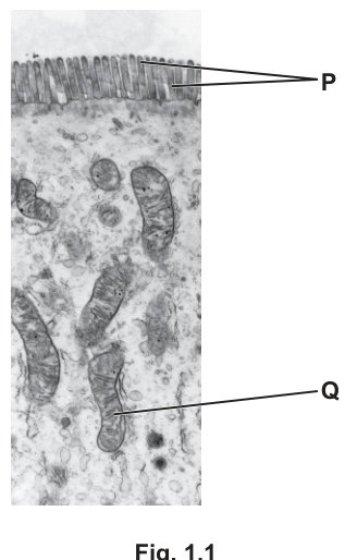
Fig. 1.1
The structures labelled P and Q in Fig. 1.1 are involved in the absorption of digested food.
Answer
Microvilli.
Microvilli
Background Concept
Cells that are specialised for absorption often increase the surface area of their exposed membrane by forming many small, finger-like projections. In the small intestine, the epithelial cells (enterocytes) of the villus have a "brush border" of these projections, each one called a microvillus (plural: microvilli). On an electron micrograph they appear as closely packed, parallel extensions of the apical plasma membrane, supported inside by actin filaments.
Understanding the Question
You are told that the cell in Fig. 1.1 is an absorptive epithelial cell from the digestive system. The structure labelled P points to the dense row of finger-like projections along the apical (free) surface of the cell. The task is simply to name this structure.
Approach
Look at the position (apical surface) and the appearance (regular, finger-like, densely packed extensions of the membrane, about 1 µm long). The structure projecting into the lumen at the apex of an absorptive epithelial cell is the microvillus.
Step-by-Step Reasoning
- The structures at P are too small and uniform to be cilia (which are longer, motile and contain a 9+2 microtubule arrangement).
- They are on the free (apical) surface of an absorptive cell, exactly where microvilli are found in the small intestine.
- Therefore P = microvilli.
Key Takeaways
- Microvilli are sub-cellular membrane projections (about 0.1 µm in diameter, ~1 µm long); cilia are much larger, motile structures.
- Microvilli massively increase the surface area of the apical membrane available for absorption of digested food.
Common Mistakes
- Writing "cilia" — cilia are longer, less densely packed and usually motile; they are not the absorptive projections of an enterocyte.
- Writing "villi" — villi are multicellular folds of the intestinal lining, visible to the naked eye; microvilli are the sub-cellular projections on the surface of each epithelial cell.
Things to Be Careful About
At A-level, the correct term is microvilli (or microvillus, singular). Avoid describing them as "hairs" or "brush border" on its own — the brush border refers to the combined appearance of all the microvilli, not the individual structure.
Answer
- Q is a mitochondrion — the site of aerobic respiration, which produces ATP.
- The ATP is used in active transport to absorb digested food against a concentration gradient.
Mitochondrion is the site of aerobic respiration so produces ATP; this ATP is used in active transport (or endocytosis) to absorb digested food.
Background Concept
Q in Fig. 1.1 points to an oval organelle with internal folded membranes (cristae) — a mitochondrion. Mitochondria are the site of aerobic respiration: glucose (and other respiratory substrates) is oxidised, with oxygen as the final electron acceptor, and the energy released is used to phosphorylate ADP to ATP on the inner membrane and cristae.
Many digested foodstuffs (e.g. glucose, amino acids, some ions) are absorbed from the intestinal lumen into the epithelial cell against their concentration gradient. This is active transport and requires energy in the form of ATP, which is supplied by adjacent mitochondria. Some larger molecules and droplets may also be taken up by endocytosis / pinocytosis, an ATP-requiring bulk-transport process.
Understanding the Question
The question tells you the cell is specialised for absorption and asks you to explain how organelle Q contributes to that absorption. The command word "explain" means you must state the mechanism, not just identify the organelle. Two marks are available: one for linking the mitochondrion to ATP, and one for stating what the ATP is used for in the context of absorption.
Approach
- Recognise Q as a mitochondrion from its double membrane and cristae visible in the TEM.
- State that the mitochondrion carries out aerobic respiration and so produces ATP.
- Apply the ATP specifically to the absorptive process: active transport of digested food against its concentration gradient (or, alternatively, endocytosis).
Step-by-Step Reasoning
- Marking point 1 — identify the role of the mitochondrion: site of aerobic respiration, producing (or providing) ATP. The mark scheme rejects the loose phrase "produces energy" — you must name ATP as the energy currency.
- Marking point 2 — say what that ATP is used for in absorption. The most precise statement is "active transport (of digested food) against a concentration gradient". "Bulk transport into the cell" / "endocytosis / pinocytosis" is also accepted as an alternative second point.
Key Takeaways
- Mitochondria are abundant in cells that carry out a lot of active transport (e.g. absorptive epithelial cells, kidney tubule cells, salt glands) because ATP demand is high.
- "Producing energy" is too vague — always specify ATP, the energy currency.
- For absorption, two ATP-requiring mechanisms exist: active transport across the membrane (for ions, glucose, amino acids) and endocytosis (for larger molecules).
Common Mistakes
- Writing "the mitochondrion produces energy" without naming ATP — this loses the first mark.
- Stating only that Q releases energy for absorption, with no link to a specific process (active transport / endocytosis) — this loses the second mark.
- Confusing the mitochondrion with a chloroplast (no thylakoids are present, and this is an animal cell).
- Writing "it carries out respiration" without specifying that the respiration is aerobic and that ATP is the product.
Things to Be Careful About
The mark scheme insists on the word "ATP" (energy currency) — "energy" alone is rejected. Tie the ATP to a specific use in the cell: active transport of digested food against a concentration gradient, or endocytosis. Both ideas earn the second mark.
In the mammalian circulatory system, red blood cells travel through different types of blood vessel as they pass from the heart to respiring tissues and back to the heart.
Fig. 2.1 shows the types of blood vessels through which red blood cells travel in the circulatory system.
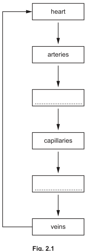
Fig. 2.1
Complete Fig. 2.1 by writing the names of the missing types of blood vessels through which red blood cells travel.
Answer
- Box between arteries and capillaries: arterioles
- Box between capillaries and veins: venules
arterioles; venules
Background Concept
Blood flows through a sequence of vessels whose structure matches their function. As blood moves away from the heart, arteries branch into smaller vessels before reaching the microscopic exchange surface of capillaries. On its return to the heart, blood is gathered from capillaries into progressively larger vessels before entering the veins.
The full pathway is: heart → artery → arteriole → capillary → venule → vein → heart.
- Arterioles are small branches of arteries with a relatively thick muscular wall that regulates blood flow into capillary beds.
- Venules are small vessels that collect blood from capillaries and join to form veins.
Understanding the Question
Fig. 2.1 is a flow diagram with two blank boxes: one between 'arteries' and 'capillaries', and another between 'capillaries' and 'veins'. The task is to name the vessel types that fit into the two gaps.
Approach
Recall the standard order of blood vessels in the closed double circulation. The vessels between arteries and capillaries must be arterioles (the smaller, muscular distribution vessels). The vessels between capillaries and veins must be venules (the small vessels that drain capillary beds into veins).
Step-by-Step Reasoning
- Between arteries and capillaries, blood is delivered to the capillary bed by smaller resistance vessels — arterioles.
- Between capillaries and veins, blood is collected from the capillary bed by small drainage vessels — venules.
- The completed sequence therefore reads: heart → arteries → arterioles → capillaries → venules → veins → heart.
Key Takeaways
- The vessel sequence around a capillary bed is always artery → arteriole → capillary → venule → vein.
- The change in vessel diameter gives a large total cross-sectional area in the capillary bed, slowing blood flow and aiding exchange.
Common Mistakes
- Writing 'artery' in the first blank — examiners look for the smaller vessel type, the arteriole.
- Writing 'vein' in the second blank — the correct answer is the smaller drainage vessel, the venule.
- Reversing the two (e.g. venule before capillaries and arteriole after capillaries); the sequence must follow blood flow away from the heart and back.
Things to Be Careful About
The mark scheme accepts either singular or plural forms ('arteriole' or 'arterioles'; 'venule' or 'venules'). Spelling matters: 'arteriole' and 'venule' are not the same as 'arterial' and 'venous'.
Water is the main component of blood. It has an important role in the transport of substances around the body.
Fig. 2.2 shows the ionic compound sodium chloride dissolving in water.
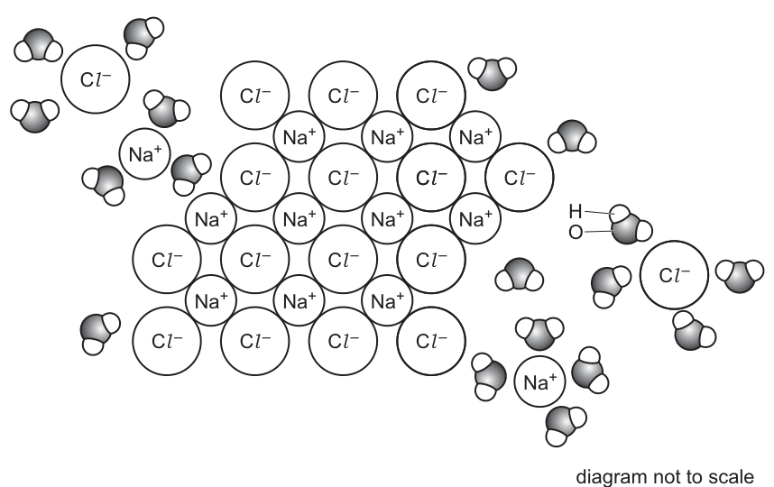
diagram not to scale
Fig. 2.2
With reference to Fig. 2.2, explain how water acts as a solvent for sodium chloride.
Answer
- Water is a polar (dipolar) molecule: the oxygen atom carries a partial negative charge () and each hydrogen atom carries a partial positive charge ().
- The of water is attracted to the positively charged sodium ions ().
- The of water is attracted to the negatively charged chloride ions ().
- Water molecules surround the ions (hydration shells) and pull the and ions apart, dissolving the sodium chloride.
Water is polar: its attracts ions and its attracts ions; water molecules surround and separate the ions, dissolving the salt.
Background Concept
A water molecule is bent (V-shaped), with the oxygen atom at the vertex and the two hydrogens on either side. Oxygen is far more electronegative than hydrogen, so the O–H bonds are polar: the bonding electrons are pulled towards oxygen, giving oxygen a partial negative charge () and each hydrogen a partial positive charge (). A molecule with separated partial charges is polar (or dipolar).
When an ionic compound such as sodium chloride is placed in water, ions at the surface of the crystal are pulled away by the water molecules. The negative ends of water are attracted to cations, and the positive ends are attracted to anions. These ion–dipole attractions are strong enough to overcome the ionic bonds holding the crystal together, so the ions separate and become surrounded by water molecules — this is the process of dissolution.
Understanding the Question
Fig. 2.2 shows the dissolving NaCl: water molecules are arranged around individual and ions, with the oxygen ends pointing towards and the hydrogen ends pointing towards . The task is to use this figure to explain the mechanism by which water dissolves the ionic compound.
Approach
Use the visible features of Fig. 2.2 (the orientation of water molecules around the two ion types) to describe:
- Why water can interact with each type of ion — because it is polar.
- Which end of the water molecule faces which ion.
- The result of these attractions — ions are pulled away from the crystal and surrounded by water.
Step-by-Step Reasoning
- Water is polar: the oxygen carries a partial negative charge and the hydrogens carry partial positive charges. (Mark scheme allows 'polar / dipolar'.)
- The negative oxygen ends () of water are attracted to the positively charged sodium ions ().
- The positive hydrogen ends () of water are attracted to the negatively charged chloride ions ().
- Water molecules collect around the ions on the surface of the crystal lattice, surrounding each ion with a hydration shell.
- These ion–dipole attractions are strong enough to break the ionic bonds in the lattice; the and ions separate from one another and become dispersed through the water — sodium chloride has dissolved.
Key Takeaways
- Polarity is what makes water a good solvent for ionic and other polar substances.
- 'Like dissolves like': polar solvents dissolve polar/ionic solutes; non-polar solutes (e.g. lipids) require non-polar solvents.
- Ions in solution are hydrated — surrounded by water molecules with the appropriate end pointing inwards.
Common Mistakes
- Saying only that 'water dissolves things' or that 'water is the solvent' — this is just restating the question and gains no marks. The question asks how, so the polarity and the specific ion–dipole attractions must be stated.
- Getting the orientation wrong: stating that water's hydrogen ends attract . The opposite charges attract — opposite charges face each other.
- Confusing partial charges () with full ionic charges (+, −). The O–H bond is covalent but polar, not ionic; the symbol (delta negative) is what shows this.
- Saying water 'breaks' or 'attacks' the lattice without naming the attraction that does so.
Things to Be Careful About
- The mark scheme treats statements of both ion–dipole attractions as implying polarity: if you correctly state that attracts and attracts , you automatically gain the polarity mark too.
- If you can only recall one mark, the examiner allows one mark for the bare idea of 'attraction between water and the ions'.
- Distinguish partial charges on the water molecule from the full ionic charges on the dissociated ions. The notation / vs / is precise and informative.
Fig. 2.3 shows a Galapagos penguin, Spheniscus mendiculus, swimming in the water.
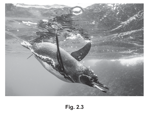
Fig. 2.3
Penguins are birds that live on land but spend a lot of time swimming underwater hunting for food. Penguins can remain underwater for up to twenty minutes. During this time they do not breathe but their tissues continue to respire.
Haemoglobin in the red blood cells of penguins has a higher affinity for oxygen than haemoglobin in other birds that do not swim underwater.
Fig. 2.4 shows the oxygen dissociation curve for a bird that does not swim underwater.
Draw a line on Fig. 2.4 to suggest the position of the oxygen dissociation curve for penguin haemoglobin.
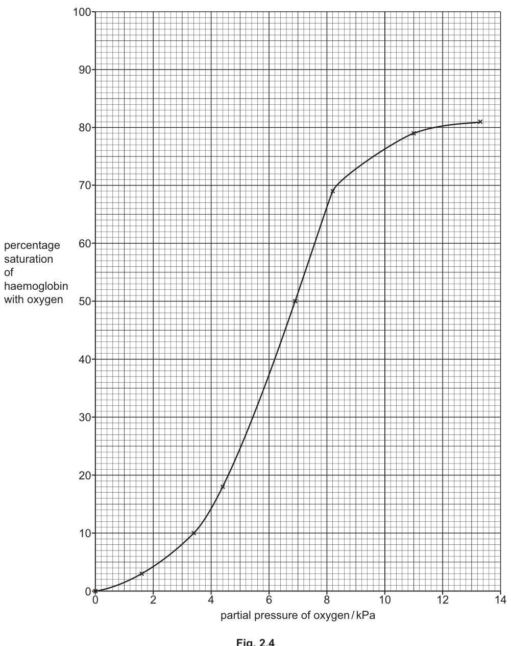
Fig. 2.4
Answer
Draw a sigmoid curve that:
- starts at the origin (0, 0),
- lies to the left of the curve already on Fig. 2.4 at every partial pressure,
- has the same general S-shape (sigmoid),
- plateaus at a similar maximum saturation (around 80–85%).
Because penguin haemoglobin has a higher affinity for oxygen, its curve reaches a given percentage saturation at a lower partial pressure of oxygen, so the whole curve is displaced to the left.
Sigmoid curve displaced to the left of the printed curve, starting at (0, 0).
Background Concept
The oxygen dissociation curve (ODC) shows the percentage saturation of haemoglobin with oxygen at different partial pressures of oxygen. It has a characteristic sigmoid (S) shape because haemoglobin is a cooperative protein: binding of the first oxygen molecule makes binding of subsequent oxygens easier, producing the steep middle portion.
A haemoglobin with a higher affinity for oxygen binds oxygen more readily, so it reaches any given percentage saturation at a lower partial pressure of oxygen. Graphically, this means the entire curve is shifted to the left of the normal curve. A haemoglobin with a lower affinity is shifted to the right.
Understanding the Question
The stem states that penguin haemoglobin has a higher affinity for oxygen than the haemoglobin of birds that do not swim. The task is to draw, on Fig. 2.4, a curve representing this higher-affinity haemoglobin. Fig. 2.4 already shows the curve for a non-diving bird — the candidate must add the penguin curve.
Approach
Recall the rule:
- higher affinity → curve shifts left
- the curve must remain sigmoid
- it must start at the origin (0, 0) — zero oxygen gives zero saturation
- the maximum saturation should be similar to that of the printed curve (haemoglobin still binds the same maximum amount of O₂)
Step-by-Step Reasoning
- The curve is sigmoid — a flat lower portion at low , a steep middle portion, and a plateau near the top.
- The curve starts at the origin (0 kPa, 0% saturation).
- Because penguin haemoglobin has a higher affinity, every percentage saturation is reached at a lower partial pressure of oxygen. The whole curve therefore lies to the left of the printed curve.
- The plateau level is similar to the printed curve (around 80–85%), because the maximum O₂-carrying capacity of haemoglobin is unchanged.
A reasonable sketch: at 4 kPa the penguin curve might be around 40–50% saturated (whereas the printed curve is about 15% at 4 kPa); at 7 kPa it might be around 80%; and so on, with the upper plateau reached by about 10–11 kPa.
Key Takeaways
- A higher-affinity haemoglobin curve is shifted to the left of the normal curve.
- The curve remains sigmoid and begins at (0, 0).
- The maximum saturation is approximately unchanged.
Common Mistakes
- Drawing the curve to the right — a rightward shift means lower affinity, the opposite of what is asked.
- Drawing a curve that does not pass through (0, 0) or has the wrong shape (e.g. a simple hyperbola).
- Drawing the curve so it sits on top of the printed curve, which says nothing about a difference in affinity.
- Drawing a curve with a much higher maximum saturation — haemoglobin still carries at most ~4 O₂ molecules per molecule, so the plateau level is similar.
Things to Be Careful About
- Both marking points must be present for full marks: the curve must (1) be displaced to the left, and (2) retain a sigmoid shape starting at (0, 0).
- Freehand drawing is acceptable on an exam paper; the examiner judges position and shape rather than penmanship.
Penguin haemoglobin is very sensitive to a decrease in pH caused by an increase in the carbon dioxide concentration in the blood.
Explain how a decrease in pH affects penguin haemoglobin, and suggest how this helps the penguin to swim underwater for a long time.
Answer
- A decrease in pH means an increase in ion concentration.
- The ions bind to haemoglobin (forming haemoglobinic acid), reducing haemoglobin's affinity for oxygen (the curve shifts to the right — the Bohr shift).
- Oxygen is therefore released from haemoglobin into the respiring tissues (especially muscles).
- This allows aerobic respiration in muscle cells to continue for longer while the penguin is underwater and not breathing, supplying ATP for muscle contraction and prolonging the dive.
Lower pH increases H+; H+ binds haemoglobin and reduces its O2 affinity (Bohr shift), releasing O2 to respiring muscles so aerobic respiration and the dive can continue.
Background Concept
Haemoglobin's affinity for oxygen is not fixed — it is sensitive to the chemical environment in the blood. The two most important modifiers are:
- concentration / pH: the Bohr effect. Active tissues produce ; in red blood cells the enzyme carbonic anhydrase converts into , which dissociates into and . The resulting drop in pH (rise in ) stabilises the deoxygenated (T) form of haemoglobin, lowering its affinity for O₂. Oxygen therefore dissociates from haemoglobin where it is needed most — in respiring tissues that are producing . Graphically, the ODC shifts to the right.
- BPG (2,3-bisphosphoglycerate): also stabilises the T-state, lowering O₂ affinity.
The reaction (or, more precisely, the formation of haemoglobinic acid: when oxygen is released) explains why a higher local pushes oxygen off haemoglobin.
Understanding the Question
The stem tells us that penguin haemoglobin is 'very sensitive' to a decrease in pH. The question has two linked parts:
- How does the lower pH affect penguin haemoglobin?
- How does this help the penguin remain underwater for twenty minutes without breathing?
The scenario is the Bohr effect, but pushed to an extreme — the penguin's haemoglobin is unusually pH-sensitive, releasing more oxygen than a typical bird's haemoglobin when rises.
Approach
Combine the Bohr effect with the diving physiology:
- During a dive, the penguin cannot breathe, but its muscles continue to respire aerobically and produce .
- The accumulating lowers the blood pH.
- Penguin haemoglobin responds strongly to this pH drop by releasing O₂.
- The released O₂ keeps aerobic respiration going in muscle, supplying ATP for swimming — and the aerobic respiration is much more efficient than anaerobic respiration, so the penguin can dive for longer.
Step-by-Step Reasoning
- More → lower pH → more : respiring tissues (muscles) produce , which (via carbonic anhydrase) increases in the blood.
- Hydrogen ions bind to haemoglobin (forming haemoglobinic acid). This is the molecular basis of the Bohr effect.
- Haemoglobin's affinity for oxygen decreases: the curve shifts to the right.
- Haemoglobin releases more oxygen to the tissues — exactly where is being produced.
- Aerobic respiration in muscle cells can continue for longer while the penguin is not breathing, providing ATP for muscle contraction and enabling the long dive.
The mark scheme accepts a statement that the curve shifts to the right (the Bohr shift) as an alternative, or that aerobic respiration continues for longer, providing ATP for muscle contraction.
Key Takeaways
- The Bohr effect is a feedback mechanism: more / lower pH → haemoglobin releases more O₂ → more aerobic respiration.
- Penguin haemoglobin is an extreme example: it is unusually pH-sensitive, so the Bohr shift is large.
- A large Bohr shift helps diving animals keep their muscles aerobic even when they are not breathing.
Common Mistakes
- Saying that pH decreases because oxygen decreases — the direction is the other way: respiring tissues produce , which lowers pH, which then releases O₂.
- Stating only that 'oxygen is released' without giving the mechanism (H⁺ binding to haemoglobin, lower affinity).
- Saying 'penguins can breathe underwater' — the stem is explicit that they cannot; the benefit is supplying tissues with oxygen already stored on haemoglobin.
- Confusing the Bohr shift with the effect of fetal haemoglobin. Fetal Hb has a left-shifted curve because of lower affinity for 2,3-BPG; the Bohr shift is a rightward displacement under low pH.
Things to Be Careful About
- The mark scheme requires both the mechanistic effect (pH change → H⁺ → lower affinity → O₂ released) and the physiological benefit (aerobic respiration can continue). Three marks are awarded across these two strands.
- 'AVP' (additional valid point) includes reference to the Bohr shift or to providing ATP for muscle contraction — these are interchangeable ways to earn the same mark.
The heart rate of a penguin decreases while it is swimming underwater.
Heart rate is regulated by a group of specialised cells in the wall of the right atrium. The activity of these cells is modified by nerve impulses.
Name the group of specialised cells in the wall of the right atrium that regulates heart rate.
Answer
Sinoatrial node (SAN).
Sinoatrial node
Background Concept
The heartbeat is myogenic — it originates within the heart itself, not from nerve impulses. The wave of electrical activity that initiates each beat begins in a small patch of specialised cardiac muscle in the wall of the right atrium, where the superior vena cava enters. This patch is the sinoatrial node (SAN), the heart's natural pacemaker.
From the SAN the wave of depolarisation spreads across both atria (causing atrial systole), reaches the atrioventricular node (AVN) at the base of the right atrium, is delayed briefly to allow the ventricles to fill, then travels down the Bundle of His, through the Purkyne fibres, and around the ventricular walls (causing ventricular systole).
Understanding the Question
The stem says the heart rate is regulated by a group of specialised cells in the wall of the right atrium, and that their activity is modified by nerve impulses (the autonomic nervous system). This describes the pacemaker, whose intrinsic rate is set by these cells and adjusted up or down by sympathetic and parasympathetic nerves.
Approach
Recall the name of the pacemaker from the cardiac conduction system: it is the sinoatrial node. The examiner will ignore the abbreviation 'SAN' — the full term must be written out.
Step-by-Step Reasoning
- The heart's natural pacemaker lies in the wall of the right atrium.
- It is a small mass of specialised cardiac muscle cells.
- Its proper name is the sinoatrial node.
Key Takeaways
- The SAN is the heart's natural pacemaker.
- It sets the intrinsic rate of the heartbeat; autonomic nerves modulate the rate up (sympathetic) or down (parasympathetic).
- The full term 'sinoatrial node' is required for the mark — abbreviations are ignored.
Common Mistakes
- Writing only 'SAN' or 'pacemaker' — the mark scheme explicitly says ignore the abbreviation; the full term is needed.
- Writing 'AVN' (which is in the septum between atria and ventricles, not the pacemaker).
- Writing 'Purkyne fibres' (these are in the ventricular walls and conduct, but do not initiate, the beat).
Things to Be Careful About
- Spelling: 'sinoatrial' (not 'sinoetrial', not 'sinatrial').
- A whole sentence is not necessary; one or two words is enough. But the precise term must appear in full.
Fig. 3.1 is a photomicrograph of a transverse section through a region of the wall of the bronchus in the gas exchange system.
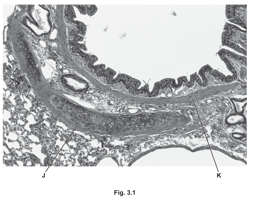
Fig. 3.1
Identify the tissues J and K shown in Fig. 3.1, and suggest how the wall of a bronchiole differs from the wall of the bronchus for these two tissues.
= ______
= ______
difference = ______
Answer
- J = cartilage
- K = smooth muscle
- Difference: cartilage is present in the wall of the bronchus but absent from the wall of a bronchiole; the bronchiole wall contains a proportionally greater amount of smooth muscle.
J = cartilage; K = smooth muscle; difference: cartilage absent (and proportionally more smooth muscle) in bronchiole wall.
Background Concept
The gas exchange system is a branching network that starts with the trachea, divides into two primary bronchi, then into smaller bronchi, and finally into bronchioles and alveoli. As the airways get smaller the wall composition changes. Cartilage (C-shaped rings in the trachea, irregular plates in the bronchi) holds the larger airways open and prevents them from collapsing during inhalation. Smooth muscle is found in the walls of both bronchi and bronchioles and can contract to narrow the airway (e.g. during an asthma attack) or relax to widen it. By the time the tubes reach the bronchioles, cartilage is no longer required because the smaller diameter and surrounding lung tissue pressure keep the airway open; instead, smooth muscle becomes relatively more important in regulating airflow.
Understanding the Question
You are given a transverse section through a bronchus wall and asked to identify two labelled tissues (J and K) and then describe one way in which the bronchiole wall differs from the bronchus wall for these tissues. The marks are: 1 mark for J, 1 mark for K, 1 mark for a single clear difference.
Approach
Use the staining and position of the labels in Fig. 3.1: J points to a pale, plate-like region of tissue — characteristic of hyaline cartilage; K points to a darker layer between the cartilage and the epithelium — the position of smooth muscle. Then recall the general trend in the gas exchange system: cartilage decreases in amount and finally disappears at the bronchioles, whereas smooth muscle becomes proportionally more important.
Step-by-Step Reasoning
- J is the large, pale, plate-shaped tissue. Hyaline cartilage stains lightly with H&E and has chondrocytes sitting in lacunae. → J = cartilage (1 mark).
- K is the darker layer between the cartilage and the lining epithelium; the cells are elongated and arranged in a band. → K = smooth muscle (1 mark).
- The general rule for the gas exchange system is that cartilage supports the larger airways but is not needed in the smaller bronchioles, so a bronchiole wall lacks cartilage. The smooth muscle layer, by contrast, is proportionally thicker in the bronchiole, allowing fine control of airflow (1 mark).
Key Takeaways
- Cartilage and smooth muscle are two key structural tissues of the gas exchange system.
- The proportion and presence of these tissues change as airways get smaller: cartilage disappears at the bronchioles; smooth muscle remains and is proportionally greater.
- Recognising tissues in histological sections relies on both staining (pale/dark) and position (lining, middle layer, outer layer).
Common Mistakes
- Confusing J with elastic fibres: cartilage is the only tissue in the wall that appears as a large, pale, plate-like region. Elastic fibres are much finer and are not what J indicates.
- Saying that bronchioles "have less smooth muscle": the mark scheme credits the ORA — proportionally more smooth muscle in the bronchiole.
- Listing several differences and diluting the answer: the mark scheme awards only one difference mark, so one well-stated point is enough.
Things to Be Careful About
- The question asks specifically about differences for tissues J and K, so a generic comment about cilia or goblet cells will be ignored (the mark scheme says I ref. to other tissues).
- The precise wording "cartilage in the bronchus but not in the bronchioles" is what earns the mark; a vague "different walls" does not.
Tuberculosis (TB) is an infectious disease that affects the human gas exchange system.
The pathogen that causes TB secretes a protein that can be detected in saliva.
Early diagnosis of TB is important in reducing the transmission of the pathogen.
Scientists have developed a test strip for TB that uses monoclonal antibodies. Monoclonal antibodies are specific in their action.
This test strip contains:
- mobile monoclonal antibodies that bind to one part of the protein secreted by the pathogen
- immobilised monoclonal antibodies.
Fig. 3.2 shows a simplified diagram of the test strip.
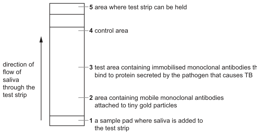
Fig. 3.2
A sample of saliva is collected and put onto the sample pad in the test strip.
The saliva moves up the test strip through area 2.
The mobile monoclonal antibodies are attached to tiny gold particles. If these antibodies collect in test area 3, a gold line becomes visible on the test strip.
A gold line that becomes visible in area 4 confirms that the test strip is working and that the results are valid.
Answer
Mycobacterium tuberculosis (also accept Mycobacterium bovis).
Mycobacterium tuberculosis (or Mycobacterium bovis).
Background Concept
Tuberculosis (TB) is a chronic infectious disease of the human gas exchange system, caused by a slow-growing, acid-fast bacterium. The species most commonly responsible for human TB is Mycobacterium tuberculosis, although Mycobacterium bovis — originally identified in cattle — can also cause TB in humans, particularly through contaminated milk. Both species belong to the genus Mycobacterium and are written in italics with a capital genus letter.
Understanding the Question
This is a one-mark, straight recall question. The expected answer is the binomial name of the pathogen, written in italics with a capital initial letter for the genus.
Approach
State the binomial name of the bacterial pathogen that causes TB, written in italics, with the genus capitalised.
Step-by-Step Reasoning
- TB is caused by a bacterium, not a virus, so a binomial with the genus Mycobacterium is required.
- The principal human pathogen is Mycobacterium tuberculosis; Mycobacterium bovis is also accepted by the mark scheme.
- 1 mark.
Key Takeaways
- TB is caused by Mycobacterium tuberculosis (or Mycobacterium bovis).
- Binomial names must be in italics, with the genus capitalised and species lower-case.
Common Mistakes
- Writing the name without italics, or with lower-case genus, which CIE may still accept, but the conventional form is italicised binomial.
- Naming the disease (tuberculosis) instead of the pathogen.
- Confusing TB with other respiratory infections such as Streptococcus pneumoniae or Bordetella pertussis.
Things to Be Careful About
- The mark scheme does not require both species — one is enough.
Answer
Antigen binding site(s) (also accept variable region).
Antigen binding site(s) / variable region.
Background Concept
An antibody (immunoglobulin) is a Y-shaped protein made of four polypeptide chains: two heavy and two light, joined by disulfide bonds. The tips of the Y carry the antigen binding sites — two identical regions formed jointly by parts of one heavy and one light chain. These tips vary in amino acid sequence between different antibodies and are therefore called the variable regions. The shape of the variable region is complementary to the shape of a specific epitope (antigenic determinant) on the antigen, and this complementarity is the basis of antibody specificity.
Understanding the Question
This is a one-mark recall question asking for the part of a monoclonal antibody that physically binds the TB protein. The mark scheme accepts either "antigen binding site(s)" or "variable region".
Approach
Recall the structure of the antibody: the tips of the Y are the antigen binding sites (variable regions); the stem is the constant region. State the correct term.
Step-by-Step Reasoning
- Each Y-shaped antibody has two identical antigen binding sites, one at the tip of each arm of the Y.
- These are formed by the variable regions of one heavy chain and one light chain.
- The mark scheme accepts either "antigen binding site(s)" or "variable region".
- 1 mark.
Key Takeaways
- Antigen binding sites / variable regions are at the tips of the antibody's Y-shape.
- Specificity of an antibody arises from the three-dimensional shape of its variable region being complementary to a specific epitope.
Common Mistakes
- Saying "the heavy chain" or "the light chain" — the binding site is formed by parts of both, not by either chain alone.
- Confusing the variable region with the constant region (which does not bind antigen).
Things to Be Careful About
- "Antigen binding site" is the clearest term; "variable region" is the alternative credit.
Saliva is added to a test strip to test for the presence of the protein secreted by the TB pathogen.
Fig. 3.3 is a diagram showing some of the molecules in area 3 of the test strip when a positive result for TB is obtained.
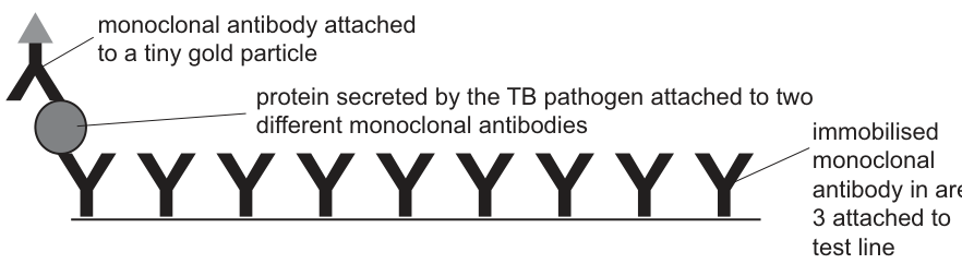
Fig. 3.3
Use the information in Fig. 3.3 to suggest and explain why this test is specific for TB.
Answer
- The immobilised monoclonal antibodies in area 3 have antigen binding sites that are complementary in shape to the protein secreted by Mycobacterium tuberculosis.
- Therefore, the gold-labelled mobile monoclonal antibodies are only held in place at area 3 when this specific TB protein is present, so a gold line appears only if the sample contains TB protein (i.e. the test is specific for TB).
The immobilised antibodies bind only the TB protein because their binding sites are complementary to it; gold-labelled antibodies are only trapped at area 3 in the presence of TB protein, so the gold line appears only when TB protein is present.
Background Concept
Monoclonal antibodies are identical antibodies produced by a single clone of B-lymphocytes hybridised with a myeloma cell. Because every antibody in the population is the same, every binding site is identical and recognises one specific epitope. This makes them ideal for diagnostic tests: a monoclonal antibody raised against a particular antigen will bind that antigen and only that antigen, even in a complex mixture such as saliva.
In a lateral-flow (test strip) sandwich immunoassay, the sample flows along the strip and meets gold-labelled mobile antibodies. If the target antigen is present, each antigen molecule is sandwiched between one mobile antibody and one immobilised antibody at the test line, trapping the gold particles and producing a visible coloured line.
Understanding the Question
You are given Fig. 3.3, which shows the test line (area 3) of the TB strip when the result is positive: a row of immobilised Y-shaped antibodies on the test line, with one molecule of TB protein bound to each, and a gold-labelled mobile antibody bound to the other side of the TB protein. The question asks you to suggest and explain why this arrangement makes the test specific for TB.
Approach
The answer has two linked parts: (1) state the molecular reason the immobilised antibody binds the TB protein (shape complementarity), and (2) explain why this means only TB produces a gold line at area 3 (only TB protein is the right shape to be sandwiched and to trap the gold-labelled antibody). The mark scheme allows "antigen for TB protein" and "TB pathogen for pathogen causing TB" as substitutes.
Step-by-Step Reasoning
- Monoclonal antibodies used in the test have binding sites with a shape complementary to a particular epitope on the TB protein.
- This complementarity means the immobilised antibody at area 3 will only bind the TB protein; other proteins in saliva (which have different shapes) will not be bound.
- A gold-labelled mobile antibody can only be held at area 3 if it is itself bound to TB protein which is in turn bound to the immobilised antibody — the sandwich shown in Fig. 3.3.
- Hence the gold line only forms when TB protein is present, which is the basis of specificity. (Any two of these points = 2 marks.)
Key Takeaways
- Antibody specificity is due to shape complementarity between the antigen binding site and the epitope.
- In a sandwich immunoassay, a positive result requires the antigen to be bound by two different antibodies simultaneously.
- Lateral-flow tests are highly specific because both the capture and the detection antibodies must recognise the same antigen.
Common Mistakes
- Saying the test is specific "because monoclonal antibodies are specific" without saying what makes them specific (complementary shape).
- Failing to link the gold line to the sandwich structure in Fig. 3.3 — the gold is visible only because it is trapped by the sandwich.
- Talking about TB generally rather than the TB protein, which is the actual molecule being detected.
Things to Be Careful About
- The mark scheme rewards both halves of the explanation: shape complementarity AND the consequence (binding only when the TB protein is there).
Area 4 contains different immobilised antibodies to those in area 3.
The mobile monoclonal antibodies bound to tiny gold particles will bind to these immobilised monoclonal antibodies in area 4.
If the test has functioned correctly, a gold line will be visible in area 4.
Suggest how the structure of immobilised monoclonal antibodies in area 3 differs from the structure of the immobilised monoclonal antibodies in area 4.
Answer
- The variable regions (antigen binding sites) of the immobilised antibodies in area 3 are a different shape from those in area 4.
- This difference arises because the antibodies have a different primary structure (amino acid sequence), giving a different tertiary structure (held together by different disulfide / hydrogen / ionic bonds), so the binding site has a different three-dimensional shape and binds a different antigen.
Different variable region shape; due to different primary structure and therefore different tertiary structure (and different bonds holding it).
Background Concept
All antibodies share the same overall Y-shaped structure of two heavy and two light chains, but the tips of the Y — the variable regions — differ greatly between antibodies. The variable region of each antibody has a unique amino acid sequence (primary structure). The variable sequence folds into a unique three-dimensional shape (tertiary structure), stabilised by disulfide bonds, hydrogen bonds, ionic bonds and hydrophobic interactions. This unique shape determines which epitope the antibody binds. Two different monoclonal antibodies therefore have different primary, tertiary and ultimately different binding-site shapes.
In this test strip, area 3 must capture the TB protein, while area 4 must capture any mobile antibody that flows past, regardless of whether it has bound TB protein. The two areas therefore need antibodies of different specificities and consequently different variable-region shapes.
Understanding the Question
The question tells you that area 4 contains different immobilised antibodies from area 3 and that the mobile gold-labelled antibody binds to the area 4 antibodies directly (it does not need the TB protein to be present). You must suggest how the structures of the area 3 and area 4 antibodies differ. The mark scheme awards two marks for any two of: different-shaped variable region/antigen binding site; different primary structure; different tertiary structure; different named bonds stabilising the tertiary structure.
Approach
Work from the functional difference (different binding specificity) to the structural basis (different variable region shape) to the molecular basis (different primary structure → different tertiary structure). Name at least one type of bond that stabilises the tertiary structure.
Step-by-Step Reasoning
- Function: the two immobilised antibodies must bind different molecules (area 3 binds TB protein; area 4 binds the mobile gold-labelled antibody directly). Therefore their antigen binding sites have different shapes.
- Shape of the binding site is determined by the tertiary structure of the variable region.
- Tertiary structure is determined by the primary structure (the amino acid sequence).
- The tertiary fold is held in place by disulfide bonds, hydrogen bonds, ionic bonds and hydrophobic interactions. Different amino acids in the sequence change which of these bonds form, giving a different fold and therefore a different binding site.
- Two of these points (e.g. different variable-region shape AND different primary structure; or different tertiary structure AND named bond) earn the two marks.
Key Takeaways
- Antibody specificity is encoded in the variable region.
- Variable-region shape is determined by the primary structure and stabilised as a tertiary structure by disulfide, hydrogen, ionic and hydrophobic bonds.
- Two monoclonal antibodies with different specificities must have different primary, tertiary and binding-site structures.
Common Mistakes
- Saying the antibodies are different "because they are monoclonal" — both are monoclonal; the difference is which antigen they bind.
- Mentioning the constant region: the constant region is the same in different antibodies of the same class; it does not determine specificity.
- Forgetting to name a type of bond (the mark scheme says "named bonds").
Things to Be Careful About
- A precise answer names both a structural level (primary or tertiary) and, where possible, the type of bond that holds the tertiary structure.
Vaccination is another way of reducing the transmission of infectious diseases such as TB. The BCG vaccine is used to help control the spread of TB. This vaccine contains a weakened strain of the pathogen that causes TB. The BCG vaccine stimulates the development of antigen-specific memory T-lymphocytes.
Explain how memory T-lymphocytes provide protection from TB in a person who has been given a BCG vaccination.
Answer
- The BCG vaccine stimulates production of antigen-specific memory T-lymphocytes, providing long-term immunity.
- On re-exposure to TB, memory T-lymphocytes recognise the TB antigen, are activated and divide to form a large clone of specific T-cells, producing a faster, stronger secondary response.
- T-helper cells release cytokines (interleukins) that increase phagocytosis, stimulate B-lymphocytes to produce antibodies, and enhance the T-killer cell response — together destroying the TB pathogen before it can establish infection.
BCG vaccination produces antigen-specific memory T-lymphocytes; on re-exposure they recognise TB antigen and mount a rapid, strong secondary response, releasing cytokines that boost phagocytosis, B-cell antibody production and T-killer cell activity.
Background Concept
The specific immune response has two arms: the cell-mediated response (T-lymphocytes) and the humoral response (B-lymphocytes and antibodies). Both are activated when a pathogen (or a vaccine mimicking a pathogen) is encountered for the first time. Some of the activated T- and B-lymphocytes differentiate into long-lived memory cells, which persist in the body for years. On a second encounter with the same antigen these memory cells are rapidly reactivated, producing a faster, larger and more effective response — the secondary response. Vaccination exploits this: a weakened or inactivated pathogen provokes the primary response and the formation of memory cells without causing the disease.
Helper T-lymphocytes (T-h cells) play a coordinating role: once activated they release cytokines such as interleukins, which stimulate B-lymphocytes, enhance phagocytosis by macrophages and support the activity of T-killer cells.
Understanding the Question
The question asks you to explain specifically how memory T-lymphocytes protect against TB in someone who has been vaccinated with BCG. The mark scheme offers six creditable points and asks for any three: long-term immunity, secondary/fast/strong response, increased numbers of specific T-lymphocytes, recognition of the foreign antigen, cytokine release, and a named downstream effect of cytokines.
Approach
Construct a logical sequence: vaccination produces memory cells → on re-exposure memory cells are activated → a rapid, large clonal expansion produces many specific T-cells → T-helper cells release cytokines → the response destroys the pathogen. Three marks require three distinct, well-articulated points from the mark scheme.
Step-by-Step Reasoning
- BCG contains weakened TB pathogens that act as antigens, stimulating the primary response and the formation of antigen-specific memory T-lymphocytes. This provides long-term immunity.
- When TB pathogens later enter the body, memory T-lymphocytes recognise the TB antigen and are activated; because they are already present in increased numbers, the secondary response is faster and stronger than a primary response would be.
- T-helper cells among the activated T-cells release cytokines (interleukins). Cytokines stimulate B-lymphocytes to produce antibodies, attract and activate macrophages (increased phagocytosis), and enhance T-killer cell activity — together eliminating the TB pathogen.
Key Takeaways
- Vaccination produces memory cells that mediate long-term, antigen-specific immunity.
- The secondary response is faster, stronger and larger than the primary response because memory cells can be activated immediately.
- T-helper cell cytokines coordinate the cellular and humoral arms of the response.
Common Mistakes
- Confusing the primary and secondary responses, or saying the secondary response is weaker.
- Writing only about B-lymphocytes and antibodies — the question is specifically about memory T-lymphocytes.
- Calling cytokines "cell-signalling molecules" only — the mark scheme ignores this and wants "cytokine(s) / interleukins".
- Failing to give an example of what cytokines do (e.g. increased phagocytosis, B-cell help, T-killer cell help).
Things to Be Careful About
- The question is about T-lymphocytes, not B-lymphocytes — keep the focus on the cell-mediated response, although B-cell help is an acceptable downstream effect of cytokine release.
- The mark scheme gives credit for a named consequence of cytokine release; pair the cytokine with its effect rather than just listing both.
The bladder is the organ in the body used to store urine.
When cells divide uncontrollably in the bladder, a tumour develops. This can lead to bladder cancer.
The BCG vaccine has been used to treat bladder cancer.
The BCG vaccine is introduced into the bladder. The tumour cells take up the weakened pathogens in the vaccine and act as antigen-presenting cells.
Answer
Endocytosis (phagocytosis is also accepted).
Endocytosis (or phagocytosis).
Background Concept
Cells take up materials across the plasma membrane in several ways. Small molecules may cross by diffusion, facilitated diffusion or active transport. Larger particles, whole cells or even pathogens cannot pass through the membrane in this way; instead, the cell surrounds the material with a portion of its plasma membrane, which then pinches off inside the cell to form a vesicle. This bulk-uptake process is called endocytosis. When the material being taken up is solid (e.g. a bacterium), the specific form is called phagocytosis ("cell eating").
Understanding the Question
This is a one-mark recall question asking for the name of the process by which tumour cells take up the weakened TB pathogens from the BCG vaccine introduced into the bladder. The pathogens are solid particles, so the process is endocytosis — phagocytosis is also accepted by the mark scheme.
Approach
Identify the general term (endocytosis) and the more specific form (phagocytosis) and choose one to write down.
Step-by-Step Reasoning
- Whole pathogens are too large to cross the membrane by diffusion or through transport proteins.
- The cell must therefore engulf them by membrane flow, forming a vesicle. The general term for this is endocytosis.
- When the material engulfed is solid (such as a bacterium), the process is called phagocytosis.
- Either term earns the mark.
Key Takeaways
- Endocytosis is the uptake of material by vesicle formation at the plasma membrane.
- Phagocytosis is the specific form of endocytosis that engulfs solid material.
- Antigen-presenting cells (such as macrophages and dendritic cells) use phagocytosis to take up pathogens for antigen presentation.
Common Mistakes
- Writing "active transport" — this is for small molecules, not whole pathogens.
- Writing "exocytosis" — this is the opposite process (release, not uptake).
- Writing "osmosis" or "diffusion" — neither describes bulk uptake of solid material.
Things to Be Careful About
- The mark scheme accepts both endocytosis and phagocytosis; either is correct.
Suggest how antigen presentation by tumour cells stimulates an immune response that leads to the destruction of the tumour cells.
Answer
- T-lymphocytes with receptors complementary in shape to the tumour antigen bind to it (clonal selection) and are activated.
- These T-lymphocytes divide by mitosis (clonal expansion) to form a clone of T-cells.
- T-killer cells among the clone recognise the tumour cells and destroy them by releasing perforins / granzymes / hydrogen peroxide (toxins) that cause tumour-cell death.
- (B-lymphocytes may also be activated by antigen-presenting tumour cells, producing antibodies that bind to the tumour cells and mark them for destruction.)
T-lymphocytes with complementary receptors bind tumour antigen (clonal selection), are activated and divide (clonal expansion); T-killer cells release perforin/granzymes and destroy the tumour cells.
Background Concept
The cell-mediated immune response is the principal defence against abnormal body cells, including virus-infected cells and tumour cells. Antigen-presenting cells (APCs) such as macrophages, dendritic cells and, in this case, the tumour cells themselves, display fragments of foreign or abnormal antigens on their surface, held in MHC (major histocompatibility complex) molecules. T-lymphocytes carry surface receptors, each specific to a single epitope. When a T-lymphocyte's receptor meets its complementary antigen on an APC, the T-cell is activated — this is clonal selection. The selected T-cell then divides rapidly by mitosis to produce a large clone — clonal expansion. The clone contains effector T-cells (T-helper cells and T-killer cells) that carry out the immune attack, and memory T-cells that provide long-term protection.
T-killer (cytotoxic) cells destroy target cells by releasing perforins, which punch holes in the target membrane, and granzymes, enzymes that enter through these holes and trigger apoptosis. They may also release hydrogen peroxide and other toxic molecules.
Understanding the Question
The question asks you to suggest, in three marks, how antigen presentation by tumour cells leads to the destruction of the tumour cells. The mark scheme lists seven possible points (any three for three marks). The expected chain is: T-cell receptor recognises tumour antigen → clonal selection → clonal expansion → T-killer cells produced → T-killer cells destroy tumour cells by a named mechanism (e.g. perforin).
Approach
Walk through the immune response in the correct order: recognition (receptor shape + clonal selection), activation and proliferation (clonal expansion), differentiation into T-killer cells, and effector function (mechanism of killing). A well-articulated answer picks three distinct points from this chain.
Step-by-Step Reasoning
- Tumour cells displaying antigen are recognised by T-lymphocytes whose surface receptors happen to be complementary in shape to that antigen. Binding activates the T-cell — this is clonal selection.
- The activated T-cell divides repeatedly by mitosis to form a clone of identical T-cells specific to the tumour antigen (clonal expansion).
- The clone includes T-killer (cytotoxic) cells, which identify and bind to other tumour cells displaying the same antigen.
- T-killer cells destroy the tumour cells by releasing perforins (which make holes in the tumour-cell membrane), granzymes (which enter through the holes and trigger apoptosis), and toxic molecules such as hydrogen peroxide.
- As a bonus point, B-lymphocytes may also be activated, producing antibodies that bind to the tumour antigens and mark the cells for destruction.
Key Takeaways
- Antigen presentation exposes tumour antigens to the immune system.
- Clonal selection ensures only T-cells with complementary receptors respond.
- Clonal expansion amplifies the response.
- T-killer cells destroy target cells via perforins, granzymes and reactive oxygen species.
Common Mistakes
- Stating that T-lymphocytes "destroy the tumour cells directly" without naming the mechanism — a specific method (perforin, granzyme, hydrogen peroxide) is required for full credit.
- Confusing T-helper cells with T-killer cells; T-helpers release cytokines, T-killers kill the target cells.
- Talking only about B-lymphocytes and antibodies; the question is about how T-cells destroy the tumour.
- Saying the tumour cells "release antigens" rather than "present" them — antigen presentation on MHC is the key concept.
Things to Be Careful About
- Pair activation with proliferation: the mark scheme specifically credits the division step (mitosis / clonal expansion).
- Name a killing mechanism; "release toxins" is too vague — use perforin, granzyme or hydrogen peroxide.
Fig. 4.1 shows the structure of sucrose, a disaccharide produced by plant cells.
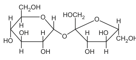
Fig. 4.1
Answer
Glycosidic (bond).
Glycosidic (bond).
Background Concept
Disaccharides such as sucrose, maltose and lactose are formed when two monosaccharides join together in a condensation reaction. The covalent bond formed between the two sugar molecules is always called a glycosidic bond. In sucrose the bond links C1 of -glucose to C2 of fructose; in maltose it links C1 of one -glucose to C4 of another; in lactose it links C1 of galactose to C4 of glucose. The nature of the bond is the same — only its position and the identity of the carbons involved differ.
Understanding the Question
The candidate is shown the structure of sucrose (Fig. 4.1) and asked simply to name the type of covalent bond joining the -glucose monomer to the fructose monomer. One mark is available.
Approach
Identify the single oxygen bridge connecting the two ring structures in the diagram and recall the specific term used for a covalent bond between two sugar units.
Step-by-Step Reasoning
- The diagram shows an oxygen atom bridging C1 of glucose to C2 of fructose.
- A covalent bond formed between two hydroxyl (–OH) groups on different sugar molecules, releasing a molecule of water, is termed a glycosidic bond.
- The mark scheme awards one mark for the single word "glycosidic".
Key Takeaways
- The covalent bond between two monosaccharides is a glycosidic bond.
- It is formed in a condensation reaction and broken by hydrolysis.
Common Mistakes
- Writing "ester bond" or "peptide bond" — these describe other biomolecule linkages and are wrong here.
- Writing "hydrogen bond" — that is the weak intermolecular force, not the covalent linkage between monomers.
Things to Be Careful About
Be precise: "glycosidic" is the single adjective. "Glucose bond" or "sugar bond" are descriptive but not the technical term required.
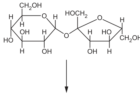
Sucrose is hydrolysed by the enzyme sucrase in the human digestive system.
The products of this hydrolysis reaction are the monosaccharides -glucose and fructose.
Complete the diagram to show the hydrolysis of sucrose to form -glucose and fructose.
Answer
Below the sucrose structure, draw the two products of hydrolysis:
-
Water — write " HO" alongside the arrow (water is a reactant in hydrolysis).
-
-glucose — six-membered ring containing O, with –OH on C1 drawn pointing below the ring (the position).
-
Fructose — five-membered ring containing O, with –CHOH groups on C1 and C6 and an –OH on C2.
Two monomers drawn correctly (α-glucose with –OH on C1 below ring; fructose with –OH on C2) and H₂O added as a reactant.
Background Concept
Hydrolysis is the reverse of a condensation reaction: a glycosidic bond is broken by the addition of a water molecule, regenerating the two original –OH groups on the monomers. For sucrose, hydrolysis by the enzyme sucrase releases one molecule of -glucose and one molecule of fructose.
It is essential to be able to recognise and draw the two monosaccharides:
- -glucose — a six-membered (pyranose) ring with the –OH on C1 on the opposite side of the ring from the –CHOH on C5 (drawn below the plane of the ring).
- Fructose — a five-membered (furanose) ring. The carbon that was involved in the glycosidic bond (C2 of fructose) must end up with a free –OH group after hydrolysis.
Understanding the Question
The candidate is given the sucrose structure with a downward arrow indicating the hydrolysis reaction and must complete the diagram to show the products. Three marks are available for correctly identifying the two monomers and showing water as a reactant.
Approach
- Add water to the reaction side of the arrow.
- Draw the six-membered -glucose ring on the left, with the –OH group on C1 in the orientation (below the ring).
- Draw the five-membered fructose ring on the right, with –OH on C2.
Step-by-Step Reasoning
- Mark 1 — labelling: clearly label one of the products as -glucose or fructose.
- Mark 2 — -glucose: redraw the glucose six-membered ring, ensuring the –OH on C1 points below the ring (the position); C6 must still bear the –CHOH group.
- Mark 3 — fructose: redraw the fructose five-membered ring, ensuring C2 now carries a free –OH group (the bond to the bridging oxygen has been replaced by –OH from water).
- Mark 4 — water: include HO somewhere near the arrow to indicate that water is a reactant.
Any three of the four points earn three marks.
Key Takeaways
- Hydrolysis = breaking a glycosidic bond using water.
- -glucose and fructose can be distinguished by ring size (6 vs 5) and by the orientation of the C1/C2 hydroxyls.
Common Mistakes
- Drawing glucose with the –OH on C1 above the ring (which would be -glucose, not ).
- Drawing fructose with the –OH on the wrong carbon — C2 must have the –OH after hydrolysis.
- Forgetting to include water on the diagram — hydrolysis explicitly requires HO.
Things to Be Careful About
- The C1 of the original -glucose must end up with an –OH (not –H) after hydrolysis.
- The ring oxygens and the carbon numbering conventions must be retained.
Plants transport sucrose from a source to a sink.
Fig. 4.2 is a scanning electron micrograph (SEM) of a transverse section through a plant tissue used to transport sucrose.
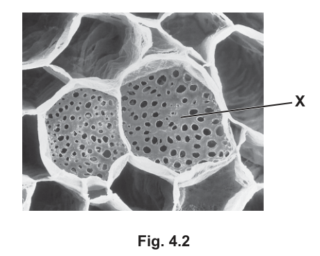
Fig. 4.2
Answer
Sieve plate.
Sieve plate.
Background Concept
Sieve tube elements are the conducting cells of phloem. They are joined end to end, and at each junction their walls are perforated by large pores to form a sieve plate. The pores allow the cytoplasmic contents (and dissolved assimilates such as sucrose) to pass from one sieve tube element to the next during mass flow.
Understanding the Question
Fig. 4.2 is a SEM of phloem in transverse section. The label X points to the perforated end wall between two adjacent sieve tube elements — a structure the candidate must name.
Approach
Recognise that the perforated end wall of a sieve tube element is the sieve plate, distinguishing it from a middle lamella or a simple cell wall.
Step-by-Step Reasoning
- The micrograph shows the lumen of several sieve tube elements with their characteristic perforated end walls.
- The end wall between two sieve tube elements, perforated by pores, is the sieve plate.
- One mark is awarded for the correct name.
Key Takeaways
- Sieve plates are the perforated end walls of sieve tube elements.
- They allow mass flow of assimilates between successive sieve tube elements.
Common Mistakes
- Writing "sieve tube" instead of "sieve plate" — the sieve tube is the whole cell, not the end wall.
- Writing "phloem" — too imprecise; phloem is the whole tissue.
Things to Be Careful About
In a SEM of phloem the sieve plate often appears as a perforated disc at the end of a sieve tube element; do not confuse it with the side walls of the element.
A scientist carried out an experiment to study carbohydrate transport in the stem of a woody plant.
Fig. 4.3 shows a plan diagram of a transverse section of the stem studied by the scientist. The position of the xylem tissue in the stem is shown.
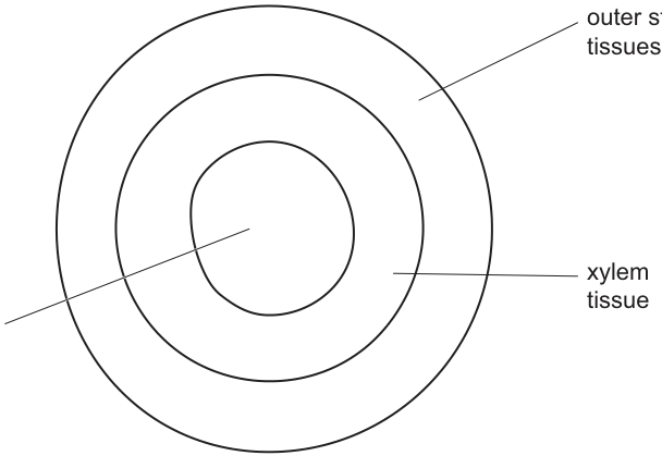
Fig. 4.3
The scientist carried out a set of experiments using plants of the same species.
In each experiment, a ring of tissue was removed from the outer stem of the plant, but the xylem tissue was left intact. This is shown in Fig. 4.4.
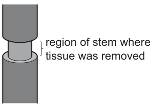
Fig. 4.4
The mass of carbohydrate transported to the lower part of the stem in 24 hours was recorded.
In each experiment a different percentage of the outer stem tissue was removed.
All other variables remained constant.
Table 4.1 shows the results of this investigation.
Table 4.1
| percentage of outer stem tissue removed | mass of carbohydrate transported to lower part of the stem in 24 hours / mg |
|---|---|
| 13 | 774 |
| 67 | 597 |
| 90 | 425 |
| 100 | 0 |
Explain the results shown in Table 4.1.
Answer
-
Carbohydrate (sucrose) is transported in the phloem sieve tubes (not in the xylem).
-
As the percentage of outer stem tissue removed increases, the amount of phloem sieve tube tissue remaining in the stem decreases.
-
With less phloem intact, less mass flow / translocation of carbohydrate can occur from the source to the sink (lower part of stem).
-
At 100 % removal, all of the phloem is removed (only xylem is left), so no carbohydrate is transported (0 mg).
-
This shows that carbohydrate is not transported in the xylem, because carbohydrate transport still occurs when the xylem is intact (and only outer tissues are removed).
Any four of these points for four marks.
Sucrose is transported in phloem; removing increasing amounts of outer tissue removes increasing amounts of phloem, reducing mass flow, until at 100 % removal (all phloem removed) transport ceases.
Background Concept
In a vascular plant, xylem carries water and dissolved mineral ions from roots to leaves, while phloem carries organic solutes (mainly sucrose) from sources (e.g. leaves, storage organs) to sinks (e.g. roots, fruits, growing tips). The translocation of sucrose in phloem occurs by mass flow through sieve tube elements. The arrangement of these tissues in a dicot stem is, from outside to inside: epidermis, cortex, phloem, cambium, xylem, pith (a ring of phloem outside a ring of xylem).
The classic "girdling" experiment (Malpighi, 1670s) removes a ring of outer stem tissue (phloem and cortex) but leaves the xylem intact. Anything produced above the ring still travels down via the xylem (water and minerals), but carbohydrate transport below the ring is interrupted, demonstrating that phloem — not xylem — carries sucrose.
Understanding the Question
A scientist removed increasing percentages of outer stem tissue (always leaving the xylem intact) and measured the mass of carbohydrate reaching the lower stem in 24 h. The candidate must explain the trend shown:
| % removed | mass transported (mg) |
|---|---|
| 13 | 774 |
| 67 | 597 |
| 90 | 425 |
| 100 | 0 |
Approach
- Identify which tissue is responsible for carbohydrate transport — phloem (sieve tubes).
- Note that the xylem is intact in all experiments, so any change in transport must be due to changes in the phloem.
- Recognise that as more outer tissue is removed, more phloem is destroyed, so less mass flow can occur.
- At 100 % removal the entire phloem ring is gone, so transport is zero.
Step-by-Step Reasoning
- Mark 1: Carbohydrate is transported in (phloem) sieve tubes (not in xylem). This is the foundational statement.
- Mark 2: As the % of outer tissue removed rises, the quantity of phloem sieve tube remaining decreases. The trend in the data is explained by progressive loss of conducting tissue.
- Mark 3: With less phloem, there is less mass flow / translocation of carbohydrate to the lower stem, hence the falling transport values.
- Mark 4: At 100 % removal, all phloem has been removed; only xylem remains. Because xylem does not transport sucrose, the value drops to 0 mg.
- Mark 5 (alternative): The result also confirms that xylem does NOT transport carbohydrate — when the xylem is intact but phloem has been removed, no carbohydrate reaches the lower stem.
- Mark 6 (alternative): Carbohydrate is moving from a source to a sink (lower stem acting as a sink in this case).
- Mark 7 (alternative): AVP — e.g. the phloem forms a continuous ring just outside the xylem, so even a small girdle breaks the entire translocation pathway; once the ring is broken at any point, transport to the lower stem ceases.
Any four points score four marks.
Key Takeaways
- Phloem transports sucrose (and other assimilates); xylem transports water and ions.
- Translocation occurs by mass flow from source to sink.
- Girdling experiments demonstrate which tissue carries which solute — a classic piece of experimental evidence.
Common Mistakes
- Saying carbohydrate is transported in the xylem — the data rule this out (transport falls to 0 when xylem is the only tissue left).
- Saying the decrease is due to wounding or stress rather than loss of phloem.
- Failing to mention mass flow / translocation.
- Not recognising that the xylem is intentionally left intact in every experiment.
Things to Be Careful About
- Quote specific data points where possible (e.g. 0 mg at 100 % removal) to support the explanation.
- Use precise terminology: phloem, sieve tubes, mass flow / translocation.
- Distinguish source (where sucrose is loaded, e.g. photosynthesising leaf) from sink (where sucrose is unloaded, e.g. root, fruit).
Sucrose is a sweet-tasting sugar found in many foods.
Some people become ill when they have sucrose in their diet. These people have a gene mutation in the gene coding for sucrase and cannot hydrolyse sucrose in the digestive system.
Scientists studying the DNA of people with this condition identified a deletion mutation in the gene coding for sucrase.
Suggest and explain why a person with this deletion mutation cannot digest sucrose.
Answer
A deletion mutation removes one or more nucleotides from the sucrase gene. This alters the base sequence of the DNA and therefore the mRNA codon sequence produced during transcription.
Depending on whether the number of nucleotides deleted is a multiple of three:
- If it is not a multiple of three, a frameshift results — every codon downstream of the deletion is read in a different frame, so the primary structure of the polypeptide is changed (different amino acid sequence from the deletion point onwards).
- This altered polypeptide cannot fold into the correct tertiary structure, so the shape of the active site is lost.
- Sucrose can therefore no longer bind to the active site and the glycosidic bond is not hydrolysed.
- The altered polypeptide may also form a premature stop codon, giving a truncated (shorter) polypeptide that is non-functional.
- The abnormal polypeptide may be recognised as faulty and degraded by the cell.
Any four well-articulated points for four marks.
A deletion mutation alters the base sequence of the sucrase gene; this changes the mRNA codon sequence, producing a polypeptide with a different primary structure that cannot form a functional active site, so sucrose cannot bind and is not hydrolysed.
Background Concept
A gene is a length of DNA that codes for a polypeptide. The base sequence of the gene is transcribed into mRNA, which is then translated by ribosomes into a polypeptide chain of specific amino acids (read three bases / codon at a time). The polypeptide then folds into a specific tertiary structure, and for an enzyme such as sucrase the active site has a precise shape complementary to its substrate (sucrose).
A deletion mutation removes one or more nucleotides from a gene. The consequences depend on how many nucleotides are removed:
- If one nucleotide (or any number not a multiple of three) is removed, every codon downstream of the deletion is shifted by one base — a frameshift. All amino acids from that point on are different and the protein is usually non-functional.
- If three (or a multiple of three) nucleotides is removed, exactly one (or more) codon(s) is lost but the reading frame is preserved; the polypeptide is shorter but the rest of the sequence is unchanged.
Understanding the Question
The question states that people who cannot digest sucrose have a deletion mutation in the gene for sucrase. The candidate must suggest (use biological reasoning to put forward plausible consequences) and explain (give reasons) why the mutant cannot digest sucrose. Four marks are available.
Approach
Walk the consequences of the mutation logically:
DNA sequence → mRNA codon sequence → polypeptide primary structure → tertiary structure / active site shape → enzyme activity.
Identify the failure point and link it back to enzyme specificity.
Step-by-Step Reasoning
The mark scheme accepts points from two strands; up to three marks from each, total four.
Strand A — what the mutation does to the gene / mRNA:
- A deletion mutation is the loss of one or more nucleotides from the gene.
- This changes the sequence of bases in the DNA of the sucrase gene.
- Therefore the sequence of codons in the mRNA transcribed from the gene is altered.
- If the deletion is not a multiple of three nucleotides, a frameshift occurs and every codon downstream is altered.
- The deletion may also generate a premature stop codon, giving a shortened (truncated) polypeptide.
Strand B — what happens to the enzyme / protein:
- The altered mRNA is translated into a polypeptide with a different primary structure (different amino acid sequence), or a truncated polypeptide.
- The polypeptide cannot fold correctly into its normal tertiary structure.
- The shape of the active site is changed, so it is no longer complementary to sucrose.
- Sucrose cannot bind to the active site, so the ES complex cannot form and the glycosidic bond is not broken.
- The non-functional polypeptide may be recognised as abnormal and degraded by cellular quality-control mechanisms.
Pick any four points from the two strands for four marks.
Key Takeaways
- A deletion mutation changes the reading frame unless exactly three (or a multiple of three) bases are lost.
- The sequence of bases in DNA → mRNA codons → amino acid sequence of polypeptide → folding into tertiary structure → active site shape → enzyme specificity.
- A single base change can ripple through the whole protein and abolish enzyme activity.
Common Mistakes
- Saying the mutation "changes the shape of the DNA" — DNA is double-stranded; the mutation changes its base sequence.
- Confusing a deletion with a substitution — a substitution changes one base to another, while a deletion removes a base.
- Saying the active site "stops working" without explaining why (shape changed; substrate no longer complementary).
- Forgetting that protein shape depends on primary structure, which depends on the base sequence.
Things to Be Careful About
- Use the precise term frameshift if the deletion is not a multiple of three.
- Distinguish primary structure (amino acid sequence) from tertiary structure (3-D folding).
- Tie the explanation back to enzyme specificity: the active site must be complementary to sucrose for the glycosidic bond to be hydrolysed.
- The question says "suggest and explain" — accept reasonable biological consequences, not just the single most likely one.
Trypsin is an enzyme which catalyses the hydrolysis of casein, a protein found in milk.
Milk that contains casein has a cloudy, white appearance. As the casein is hydrolysed by trypsin, the milk changes in appearance to a clear (transparent), colourless solution.
A student carried out an experiment to investigate the effect of enzyme concentration on the rate at which trypsin hydrolyses casein.
The student added a solution of trypsin to a sample of milk and recorded the time taken for the milk to become transparent. The student repeated the experiment with different concentrations of trypsin. All other variables were kept constant.
Fig. 5.1 shows the results from the experiment.
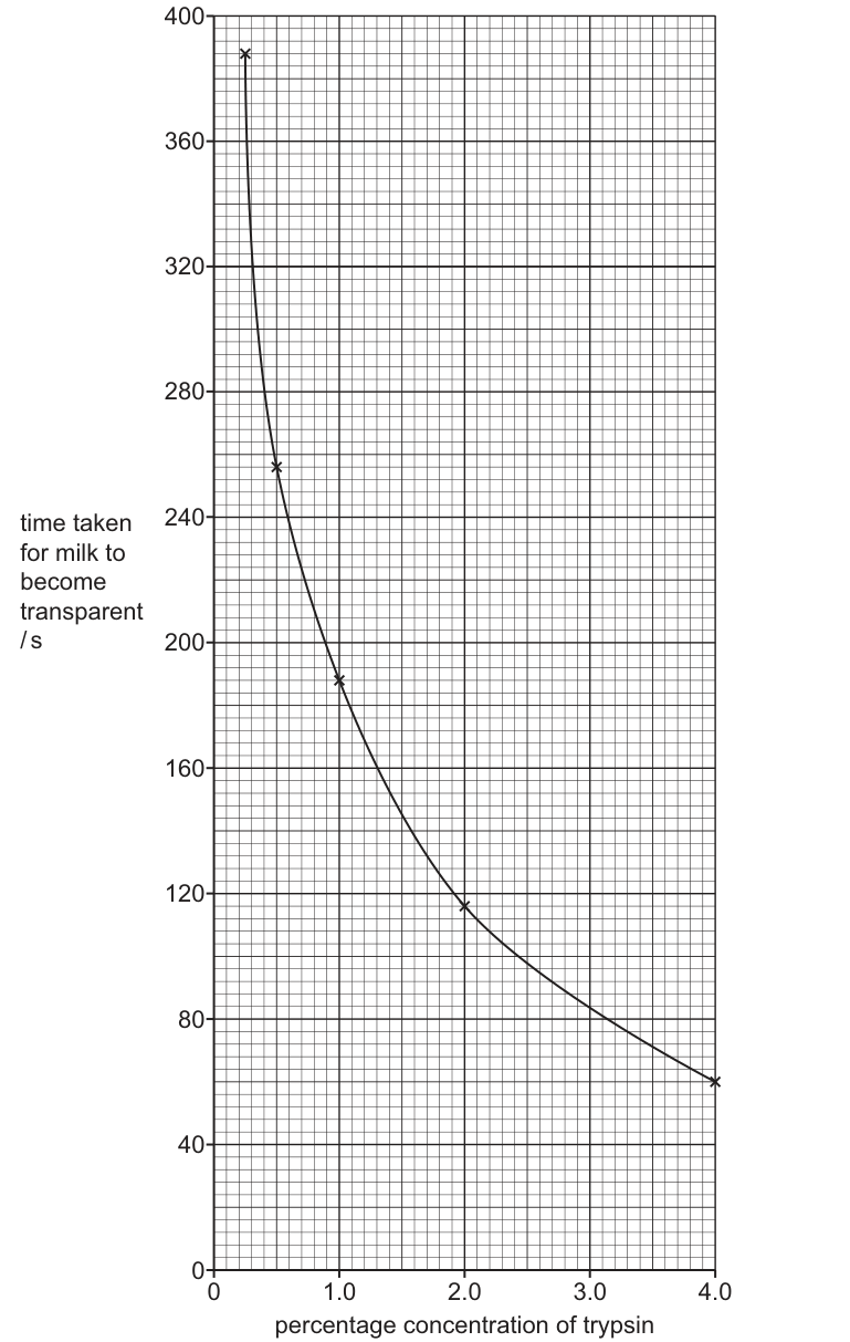
Fig. 5.1
When the concentration of trypsin increases from 2.0% to 4.0%, the time taken for the milk to become transparent decreases by 48%.
Calculate the percentage decrease in the time taken for milk to become transparent when the concentration of trypsin increases from 0.25% to 0.5%.
Write your answer to the nearest whole number.
percentage decrease = ______
Working
From Fig. 5.1:
- time at 0.25% trypsin = 385 s
- time at 0.5% trypsin = 255 s
Answer
percentage decrease = 34
34
Background Concept
When analysing enzyme experiments, the rate of reaction is often reported as 1/time (the time taken for a defined end-point). Percentage change is a standard way to compare the size of an effect at different parts of a curve:
The "original" value is the starting point, not the smaller of the two. The answer is rounded to a whole number as instructed.
Understanding the Question
Fig. 5.1 plots time taken (s) on the y-axis against percentage concentration of trypsin on the x-axis, with five data points: (0.25, 385), (0.5, 255), (1.0, 188), (2.0, 116) and (4.0, 60). The question asks specifically about the change between 0.25% and 0.5%, so only those two values are needed.
Approach
- Read the two y-values from the graph at x = 0.25 and x = 0.5.
- Subtract the new (smaller) time from the original (larger) time to find the absolute decrease.
- Divide by the original time and multiply by 100 to convert to a percentage.
- Round to the nearest whole number.
Step-by-Step Reasoning
- At 0.25% trypsin, the milk takes 385 s to go transparent.
- At 0.5% trypsin, the milk takes 255 s.
- The absolute decrease is 385 − 255 = 130 s.
- As a percentage of the original (385 s): 130 ÷ 385 = 0.3377...
- Multiplied by 100 = 33.77%, which rounds to 34%.
The mark scheme confirms 34% as the single correct whole-number answer.
Key Takeaways
- Always use the starting (earlier) value as the denominator in a percentage change calculation.
- Even a small move along a steep part of the curve produces a large percentage change — that is what the question is checking.
Common Mistakes
- Dividing by the smaller value (255) instead of the original (385), which would give ≈ 51%.
- Using 4.0% (60 s) instead of 0.5% (255 s) because the problem statement mentions 2.0% → 4.0% — read the actual part you are answering.
- Reporting 33% instead of 34% by truncating rather than rounding.
Things to Be Careful About
The mark scheme accepts only the whole number 34; do not give a decimal. There is no error-carried-forward from any earlier part because none of the values are calculated.
Answer
- At low trypsin concentrations there are not enough active sites / trypsin molecules to deal with all the casein substrate ;
- as the concentration of trypsin increases, the number of active sites available increases ;
- so there are more successful collisions between trypsin and casein per unit time ;
- more enzyme–substrate complexes are formed per unit time ;
- the rate of hydrolysis of casein increases, so the time taken for the milk to become transparent decreases ;
- at high concentrations the substrate (casein) becomes the limiting factor, so the curve levels off ;
- when the milk goes transparent, the casein has been fully hydrolysed to amino acids / smaller peptides.
See working
Background Concept
Enzymes are biological catalysts that speed up reactions by binding their substrate at the active site to form an enzyme–substrate (ES) complex. The substrate is converted to product, which then leaves the active site, freeing it for another substrate molecule. The rate of an enzyme-catalysed reaction therefore depends on:
- how many enzyme (active site) molecules are present, and
- how often each active site is productively occupied.
When substrate is in excess, enzyme concentration is the limiting factor: doubling the enzyme roughly doubles the rate, because more active sites are available to form ES complexes per unit time. Eventually the substrate itself becomes limiting, and further increases in enzyme concentration have little effect — this is the plateau region of a typical rate-vs-enzyme-concentration curve.
Understanding the Question
The graph shows time taken (s) on the y-axis (so the inverse of rate) versus percentage concentration of trypsin. As trypsin concentration rises, time falls steeply at first and then levels off — the classic hyperbolic shape that goes with a rate-vs-enzyme-concentration plot when read the other way up. The question wants the biological explanation of that shape: why does more trypsin make the reaction faster, and why does the improvement get smaller at higher concentrations?
Approach
- Begin with the active site: more enzyme = more active sites.
- Move to the kinetic consequence: more active sites mean more successful collisions with casein per unit time.
- Then state the chemical consequence: more ES complexes per unit time, so more casein hydrolysed per unit time.
- Finally, link the chemistry back to what the student actually sees: faster hydrolysis → shorter time to transparency.
- Mention the plateau: at high enzyme concentration the substrate (casein) is the limiting factor.
Step-by-Step Reasoning
- At low trypsin concentrations there are insufficient active sites to accommodate all the casein molecules. Substrate molecules must wait for an active site to become free, which slows the overall rate.
- As trypsin concentration increases, the total number of active sites available at any instant increases, so more casein molecules can be bound at once.
- More active sites → more successful enzyme–substrate collisions per unit time, increasing the number of ES complexes formed per unit time.
- Each ES complex leads to hydrolysis of casein into smaller peptides/amino acids, so a greater number of ES complexes per unit time means a higher rate of casein breakdown.
- Higher rate = less time needed to hydrolyse all of the casein, which is exactly what the graph shows: shorter time to transparency at higher trypsin concentrations.
- At high concentrations the curve begins to level off because casein (the substrate) is now the limiting factor — there is simply not enough casein left to keep all the active sites busy, so adding more trypsin makes little further difference.
- The end-point itself is the complete hydrolysis of casein: when the milk becomes transparent, all of the casein has been broken down and the solution contains only amino acids / small peptides (no longer the light-scattering casein colloid).
Key Takeaways
- Always tie "more enzyme" to "more active sites" — that is the mark-scheme phrasing.
- "More active sites" → "more successful collisions" → "more ES complexes per unit time" → "higher rate" is the standard chain of reasoning.
- A curve that levels off is telling you the other reactant (substrate) is now the limiting factor.
Common Mistakes
- Saying "more enzyme means a faster reaction" with no active site/collision/ES-complex link — this is too vague to score.
- Talking about "more collisions" without specifying successful collisions, or failing to mention the active site.
- Confusing this graph (time vs concentration) with a rate vs concentration graph and saying "rate increases" without explaining that time decreases.
- Not mentioning the plateau / substrate limitation at high concentrations — examiners often look for this insight.
Things to Be Careful About
- The mark scheme accepts "enzyme" for trypsin and "substrate" for casein, so wording is flexible, but the key concepts (active site, ES complex, successful collisions, limiting factor) are not.
- You need both the cause (more active sites) and the consequence (more ES complexes per unit time) for full credit — stating one without the other usually caps marks at 2–3.
- The question says "explain the results", so referring back to the specific trend shown in Fig. 5.1 (time falling steeply then levelling off) earns the link-back marks.
Trypsin has the potential to be used in a wide range of industrial processes.
The use of immobilised enzymes in industrial processes has many advantages.
Scientists investigated the effect of temperature on the activity of trypsin immobilised on the surface of a material and trypsin free in solution.
Table 5.1 shows the results of the investigation.
Table 5.1
| temperature / °C | percentage of maximum activity of immobilised trypsin | percentage of maximum activity of trypsin free in solution |
|---|---|---|
| 25 | 60 | 100 |
| 35 | 85 | 100 |
| 45 | 98 | 80 |
| 55 | 95 | 20 |
| 65 | 100 | 5 |
State a reason for the difference in percentage of maximum activity of immobilised trypsin and trypsin free in solution at 25°C.
Answer
- Immobilising the trypsin may have altered the tertiary structure / shape of the active site, reducing its activity at 25 °C ;
- (alternatively) the material to which trypsin is attached may not be inert and may have an inhibitory effect at lower temperatures ;
- (alternatively) immobilising may have covered part of the active site of some enzyme molecules ;
- (alternatively) trypsin free in solution has an increased chance of collision with substrate, giving higher activity.
Immobilisation may have altered the tertiary structure / shape of the active site of trypsin, reducing its activity compared to the free enzyme at 25 °C.
Background Concept
Immobilised enzymes are enzymes that have been attached to or trapped within an insoluble support (e.g. alginate beads, a resin surface, a membrane). This is widely used in industry because it makes the enzyme easy to recover, reuse and separate from the product. However, the act of immobilisation is not biologically neutral:
- The chemical attachment or physical entrapment can distort the tertiary structure of the enzyme, subtly changing the shape of the active site.
- The support may physically block part of the active site of some enzyme molecules.
- The support may not be chemically inert — it can interact with the substrate, the product or the enzyme itself, sometimes inhibiting activity.
- An immobilised enzyme cannot tumble freely, so its collision frequency with substrate is lower than that of a free enzyme in solution.
Any of these effects can lower the observed activity of the immobilised enzyme relative to the free enzyme, especially at temperatures well below the optimum where small losses in active-site efficiency are most visible.
Understanding the Question
Table 5.1 shows that at 25 °C the free trypsin reaches 100% of its maximum activity, while the immobilised trypsin only reaches 60%. The question is: why is the immobilised form less active at this low temperature? It is a "state a reason" question, so one clear, well-justified point is enough.
Approach
Pick the most biologically defensible reason and state it concisely. The strongest answers refer to a structural change caused by the attachment process; the kinetic (collision-frequency) argument is also accepted.
Step-by-Step Reasoning
- Why is the free enzyme at 100% at 25 °C? Because 25 °C is below the optimum (45 °C in this experiment) but not low enough to limit the reaction severely when substrate is plentiful and the enzyme is fully active.
- Why is the immobilised form only at 60%? The most common accepted explanations are:
- Immobilisation alters the tertiary structure of some enzyme molecules, slightly distorting the active site so substrate binds less efficiently.
- The support material is not inert and chemically interferes with the enzyme at lower temperatures.
- The support covers part of the active site of some trypsin molecules, lowering the proportion of usable active sites.
- The immobilised enzyme has a lower frequency of successful collisions with substrate than a free enzyme in solution, because it is held in place.
- Any one of these, stated clearly, is the full mark.
Key Takeaways
- Immobilisation is a useful industrial tool, but it can compromise enzyme activity for several distinct reasons (structural, chemical, kinetic).
- When comparing two forms of the same enzyme, the differences almost always come down to either structure (active site shape) or mobility (collision frequency).
Common Mistakes
- Saying only that the immobilised enzyme is "less active" without explaining the mechanism (active site / tertiary structure / collision).
- Confusing the immobilised enzyme with a denatured enzyme — at 25 °C neither form is denatured, so denaturation is not the right explanation here.
- Citing advantages of immobilisation (reusability, etc.) — the question asks why activity is lower, not why immobilisation is useful.
Things to Be Careful About
- The question is "state a reason", not "explain" — one clear, mechanism-based point is enough for the mark.
- The mark scheme lists five acceptable alternatives; pick the one you can express most precisely.
Suggest and explain why the percentage of maximum activity of immobilised trypsin at 55°C is higher than the percentage of maximum activity of trypsin free in solution at 55°C.
Answer
- Immobilisation stabilises the tertiary structure of trypsin / protects the enzyme from thermal denaturation at 55 °C ;
- therefore the active site of the immobilised enzyme retains its shape, whereas the free enzyme is denatured ;
- (and) the support restricts the vibration / movement of the enzyme molecules, reducing the chance of bonds in the tertiary structure (e.g. hydrogen / ionic bonds) breaking at 55 °C.
Immobilisation stabilises the tertiary structure of trypsin, protecting it from denaturation at 55 °C so the active site retains its shape, whereas free trypsin is denatured at this temperature.
Background Concept
At temperatures well above the optimum, an enzyme's tertiary structure begins to break down. The hydrogen, ionic and disulfide bonds that hold the polypeptide chain in its precise 3-D shape are disrupted by the increased kinetic energy of the molecule, the active site loses its specific shape, and the enzyme is said to be denatured. Denaturation is usually irreversible.
When an enzyme is immobilised, the support it is attached to can:
- hold the enzyme in a fixed orientation, restricting the vibration of the polypeptide chain;
- provide additional physical or chemical stabilisation of the tertiary structure;
- make the enzyme more thermally stable so it retains activity at temperatures that would denature the free enzyme.
This is one of the main industrial advantages of immobilisation: the enzyme can be used at higher temperatures (often giving faster reaction rates) without being destroyed.
Understanding the Question
Table 5.1 shows the contrast at 55 °C:
- Immobilised trypsin = 95% of maximum activity (still nearly fully functional)
- Free trypsin = 20% of maximum activity (largely denatured)
The question asks the candidate to suggest and explain this difference. "Suggest" means put forward a hypothesis; "explain" means justify it biologically.
Approach
- Identify what happens to free trypsin at 55 °C: it denatures (its tertiary structure breaks down and the active site loses its shape).
- Identify what is different about immobilised trypsin: the support stabilises it.
- Explain how the support stabilises it: by holding the enzyme in place and reducing vibration, the bonds (H-bonds, ionic bonds) in the tertiary structure are less likely to break.
Step-by-Step Reasoning
- At 55 °C, free trypsin molecules have high kinetic energy. The vibrations of the polypeptide chain break the hydrogen and ionic bonds that hold the tertiary structure together.
- This causes the active site to change shape, so substrate can no longer bind efficiently — the enzyme is denatured and its activity falls to 20%.
- Immobilised trypsin is physically held in place by the support material. This restricts the vibration of the enzyme and provides extra structural support.
- As a result, the hydrogen and ionic bonds in the tertiary structure are less likely to break, the active site retains its shape, and the enzyme remains active (95%).
- In short, immobilisation protects the enzyme from thermal denaturation by stabilising the tertiary structure.
Key Takeaways
- Denaturation = loss of tertiary structure = loss of active site shape = loss of activity.
- Immobilisation improves thermal stability, which is a major industrial advantage because higher temperatures usually mean faster reaction rates.
- The explanation must connect the support → reduced vibration / structural stabilisation → bonds not breaking → active site shape preserved.
Common Mistakes
- Saying only that "immobilised enzymes are more stable" without saying what is being stabilised (the tertiary structure / the active site shape) or how (reduced vibration, bond protection).
- Confusing this with the 25 °C part — at 55 °C the free enzyme is the one in trouble, not the immobilised one.
- Citing a different advantage of immobilisation (e.g. reusability) that does not explain the temperature result.
- Saying the immobilised enzyme "works better at high temperatures" without referencing denaturation of the free enzyme.
Things to Be Careful About
- The mark scheme credits both the protection from denaturation and the mechanism (bond stability / reduced vibration). A candidate who only gives one of the two will only get 1 of the 2 marks.
- "Thermal stability" or "resistance to denaturation" is the key phrase — write it down.
Fig. 6.1 shows a plant cell in a stage of mitosis.
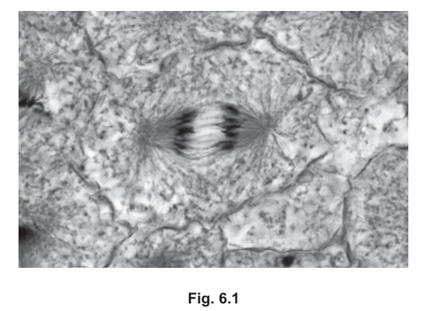
Fig. 6.1
Some of the structures shown in Fig. 6.1 contain DNA.
Use a line labelled D on Fig. 6.1 to indicate one of these structures.
Answer
A line labelled D drawn from the label to one of the dark, V-shaped chromatids in either of the two groups near the poles of the cell. Each chromatid (a single chromosome consisting of one DNA molecule packaged with histones) is the structure that contains DNA.
Line labelled D pointing to one chromatid (chromosome).
Background Concept
During mitosis the genetic material is condensed into discrete chromosomes. Before S-phase each chromosome consists of a single DNA molecule; after DNA replication each chromosome is made of two identical sister chromatids joined at the centromere. Each chromatid is therefore one DNA molecule tightly packaged with histone proteins. In a stained micrograph, the chromatids appear as the dark, compact bodies.
Understanding the Question
The candidate is shown Fig. 6.1, a micrograph of a plant cell in anaphase of mitosis, in which two groups of dark chromosomes are visible near opposite poles with spindle fibres stretching between them. The instruction is to use a labelled line on the printed figure to indicate one structure that contains DNA. Any chromosome/chromatid in the cell is a valid answer because chromosomes are the only DNA-containing structures visible at this stage — the spindle fibres, the cell wall and the cytoplasm do not contain DNA.
Approach
Look for the most clearly visible, darkly stained, compact body in the micrograph — these are the chromatids (sister chromosomes). Draw a straight line from a clearly written "D" label to one of them, ending with a small arrow or dot on the chromatid. Avoid pointing to spindle fibres, the cell wall or empty cytoplasm.
Step-by-Step Reasoning
- Identify DNA-containing structures. In a mitotic cell, DNA is contained in the chromosomes/chromatids only.
- Locate the chromatids. In Fig. 6.1 the dark, V- or rod-shaped bodies at each pole are the sister chromatids that have just separated.
- Draw the line. Place a clear "D" outside the cell (to the right or above the cell body) and draw a straight line ending on one chromatid.
- Why not the spindle? Spindle fibres are made of microtubules (tubulin protein), not DNA. The cell wall is cellulose. Neither contains DNA.
Key Takeaways
- DNA in a mitotic cell is located exclusively in the chromosomes/chromatids.
- On a stained micrograph, chromosomes are the smallest, darkest, most sharply defined bodies.
- Annotating a printed figure precisely (label outside, line touching the structure) is a routine exam skill.
Common Mistakes
- Labelling the spindle fibres or the cell wall — these contain no DNA.
- Drawing the line too vaguely (e.g. pointing to a region rather than to a single chromatid).
- Putting the label inside the cell where it obscures detail.
Things to Be Careful About
- "Chromosome" and "chromatid" are both acceptable, but the line should touch one clearly visible body, not a region of cytoplasm.
Answer
Anaphase.
Anaphase
Background Concept
Mitosis is conventionally divided into four (sometimes five) stages — prophase, metaphase, anaphase and telophase — defined by the position and behaviour of the chromosomes and spindle:
- Prophase: chromosomes condense; spindle forms.
- Metaphase: chromosomes line up on the equator.
- Anaphase: sister chromatids separate at the centromere and are pulled to opposite poles by shortening spindle fibres.
- Telophase: chromatids arrive at the poles; nuclear envelopes re-form.
Understanding the Question
Fig. 6.1 shows two distinct groups of dark chromatids at opposite ends of the cell, with spindle fibres between them. The candidate has to recognise this as a particular mitotic phase. The visual signature of two separated groups moving apart is unique to one stage.
Approach
Match what is seen in the micrograph to the defining feature of each stage. The defining feature here — two separate, equal groups of chromatids moving towards opposite poles — fits only anaphase.
Step-by-Step Reasoning
- The chromatids are NOT aligned in a single line across the middle — so this is not metaphase.
- The chromatids are in TWO groups at opposite poles, with spindle fibres between them — this is the classic anaphase picture.
- Cytokinesis has not visibly occurred (no cleavage furrow dividing the cell into two) — so this is not yet telophase.
- Therefore the stage is anaphase.
Key Takeaways
- Anaphase is identified by separation of sister chromatids and their movement towards opposite poles.
- Spindle fibres shortening (microtubule depolymerisation) is the mechanism that drags chromatids poleward.
Common Mistakes
- Confusing anaphase with telophase because in telophase chromatids are also at the poles — but in telophase the nuclear envelope is reforming and cytokinesis is occurring.
- Confusing anaphase with metaphase by misreading the central grouping as a metaphase plate.
Things to Be Careful About
- Spelling: "anaphase", not "anaphase" with a stray letter. Pronouncing it does not matter; spelling does.
Colchicine is a chemical used by scientists to study mitosis. This chemical inhibits the organisation of the microtubules in prophase of mitosis.
The cell shown in Fig. 6.1 had not been treated with colchicine.
Explain the evidence in Fig. 6.1 that shows the cell had not been treated with colchicine.
Answer
- A fully formed spindle / spindle fibres are visible in Fig. 6.1 — colchicine prevents spindle formation in prophase, so the presence of a spindle shows it was not treated.
- Sister chromatids have separated and are moving to opposite poles because spindle fibres have contracted (microtubules have shortened) and pulled them apart; this can only happen if a functional spindle is present.
- The cell has reached anaphase, so prophase and metaphase have been completed normally — colchicine-treated cells would be arrested in prophase and could not reach anaphase.
Spindle fibres are visible and chromatids are attached to them / have separated because the spindle fibres have contracted, showing the cell has proceeded normally through prophase and metaphase to anaphase.
Background Concept
The mitotic spindle is built from microtubules (tubulin polymers) that assemble during prophase from the two centrosomes (poles). Kinetochore microtubules attach to the centromere of each chromatid; when they shorten during anaphase they pull sister chromatids apart toward opposite poles. Colchicine binds to tubulin and prevents microtubule polymerisation, so in colchicine-treated cells no spindle can form. The cell therefore arrests in (or before) prophase, the chromosomes cannot attach to anything, sister chromatids never separate, and the cell cannot enter anaphase.
Understanding the Question
The question states explicitly that colchicine inhibits microtubule organisation in prophase and that Fig. 6.1 had not been treated with colchicine. The candidate has to look at Fig. 6.1 and pick out features that would be impossible without a functional spindle. Any two such features earn the two marks.
Approach
Reason backwards: if colchicine blocks microtubule assembly, then any feature of the micrograph that depends on microtubules is evidence AGAINST colchicine treatment. Visible spindle fibres, chromatids attached to those fibres, separated chromatids moving poleward, and the cell having reached anaphase all fit this reasoning.
Step-by-Step Reasoning
- Spindle is present. Fine fibres can be seen between the two poles of the cell. These are microtubules, and they could not have formed if colchicine had been applied — so the spindle itself is evidence.
- Chromatids are attached to the spindle. Each chromatid at the poles is connected to a pole by a fibre; without microtubules there would be no attachment and no movement.
- Sister chromatids have separated. Separation requires spindle shortening. A colchicine-treated cell would show condensed chromosomes still paired at prophase, not two separated groups.
- The cell is in anaphase. Reaching anaphase means prophase and metaphase were completed normally. Colchicine arrests cells in prophase, so a colchicine-treated cell could not be in anaphase.
Any two of these four points are credited.
Key Takeaways
- Colchicine blocks microtubule polymerisation → no spindle → mitosis halted at prophase.
- Visible spindle, chromatid–spindle attachment, separated chromatids and progression past metaphase are all evidence of normal, uninhibited mitosis.
- Reasoning "if X were inhibited, Y would not occur; Y is occurring, so X is not inhibited" is the general pattern for questions about inhibitors.
Common Mistakes
- Stating only "spindle is visible" without linking it to microtubules (the mark scheme requires the spindle/microtubule link).
- Saying "chromosomes have separated" without explaining that this happened because spindle fibres contracted (mechanism is required for full credit).
- Vague answers such as "the cell is dividing normally" — not specific to what is visible in Fig. 6.1.
- Implying colchicine affects DNA directly — it does not; it affects microtubules.
Things to Be Careful About
- The question asks for EVIDENCE in Fig. 6.1, so reference what is actually visible (spindle, separated chromatids, position at poles), not generic statements about cell division.
- Either "spindle fibres have contracted" or "microtubules have disassembled" is acceptable mechanistic language — the mark scheme uses "AW" (accept any wording) for this point.