Biology 9700/13 — October/November 2024
Cambridge AS Level · Multiple Choice (AS Level) · answer key with instant marking and worked solutions
Topics Cell Structure · Transport in Plants · Biological Molecules · Transport in Mammals · Nucleic Acids and Protein Synthesis · Enzymes · +5 more
Tap an option under each question to check it — your score builds as you go.
Which steps are needed to find the actual width of a xylem vessel viewed in transverse section using a objective lens?
1 Convert from to by multiplying by .
2 Calibrate the eyepiece graticule using a stage micrometer on a objective lens.
3 Measure the width of the xylem vessel using an eyepiece graticule.
4 Multiply the number of eyepiece graticule units by the calibration of the eyepiece graticule.
Options
A 1, 2, 3 and 4
B 1 and 2 only
C 2, 3 and 4 only
D 3 and 4 only
Working
- Step 1 is wrong. To convert mm to µm you multiply by (because ), not by .
- Step 2 is wrong. The eyepiece graticule must be calibrated on the same objective lens that will be used for the measurement. The specimen is being viewed with a objective, so the stage micrometer calibration must also be carried out on the objective, not on a objective.
- Step 3 is correct. The width of the xylem vessel is measured by counting the number of eyepiece graticule divisions it spans.
- Step 4 is correct. Each eyepiece graticule division at that objective represents a known length (the calibration value, in µm); multiplying the number of divisions by this calibration gives the actual width of the vessel.
Only steps 3 and 4 are required.
Answer
D
D
Background Concept
A light microscope has two interchangeable scales for measuring specimens:
- An eyepiece graticule is a small glass disc with a scale etched onto it, placed inside the eyepiece. It appears superimposed on the specimen, so the number of graticule divisions a specimen spans can be read directly. Critically, the eyepiece graticule is fixed — the same graticule is used at every objective.
- A stage micrometer is a slide with a scale of known length (usually 1 mm divided into 100 parts, so each division = 10 µm) and is used to calibrate the eyepiece graticule.
Because the apparent size of the graticule divisions changes with magnification, the calibration value (the actual length, in µm, of one eyepiece graticule division) is different for every objective lens. The eyepiece graticule must therefore be calibrated at each objective being used, not once for the whole microscope.
To convert the eyepiece reading into an actual length:
Unit conversions commonly used:
- (so mm → µm is a multiply by )
Understanding the Question
The question describes a practical task: a student wants to find the actual width of a xylem vessel seen in transverse section, viewed through a objective lens. Four possible procedural steps are listed, and the candidate must identify which of them are needed.
The command word is "needed" — only the steps that are correct AND necessary for the measurement should be selected. The question is testing two distinct microscopy ideas:
- The correct unit conversion between mm and µm.
- The rule that an eyepiece graticule must be calibrated on the same objective as the one used for measuring.
Approach
Walk through each numbered step in turn, decide whether it is required (and correct), and then look at the answer options. The correct combination is the set of steps that are both needed and correct; any step that is wrong or unnecessary removes that option.
Step-by-Step Reasoning
Step 1 — "Convert from mm to µm by multiplying by ."
- This is incorrect. , so to go from mm → µm you multiply by , not . Multiplying by would actually take you from µm → mm.
- Therefore step 1 is wrong and not needed.
- This rules out option A (which includes 1) and option B (1 and 2 only).
Step 2 — "Calibrate the eyepiece graticule using a stage micrometer on a objective lens."
- This is incorrect because the calibration must be carried out at the same objective used for the measurement. The measurement here is on a objective, so the stage micrometer must be used on the objective to find the µm-per-division value at that magnification. Calibrating on a objective would give a calibration value valid only at , which is not what we need.
- Therefore step 2 is wrong and not needed.
- This rules out option B (1 and 2 only) and any option that includes 2.
Step 3 — "Measure the width of the xylem vessel using an eyepiece graticule."
- Correct. The eyepiece graticule is the ruler used to take a reading across the xylem vessel. This step is necessary.
Step 4 — "Multiply the number of eyepiece graticule units by the calibration of the eyepiece graticule."
- Correct. The number of graticule divisions spanned by the vessel is multiplied by the calibration value (µm per division at the objective) to give the actual width in µm. This step is necessary.
Only steps 3 and 4 are correct, so the answer is option D.
Key Takeaways
- An eyepiece graticule must be calibrated with a stage micrometer at each objective lens separately; the calibration at cannot be used at .
- The actual size of a structure is obtained by: .
- The conversion is a common error point — multiplying instead of dividing (or vice versa) is easy to do under exam pressure.
Common Mistakes
- Converting mm to µm by multiplying by (or equivalently, by 0.001). This produces a result 1 000 000 times too small. The correct operation is to multiply by (or 1000).
- Calibrating the eyepiece graticule on a different objective to the one used for measurement. This is a frequent error because students remember "you must calibrate the graticule" but forget that the calibration is magnification-specific.
- Forgetting to multiply the graticule reading by the calibration value, leaving the answer in eyepiece graticule units, which have no absolute meaning.
Things to Be Careful About
- The eyepiece graticule is in the eyepiece (and so is magnification-dependent only through the objective). The stage micrometer is on a slide.
- Always read off the calibration in the same units you want the answer in (e.g. µm if the answer is wanted in µm).
- Magnification formulae and eyepiece-graticule calibrations are different things. Magnification = image size / actual size, but you do not need the magnification of the microscope to use the calibrated graticule.
- This question references a objective — candidates sometimes confuse the objective magnification with the eyepiece magnification, but only the objective needs to match the calibration here.
Which cell structure is found in human cells and typical plant cells?
Options
A 70S ribosomes
B cilia
C plasmodesmata
D tonoplast
Working
Both human (animal) cells and typical plant cells possess mitochondria, which contain 70S ribosomes. Cilia are restricted to certain animal cells, plasmodesmata are exclusive to plant cells, and the tonoplast is the vacuolar membrane found only in plant cells.
Answer
A
A
Background Concept
Ribosomes are the sites of protein synthesis in all cells. They are classified by their sedimentation coefficient (measured in Svedberg units, S), which reflects their size and mass:
- 70S ribosomes are smaller and are found in prokaryotes (bacteria and archaea).
- 80S ribosomes are larger and are found in the cytoplasm of eukaryotic cells.
However, eukaryotic cells are not entirely free of 70S ribosomes. The endosymbiotic theory proposes that mitochondria (and chloroplasts in plants) evolved from engulfed prokaryotes. As a result, these organelles retain their own ribosomes, which are 70S — providing strong evidence for their bacterial ancestry. So every eukaryotic cell, both animal and plant, contains 70S ribosomes inside its mitochondria.
The other structures in the options have much more restricted distributions:
- Cilia are microtubule-based projections found in some animal cells (e.g., respiratory epithelium, oviduct) and in some protists, but not in typical plant cells, which have a rigid cellulose cell wall.
- Plasmodesmata are cytoplasmic channels穿过 plant cell walls, allowing communication between adjacent plant cells. They are entirely absent from animal cells.
- The tonoplast is the selectively permeable membrane surrounding the large central vacuole of plant cells. Animal cells lack both a large central vacuole and a tonoplast.
Understanding the Question
This is a single-best-answer MCQ asking which structure occurs in BOTH human (animal) cells and typical plant cells. The question is testing whether you know that the presence of 70S ribosomes extends beyond prokaryotes to eukaryotic organelles, while the other three options are features exclusive to one cell type or the other.
Approach
For each option, ask: "Is this found in animal cells? Is it found in plant cells?" Only the structure that appears in both gets the mark. The trick is that 70S ribosomes are typically associated with prokaryotes, so many students dismiss option A — but eukaryotic mitochondria contain 70S ribosomes, so this option is correct.
Step-by-Step Reasoning
- Option A — 70S ribosomes: Mitochondria are present in both human cells and plant cells. Their matrices contain 70S ribosomes (inherited from their prokaryotic ancestors). ✓ Present in both.
- Option B — cilia: Found in some animal cells (e.g., lining the trachea and oviducts) but absent from typical plant cells. ✗ Not in both.
- Option C — plasmodesmata: Unique to plant cells, traversing the cell wall between adjacent cells. ✗ Not in both.
- Option D — tonoplast: Membrane of the plant central vacuole; no equivalent in animal cells. ✗ Not in both.
Only option A is found in both human and typical plant cells.
Key Takeaways
- Eukaryotic cells contain BOTH 80S ribosomes (in the cytoplasm) AND 70S ribosomes (in mitochondria, and in chloroplasts for plant cells).
- The 70S ribosomes in eukaryotic organelles are evidence for the endosymbiotic origin of these organelles from prokaryotes.
- Cilia, plasmodesmata, and tonoplast are all cell-type-specific structures and cannot be the answer to "found in both."
Common Mistakes
- Choosing B, C, or D because they are seen more readily as features of one cell type — forgetting the question asks for a structure in BOTH.
- Rejecting A because 70S ribosomes are "prokaryotic," not realising mitochondria are an exception in eukaryotic cells.
- Confusing 70S and 80S ribosomes (mixing up the numbers or the cell types they are associated with).
Things to Be Careful About
- The question specifies "typical" plant cells, so unusual plant cells (e.g., motile gametes of some algae with flagella) are not relevant.
- Remember that the 70S vs 80S distinction applies to ribosomes INSIDE organelles vs in the cytoplasm, not to whole cells.
- The mark scheme may use "S" (Svedberg) without further explanation — be familiar with this unit.
Which statements are correct for a typical prokaryotic cell?
1 It contains 70S ribosomes.
2 It contains a cellulose cell wall.
3 It contains circular DNA.
4 It is up to in diameter.
Options
A 1, 2 and 3
B 1, 2 and 4
C 1, 3 and 4
D 2, 3 and 4
Working
Evaluate each statement against features of a typical prokaryotic cell:
- Statement 1 — 70S ribosomes: Prokaryotes have 70S ribosomes (smaller than the 80S ribosomes of eukaryotes). ✓
- Statement 2 — Cellulose cell wall: Bacterial cell walls are made of peptidoglycan (murein), not cellulose. Cellulose is found in plant cell walls. ✗
- Statement 3 — Circular DNA: Prokaryotes have a single, circular DNA molecule located in the nucleoid region (no membrane-bound nucleus). ✓
- Statement 4 — Up to 5 µm in diameter: Typical prokaryotes are ~1–5 µm in diameter, much smaller than typical eukaryotic cells (10–100 µm). ✓
Correct statements: 1, 3 and 4.
Answer
C
C
Background Concept
A typical prokaryotic cell (e.g. a bacterium) is far smaller and structurally simpler than a eukaryotic cell. Its defining features include:
- No membrane-bound nucleus: the genetic material sits in a region called the nucleoid, not enclosed by a nuclear envelope.
- A single, circular DNA molecule: this is the main chromosome; many bacteria also carry small circular plasmids.
- 70S ribosomes: smaller than the 80S ribosomes found in the cytoplasm of eukaryotic cells. (S = Svedberg unit, a measure of how a particle sediments in a centrifuge; it reflects size/shape/density, not mass alone.)
- Cell wall made of peptidoglycan (murein): a mesh of sugar chains cross-linked by short peptides. This is chemically very different from the cellulose of plant cell walls or the chitin of fungal cell walls.
- Small size: typically 1–5 µm in diameter (compared to 10–100 µm for eukaryotic cells), which gives a high surface-area-to-volume ratio and supports rapid exchange of materials.
Understanding the Question
This is a multiple-choice question that lists four statements and asks which combination correctly describes a typical prokaryotic cell. Each statement must be evaluated against the standard list of prokaryotic features. The command word is implicit ("which statements are correct"), so the task is purely one of recognition and elimination.
Approach
The cleanest method is to rule each statement true or false one by one, then check which answer option contains exactly the true ones.
Step-by-Step Reasoning
-
Statement 1 — 70S ribosomes: TRUE. Prokaryotic ribosomes have a sedimentation coefficient of 70S (made of 50S and 30S subunits), in contrast to the 80S ribosomes (60S + 40S) in eukaryotic cytoplasm. This difference is exploited by antibiotics such as streptomycin and tetracyclines, which selectively inhibit bacterial protein synthesis.
-
Statement 2 — cellulose cell wall: FALSE. Bacterial walls contain peptidoglycan, not cellulose. Cellulose (a β-1,4-linked glucose polymer) is the main structural polysaccharide of plant cell walls and is also made by some algae. Conflating bacterial and plant walls is one of the most common errors in this topic.
-
Statement 3 — circular DNA: TRUE. The bacterial chromosome is a single, closed-circle dsDNA molecule, supercoiled and packaged in the nucleoid. Plasmids are also circular (and much smaller). Eukaryotic chromosomes, by contrast, are linear and packaged with histones into chromatin.
-
Statement 4 — up to 5 µm in diameter: TRUE. Typical bacteria range from about 0.5 µm to 5 µm, so "up to 5 µm" is a correct upper-bound descriptor. Eukaryotic cells are roughly an order of magnitude larger, which is part of why prokaryotes rely on structures such as mesosomes, infoldings of the plasma membrane, to compensate for their small volume.
The true statements are 1, 3 and 4 — this matches option C.
Key Takeaways
- Memorise the standard prokaryote vs eukaryote comparison; it is tested repeatedly across all AS papers.
- Remember the three classic differences: ribosome size (70S vs 80S), DNA organisation (circular vs linear with histones), and nuclear status (no membrane-bound nucleus vs membrane-bound nucleus).
- Be careful with the cell-wall chemical: bacteria → peptidoglycan; plants → cellulose; fungi → chitin.
Common Mistakes
- Choosing an option containing statement 2 (cellulose cell wall) because cellulose is a "cell-wall material". This is wrong: cellulose is the plant cell-wall material. Bacterial walls are peptidoglycan.
- Confusing 70S and 80S ribosomes, or thinking prokaryotes have no ribosomes at all.
- Believing prokaryotes are the same size as eukaryotes; they are about 10× smaller in linear dimension.
Things to Be Careful About
- "S" in 70S / 80S is a Svedberg unit — it is not additive across subunits (a 50S + 30S = 70S ribosome, not 80S), and you do not need to derive it, only recall it.
- "Up to 5 µm" is a correct upper limit; stating "5 µm" alone as a typical value is also acceptable, but be ready to defend it against distractors giving 20 µm or 100 µm (eukaryotic values).
- Plasmid DNA is also circular, but the question refers to the main chromosome, which is the standard circular DNA referred to as a defining prokaryotic feature.
The diagram shows a typical animal cell.
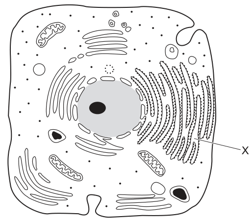
What is the function of the membrane system labelled X?
Options
A lipid synthesis only
B protein synthesis and transport
C protein synthesis only
D protein transport only
Working
X labels the rough endoplasmic reticulum (RER) — a network of flattened membranes whose cytoplasmic surface is studded with ribosomes (the small dots shown on the membrane). The attached ribosomes are the site of protein synthesis, and the RER membrane system then transports these newly synthesised proteins through its lumen and packages them into vesicles that bud off to carry the proteins to the Golgi apparatus.
Answer
B
B
Background Concept
The endoplasmic reticulum (ER) is a system of flattened membrane-bound sacs (cisternae) continuous with the outer nuclear envelope. It exists in two forms:
- Rough ER (RER) — studded with ribosomes on its cytoplasmic face. The ribosomes synthesise proteins, which are threaded into the lumen of the RER as they are made. The RER then transports and begins to modify (e.g. by glycosylation and folding) these proteins before packaging them into transport vesicles that bud off to the Golgi apparatus.
- Smooth ER (SER) — lacks ribosomes. It is the site of lipid and steroid synthesis, and also handles detoxification and Ca²⁺ storage in some cell types.
Because the RER carries out both the synthesis of proteins (at its surface ribosomes) and their transport (through the lumen and into vesicles), its function cannot be reduced to either activity alone.
Understanding the Question
The diagram shows a typical animal cell with several membrane systems visible — a stack of curved cisternae on the right (Golgi apparatus), a network nearer the nucleus studded with small dots, and mitochondria elsewhere. The label X points specifically to the ribosome-studded membrane network, i.e. the rough endoplasmic reticulum. The question asks for its function, and the four options test whether the candidate knows the full role of the RER or only part of it.
Approach
- Identify the organelle X is pointing to by its visible features: flattened cisternae with dots (ribosomes) on the outer surface, located near the nucleus. This is the rough ER.
- Recall the functions of the rough ER: (a) provides a surface for ribosomes to attach and synthesise proteins; (b) transports those proteins through the lumen and into vesicles destined for the Golgi.
- Select the option that names both functions — that is the complete and therefore correct answer.
Step-by-Step Reasoning
- Option A (lipid synthesis only) describes the function of the smooth ER, not the rough ER. The RER's defining feature — ribosomes — is irrelevant to lipid synthesis. Reject.
- Option C (protein synthesis only) captures half the story. The attached ribosomes do synthesise proteins, but those proteins must then be transported through the membrane system to reach their destination. Saying "protein synthesis only" under-describes the RER. Reject.
- Option D (protein transport only) captures the other half. The RER does transport proteins, but it is also the site where those proteins are first made (by the surface ribosomes). Reject.
- Option B (protein synthesis and transport) captures both halves together. The ribosomes attached to the RER synthesise proteins, and the RER membrane network then transports them. This is the complete description of the RER's role in the secretory pathway. Accept.
Key Takeaways
- The dots on a membrane in a cell diagram represent ribosomes and identify the structure as rough ER.
- Rough ER has a dual function: it hosts protein synthesis (via attached ribosomes) and transports those proteins onwards to the Golgi.
- Always pick the option that names the complete function of an organelle — questions like this are designed so that each "only" or single-activity option is a deliberate trap.
Common Mistakes
- Confusing rough and smooth ER and choosing the lipid-synthesis option (A) — lipid synthesis is the job of the SER, which has no ribosomes.
- Choosing an option that lists only one of the RER's two functions (C or D). The RER is not just a place for ribosomes to sit; the membrane network itself plays an active role in moving and modifying proteins.
- Confusing the RER with the Golgi apparatus. The Golgi is shown as a separate stack of curved cisternae on the right of the diagram and is the next stop in the pathway, not the site of initial protein synthesis.
Things to Be Careful About
- Read the option wording exactly: the words "only" and "and" are doing real work. "Protein synthesis and transport" (B) is correct; "protein synthesis only" (C) and "protein transport only" (D) are each incomplete.
- Ribosomes free in the cytoplasm also make proteins, but proteins destined for secretion, lysosomes, or the plasma membrane are made on the RER, which is why the RER is integral to the secretory pathway — synthesis and onward transport in one location.
What describes a lysosome?
Options
A a vesicle containing enzymes, enclosed by a double membrane, that is budded off the endoplasmic reticulum
B a vesicle containing hydrolytic enzymes and surrounded by a single membrane, found only in phagocytes
C a vesicle enclosed by a single membrane, containing several different hydrolytic enzymes that may act inside or outside the cell
D a vesicle surrounded by a double membrane, containing enzymes which can hydrolyse damaged organelles in a cell
Answer
A lysosome is a vesicle bounded by a single membrane, containing several different hydrolytic enzymes. These enzymes can digest material taken into the cell (inside the cell, within the vesicle) and can also be released to act outside the cell (e.g. in extracellular digestion of material released by, or damaged on, the cell surface).
C
C
Background Concept
A lysosome is a membrane-bound organelle found in eukaryotic cells. It is essentially a small spherical vesicle whose lumen contains a cocktail of hydrolytic enzymes (acid hydrolases) that work best at an acidic pH (around pH 4.5–5.0), maintained by H⁺ pumps in the surrounding membrane.
Three structural points are central to this question:
- Number of membranes. A lysosome is bounded by a single phospholipid bilayer. It is not a double-membrane organelle like the nucleus, mitochondrion or chloroplast.
- Contents. It contains a variety of hydrolytic enzymes (proteases, lipases, nucleases, glycosidases, etc.), capable of breaking down proteins, lipids, nucleic acids and carbohydrates.
- Origin. Lysosomes are formed by the budding of vesicles from the Golgi apparatus (not the endoplasmic reticulum), although hydrolytic enzymes are synthesised on the rough endoplasmic reticulum and then transported via the Golgi.
Functionally, lysosomes digest:
- material taken into the cell by endocytosis or phagocytosis (intracellular digestion);
- the cell's own damaged organelles during autophagy;
- and, when the lysosome fuses with the plasma membrane, material outside the cell (extracellular digestion, e.g. osteoclasts digesting bone matrix, or sperm acrosome enzymes digesting the zona pellucida — although the acrosome is technically a related lysosome-derived vesicle).
Lysosomes are present in most eukaryotic cells, not only phagocytes. Although phagocytes (e.g. macrophages, neutrophils) have particularly abundant lysosomes because they engulf and destroy pathogens, almost all cells that need to recycle macromolecules possess them.
Understanding the Question
The stem asks which statement describes a lysosome. The command word is "describes", so the answer must correctly identify both a structural feature (membrane, contents) and a functional feature (where the enzymes act). Each distractor contains one or more correct-sounding ideas combined with at least one factual error.
Approach
Compare each option point-by-point against the three core facts (single membrane; several hydrolytic enzymes; acts inside or outside the cell). Eliminate any option that conflicts on any of these points.
Step-by-Step Reasoning
- Option A claims lysosomes are "enclosed by a double membrane, that is budded off the endoplasmic reticulum". The membrane number is wrong (it is single), and the origin is also wrong (they bud from the Golgi). → Reject.
- Option B says "surrounded by a single membrane, found only in phagocytes". The membrane part is correct, but the "only in phagocytes" restriction is wrong — lysosomes occur in most eukaryotic cells. → Reject.
- Option C states "a vesicle enclosed by a single membrane, containing several different hydrolytic enzymes that may act inside or outside the cell". All three components match: single membrane, multiple hydrolytic enzymes, action both intra- and extracellularly. → Correct.
- Option D claims a "double membrane, containing enzymes which can hydrolyse damaged organelles". The double-membrane claim is wrong; also, this option only describes autophagy and ignores extracellular roles. → Reject.
Hence the correct option is C.
Key Takeaways
- A lysosome = single-membrane vesicle + several hydrolytic enzymes.
- Functions span intracellular digestion, autophagy, and (after exocytosis) extracellular digestion.
- Lysosomes are formed from the Golgi, not the ER.
- Lysosomes are widely distributed in eukaryotic cells — they are not unique to phagocytes.
Common Mistakes
- Confusing lysosomes with double-membrane organelles (mitochondria, chloroplasts, nucleus).
- Attributing the "only in phagocytes" restriction; phagocytes do have many lysosomes, but so do most other cell types.
- Saying lysosomes bud from the rough endoplasmic reticulum; the enzymes are made on the rER, but the lysosome itself is a Golgi-derived vesicle.
- Describing lysosomes purely as the cell's "recycling centre" without acknowledging the extracellular role.
Things to Be Careful About
- "Hydrolytic enzymes" is the precise term — not just "enzymes", because not all enzymes hydrolyse substrates.
- Always check membrane number in organelle descriptions: a single error on this point invalidates an option.
- "May act inside or outside the cell" is the breadth that distinguishes C from a narrower but technically true answer about autophagy alone.
The diagram shows a triglyceride.
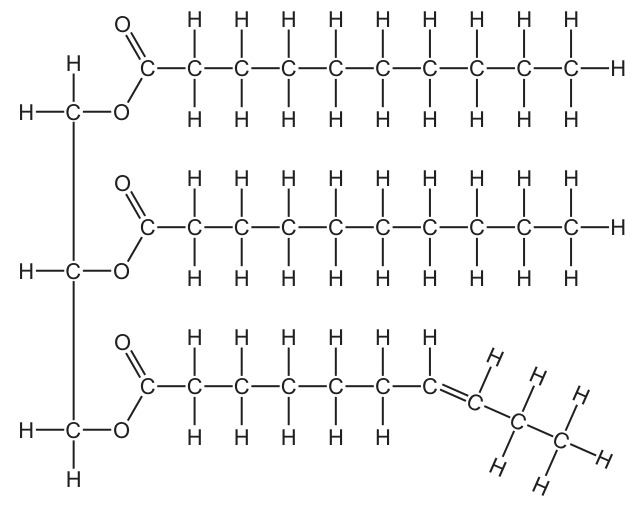
What will be the products after hydrolysis of this triglyceride?
Options
A 1 molecule of glycogen, 3 unsaturated fatty acids and 3 molecules of water
B 1 molecule of glycerol, 2 saturated fatty acids and 1 unsaturated fatty acid
C 1 molecule of glycogen, 2 saturated fatty acids and 1 unsaturated fatty acid
D 1 molecule of glycerol, 1 saturated fatty acid, 2 unsaturated fatty acids and 3 molecules of water
Working
A triglyceride consists of a glycerol backbone bonded to three fatty acid chains via ester bonds. Hydrolysis of each ester bond cleaves the linkage, regenerating the free fatty acid and the –OH on glycerol. Water is consumed (a reactant), not released.
Reading Fig. 6.1:
- The top two chains contain only C–C single bonds → 2 saturated fatty acids.
- The bottom chain contains one C=C double bond → 1 unsaturated fatty acid.
- The three-carbon backbone is glycerol (not glycogen, which is a polysaccharide).
Answer
B
B
Background Concept
A triglyceride is a lipid made of one glycerol molecule esterified to three fatty acid chains. Each fatty acid is joined to glycerol through an ester bond formed by a condensation reaction (in which a molecule of water is released). Triglycerides are therefore the main storage lipid in animals and plants, and are broken down during digestion by lipase enzymes.
Fatty acid terminology:
- A saturated fatty acid has only C–C single bonds in its hydrocarbon chain; it is "saturated" with hydrogen.
- An unsaturated fatty acid contains at least one C=C double bond, which introduces a kink in the chain and lowers the melting point.
Hydrolysis is the reverse of condensation. Adding a molecule of water across each ester bond splits the bond, regenerating the original –OH groups. The products of complete hydrolysis of a triglyceride are therefore:
Water is a reactant, not a product.
Understanding the Question
This is a Paper 1 multiple choice item. The candidate is shown a drawn triglyceride (Fig. 6.1) and must:
- Identify the monomer backbone (glycerol) as distinct from any carbohydrate (e.g. glycogen).
- Count how many of the three chains are saturated versus unsaturated by inspecting the C–C bonds.
- Recognise the products of hydrolysis (water is consumed, not produced).
The command word is "what will be the products", so the answer is a list of the chemical species released, not a description of the mechanism.
Approach
First, translate the drawing into chemical facts:
- The three-carbon vertical backbone = glycerol.
- The two upper chains are straight (no kinks, all C–C) = saturated fatty acids.
- The lower chain has a C=C (drawn as a double line between two carbons, producing a kink) = unsaturated fatty acid.
- Then apply the hydrolysis rule: ester bonds are split by water, releasing one glycerol + three fatty acids, with water as a reactant.
Finally, scan the four options:
- Options A and C mention glycogen, an immediate disqualifier — glycogen is a glucose polymer, not related to triglyceride hydrolysis.
- Options A and D include 3 molecules of water as a product — also wrong, because water is consumed in hydrolysis.
- Option B lists 1 glycerol + 2 saturated fatty acids + 1 unsaturated fatty acid, with no water, exactly matching the drawn molecule and the chemistry.
Step-by-Step Reasoning
-
Identify the backbone. The vertical C–C–C structure in Fig. 6.1 is a three-carbon alcohol, glycerol. Glycogen is a branched polymer of glucose, and would not appear in a lipid structure.
-
Classify the three chains.
- Top chain: all C–C single bonds → saturated.
- Middle chain: all C–C single bonds → saturated.
- Bottom chain: contains a C=C double bond (visible as a double line) → unsaturated.
-
Apply hydrolysis stoichiometry. Three ester bonds must be broken, so the products are 1 glycerol + 3 fatty acids. No water molecules are produced; in fact three water molecules are consumed.
-
Match to the options. Only option B is consistent: 1 glycerol, 2 saturated fatty acids, 1 unsaturated fatty acid, with no water produced.
Key Takeaways
- A triglyceride = glycerol + 3 fatty acids (linked by 3 ester bonds).
- Hydrolysis is the reverse of condensation: water is a reactant, not a product.
- Saturated vs unsaturated fatty acids is decided by the presence/absence of C=C double bonds in the chain.
- "Glycogen" in a lipid answer is a clear red flag — it is a polysaccharide, not a lipid subunit.
Common Mistakes
- Choosing A or C by confusing glycerol (a 3-carbon alcohol) with glycogen (a branched glucose polymer). These are very different molecules.
- Choosing A or D by thinking hydrolysis releases water; the opposite is true — water is added across each ester bond.
- Counting the chains incorrectly by misreading the double bond in the bottom chain and calling it saturated (or missing the double bond entirely and counting three saturated chains).
Things to Be Careful About
- Read the structural formula carefully: a C=C double bond is shown as two parallel lines between the carbons and usually causes a visible kink in the chain.
- "Hydrolysis" always means water is consumed to break a bond. If an option lists water as a product of hydrolysis, it is wrong (it would be a product of a condensation reaction).
- Glycerol has three carbons; glycogen is a large polymer — they cannot be the same molecule.
Which description lists all the components of a human haemoglobin molecule?
Options
A four polypeptides that are all the same and one haem group
B four polypeptides that are not all the same and one haem group
C four polypeptides that are all the same and four haem groups
D four polypeptides that are not all the same and four haem groups
Working
Human adult haemoglobin (HbA) has a quaternary structure made of:
- 4 polypeptide chains — specifically -chains and -chains, so the four polypeptides are not all the same
- 4 haem groups — one non-protein prosthetic group per polypeptide chain, each containing a central ion that binds one molecule
Matching these facts to the options:
- A — wrong: chains are not all the same, and there are 4 haem groups, not 1
- B — wrong: there are 4 haem groups, not 1
- C — wrong: the chains are not all the same (2α + 2β)
- D — correct: four polypeptides that are not all the same AND four haem groups
Answer
D
D
Background Concept
Haemoglobin is the oxygen-transport protein inside red blood cells and the textbook example of a globular protein with quaternary structure. Quaternary structure means a functional protein is built from more than one polypeptide chain held together by hydrogen bonds, ionic bonds and hydrophobic interactions between R groups.
The common adult form, HbA, is made of four polypeptide subunits:
- 2 α-globin chains (141 amino acids each)
- 2 β-globin chains (146 amino acids each)
Because the α and β chains have different amino acid sequences, the four polypeptides are not all the same. (Foetal haemoglobin, HbF, is 2α + 2γ — still two chain types, not all the same.)
Nestled inside a hydrophobic pocket of each polypeptide is a prosthetic haem group: a flat porphyrin ring with a central ion. Each reversibly binds one molecule, so the stoichiometry is one haem group per polypeptide chain. A complete haemoglobin therefore carries four haem groups, and up to four molecules.
Understanding the Question
The command word "lists" is a low-demand word here — the candidate just has to pick the option whose description fully matches the composition of a human haemoglobin molecule. Two facts must be correct simultaneously:
- The number and identity of the polypeptide chains (four, but not all the same)
- The number of haem groups (four)
A wrong choice on either fact loses the mark.
Approach
Recall the two structural facts above and test each option against both. Any option that is wrong on either count is rejected; the one that is right on both is the answer.
Step-by-Step Reasoning
- Eliminate options that say the polypeptides are all the same. Adult haemoglobin has 2α + 2β — two chain types — so "all the same" is incorrect. This rules out A and C.
- Eliminate options that say there is only one haem group. Each of the four chains has its own haem group, giving four haem groups per molecule. This rules out A and B.
- The only option consistent with both facts is D: four polypeptides that are not all the same and four haem groups.
Key Takeaways
- Adult human haemoglobin = polypeptide chains + 4 haem groups.
- The four chains are not all identical → this is the basis of haemoglobon's quaternary structure.
- Each chain binds one haem group → one per haem → up to four per haemoglobin.
- A common confusion is with myoglobin, the single-chain, single-haem oxygen-storage protein in muscle. Haemoglobin ≠ myoglobin.
Common Mistakes
- Saying all four chains are identical — wrong; the α- and β-chains differ in length and amino acid sequence.
- Saying there is only one haem group — wrong; each of the four chains has its own haem prosthetic group.
- Confusing haemoglobin with myoglobin. Myoglobin has one polypeptide and one haem; haemoglobin has four of each.
- Assuming "not all the same" implies more than two types — only two types exist (α and β), but they are not all the same, which is all the question needs.
Things to Be Careful About
- The question asks about human haemoglobin. Sickle-cell haemoglobin (HbS) is also 2α + 2β and still has four haem groups, so the structural count is unchanged even when the β-chain carries a point mutation.
- The haem group is a prosthetic (non-protein) component, but it is still part of the whole haemoglobin molecule and must be counted in this kind of question.
- The 1:1 stoichiometry between haem groups and molecules is why haemoglobin's oxygen-binding curve plateaus at four per Hb and shows the characteristic sigmoidal (S-shaped) cooperativity.
HIV-1 protease is an enzyme produced by the HIV virus.
Two identical chains of 99 amino acids form the enzyme. In each chain, amino acids 25, 26 and 27 in the sequence form part of the active site.
Which orders of protein structure control the shape of the active site?
Options
A primary, secondary, tertiary and quaternary
B primary, secondary and tertiary only
C primary and quaternary only
D quaternary only
Working
The shape of the active site depends on how the polypeptide chain folds and how chains associate.
- Primary structure determines the sequence of amino acids (including 25, 26, 27), which in turn dictates all higher levels of folding.
- Secondary structure (α-helices and β-pleated sheets, held by hydrogen bonds) contributes the local folding patterns that position the active-site residues.
- Tertiary structure is the overall 3-D folding of each single chain, which directly creates the active-site pocket.
- Quaternary structure is the association of the two identical chains; in HIV-1 protease the active site lies at the interface between the two subunits, so the dimer arrangement is essential for the active-site shape.
All four levels of structure therefore control the shape of the active site.
Answer
A
A
Background Concept
Proteins have up to four levels of structure:
- Primary (1°) structure — the linear sequence of amino acids joined by peptide bonds. The side-chain (R-group) chemistry of each residue is set here, and the sequence dictates every higher level of folding.
- Secondary (2°) structure — local, regular folding patterns such as α-helices and β-pleated sheets, stabilised mainly by hydrogen bonding between the backbone N–H and C=O groups.
- Tertiary (3°) structure — the overall 3-D folding of a single polypeptide chain, held by interactions between R-groups: hydrogen bonds, ionic bonds, disulfide bridges and hydrophobic interactions.
- Quaternary (4°) structure — the assembly of two or more polypeptide chains (subunits) into the functional protein. Not all proteins have quaternary structure (e.g. myoglobin is monomeric); those that do are often called multimeric — dimeric, trimeric, etc.
An enzyme's active site is the pocket where substrate binds. Its precise 3-D shape depends on the side chains being held in exactly the right positions, which in turn depends on every level of structure that contributes to that geometry.
Understanding the Question
The stem tells us that HIV-1 protease:
- is made of two identical chains (a homodimer) of 99 amino acids;
- has amino acids 25, 26 and 27 of each chain contributing to the active site.
The question asks which orders of protein structure control the shape of the active site. The candidate must decide whether one, several, or all four levels are needed.
Approach
Work through each level and ask: "Does this level contribute to the geometry of the active site in this enzyme?" If yes, include it. Because this enzyme is a dimer, the quaternary-structure question is the key discriminator between options A and B.
Step-by-Step Reasoning
-
Primary structure — yes. Without the correct sequence (including residues 25–27) the side chains in the active site simply would not be there. Primary structure is the template for everything above it. The active-site shape cannot be defined without it. Include 1°.
-
Secondary structure — yes. α-helices and β-sheets in each subunit position the active-site residues relative to one another. Local folding patterns therefore contribute to the geometry of the pocket. Include 2°.
-
Tertiary structure — yes. The overall 3-D fold of each single chain creates the cleft into which the substrate fits. Tertiary structure is what physically sculpts the active site in a single-subunit enzyme, and it does the same here within each chain. Include 3°.
-
Quaternary structure — yes, and this is the crucial point. HIV-1 protease is a homodimer: two identical chains associate. The active site is formed at the interface between the two subunits, with residues 25–27 from each chain contributing. The dimer arrangement (quaternary structure) is what brings those residues from the two chains into the correct relative positions to form a functional active site. If the two chains did not associate, there would be no active site. Include 4°.
All four orders of protein structure are required → option A.
Why the other options are wrong
- B (primary, secondary, tertiary only) — this would be correct for a monomeric enzyme. It misses the fact that, in this dimer, the active site is completed only when the two chains come together, which is quaternary structure.
- C (primary and quaternary only) — ignores the local (2°) and overall (3°) folding that actually shape the pocket within each chain.
- D (quaternary only) — the active site is not produced by subunit association alone; the chains must first be correctly folded at the 1°, 2° and 3° levels.
Key Takeaways
- An active site is a 3-D feature, so it is influenced by every level of structure that contributes to 3-D shape.
- A dimeric (or any multimeric) enzyme has its active site completed by the association of subunits, so quaternary structure is essential — this is the common discriminator in MCQs of this type.
- "Control the shape" means "have an effect on the geometry" — and primary structure controls everything else because it dictates the sequence that folds.
Common Mistakes
- Choosing B because tertiary structure "makes the active site" — forgetting that, in a multimeric protein, the active site is at a subunit interface and so also requires quaternary structure.
- Choosing D because the question emphasises "two identical chains" — assuming the chains alone, without internal folding, are enough. A chain must first fold (1°–3°) before it can contribute to a functional interface.
- Choosing C by mixing up which level is most "important" rather than which levels are actually required.
Things to Be Careful About
- "Identical chains" is a clue that the enzyme is a homodimer and therefore has quaternary structure.
- "Part of the active site" rather than "the active site" is deliberate wording — it signals that residues from more than one chain contribute, reinforcing the need for quaternary structure.
- A common exam trap: assume quaternary structure is only relevant if the question mentions multiple, different chains. In fact, even identical chains associating count as quaternary structure.
Which statements are correct reasons for how animals cool down in hot environments using latent heat of vaporisation of water?
1 Some animals lie down and roll in wet soil.
2 Fish move into deeper water.
3 Some animals lick their fur to make it wet.
4 Some animals breathe quickly with a wet tongue hanging out of their mouth.
Options
A 1, 2 and 3
B 1, 2 and 4
C 1, 3 and 4
D 2, 3 and 4
Working
Latent heat of vaporisation is the energy required to convert water from liquid to gas. When water evaporates from a surface, it absorbs this energy (as heat) from that surface, causing cooling.
Evaluate each statement:
- Lying in wet soil – water from the soil evaporates from the animal's body surface, absorbing latent heat → cools the animal. ✓
- Fish moving into deeper water – this is behavioural avoidance of warmer surface water; no evaporation is involved because the fish is submerged. ✗
- Licking fur to make it wet – the saliva evaporates from the fur, absorbing latent heat → cools the animal. ✓
- Panting with a wet tongue – water on the tongue and moist surfaces of the mouth/upper respiratory tract evaporates, absorbing latent heat → cools the animal. ✓
Correct statements: 1, 3 and 4.
Answer
C
C
Background Concept
Latent heat of vaporisation is the large amount of energy required to change water from a liquid to a gas (vapour) without a change in temperature. At body temperature (~37 °C for mammals), the latent heat of vaporisation of water is approximately — an enormous amount of energy per unit mass. When water evaporates from a surface, this energy must come from the surroundings — i.e. from the body of the animal — so the surface that loses the water is cooled.
For evaporative cooling to occur, two conditions must be met:
- There must be liquid water present on (or accessible to) the body surface where it can evaporate.
- The water must be able to evaporate — i.e. it must not be in an environment that is already saturated with water vapour, and the animal must have a route for the vapour to escape.
Many mammals exploit this principle behaviourally (e.g. licking fur, panting) to lose heat when environmental temperature approaches or exceeds body temperature and radiation/convection become ineffective.
Understanding the Question
The question presents four animal behaviours, each of which is loosely related to dealing with heat. It asks specifically which behaviours achieve cooling by evaporation of water (using its latent heat of vaporisation). The keyword here is the mechanism — only behaviours that involve water evaporating count; behaviours that simply move the animal to a cooler place, or rely on conduction/convection, are not examples of evaporative cooling.
This is a "select correct statements" MCQ; the answer is the option that includes every correct statement and excludes every incorrect one.
Approach
For each numbered statement, ask:
- Is water present in a form that can evaporate from the animal's surface?
- If so, will that evaporation absorb latent heat from the animal and therefore cool it?
If yes → the statement is a correct reason. If the behaviour cools the animal by a different mechanism (e.g. moving to a cooler environment) or doesn't involve evaporation, it is wrong.
Step-by-Step Reasoning
Statement 1 — Lying in wet soil.
The soil is wet, so the animal's body surface (and fur, if present) becomes coated with water. That water then evaporates into the air, absorbing latent heat from the animal's body. ✓ Correct reason.
Statement 2 — Fish moving into deeper water.
Deeper water is usually cooler than surface water in a hot environment, so the fish does become cooler. However, this cooling is achieved by moving to a cooler environment — the fish is already in water, so no evaporation is occurring (the surrounding water is essentially at the same temperature as the fish and cannot accept more water vapour to any meaningful extent). This is conductive/environmental avoidance, not evaporative cooling. ✗ Incorrect reason for this question.
Statement 3 — Licking fur to make it wet.
The saliva deposited on the fur is water. As it evaporates, it absorbs latent heat from the skin and fur, cooling the animal. This is a well-known behaviour (e.g. in dogs, kangaroos) that exploits evaporative cooling. ✓ Correct reason.
Statement 4 — Panting with a wet tongue.
Many mammals (dogs, some marsupials) pant with the tongue out. The tongue is kept wet with saliva. As the animal breathes quickly, air flows over the wet tongue, evaporating the water and absorbing latent heat from the tongue and from blood circulating close to the surface. This is a classic example of evaporative cooling. ✓ Correct reason.
Statements 1, 3 and 4 are correct; statement 2 is not. The only option containing exactly 1, 3 and 4 is C.
Key Takeaways
- Latent heat of vaporisation of water is a powerful cooling mechanism because so much energy is needed to turn liquid water into vapour.
- For evaporation to cool an animal, water must be on or near a body surface that is in contact with air that is not saturated with water vapour.
- Be careful to distinguish evaporative cooling (uses latent heat) from conduction/convection cooling (heat transfer to a cooler environment by direct contact or by movement of air/water) and from behavioural avoidance (simply moving away from heat).
- Submerged aquatic animals cannot use evaporative cooling for thermoregulation; they rely on behavioural selection of cooler water.
Common Mistakes
- Choosing option B (1, 2, 4) by including statement 2. Students may think "the fish is in cooler water so it's cooler" without noticing that the question specifies evaporative cooling. Fish do not cool by evaporation.
- Confusing sweating/panting with the wrong mechanism. Sweating and panting cool the body because water evaporates from the skin/mouth surface, taking latent heat with it. They are not cooling the body by "exhaling hot air" — although some heat is lost in the warm exhaled air, the dominant mechanism is evaporation.
- Assuming licking fur is for cleaning or grooming only. In many mammals, fur-licking is a deliberate thermoregulatory behaviour that uses saliva evaporation.
- Forgetting that latent heat of vaporisation applies to any water source on the body — soil water, saliva, sweat, urine or pond water all work the same way.
Things to Be Careful About
- Read the command word carefully: this question specifically asks for reasons using latent heat of vaporisation, not just any cooling method.
- Do not be misled by the realism of a behaviour — a behaviour may genuinely help an animal cope with heat (e.g. fish going deeper) but still be wrong for this question because it uses a different mechanism.
- For "select correct statements" MCQs, the right answer is the one that includes all and only the correct statements; an option missing one correct statement is wrong, even if it includes no incorrect ones.
Two different enzymes, P and Q, are investigated to find the optimum pH for each enzyme. The results show that P works only in acidic conditions. Q has an optimum pH which is slightly alkaline.
Which graph shows the correct results for P and Q?
Options
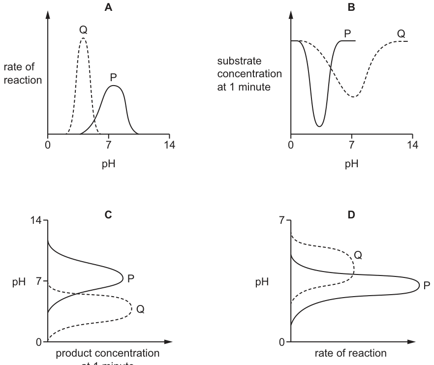
Working
Enzyme P works only in acidic conditions, so its highest rate must occur at pH < 7. Enzyme Q has an optimum pH that is slightly alkaline, so its highest rate must occur at pH > 7.
- A: Q peaks at acidic pH and P peaks at pH 7 — both wrong.
- B: Substrate concentration at 1 minute is on the y-axis. A dip (low remaining substrate) corresponds to a fast reaction. P dips at acidic pH and Q dips at slightly alkaline pH — correct.
- C: P peaks at pH 7 (neutral), not acidic — wrong for P.
- D: The pH axis only extends to 7, so it cannot show Q's alkaline optimum — wrong.
Answer
B
B
Background Concept
Enzymes are globular proteins whose catalytic activity depends on the precise 3-D shape of their active site. Each enzyme has an optimum pH at which the ionisation state of the active-site amino acid side chains (e.g. –COOH, –NH₂) and of the substrate allows the enzyme–substrate complex to form most readily. Away from the optimum, hydrogen and hydroxide ions disrupt ionic and hydrogen bonds in the active site, distorting its shape and lowering the reaction rate. Extreme pH usually denatures the enzyme permanently.
Different enzymes are adapted to the conditions where they normally work:
- Pepsin (stomach) has an optimum around pH 2 (very acidic).
- Trypsin (small intestine) has an optimum around pH 8 (slightly alkaline).
- Most cytoplasmic enzymes work near pH 7.
The question describes two enzymes:
- P — only active in acidic conditions (optimum pH < 7).
- Q — optimum slightly alkaline (optimum pH just above 7).
Understanding the Question
We are shown four graphs (A–D), each plotting some measure of enzyme activity against pH. We must identify the graph whose shape is consistent with P peaking (highest activity) at acidic pH and Q peaking (highest activity) at a slightly alkaline pH, with the full pH scale 0–14 displayed so both optima are visible.
The command word is implicit ("Which graph shows…"), so the answer is simply the letter of the correct option. The marks reward choosing the right graph, not lengthy reasoning.
Approach
For each option, check two things:
- Does the axis range cover both acidic and alkaline pH? (It must reach above 7, since Q is slightly alkaline.)
- Where does each curve reach its maximum activity? (P must peak at pH < 7; Q must peak just above pH 7.)
The subtle trap is graphs that use an inverted measure of rate: graph B plots the substrate concentration remaining after 1 minute on the y-axis. A lower value on this axis means more substrate has been converted to product, which means a faster reaction. So the maxima of rate are the minima (dips) of the curve in B, not the peaks.
Step-by-Step Reasoning
Graph A — Rate of reaction vs pH (0–14).
- Curve Q peaks at pH < 7 (acidic).
- Curve P peaks at pH ≈ 7 (neutral).
- This is the reverse of what the question states. Reject.
Graph B — Substrate concentration at 1 minute vs pH (0–14).
- The y-axis is remaining substrate, so a low point = high rate.
- Curve P dips (lowest remaining substrate, hence highest rate) at pH < 7 ✓ — matches "P works only in acidic conditions".
- Curve Q dips at pH > 7, slightly alkaline ✓ — matches "Q has an optimum pH which is slightly alkaline".
- The full pH range 0–14 is shown, so both optima are visible.
- Accept: B.
Graph C — pH (y) vs product concentration at 1 minute (x).
- The axes are transposed (pH on the y-axis), but the underlying shapes can still be read.
- P peaks at pH ≈ 7 (neutral) — this does not satisfy "P works only in acidic conditions".
- Q peaks at slightly alkaline pH — correct, but P is wrong, so the whole option is wrong.
- Reject.
Graph D — pH (y, 0–7) vs rate of reaction (x).
- The pH axis only extends from 0 to 7, so the graph is physically incapable of showing Q's alkaline optimum. Even if the shapes within the range were correct, the data for Q is missing.
- Reject.
Only graph B correctly displays both enzymes' optima on an appropriate pH range.
Key Takeaways
- The shape of a rate–pH curve is determined by where the enzyme's active-site ionisation state is optimal.
- A "low remaining substrate" or "high product formed" at a given pH both indicate a fast reaction — be alert to which way the y-axis runs.
- When judging a graph, always check both the location of the maximum and the range of the independent variable.
Common Mistakes
- Reading the y-axis in graph B the wrong way and concluding that the peaks (high remaining substrate) are where the enzymes are fastest. The dips are the maxima of rate.
- Choosing A because it has the "classic" bell-shape; this confuses P and Q's optima.
- Choosing D because the curves look reasonable within the limited pH range; D cannot show an alkaline optimum and so is incomplete.
- Confusing P and Q: the labels (acidic P, alkaline Q) must be tracked carefully because the names in graphs A and C are swapped relative to the question.
Things to Be Careful About
- "Slightly alkaline" means pH just above 7 (e.g. 7.5–8), not strongly alkaline (e.g. pH 12).
- "Only in acidic conditions" means P should have negligible activity at pH 7 and above — its curve should fall to baseline before pH 7.
- An axis that does not span 0–14 cannot fully characterise both an acidic and a (slightly) alkaline optimum.
A student investigated the effect of substrate concentration on the rate of an enzyme-catalysed reaction.
The student plotted the results in a graph.
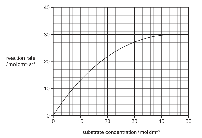
What is the for this enzyme-catalysed reaction?
Options
A
B
C
D
Working
is the substrate concentration at which the reaction rate equals .
From the graph:
Reading across from on the y-axis to the curve and down to the x-axis gives a substrate concentration of approximately .
is a concentration, so its units are (not ).
Answer
A
A
Background Concept
For many enzymes, the relationship between reaction rate () and substrate concentration () follows the Michaelis–Menten model:
As increases, rises steeply at first (almost first-order kinetics) and then plateaus at a maximum rate, , where the enzyme is saturated — every active site is occupied and the rate is limited only by how fast the enzyme can turn substrate into product.
Two key constants describe the curve:
- — the maximum (plateau) rate.
- — the Michaelis constant, defined as the substrate concentration at which the reaction rate is exactly half of . It is an inverse measure of an enzyme's affinity for its substrate: a low means the enzyme reaches half-maximum speed at a low (high affinity); a high means it needs a lot of substrate to get there (low affinity).
Critically, is a concentration, not a rate, so it carries the same units as (typically ). It is not measured in .
Understanding the Question
The question gives a Michaelis–Menten curve with reaction rate (y-axis, ) plotted against substrate concentration (x-axis, ). The curve plateaus at . We must read off the substrate concentration at which the rate equals half this plateau value.
The distractor options all share the same number (, , , ) but mix up concentration and rate units. The biology is in knowing that is read off the x-axis (a concentration) at a specific y-value (half of ).
Approach
- Find from the plateau of the curve.
- Calculate .
- Move horizontally from this rate to the curve, then vertically down to the x-axis to read the substrate concentration — that concentration is .
- Make sure the chosen option has concentration units (), not rate units.
Step-by-Step Reasoning
Step 1 —
The curve flattens at the right-hand side at a reaction rate of . So .
Step 2 — Half
Step 3 — Find at this rate on the curve
Draw a horizontal line from on the y-axis across to the curve. Where it meets the curve, drop a vertical line down to the x-axis. The reading there is about .
Step 4 — Check the units
is a substrate concentration, so its units are .
Therefore , which matches option A.
Why the other options are wrong:
- B () — this is the rate at which is read off the curve, not a concentration. The units are also rate units, not concentration units.
- C () — this is itself, again with rate units.
- D () — this is just the upper limit of the x-axis; the curve has already plateaued well before this point.
Key Takeaways
- is the substrate concentration at which .
- is always a concentration, with the same units as the substrate concentration axis.
- A small = high enzyme–substrate affinity; a large = low affinity.
- When asked to read from a graph: find , halve it, then go across to the curve and down to the x-axis.
Common Mistakes
- Giving a rate (with ) for . is a concentration; rate units belong on , not on .
- Reporting as . These are two different constants; is the maximum rate, is the substrate concentration that gives half of it.
- Picking an x-value at the plateau. The curve flattens around , so any value in that range is well past .
Things to Be Careful About
- Always draw the construction lines on the graph (horizontal at , then vertical down to the x-axis) — eyeballing the curve at the half-way height of the plateau is not accurate enough.
- Quote with the same units as the x-axis. If the x-axis is in , the is in , not .
Which statements could be used to describe enzyme molecules and antibody molecules?
1 Hydrogen bonds stabilise the structure of the protein and are important for it to function efficiently.
2 Hydrophilic R-groups point in to the centre of the molecule and cause it to curl into a spherical shape.
3 The tertiary structure of the protein molecule plays an important role in the functioning of the protein.
Options
A 1, 2 and 3
B 1 and 2 only
C 1 and 3 only
D 2 and 3 only
Working
Enzymes and antibodies are both globular proteins, so any correct statement must apply to both.
Statement 1: Hydrogen bonds are one of the interactions that stabilise the tertiary structure of globular proteins. This specific 3D shape is required for the active site of an enzyme and the antigen-binding site of an antibody to work efficiently. True for both.
Statement 2: In a globular protein, hydrophobic (not hydrophilic) R-groups point inwards to the centre of the molecule, while hydrophilic R-groups point outwards into the surrounding aqueous environment. This statement has the orientation reversed. False for both.
Statement 3: The function of both enzymes (substrate binding at the active site) and antibodies (antigen binding at the variable region) depends on the specific 3D shape of the tertiary structure. True for both.
Statements 1 and 3 only apply to both enzyme and antibody molecules.
Answer
C
C
Background Concept
Enzymes and antibodies are both globular proteins — proteins whose polypeptide chain folds into a compact, roughly spherical shape. This folding is driven by the behaviour of the R-groups (side chains) on the amino acids:
- Hydrophobic R-groups are repelled by water and so cluster inwards, away from the aqueous cytoplasm or tissue fluid.
- Hydrophilic R-groups are attracted to water and so project outwards into the surrounding environment, often forming further hydrogen bonds with water molecules.
This R-group distribution produces the tertiary structure — the overall 3D shape of a single polypeptide chain. The tertiary structure is held in place by several types of bond and interaction, including hydrogen bonds, ionic bonds, disulfide bridges and hydrophobic interactions.
The 3D shape of a globular protein is not just incidental — it is the very thing that allows the protein to function:
- In an enzyme, the active site is a specific pocket of shape and charge complementary to the substrate.
- In an antibody (immunoglobulin), the variable regions at the tips of the Y-shape form antigen-binding sites whose shape is complementary to a specific epitope on an antigen.
Hydrogen bonds are particularly important: although individually weak, in large numbers they hold α-helices, β-pleated sheets and the overall fold in place. Disrupting hydrogen bonds (e.g. by heat or extremes of pH) causes denaturation, where the tertiary structure is lost and the protein can no longer function.
Understanding the Question
This MCQ asks which statements correctly describe both enzyme molecules and antibody molecules. Because both are globular proteins, the test is really about which statements are true of globular proteins in general — with one statement (Statement 2) deliberately containing an error to catch the unwary.
The command word is implicit but clear: identify which statements could be applied to BOTH protein types.
Approach
Take each statement in turn, decide whether it is true for a globular protein in general, and therefore true for both enzymes and antibodies. Use the distinction between hydrophobic and hydrophilic R-group behaviour as the key test for Statement 2.
Step-by-Step Reasoning
Statement 1 — "Hydrogen bonds stabilise the structure of the protein and are important for it to function efficiently."
- Hydrogen bonds form between slightly δ⁺ hydrogen atoms and slightly δ⁻ oxygen or nitrogen atoms. In a globular protein, they are abundant between backbone C=O and N–H groups (stabilising α-helices and β-sheets) and between polar R-groups.
- These bonds collectively hold the tertiary structure in the precise shape needed for the active site of an enzyme or the antigen-binding site of an antibody to be functional.
- Verdict: TRUE for both.
Statement 2 — "Hydrophilic R-groups point in to the centre of the molecule and cause it to curl into a spherical shape."
- This is the classic trap. The statement is back to front:
- Hydrophilic (water-loving) R-groups point outwards, towards the surrounding water, where they can form hydrogen bonds with water molecules.
- Hydrophobic (water-fearing) R-groups point inwards, away from water — this is what drives the polypeptide to fold into a compact, spherical shape (the hydrophobic effect).
- Because the statement swaps these two, it is false.
- Verdict: FALSE for both.
Statement 3 — "The tertiary structure of the protein molecule plays an important role in the functioning of the protein."
- Enzyme function depends on the specific 3D shape of the active site (lock-and-key or induced-fit).
- Antibody function depends on the specific 3D shape of the antigen-binding site at the tips of the variable regions.
- Both lose function if the tertiary structure is disrupted by denaturation.
- Verdict: TRUE for both.
So the correct statements are 1 and 3 only, which corresponds to option C.
Key Takeaways
- Both enzymes and antibodies are globular proteins whose function depends on tertiary structure.
- In globular proteins: hydrophobic R-groups point INWARDS; hydrophilic R-groups point OUTWARDS — never the reverse.
- Hydrogen bonds (and ionic, disulfide and hydrophobic interactions) stabilise tertiary structure; losing them = denaturation = loss of function.
Common Mistakes
- Reversing hydrophilic and hydrophobic R-group positions in Statement 2 — this is the most common error and the one the question is designed to test. Remember: "philic = outward-facing (loves water outside), phobic = inward-facing (fears water outside)."
- Assuming antibodies are not really proteins — they are; immunoglobulins are globular proteins with quaternary structure (four polypeptide chains held together by disulfide bonds).
- Forgetting that hydrogen bonds are individually weak but collectively important, so the statement is correct even though one H-bond by itself would not stabilise a whole protein.
Things to Be Careful About
- Read each statement precisely — Statement 2 contains a single wrong word ("hydrophilic" instead of "hydrophobic") that flips the answer.
- The question demands statements that apply to BOTH enzymes and antibodies, not just one. Always check both before selecting.
- A descriptive answer about proteins in general can still be wrong if one detail is reversed, as in Statement 2.
Which description identifies a reversible, non-competitive enzyme inhibitor?
Options
A It can attach to the active site.
B It can attach to a site other than the active site.
C It can attach to the active site and another site simultaneously.
D It can attach to either the active site or another site.
Working
A non-competitive inhibitor binds reversibly to a site on the enzyme other than the active site (an allosteric site), changing the shape of the active site so the substrate can no longer bind. It does not occupy the active site itself.
- A — describes a competitive inhibitor (binds the active site).
- B — correct: a non-competitive inhibitor attaches to a site other than the active site.
- C — wrong: it binds one site at a time, not both simultaneously.
- D — wrong: this would describe an inhibitor that can act by either mechanism, not a defined non-competitive one.
Answer
B
B
Background Concept
Enzymes are globular proteins with an active site whose specific shape is complementary to the substrate. Enzyme inhibitors are molecules that reduce enzyme activity, and they fall into two main categories defined by WHERE they bind and HOW they interact with the enzyme.
- Competitive inhibitors have a shape similar to the substrate and bind to the active site directly, blocking substrate access. Their effect can be overcome by increasing substrate concentration.
- Non-competitive inhibitors bind to a site other than the active site (an allosteric site). This binding alters the tertiary structure of the enzyme, distorting the active site so the substrate can no longer bind effectively. Increasing substrate concentration does NOT overcome the effect because the inhibitor and substrate are not competing for the same site.
Inhibitors are also classified as reversible (they bind by weak interactions and can dissociate) or irreversible (they form strong covalent bonds and permanently disable the enzyme). A reversible, non-competitive inhibitor therefore binds at an allosteric site via weak interactions and can detach, restoring enzyme function.
Understanding the Question
This is a multiple-choice question (Paper 1 style) asking the candidate to pick the single statement that correctly describes a reversible, non-competitive inhibitor. The command word is "identifies", so the answer should pinpoint the defining location of binding. The data given is the four options themselves.
Approach
Recall the two defining features of a non-competitive inhibitor:
- It binds somewhere other than the active site.
- Its binding is reversible (by weak interactions such as hydrogen bonds and van der Waals forces).
The question emphasises "reversible" and "non-competitive", so the answer must be the option that correctly captures the binding location. Walk through each option against the definition.
Step-by-Step Reasoning
- Option A — "It can attach to the active site." This is the defining feature of a competitive inhibitor, not a non-competitive one. Reject.
- Option B — "It can attach to a site other than the active site." This is the textbook description of a non-competitive inhibitor: it binds to an allosteric site. Because the stem specifies "reversible", the binding is via weak, non-covalent interactions, so the inhibitor can detach and the enzyme can resume function. Accept.
- Option C — "It can attach to the active site and another site simultaneously." A non-competitive inhibitor binds to ONE specific allosteric site; it does not simultaneously occupy both the active site and another site. The "simultaneously" clause makes this description physically wrong. Reject.
- Option D — "It can attach to either the active site or another site." This describes a hypothetical inhibitor that can act by either mechanism, which is not the definition of a non-competitive inhibitor. A non-competitive inhibitor ONLY binds away from the active site. Reject.
Key Takeaways
- A non-competitive inhibitor binds to a site other than the active site (allosteric site).
- A competitive inhibitor binds to the active site.
- Reversible inhibitors bind through weak interactions and can dissociate; irreversible inhibitors form permanent (usually covalent) bonds.
- Increasing substrate concentration overcomes competitive inhibition but NOT non-competitive inhibition (since the inhibitor and substrate do not compete for the same site).
Common Mistakes
- Confusing non-competitive with competitive inhibition and choosing A (active site binding).
- Choosing D because it sounds "flexible" — but the question asks for the description of a non-competitive inhibitor specifically, which only binds the allosteric site.
- Thinking that "reversible" implies the inhibitor can move between sites — reversibility refers to the ability to dissociate from its single binding site, not to swap between sites.
Things to Be Careful About
- Read the question carefully: "reversible, non-competitive" specifies BOTH properties. Reversibility on its own would also fit competitive inhibitors, so the binding site is the key distinguishing feature here.
- "Allosteric site" is the precise term for a site other than the active site; using "another site" in the answer is acceptable as long as the meaning is clear.
Before mitochondria are extracted from cells for microscopy, they are usually kept in a sucrose solution.
Why is the sucrose solution used?
Options
A to act as a solvent
B to enable the rate of reaction of the mitochondria to be determined
C to prevent the mitochondria from changing in dimension
D to provide a source of energy
Working
The sucrose solution at is approximately isotonic with the contents of the mitochondrion (i.e. it has the same water potential, , as the mitochondrial matrix). Because water potential is equal inside and outside, there is no net movement of water across the mitochondrial membranes by osmosis, so the organelle does not swell or shrink and its dimensions are preserved for microscopy.
Answer
C
C
Background Concept
When a cell or organelle is placed in a solution, water moves across its partially permeable membrane by osmosis. The direction and extent of this movement depend on the water potential () gradient between the inside of the organelle and the surrounding solution:
- In a hypotonic solution (higher/less negative outside than inside), water enters the organelle, causing it to swell and possibly burst (lysis).
- In a hypertonic solution (lower/more negative outside than inside), water leaves the organelle, causing it to shrink and its membrane to shrivel (crenation in animal cells, plasmolysis in plant cells).
- In an isotonic solution, inside equals outside, so there is no net movement of water and the organelle retains its normal shape and size.
Sucrose is used because it is a small, soluble molecule that does not cross biological membranes readily. A sucrose solution therefore acts as an effective osmotic agent: it sets the water potential of the external medium without entering the organelle. The concentration of is empirically the value that is isotonic with the cytoplasm of many cells, including the matrix of mitochondria isolated from typical mammalian tissues such as liver.
Understanding the Question
The question describes the first step in preparing mitochondria for microscopy — extracting them from cells. Before the organelle can be observed, it must be kept in a medium that does not damage it. The question asks why the standard medium used in this procedure is sucrose, and offers four possible reasons, only one of which is correct.
The key biological idea is that isolating an organelle from its native cytoplasm exposes it to a new external solution; if that solution is not water-potential balanced, the organelle will gain or lose water and change shape. Because microscopy is used to study the structure and dimensions of mitochondria, those dimensions must be preserved.
Approach
The approach is to evaluate each option against the property of the sucrose solution:
- A (solvent): water is the solvent in most cell-fractionation media; sucrose is a solute, not a solvent — this is wrong.
- B (enable rate of reaction): sucrose is not a substrate for mitochondrial respiration, and the question concerns microscopy, not respirometry — wrong.
- C (prevent change in dimension): the sucrose solution is isotonic, so no osmotic water movement changes the organelle's size or shape — this matches the biology.
- D (source of energy): sucrose is a disaccharide and not directly used by isolated mitochondria; the preparation is for microscopy, not to fuel respiration — wrong.
The correct reasoning rests on the isotonic, osmotically protective role of the sucrose medium.
Step-by-Step Reasoning
- The mitochondrion is bounded by a double membrane that is partially permeable to water.
- If the mitochondrion is placed in pure water, the external is while the internal is negative (e.g. around ); water rushes in, the organelle swells, and its dimensions distort.
- If placed in a much more concentrated solution, the mitochondrion loses water and shrinks, again distorting its dimensions.
- The sucrose solution has a water potential that approximately matches that of the mitochondrial matrix, so across the membranes and there is no net osmotic flow of water.
- With no net water movement, the mitochondrion retains its normal shape, size and internal structure, allowing accurate microscopy.
- Therefore the function of the sucrose solution is to keep the dimensions of the mitochondria unchanged — option C.
Key Takeaways
- Isolated organelles must be kept in an isotonic medium to prevent osmotic damage during cell fractionation.
- A sucrose solution is the standard isotonic medium for mitochondria because sucrose does not cross the membrane and its matches that of the mitochondrial matrix.
- Distortion of organelle dimensions by osmosis would make microscopy observations unreliable.
Common Mistakes
- Choosing A because sucrose "dissolves" things — sucrose is the solute; the solvent is water, which is already present.
- Choosing B because mitochondria carry out aerobic respiration — but the sucrose is not metabolised by isolated mitochondria in a microscopy prep, and the question is about preserving structure, not measuring respiration.
- Choosing D because sucrose is a sugar and therefore "energy-providing" — a common conflation; isolated mitochondria in microscopy buffers are not respiring, and sucrose is not their direct substrate anyway.
Things to Be Careful About
- Make sure the answer uses the precise idea of isotonic / equal water potential, not vague phrases like "same concentration as the cell".
- The sucrose is acting osmotically (it cannot cross the membrane), not chemically — do not describe it as a nutrient or fuel in this context.
- The question is about preserving dimensions (size and shape) for microscopy; answers about "preventing damage" in general are too vague to score the mark.
The photomicrograph shows a type of blood cell.
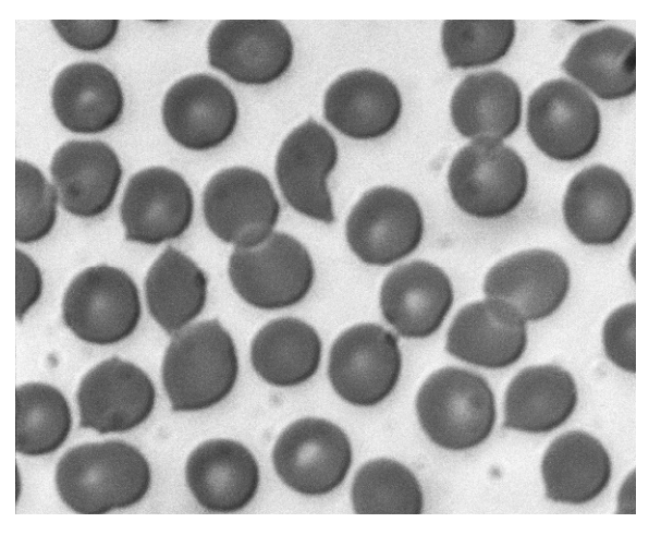
Which statements about these cells are correct?
1 Oxygen diffuses through the phospholipid bilayer.
2 Sodium ions diffuse through the phospholipid bilayer.
3 Water passes in and out of these cells by osmosis.
Options
A 1, 2 and 3
B 1 and 2 only
C 1 and 3 only
D 2 and 3 only
Working
The photomicrograph shows red blood cells (erythrocytes) — biconcave discs with no nucleus.
- Statement 1: O₂ is a small, non-polar molecule, so it diffuses through the phospholipid bilayer directly. ✓ Correct.
- Statement 2: Na⁺ is a charged ion; it cannot pass through the hydrophobic phospholipid bilayer and must pass via channel/carrier proteins. ✗ Incorrect.
- Statement 3: Water crosses the plasma membrane by osmosis (via aquaporins and to a small extent through the bilayer). ✓ Correct.
Answer
C
C
Background Concept
The plasma membrane is described by the fluid mosaic model. Its fundamental structure is a phospholipid bilayer: phospholipid molecules have hydrophilic phosphate heads facing the aqueous environment on either side, and hydrophobic fatty-acid tails pointing inward. This creates an internal hydrophobic core.
The chemical nature of a substance determines whether it can cross this hydrophobic core unaided:
- Small, non-polar molecules (e.g. O₂, CO₂, steroid hormones) dissolve in the bilayer and diffuse straight through it.
- Charged ions and large polar molecules (e.g. Na⁺, K⁺, Cl⁻, glucose, amino acids) cannot pass through the hydrophobic core and require channel proteins (facilitated diffusion) or carrier proteins (facilitated diffusion or active transport).
- Water is a small polar molecule. It crosses the membrane by osmosis — mostly through specialised channel proteins called aquaporins, and to a limited extent directly through the bilayer.
The cells in Fig. 15.1 are red blood cells (erythrocytes) — biconcave discs about 7–8 µm across, with no nucleus, packed with haemoglobin. Their membranes are typical plasma membranes and obey these general permeability rules.
Understanding the Question
The question gives a photomicrograph of red blood cells and asks which of three statements about how substances cross the membrane of these cells are correct. Each statement is a claim about a different transport mechanism, so each must be evaluated against the permeability rules above.
Approach
For each statement, ask: what kind of molecule is being described, and does that type of molecule cross the phospholipid bilayer unaided?
- O₂ → small non-polar → yes, through bilayer.
- Na⁺ → charged ion → no, needs protein channels.
- Water → moves by osmosis → yes.
Step-by-Step Reasoning
Statement 1 — "Oxygen diffuses through the phospholipid bilayer."
O₂ is small and non-polar. The hydrophobic interior of the bilayer does not impede it, and O₂ dissolves in and crosses the membrane by simple diffusion. Correct.
Statement 2 — "Sodium ions diffuse through the phospholipid bilayer."
Na⁺ carries a positive charge and is surrounded by a hydration shell of water molecules. It cannot pass through the hydrophobic fatty-acid tails of the bilayer directly. In reality, Na⁺ crosses red-cell membranes via specific channel proteins (e.g. the epithelial sodium channel, ENaC) or is pumped by the Na⁺/K⁺-ATPase. Incorrect as written because the statement says it crosses the phospholipid bilayer (i.e. without a protein).
Statement 3 — "Water passes in and out of these cells by osmosis."
Water moves across the red-cell membrane by osmosis — the net movement of water molecules from a region of higher water potential to a region of lower water potential across a partially permeable membrane. In red cells this is mainly through aquaporin-1 (AQP1) channels, with a small component of direct diffusion through the bilayer. Correct.
So statements 1 and 3 are correct and 2 is not — answer C.
Key Takeaways
- The phospholipid bilayer is freely permeable to small non-polar molecules (O₂, CO₂) but impermeable to ions and large polar molecules.
- Ions (Na⁺, K⁺, Cl⁻, Ca²⁺) always require membrane transport proteins — they never diffuse "through the phospholipid bilayer" by themselves.
- Water crosses membranes by osmosis, primarily through aquaporins.
- Whenever an MCQ says an ion "diffuses through the phospholipid bilayer", treat it as wrong unless a specific protein-mediated route is also described.
Common Mistakes
- Picking A because all three statements describe real processes (oxygen does diffuse, sodium ions do cross the membrane, water does move by osmosis) — but statement 2 is wrong because it specifies through the phospholipid bilayer rather than through a protein channel.
- Picking B by assuming water does not move by osmosis in red cells — it does, very readily (this is in fact the basis for the clinical osmotic-fragility test).
- Picking D by thinking that water cannot cross a membrane that is "impermeable to ions" — the bilayer is selectively permeable: it blocks ions but lets water through.
Things to Be Careful About
- Read the wording precisely: the question specifies the phospholipid bilayer in statements 1 and 2. Anything charged (ions, polar molecules) cannot cross the bilayer alone.
- "Diffuses" in the context of membranes can mean either simple diffusion through the bilayer or facilitated diffusion through proteins — but in this style of question it is used in its strict sense of passing directly through the bilayer.
- Osmosis is a special case of diffusion (of water); it is correct to say water moves by osmosis across a plasma membrane, even though a small amount also moves by simple diffusion through the bilayer.
Which row is correct for parts of a phospholipid molecule?
Options
| can be saturated or unsaturated | can also be found in a triglyceride | |
|---|---|---|
| A | head | tail |
| B | tail | head |
| C | head | head |
| D | tail | tail |
Working
A phospholipid has a hydrophilic phosphate head and two hydrophobic fatty acid tails.
- The head is a phosphate group; it is not a fatty acid, so it cannot be saturated or unsaturated, and it is not found in a triglyceride.
- The tails are fatty acid chains, which can be saturated (no C=C double bonds) or unsaturated (one or more C=C double bonds). Triglycerides are also built from fatty acid chains attached to glycerol, so the tails are the part that is shared with a triglyceride.
Answer
D
D
Background Concept
A phospholipid is built from a glycerol backbone, a phosphate group (the polar "head") and two fatty acid chains (the non-polar "tails"). Because the head is hydrophilic and the tails are hydrophobic, phospholipids arrange themselves into bilayers in aqueous environments — the structural basis of all biological membranes.
A triglyceride is built from a glycerol backbone esterified to three fatty acid chains. It contains no phosphate group, so it has no "head" in the phospholipid sense — only fatty acid tails.
Saturation describes the fatty acid tails, not the head. A fatty acid is saturated when its carbon chain contains only single C–C bonds (saturated with hydrogens, e.g. in many animal fats), and unsaturated when it contains one or more C=C double bonds, which introduce kinks into the chain (e.g. in many plant oils).
Understanding the Question
This MCQ presents a 2 × 2 grid of statements about the head and tail of a phospholipid:
- Whether that part "can be saturated or unsaturated"
- Whether that part "can also be found in a triglyceride"
The candidate must pick the row (A, B, C or D) where both statements in that row are correct.
Approach
Decide independently for each of the two parts of a phospholipid (head vs tail) which statement applies, then locate the row matching that pair.
Step-by-Step Reasoning
- Head (phosphate group):
- Saturation? No — saturation is a property of fatty acid chains, and the head is not a fatty acid. ⇒ False.
- Found in a triglyceride? No — triglycerides have glycerol + three fatty acids, no phosphate. ⇒ False.
- Tail (fatty acid chain):
- Saturation? Yes — fatty acids can be saturated or unsaturated. ⇒ True.
- Found in a triglyceride? Yes — triglycerides consist of three fatty acid chains attached to glycerol. ⇒ True.
So the correct pair is: tail and tail. Looking at the table, this is row D.
Eliminating the others:
- A (head, tail): the head is not saturated/unsaturated.
- B (tail, head): the head is not in a triglyceride.
- C (head, head): both statements about the head are wrong.
Key Takeaways
- The phospholipid "head" is the phosphate group; the "tails" are the two fatty acid chains.
- Saturation/unsaturation applies to fatty acid chains, not to phosphate.
- Both phospholipids and triglycerides contain fatty acid chains, but only phospholipids contain a phosphate group.
Common Mistakes
- Thinking the phosphate head is a fatty acid and can therefore be "saturated or unsaturated" — saturation is defined by C–C single vs C=C double bonds in hydrocarbon chains, not by the phosphate group.
- Confusing the structure of a triglyceride (3 fatty acids + glycerol) with a phospholipid (2 fatty acids + phosphate + glycerol), and so wrongly selecting "head" for the part found in a triglyceride.
- Picking B because the tail is correctly identified as saturated/unsaturated, without checking the second column.
Things to Be Careful About
- "Saturated/unsaturated" is a property of the fatty acid tails only, never of the head.
- The shared feature between phospholipids and triglycerides is the fatty acid chain (the tail), not the glycerol or the head — glycerol is the backbone in both, but the question is about which part of the phospholipid reappears in a triglyceride, and the distinctive shared component is the fatty acid chain.
- The mark scheme accepts only row D; partial credit is not available on a single-letter MCQ.
A student filled dialysis tubing with a sucrose solution and knotted both ends. This formed a cylinder with a length of and a radius of .
What is the surface area to volume ratio for this cylinder of dialysis tubing?
Options
A
B
C
D
Working
For a cylinder of length and radius :
Substituting and :
Answer
C
C
Background Concept
The surface area to volume ratio (SA:V) is a key idea in biology because it governs how efficiently a cell or small structure can exchange materials (such as gases, nutrients and waste) with its surroundings across its surface. As an object gets larger, its volume grows faster than its surface area, so its SA:V falls. Single-celled organisms and many exchange surfaces in larger organisms (alveoli, villi, root hairs) keep SA:V high by being small or having elaborate shapes.
A cylinder is a useful simple model for a tube-like structure such as dialysis tubing, a capillary or a piece of gut. The two key formulas are:
- Surface area (including both circular ends):
- Volume:
Understanding the Question
We are told a dialysis-tubing sac has been tied into a cylinder shape. Its length (height) is and its radius is . The question asks us to work out the SA:V ratio of this cylinder.
The command word is "What is…", and the four options are all expressed as ratios to , so we just need to compute the numerical SA:V and pick the matching option.
Approach
Apply the standard cylinder formulas, substitute and , cancel the common factor of and simplify to a single number. The factor of cancels in the ratio, so we never actually need a numerical value of .
Step-by-Step Reasoning
- Identify the two ends (each an area of ) and the curved side (circumference length ):
- Substitute and :
- Calculate the volume:
- Form the ratio. The cancels:
- Compare to the options: matches option C.
Key Takeaways
- Always include the two end caps when a structure is described as a closed cylinder (a tied-off sac of dialysis tubing is closed at both ends).
- When forming a ratio, units must agree. The cancels here, but the surface area is in and the volume in ; the ratio is genuinely dimensionless.
- The cancels in any SA:V for a cylinder, so it never needs to be evaluated.
- A SA:V of is fairly low — consistent with a relatively large object. This is why real exchange surfaces (alveoli, villi) are highly folded: to keep the effective SA:V high.
Common Mistakes
- Forgetting the two circular ends and only using the curved surface . This gives , which is option B — a classic distractor.
- Using the diameter () instead of the radius.
- Halving the surface area "because it is only exchanging through the membrane" — not what the question asks; it asks for the SA of the whole cylinder.
- Squaring the height instead of the radius in the volume formula.
Things to Be Careful About
- Read "length" and "radius" carefully — for a cylinder the "height" and "length" mean the same thing and both are measured along the axis.
- The question does not say "surface area to volume" written in the form because the answer is greater than 1; the correct SA:V is , not (option A, which would arise from accidentally dividing volume by surface area).
- Show enough working to confirm the formula, but the ratio itself is what the mark rewards.
Which process does not involve mitosis?
Options
A asexual reproduction
B growth of unicellular organisms
C repair of tissues by cell replacement
D replacement of damaged or dead cells
Working
Mitosis is required for:
- Asexual reproduction (e.g. binary fission, budding) — produces genetically identical new individuals.
- Repair of tissues — new cells replace damaged ones.
- Replacement of damaged or dead cells — new cells are produced.
A unicellular organism grows by increasing the size of its single cell through the uptake of nutrients and metabolic activity, not by cell division. Mitosis in a unicellular organism produces a new individual, not a larger version of the existing one.
Answer
B
B
Background Concept
Mitosis is the division of a nucleus to produce two genetically identical daughter nuclei. It is followed by cytokinesis, which divides the cytoplasm. In multicellular organisms mitosis is essential for increasing cell number during growth, replacing worn-out or damaged cells, and repairing tissues. In unicellular organisms (and in asexual reproduction generally) mitosis is the mechanism that produces a new individual from a pre-existing one.
However, the word growth has two distinct meanings:
- Growth in size of a cell — an increase in mass/volume through the synthesis of new cytoplasm, organelles and macromolecules driven by metabolism. No division is involved.
- Growth in number — an increase in the number of individuals or cells, which does require cell division.
Understanding the Question
The question asks which option describes a process that does not use mitosis. We must read each option carefully and decide whether it involves nuclear division.
- A — Asexual reproduction: e.g. budding in yeast, binary fission in Amoeba. The parent cell divides to produce a new, genetically identical individual. ✓ Mitosis.
- B — Growth of unicellular organisms: a single-celled organism increases in size by taking up nutrients and synthesising more cytoplasm. It does not divide to grow larger. ✗ No mitosis.
- C — Repair of tissues by cell replacement: damaged tissue is restored by producing new cells from neighbouring ones that divide. ✓ Mitosis.
- D — Replacement of damaged or dead cells: new cells are produced to replace those that have been lost (e.g. red blood cells, skin cells, gut epithelial cells). ✓ Mitosis.
Approach
Identify the key distinction: mitosis is about producing new cells, not about making existing cells bigger. A unicellular organism grows larger without dividing; only when it reproduces does it undergo mitosis.
Step-by-Step Reasoning
- Mitosis produces two daughter cells from one parent cell, each genetically identical to the parent.
- In option A, asexual reproduction requires a parent to produce offspring, so mitosis is used.
- In options C and D, repair and replacement both depend on the production of new cells, so mitosis is used.
- In option B, "growth of unicellular organisms" refers to a single cell getting larger — this is an increase in cell size via metabolism, not cell division, so mitosis is not required.
- Therefore B is the process that does not involve mitosis.
Key Takeaways
- Mitosis is for cell multiplication, not for cell enlargement.
- Unicellular organisms grow in size by metabolic accumulation of material; they divide by mitosis only to reproduce.
- Asexual reproduction, tissue repair, and cell replacement all depend on mitotic cell division.
Common Mistakes
- Confusing the growth of a unicellular organism (increase in cell size) with the population growth of unicellular organisms (increase in cell number). The question uses the singular sense — "growth of unicellular organisms" — meaning how an individual cell gets bigger.
- Assuming all four options must involve mitosis because the topic is "the mitotic cell cycle". The question deliberately tests the exception.
Things to Be Careful About
- Read the precise wording: "growth of unicellular organisms", not "growth of multicellular organisms" (which would involve mitosis in tissues such as the meristems or growth plates).
- Distinguish between a process and its result. Mitosis is a process of nuclear division; the question is asking which listed scenario is not brought about by that process.
The photomicrograph shows cells in an onion root tip.
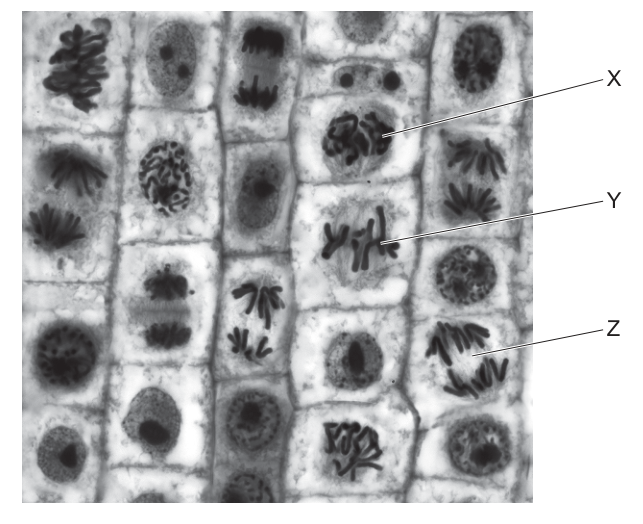
Which labelled cells are in anaphase, metaphase and prophase?
Options
| anaphase | metaphase | prophase | |
|---|---|---|---|
| A | X | Y | Z |
| B | Y | Z | X |
| C | Z | X | Y |
| D | Z | Y | X |
Working
Look at the chromosome arrangement in each labelled cell:
- X — chromosomes are condensed and visible as thread-like structures scattered in the nucleus, with no nuclear envelope. This is prophase.
- Y — chromosomes are lined up along the equator (metaphase plate) of the cell, each attached to spindle fibres. This is metaphase.
- Z — sister chromatids have separated and are being pulled to opposite poles of the cell, forming two distinct groups. This is anaphase.
So: anaphase = Z, metaphase = Y, prophase = X.
Answer
D
D
Background Concept
Mitosis is a continuous process, but biologists divide it into four named stages based on the appearance and position of the chromosomes. In an onion root tip, which is a classic site for studying mitosis because the cells at the tip are actively dividing, you can see all the stages side by side in a single longitudinal section. The key events to recognise are:
- Prophase — the chromatin condenses into visible, thick, thread-like chromosomes (each consisting of two sister chromatids joined at the centromere). The nuclear envelope breaks down and a spindle begins to form.
- Metaphase — the chromosomes, still consisting of two sister chromatids, are pulled by spindle fibres and line up along the equator (the metaphase plate) of the cell, perpendicular to the two poles.
- Anaphase — the centromeres split and the sister chromatids are pulled apart, moving towards opposite poles of the cell. They now appear as two separated groups of single-chromatid chromosomes, often V- or J-shaped because the centromere leads the way.
- Telophase — chromatids arrive at the poles, decondense, and new nuclear envelopes form; cytokinesis usually follows.
Understanding the Question
This is a multiple-choice identification question. The photomicrograph (Fig. 19.1) shows several onion root tip cells in different stages of mitosis, with three of them labelled X, Y and Z. You are asked to match each label to the correct stage name (anaphase, metaphase or prophase) and then select the option that lists the three matches in the requested order: anaphase, metaphase, prophase.
The command word is implicit — "identify" — so the mark depends on correctly reading the chromosome arrangement in each cell.
Approach
The reliable way to tell these three stages apart on a micrograph is to ask, for each labelled cell, two questions in order:
- Are the chromosomes in one group or in two groups? One group = prophase or metaphase; two groups (moving apart) = anaphase.
- If one group, is it arranged in a line (equator) or scattered? A line across the middle = metaphase; scattered with no clear order = prophase.
Step-by-Step Reasoning
- Cell X: the chromosomes appear as a tangled, condensed mass within the cell. They are not aligned and have not separated. This is prophase.
- Cell Y: the chromosomes form a clear single line/row across the middle of the cell, at right angles to a line drawn between the two poles. This single equatorial alignment is the defining feature of metaphase.
- Cell Z: the chromosomes are clearly in two distinct groups at opposite ends of the cell, with the chromatids pointing back towards the equator — the classic V-shape produced by centromeres being pulled poleward first. This is anaphase.
Substituting into the requested order: anaphase = Z, metaphase = Y, prophase = X. That corresponds to row D in the table of options.
Key Takeaways
- Prophase = condensed, scattered chromosomes; nuclear envelope gone.
- Metaphase = chromosomes lined up at the equator (one neat row).
- Anaphase = sister chromatids separated, forming two groups moving to opposite poles.
- On a micrograph, focus on chromosome position and grouping, not on the rest of the cell, which can look similar.
Common Mistakes
- Confusing anaphase with telophase: both show two groups of chromosomes, but in telophase the chromosomes have reached the poles and begun to decondense, and a new nuclear envelope may be visible. In anaphase the two groups are still moving and the chromatids are still condensed.
- Confusing prophase with interphase cells: in true interphase the chromosomes are NOT visible as discrete threads (the nucleus shows a more uniform appearance with a nucleolus), whereas in prophase the chromosomes are condensed and clearly visible.
- Mistaking a side-on view of metaphase (where chromosomes form a line) for anaphase — anaphase always shows two groups, not one.
Things to Be Careful About
- Always look at more than one feature: chromosome shape (two chromatids vs one), chromosome position (scattered / equatorial / two groups) and whether a nuclear envelope or nucleolus is visible.
- In onion root tip preparations, cells are not all synchronised, so neighbouring cells in the same field of view are usually in different stages — use this to your advantage by comparing each labelled cell against its neighbours.
- The question is asking for the order anaphase, metaphase, prophase in the options, not the biological order of the stages. Read the column headings carefully before selecting.
The diagram shows an outline of the mitotic cell cycle.
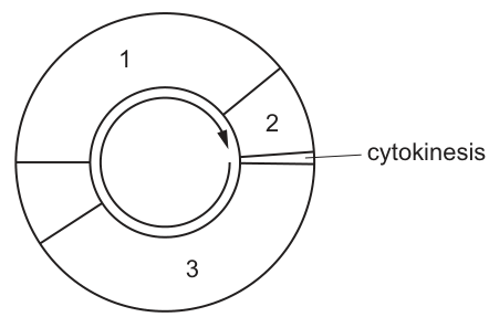
Which numbered stages of the cell cycle include a period of time when each chromosome consists of sister chromatids joined by a centromere?
Options
A 1 and 2
B 1 and 3
C 2 and 3
D 2 only
Working
In the cell cycle diagram:
- Stage 1 = part of interphase that includes the S phase (DNA replication) and G2. After S phase, each chromosome consists of two sister chromatids joined at the centromere, and this persists through G2.
- Stage 2 = mitosis. During prophase and metaphase, chromosomes are condensed with sister chromatids joined at the centromere. Chromatids only separate at anaphase, so sister chromatids are present for most of stage 2.
- Stage 3 = G1 of interphase. DNA replication has not yet occurred, so each chromosome is a single chromatid — no sister chromatids are present.
Therefore, stages 1 and 2 both include a period when each chromosome consists of sister chromatids joined by a centromere.
Answer
A
A
Background Concept
A chromosome is a single, long DNA molecule packaged with histone proteins. Before DNA replication, each chromosome is a single chromatid — one DNA double helix. During S phase of interphase, the DNA is replicated semi-conservatively, producing two identical DNA double helices attached at a constricted region called the centromere. From this point on, the chromosome is described as having two sister chromatids joined at the centromere.
Sister chromatids persist as a joined pair through:
- The rest of interphase (G2)
- Prophase
- Metaphase
- The early part of anaphase (before separation)
They finally separate at anaphase, when the centromere splits and each chromatid is pulled to an opposite pole, becoming an individual chromosome in its own right.
During G1 of interphase (before S phase) and at the very end of telophase/cytokinesis (after separation), each chromosome exists as a single chromatid only.
Understanding the Question
The question shows a circular cell cycle diagram with three numbered stages and asks which of them include any period during which the chromosomes exist as sister chromatids joined at the centromere. The mark-scheme image description indicates that:
- Stage 1 = part of interphase (S phase and G2)
- Stage 2 = mitosis (with cytokinesis following)
- Stage 3 = G1 of interphase
The command word is implicit — we need to decide which option correctly identifies the stages that contain sister chromatids at some point within them.
Approach
The strategy is to map each labelled stage onto the underlying molecular events:
- Does the stage include the S phase (when chromatids are first produced)?
- Or does the stage include prophase/metaphase/early anaphase (when joined chromatids are visible)?
If yes to either, the stage is part of the answer. If the stage consists only of G1 (before replication), it is not part of the answer.
Step-by-Step Reasoning
Stage 1 (S phase + G2 of interphase): DNA replication occurs during S phase, so from the moment S phase begins until the end of G2, every chromosome is composed of two sister chromatids held together at the centromere. Stage 1 therefore does include a period of joined sister chromatids.
Stage 2 (Mitosis): During prophase, chromosomes condense and each clearly shows two sister chromatids joined at the centromere. They remain joined through metaphase, lined up at the equator. They only separate at the start of anaphase, when the centromere divides. Therefore, throughout prophase, metaphase, and the start of anaphase, sister chromatids are present. Stage 2 does include a period of joined sister chromatids.
Stage 3 (G1 of interphase): This is the gap phase before DNA replication. No replication has occurred, so each chromosome is a single, unreplicated chromatid. There are no sister chromatids to be joined. Stage 3 does not include a period of joined sister chromatids.
The stages that qualify are 1 and 2, corresponding to option A.
Key Takeaways
- Sister chromatids are created during S phase of interphase and persist as a joined pair through G2, prophase, and metaphase.
- They separate at anaphase, becoming individual chromosomes.
- G1 is the only phase during which no sister chromatids exist.
- A cell cycle diagram divides interphase into sub-phases; you must identify which numbered region corresponds to G1 (the unreplicated state) to answer questions like this.
Common Mistakes
- Choosing B (1 and 3): Treating stage 3 as if it contained replicated chromosomes. Stage 3 is G1 — replication has not yet happened.
- Choosing C (2 and 3): Assuming G1 still contains replicated chromosomes. G1 occurs before S phase.
- Choosing D (2 only): Forgetting that S phase already produces sister chromatids, so part of interphase (stage 1) also contains them.
Things to Be Careful About
- A chromosome with sister chromatids is often described in textbooks as an X-shaped structure — the cross is the centromere, the two arms of the X are the chromatids.
- After anaphase, the separated chromatids are no longer called sister chromatids; they are individual (single-chromatid) chromosomes.
- The terms "chromatid" and "chromosome" can be confusing because a single chromatid before replication and a single chromatid after separation are both legitimate uses; the key is the joined, two-chromatid configuration that exists between S phase and anaphase.
- The diagram's numbering is non-standard — stage 3 is the G1 portion only, not the whole of interphase — so do not assume the largest section is G1; instead, rely on the image's description.
The diagram shows part of a nucleic acid molecule.
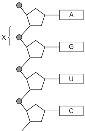
What is the name of bond X?
Options
A glycosidic bond
B hydrogen bond
C peptide bond
D phosphodiester bond
Working
In a nucleic acid, adjacent nucleotides are joined by a covalent bond between the phosphate group of one nucleotide and the sugar (ribose in RNA) of the next. This bond is called a phosphodiester bond.
- A glycosidic bond joins sugars to other groups in carbohydrates.
- A hydrogen bond joins complementary bases across the two strands of DNA (or within RNA secondary structure), not the backbone.
- A peptide bond joins amino acids in proteins.
Answer
D
D
Background Concept
A nucleic acid (DNA or RNA) is a polymer of nucleotides. Each nucleotide has three components:
- a pentose sugar (deoxyribose in DNA, ribose in RNA)
- a phosphate group attached to the 5' carbon of the sugar
- a nitrogenous base attached to the 1' carbon of the sugar (A, G, C, T in DNA; A, G, C, U in RNA)
Nucleotides are linked into a chain by a covalent bond that forms between the phosphate group of one nucleotide and the 3' carbon of the sugar of the next nucleotide. Because this single linkage involves a phosphate flanked by two ester bonds (one to each sugar), it is called a phosphodiester bond. The repeating sugar-phosphate-sugar-phosphate chain forms the sugar-phosphate backbone of the nucleic acid.
A different kind of bond, the hydrogen bond, holds the two strands of DNA together across the middle, between complementary base pairs (A–T, 2 H-bonds; G–C, 3 H-bonds). Hydrogen bonds are weak and easily broken; the phosphodiester bonds of the backbone are strong covalent bonds that give the strand its structural integrity.
Understanding the Question
Fig. 21.1 shows a single strand of RNA (U is present, confirming RNA rather than DNA). Four nucleotides are drawn with their sugars shown as pentagons, phosphates as small circles, and the bases labelled A, G, U, C. Label X points to the bond between the phosphate of one nucleotide and the sugar of the next — i.e. the bond that builds the sugar-phosphate backbone.
The command word is "name", and the question requires identifying which of the four named bond types corresponds to this linkage.
Approach
Recognise the position of bond X in the diagram (between a phosphate and a sugar, joining two adjacent nucleotides along the backbone). Match this to the known bond type for that linkage.
Step-by-Step Reasoning
- Bond X is the covalent bond between the phosphate group (small circle) of one nucleotide and the sugar (pentagon) of the next nucleotide.
- By definition, a bond between a phosphate group and two sugar hydroxyls (one on each side, hence "di-ester") is a phosphodiester bond.
- Eliminate the distractors:
- Glycosidic bond (A) — found in carbohydrates, joining one sugar to another (e.g. maltose, sucrose) or a sugar to a nitrogenous base within a single nucleotide. Not the inter-nucleotide linkage in a nucleic acid.
- Hydrogen bond (B) — joins complementary bases ACROSS the two strands of DNA, not along a single backbone. The figure shows only one strand, and X is clearly a covalent bond on the backbone.
- Peptide bond (C) — found in proteins, joining amino acids. Nucleic acids contain no peptide bonds.
- The correct answer is therefore D, the phosphodiester bond.
Key Takeaways
- The sugar-phosphate backbone of DNA and RNA is held together by phosphodiester bonds (covalent, strong).
- Complementary base pairing across two strands uses hydrogen bonds (weak, many together hold the strands but can be separated in replication/transcription).
- The bases (A, T/U, G, C) project sideways from the backbone; do not confuse a base with the backbone itself.
- Presence of U (not T) in the figure identifies the molecule as RNA, but the phosphodiester backbone chemistry is the same in both DNA and RNA.
Common Mistakes
- Choosing hydrogen bond because nucleic acids are famous for base pairing. Hydrogen bonds are between bases across the double helix, not along the backbone.
- Choosing glycosidic bond — this is the bond between the sugar and the base within a single nucleotide (the N-glycosidic bond), not between nucleotides.
- Choosing peptide bond — confusing nucleic acid structure with protein structure.
Things to Be Careful About
- Read the position of label X carefully: it sits on the sugar-phosphate-sugar linkage, NOT on a base pair.
- The molecule is RNA here (U is shown), but the bond type is identical in DNA and RNA. Do not be distracted by the U.
- The question rewards the name of the bond, so use the exact term "phosphodiester bond" in any free-response version.
XNA is a laboratory-made nucleic acid. XNA is made of nucleotides in which one component has been replaced by chemical X. The chemical X is organic but is not found in nature. The part of the molecule responsible for coding is not changed.
Which component of a DNA or RNA nucleotide has been replaced by the organic chemical X?
Options
A five-carbon sugar
B phosphate group
C purine base
D pyrimidine base
Working
A nucleotide contains three components: a five-carbon (pentose) sugar, a phosphate group, and a nitrogenous base.
- The nitrogenous bases (purines and pyrimidines) carry the genetic code, so the bases cannot be the replaced part (the 'coding' component is unchanged). This eliminates C and D.
- The phosphate group is inorganic (no carbon–hydrogen framework), so it cannot be replaced by an organic chemical X. This eliminates B.
- The five-carbon sugar is organic and is not the coding component, so it can be replaced by the organic, non-natural chemical X.
Answer
A
A
Background Concept
Every DNA and RNA nucleotide is built from three components joined together:
- A five-carbon (pentose) sugar – deoxyribose in DNA, ribose in RNA. Both are organic molecules built on a carbon framework with hydroxyl (–OH) groups.
- A phosphate group – attached to the 5′ carbon of the sugar. The phosphate group is inorganic (it contains no carbon–hydrogen framework; it is a PO₄ unit).
- A nitrogenous base – either a purine (adenine, guanine) or a pyrimidine (cytosine, thymine in DNA; cytosine, uracil in RNA). The bases are organic and it is the sequence of bases that stores the genetic code.
XNA (xeno-nucleic acid) is a family of laboratory-synthesised nucleic-acid analogues. The defining feature of an XNA is that the pentose sugar is replaced by a different, often unnatural, sugar-like molecule — for example, threose in TNA (threose nucleic acid), hexitol in HNA, or cyclohexene in CeNA. The bases are still the familiar A, T/U, G, C, so the 'coding' component of the molecule is preserved, and the polymer can still pair up in a double-helix-like fashion.
Understanding the Question
The question gives three clues about chemical X and the replaced component:
- XNA is built from nucleotides → the question is about one of the three standard nucleotide components.
- The 'part of the molecule responsible for coding is not changed' → the genetic information is carried by the sequence of bases, so the bases must still be the natural ones. This rules out the purine and pyrimidine options.
- Chemical X is organic → an organic compound contains a carbon–hydrogen framework. This rules out the phosphate group, which is inorganic.
- Chemical X is not found in nature → consistent with a laboratory-made sugar analogue.
The only component that is organic, is not the coding element, and can be swapped for a non-natural alternative is the five-carbon sugar.
Approach
Eliminate the options systematically using the two strongest clues:
- The 'coding' clue removes both base options (C and D).
- The 'organic' clue removes the phosphate option (B).
- By elimination, the replaced component must be the sugar (A).
Step-by-Step Reasoning
- A nucleotide's coding function resides in the order of its nitrogenous bases (the four bases function like letters of a genetic alphabet). Because the question states the coding part is unchanged, the bases must still be the natural purines and pyrimidines. So neither C nor D can be correct.
- The phosphate group is PO₄³⁻ — it has no carbon in it and is therefore inorganic. Because chemical X is described as organic, the phosphate cannot be the replaced component. So B is incorrect.
- The pentose sugar (deoxyribose or ribose) is organic, is not the information-carrying part of the molecule, and has known laboratory-made analogues (the very basis of XNA). Therefore the five-carbon sugar is the part replaced by chemical X.
Key Takeaways
- A nucleotide = pentose sugar + phosphate + nitrogenous base.
- The sequence of bases is what codes for proteins; the sugar–phosphate backbone is a structural scaffold.
- The phosphate group is inorganic; the sugar and bases are organic.
- XNA is a real class of molecules in which the natural pentose is replaced by an alternative sugar-like molecule while the bases are kept the same.
Common Mistakes
- Choosing C or D because 'the bases are part of the molecule' — forgetting that the question specifically says the coding part is unchanged.
- Choosing B because the phosphate 'sounds' replaceable — failing to recall that the phosphate is inorganic and so cannot be replaced by an organic chemical X.
- Confusing which component carries the code: the sugar–phosphate backbone is structural, while the bases carry the genetic information.
Things to Be Careful About
- Read both clues (organic AND coding part unchanged) before answering; either clue alone leaves two viable options, but together they uniquely identify the sugar.
- The 'organic' vs 'inorganic' distinction is a key discriminator in this question: organic = contains C–H; inorganic (like phosphate) does not.
- 'Coding' in molecular biology refers to the base sequence, not the sugar or the backbone.
Which strand of DNA does RNA polymerase bind to?
Options
A lagging strand
B leading strand
C non-transcribed strand
D template strand
Answer
During transcription, RNA polymerase binds to the template strand of DNA. This is the strand that is read in the direction to synthesise a complementary mRNA molecule (built in the direction). The other strand, the coding/sense strand, is not used as a template.
D
D
Background Concept
DNA in a cell is double-stranded, but only one of the two strands is read when a gene is transcribed. The strand that is read is called the template strand (also called the antisense strand or transcribed strand). The complementary strand is called the coding strand (or sense/non-transcribed strand), and its sequence matches the mRNA that is produced (with T in place of U).
RNA polymerase is the enzyme that catalyses transcription. It binds to a specific region of DNA (the promoter) and then moves along the template strand, reading its base sequence in the direction. As it moves, it uses the template strand as a guide to build a complementary mRNA strand, which is synthesised in the direction using complementary base pairing (A–U, T–A, G–C, C–G).
Understanding the Question
The question asks which DNA strand is bound by RNA polymerase during transcription. The four options are terms that students often confuse: "lagging/leading strand" (replication terminology, not transcription), "non-transcribed strand" (synonymous with the coding strand, which is the OPPOSITE of the template strand), and "template strand" (the correct answer).
Approach
Recall that the enzyme responsible for transcription (RNA polymerase) must use a strand as a physical template from which to read the base sequence. Identify the strand that is physically read by the polymerase and discard the other options based on what they mean in molecular biology.
Step-by-Step Reasoning
- RNA polymerase catalyses transcription, not replication. Therefore, the options "lagging strand" and "leading strand" are distractors referring to DNA replication. RNA polymerase does not bind to either of these.
- The "non-transcribed strand" is the strand that is NOT used as a template. Since RNA polymerase binds to and reads the template strand, it does NOT bind to the non-transcribed strand. Option C is therefore incorrect.
- The template strand (also called the antisense strand) is the strand that RNA polymerase binds to. The polymerase reads this strand's bases in the direction and synthesises a complementary mRNA in the direction.
- Therefore, the correct answer is D — template strand.
Key Takeaways
- RNA polymerase binds to and reads the template (antisense) strand during transcription.
- The template strand is read in the direction; mRNA is synthesised in the direction.
- The other strand (coding/sense/non-transcribed strand) has the same sequence as the mRNA (with T instead of U) but is not the one used as a template.
- "Leading/lagging strand" are terms from DNA replication, not transcription — a common source of confusion.
Common Mistakes
- Choosing A or B (leading/lagging strand): these describe how DNA polymerase replicates the two strands of DNA during replication, not what RNA polymerase does during transcription.
- Choosing C (non-transcribed strand): this is the coding/sense strand, which is NOT read by RNA polymerase; it has the same sequence as the mRNA but is not used as a template.
- Confusing the term "template strand" with the mRNA itself — the template strand is part of the DNA double helix, not the RNA product.
Things to Be Careful About
- Be precise with terminology: the strand read by RNA polymerase is the template strand, not the "sense strand" or "coding strand".
- "Antisense strand" and "template strand" refer to the same DNA strand.
- The terms "leading/lagging strand" should be reserved for DNA replication; using them in the context of transcription is incorrect.
Which processes occur during the formation of messenger RNA?
1 condensation
2 polymerisation
3 replication
4 transcription
Options
A 1, 2 and 3
B 1, 2 and 4
C 1, 3 and 4
D 2, 3 and 4
Working
Messenger RNA (mRNA) is synthesised by transcription (4), in which a complementary RNA strand is built against a DNA template strand.
During synthesis, free RNA nucleotides are joined together by condensation reactions (1) that form phosphodiester bonds, releasing water. This joining of monomers into a polymer is also polymerisation (2).
Replication (3) is the copying of DNA to produce DNA, not RNA, so it does not occur during mRNA formation.
Therefore, processes 1, 2 and 4 occur.
Answer
B
B
Background Concept
Messenger RNA (mRNA) is a single-stranded nucleic acid built from the four RNA nucleotides (containing adenine, guanine, cytosine and uracil, with uracil replacing thymine). It is made from a DNA template during a process called transcription, which is catalysed by the enzyme RNA polymerase inside the nucleus of eukaryotic cells.
Individual nucleotides are joined together into a chain by condensation reactions, in which a hydroxyl group on the sugar of one nucleotide bonds to the phosphate group of the next, forming a phosphodiester bond and releasing a molecule of water. Because the product is a long chain of nucleotide monomers, this joining together is also a polymerisation reaction.
Replication is an entirely different process: it is the copying of DNA to produce a new DNA molecule (semi-conservative replication, using DNA polymerase). It does not occur during the formation of mRNA.
Understanding the Question
This is a multiple-choice question asking which of the four named processes happen while mRNA is being made. The key is to identify which processes are specifically part of mRNA synthesis, and which belong to a different process (DNA replication).
Approach
Go through each numbered process and ask whether it occurs during mRNA synthesis:
- 1 condensation – Yes, joins nucleotides together.
- 2 polymerisation – Yes, builds the nucleotide polymer.
- 3 replication – No, this copies DNA into DNA.
- 4 transcription – Yes, this is the name of the mRNA synthesis process itself.
Processes 1, 2 and 4 are correct, which matches option B.
Step-by-Step Reasoning
- Condensation (1): When RNA nucleotides are joined, a phosphodiester bond is formed between the 3'–OH of one sugar and the 5'–phosphate of the next, releasing water. This is a condensation reaction. ✓
- Polymerisation (2): A long chain of nucleotide monomers forms an mRNA polymer; the joining reaction is therefore polymerisation. ✓
- Replication (3): This is the production of new DNA from a DNA template using DNA polymerase. It is not part of mRNA synthesis. ✗
- Transcription (4): This is the formal name for the synthesis of an RNA molecule (including mRNA) from a DNA template, catalysed by RNA polymerase. ✓
Selecting 1, 2 and 4 gives option B.
Key Takeaways
- mRNA is made by transcription.
- Nucleotides are joined by condensation (phosphodiester bonds) — this is also polymerisation.
- Replication is DNA → DNA, not part of mRNA synthesis.
Common Mistakes
- Selecting option D (2, 3 and 4) by confusing replication with transcription.
- Forgetting that condensation and polymerisation both refer to the same nucleotide-joining step and are both credited.
- Confusing mRNA synthesis with translation, which is the next step in which ribosomes read the mRNA.
Things to Be Careful About
- Note that "replication" specifically means DNA copying itself; transcription is the term reserved for DNA-directed RNA synthesis.
- "Polymerisation" describes the overall build-up of the chain; "condensation" describes the type of bond-forming reaction — both are valid, complementary descriptions of the same event.
The anticodon for the amino acid tryptophan is ACC.
What shows a template DNA sequence that includes the DNA triplet for tryptophan?
Options
A UGG CGU CCG
B GCU GAC ACG
C CTT TGG ATG
D CCT ACC CAT
Working
- Anticodon (tRNA) for tryptophan = ACC.
- The anticodon pairs with the mRNA codon, so the mRNA codon for tryptophan is the complement: 5′-UGG-3′ (A↔U, C↔G, C↔G).
- The mRNA codon was transcribed from the template (antisense) DNA strand. Complementing UGG and replacing U with T gives the template DNA triplet: ACC.
- Option D (CCT ACC CAT) is the only DNA sequence containing the template triplet ACC; options A and B contain U (so they are RNA), and option C contains TGG (the coding/sense strand, not the template).
Answer
D
D
Background Concept
DNA codes for proteins via a two-stage process: transcription (DNA → mRNA in the nucleus) and translation (mRNA → polypeptide at the ribosome). Only one of the two DNA strands — the template (antisense) strand — is read by RNA polymerase. The other strand, the coding (sense) strand, has the same sequence as the mRNA except that thymine (T) replaces uracil (U).
Base-pairing rules:
- DNA–DNA: A–T, C–G
- DNA–RNA (during transcription): A–U, T–A, C–G, G–C
- RNA–RNA (codon–anticodon, antiparallel): A–U, C–G
Tryptophan has a single codon: 5′-UGG-3′ (this is the only amino acid with just one codon, which makes questions on Trp particularly common in exams).
Understanding the Question
We are given the tRNA anticodon for tryptophan (ACC) and asked to identify a template DNA sequence that contains the DNA triplet coding for tryptophan. The trap answers include RNA sequences (containing U) and the coding/sense DNA strand — so we must (i) convert the anticodon to an mRNA codon, then (ii) convert that codon to the template DNA strand, and (iii) check that the option is DNA, not RNA.
Approach
- Step 1: anticodon → mRNA codon (complement, antiparallel).
- Step 2: mRNA codon → template DNA triplet (complement, replace U with T).
- Step 3: scan each option for that template DNA triplet and confirm the option is DNA (T, not U).
Step-by-Step Reasoning
Step 1 — Anticodon to mRNA codon.
Anticodon (3′-) ACC (-5′) pairs with codon (5′-) UGG (-3′). The mRNA codon for tryptophan is therefore UGG. (Tryptophan is unique in having only this single codon, which is a useful cross-check.)
Step 2 — mRNA codon to template DNA.
The template (antisense) DNA strand is complementary to the mRNA codon, with T replacing U:
- mRNA: 5′-U G G-3′
- DNA: 3′-A C C-5′ (or 5′-CCA-3′ when read conventionally)
So the template DNA triplet for tryptophan, written 3′→5′ as it pairs with the mRNA, is ACC.
Step 3 — Evaluate the options.
- A. UGG CGU CCG — contains U, so this is RNA, not DNA. Reject.
- B. GCU GAC ACG — contains U, so this is RNA. Reject.
- C. CTT TGG ATG — this is DNA, but TGG is the coding (sense) strand sequence (same as mRNA but with T). It is not the template strand. Reject.
- D. CCT ACC CAT — this is DNA, and the middle triplet is ACC, which is the template strand triplet for tryptophan. ✓
Hence the answer is D.
Key Takeaways
- Anticodon ↔ codon pairing is antiparallel and complementary; the anticodon of a tRNA is the complement of the mRNA codon it recognises.
- The template (antisense) DNA strand is the complement of the mRNA (with T instead of U); the coding (sense) strand matches the mRNA sequence (with T instead of U).
- A quick sanity check in these questions: any option with U is RNA, not DNA — instant reject.
- Tryptophan codon: 5′-UGG-3′ (the only codon for Trp).
Common Mistakes
- Confusing the template and coding strands. The template has ACC (complementary to mRNA); the coding strand has TGG (same as mRNA). Option C, with TGG, is a common trap.
- Failing to apply antiparallel reading. The exam rarely penalises this if the triplet itself is correct, but be alert to direction if the triplet appears reversed (e.g. CCA instead of ACC).
- Forgetting that DNA uses T not U. Options A and B contain U and must be rejected as RNA.
Things to Be Careful About
- Read the question wording carefully: it specifies template DNA, not just any DNA.
- When writing the template DNA, the bases are complementary to the mRNA (A↔U, C↔G) and U is replaced by T — do both substitutions.
- Tryptophan is the rare amino acid with a single codon (UGG); this is a useful cross-check when answering.
A student drew a plan diagram of some plant tissue.
The diagram is shown. It contains a mistake in the way that it is drawn.
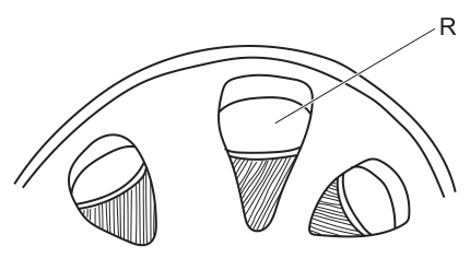
The maximum width of R is on the diagram and its actual width is .
The student was asked to draw a scale bar on their diagram.
Which row is correct?
Options
| part of the plant drawn | location of drawing mistake | length of scale bar in to correctly represent | |
|---|---|---|---|
| A | root | phloem | 6 |
| B | root | xylem | 170 |
| C | stem | phloem | 170 |
| D | stem | xylem | 6 |
Working
Identify the organ. The vascular bundles are arranged in a ring near the epidermis, so this is a transverse section of a dicot stem (a root would show a central xylem star/cylinder, not a ring of bundles).
Locate the drawing mistake. In a real dicot stem each vascular bundle has xylem on the inside (toward the centre) and phloem on the outside (toward the epidermis). The diagram has these reversed, so the mistake is in the xylem (drawn in the wrong position).
Scale bar length.
So 1 µm of actual tissue = 60 µm on the diagram, and a 100 µm scale bar must be:
Answer
D
D
Background Concept
A plan diagram is a low-magnification outline drawing that shows the distribution of tissues in a specimen without drawing individual cells. For a plant organ, two features are usually enough to identify what is being drawn:
- Dicot stem (TS): vascular bundles arranged in a single ring just inside the epidermis. Within each bundle, xylem lies on the inside (toward the centre of the stem) and phloem on the outside (toward the epidermis). The ground tissue between the bundles and the epidermis is the cortex; the tissue enclosed by the ring of bundles is the pith.
- Dicot root (TS): a central xylem star or cross (with phloem strands between the arms of the star), surrounded by a ring of pericycle, with the cortex and epidermis on the outside. There is no ring of separate vascular bundles and no pith in the typical dicot root.
A scale bar is a short line on a drawing that represents a stated real length, so the reader can measure structures on the diagram against a known reference. To work out the length of a scale bar you first need the magnification of the drawing:
Understanding the Question
The student has drawn a plan diagram (Fig. 26.1) of a plant organ, but the drawing contains an error. We are given two numerical facts about the bundle labelled R — its maximum width is 15 mm on the diagram, and its true width in the specimen is 250 µm. We have to pick the row of the table that correctly states (i) which organ was drawn, (ii) where the drawing mistake lies, and (iii) what length a 100 µm scale bar should be on this diagram. Each of the three must be correct for the answer to be right.
Approach
There are three independent decisions to make, in order:
- Tissue type — look at how the vascular bundles are arranged: ring near the edge → dicot stem; central star → root.
- Where the mistake is — recall the correct inside/outside arrangement within a vascular bundle and see which tissue is in the wrong place.
- Scale bar length — convert the image/actual pair into a magnification, then multiply by the desired real length (100 µm), converting the answer back to mm.
Step-by-Step Reasoning
Step 1 — Identify the organ.
The figure shows three vascular bundles sitting in a ring just inside an outer boundary (epidermis + cortex). This is the unmistakable signature of a dicot stem in transverse section. A root would not show a ring of separate bundles; it would show a central xylem core. This already rules out options A and B.
Step 2 — Locate the drawing mistake.
In a real dicot stem each bundle has phloem on the outside and xylem on the inside. In the diagram the shaded region of bundle R (the hatched triangle) is on the inside of the bundle, i.e. toward the centre of the stem — that is the xylem position drawn in the wrong place. The error therefore lies in the xylem. This rules out option C (which blames the phloem) and confirms option D.
Step 3 — Calculate the scale bar for 100 µm.
First find the magnification using bundle R:
So 1 µm of real tissue has been drawn as 60 µm on the page. A 100 µm scale bar therefore needs to be:
Option D gives 6 mm — consistent with the calculation. (Option C's 170 mm is a trap obtained by forgetting to convert 15 mm into µm before dividing: 15/250 = 0.06, not 60.)
All three parts of row D match, so the answer is D.
Key Takeaways
- A ring of vascular bundles near the epidermis = dicot stem; a central xylem star = root.
- In a stem bundle, remember the mnemonic "Phloem Out, XyIn" (Phloem Outside, Xylem Inside).
- To size a scale bar, always work in one set of units (convert mm to µm or vice versa) and remember the magnification = image/actual, so scale bar on page = actual length × magnification.
Common Mistakes
- Confusing stem and root from a quick glance — the ring arrangement is the key feature; the relative size of cortex vs pith is secondary.
- Saying the mistake is "in the phloem" because the phloem looks unusual, when in fact the phloem is in the right place and the xylem has been drawn on the wrong side of the bundle.
- Computing the scale bar as without converting units first, or computing and getting 170 mm — both arise from mixing mm and µm.
- Forgetting to convert the final 6 000 µm back into mm when reading off the answer in the table.
Things to Be Careful About
- Always convert to the same unit before dividing; is meaningless until one is changed to the other.
- The magnification is 60×, not 0.06× — a common slip is to put the small number on top.
- Read the question carefully: it asks for the length representing 100 µm, not 250 µm (250 µm would give 15 mm, i.e. the whole width of R).
- "Xylem on the outside" is the descriptive clue for the error: name the tissue that is misplaced, not just "the bundle is wrong".
Four students have drawn and labelled part of a structure seen on an electron micrograph.
Which student has labelled their drawing correctly?
Options
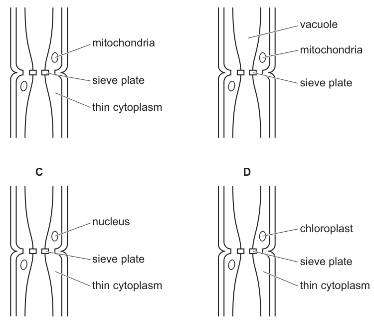
Working
A mature sieve tube element loses most of its organelles at maturity:
- it has no nucleus
- it has no (or greatly reduced) vacuole
- it has no ribosomes and no chloroplasts
- it retains only a thin layer of cytoplasm lining the cell
Its associated companion cell retains a full complement of organelles, including a nucleus and mitochondria, supplying ATP to the sieve tube element via plasmodesmata. End-to-end sieve tube elements are joined by a sieve plate.
Evaluating each option:
- A — mitochondria in the companion cell ✓, sieve plate ✓, thin cytoplasm in the sieve tube element ✓ — all correct.
- B — sieve tube elements do not have a large vacuole and do not contain mitochondria. ✗
- C — sieve tube elements have no nucleus (the nucleus is in the companion cell). ✗
- D — sieve tube elements do not contain chloroplasts. ✗
Answer
A
A
Background Concept
Phloem is the plant tissue that translocates assimilates (mainly sucrose) from sources (e.g. photosynthetic leaves, storage organs) to sinks (e.g. roots, fruits, growing tips). Its conducting elements are sieve tube elements (also called sieve tube members), joined end-to-end into sieve tubes. Each sieve tube element is closely associated with one or more companion cells, derived from the same mother cell by unequal division.
Mature sieve tube elements are highly modified for mass flow:
- They lose their nucleus during differentiation.
- They lose their ribosomes and most other organelles (Golgi, etc.).
- The central vacuole is greatly reduced or absent; the cell is filled with a thin layer of cytoplasm at the periphery.
- They retain a plasma membrane and some smooth ER.
- The cross-walls between adjacent sieve tube elements are perforated to form the sieve plate.
- They remain alive but are metabolically dependent on the adjacent companion cell.
The companion cell keeps a full set of organelles — a nucleus, mitochondria, ribosomes, Golgi and a normal vacuole. It is connected to the sieve tube element by numerous plasmodesmata, through which ATP, proteins and other molecules are passed to the sieve element.
Understanding the Question
The question shows four drawings of a longitudinal section through a phloem sieve tube element with its companion cell, each with a different set of labels. The candidate must decide which set of labels correctly reflects phloem ultrastructure.
The key biological point being tested: a mature sieve tube element has almost no organelles of its own — its metabolic support comes from the adjacent companion cell.
Approach
For each option, check every label against the real structure:
- Where is the mitochondrion drawn — in the sieve tube element or in the companion cell?
- Is the sieve plate correctly placed at the junction between two sieve tube elements?
- Is "thin cytoplasm" correctly applied to the sieve tube element?
- Are any impossible structures (nucleus, large vacuole, chloroplast) claimed to be inside the sieve tube element?
Reject any option that places a nucleus, large vacuole, or chloroplast inside the sieve tube element, or that places mitochondria inside the sieve tube element. Accept the option that places the metabolically active organelles in the companion cell and correctly describes the sieve tube element as having only thin cytoplasm and a sieve plate.
Step-by-Step Reasoning
Option A
- Mitochondrion labelled in the companion cell — correct, the companion cell retains mitochondria to produce ATP for both cells.
- Sieve plate labelled at the junction between two sieve tube elements — correct.
- Thin cytoplasm labelled inside the sieve tube element — correct, this is exactly the reduced content of a mature sieve tube element.
All three labels are anatomically accurate → A is correct.
Option B
- A large vacuole is labelled inside the sieve tube element — wrong; mature sieve tube elements have lost their large central vacuole.
- Mitochondria labelled inside the sieve tube element — wrong; they reside in the companion cell.
- Sieve plate is correct, but the other two errors disqualify this option.
Option C
- A nucleus is labelled inside the sieve tube element — wrong; the nucleus was lost during differentiation. The nucleus is in the companion cell.
- Sieve plate and thin cytoplasm are correct, but the nuclear label alone makes the drawing incorrect.
Option D
- A chloroplast is labelled inside the sieve tube element — wrong; sieve tube elements do not photosynthesise and do not retain chloroplasts at maturity.
- The other two labels are correct, but the chloroplast label disqualifies this option.
Key Takeaways
- A mature sieve tube element is essentially a hollow tube of cytoplasm: no nucleus, no (or very reduced) vacuole, no ribosomes, no chloroplasts, no mitochondria — only thin cytoplasm, a plasma membrane and the sieve plate at each end wall.
- The companion cell carries out the metabolism: it has a nucleus, mitochondria, ribosomes and a normal vacuole, and supplies the sieve tube element with ATP through plasmodesmata.
- This division of labour is what makes mass flow in the phloem possible: the conducting tube is kept clear of obstructions, while the metabolic machinery is housed alongside in the companion cell.
Common Mistakes
- Assuming the sieve tube element, because it is alive, must contain a nucleus and a normal vacuole. The element is alive but has shed its nucleus and most organelles during differentiation.
- Confusing xylem vessel elements (dead, hollow, lignified tubes with no cytoplasm at all) with sieve tube elements (alive, with thin cytoplasm, no nucleus).
- Placing chloroplasts in phloem cells. Sieve tube elements are not photosynthetic; even in green stems, mature sieve elements do not retain chloroplasts.
- Labelling the mitochondrion in the wrong cell of the pair. Many students place it inside the larger sieve tube element because it looks more prominent in the drawing.
Things to Be Careful About
- A "vacuole" inside a mature sieve tube element is not present in the way it is in a typical plant cell; if a large central vacuole is labelled, the option is wrong.
- The nucleus always belongs to the smaller companion cell in the drawing — never to the sieve tube element.
- The sieve plate is the perforated cross-wall at each end of a sieve tube element, marking where one element meets the next — not the lateral wall.
- "Thin cytoplasm" is the correct description of the residual contents of a mature sieve tube element; the cell is not "empty" but it is far from full of cytoplasm.
The diagram shows some structures used for transport in the phloem.
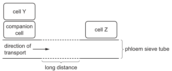
Which statement is correct?
Options
A Cell Y is the sink cell.
B Cell Z can be above or below cell Y in the plant.
C Cell Z must be in a photosynthetic tissue.
D Cell Y allows glucose to move to the sieve tube via the companion cell.
Working
The arrow shows the direction of assimilate (sucrose) flow is from the companion-cell region (where cell Y is) towards cell Z. Therefore:
- Cell Y = source (loading assimilates into the phloem)
- Cell Z = sink (unloading assimilates from the phloem)
A – incorrect. Cell Y is the source, not the sink.
B – correct. A sink can be above the source (e.g. growing shoot apex, young leaves, fruits, seeds) or below it (e.g. roots, rhizomes). Phloem transport occurs in whichever direction the source–sink gradient dictates, so cell Z may lie either above or below cell Y.
C – incorrect. Cell Z is the sink; sinks are usually non-photosynthetic tissues (roots, storage organs, developing fruits) that consume or store assimilates.
D – incorrect. The sugar loaded into the sieve tube via the companion cell is sucrose, not glucose.
Answer
B
B
Background Concept
Phloem translocates the organic products of photosynthesis — chiefly sucrose — from sites of production or mobilisation (sources) to sites of utilisation or storage (sinks).
- Source examples: photosynthetic mesophyll cells in mature leaves, storage tissues releasing reserves (e.g. a germinating seed's cotyledons), or storage roots in spring.
- Sink examples: roots, root tips, young developing leaves, apical and axillary buds, flowers, fruits, seeds, tubers and other storage organs.
Sucrose moves from a source mesophyll cell into a companion cell, which then actively loads it into the adjacent sieve tube element using a sucrose–H⁺ symporter driven by a proton pump. Inside the sieve tube, the sucrose solution flows by mass flow along a hydrostatic pressure gradient (high at the source, low at the sink) generated by these loading/unloading events.
A key feature of phloem transport is that its direction is not fixed: it follows the source-to-sink gradient. A single plant can therefore have phloem moving upwards (to apical buds, fruits or growing leaves above the source) and downwards (to roots or storage organs below the source) simultaneously, depending on where the sources and sinks are at that developmental stage.
Understanding the Question
The diagram shows:
- Cell Y with a companion cell, feeding into the sieve tube.
- An arrow along the sieve tube pointing towards cell Z.
- A bracket labelled "long distance" along the sieve tube.
Because the arrow points from the companion-cell region towards cell Z, cell Y is upstream of the flow (the source) and cell Z is downstream (the sink). The question asks which of four statements about these cells and the system is correct.
Approach
Resolve the source–sink identity from the direction arrow, then test each option against what is known about phloem loading, the identity of the transported sugar, and the variable location of sinks in the plant body.
Step-by-Step Reasoning
Option A — "Cell Y is the sink cell."
The transport arrow moves away from cell Y's region. Material is being loaded into the sieve tube near cell Y and carried towards cell Z. Therefore cell Y is the source, not the sink. A is wrong.
Option B — "Cell Z can be above or below cell Y in the plant."
Cell Z is the sink. Sinks include roots and underground storage organs (below the source leaf) as well as apical buds, young leaves, flowers, fruits and seeds (above the source leaf). Phloem mass flow simply follows the pressure gradient from source to sink, so the sink can be located either above or below the source. B is correct.
Option C — "Cell Z must be in a photosynthetic tissue."
Photosynthetic (mature mesophyll) tissue is a typical source, not a sink. Cell Z, the sink, is more often non-photosynthetic — roots, tubers, developing fruits, etc. C is wrong.
Option D — "Cell Y allows glucose to move to the sieve tube via the companion cell."
The sugar translocated in the phloem is sucrose (a disaccharide of glucose + fructose). Glucose is not the major transported form because sucrose is non-reducing, more chemically stable in transit, and is the form in which plants move fixed carbon. The wording "glucose" disqualifies this statement. D is wrong.
Key Takeaways
- Sources load assimilates into the phloem; sinks unload them.
- The transported sugar is sucrose, not glucose.
- Companion cells perform the active loading of sucrose into sieve tube elements.
- Phloem direction is dictated by the source–sink gradient, so a sink can be above, below or on the same level as a source, and directions can reverse seasonally (e.g. from roots to shoots in spring when stored reserves are mobilised).
Common Mistakes
- Confusing the source and sink based on the diagram's layout rather than the direction arrow.
- Believing phloem always flows downwards — it follows the source-to-sink pressure gradient, which can be in any direction.
- Stating "glucose" instead of "sucrose" as the sugar in phloem — this is one of the most common terminology errors in this topic.
- Assuming sinks must be photosynthetic — they are usually non-photosynthetic, net importers of assimilate.
Things to Be Careful About
- Read the direction arrow, not the spatial position, when deciding which end of a phloem diagram is source and which is sink.
- The wording "can be above or below" is the key clue in option B; remember that phloem is not unidirectional like xylem.
- A "source" or "sink" status can change with season and developmental stage (e.g. a developing leaf is first a sink, then becomes a source), so do not equate source/sink with a fixed cell type.
The diagram shows a section through a root of a dicotyledonous plant.
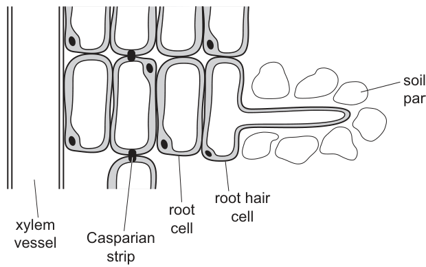
Which statement correctly describes the movement of water and solutes through this root?
Options
A A layer of suberin causes water and solutes to move from the apoplast pathway into the symplast pathway.
B A layer of suberin causes water and solutes to move from the symplast pathway into the apoplast pathway.
C The tonoplast causes water and solutes to move from the symplast pathway into the apoplast pathway.
D The tonoplast causes water and solutes to move from the apoplast pathway into the symplast pathway.
Working
Water enters the root via two pathways:
- The apoplast pathway — through cell walls and intercellular spaces (without crossing any plasma membrane).
- The symplast pathway — through the cytoplasm of cells, passing from cell to cell via plasmodesmata.
At the endodermis, the Casparian strip is a band of suberin deposited in the radial and transverse walls. Suberin is waterproof and impermeable to dissolved solutes, so it blocks the apoplast pathway at this point. To continue inward toward the xylem, water and mineral ions must cross the plasma membrane of an endodermal cell and enter the symplast.
Answer
A
A
Background Concept
Water moving from the soil into the xylem of a root can take two parallel routes:
- Apoplast pathway: water and dissolved solutes travel through the porous cell walls and intercellular spaces without ever crossing a plasma membrane. This is a rapid, low-resistance route through the cortex.
- Symplast pathway: water moves from cell to cell through the cytoplasm, crossing the plasma membrane once at the outer face of the epidermis and then passing between cells via plasmodesmata (cytoplasmic continuities).
A small amount of water also takes the vacuolar pathway (crossing the tonoplast to move through vacuoles), but this is usually grouped with the symplast.
The endodermis is the innermost layer of the cortex, surrounding the vascular tissue. Its cell walls are impregnated with suberin in a continuous ring called the Casparian strip. Suberin is a waxy, hydrophobic material that is impermeable to water and dissolved solutes.
Understanding the Question
The question shows a root cross-section including the root hair cell, cortex (root cells), endodermis with the Casparian strip, and the xylem. It asks which statement correctly describes what forces water and solutes to switch pathways on their way to the xylem. The command word is implicit in the MCQ format — we must pick the statement that is biologically accurate about the apoplast/symplast switch.
Approach
The key is to identify:
- The structure that interrupts the apoplast pathway (the Casparian strip, made of suberin in the endodermis).
- The direction of switching it forces (apoplast → symplast, because the symplast is the only remaining option once the apoplast is blocked).
- The role of the tonoplast is different — it is the membrane around the vacuole, not a barrier in the cell wall — so any option using "tonoplast" as the cause of pathway switching is wrong.
Step-by-Step Reasoning
- Water enters the root hair cell and crosses the cortex largely via the apoplast (cell walls).
- On reaching the endodermis, the suberised Casparian strip seals the cell walls, so apoplastic flow stops.
- To continue into the stele and the xylem, water and solutes must cross the plasma membrane of an endodermal cell, thereby entering the symplast. This transition allows the plant to control which ions enter the xylem (the membrane is selectively permeable).
- Therefore the suberin layer causes movement from the apoplast into the symplast — option A.
- Option B is the wrong direction.
- Options C and D wrongly attribute the switching to the tonoplast, which is the vacuolar membrane and is not what blocks the apoplast at the endodermis.
Key Takeaways
- The Casparian strip is a ring of suberin in the radial and transverse walls of endodermal cells.
- It is impermeable to water and solutes, so it blocks the apoplast pathway at the endodermis.
- Water and solutes are forced to cross a plasma membrane and continue via the symplast, giving the plant control over what enters the xylem.
Common Mistakes
- Reversing the direction of switching and choosing B (apoplast → symplast is correct, not symplast → apoplast).
- Confusing the Casparian strip (suberin in cell walls) with the tonoplast (membrane around the vacuole). The tonoplast is involved in the vacuolar pathway, not in forcing the apoplast–symplast switch.
- Thinking the apoplast pathway goes through the Casparian strip; it is blocked there.
Things to Be Careful About
- The endodermis is the innermost layer of the cortex, just outside the pericycle and vascular tissue.
- The Casparian strip is a property of the cell wall (suberin in the wall), not of the plasma membrane.
- The selective barrier function at the endodermis is the reason plants can regulate the mineral composition of xylem sap.
The graph shows the effect of different partial pressures of carbon dioxide () on the oxygen dissociation curve for haemoglobin.
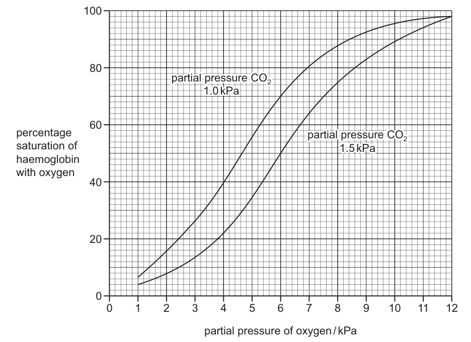
What is the change in percentage oxygen saturation of haemoglobin at a partial pressure of oxygen of as the partial pressure of carbon dioxide changes from to ?
Options
A
B
C
D
Working
At :
- On the CO₂ curve (upper curve): percentage saturation ≈
- On the CO₂ curve (lower curve): percentage saturation ≈
Answer
B
B
Background Concept
Haemoglobin is a globular protein in red blood cells that binds oxygen in the lungs and releases it in respiring tissues. The relationship between the partial pressure of oxygen () and the percentage of haemoglobin binding sites occupied by oxygen is shown by the oxygen dissociation curve — a sigmoid (S-shaped) curve.
The shape arises from the cooperative binding of O₂: once one O₂ molecule binds, it becomes easier for the next three to bind (steep middle portion), and once the first O₂ leaves, the others follow more easily (steep lower portion).
The position of the curve is shifted by several factors, the most important being and pH. An increase in (or a fall in pH) shifts the curve to the right — this is the Bohr shift. A rightward shift means that, at any given , haemoglobin holds less oxygen and therefore releases more. This is physiologically important: respiring tissues produce CO₂, so locally the curve shifts right, unloading more O₂ exactly where it is needed.
Understanding the Question
The question gives a graph with two oxygen dissociation curves:
- An upper curve at
- A lower curve at (the Bohr-shifted curve)
We must read the percentage saturation on both curves at and find the change when increases from to . A negative answer is expected because the saturation decreases when the curve shifts right.
Approach
- Draw a vertical line at on the x-axis.
- Read where this line meets the upper ( CO₂) curve — this gives the higher saturation value.
- Read where this line meets the lower ( CO₂) curve — this gives the lower saturation value.
- Subtract: (value on lower curve) − (value on upper curve). The result should be negative because saturation falls as rises.
Step-by-Step Reasoning
- At on the upper curve ( CO₂): the curve passes through approximately 80% saturation.
- At on the lower curve ( CO₂): the curve passes through approximately 60% saturation.
- Change in saturation = (saturation at CO₂) − (saturation at CO₂) = .
- This agrees with the biological principle of the Bohr shift: higher CO₂ → lower affinity of haemoglobin for O₂ → reduced saturation at any given .
A -20% change is the correct magnitude, matching option B.
Key Takeaways
- The Bohr shift describes the rightward displacement of the oxygen dissociation curve as rises (or pH falls).
- A rightward shift means haemoglobin has a lower affinity for O₂ at any given , so it unloads more oxygen — exactly what is needed in actively respiring tissues.
- Reading values from dissociation curves is a core Paper 1 / Paper 2 skill: always draw a vertical line at the given and read the corresponding saturation, then compare curves at the same .
Common Mistakes
- Reading the wrong curve: confusing the upper and lower curves leads to a positive change of +20% rather than −20%, picking option C instead of B.
- Reading at the wrong : mislocating 6 kPa on the x-axis (it sits roughly halfway along) gives a wildly different saturation.
- Ignoring the sign of the change: the question asks for the change, so the sign matters. As rises, saturation falls — the change must be negative.
- Choosing 46% (option D): this would be the saturation on the upper curve minus ~34% (a common misread on the lower curve at ).
Things to Be Careful About
- Always quote percentage saturation with the % sign and with the correct sign when describing a change.
- The two curves are at the same range; only differs, so the comparison must be made vertically (same x-value, different y-values).
- Recognise that on this graph the rightward shift of the lower curve is the visual signature of the Bohr effect — confirming qualitatively that the answer should be negative before you even read the numbers.
What happens in the heart at the start of ventricular diastole?
Options
A The semilunar valves open.
B The atrioventricular valves open.
C The pressure in the atria rises above the pressure in the ventricles.
D The pressure in the left atrium rises more than the pressure in the right atrium.
Working
Ventricular diastole begins when the ventricular muscle relaxes, so the pressure inside the ventricles falls. As soon as ventricular pressure drops below atrial pressure, the atrioventricular (bicuspid and tricuspid) valves are forced open and blood flows from the atria into the ventricles.
- A is wrong: the semilunar (aortic and pulmonary) valves close at the start of ventricular diastole, they do not open.
- C is wrong: although atrial pressure does exceed ventricular pressure at this point, the question asks what happens in the heart — the opening of the AV valves is the defining event.
- D is wrong: there is no reason for left atrial pressure to rise more than right atrial pressure at this moment.
Answer
B
B
Background Concept
The cardiac cycle is the repeating sequence of contraction (systole) and relaxation (diastole) of the heart chambers, coordinated so that blood flows in one direction. Four valves enforce this one-way flow:
- Atrioventricular (AV) valves — the bicuspid (mitral) valve on the left and the tricuspid valve on the right, lying between each atrium and its ventricle.
- Semilunar valves — the aortic valve (left ventricle to aorta) and the pulmonary valve (right ventricle to pulmonary artery).
Valves are passive: they open and close purely in response to pressure differences on either side. When pressure is higher on the "upstream" side, the valve is forced open; when pressure is higher on the "downstream" side, it snaps shut (preventing backflow). The cardiac cycle is therefore driven by the pressure changes that result from muscular contraction and relaxation.
The cycle has three useful reference points:
- Atrial systole — atria contract and push the remaining blood into the ventricles.
- Ventricular systole — ventricles contract; AV valves close ("lub"), then pressure rises above that in the arteries and the semilunar valves open, ejecting blood.
- Ventricular diastole — ventricles relax; pressure inside them falls below atrial pressure, so the AV valves open, allowing the ventricles to refill.
Understanding the Question
The stem asks what happens at the start of ventricular diastole — that is, the very moment the ventricles begin to relax after having just ejected blood. The candidate must identify which of the four statements correctly describes the situation at that instant.
The four options test:
- A: an action of the semilunar valves,
- B: an action of the AV valves,
- C: a pressure relationship between atria and ventricles,
- D: a pressure difference between the left and right atria.
Only one of these is the event that characterises the start of ventricular diastole.
Approach
The cleanest way to decide is to work out which valves are doing what at the start of ventricular diastole, by tracking the pressure in the chambers and great vessels across the transition from systole to diastole.
At the end of ventricular systole:
- Ventricular pressure is high (it has just ejected blood through the open semilunar valves).
- As the ventricle relaxes, ventricular pressure falls rapidly.
- The moment ventricular pressure drops below the pressure in the connected arteries (aorta/pulmonary artery), the semilunar valves snap shut (this is the "dub" of the heartbeat).
- Ventricular pressure continues to fall. The instant it drops below atrial pressure, the AV valves are pushed open by the higher atrial pressure, and blood begins to refill the ventricles.
So the defining valve event at the start of ventricular diastole is the opening of the AV valves.
Step-by-Step Reasoning
- Option A — "The semilunar valves open." This describes the start of ventricular ejection (during ventricular systole), not the start of diastole. At the start of diastole the semilunar valves close. Wrong.
- Option B — "The atrioventricular valves open." Correct. As ventricular pressure falls below atrial pressure at the start of ventricular diastole, the higher atrial pressure forces the AV valves open, refilling the ventricles.
- Option C — "The pressure in the atria rises above the pressure in the ventricles." Although the pressure relationship described is true at this moment (atrial pressure is higher than ventricular pressure, which is why the AV valves open), the statement is misleading because atrial pressure does not suddenly "rise" — ventricular pressure is what has fallen below atrial pressure. More importantly, the defining event in the heart at the start of ventricular diastole is the valve opening (B), not the pressure change itself. CIE mark schemes typically want the structural event (valve opening) as the answer to "what happens in the heart".
- Option D — "The pressure in the left atrium rises more than the pressure in the right atrium." There is no reason to single out the left atrium here; the cardiac cycle is essentially symmetrical on both sides in this respect. Wrong.
Therefore B is the correct option.
Key Takeaways
- The cardiac cycle is driven by pressure changes; valves are passive and respond to those changes.
- Ventricular diastole starts when the ventricular muscle relaxes: ventricular pressure falls, the semilunar valves close, then once atrial pressure exceeds ventricular pressure the AV valves open and the ventricles refill.
- "Lub" = AV valves closing (start of ventricular systole); "dub" = semilunar valves closing (start of ventricular diastole).
- A pressure statement is not the same as an event; on CIE MCQs, "what happens in the heart" is usually answered by naming the structural event (valve opening/closing).
Common Mistakes
- Choosing A by confusing the start of ventricular systole (semilunar valves open) with the start of ventricular diastole (semilunar valves close, AV valves open).
- Choosing C because the pressure relationship is true in a literal sense — but the event being asked about is the opening of the AV valves, and the atrial pressure does not actively "rise" at this moment; it is the ventricular pressure that has fallen.
- Forgetting that the AV valves are called the bicuspid/mitral (left) and tricuspid (right) valves, while the semilunar valves are the aortic and pulmonary valves — mixing up these names is a common source of lost marks on longer-answer cardiac-cycle questions.
Things to Be Careful About
- Be precise about timing within the cycle: "start of ventricular diastole" is the moment the ventricles begin to relax, not the later filling phase.
- Valve action is a consequence of pressure changes, not an active process — a good answer describes the pressure change that forces the valve open or shut.
- On CIE multiple-choice papers, MCQ command words are interpreted strictly: "what happens in the heart" expects a structural/functional event such as a valve opening, not a description of a pressure gradient in isolation.
The electron micrograph shows a longitudinal section through a blood vessel.
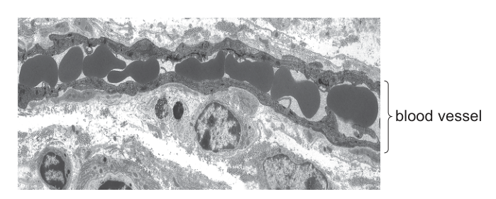
Which type of blood vessel is shown?
Options
A arteriole
B capillary
C vein
D venule
Working
The electron micrograph shows a longitudinal section through a very small blood vessel. The wall consists of a single layer of flattened endothelial cells (no smooth muscle, no elastic fibres, no connective tissue layers). The lumen is only marginally wider than a red blood cell, so the red blood cells are squeezed through the vessel in single file.
These two features — a wall of just one endothelial cell thick and red cells travelling in single file — are diagnostic of a capillary.
Answer
B
B
Background Concept
Blood vessels form a closed transport system. The three principal types — arteries, veins and capillaries — are built differently because they do different jobs:
- Arteries and arterioles carry blood away from the heart at high pressure. Their walls are thick and contain smooth muscle, elastic fibres and an outer layer of connective tissue. Arterioles still have several layers of smooth muscle in the tunica media and a clearly multilayered wall.
- Veins and venules carry blood back to the heart at low pressure. Their walls are thinner than equivalent arteries but still contain smooth muscle and an outer tunica adventitia of connective tissue; many veins also have valves to prevent backflow. Venules are the smallest veins and may have a lumen noticeably larger than a red blood cell, with at least some smooth muscle in the wall.
- Capillaries are the site of exchange between blood and surrounding tissues. To minimise the diffusion distance for oxygen, carbon dioxide, nutrients and wastes, their wall is reduced to a single layer of squamous (flattened) endothelial cells sitting on a basement membrane, with an internal diameter of only about – — just large enough for a red blood cell (about across) to squeeze through. As a result, red cells travel through capillaries in single file, which maximises their surface-area-to-volume ratio for gas exchange.
Understanding the Question
The question presents an electron micrograph of a longitudinal section through a small blood vessel and asks the candidate to name the type of vessel. The image is the only source of evidence, so the answer must come from identifying the structural features visible at high magnification.
The command word is which type, so a single correct option is required, supported by the observable features in the micrograph.
Approach
Look for the two diagnostic structural clues that separate a capillary from every other vessel type:
- Wall thickness and composition — is there only a single layer of cells (endothelium), or are there additional layers of smooth muscle, elastic tissue or connective tissue?
- Lumen diameter relative to a red blood cell — are the red cells flowing in single file, or in multiple columns?
If both clues point the same way, the identification is secure.
Step-by-Step Reasoning
- The electron micrograph shows the vessel wall as a single, very thin layer of flattened cells. There is no evidence of a tunica media with smooth muscle, no elastic lamina, and no outer connective-tissue sheath. This rules out an arteriole (which would show at least one, often two, layers of smooth muscle in the wall) and a venule or vein (which would also show smooth muscle and an outer adventitia).
- The lumen is so narrow that the dark, biconcave red blood cells inside it sit end-to-end in a single line; the cells are visibly deformed as they pass through. This is the textbook image of red cells in single file and is only possible when the lumen is roughly the diameter of one red blood cell — a defining feature of a capillary.
- Both features together therefore identify the vessel as a capillary.
Key Takeaways
- A capillary's wall is one endothelial cell thick with a basement membrane — nothing else.
- The capillary lumen is only slightly wider than a red blood cell, so red cells pass through in single file.
- These features make capillaries ideal for rapid exchange of materials between blood and tissues (short diffusion distance, large surface-area-to-volume ratio of each red cell).
- On an EM, the absence of smooth muscle, elastic fibres and a connective-tissue layer is the single most reliable way to distinguish a capillary from a small arteriole or venule.
Common Mistakes
- Choosing A (arteriole) because the vessel is small. Size alone is misleading — the decisive feature is the composition of the wall, not its diameter.
- Choosing D (venule) because venules are also small. Venules already have some smooth muscle and a wider lumen in which several red blood cells can travel side by side.
- Choosing C (vein) because the wall looks thin in absolute terms. Even a small vein still has a multilayered wall and a lumen far larger than a red blood cell.
Things to Be Careful About
- On an EM of a longitudinal section, the endothelium appears as a thin ribbon of cells running along the top and bottom of the lumen; do not mistake this for connective tissue.
- "Thin wall" is not the same as "one cell thick" — arterioles and venules also look thin compared with a large artery, but they still have several cell layers in cross-section.
- The single-file arrangement of red cells is only seen in capillaries; in any other vessel the red cells form a wider, packed column.
A student drew a sketch to show the formation of tissue fluid.
Which label is correct?
Options
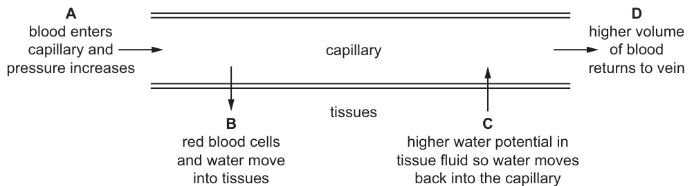
Working
- A: Incorrect — blood pressure is highest at the arterial end and decreases along the capillary; it does not increase.
- B: Incorrect — red blood cells remain inside the capillary; only plasma and small dissolved solutes (water, ions, glucose, urea) leave at the arterial end.
- C: Correct — at the venous end hydrostatic pressure has fallen; the blood has a lower water potential than the tissue fluid (due to plasma proteins) so water moves back into the capillary by osmosis.
- D: Incorrect — not all of the fluid that left the capillary returns to the vein; the excess (~10%) is drained away by the lymphatic system.
Answer
C
C
Background Concept
Tissue fluid is the fluid that surrounds the cells of the body. It forms when blood plasma is forced out of the capillaries under pressure and bathes the cells, supplying them with oxygen, glucose, amino acids, ions and other small solutes, and removing carbon dioxide and other waste products.
The formation of tissue fluid depends on two opposing forces acting across the thin, porous wall of the capillary:
- Hydrostatic (blood) pressure — this is the physical pressure of the blood pushing outwards against the capillary wall. It is highest at the arterial end of the capillary and falls along its length.
- Water potential — the water potential of the blood plasma is reduced by the presence of plasma proteins (especially albumin), which cannot leave the capillary. Tissue fluid lacks these large proteins, so it has a higher (less negative) water potential than the plasma inside the capillary.
At the arterial end, hydrostatic pressure is high and exceeds the inward pull of the water potential gradient. The net result is that fluid is forced out of the capillary through the small gaps between the endothelial cells, forming tissue fluid. Cells and most plasma proteins stay behind in the blood.
At the venous end, hydrostatic pressure has fallen below the water potential gradient, so water re-enters the capillary by osmosis, carrying dissolved waste products. Not all of the tissue fluid returns to the blood; about 10% is taken up by the lymph capillaries and eventually returned to the bloodstream via the thoracic duct.
Understanding the Question
The question shows the student's sketch of a capillary bed and four statements (A–D) describing different features of tissue fluid formation. The task is to identify which single labelled statement is biologically correct. This is a multiple-choice question, so only one option is fully correct.
Approach
Go through each option in turn and check it against the known biology of tissue fluid formation. Reject any option containing a specific false claim (e.g. an incorrect direction of movement, or movement of a particle that cannot cross the capillary wall).
Step-by-Step Reasoning
Option A — 'blood enters capillary and pressure increases'.
This is wrong. Blood pressure is at its maximum as it enters the capillary from the arteriole, and the hydrostatic pressure actually falls progressively along the length of the capillary. Stating that pressure 'increases' misrepresents the pressure gradient.
Option B — 'red blood cells and water move into tissues'.
This is wrong because the capillary wall is impermeable to red blood cells. The gaps between the endothelial cells are only large enough to allow plasma and small solutes (water, glucose, amino acids, ions, urea) to pass. Red blood cells are far too large (~7 µm) to leave the capillary. Only water and dissolved substances form tissue fluid.
Option C — 'higher water potential in tissue fluid so water moves back into the capillary'.
This is correct. By the time blood reaches the venous end, hydrostatic pressure has dropped and the dominant force is the water potential gradient: tissue fluid has a higher (less negative) water potential than plasma because it lacks plasma proteins. Water therefore moves from the tissue fluid back into the capillary by osmosis, down the water potential gradient. This is the standard CIE account of the return of fluid at the venous end.
Option D — 'higher volume of blood returns to vein'.
This is wrong. More fluid leaves the capillary at the arterial end than returns at the venous end. The excess fluid (~10% of the volume filtered out) is drained by the lymphatic system and eventually returned to the bloodstream via the subclavian veins. The volume of blood in the vein downstream of the capillary is therefore less than at the arterial end, not higher.
Only option C contains a fully correct statement.
Key Takeaways
- Tissue fluid is formed from plasma forced out at the arterial end by high hydrostatic pressure.
- Red blood cells and most plasma proteins remain inside the capillary; only small solutes and water leave.
- At the venous end, water re-enters the capillary by osmosis because tissue fluid has a higher water potential than the plasma.
- The lymphatic system collects the excess tissue fluid that does not return directly to the capillary.
Common Mistakes
- Saying that 'pressure increases' along the capillary — in reality it decreases from arterial to venous end.
- Stating that red blood cells, white blood cells or plasma proteins leave the capillary — only small solutes do.
- Confusing the direction of the water potential gradient: water moves from high water potential (tissue fluid) to low water potential (plasma with its dissolved proteins).
- Believing that all of the filtered fluid returns to the vein — about 10% is returned via the lymph.
Things to Be Careful About
- 'Water potential' is the precise term — avoid 'water concentration' or 'water moves down its concentration gradient', as these are imprecise for an A-level answer.
- The water potential of plasma is lower (more negative) than that of tissue fluid because of plasma proteins, not because of any ion difference.
- Direction of movement at each end is determined by whichever of the two opposing forces (hydrostatic pressure vs water potential gradient) is greater at that point.
Which structures are present in the trachea and also in all bronchioles?
1 cartilage
2 smooth muscle
3 epithelial cells
Options
A 1, 2 and 3
B 1 only
C 2 and 3 only
D 2 only
Working
- Trachea: contains C-shaped cartilage, smooth muscle (in the posterior wall between the ends of the C-rings), and ciliated epithelial cells.
- Bronchioles: contain smooth muscle and epithelial cells, but no cartilage (cartilage is absent from bronchioles — this is what structurally distinguishes them from bronchi).
- Structures present in both trachea and all bronchioles: smooth muscle (2) and epithelial cells (3). Cartilage (1) is present in the trachea but absent from bronchioles.
Answer
C
C
Background Concept
The human gas exchange system is a branching tube network: trachea → bronchi → bronchioles → alveoli. As the airways branch and narrow, their wall structure changes. Three components are important here:
- Cartilage: rigid supporting tissue. In the trachea it forms incomplete (C-shaped) rings that keep the airway open. Cartilage is present in the trachea and bronchi, but is absent from bronchioles — bronchioles rely on smooth muscle tone and surrounding lung tissue to keep them patent.
- Smooth muscle: allows the airway diameter to be regulated (bronchoconstriction/bronchodilation). It is present in the trachea (between the open ends of the C-shaped cartilage rings, in the posterior wall) and throughout the bronchioles, where it is relatively more prominent because there is no cartilage.
- Epithelial cells: line the luminal surface of the entire airway. In the trachea and larger bronchioles this is a pseudostratified ciliated columnar epithelium with goblet cells; in smaller bronchioles it becomes simple ciliated cuboidal. Epithelial cells are present at all levels, including the trachea and every bronchiole.
Understanding the Question
This is a "which of the following" MCQ testing whether the candidate can identify the structural components common to the trachea and to all bronchioles. The word "all" is important: the answer must hold for the smallest terminal bronchioles, not just the larger ones.
Approach
Go through each numbered structure and decide: present in trachea? Present in (all) bronchioles? A structure must be present at both levels to be included in the answer.
Step-by-Step Reasoning
- Cartilage (1): present in the trachea as C-shaped rings. Absent in bronchioles. Therefore cartilage is not in both. → excluded.
- Smooth muscle (2): present in the trachea (posterior wall between cartilage ends) and in all bronchioles (where it is the main structural support of the wall). → present in both. ✓
- Epithelial cells (3): the entire respiratory tract, from trachea down to terminal bronchioles, is lined by epithelial cells. → present in both. ✓
Only statements 2 and 3 are correct in both locations, so the answer is C (2 and 3 only).
Key Takeaways
- The defining structural feature that separates bronchi from bronchioles is the loss of cartilage and an increase in the relative amount of smooth muscle.
- Smooth muscle and an epithelial lining are features of the entire conducting airway.
- The wall of every airway is also elastic to a degree, and goblet cells/cartilage are progressively lost as the tubes narrow.
Common Mistakes
- Choosing A (1, 2 and 3): thinking cartilage extends all the way down. Cartilage stops where the bronchi end; bronchioles have no cartilage.
- Choosing D (2 only): forgetting that the trachea (and all bronchioles) is lined by an epithelium. Epithelial cells are present at every level of the conducting airway.
- Choosing B (1 only): missing both that cartilage is absent in bronchioles and that smooth muscle and epithelium are shared features.
Things to Be Careful About
- The trachea does have smooth muscle — it sits in the posterior wall between the open ends of the C-shaped cartilage rings. Don't assume "trachea = cartilage only".
- The smallest terminal bronchioles still have an epithelial lining and a smooth muscle layer, even when cartilage and goblet cells have been lost.
The photomicrograph shows a section through part of the gas exchange system.
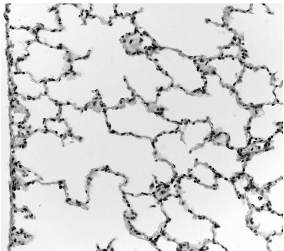
Which tissue can be seen?
Options
A cartilage
B ciliated epithelium
C smooth muscle
D squamous epithelium
Working
The photomicrograph shows numerous air spaces (alveoli) separated by very thin walls. Each alveolar wall consists of a single layer of flattened cells with darkly stained, elongated nuclei — the characteristic appearance of squamous epithelium. This thin lining provides the short diffusion distance required for gas exchange between the alveolar air and the blood in the surrounding capillaries.
- Cartilage (A) would appear as cells in lacunae within a dense matrix — not seen here.
- Ciliated epithelium (B) would show a columnar layer with apical cilia and goblet cells — not seen here.
- Smooth muscle (C) would show elongated, spindle-shaped cells with central nuclei in bundles — not seen here.
- Squamous epithelium (D) matches the thin, flattened cells lining the alveoli.
Answer
D
D
Background Concept
The gas exchange system ends in the alveoli — millions of tiny, thin-walled air sacs where O₂ enters the blood and CO₂ leaves it. To allow rapid diffusion, the barrier between alveolar air and pulmonary capillary blood must be extremely thin, and it is lined by squamous (pavement) epithelium: a single layer of flat, scale-like cells with flattened, elongated nuclei.
The other tissues in the gas exchange system each have distinctive appearances:
- Cartilage — chondrocytes sitting in lacunae within a dense extracellular matrix; in the trachea and bronchi it forms C-shaped or irregular supporting rings.
- Ciliated epithelium — tall columnar cells with cilia on their apical surface, plus goblet cells; it lines the trachea and most of the bronchi, sweeping mucus upwards.
- Smooth muscle — bundles of elongated, spindle-shaped cells with a single central nucleus; found in the walls of the bronchi and bronchioles, controlling airway diameter.
Understanding the Question
The question shows a single light-microscope image of lung tissue. The command word is implicit ("Which tissue can be seen?"), so the candidate must identify the one tissue type that is clearly visible, based on the structural features observable in the image.
Approach
- Scan the image for the dominant structural feature — here, the many open spaces (alveoli) separated by thin partitions.
- Examine the partitions closely to see what kind of cells form them.
- Match the observed cell shape and arrangement to one of the four named tissues.
Step-by-Step Reasoning
- The micrograph is filled with roughly circular, empty-looking spaces of varying sizes: these are the alveoli cut in section.
- The "walls" between alveoli are extremely thin — only one cell thick. The cells visible in those walls have flattened, elongated, darkly stained nuclei, the hallmark of squamous epithelium.
- There is no thick, dense, glossy matrix containing cells in lacunae → not cartilage.
- There is no columnar layer, no apical cilia, no goblet cells → not ciliated epithelium.
- There are no bundles of elongated, spindle-shaped cells with central cigar-shaped nuclei → not smooth muscle.
- The single layer of flattened cells lining each alveolus is squamous epithelium, which is exactly what option D describes.
Key Takeaways
- Alveoli are lined by squamous epithelium; its thinness is the structural feature that makes efficient gas exchange possible (Fick's law — short diffusion distance).
- Recognising tissues from micrographs is a core Paper 1 / Paper 2 / Paper 3 skill: look at cell shape, arrangement, and any distinguishing features (cilia, lacunae, spindle shape, flattened nuclei).
Common Mistakes
- Choosing B (ciliated epithelium) because the gas exchange system is often associated with cilia — but cilia are in the trachea/bronchi, not the alveoli.
- Choosing A (cartilage) because cartilage supports airways — but the image shows no lacunae or matrix.
- Choosing C (smooth muscle) if darker, denser regions are mistaken for muscle — but smooth muscle is found in airway walls, not lining alveoli.
Things to Be Careful About
- Squamous epithelium here is sometimes called "alveolar epithelium" or "pavement epithelium"; all three names refer to the same single layer of flat cells.
- In a micrograph, the very dark, elongated nuclei running along the alveolar walls are the most reliable visual clue — they look like dashes bordering each air space.
What is the function of the goblet cells in the gas exchange system?
Options
A to increase the surface area
B to move mucus
C to release mucus
D to trap dust and pathogens
Working
Goblet cells are modified epithelial cells that secrete (release) mucus onto the surface of the airways. Cilia on neighbouring ciliated cells then beat to move the mucus, and the mucus itself traps dust and pathogens — so releasing mucus is the goblet cell's role, while moving it and trapping particles are performed by other structures.
Answer
C
C
Background Concept
The gas exchange system (trachea, bronchi, bronchioles) is lined by a specialised epithelium. Two cell types work together to keep the airways clean:
- Goblet cells are modified columnar epithelial cells packed with mucinogen granules. They secrete mucus onto the airway surface.
- Ciliated cells bear apical cilia that beat in a coordinated wave to move the mucus (and anything trapped in it) upwards, away from the lungs, towards the pharynx where it is swallowed.
The mucus layer is sticky and traps inhaled particles such as dust, bacteria and viruses. The combination — sticky mucus plus an escalator of beating cilia — is called the mucociliary escalator and is a key non-specific defence of the respiratory tract.
Understanding the Question
The question asks for the function of the goblet cells specifically, with four options that each describe something the airways do. The distractors are plausible because they describe real, related events in the gas exchange system — but they are performed by other structures, not by the goblet cells themselves.
Approach
Link each option to the structure that actually carries out that role, then pick the option that names what the goblet cell does.
Step-by-Step Reasoning
- A — to increase the surface area. This is the role of alveoli (and of microvilli in the gut). Goblet cells are not a surface-area-increasing feature — reject.
- B — to move mucus. This is performed by the cilia of ciliated cells, which beat rhythmically to propel the mucus layer. Goblet cells have no motile apparatus — reject.
- C — to release mucus. Goblet cells are mucus-secreting cells. Their cytoplasm is full of mucin-containing secretory vesicles that release mucus onto the epithelial surface by exocytosis. ✓
- D — to trap dust and pathogens. This is the function of the mucus itself, not of the cells that secrete it. The sticky mucin glycoproteins physically trap inhaled particles — reject.
The correct answer is therefore C.
Key Takeaways
- Goblet cells secrete mucus; ciliated cells move it; mucus traps debris.
- These three roles are often confused in MCQs because they form a single integrated system (the mucociliary escalator).
- Always match the cell name to its precise verb of action: goblet → secrete/release; ciliated → beat/move; mucus → trap.
Common Mistakes
- Choosing B (move mucus) by attributing the action of cilia to goblet cells.
- Choosing D (trap dust and pathogens) by confusing the function of the secretion (mucus) with the function of the cell that makes it (goblet cell).
- Choosing A (increase surface area) when thinking vaguely of "lots of cells in the epithelium" without recalling that surface-area maximisation is the job of the alveoli.
Things to Be Careful About
- "Mucus" and "mucous" are easy to mix up: mucus is the noun (the secretion), mucous is the adjective (e.g. mucous membrane, mucous gland).
- Goblet cells are found throughout the respiratory tract but are most numerous in the trachea and bronchi; in the smaller bronchioles mucus is secreted mainly by club (Clara) cells instead.
- Watch for command-word traps: the question asks for the function of the cells, not of the mucus they produce.
Which organisms have cell walls that can be affected by penicillin?
1 bacteria
2 protoctists
3 viruses
Options
A 1 and 2
B 1 and 3
C 1 only
D 2 and 3
Working
Penicillin inhibits the formation of peptidoglycan cross-links in bacterial cell walls, weakening them so the bacterium takes up water and lyses.
- 1 (bacteria): have peptidoglycan cell walls → affected by penicillin ✓
- 2 (protoctists): may have cell walls, but they are made of cellulose/silica/etc., not peptidoglycan → not affected by penicillin ✗
- 3 (viruses): have no cell wall (only a protein capsid, and in some cases a lipid envelope) → not affected by penicillin ✗
Only statement 1 is correct.
Answer
C
C
Background Concept
Antibiotics are chemicals that kill or inhibit the growth of microorganisms. Different antibiotics target different structures or processes; penicillin is one of the β-lactam antibiotics and works specifically by inhibiting the enzyme transpeptidase (also called penicillin-binding protein). This enzyme cross-links the peptide side-chains of peptidoglycan, the rigid mesh-like polymer that gives bacterial cell walls their strength. When transpeptidase is blocked, the wall cannot be cross-linked, water enters the cell, and the bacterium bursts (lysis).
Because peptidoglycan is unique to bacteria, penicillin (and other β-lactams) selectively damages bacterial cells while leaving host cells — which have no peptidoglycan — unharmed. This is the basis of selective toxicity.
It is essential to remember that:
- Viruses are non-cellular. They consist of a nucleic acid core (DNA or RNA) enclosed in a protein capsid, sometimes wrapped in a host-derived envelope. They have no cell wall and no peptidoglycan, so no antibiotic can act on a cell wall that does not exist.
- Protoctists (e.g. Plasmodium, Trypanosoma, Amoeba) are mostly single-celled eukaryotic organisms. Some protoctists (the algae) do have cell walls, but these are made of cellulose or other materials, not peptidoglycan, so penicillin does not affect them.
Understanding the Question
This is a multiple-choice question testing two facts at once:
- Which organisms on the list actually possess cell walls?
- Of those with walls, which contain the peptidoglycan target of penicillin?
The command word "which organisms have cell walls that can be affected by penicillin" requires both conditions to be met simultaneously.
Approach
Step through each numbered organism and apply two filters in turn:
- Filter 1: Does it have a cell wall at all?
- Filter 2: If yes, is that wall made of peptidoglycan?
Only organisms passing both filters are credited.
Step-by-Step Reasoning
-
Bacteria (1): All bacteria have a peptidoglycan cell wall (Gram-positive bacteria have a thick layer; Gram-negative bacteria have a thinner layer plus an outer membrane). Penicillin binds transpeptidase → wall weakens → cell lysis. ✓ Affected.
-
Protoctists (2): Animal-like protoctists (e.g. Amoeba, Plasmodium) have no cell wall at all. Plant-like protoctists (algae) do have walls, but they are made of cellulose, not peptidoglycan. Either way, the wall is not a penicillin target. ✗ Not affected.
-
Viruses (3): Viruses are not cells. They possess a protein capsid (and sometimes a lipid envelope), but no cell wall and no peptidoglycan. Antibiotics, including penicillin, cannot act on viruses — this is a classic syllabus point. ✗ Not affected.
Only statement 1 is correct, so the answer is C (1 only).
Key Takeaways
- Penicillin kills bacteria by blocking peptidoglycan cross-linking in their cell walls.
- Antibiotics (including penicillin) do not work against viruses, because viruses lack the cellular structures (wall, ribosomes, etc.) that antibiotics target.
- "Having a cell wall" is not enough — the wall must contain peptidoglycan for penicillin to act.
Common Mistakes
- Choosing A (1 and 2): assuming that because some protoctists (algae) have cell walls, penicillin must act on them. Forget that the wall composition — not just its presence — determines susceptibility.
- Choosing B (1 and 3): confusing the protein capsid of a virus with a cell wall. A capsid is not a wall, and it contains no peptidoglycan.
- Choosing D (2 and 3): misreading the question and excluding bacteria, perhaps because of confusion between fungal cell walls (which contain chitin, not peptidoglycan) and protoctist walls.
Things to Be Careful About
- Penicillin affects bacteria, not fungi, despite both having cell walls — fungal walls are made of chitin/glucan and are not a target of penicillin.
- The classic syllabus phrase to remember is: "antibiotics do not affect viruses."
- Selective toxicity depends on the target molecule (peptidoglycan) being unique to the pathogen, not merely on the presence of any wall.
Each year, there are 462 000 deaths from malaria in children under 5 years old globally.
Insecticide-treated nets could prevent of malaria cases and reduce deaths of children under 5 years old by .
How many children could be saved by using insecticide-treated nets?
Options
A 41 580
B 83 160
C 231 000
D 332 640
Working
Number of children's lives saved = total deaths × percentage reduction
Answer
B
B
Background Concept
Malaria is a parasitic disease caused by Plasmodium species (most notably Plasmodium falciparum) and transmitted by the bite of infected female Anopheles mosquitoes. It remains one of the world's most serious infectious diseases, with young children in sub-Saharan Africa bearing the heaviest burden. Public-health interventions target different stages of the parasite's life cycle: insecticide-treated nets (ITNs) reduce human–mosquito contact at night, indoor residual spraying kills mosquitoes that rest on treated walls, antimalarial drugs treat the infection, and (in some regions) vaccines provide additional protection. The question gives you two statistics that allow you to quantify the impact of one specific intervention — ITNs — on under-5 mortality.
Understanding the Question
The question provides:
- 462,000 annual deaths from malaria in children under 5 years old.
- ITNs prevent 50% of malaria cases (a separate figure — not the one we need).
- ITNs reduce deaths in this age group by 18%.
The command word is implicit but the task is to calculate how many children could be saved. The trap in this question is the 50% figure, which relates to cases rather than deaths, and which would give 231,000 (option C) if misapplied.
Approach
Use the percentage-of-a-total formula. The relevant percentage is the 18% reduction in deaths, and the relevant total is the 462,000 under-5 deaths. Multiply, being careful to use the deaths figure (not the cases figure) and the 18% figure (not the 50% figure).
Step-by-Step Reasoning
- Identify the base figure: 462,000 deaths.
- Identify the percentage to apply: 18% (the reduction in deaths, not the 50% reduction in cases).
- Convert 18% to a decimal: 0.18.
- Multiply: 462,000 × 0.18.
- Calculate: 462,000 × 0.18 = 83,160.
For verification: 462,000 × 0.10 = 46,200 and 462,000 × 0.08 = 36,960. Sum = 46,200 + 36,960 = 83,160. ✓
Option C (231,000) is what you get if you mistakenly use the 50% cases figure (462,000 × 0.50 = 231,000). Option D (332,640) is 462,000 × 0.72, which has no clear link to the figures given. Option A (41,580) is half of 83,160 — the result if you divide 18% by 2 instead of multiplying. The correct answer is therefore B.
Key Takeaways
- Always read carefully which statistic applies to which outcome (cases vs deaths).
- A percentage-of-a-total calculation requires only one multiplication: total × (percentage ÷ 100).
- The distractor values map to predictable wrong moves (using the wrong percentage, halving the answer), which is a clue to which mistake the examiner is testing.
Common Mistakes
- Using the 50% (cases prevented) figure instead of the 18% (deaths reduced) figure → gives 231,000 (option C).
- Dividing instead of multiplying, or moving the decimal the wrong way → gives 41,580 (option A).
- Multiplying by 72% (1 − 0.18 = 0.82 would be deaths remaining; 0.72 has no basis) → gives 332,640 (option D).
Things to Be Careful About
- Match the quantity (deaths) with the percentage that refers to that quantity (18%).
- Quoting the answer to the same number of significant figures as the given data — 462,000 (3 s.f.) and 18% (2 s.f.) give an answer best reported as 83,160 (3 s.f.) or, rounded, 83,200.
- The answer is a whole number of children, not a percentage.
Which row is correct?
Options
| involved in phagocytosis | secrete antibodies | |
|---|---|---|
| A | T-lymphocytes | B-lymphocytes |
| B | T-lymphocytes | T-lymphocytes |
| C | B-lymphocytes | B-lymphocytes |
| D | B-lymphocytes | T-lymphocytes |
Working
- B-lymphocytes secrete antibodies (the cell-mediated producers of immunoglobulins in the humoral response), so the correct row must have B-lymphocytes in the 'secrete antibodies' column.
- That leaves options A and C.
- T-lymphocytes are the cells involved in phagocytosis, so the correct row must have T-lymphocytes in the 'involved in phagocytosis' column.
- Only option A satisfies both conditions: T-lymphocytes (phagocytosis) paired with B-lymphocytes (secrete antibodies).
Answer
A
A
Background Concept
The adaptive immune response relies on two main classes of lymphocyte, both produced in the bone marrow but maturing in different sites:
- B-lymphocytes mature in the bone marrow. When activated by binding to their specific antigen (usually with help from a helper T-lymphocyte), they proliferate and differentiate into plasma cells. Plasma cells are essentially antibody factories — they secrete large quantities of immunoglobulins (antibodies) that circulate in the blood and lymph, binding to the matching antigen to mark it for destruction (opsonisation, neutralisation, complement activation).
- T-lymphocytes mature in the thymus. They do not secrete antibodies. Instead, different sub-types carry out cell-mediated responses: helper T-cells (Th) release cytokines that activate B-cells and macrophages; cytotoxic T-cells (Tc) directly kill infected cells; and some T-cells assist in the phagocytic clearance of pathogens by recruiting and activating macrophages at the site of infection.
Phagocytosis itself — the engulfment and digestion of pathogens — is carried out primarily by phagocytes (macrophages and neutrophils), which are not lymphocytes. However, the question asks which lymphocytes are involved in the phagocytic process, and T-lymphocytes (especially helper T-cells) play a key coordinating role by activating macrophages and promoting their phagocytic activity.
Understanding the Question
The question presents a 2×2 grid pairing two roles — involved in phagocytosis and secrete antibodies — with the two lymphocyte types (B- and T-lymphocytes). It asks the candidate to identify the single row in which each cell type is matched with its correct function. It is testing the core distinction that B-lymphocytes secrete antibodies while T-lymphocytes are involved in the cell-mediated/phagocytic arm of the response.
Approach
- Recall which lymphocyte secretes antibodies — this is the strongest, most secure fact.
- Use that fact to eliminate rows in which B-lymphocytes are not placed in the antibody column.
- Of the remaining rows, pick the one consistent with T-lymphocytes being involved in phagocytosis.
Step-by-Step Reasoning
-
Step 1 — Anchor on the secure fact. B-lymphocytes (specifically the plasma cells they differentiate into) are the antibody-secreting cells. This is non-negotiable and is the single most heavily tested fact about B-cells.
-
Step 2 — Eliminate rows.
- Row B places T-lymphocytes in the antibody column — wrong, T-cells do not secrete antibodies. Eliminate B.
- Row D places T-lymphocytes in the antibody column — same error. Eliminate D.
- Rows A and C both place B-lymphocytes in the antibody column, so both pass this filter.
-
Step 3 — Differentiate A from C. The remaining distinction is which cell is "involved in phagocytosis".
- Row C claims B-lymphocytes are involved in phagocytosis — they are not; their role is antibody production.
- Row A claims T-lymphocytes are involved in phagocytosis — T-cells (particularly helper T-cells) coordinate and activate the phagocytic response by macrophages, so this is the accepted answer.
-
Conclusion. Only row A correctly pairs each lymphocyte type with its role: T-lymphocytes with the phagocytic arm and B-lymphocytes with antibody secretion.
Key Takeaways
- B-lymphocytes → antibodies (plasma cells). This is the headline role of B-cells in humoral immunity.
- T-lymphocytes → cell-mediated responses, including coordination of phagocytosis (via helper T-cells activating macrophages) and direct killing of infected cells (via cytotoxic T-cells).
- In multiple-choice tables, locking onto the single most secure fact and using it to eliminate options is a reliable strategy.
Common Mistakes
- Confusing T-cells with antibody secretors. Some students think T-cells "help with" antibodies and therefore secrete them. They do not — only plasma cells (derived from B-lymphocytes) secrete antibodies.
- Assuming B-cells are phagocytes. B-cells are not phagocytes; they bind antigens via surface immunoglobulin, then differentiate into antibody-secreting plasma cells.
- Mixing up the column meanings. Careless reading can lead to choosing a row where the labels are reversed relative to the cell types in the question's header.
Things to Be Careful About
- "Involved in phagocytosis" is not the same as "is a phagocyte". The strict phagocytes are macrophages and neutrophils (myeloid lineage). Lymphocytes are lymphoid lineage, but T-cells participate in the phagocytic process indirectly through cytokine release and macrophage activation, which is the sense in which the mark scheme accepts A.
- Remember the cell lineage: B = Bone marrow; T = Thymus. A mnemonic to lock in the antibody fact is "B-cells Build antibodies".
Four steps that occur during the primary immune response to a pathogen are listed.
P B-lymphocytes divide by mitosis.
Q T-helper cells interact with macrophages that have digested pathogens.
R Plasma cells secrete antibodies.
S T-helper cells activate other lymphocytes.
These steps can be arranged in the sequence in which they occur.
Which step occurs third in the sequence?
Options
A P
B Q
C R
D S
Working
The correct sequence in the primary immune response is:
- Q – T-helper cells interact with macrophages that have digested the pathogen (antigen presentation).
- S – T-helper cells then activate other lymphocytes (B-lymphocytes).
- P – Activated B-lymphocytes divide by mitosis (clonal expansion) to form a clone of plasma cells.
- R – Plasma cells secrete antibodies.
The third step is P – B-lymphocytes divide by mitosis.
Answer
A
A
Background Concept
The primary immune response is the body's first encounter with a specific antigen (a molecule recognised as foreign). It involves a coordinated sequence of cellular events in which different types of white blood cell (leucocyte) communicate and act. The key cell types are:
- Macrophages – phagocytose (engulf and digest) pathogens at the site of infection, then present fragments of the pathogen's antigens on their surface.
- T-helper cells (Th cells) – recognise the antigen presented by the macrophage, become activated, and release cytokines that activate other lymphocytes.
- B-lymphocytes – once activated by a T-helper cell, divide rapidly by mitosis to form a large clone (clonal expansion). Most of these differentiate into plasma cells; a few become memory cells.
- Plasma cells – the antibody-secreting effector cells; they produce specific antibodies that bind to the antigen and help destroy the pathogen.
Understanding the Question
The question gives four events (P, Q, R, S) that all occur during the primary immune response and asks you to identify the one that comes third in the correct sequence. The correct answer is the option corresponding to the third event in the order.
Approach
To answer this, you need to recall the logical flow of the cellular events:
- The macrophage must first process the pathogen before any T-cell interaction can occur.
- The T-helper cell must then communicate with, and activate, the B-lymphocyte.
- The B-lymphocyte must multiply (clonal expansion) before antibody-producing cells can exist in large numbers.
- The plasma cells (the differentiated product of B-cell division) finally secrete antibodies.
Number these 1–4 and match them to the letters given.
Step-by-Step Reasoning
- Q (first): T-helper cells can only interact with macrophages after the macrophage has already engulfed and digested the pathogen, exposing antigen on its surface. This is the antigen-presenting step, so Q must come first.
- S (second): Once a T-helper cell has recognised the presented antigen, it releases cytokines that activate other lymphocytes, particularly B-lymphocytes. So S follows Q.
- P (third): An activated B-lymphocyte cannot secrete antibodies itself. It must first undergo clonal expansion – many rounds of mitosis – to produce a clone of cells, the majority of which differentiate into plasma cells. So P is the third step.
- R (fourth): Only after the B-lymphocytes have divided and differentiated do the resulting plasma cells secrete antibodies. So R is the final step.
The sequence Q → S → P → R places P in the third position, so the answer is A.
Key Takeaways
- The order of the primary response is: macrophage presents antigen → T-helper cell activates B-lymphocyte → B-lymphocyte divides by mitosis (clonal expansion) → plasma cells secrete antibodies.
- Clonal expansion must precede antibody secretion because a single activated B-cell is not enough to mount an effective response – many plasma cells are needed to produce sufficient antibody.
- T-helper cells are the "coordinators" of the specific immune response; without their activation, neither B- nor T-cytotoxic cells would respond effectively.
Common Mistakes
- Placing R (plasma cells secrete antibodies) third – this is the most common error. Students remember that antibodies are the end-product and assume they appear soon after B-cell activation, forgetting that the activated B-cell must first multiply.
- Placing S (T-helper cells activate other lymphocytes) third – this ignores the fact that B-lymphocyte division is a consequence of, and therefore occurs after, T-helper cell activation.
- Placing P first – B-lymphocytes cannot begin to divide until they have been activated by a T-helper cell (T-dependent activation).
Things to Be Careful About
- "Plasma cells" are the differentiated, antibody-secreting form of a B-lymphocyte – they arise from the dividing B-lymphocyte, so they cannot secrete antibodies before the division step (P) has occurred.
- The interaction in Q is between the T-helper cell and a macrophage that has already digested the pathogen – this is antigen presentation, not phagocytosis itself.
- Remember the order: macrophage presents → T-helper activates B-cell → B-cell divides (clonal expansion) → plasma cells secrete antibody.
Your score so far
Answer a question to start scoring
Your marks add up here as you work through the paper.