Biology 9700/12 — October/November 2024
Cambridge AS Level · Multiple Choice (AS Level) · answer key with instant marking and worked solutions
Topics Cell Structure · Biological Molecules · Cell Membranes and Transport · The Mitotic Cell Cycle · Nucleic Acids and Protein Synthesis · Transport in Mammals · +5 more
Tap an option under each question to check it — your score builds as you go.
A light microscope is used to observe two structures that are apart on the slide.
What is the actual distance between the two structures when the magnification is changed from to ?
Options
A
B
C
D
Working
Magnification changes the image size of a specimen, not its actual size.
Therefore, regardless of whether the magnification is or , the actual distance between the two structures on the slide remains the same:
Answer
C
C
Background Concept
A light microscope produces a magnified image of a specimen. The key relationship is:
or rearranged:
Crucially, changing the magnification changes the size of the image you see, not the size of the specimen itself. The actual size of the object is a fixed property — it is determined by the object, not by the lens. What the microscope does is produce a larger (or smaller) image of that fixed object.
Units used in microscopy include millimetres (), micrometres () and nanometres ():
Understanding the Question
The question describes two structures that are apart on the slide. The stem tells us the magnification is being changed from to , and then asks for the actual distance between the structures.
This is a conceptual trap. Many students will instinctively try to do a calculation — perhaps to find the image size at each magnification, or to divide/multiply the distance by a magnification factor. But the question specifically asks for the actual distance, which is a property of the specimen and does not change when the magnification is changed.
The command word is effectively "identify" or "state" — what is the actual distance, given that the two structures are apart on the slide?
Approach
The approach is to recognise that the question is testing a single concept: actual size is independent of magnification. No calculation involving or is needed. The actual distance on the slide is simply the value given in the question, .
To confirm: at , the image distance would be . At , the image distance would be . The image gets larger, but the actual specimen stays the same size.
Step-by-Step Reasoning
- The two structures are described as apart on the slide — this is the actual size of the specimen.
- The question asks for the actual distance between the structures.
- Changing the magnification from to changes the apparent (image) size of the structures under the microscope, but does not alter the actual distance between them on the slide.
- Therefore, the actual distance remains , regardless of the magnification used.
- The unit matches option C, so the correct answer is C.
Key Takeaways
- Actual size is a property of the specimen and is fixed; image size is what the microscope shows you and depends on magnification.
- The magnification equation relates these three quantities: .
- Whenever a question asks about the "actual" size or "actual" distance, do not multiply or divide by the magnification — the answer is the value given in the question (or that you measure on the actual specimen).
- Be comfortable converting between , and — .
Common Mistakes
- Multiplying by a magnification factor: A common wrong approach is to take (option D) on the assumption that the distance scales up by the factor of magnification change (). This is incorrect because actual size does not change with magnification.
- Converting incorrectly: Some students may convert to and then look for a matching option, getting confused. The unit must match the answer given — option C correctly quotes the distance in .
- Picking B (): This may come from incorrectly dividing by 100, or from a muddled conversion. Always check unit conversions carefully.
Things to Be Careful About
- The word "actual" in the question is the key. It signals that no magnification calculation is required.
- The question stem gives the actual distance explicitly ( on the slide). If the question had asked for the image distance at , the answer would have been , but that is not one of the options and is not what is asked.
- Remember the hierarchy of units: is smaller than , which is smaller than . To convert to , divide by 1000; to convert to , multiply by 1000.
A cell is shown in the micrograph.
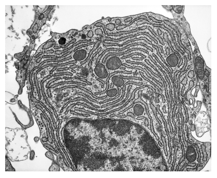
Which statement explains how it is possible to identify the type of microscope used to produce the micrograph?
Options
A The nucleus is visible, so an electron microscope was used.
B The endoplasmic reticulum is not visible, so a light microscope was used.
C Chloroplasts are visible, so a light microscope was used.
D Ribosomes are visible, so an electron microscope was used.
Working
Ribosomes are only about 20–30 nm in diameter, which is below the resolution of a light microscope (~200 nm). Therefore ribosomes can only be seen using an electron microscope. The micrograph clearly shows individual ribosomes studding the rough endoplasmic reticulum, which confirms an electron microscope was used.
- A is wrong: nuclei are easily visible with a light microscope (they are typically 5–10 µm across).
- B is wrong: the endoplasmic reticulum is clearly visible in the micrograph, so this statement is factually incorrect.
- C is wrong: no chloroplasts are visible, and chloroplasts are large enough to be seen with a light microscope anyway.
Answer
D
D
Background Concept
The key concept is the resolving power of a microscope, which is the smallest distance between two objects at which they can still be distinguished as separate. The resolution of a light microscope is limited by the wavelength of visible light to approximately 200 nm. The resolution of an electron microscope is much greater because electrons have a much shorter wavelength, allowing it to resolve structures as small as about 0.1–0.2 nm.
This means that any organelle larger than 200 nm can be seen with a light microscope, while organelles smaller than 200 nm can only be visualised with an electron microscope. Common organelle size benchmarks for this topic:
- Nucleus (~5–10 µm): visible with light microscope
- Mitochondrion (~0.5–1 µm): visible with light microscope
- Chloroplast (~5–10 µm): visible with light microscope
- Endoplasmic reticulum (cisternae, ~50–100 nm wide): at the limit; usually seen clearly only with electron microscope
- Ribosome (~20–30 nm): electron microscope only
Understanding the Question
The question presents a micrograph and asks which statement correctly identifies the microscope type from the image. The image (Fig. 2.1) is an electron micrograph showing extensive rough endoplasmic reticulum with numerous small dark dots (ribosomes) studding the membrane surfaces, a large dark nucleus, and scattered mitochondria. The task is to pick the option that uses a feature whose mere visibility proves which type of microscope was used.
Approach
For a statement to identify the microscope type, the feature cited must be:
- Actually visible in the image, and
- Only visible with one specific type of microscope (i.e. smaller than the resolution limit of the other type).
Check each option against both criteria.
Step-by-Step Reasoning
- Option A — nucleus visible → electron microscope. The nucleus is several micrometres across, far larger than the ~200 nm resolution of a light microscope. Nuclei are routinely seen in light-microscope images (e.g. onion epidermis cells). So a visible nucleus does NOT prove an electron microscope was used. Reject.
- Option B — endoplasmic reticulum not visible → light microscope. Two problems. First, the endoplasmic reticulum IS clearly visible in the micrograph (the extensive membrane network filling the cytoplasm), so the statement is factually wrong. Second, even if it were absent, absence of evidence is not evidence — you cannot conclude the microscope type from a structure not being shown. Reject.
- Option C — chloroplasts visible → light microscope. No chloroplasts are visible in this image (the cell is a secretory-type animal cell). Furthermore, chloroplasts are large enough to be seen with both light and electron microscopes, so their visibility would not uniquely identify a light microscope. Reject.
- Option D — ribosomes visible → electron microscope. Ribosomes are roughly 20–30 nm in diameter. This is well below the resolution limit of a light microscope (~200 nm), so a light microscope cannot resolve individual ribosomes. The fact that the small dark dots on the rough ER membranes are clearly visible in Fig. 2.1 can only be explained by the use of an electron microscope. Correct.
Key Takeaways
- Resolution (not magnification) determines what can be seen. High magnification with poor resolution just produces a larger blurry image.
- Light microscope resolution ≈ 200 nm; electron microscope resolution ≈ 0.1–0.2 nm.
- Structures unique to electron micrographs (i.e. sub-200 nm): ribosomes, detailed ER structure, individual cristae of mitochondria, viruses.
- Structures visible with light microscopes: nucleus, chloroplasts, mitochondria (as small shapes), vacuoles, cell walls.
Common Mistakes
- Confusing magnification with resolution. A light microscope can be made to magnify hugely, but it still cannot resolve ribosomes — this is a very common exam trap.
- Reasoning from absence. Saying "feature X is not visible, therefore it must be a light microscope" is invalid; you can never be sure that a structure is truly absent from a sample just because it does not appear in one image.
- Assuming a visible nucleus implies electron microscopy. Nuclei are large and easily seen with light microscopes; this is a classic distractor in this type of question.
Things to Be Careful About
- The presence of dark patches on the membranes in this image is the diagnostic feature — those are ribosomes, and their visibility is the giveaway.
- Always check that the organelle mentioned in the option is actually present in the image, as well as being below the resolution of one of the microscope types.
Which statement supports the fact that mature plant cells contain organelles that carry out the same role as lysosomes?
Options
A A range of hydrolytic enzymes can be found within mature plant vacuoles.
B Glycogen, found within vesicles, can be hydrolysed to glucose molecules.
C Double membrane-bound vesicles are formed from plant Golgi bodies.
D Vesicles, formed from the cell surface membrane, contain enzymes.
Working
Lysosomes are membrane-bound organelles that contain hydrolytic (digestive) enzymes used to break down unwanted molecules, organelles, or engulfed pathogens within the cell. Mature plant cells do not contain obvious lysosomes, but the large central vacuole performs the same role because it too contains a range of hydrolytic enzymes capable of digesting materials internally.
Evaluating each option:
- A — Correct: hydrolytic enzymes within mature plant vacuoles provide the same digestive function as lysosomal enzymes in animal cells.
- B — Incorrect: glycogen is an animal storage polysaccharide; plant cells store carbohydrate as starch, and the statement is unrelated to lysosomal function.
- C — Incorrect: double-membrane-bound vesicles describe mitochondria/chloroplast-like structures, not digestive organelles.
- D — Incorrect: vesicles formed from the cell surface membrane are endocytic, not lysosomal.
Answer
A
A
Background Concept
Lysosomes are small, spherical, membrane-bound organelles found in animal cells. They are formed from vesicles that bud off the Golgi apparatus (or, in some descriptions, the rough endoplasmic reticulum) and contain a cocktail of hydrolytic enzymes — proteases, nucleases, lipases and carbohydrases — active at the acidic internal pH (around pH 4.5–5.0), maintained by H⁺-ATPases in the lysosomal membrane. Their role is intracellular digestion: they break down engulfed material delivered by endocytosis, recycle worn-out organelles during autophagy, and, in some immune cells, digest pathogens after phagocytosis.
Plant cells do not usually show discrete lysosomes, but they still need a compartment in which hydrolysis can occur safely — releasing these powerful enzymes into the cytosol would digest the cell itself. This compartment is the large central vacuole of the mature plant cell. Tonoplast (the vacuolar membrane) keeps the hydrolytic enzymes separate from the cytoplasm, and the lumen of the vacuole becomes mildly acidic, allowing the same digestive processes to occur. The vacuole additionally stores water, ions, pigments and waste products, but its enzyme content means it is functionally the plant's lysosome.
Understanding the Question
The command is to identify the statement that supports the idea that mature plant cells contain organelles doing the same job as lysosomes. The question is testing whether the candidate knows which plant organelle carries the hydrolytic enzymes and recognises that this is sufficient evidence of a shared function. The distractor statements are designed to look plausible by mentioning vesicles, membranes or enzymes but to be wrong in one crucial biological detail.
Approach
First, recall the defining feature of a lysosome: a membrane-bound vesicle containing hydrolytic enzymes. Then, identify which plant structure contains hydrolytic enzymes in a membrane-bound compartment. Eliminate the distractors by checking the biology of each claim (storage polysaccharide in plants, membrane count of common organelles, origin of endocytic vesicles).
Step-by-Step Reasoning
- Option A states that mature plant vacuoles contain a range of hydrolytic enzymes. This is true — the central vacuole of a mature plant cell holds acid hydrolases that digest proteins, nucleic acids and other macromolecules, fulfilling the same intracellular digestive role as animal lysosomes. The vacuole's membrane (tonoplast) acts as the boundary that keeps these enzymes compartmentalised, exactly as the lysosomal membrane does. This is the supporting statement.
- Option B mentions glycogen in vesicles. Glycogen is an animal storage polysaccharide (mainly in liver and muscle); plants store carbohydrate as starch in amyloplasts, not glycogen. Even if such a vesicle existed, hydrolysis of glycogen to glucose is a metabolic mobilisation, not the broad hydrolytic/digestive role characteristic of lysosomes. Rejected.
- Option C describes double-membrane-bound vesicles formed from plant Golgi bodies. Golgi-derived vesicles are single-membrane-bound; double membranes are characteristic of mitochondria and chloroplasts (and of the nuclear envelope), and these organelles do not perform lysosomal digestion. Rejected.
- Option D describes vesicles formed from the cell surface membrane. These are endocytic vesicles, which deliver material to lysosomes in animal cells — they are not themselves lysosomes and do not, in plants, contain the full hydrolytic enzyme complement. Rejected.
Key Takeaways
- Lysosomes = membrane-bound vesicles containing hydrolytic enzymes for intracellular digestion.
- Mature plant cells lack obvious lysosomes; the central vacuole performs the equivalent function because it contains a similar set of hydrolytic enzymes within a membrane (the tonoplast).
- When comparing plant and animal cells, do not look for identical structures — look for structures performing the same function (e.g. cell wall ↔ no animal equivalent, but vacuole ↔ multiple animal roles including that of the lysosome).
- Be alert to distractors using correct-sounding vocabulary (membranes, vesicles, enzymes) but applied to the wrong organelle or the wrong species.
Common Mistakes
- Confusing plant and animal storage carbohydrates (starch vs glycogen).
- Equating any vesicle containing an enzyme with a lysosome — the range of hydrolytic enzymes and the membrane-bound compartment are the defining features.
- Assuming "double-membrane-bound" implies a digestive organelle — this describes mitochondria/chloroplasts, not lysosome-equivalents.
- Choosing D because it mentions enzymes in vesicles, without noticing that endocytic vesicles are transport, not digestive, compartments.
Things to Be Careful About
- Use the precise CIE term hydrolytic enzymes when describing lysosomal contents — "digestive enzymes" is informal and the more specific phrase is preferred.
- Remember the tonoplast as the membrane bounding the plant vacuole, paralleling the lysosomal membrane.
- Do not credit glycogen as a plant cell component under any circumstances — this is a reliable mark-scheme rejection in CIE mark schemes.
Which cell structures may contain cisternae?
Options
| chloroplast | endoplasmic reticulum | Golgi body | mitochondrion | |
|---|---|---|---|---|
| A | ✓ | ✓ | ✓ | ✗ |
| B | ✓ | ✗ | ✗ | ✓ |
| C | ✗ | ✓ | ✓ | ✗ |
| D | ✗ | ✓ | ✗ | ✓ |
key
✓ = may contain cisternae
✗ = does not contain cisternae
Working
A cisterna is a flattened, fluid-filled, membrane-bound sac. The cell structures whose membranes are organised into flattened sacs are:
- Endoplasmic reticulum (ER) ✓ — formed of flattened membrane-bound cisternae (rough ER has ribosomes; smooth ER does not).
- Golgi body ✓ — a stack of flattened membrane-bound cisternae, with vesicles budding from the ends.
The other two organelles do not have cisternae:
- Chloroplast ✗ — the internal flattened sacs are thylakoids (stacked into grana), not cisternae.
- Mitochondrion ✗ — the inner membrane is folded into cristae, not flattened cisternae.
The combination is therefore: chloroplast ✗, ER ✓, Golgi ✓, mitochondrion ✗.
Answer
C
C
Background Concept
A cisterna (plural: cisternae) is a flattened, closed, fluid-filled sac bounded by a single membrane. Several membrane-bound organelles build their internal architecture from stacks or networks of cisternae, while others use different membrane arrangements (tubules, vesicles, or infoldings). Being able to identify which is which is a basic ultrastructure skill.
The four organelles in the question each have a characteristic internal membrane arrangement:
- Rough endoplasmic reticulum (RER) / Smooth endoplasmic reticulum (SER): a continuous network of flattened membranous sacs (cisternae) connected to the nuclear envelope. RER has ribosomes on its cytoplasmic face; SER does not.
- Golgi body (Golgi apparatus): a stack of 3–10 flattened membranous sacs (cisternae), with a cis face (receiving side, facing the ER) and a trans face (shipping side, releasing vesicles).
- Chloroplast: the internal membrane system consists of flattened green-pigmented discs called thylakoids, which stack into grana (singular: granum). The fluid surrounding them is the stroma. These are NOT called cisternae.
- Mitochondrion: the inner membrane is thrown into folds called cristae (which greatly increase surface area for the electron-transport chain and ATP synthase). These are tubular infoldings, not flattened sacs, so they are not cisternae.
Understanding the Question
The question is a multiple-choice check on terminology. It lists four organelles and asks which of them "may contain cisternae". The candidate must know the precise internal structure of each organelle and recognise that the word cisterna applies to the ER and Golgi only. The matrix format (✓/✗) is testing for one correct combination across the four options.
Approach
Match each organelle against the strict definition of a cisterna — a flattened, fluid-filled, membrane-bound sac. Discard any organelle whose internal membranes are organised differently (thylakoids in chloroplasts, cristae in mitochondria), and select the option that places ✓ against ER and Golgi and ✗ against chloroplast and mitochondrion.
Step-by-Step Reasoning
- Endoplasmic reticulum: The RER/SER consists of flattened sacs (cisternae) continuous with the nuclear envelope. ✓ contains cisternae.
- Golgi body: Each Golgi stack is built from flattened membranous sacs (cisternae). ✓ contains cisternae.
- Chloroplast: Internal membranes form thylakoids/grana, suspended in stroma. ✗ does not contain cisternae — the term is reserved for ER/Golgi-type sacs.
- Mitochondrion: The inner membrane folds into cristae. ✗ does not contain cisternae.
Reading the table:
- A: chloroplast ✓, ER ✓, Golgi ✓, mitochondrion ✗ — wrong (chloroplast does not have cisternae).
- B: chloroplast ✓, ER ✗, Golgi ✗, mitochondrion ✓ — wrong on all four counts.
- C: chloroplast ✗, ER ✓, Golgi ✓, mitochondrion ✗ — correct.
- D: chloroplast ✗, ER ✓, Golgi ✗, mitochondrion ✓ — wrong (mitochondrion does not have cisternae; Golgi does).
Key Takeaways
- Cisternae = flattened, closed, membrane-bound sacs. Found in the ER and Golgi body.
- Thylakoids/grana are the flattened internal discs of chloroplasts — different name, different organelle.
- Cristae are the infoldings of the mitochondrial inner membrane — also not cisternae.
- The term cisterna is sometimes loosely used in textbooks for chloroplast/mitochondrial compartments, but in CIE A-level Biology it is restricted to ER and Golgi.
Common Mistakes
- Calling thylakoids "chloroplast cisternae" — common slip; they are functionally and structurally distinct.
- Calling cristae "mitochondrial cisternae" — cristae are tubular/sheet-like infoldings, not stacked sacs.
- Confusing the Golgi with the ER; both have cisternae but in different arrangements (Golgi = discrete stack with a defined cis–trans polarity; ER = continuous network).
Things to Be Careful About
- The question uses the word "may contain" — so any organelle that is capable of having cisternae scores ✓, even if the section happens to show a different feature.
- Watch the matrix direction: ✓ = may contain, ✗ = does not contain. Reversing this interpretation will systematically pick the wrong option.
- The nuclear envelope is also continuous with the ER and is sometimes described as a single cisterna, but the question does not include it, so the answer depends only on the four organelles listed.
The diagram shows some cell structures of one type of cell.
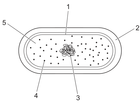
Which labelled cell structures are present in typical eukaryotic cells and typical bacterial cells?
Options
A 1, 2, 3 and 4
B 1, 3, 4 and 5
C 1, 2 and 3 only
D 3, 4 and 5 only
Working
The labelled structures in the prokaryotic cell are:
- 1 – circular DNA (nucleoid)
- 2 – plasmid
- 3 – ribosomes
- 4 – cytoplasm
- 5 – cell wall
Checking which are present in typical eukaryotic cells as well:
- 1 (circular DNA): present — eukaryotes have linear nuclear DNA, but their mitochondria and chloroplasts contain circular DNA.
- 2 (plasmid): absent — plasmids are characteristic of bacteria (not typical eukaryotes).
- 3 (ribosomes): present — both cell types have ribosomes (eukaryotic ribosomes are larger, but they are present).
- 4 (cytoplasm): present — both cell types have cytoplasm.
- 5 (cell wall): present — plant and fungal cells (typical eukaryotes alongside animal cells) possess cell walls, and so do bacteria.
Structures present in both typical eukaryotic and typical bacterial cells: 1, 3, 4 and 5.
Answer
B
B
Background Concept
All cells share a small set of core features: a plasma membrane, cytoplasm, ribosomes (for protein synthesis), and DNA (carrying the genetic information). Beyond these, prokaryotic and eukaryotic cells differ in important ways:
- Prokaryotic cells (bacteria) are small (typically 1–5 µm), lack a true nucleus, and usually have a single circular chromosome located in a region called the nucleoid. They commonly carry small rings of DNA called plasmids in addition to the main chromosome, and almost all have a cell wall made of peptidoglycan. They have 70S ribosomes (made of a 50S and a 30S subunit) free in the cytoplasm.
- Eukaryotic cells are larger and have a membrane-bound nucleus containing linear chromosomes. Their ribosomes (80S) are larger and can be free in the cytoplasm or bound to the endoplasmic reticulum. Many eukaryotic cells — including plant, fungal, and many microbial cells — have a cell wall (cellulose in plants, chitin in fungi). Eukaryotic cells also contain membrane-bound organelles such as mitochondria, and in plants, chloroplasts.
A subtle but important point: although the main nuclear DNA of a eukaryote is linear, the DNA inside mitochondria and chloroplasts is circular, because these organelles evolved from free-living prokaryotes via endosymbiosis. So a typical eukaryotic cell does contain circular DNA — just not in its nucleus.
Understanding the Question
The question shows a labelled diagram of a prokaryotic (bacterial) cell and asks which of the five labelled structures are present in both typical eukaryotic cells and typical bacterial cells. This is a "shared features" question, so the correct answer is the option whose structures are not unique to bacteria.
The diagram labels are:
- 1 → circular DNA (nucleoid)
- 2 → plasmid
- 3 → ribosomes
- 4 → cytoplasm
- 5 → cell wall
Approach
Go through each label and decide: Is this structure found in a typical eukaryotic cell as well? Discard any label whose structure is essentially bacterial-only, then choose the matching option.
Step-by-Step Reasoning
-
Label 1 — circular DNA: Eukaryotic cells have linear DNA in the nucleus, but their mitochondria (and chloroplasts in plants) contain circular DNA. So a typical eukaryotic cell does contain circular DNA. Present in both. ✓
-
Label 2 — plasmid: Plasmids are small, self-replicating circular DNA molecules found mainly in bacteria (and in a few yeast strains used in biotechnology). They are not a feature of typical eukaryotic cells. Bacterial only. ✗
-
Label 3 — ribosomes: Both cell types need to make proteins, so both have ribosomes. Eukaryotic ribosomes (80S) are larger than bacterial ones (70S), but ribosomes as a structure are present in both. Present in both. ✓
-
Label 4 — cytoplasm: The cytoplasm (including cytosol and, in eukaryotes, cytoplasmic organelles) is a universal feature of all cells. Present in both. ✓
-
Label 5 — cell wall: Bacteria have cell walls (peptidoglycan). Many typical eukaryotes also have cell walls — plant cells (cellulose), fungal cells (chitin), and most algae. The phrase "typical eukaryotic cell" in this question encompasses plant and animal cells; with plants/fungi included, cell walls are present in both groups. Present in both. ✓
The structures present in both are 1, 3, 4 and 5. This matches option B.
Key Takeaways
- The features that all cells share are: plasma membrane, cytoplasm, ribosomes, and DNA.
- Plasmids are a bacterial feature and are the strongest "bacteria-only" tell in this question — that is why option B (which excludes 2) is correct.
- Circular DNA in eukaryotes is found in mitochondria and chloroplasts, not the nucleus — this is a key fact from endosymbiotic theory.
- Cell walls are not unique to plants; bacteria, fungi, and algae also have them, but typical animal cells do not.
Common Mistakes
- Choosing D (3, 4, 5 only): forgetting that mitochondria and chloroplasts contain circular DNA, so label 1 should also be credited.
- Choosing A (1, 2, 3, 4): assuming plasmids exist in typical eukaryotic cells, or missing that the question is asking for structures shared with eukaryotes (a plasmid is a "bacteria-only" structure).
- Choosing C (1, 2, 3 only): excluding the cytoplasm or the cell wall for an unclear reason.
- Confusing "typical eukaryotic cell" with "animal cell": animal cells lack a cell wall and have no circular DNA, but "typical" eukaryotes include plant and fungal cells, which change the answer.
Things to Be Careful About
- The wording "present in typical eukaryotic cells and typical bacterial cells" means both must have it — a structure found in only one cell type is excluded.
- Circular DNA ≠ plasmid: both are circular, but plasmids are small, extrachromosomal, and largely bacterial; circular DNA as a feature is broader.
- The size of the ribosome (70S vs 80S) is a difference between the two cell types, but the presence of ribosomes is shared — do not be misled into thinking eukaryotes lack ribosomes.
- "Cell wall" is a structural category, not a specific molecule: bacterial walls (peptidoglycan) and plant walls (cellulose) are chemically different but satisfy the same structural description.
The diagram shows part of a collagen fibril made of collagen triple helices. The collagen triple helices are linked to each other by one type of bond. This bond is labelled as X in the diagram.
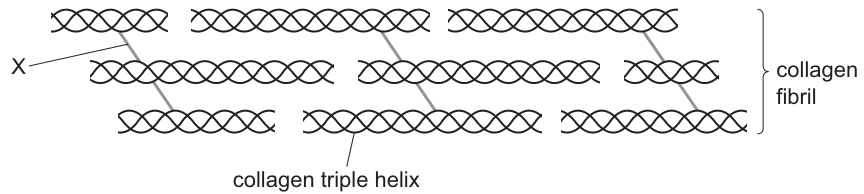
What is bond X?
Options
A covalent bond
B disulfide bond
C hydrogen bond
D peptide bond
Working
Within a single collagen triple helix the three polypeptide chains are held together by hydrogen bonds. Bond X, however, is shown as a cross-link between adjacent triple helices in the collagen fibril. These inter-fibril cross-links are covalent bonds, formed between modified lysine / hydroxylysine residues (catalysed by the enzyme lysyl oxidase). They are not hydrogen bonds (those are within the triple helix), disulfide bonds (no cysteine involvement here), or peptide bonds (those join amino acids within a single chain).
Answer
A
A
Background Concept
Collagen is the most abundant fibrous protein in animals. Its basic structural unit is the tropocollagen molecule, which consists of three polypeptide (α) chains wound around each other into a right-handed triple helix (sometimes called tropocollagen). Each α chain has a repeating sequence in which every third residue is glycine; the small glycine residues pack into the centre of the triple helix, allowing the three chains to lie close together.
Two different kinds of bond are involved in stabilising collagen:
- Hydrogen bonds — run within the triple helix, between the backbone N–H of one chain and the C=O of a neighbouring chain. They stabilise the three α chains as a single triple helix.
- Covalent cross-links — form between the side chains of adjacent tropocollagen molecules once they have been assembled into a fibril. The enzyme lysyl oxidase oxidatively deaminates selected lysine and hydroxylysine side chains to form reactive aldehydes, which then condense with neighbouring lysine/hydroxylysine residues to give a strong covalent bond. These covalent cross-links tie many triple helices together into the staggered array seen in a collagen fibril and ultimately into a collagen fibre.
The diagram in Fig. 6.1 shows the classic staggered arrangement of triple helices within a fibril, with the cross-links drawn as the grey bars labelled X.
Understanding the Question
The question shows a collagen fibril and points to bond X, which is positioned between two adjacent triple helices (not within a single one). The question simply asks what type of bond this is. The candidate must therefore identify the inter-helix cross-link — and crucially distinguish it from the hydrogen bonds that hold the three chains of one triple helix together.
Approach
Ask: where in the structure is the bond located? If the bond is between separate triple helices in the fibril, it must be a covalent cross-link. If it were within a single triple helix it would be a hydrogen bond. Then eliminate the distractors that do not fit collagen cross-links (disulfide requires cysteine, peptide bonds join amino acids within a single chain).
Step-by-Step Reasoning
- Locate bond X in the figure: it bridges two separate triple helices — it is an inter-helix bond, not an intra-helix bond.
- Hydrogen bonds (option C) hold the three chains of one triple helix together internally, so this is not the bond shown.
- Peptide bonds (option D) join amino acids within a single polypeptide chain and are formed during translation; they are not drawn as cross-links between separate molecules.
- Disulfide bonds (option B) are covalent, but they form specifically between two cysteine residues. Collagen's cross-links are not disulfide bonds — they form between modified lysine / hydroxylysine side chains.
- The inter-helix cross-links of collagen are covalent bonds formed by the enzyme lysyl oxidase acting on lysine/hydroxylysine side chains. Option A is therefore correct.
Key Takeaways
- Within a collagen triple helix → hydrogen bonds (between the three α chains).
- Between collagen triple helices in a fibril → covalent cross-links (between lysine / hydroxylysine side chains, made via lysyl oxidase).
- Collagen is a fibrous protein whose strength comes from both regular hydrogen bonding and covalent cross-linking — a good example of structure relating to function (high tensile strength in tendons, bone, skin, blood-vessel walls).
Common Mistakes
- Choosing C (hydrogen bond) because the candidate associates collagen with hydrogen bonding without distinguishing where the hydrogen bonds are.
- Choosing B (disulfide bond) because the candidate associates "strong structural bond" with disulfide, without checking that disulfide requires cysteine residues (collagen has very few cysteines).
- Choosing D (peptide bond) because the candidate confuses the inter-chain cross-link with the bonds that join amino acids along the chain.
Things to Be Careful About
- The mark scheme credits covalent bond as the general category; specifying "disulfide" is not accepted for the inter-fibril cross-links in collagen.
- Read the diagram carefully: bond X is between two triple helices, not within one. The question hinges on this distinction.
- Do not credit answers that describe the bond in functional terms only (e.g. "strong bond") without naming the correct type.
The table shows some information about the polypeptides that make up haemoglobin.
| -globin | -globin | |
|---|---|---|
| total number of amino acid residues in polypeptide chain | 141 | 146 |
| position of amino acid cysteine in polypeptide chain | 104 | 93 and 112 |
Scientists studied the region of the -globin polypeptide chain containing the amino acid cysteine at position 93. They found that:
● this region faces outwards when no oxygen is attached to the haem group
● this region faces inwards when oxygen is attached to the haem group
● replacing cysteine with a different amino acid reduces the Bohr shift.
What can be concluded from the information about cysteine in haemoglobin?
Options
A More than 1% of the amino acids in one haemoglobin protein are cysteine.
B In -globin, there is a cysteine closer to the end of the polypeptide chain with an unreacted carboxyl group than in -globin.
C The replacement of the cysteine at position 93 in -globin decreases the affinity of haemoglobin for oxygen at low pH.
D The binding of oxygen to the haem group causes the region of -globin containing cysteine at position 93 to become more hydrophilic.
Working
A haemoglobin molecule is a tetramer: .
Total amino acid residues:
Total cysteine residues (1 per α-chain, 2 per β-chain):
Percentage of cysteine:
So statement A is true.
Check the other options:
- B: Distance from C-terminus. α-cysteine at 104 → residues from C-terminus. The nearer β-cysteine at 112 → residues from C-terminus. β-globin has the cysteine closer to the C-terminus, so B is false.
- C: Reducing the Bohr shift means haemoglobin affinity at low pH falls less than normal — affinity at low pH is higher, not lower. So C is false.
- D: When O binds, the cysteine-93 region moves inwards (toward the hydrophobic haem pocket), so the region becomes more hydrophobic, not more hydrophilic. So D is false.
Answer
A
A
Background Concept
Haemoglobin is a globular protein with quaternary structure: four polypeptide chains — two -globin (141 residues each) and two -globin (146 residues each) — each carrying one haem prosthetic group that binds .
Cysteine is an amino acid whose side chain terminates in a thiol () group. It is relatively non-polar and is often found in the hydrophobic interior of globular proteins, where two cysteines can form a disulfide () bridge that stabilises tertiary structure.
The Bohr shift describes how a fall in pH (rise in concentration) reduces haemoglobin's affinity for , causing the oxygen dissociation curve to shift to the right. This is physiologically important because actively respiring tissues (which produce and therefore lower the local pH) cause haemoglobin to release more readily.
Understanding the Question
We are told the positions of every cysteine in - and -globin, plus three findings about the cysteine-93 region of -globin:
- It faces outwards when no is bound.
- It faces inwards when is bound.
- Replacing this cysteine reduces the Bohr shift.
We must decide which of four statements can be concluded from this information.
Approach
A conclusion must be directly supported by the given data (or by simple calculation from it). For each option, test whether the data forces the conclusion to be true.
Step-by-Step Reasoning
Option A — percentage of cysteine in one haemoglobin.
A single haemoglobin molecule contains amino acid residues, with cysteines. The percentage is
which is indeed greater than 1%. A is supported by calculation — TRUE.
Option B — position of cysteine relative to the C-terminus.
The C-terminus is residue 1 from the C-terminal end. Counting back from the C-terminus:
- -globin cysteine (position 104): residues from C-terminus.
- -globin cysteines (positions 93 and 112): the nearer one (112) is residues from C-terminus.
So the -globin cysteine at 112 is closer to the C-terminus than the -globin cysteine. Option B reverses this — FALSE.
Option C — Bohr shift direction.
"Replacing cysteine reduces the Bohr shift" means the pH-induced drop in affinity is smaller, i.e. at low pH the affinity is higher than in the wild-type protein. The option claims affinity at low pH decreases — opposite of what the data imply. FALSE.
Option D — change in environment of cysteine-93.
When binds, the cysteine-93 region moves inwards, into the hydrophobic pocket that contains the haem group. This means the local environment becomes more hydrophobic, not more hydrophilic. FALSE.
Only option A follows directly from the data.
Key Takeaways
- Haemoglobin is — any whole-protein calculation must multiply chain data by 2.
- "Faces inwards" in a globular protein means a hydrophobic interior; "faces outwards" means exposed to the aqueous cytosol.
- The Bohr shift = reduced affinity at low pH. Anything that reduces the Bohr shift therefore raises the affinity at low pH.
- Always cross-check a numeric claim by calculation before accepting it.
Common Mistakes
- Forgetting the tetramer. Treating "one haemoglobin" as a single chain gives 1/141 ≈ 0.7% or 2/146 ≈ 1.4%, which scatters students away from the correct >1% conclusion.
- Confusing the Bohr shift direction. Students who do not distinguish "reducing the Bohr shift" from "removing the Bohr shift entirely" may wrongly pick C.
- Treating cysteine as hydrophilic. The group is only weakly polar; the residue is generally classed as hydrophobic and is buried inside globular proteins.
- Reading positions from the wrong end. Polypeptide positions are conventionally numbered from the N-terminus. Distance from the C-terminus = (chain length − position number).
Things to Be Careful About
- Use and chain lengths exactly as given; do not round.
- A single haemoglobin has six cysteines in total (2 in + 4 in ).
- When asked what "can be concluded", look for the statement that is logically forced by the data, not merely consistent with it.
- "Faces inwards" in a haemoglobin subunit means towards the haem pocket — a hydrophobic environment.
Which feature of glycogen distinguishes it from starch?
Options
A All glycogen molecules are highly branched.
B All glycogen molecules are polysaccharides.
C All glycogen molecules contain -glucose.
D All glycogen molecules contain 1,4-glycosidic bonds.
Working
Starch is a mixture of amylose (unbranched) and amylopectin (branched, but with fewer and longer branches than glycogen). Glycogen, however, is always highly branched with 1,6-glycosidic bonds every 8–12 glucose units. Therefore the only feature that genuinely distinguishes ALL glycogen molecules from starch is that every glycogen molecule is highly branched.
- B is wrong: starch is also a polysaccharide.
- C is wrong: both are made of α-glucose.
- D is wrong: both contain 1,4-glycosidic bonds.
Answer
A
A
Background Concept
Glycogen and starch are both storage polysaccharides made from α-glucose monomers linked by 1,4-glycosidic bonds, with branches formed through 1,6-glycosidic bonds. Their difference lies in the degree and frequency of branching:
- Starch (in plants) is a mixture of two polymers: amylose, an unbranched chain of α-glucose joined only by 1,4-bonds, and amylopectin, which is branched but with side chains every 24–30 glucose units.
- Glycogen (in animals and fungi) is structurally similar to amylopectin but much more highly branched, with side chains arising roughly every 8–12 glucose units, and the molecule is more compact.
The extensive branching of glycogen gives many free ends from which glucose can be released rapidly during metabolism, which is why it is the main storage polysaccharide in animals.
Understanding the Question
The question asks for the feature that distinguishes glycogen from starch — in other words, the one property that is true of ALL glycogen molecules but NOT true of ALL starch molecules. A wrong answer would be a property shared by both.
Approach
Compare each option against the structure of starch to see whether it is also a property of starch. The option that is true only of glycogen is the answer.
Step-by-Step Reasoning
- A – All glycogen molecules are highly branched. True. Every glycogen molecule is a heavily branched structure with 1,6-branches every 8–12 residues. Starch is NOT entirely highly branched: amylose has no branches at all. This property therefore distinguishes glycogen from starch. ✓
- B – All glycogen molecules are polysaccharides. True, but starch is also a polysaccharide. Not distinguishing. ✗
- C – All glycogen molecules contain α-glucose. True, but starch is also built from α-glucose monomers. Not distinguishing. ✗
- D – All glycogen molecules contain 1,4-glycosidic bonds. True, but starch (including unbranched amylose) also contains 1,4-glycosidic bonds. Not distinguishing. ✗
Only option A isolates a structural feature unique to glycogen.
Key Takeaways
- Glycogen and starch share: α-glucose monomers, 1,4-glycosidic bonds, and a polysaccharide nature.
- The defining difference is branching: glycogen is always highly branched; starch is a mix of unbranched amylose and moderately branched amylopectin.
- Greater branching = more terminal ends = faster mobilisation of glucose when required.
Common Mistakes
- Choosing D because "1,4-bonds" sounds distinctive — forgetting that amylose is entirely 1,4-bonded and unbranched.
- Choosing C because α-glucose is the monomer — failing to note that starch is also made of α-glucose.
- Confusing glycogen with cellulose: cellulose uses β-glucose, not α-glucose, but starch does not.
Things to Be Careful About
- "Highly branched" must be qualified — amylopectin is branched, just less so than glycogen. The mark-bearing word is highly.
- The question says "distinguishes", so any property also true of starch cannot earn the mark.
- Do not confuse branching (1,6-bonds) with the main-chain linkage (1,4-bonds); both polymers contain 1,4-bonds but differ in how many 1,6-branches they carry.
The diagram shows a biological molecule.
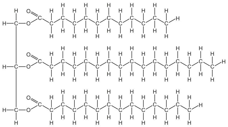
Which molecules would be produced if this biological molecule was hydrolysed?
Options
A amino acids and glycerol only
B amino acids, glycerol and water
C fatty acids and glycerol only
D fatty acids, glycerol and water
Working
The diagram shows a glycerol backbone (three carbons on the left, each with –OH groups in the original molecule) linked via three ester bonds to three long hydrocarbon chains (saturated fatty acids). This is a triglyceride.
Hydrolysis breaks the three ester bonds. Because hydrolysis means "splitting with water", a water molecule is consumed (H added to one fragment, OH to the other) for each bond broken. Water is therefore a reactant, not a product.
The products of hydrolysing a triglyceride are:
- 3 fatty acids
- 1 glycerol
Answer
C
C
Background Concept
A triglyceride (triacylglycerol) is a lipid formed from one glycerol molecule (a 3-carbon alcohol) and three fatty acid molecules. Each fatty acid is joined to the glycerol by an ester bond, which forms when the –OH of glycerol's hydroxyl group reacts with the –COOH of a fatty acid, releasing a molecule of water (a condensation reaction).
Hydrolysis is the reverse of condensation. The word literally means "splitting with water" ( = water, = splitting). To break an ester bond, a water molecule is added across the bond: the –H joins one side and the –OH joins the other. Water is therefore a reactant in hydrolysis, not a product.
Understanding the Question
Fig. 9.1 shows a structural formula: on the left, a vertical three-carbon glycerol backbone; extending to the right, three long hydrocarbon chains attached through groups (the ester linkages). The hydrocarbon chains are made of repeating units ending in , indicating they are saturated fatty acids. The molecule shown is therefore a triglyceride.
The question asks what is produced when this molecule is hydrolysed. The key trap is option B/D, which include water as a product — water is needed to drive hydrolysis, so it cannot also be a product of it.
Approach
- Identify the molecule from its structure.
- Recall the products of hydrolysing that class of molecule.
- Decide whether water is consumed or released, and eliminate any option that lists water as a product.
Step-by-Step Reasoning
- Step 1 — Identify the molecule: A central three-carbon unit (glycerol) attached by ester bonds () to three long hydrocarbon chains = a triglyceride.
- Step 2 — Apply hydrolysis: Hydrolysing the three ester bonds releases the three fatty acid chains from the glycerol backbone.
- Step 3 — Account for water: Hydrolysis requires one water molecule per ester bond broken. Water is a reactant, so it is not in the list of products.
- Step 4 — Match to options: Products = fatty acids + glycerol only → C.
Eliminating distractors:
- A (amino acids and glycerol): Amino acids are the monomers of proteins (joined by peptide bonds), not lipids. Wrong class of molecule.
- B (amino acids, glycerol, water): Amino acids are wrong, and water is consumed, not produced.
- D (fatty acids, glycerol, water): Fatty acids and glycerol are correct, but water is a reactant, not a product.
- C (fatty acids and glycerol only): Correct.
Key Takeaways
- A triglyceride = glycerol + 3 fatty acids, linked by 3 ester bonds.
- Hydrolysis of a triglyceride gives fatty acids and glycerol only.
- Water is a reactant in hydrolysis (it is split across the bond), never a product.
- The reverse synthesis reaction (condensation/esterification) releases water.
Common Mistakes
- Confusing hydrolysis with condensation: students often put water in the products list because they remember that water is involved in the reaction. The direction of the reaction determines whether water is consumed or released.
- Confusing triglycerides with proteins: option A tempts students who misread the structure as amino acids joined together.
- Forgetting that each triglyceride has three fatty acids, not one — but this does not change the identity of the product types.
Things to Be Careful About
- Ester bonds () are the markers of a lipid; peptide bonds () mark a protein.
- Saturated vs unsaturated fatty acids: the presence of double bonds would indicate unsaturated fatty acids, but this does not change the hydrolysis products.
- The terminology "hydrolysed" implies that an aqueous (often acidic or enzymic) environment is breaking bonds with water; water is the reagent, not the product.
A mixture of glucose and starch solutions was placed in a length of dialysis (Visking) tubing and the tubing sealed. The tubing was then placed in a boiling tube containing distilled water. Two samples were immediately removed from this water (time 0 minutes) and tested with either iodine solution or Benedict’s solution. This was repeated at 10 minute intervals for 30 minutes.
The iodine solution gave an orange-brown colour each time.
The table shows the results of the Benedict’s test.
| time / minutes | ||||
|---|---|---|---|---|
| 0 | 10 | 20 | 30 | |
| colour produced by Benedict’s test | blue | green | yellow | red |
What may be concluded from these results?
1 The pores in the Visking tubing are too small for a starch molecule to pass through.
2 Glucose diffuses through the Visking tubing down a diffusion gradient.
3 Water diffuses into the Visking tubing.
Options
A 1, 2 and 3
B 1 and 2 only
C 1 and 3 only
D 2 and 3 only
Working
- The iodine solution stayed orange–brown at every time point, so no starch passed out of the tubing. This shows the pores of the Visking tubing are smaller than a starch molecule. Statement 1 is supported.
- The Benedict's test on the water outside changed from blue (0 min) to green (10 min) to yellow (20 min) to red (30 min), showing the concentration of reducing sugar in the water increased over time. Glucose has moved from inside the tubing (high concentration) to the water outside (low concentration), i.e. down a diffusion gradient. Statement 2 is supported.
- The experiment does not record any measurement of water movement into the tubing (e.g. change in mass or volume), so statement 3 cannot be concluded from these results.
Answer
B
B
Background Concept
Dialysis (Visking) tubing is a partially permeable membrane with pores of a defined size. Small molecules such as water and monosaccharides (e.g. glucose) can pass through the pores, whereas large molecules such as starch and proteins cannot. Whether a substance moves through depends on both its molecular size relative to the pore size and the direction of its concentration gradient.
Two biochemical tests are used here to detect these molecules in the water outside the tubing:
- Iodine solution turns blue–black in the presence of starch and remains orange–brown when starch is absent.
- Benedict's reagent is a qualitative test for reducing sugars. A negative result is blue; positive results range from green (low concentration) through yellow and orange to brick-red (high concentration) when heated.
Diffusion is the net movement of molecules from a region of higher concentration to a region of lower concentration, down a concentration gradient. No membrane protein or ATP is required; molecules simply pass through the phospholipid bilayer or, as here, through pores in the membrane.
Understanding the Question
A sealed Visking tubing bag contains a mixture of glucose and starch. The bag sits in distilled water. Samples of the surrounding water are taken at 0, 10, 20 and 30 minutes and tested with iodine and with Benedict's reagent. We must decide which of the three stated conclusions are supported by the recorded results.
The key command is "may be concluded" — a conclusion must be directly justified by the evidence. A statement that is biologically plausible but not actually demonstrated by the data must be rejected.
Approach
Evaluate each statement against the two pieces of evidence:
- What does the persistent orange–brown iodine result tell us about whether starch is crossing the membrane?
- What does the colour change in the Benedict's test tell us about whether a reducing sugar is crossing the membrane?
- Is any information given about water movement across the membrane?
Only statements supported by the actual measurements can be credited.
Step-by-Step Reasoning
Statement 1: "The pores in the Visking tubing are too small for a starch molecule to pass through."
The iodine test on the surrounding water stayed orange–brown at 0, 10, 20 and 30 minutes. If starch had escaped from the tubing, the water would have turned blue–black. The absence of any blue–black colour means no detectable starch crossed the membrane — consistent with starch being too large to fit through the pores. Supported.
Statement 2: "Glucose diffuses through the Visking tubing down a diffusion gradient."
Benedict's test on the surrounding water went:
- 0 min: blue (no reducing sugar)
- 10 min: green (a little reducing sugar)
- 20 min: yellow (more reducing sugar)
- 30 min: red (lots of reducing sugar)
The progressive increase in reducing-sugar concentration outside the tubing is exactly the pattern expected if glucose (a reducing sugar) is moving from the high concentration inside the tubing into the distilled water, where its concentration started at zero. This is diffusion down a concentration gradient, with no ATP or carrier protein required. Supported.
Statement 3: "Water diffuses into the Visking tubing."
The data record the colour of the water outside the tubing at each time point. Nothing is reported about the mass, volume or appearance of the tubing itself, so there is no direct evidence about the direction or rate of water movement. The statement may be true in reality, but it cannot be concluded from these particular results. Not supported by the data.
Only statements 1 and 2 are supported, giving answer B.
Key Takeaways
- A negative control (here, distilled water) is essential: the Benedict's test being blue at time 0 confirms the change later on is due to glucose leaving the tubing, not contaminating sugar in the water.
- The semi-quantitative nature of Benedict's test (blue → green → yellow → red) lets you follow the progression of a diffusion process over time, not just its presence or absence.
- "May be concluded" is a strict phrase: a conclusion must be directly justified by the data collected. Plausible biology that the experiment did not measure does not earn the mark.
- The molecular size cut-off of Visking tubing (~1–2 nm pores) explains why monosaccharides and water pass through while polysaccharides and proteins do not.
Common Mistakes
- Choosing A (1, 2 and 3) because water does enter the tubing in reality, forgetting that the experiment did not measure water movement.
- Choosing D (2 and 3 only) by misreading the iodine result as a faint blue and concluding starch did cross — but the question explicitly states the colour was orange–brown throughout.
- Confusing the Benedict's colour scale: thinking green indicates a negative result, when it actually shows a small amount of reducing sugar.
- Assuming that an increasing Benedict's colour means more starch is present — Benedict's detects reducing sugars, not starch.
Things to Be Careful About
- Read the question's data carefully: the iodine colour is given as orange–brown, the same as the reagent's original colour — a negative result for starch.
- Benedict's reagent must be heated to give a positive result; a colour change without heating is unreliable.
- "Down a diffusion gradient" specifically means from high to low concentration — make sure both regions (inside the tubing and the surrounding water) are described.
- Do not credit a conclusion that requires an extra measurement (here, of water movement) that the experiment did not make.
Which molecules are globular proteins?
1 amylase
2 haemoglobin
3 DNA polymerase
Options
A 1, 2 and 3
B 1 and 2 only
C 1 and 3 only
D 2 only
Working
Amylase is an enzyme, so it has a compact, roughly spherical shape that creates an active site — therefore globular.
Haemoglobin is the textbook example of a globular protein: its four polypeptide chains fold into a compact, soluble shape that binds oxygen.
DNA polymerase is an enzyme that synthesises new DNA strands; like other enzymes it is globular, with an active site that binds DNA substrates.
All three (1, 2 and 3) are globular proteins.
Answer
A
A
Background Concept
Proteins are classified by their overall three-dimensional shape into two broad groups: fibrous and globular.
- Fibrous proteins have long, parallel polypeptide chains that form fibres or sheets. They are usually insoluble in water and have structural roles. Examples are collagen (tendons, bone, skin) and keratin (hair, nails).
- Globular proteins have polypeptide chains that fold back on themselves into a compact, roughly spherical shape. They are usually soluble in water and have functional roles such as catalysis, transport, signalling, or recognition. Examples include enzymes (amylase, DNA polymerase), transport proteins (haemoglobin), antibodies, and many hormones (e.g. insulin).
Globular proteins are stabilised by hydrophobic interactions that tuck non-polar R groups into the protein's interior, while polar/charged R groups remain on the outside, interacting with water — this is what makes them soluble. Within a globular protein, pockets or grooves form the active site of an enzyme, the oxygen-binding haem group pocket of haemoglobin, or the DNA-binding site of DNA polymerase.
Understanding the Question
The question gives a short list of three named molecules and asks which of them are globular proteins. The mark scheme's answer is A (1, 2 and 3), meaning all three. The candidate must know the shape/function classification of each molecule.
Approach
For each molecule, decide:
- Is it a protein at all?
- If yes, is it globular (compact, soluble, functional) or fibrous (long, insoluble, structural)?
The classification comes from the molecule's function: anything whose function depends on a specific 3D active site or binding pocket is globular; anything whose function depends on tensile strength along fibres is fibrous.
Step-by-Step Reasoning
1. Amylase (statement 1)
Amylase is a digestive enzyme that hydrolyses the glycosidic bonds in starch. Like all enzymes, it folds into a compact globular shape so that its active site can bind starch substrate. Globular.
2. Haemoglobin (statement 2)
Haemoglobin is the oxygen-carrying pigment in red blood cells. It is composed of four polypeptide chains (two α and two β) wrapped around a haem group that binds O₂. Its soluble, compact structure is the classic globular conformation — the syllabus names haemoglobin explicitly as a globular example (paired with collagen as the fibrous example). Globular.
3. DNA polymerase (statement 3)
DNA polymerase is the enzyme that synthesises new DNA strands during replication. It folds into a globular shape so that its active site can grip the DNA template strand and incoming nucleotides, catalysing phosphodiester bond formation. Like all enzymes, it is globular. Globular.
All three are globular proteins, so the correct option is A.
Key Takeaways
- Globular proteins are compact, water-soluble, and have functional roles (catalysis, transport, signalling).
- Fibrous proteins are long, insoluble, and have structural roles.
- All enzymes (including amylase and DNA polymerase) are globular — their function depends on a specific 3D active site.
- Haemoglobin is the textbook example of a globular protein; collagen is the textbook example of a fibrous protein.
Common Mistakes
- Assuming any large protein is fibrous — in fact, most named metabolic proteins (enzymes, transport proteins, antibodies, hormones) are globular.
- Confusing DNA (a nucleic acid, not a protein) with DNA polymerase (an enzyme that is a protein). The question is about DNA polymerase, which is a protein, not DNA itself.
- Thinking "polymerase" refers to a structural role because of the word "polymer" — the suffix here just means it makes polymers; the protein itself is globular.
Things to Be Careful About
- Read the molecule name carefully: "DNA polymerase" is the enzyme, not DNA. If the question had asked about DNA itself, the answer would be different (DNA is a nucleic acid, not a protein at all).
- Haemoglobin is sometimes mistakenly called fibrous because of the word "globin" sounding structural — but the globin folds are arranged into the compact, soluble globin fold, hence globular.
- The classification is about overall shape, not size: a very large protein can still be globular if its chains fold into a compact form.
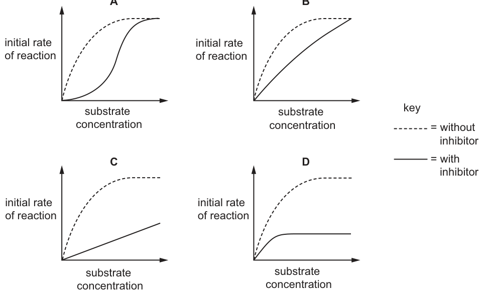
The initial rate of a reaction catalysed by an enzyme was measured at various substrate concentrations.
Which graph shows the effect of a low concentration of non-competitive inhibitor on the reaction?
Options
Working
A non-competitive inhibitor binds to a site other than the active site, distorting the active site so substrate can no longer bind effectively. Because it does not compete with substrate, increasing substrate concentration cannot overcome the inhibition, so falls. The remaining functional enzyme molecules have an unchanged affinity for substrate, so is unchanged. The graph must therefore show a lower plateau () at roughly the same as the uninhibited curve.
- A: sigmoidal with-inhibitor curve → characteristic of allosteric/cooperative binding, not standard non-competitive inhibition.
- B: same , higher → competitive inhibition.
- C: linear, no plateau → not a realistic enzyme kinetics curve.
- D: lower at the same → non-competitive inhibition.
Answer
D
D
Background Concept
Enzyme kinetics describes how the initial rate of an enzyme-catalysed reaction varies with substrate concentration. For a typical (Michaelis–Menten) enzyme, the curve is hyperbolic: as substrate concentration rises, the rate increases steeply and then levels off at a maximum value, , when the active sites are saturated. The substrate concentration at which the rate is half of is the Michaelis constant, , an inverse measure of the enzyme's affinity for its substrate.
Inhibitors slow the reaction. The two classic types give distinct kinetic fingerprints on a rate–substrate concentration plot:
- Competitive inhibitor binds reversibly to the active site, directly competing with substrate. At low substrate concentrations the inhibition is strong, but at very high substrate concentrations substrate out-competes the inhibitor, so the original is reached again. Result on the graph: unchanged, increased (curve shifted to the right).
- Non-competitive inhibitor binds to a different site (an allosteric site) and distorts the active site so the affected enzyme molecules can no longer turn substrate into product, regardless of how much substrate is present. Adding more substrate cannot rescue these enzyme molecules, so the effective falls. The enzyme molecules that are still active have the same intrinsic affinity for substrate, so is unchanged. Result on the graph: lower plateau () at the same .
Understanding the Question
The question asks which of the four sketches shows the effect of a low concentration of a non-competitive inhibitor on a rate–substrate concentration plot, compared with the uninhibited control (the dashed curve). The task is to identify the kinetic signature of non-competitive inhibition: a lower with an unchanged .
Approach
Look at each graph and ask two questions:
- Does the plateau () change?
- Does the substrate concentration giving half () change?
Match the pattern to the definition of non-competitive inhibition (lower , same ).
Step-by-Step Reasoning
- Graph A – the with-inhibitor curve is sigmoidal while the control is hyperbolic, both reaching the same plateau. A sigmoidal shape implies cooperativity between substrate-binding sites (e.g. an allosteric enzyme), not standard Michaelis–Menten non-competitive inhibition. This is not the right pattern for a simple non-competitive inhibitor.
- Graph B – the with-inhibitor curve reaches the same as the control but is shifted to the right, so a higher substrate concentration is needed to reach half ( is higher). This is the textbook signature of competitive inhibition, where extra substrate overcomes the inhibitor. Wrong type.
- Graph C – the with-inhibitor curve is essentially a straight line with no clear plateau, and the slope is much lower than the control. This does not represent a real enzyme kinetics curve under simple non-competitive inhibition; it would imply that is not reached at any practical substrate concentration, which is not what non-competitive inhibition produces.
- Graph D – the with-inhibitor curve plateaus at a much lower rate than the control, but the half-maximal substrate concentration () is essentially the same. This is exactly the kinetic signature of non-competitive inhibition: is reduced, is unchanged. Correct.
Key Takeaways
- Non-competitive inhibition lowers ; competitive inhibition does not.
- Non-competitive inhibition leaves unchanged; competitive inhibition increases .
- Non-competitive inhibitors bind at a site other than the active site, so increasing substrate concentration cannot overcome the inhibition.
Common Mistakes
- Confusing B with non-competitive inhibition because the curve is "shifted". The shift in B is a rightward shift (higher ) at unchanged — that is competitive.
- Choosing A because the curve looks "different". Sigmoidal kinetics describe cooperative/allosteric behaviour, not standard non-competitive inhibition.
- Choosing C because it "looks inhibited". A true non-competitive inhibition curve still plateaus; it just plateaus lower.
Things to Be Careful About
- "Low concentration of inhibitor" still produces a clearly lower on the plot — non-competitive inhibition reduces the effective number of functional active sites.
- The uninhibited dashed curve is the reference; always compare the solid curve's plateau height () and half-saturation point () to the dashed curve before deciding.
- Don't be misled by the visual steepness of the rising phase; the diagnostic features are and , not the initial slope.
Gout is a type of arthritis in which small uric acid crystals form inside and around the joints. It causes sudden attacks of severe pain and swelling.
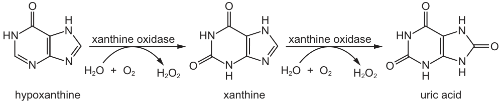
The diagram shows how uric acid is formed from hypoxanthine catalysed by the enzyme xanthine oxidase.
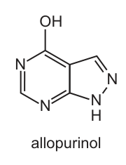
Gout can be treated using a drug called allopurinol which has a similar shape to hypoxanthine.
What can be concluded from this information about how allopurinol prevents the formation of uric acid?
Options
A It binds to the active site of xanthine oxidase instead of hypoxanthine, resulting in reduced production of uric acid.
B It binds to another part of xanthine oxidase and this changes the shape of the active site.
C It disrupts the hydrogen bonds within xanthine oxidase so it denatures and the active site is no longer complementary to hypoxanthine and xanthine.
D It hydrolyses the peptide bonds within xanthine oxidase to change the shape of the active site.
Working
Allopurinol has a similar shape to hypoxanthine (the substrate of xanthine oxidase). Because of this structural similarity, allopurinol fits into the active site of xanthine oxidase and competes with hypoxanthine for binding. This is competitive inhibition. When allopurinol occupies the active site, hypoxanthine cannot bind, so it is not converted to xanthine and then to uric acid, reducing uric acid production.
- B describes non-competitive inhibition, but the shape similarity to the substrate indicates binding at the active site.
- C is incorrect: allopurinol does not denature the enzyme; it binds reversibly to the active site.
- D is incorrect: allopurinol is not a protease and does not hydrolyse peptide bonds.
Answer
A
A
Background Concept
Enzymes are biological catalysts that speed up reactions by lowering the activation energy. Each enzyme has a specific 3D shape, including an active site where the substrate binds. Inhibitors are molecules that reduce enzyme activity and can act in several ways:
- Competitive inhibitors have a shape similar to the substrate and bind to the active site, directly competing with the substrate. They are reversible and their effect can be overcome by increasing substrate concentration.
- Non-competitive inhibitors bind to a site other than the active site (an allosteric site), changing the shape of the active site so the substrate can no longer bind effectively. Their effect cannot be overcome by adding more substrate.
- Irreversible inhibitors permanently damage the enzyme, often by forming covalent bonds with amino acid side chains in the active site. This is distinct from denaturation, which is the unfolding of the entire protein caused by disruption of hydrogen bonds and other interactions under extreme conditions such as high temperature or extreme pH.
The key diagnostic feature of competitive inhibition is structural similarity between the inhibitor and the substrate — this is the clue that the inhibitor will fit into the active site.
Understanding the Question
The stem tells us that uric acid (the substance that causes gout crystals) is produced from hypoxanthine via the enzyme xanthine oxidase. We are then told that allopurinol has a similar shape to hypoxanthine and is used to treat gout. The question asks us to deduce how allopurinol prevents uric acid formation based on this information. This is a multiple-choice question requiring us to identify the correct mechanism of inhibition.
Approach
Look at the structural clue (allopurinol similar to hypoxanthine) and match it to the appropriate type of inhibition. Then evaluate each option to see which one correctly describes that mechanism while excluding the alternatives.
Step-by-Step Reasoning
- Identify the structural clue. From the figures, allopurinol and hypoxanthine both contain a fused two-ring purine structure with similar nitrogen positions. This is a clear visual indicator of molecular mimicry.
- Link structure to mechanism. Because allopurinol mimics the substrate's shape, it will fit into the active site of xanthine oxidase that normally binds hypoxanthine.
- Apply the concept of competitive inhibition. When allopurinol occupies the active site, hypoxanthine is prevented from binding, and the conversion of hypoxanthine → xanthine → uric acid is reduced. This lowers uric acid levels and treats gout.
- Confirm against the options:
- Option A states that allopurinol binds to the active site instead of hypoxanthine, reducing uric acid production. This is exactly competitive inhibition. ✓
- Option B describes non-competitive inhibition (binding elsewhere, changing the active site shape). This would not require the molecule to look like the substrate, and is inconsistent with the structural clue. ✗
- Option C claims allopurinol denatures xanthine oxidase by disrupting hydrogen bonds. Denaturation is irreversible and typically caused by heat or pH extremes — not by a drug that reversibly occupies an active site. ✗
- Option D claims allopurinol hydrolyses peptide bonds. Allopurinol is not a protease, and there is no information suggesting it breaks peptide bonds. This is biochemically incorrect. ✗
Key Takeaways
- Structural similarity between inhibitor and substrate → competitive inhibition at the active site.
- Non-competitive inhibitors do not need to resemble the substrate because they bind at a separate allosteric site.
- Competitive inhibition is reversible and is overcome by increasing substrate concentration.
- A useful diagnostic question: "Does the inhibitor look like the substrate?" If yes, it almost certainly acts at the active site.
Common Mistakes
- Choosing B because it is a common misconception that all inhibitors change the shape of the active site. The structural similarity points to competitive, not non-competitive, inhibition.
- Choosing C by confusing reversible competitive inhibition with irreversible denaturation. Drugs that act by molecular mimicry are not denaturants.
- Choosing D by misinterpreting "changes the shape of the active site" as involving peptide bond hydrolysis. Hydrolysing peptide bonds is what proteases do — and there is no evidence here that allopurinol behaves as one.
Things to Be Careful About
- Always read the structural information in the stem before answering. The phrase "similar shape to hypoxanthine" is the decisive clue for competitive inhibition.
- Remember that competitive inhibition is reversible — the inhibitor does not destroy or denature the enzyme.
- Distinguish carefully between an inhibitor (which binds and blocks) and a denaturant (which unfolds the protein permanently).
Phospholipids are formed in a similar way to triglycerides.
A sample contained six phospholipid molecules.
● The molecular weight of a phosphate ion is .
● The molecular weight of each individual fatty acid in this sample was found to be .
● The molecular weight of glycerol is .
● The molecular weight of water is .
What is the molecular weight of the sample in ?
Options
A 2598
B 4182
C 4506
D 4830
Working
A phospholipid is formed from:
- 1 glycerol
- 2 fatty acids
- 1 phosphate ion
3 condensation reactions occur, releasing 3 water molecules (2 when the fatty acids bond to glycerol, 1 when the phosphate group bonds to glycerol).
For 6 phospholipid molecules:
Answer
B
B
Background Concept
A phospholipid is structurally similar to a triglyceride (a glycerol molecule with three fatty acids attached) except that only two of glycerol's three hydroxyl groups are esterified to fatty acids. The third hydroxyl is esterified to a phosphate group (often with an additional alcohol such as choline attached, but in this question we treat the polar head simply as a phosphate ion, PO₄³⁻, with a molecular weight of 95 g mol⁻¹).
Each time a hydroxyl group reacts with a carboxyl (fatty acid) or with the phosphate, a condensation reaction takes place and one molecule of water is released. So the formation of one phospholipid from glycerol, two fatty acids and one phosphate ion produces three water molecules (two from the fatty-acid + glycerol esterifications, one from the phosphate + glycerol esterification).
Understanding the Question
The question gives the molecular weights of the four building blocks (glycerol 92, one fatty acid 282, phosphate ion 95, water 18) and asks for the molecular weight of a sample containing six phospholipid molecules. Because three water molecules are lost during the formation of each phospholipid, they must be subtracted from the total mass of the components.
Approach
- Add up the molecular weights of the components of a single phospholipid: 1 × glycerol + 2 × fatty acids + 1 × phosphate ion.
- Subtract the mass of the three water molecules released during condensation.
- Multiply by 6 to get the mass of the sample.
Step-by-Step Reasoning
Components of one phospholipid:
- 1 × glycerol = 1 × 92 = 92 g mol⁻¹
- 2 × fatty acid = 2 × 282 = 564 g mol⁻¹
- 1 × phosphate ion = 1 × 95 = 95 g mol⁻¹
- Sum of components = 92 + 564 + 95 = 751 g mol⁻¹
Water lost in the three condensation reactions:
- 3 × water = 3 × 18 = 54 g mol⁻¹
Mass of one phospholipid:
Mass of six phospholipid molecules:
This matches option B.
Key Takeaways
- A phospholipid has the same backbone as a triglyceride (glycerol) but only two fatty-acid tails; the third position carries a phosphate group.
- Forming each ester (or phosphoester) bond releases one water molecule; the total number of condensation steps determines how much mass to subtract.
- Always remember to subtract water when calculating the molecular weight of any molecule built by condensation.
Common Mistakes
- Treating the phospholipid like a triglyceride and using three fatty acids (this gives 4500, close to option C, but wrong).
- Forgetting to subtract the three water molecules (gives 4506, option C).
- Subtracting only two waters instead of three.
- Multiplying the mass of a single phospholipid by a number other than six.
Things to Be Careful About
- "Phosphate ion" (PO₄³⁻) at 95 g mol⁻¹ is what the question provides; do not confuse it with "phosphorus" (31) or "phosphoric acid, H₃PO₄" (98).
- The condensation reactions occur between the hydroxyl groups of glycerol and: (a) the carboxyl groups of two fatty acids, and (b) the hydroxyl groups of phosphoric acid. Count carefully — three bonds = three waters.
- Keep the units consistent (g mol⁻¹) and remember that the question asks for the molecular weight of the sample, not of a single molecule.
Which statements about phospholipids in cell surface membranes are correct?
1 Fatty acid tails allow most ions to pass through the membrane.
2 Hydrophobic tails point inwards facing each other.
3 All polar heads face the cytoplasm.
4 The phospholipids help with the flexibility of the membrane.
Options
A 1, 2 and 3
B 1 and 3 only
C 2, 3 and 4
D 2 and 4 only
Working
Evaluate each statement against the structure of the phospholipid bilayer:
- Statement 1 is incorrect: the fatty acid tails are hydrophobic, so they prevent ions (which are charged) from passing through the membrane.
- Statement 2 is correct: the hydrophobic tails point inwards, facing each other, away from the aqueous environments on either side.
- Statement 3 is incorrect: polar heads face both the cytoplasm and the extracellular fluid — not just the cytoplasm.
- Statement 4 is correct: phospholipids can move laterally within the bilayer, giving the membrane its flexibility (fluidity).
Correct statements: 2 and 4.
Answer
D
D
Background Concept
The cell surface membrane is organised as a phospholipid bilayer according to the fluid mosaic model. Each phospholipid has two distinct regions:
- A polar (hydrophilic) head — the phosphate group, which is attracted to water.
- Two non-polar (hydrophobic) fatty acid tails — which are repelled by water.
In an aqueous environment, the phospholipids arrange themselves so that the hydrophobic tails are buried in the interior, shielded from water, while the hydrophilic heads face outwards into the watery surroundings on both sides. This produces the stable bilayer seen in every cell membrane.
The bilayer is fluid: individual phospholipid molecules can move laterally (side-to-side) and, less commonly, flip between the two layers. This gives the membrane its flexibility. The hydrophobic core also acts as a barrier to charged particles (ions) and large polar molecules, which is why membrane transport proteins are needed for these substances.
Understanding the Question
This is a multiple-choice question asking you to identify which of four statements about phospholipids in the cell surface membrane are correct. The mark scheme rewards only the combination of statements that are biologically accurate, so each statement must be judged on its own merits before the correct option can be chosen.
Approach
For each numbered statement, decide whether it is consistent with the structure and properties of the phospholipid bilayer. Eliminate the false statements and check which option contains only the true ones.
Step-by-Step Reasoning
-
Statement 1 — "Fatty acid tails allow most ions to pass through the membrane."
Ions are charged. The fatty acid tails are hydrophobic (non-polar), so they repel charged particles. The hydrophobic core of the bilayer therefore prevents ions from passing through directly. Statement 1 is false. -
Statement 2 — "Hydrophobic tails point inwards facing each other."
Because the tails are hydrophobic, they orient themselves away from the watery cytoplasm and extracellular fluid, pointing inwards and meeting in the middle of the bilayer. Statement 2 is true. -
Statement 3 — "All polar heads face the cytoplasm."
The polar (hydrophilic) heads face both aqueous environments: the cytoplasm on the inner face of the membrane and the tissue fluid / extracellular environment on the outer face. They do not all face the cytoplasm. Statement 3 is false. -
Statement 4 — "The phospholipids help with the flexibility of the membrane."
Phospholipids can move laterally within their own layer and the bilayer has a fluid character. This allows the membrane to bend, flex and reseal, contributing to its flexibility. Statement 4 is true.
The correct statements are therefore 2 and 4 only, which corresponds to option D.
Key Takeaways
- Hydrophobic fatty acid tails form the interior of the bilayer and act as a barrier to ions and other polar substances.
- Hydrophilic phosphate heads face both the cytoplasm and the extracellular fluid.
- The lateral movement of phospholipids gives the membrane its fluidity / flexibility.
- Ions and polar molecules require membrane transport proteins to cross the phospholipid bilayer.
Common Mistakes
- Confusing the role of the hydrophobic tails — they block ions rather than allow them through. A common error is to assume the membrane interior is water-permeable to everything.
- Forgetting the outer surface — saying polar heads "face outwards" without specifying that they face both the cytoplasm and the extracellular fluid. Statement 3 trips students up by only mentioning the cytoplasm.
- Ignoring membrane fluidity — flexibility is one of the key properties of the bilayer; treating the membrane as a rigid barrier is incorrect.
Things to Be Careful About
- Read each statement carefully: small words like "all" or "most" can change a true idea into a false one.
- Remember that both faces of the membrane are in contact with an aqueous environment (cytoplasm inside, tissue fluid outside).
- The hydrophobic core is specifically what makes the membrane selectively permeable to small non-polar molecules (e.g. O₂, CO₂) but not to ions or large polar molecules.
In an experiment, pieces of onion epidermis are put into three different concentrations of sucrose solutions, P, Q and R. The pieces of onion are left for an hour and then examined using the low power of a light microscope.
Each diagram shows one cell from the epidermis that was placed in each of the sucrose concentrations.

What explains the appearance of cells in solution Q?
Options
A The concentration of solution Q is equal to the concentration of the solutes in the cell sap.
B The cytoplasm has the same concentration of sucrose as solution Q.
C The water potential of the cytoplasm is equal to the water potential of the vacuole.
D The water potential of the cell sap is equal to the water potential of solution Q.
Working
At incipient plasmolysis the protoplast has just begun to pull away from the cell wall. Water has moved out of the vacuole by osmosis down the water potential gradient until the water potential of the cell sap (vacuole) has fallen to the same value as the water potential of the external solution Q. Equilibrium has been reached at the point where the protoplast just loses contact with the wall.
- A — Refers to "concentration of solutes" rather than water potential, and at incipient plasmolysis the external solution is actually slightly more negative than the original cell sap (otherwise no net water loss would have occurred).
- B — Sucrose does not enter the cell, so the cytoplasm does not contain sucrose at the same concentration as solution Q.
- C — True of any cell at equilibrium, but does not explain why incipient plasmolysis is occurring specifically in Q.
- D — Correctly states that the water potential of the cell sap equals the water potential of solution Q, which defines the incipient plasmolysis state.
Answer
D
D
Background Concept
Osmosis is the net movement of water molecules across a partially permeable membrane from a region of higher (less negative) water potential to a region of lower (more negative) water potential.
Water potential (Ψ) is the tendency of water to move from one place to another. Pure water has Ψ = 0 kPa; adding solute makes Ψ more negative. The water potential of a plant cell is determined mainly by the dissolved solutes in the cell sap (vacuole), with smaller contributions from the cytoplasm and any wall pressure.
When a plant cell is placed in a solution:
- If the external Ψ is less negative than the cell sap, water enters → the protoplast pushes firmly against the cell wall → the cell is turgid (as in P).
- If the external Ψ is more negative than the cell sap, water leaves → the protoplast shrinks and pulls away from the cell wall → plasmolysis (as in R).
- At the transition point, the water potential of the cell sap exactly equals the water potential of the external solution. The protoplast has just lost enough water that it begins to detach from the wall at the corners. This stage is called incipient plasmolysis (as in Q).
Understanding the Question
Fig. 16.1 shows three onion epidermal cells after one hour in sucrose solutions P, Q and R. The candidate must identify the state of cell Q — incipient plasmolysis — and choose the statement that correctly explains why it looks the way it does.
The command word is "explains", so the chosen answer must give the underlying biophysical reason, not just a description. Three of the four options refer to genuine concepts (concentration, sucrose in the cytoplasm, water potential inside the cell) but only one correctly applies the idea of water potential equilibrium to the specific incipient-plasmolysis state.
Approach
- Identify the state of cell Q from the diagram: the protoplast is just beginning to pull away from the wall (incipient plasmolysis).
- Recall what defines incipient plasmolysis biophysically: the water potential of the cell sap has fallen (by loss of water) until it equals the water potential of the surrounding solution, but the cell wall no longer exerts inward pressure to oppose further water loss.
- Test each option against this definition; reject any that misuse "concentration" instead of water potential, any that put sucrose inside the cell, and any that describe a general cell property unrelated to the incipient-plasmolysis state.
Step-by-Step Reasoning
- Cell P is turgid — solution P has a higher (less negative) water potential than the cell sap, so water has entered and the protoplast is pressed hard against the cell wall.
- Cell Q is at incipient plasmolysis — water has left the vacuole until the protoplast has shrunk just enough to begin detaching from the wall at the corners. At this point the water potential of the cell sap has dropped to match the water potential of solution Q; no net water movement would occur if the system were left undisturbed.
- Cell R is fully plasmolysed — solution R has a much more negative water potential than the cell sap, so the protoplast has lost a large volume of water and is rounded up in the middle of the wall.
Now to the options:
- A speaks of "concentration of solutes" being equal. Solute concentration and water potential are inversely related but not identical (water potential also depends on pressure). More importantly, incipient plasmolysis is defined in terms of water potential, and the external solution is, by definition, slightly more concentrated than the original cell sap — otherwise no net water loss would have happened.
- B is wrong because sucrose cannot cross the tonoplast or plasma membrane in significant amounts in this timescale, so the cytoplasm never contains sucrose at the same concentration as the bath solution.
- C is a true general statement (cytoplasm and vacuole are in water-potential equilibrium in a healthy cell), but it is not what is special about cell Q — it would also be true of P and R before plasmolysis. It does not explain incipient plasmolysis.
- D correctly identifies the defining condition of incipient plasmolysis: the water potential of the cell sap has become equal to the water potential of solution Q.
Key Takeaways
- Incipient plasmolysis is the exact moment at which the water potential of the cell sap equals the water potential of the external solution; the protoplast has just begun to detach from the wall.
- The three stages shown (P, Q, R) correspond to: external Ψ > cell sap Ψ (turgid), external Ψ = cell sap Ψ (incipient plasmolysis), and external Ψ < cell sap Ψ (fully plasmolysed).
- "Water potential" is the correct term to use when comparing cell and solution; "concentration" alone is imprecise because it ignores pressure potential.
- The plasma membrane and tonoplast are freely permeable to water but not to sucrose, so sucrose stays outside the cell — water moves, not solute.
Common Mistakes
- Choosing A because it "sounds right": students often say "the concentrations are equal" without specifying that the comparison must be of water potential (which combines solute and pressure components), not raw concentration.
- Choosing B through a misunderstanding of membrane permeability — sucrose does not enter the cytoplasm, so its concentrations inside and outside cannot be equal.
- Choosing C because it is a true statement about cells in general, without recognising that the question asks specifically about what is uniquely true of the incipient-plasmolysis state in solution Q.
- Confusing incipient plasmolysis with full plasmolysis — incipient means just beginning; the protoplast is barely detached, not fully shrunken.
Things to Be Careful About
- Always use the term water potential (Ψ), not "concentration", when comparing cell and solution.
- "Cell sap" refers specifically to the contents of the vacuole; do not equate it with the cytoplasm.
- The plasma membrane and tonoplast are both partially permeable; the cell wall is fully permeable and plays no role in determining the point of incipient plasmolysis.
- Pressure potential (Ψ_p) inside a turgid cell is positive; incipient plasmolysis is defined as the state in which Ψ_p ≈ 0, so Ψ_cell ≈ Ψ_solute. This is why the water potential of the cell sap — not just its solute potential — is what equals the external Ψ.
The electron micrograph shows rod-shaped bacteria.
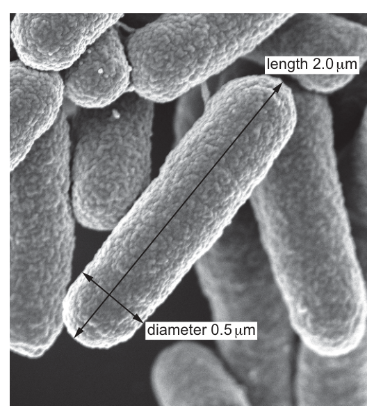
The actual length of the bacterium is and the diameter is . Assume that the bacterium is cylinder-shaped.
What is the surface area to volume ratio of the bacterium?
Options
A
B
C
D
Working
Treat the bacterium as a cylinder with radius and height (height = length).
Surface area of a closed cylinder:
Volume of a cylinder:
Surface area to volume ratio:
Answer
D
D
Background Concept
The surface area to volume (SA:V) ratio is a fundamental idea in cell biology. It describes how much membrane a cell has relative to its internal volume. A high SA:V ratio means a cell can exchange materials (oxygen, nutrients, waste, heat) with its surroundings quickly enough to service its entire cytoplasm. As cells grow larger, volume increases faster than surface area, so the SA:V ratio falls — this is why cells stay small or become flattened/elongated.
The mathematical formulas you need:
- Closed cylinder: (two circular ends + curved side)
- Cylinder volume:
The unit of SA:V is , which simplifies to , but conventionally the ratio is reported as a pure number such as .
Understanding the Question
The electron micrograph shows a rod-shaped bacterium. We are told to model it as a perfect cylinder with:
- Length (height of cylinder):
- Diameter: , giving radius
We must calculate SA:V and pick the matching option (A–D). This is a pure numeracy question testing whether you can apply the cylinder formulae correctly.
Approach
- Convert diameter to radius.
- Substitute and into the cylinder SA and volume formulae.
- Divide SA by V to get the ratio.
- Match against the four options.
Step-by-Step Reasoning
Step 1 — find r:
Step 2 — surface area:
The curved side dominates (much longer than the diameter), so most of the area is the lateral surface, not the two end caps.
Step 3 — volume:
Step 4 — ratio:
So , which matches option D.
Key Takeaways
- Always halve the diameter to get the radius before substituting into .
- A cylinder's SA has two terms — end caps () and lateral surface (). Forgetting the end caps is a common error.
- A long, thin shape (like this rod-shaped bacterium) has a higher SA:V than a sphere of the same volume, which is one reason many bacteria are rod-shaped.
- Because cancels in the ratio, you can in fact skip it: . For : — the same answer, faster.
Common Mistakes
- Using diameter instead of radius — this gives , matching no option, so the error is obvious.
- Forgetting the two end caps () — gives SA = , SA:V = , which is the distractor C. This is the most plausible trap on this question.
- Using the sphere formula — gives a totally different ratio and is rejected.
Things to Be Careful About
- Keep units consistent: here everything is in , so SA is in and V in . The ratio collapses to a pure number.
- Quoting to 2 significant figures (matching the data) gives , not without a decimal place.
- The "9.0" in option D and the "8.0" in option C differ only because of whether the two circular ends are included — being able to derive both quickly is a useful double-check.
Chickens have 78 chromosomes in the nucleus of a body cell.
How many DNA molecules are there in a chicken body cell at the start of prophase of mitosis?
Options
A 46
B 78
C 92
D 156
Working
A chicken body cell has 78 chromosomes. During S phase of interphase, DNA replication occurs, so by the start of prophase each chromosome consists of two sister chromatids, each containing one DNA molecule.
Answer
D
D
Background Concept
A chromosome is a single, very long DNA molecule (wrapped with histone proteins) that carries genetic information. Before DNA replication, each chromosome contains one DNA molecule. During S phase of interphase, the entire genome is replicated, producing two identical copies of each DNA molecule. Once replicated, the chromosome is said to be made of two sister chromatids held together at the centromere, and each chromatid is one DNA molecule.
The key point is that the number of DNA molecules and the number of chromosomes do not always match:
- Before S phase / after telophase/cytokinesis: chromosomes = DNA molecules (each is a single, unreplicated chromatid).
- After S phase until anaphase (i.e. during prophase and metaphase of mitosis): DNA molecules = 2 × chromosome number, because every chromosome has been replicated into two chromatids.
Understanding the Question
The question states a chicken body cell has 78 chromosomes. It then asks for the number of DNA molecules specifically at the start of prophase of mitosis.
The trap is that the obvious answer of 78 is wrong: at the start of prophase, DNA replication has already happened during the preceding S phase, so every chromosome now exists as a pair of sister chromatids.
The command word is "How many" — a single numerical answer is required. This is a multiple choice question and the correct option is the one that correctly counts the DNA molecules after replication.
Approach
- Recognise the cell cycle position: start of prophase lies immediately after S phase of interphase, when DNA replication is complete.
- State the relationship at this stage: each replicated chromosome = 2 chromatids = 2 DNA molecules.
- Multiply the given chromosome number by 2.
- Select the matching option.
Step-by-Step Reasoning
- The chicken cell has 78 chromosomes. In a G1 cell this would also mean 78 DNA molecules, because each chromosome is a single chromatid.
- During S phase, every DNA molecule is replicated by semi-conservative replication. Each chromosome therefore now consists of two sister chromatids, joined at the centromere.
- At the start of prophase, the chromosomes condense and become visible. Counting chromosomes still gives 78 (the centromere holds two chromatids together, so it is still counted as one chromosome), but counting DNA molecules gives 2 per chromosome.
- The correct option is D: 156.
Why the distractors are wrong:
- A (46): 46 is the human diploid chromosome number — irrelevant to this question and not the chicken's number.
- B (78): This is the chromosome number, not the number of DNA molecules at this stage. It is the most tempting wrong answer because the question gives 78 as a stated fact.
- C (92): This is 2 × 46 — a doubling of the human chromosome number; it applies the right idea (×2) to the wrong number.
Key Takeaways
- Chromosome number ≠ DNA molecule number, except in unreplicated cells (before S phase or after cytokinesis).
- After S phase and through to anaphase, DNA molecules = 2 × chromosome number, because each chromosome has two sister chromatids.
- Knowing the cell-cycle position is essential when asked about DNA content.
Common Mistakes
- Equating chromosome number with DNA molecule number — this gives 78 (option B) and is the most common error.
- Forgetting that DNA replication occurs in interphase, before prophase begins.
- Confusing chromatids with chromosomes and so multiplying the wrong quantity by 2.
Things to Be Careful About
- Always check the stage of the cell cycle in the question. "Start of prophase" specifically means after S phase and before anaphase separation.
- Distinguish carefully between chromatid (one DNA molecule) and chromosome (one unreplicated, or two replicated chromatids joined at a centromere).
- Do not let the number 46 (human) lure you into picking option A; it is not a generic biological constant.
The photomicrograph shows cells undergoing mitosis.
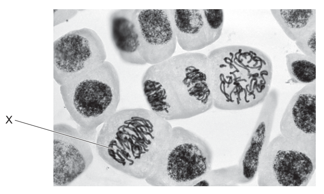
Which statement describes what will happen next in cell X?
Options
A Chromatin coils up tightly and the nuclear envelope breaks down.
B Chromosomes line up along the equator of the cell and attach to the spindle.
C Sister chromatids move towards opposite poles, pulled by the spindle fibres.
D Spindle fibres break down and the cell prepares for cytokinesis.
Working
In cell X, the chromosomes are clearly condensed (visible as dark, thread-like structures) but are scattered through the cell and are not aligned at the equator. The nuclear envelope is no longer intact. This appearance is characteristic of prophase.
The mitotic sequence is:
prophase → metaphase → anaphase → telophase
Therefore the next event in cell X is metaphase, in which the chromosomes line up along the equator of the cell and attach to the spindle fibres.
- A describes prophase (what is already happening / has just finished in X).
- C describes anaphase (two stages later).
- D describes telophase / cytokinesis (three stages later).
Answer
B
B
Background Concept
Mitosis is a continuous process, but biologists divide it into four named stages according to what the chromosomes are doing. In order:
- Prophase – chromatin condenses into visible, rod-shaped chromosomes; each chromosome consists of two sister chromatids joined at the centromere. The nuclear envelope breaks down and the spindle begins to form.
- Metaphase – chromosomes, pushed by spindle fibres, line up along the equator (metaphase plate) of the cell, with spindle fibres attached to the centromere of each chromosome.
- Anaphase – the centromeres split and sister chromatids are pulled to opposite poles by the shortening spindle fibres.
- Telophase – chromatids reach the poles, decondense, new nuclear envelopes form, and the spindle breaks down; cytokinesis (division of the cytoplasm) usually follows.
Because each stage is identified by chromosome appearance, a stained micrograph lets you place a cell in the cycle. Root tip meristems are classic material because cells are dividing rapidly and chromosomes are easily stained.
Understanding the Question
The photomicrograph shows several plant root tip cells in different stages of the cell cycle. Cell X is highlighted: the chromosomes are clearly condensed and visible as dark, tangled, thread-like structures, but they are not lined up at any equator, and the nucleus is no longer bounded by a clear envelope. This places cell X in prophase.
The question asks what happens next in cell X, i.e. the stage that immediately follows the one shown.
Approach
- Identify the current stage of cell X from chromosome appearance.
- Recall the order of mitotic stages (prophase → metaphase → anaphase → telophase).
- Match the description of each option to a stage and pick the one that immediately follows prophase.
Step-by-Step Reasoning
- Cell X shows prophase: condensed chromosomes are visible but scattered; the nuclear envelope is breaking down. This is the very early appearance after chromatin condensation.
- Stage immediately after prophase = metaphase: chromosomes are aligned along the equator and spindle fibres attach to their centromeres. This is exactly what option B states.
- Why the others are wrong:
- A describes prophase events (chromatin coiling and nuclear envelope breakdown) — these are currently happening in cell X, not what happens next.
- C describes anaphase — chromatid separation. This is two stages later, after metaphase has been completed.
- D describes telophase / cytokinesis — the very last events. This is three stages later.
Only option B describes the next event, metaphase.
Key Takeaways
- Recognising mitotic stages on a slide depends on chromosome position: scattered (prophase), equatorial line (metaphase), separating to poles (anaphase), at poles reforming nuclei (telophase).
- The correct sequence is fixed: prophase → metaphase → anaphase → telophase → cytokinesis.
- "Next" in a mitotic question always means the stage that immediately follows the one depicted.
Common Mistakes
- Confusing prophase with metaphase because both show chromosomes. The giveaway is alignment: in prophase chromosomes are scattered, in metaphase they form a single line across the equator.
- Reading the photomicrograph quickly and assuming cell X is in metaphase because chromosomes are visible — they must actually be lined up to be metaphase.
- Selecting option A because it is "what is happening" rather than "what happens next". The question explicitly asks for the next event.
Things to Be Careful About
- Always state both the position of the chromosomes and the state of the nuclear envelope when justifying a stage; the mark scheme usually requires both ideas.
- Spindle fibres are usually not visible in routine light-microscope preparations of root tips (they are not stained), so do not rely on seeing them to identify a stage — use chromosome position instead.
- Cytokinesis is technically separate from mitosis; option D is best read as describing the end of mitosis / beginning of cytokinesis.
Which processes occur in bone marrow cells that are in a mitotic cell cycle?
1 Phosphate groups bind to ADP molecules to form ATP.
2 Bonds form between nucleotides in a DNA strand.
3 Hydrogen bonds form between tRNA anticodons and mRNA codons.
Options
A 1, 2 and 3
B 1 and 2 only
C 1 and 3 only
D 2 only
Working
Bone marrow cells in the mitotic cell cycle are actively dividing and continue to perform all normal cellular processes:
- Statement 1 — ATP synthesis (oxidative phosphorylation in mitochondria and substrate-level phosphorylation in glycolysis/the Krebs cycle) occurs continuously in all living cells via respiration. Dividing cells require ATP for spindle formation, chromosome movement and cytokinesis. ✓
- Statement 2 — During S phase of interphase, DNA replication occurs. Phosphodiester bonds form between adjacent nucleotides, catalysed by DNA polymerase. S phase is part of the mitotic cell cycle. ✓
- Statement 3 — Translation occurs continuously in all cells, including bone marrow cells, which need proteins for division and function. Hydrogen bonds form between tRNA anticodons and mRNA codons at the ribosome. ✓
All three processes occur.
Answer
A
A
Background Concept
Bone marrow is the site of haematopoiesis — the production of blood cells. Red blood cells, white blood cells and platelets are all generated from haematopoietic stem cells through repeated rounds of mitosis followed by differentiation. Bone marrow is therefore one of the most mitotically active tissues in the body.
The mitotic cell cycle consists of:
- Interphase: a long period comprising G1 (cell growth), S (DNA replication) and G2 (preparation for mitosis)
- Mitosis: prophase, metaphase, anaphase, telophase — division of the nucleus
- Cytokinesis: division of the cytoplasm to produce two daughter cells
Throughout the cell cycle, the cell must continue to perform its normal metabolic activities: respiration to produce ATP, transcription and translation for protein synthesis, and (specifically in S phase) DNA replication.
Understanding the Question
This is a multiple-choice question testing whether you recognise that cells in the mitotic cell cycle continue to perform all of the routine processes of living cells, in addition to the specific event of DNA replication. You must assess each of the three statements and decide whether the described process occurs in a bone marrow cell that is in the mitotic cell cycle.
Approach
For each statement, ask two questions: (1) Is this a process that occurs in living cells? (2) Is it consistent with a cell being in the mitotic cell cycle? If the answer to both is yes, the statement is correct. If any statement is true, that process must be happening in the cell.
Step-by-Step Reasoning
Statement 1 — Phosphate groups bind to ADP molecules to form ATP
ATP synthesis happens continuously in every living cell through cellular respiration:
- Oxidative phosphorylation at the inner mitochondrial membrane (electron transport chain)
- Substrate-level phosphorylation in the cytoplasm (glycolysis) and in the mitochondrial matrix (Krebs cycle)
Bone marrow cells are no exception. Even during mitosis itself they require ATP for spindle fibre assembly, chromosome movement at anaphase, and the membrane dynamics of cytokinesis. ✓
Statement 2 — Bonds form between nucleotides in a DNA strand
This describes the formation of phosphodiester bonds between adjacent nucleotides during DNA replication, catalysed by DNA polymerase. DNA replication occurs in S phase of interphase, which is part of the mitotic cell cycle. The cell must copy its entire genome before it can divide. ✓
Statement 3 — Hydrogen bonds form between tRNA anticodons and mRNA codons
This describes translation — the assembly of a polypeptide at the ribosome, where a tRNA carrying the correct amino acid pairs its anticodon with the corresponding mRNA codon by complementary base pairing (held together by hydrogen bonds). Translation occurs continuously in every living cell because proteins are needed at all times. Bone marrow cells are particularly active in protein synthesis — all cells need new proteins for growth and division, and the B-lymphocytes that mature in bone marrow can produce huge quantities of antibody protein. ✓
All three statements are correct, so the answer is A (1, 2 and 3).
Key Takeaways
- A cell in the mitotic cell cycle is alive and metabolically active. Respiration and protein synthesis do not stop during the cell cycle — they continue throughout.
- DNA replication is a specific scheduled event of the cell cycle (S phase of interphase), and is essential if mitosis is to produce two genetically identical daughter cells.
- Bone marrow is highly mitotically active because blood cells have short lifespans and must be replaced constantly.
- Do not confuse transcription (DNA template → mRNA, catalysed by RNA polymerase) with translation (mRNA → polypeptide, catalysed by ribosomes with tRNA). Only translation involves tRNA anticodons binding to mRNA codons.
Common Mistakes
- Choosing D (2 only): students may think that a cell in the mitotic cell cycle only carries out DNA replication, forgetting that respiration and translation are continuous.
- Choosing B (1 and 2 only): students may forget that translation (and therefore codon–anticodon pairing) is a routine process that happens in all cells at all times.
- Confusing transcription with translation: only translation involves tRNA anticodons. Transcription involves RNA polymerase reading the DNA template strand, with no tRNA involvement.
- Thinking DNA replication happens during mitosis itself: DNA replication occurs in S phase, which is part of interphase (not part of mitosis). It is, however, part of the mitotic cell cycle.
Things to Be Careful About
- The "mitotic cell cycle" includes interphase, not just mitosis. S phase — where DNA replication occurs — sits within interphase, before the visible stages of mitosis.
- The bond type matters: phosphodiester bonds join adjacent nucleotides within a DNA strand; hydrogen bonds hold the codon–anticodon pair together during translation.
- A bone marrow cell in the mitotic cell cycle is performing all of its routine life activities in addition to preparing for division. None of respiration, translation, or DNA replication is "switched off" during the cell cycle.
The enzyme telomerase prevents loss of telomeres after many mitotic cell cycles.
Which cells need to transcribe telomerase enzyme?
1 stem cells
2 activated memory B-lymphocytes
3 helper T-lymphocytes secreting cytokines
Options
A 1, 2 and 3
B 1 and 2 only
C 1 and 3 only
D 2 and 3 only
Working
- Stem cells divide repeatedly throughout life to replenish tissues, so they need telomerase to maintain telomere length.
- Activated memory B-lymphocytes can be re-activated many times across an organism's lifetime and undergo clonal expansion, requiring telomerase.
- Helper T-lymphocytes that are already secreting cytokines are short-lived effector cells; they do not need to maintain telomere length for many future divisions.
Answer
B
B
Background Concept
Telomeres are repetitive, non-coding DNA sequences (TTAGGG in humans) found at the ends of linear chromosomes. They protect coding DNA from being lost and prevent chromosome ends from being recognised as DNA damage. However, because DNA polymerase cannot fully replicate the 3′ end of a lagging strand, a small amount of telomeric DNA is lost with every round of DNA replication in mitosis.
Telomerase is a ribonucleoprotein enzyme that adds telomeric repeats back onto chromosome ends using its own RNA template, counteracting this shortening. Without telomerase, telomeres progressively shorten and eventually trigger cell-cycle arrest or apoptosis (the Hayflick limit).
Telomerase is therefore required primarily by cells that must divide many times over an extended period:
- Germline cells and stem cells (which replenish tissues throughout life)
- Some activated lymphocytes that undergo repeated rounds of clonal expansion
- Most cancer cells (around 85–90% show reactivated telomerase)
Differentiated somatic cells that do not need to divide further generally do not express telomerase.
Understanding the Question
The stem reminds us that telomerase prevents telomere loss during repeated mitotic cycles. The question asks which of the three listed cell types must transcribe (express) telomerase. The answer depends on whether each cell type undergoes many rounds of division in the future.
Approach
For each cell type, decide whether it must divide many times in its remaining lifetime. If yes, it needs telomerase; if not, it does not.
Step-by-Step Reasoning
-
Stem cells (statement 1): Adult stem cells in tissues such as bone marrow, intestinal epithelium, and skin continuously divide to replace lost or damaged cells over an organism's entire lifespan. Without telomerase their telomeres would shorten rapidly and they would exhaust their division potential. Stem cells DO need telomerase. ✓
-
Activated memory B-lymphocytes (statement 2): Memory B-cells can persist for decades and, on re-exposure to the same antigen, rapidly proliferate (clonal expansion) to produce large numbers of plasma cells. Each activation involves many mitotic divisions, so telomere maintenance is essential. Memory B-lymphocytes DO need telomerase. ✓
-
Helper T-lymphocytes secreting cytokines (statement 3): A helper T-cell that is actively secreting cytokines is an effector T-cell — it has already proliferated and differentiated following antigen stimulation. Effector cells are short-lived (days to weeks) and do not need to undergo extensive further division. Telomerase activity is not required for these cells. ✗
Therefore statements 1 and 2 are correct, but not 3. This matches option B.
Key Takeaways
- Telomerase is expressed by cells that must divide many times.
- Stem cells (for tissue renewal) and memory lymphocytes (for long-term immune memory) both need telomerase.
- Effector lymphocytes (e.g. cytokine-secreting helper T-cells, plasma cells) are terminally differentiated, short-lived cells that do not require telomerase.
- ~90% of cancer cells also reactivate telomerase, allowing them to divide indefinitely.
Common Mistakes
- Assuming all lymphocytes need telomerase because they divide. The distinction is between memory cells (long-lived, will divide again) and effector cells (short-lived, work then die).
- Forgetting that stem cells must divide throughout life and therefore need active telomere maintenance.
- Confusing helper T-lymphocytes (CD4+) with memory T-cells; only the memory subset has long-term proliferation potential.
Things to Be Careful About
- The question specifies "transcribe telomerase enzyme" — so the cell needs the gene actively expressed, not just the enzyme inherited from a parent cell.
- "Activated memory B-lymphocytes" specifically describes the proliferating state — these are the cells that benefit from telomerase.
- Helper T-cells that have already differentiated into cytokine-secreting effectors are not the population that needs ongoing telomere maintenance; their proliferation occurred earlier, in the clonal expansion phase.
A polypeptide molecule contains the amino acid sequence:
glycine – leucine – lysine – valine.
The table shows DNA triplets for these amino acids.
| glycine | leucine | lysine | valine |
|---|---|---|---|
| CCC | GAA | TTT | CAA |
Which tRNA anticodons are needed for the synthesis of this polypeptide?
Options
A CCC GAA TTT CAA
B CCC GAA UUU CAA
C GGG CUU AAA GUU
D GGG CUU UUU GUU
Working
The DNA triplets given are from the template (antisense) strand that is transcribed.
-
Transcribe DNA template to mRNA (complementary base pairing, A↔U, G↔C):
- CCC → GGG
- GAA → CUU
- TTT → AAA
- CAA → GUU
- mRNA codons: GGG – CUU – AAA – GUU
-
The tRNA anticodon is complementary to the mRNA codon (and antiparallel). Because tRNA is RNA, any T in DNA is replaced by U:
- GGG → CCC
- CUU → GAA
- AAA → UUU
- GUU → CAA
- tRNA anticodons: CCC – GAA – UUU – CAA
Answer
B
B
Background Concept
Three nucleic acid molecules cooperate to make a protein:
- DNA is the master copy in the nucleus. Genes are read 3′ → 5′ along the template (antisense) strand by RNA polymerase. The other strand is the coding/sense strand and has the same sequence as the mRNA (with T instead of U).
- mRNA is a single-stranded copy of the gene. It is built by complementary base pairing against the template: A pairs with U (in RNA), T pairs with A, G pairs with C, C pairs with G. Every three bases (a codon) codes for one amino acid.
- tRNA is the adaptor molecule. At one end it carries an amino acid; at the other end it has a three-base anticodon that is complementary to the mRNA codon. tRNA is RNA, so it contains uracil (U), not thymine (T).
The codon–anticodon pairing is antiparallel and obeys the same base-pairing rules, so a codon 5′-AAA-3′ is read by an anticodon 3′-UUU-5′ (often written 5′-UUU-3′ for the anticodon alone).
Understanding the Question
The question gives a short polypeptide and a table of DNA triplets said to code for its four amino acids. It then asks which set of tRNA anticodons is required to translate the mRNA that comes from these DNA triplets. This is a multiple-choice question with four options.
The command word is implicit ("which… are needed?") — the candidate must select the option that correctly lists the four tRNA anticodons in the order glycine, leucine, lysine, valine.
Approach
A two-step base-pairing chain is required:
- Decide which strand the DNA table represents. In Cambridge A-Level Biology, when a table lists DNA triplets "for" amino acids, the convention is that the triplets shown are the template (antisense) strand read by RNA polymerase. (Sometimes these problems give the coding strand; the answer key is the only way to be sure, but the standard convention is template.)
- Derive the mRNA codons by complementing the template (A→U, T→A, G→C, C→G).
- Derive the tRNA anticodons by complementing the mRNA codons, remembering that the anticodon is RNA (U, not T).
The order of the four anticodons must match the order of the four amino acids in the polypeptide.
Step-by-Step Reasoning
Step 1 — Template DNA (as given):
- Glycine: 5′-CCC-3′
- Leucine: 5′-GAA-3′
- Lysine: 5′-TTT-3′
- Valine: 5′-CAA-3′
Step 2 — Transcribe to mRNA (complement, T→U in the new strand):
| Template DNA | mRNA codon |
|---|---|
| CCC | GGG (Gly) |
| GAA | CUU (Leu) |
| TTT | AAA (Lys) |
| CAA | GUU (Val) |
So the mRNA reads 5′-GGG-CUU-AAA-GUU-3′.
Step 3 — Derive the tRNA anticodons (complement of mRNA, written in the 5′→3′ direction of the tRNA):
- mRNA GGG → anticodon CCC
- mRNA CUU → anticodon GAA
- mRNA AAA → anticodon UUU (this is the U-for-T trap; the DNA template had T but the anticodon must contain U because tRNA is RNA)
- mRNA GUU → anticodon CAA
tRNA anticodons: CCC – GAA – UUU – CAA.
That matches option B.
Why the other options are wrong:
- A (CCC GAA TTT CAA): uses the DNA template triplets as anticodons directly, but ignores that the mRNA is a complement of the template, and wrongly uses TTT instead of UUU in an RNA molecule.
- C (GGG CUU AAA GUU): these are the mRNA codons, not the tRNA anticodons. The student forgot to take the complement of the mRNA.
- D (GGG CUU UUU GUU): a mixture — partly the mRNA codons and partly an attempted complement, but applied inconsistently. It still fails to complement CUU, AAA, and GUU properly, and uses UUU where AAA's true complement is UUU only if read correctly — but the rest of the entries are wrong.
Key Takeaways
- The chain template DNA → mRNA → tRNA anticodon requires two rounds of complementary base pairing.
- The tRNA anticodon is complementary to the mRNA codon, not to the DNA template.
- tRNA is RNA, so its bases are A, U, G, C — never T. Where you would write a T, write a U.
- The order of the anticodons in the answer must follow the order of the amino acids in the polypeptide.
Common Mistakes
- Writing the DNA template triplets directly as the answer (option A) because the table "shows DNA for each amino acid". This is the single most common error.
- Confusing the mRNA codon with the tRNA anticodon and writing the mRNA sequence (option C).
- Forgetting that tRNA contains U, not T, so the third anticodon must be UUU, not TTT.
- Assuming the table shows the coding (sense) strand instead of the template strand, which would give a different set of mRNA codons and hence different anticodons. By CIE convention in this kind of question, treat the DNA as the template strand.
Things to Be Careful About
- Always write the tRNA anticodon as the complement of the mRNA codon, never of the DNA triplet.
- Read the option carefully: B contains UUU (with a U); if you see a U in an option you know it must be a tRNA (RNA), and the rest of the entries should also be examined for the same RNA convention.
- Keep the four anticodons in the order specified by the polypeptide (glycine, leucine, lysine, valine); reordering any one of them would still give the wrong answer even if the individual bases were correct.
The diagram shows a section of a strand of DNA.
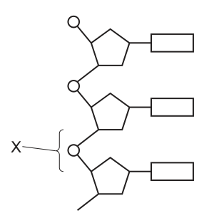
Which type of bond is labelled X?
Options
A glycosidic
B hydrogen
C peptide
D phosphodiester
Working
The diagram shows a single strand of DNA. Each nucleotide has a phosphate (circle), a deoxyribose sugar (pentagon) and a base (rectangle). Bond X joins the phosphate of one nucleotide to the deoxyribose sugar of the next nucleotide along the strand. In DNA, this backbone linkage is a phosphodiester bond (phosphate to the 3′-OH of the adjacent sugar).
Answer
D
D
Background Concept
A DNA strand is a polymer of deoxyribonucleotides. Each nucleotide contains three parts: a phosphate group, a deoxyribose sugar, and a nitrogenous base (A, T, G or C). Three different kinds of bond are involved in DNA structure, and the exam frequently tests whether you can name which is which.
- Phosphodiester bond: links the phosphate of one nucleotide to the 3′-OH of the deoxyribose of the next nucleotide. These are the covalent bonds that form the sugar–phosphate backbone of each strand.
- Hydrogen bond: links complementary bases across the two strands of the double helix (A with T via 2 H-bonds, G with C via 3 H-bonds).
- Glycosidic bond: joins the nitrogenous base to the 1′ carbon of the sugar within a single nucleotide.
Peptide bonds are not part of DNA at all — they join amino acids in proteins.
Understanding the Question
Fig. 23.1 shows three nucleotides of one DNA strand. The phosphate–sugar backbone runs down the left (the small circles are phosphates and the pentagons are sugars), and the bases project to the right (the rectangles). The label X points to the bond between the phosphate of one nucleotide and the sugar of the adjacent nucleotide, which is exactly the backbone linkage.
Approach
Match the location of X — between the phosphate of one nucleotide and the sugar of the next — to the correct bond type from the options, ruling out the bonds that occur elsewhere or in different biomolecules.
Step-by-Step Reasoning
- A (glycosidic) is wrong: a glycosidic bond holds the base to the sugar inside a single nucleotide, not between two nucleotides.
- B (hydrogen) is wrong: hydrogen bonds hold complementary bases together across the two strands of the double helix, not along a single strand.
- C (peptide) is wrong: peptide bonds are found in proteins, not in nucleic acids.
- D (phosphodiester) is correct: this is the covalent bond that joins the phosphate of one nucleotide to the 3′-OH of the deoxyribose of the next nucleotide, building the sugar–phosphate backbone of DNA.
Key Takeaways
- Sugar–phosphate backbone = phosphodiester bonds.
- Two strands held together = hydrogen bonds between complementary bases.
- Sugar–base attachment within a nucleotide = glycosidic bond.
- Peptide bonds belong to proteins, never to DNA or RNA.
Common Mistakes
- Choosing B (hydrogen) because "hydrogen bonds are in DNA" is a common trap — hydrogen bonds are between the two strands, not within one strand.
- Choosing A (glycosidic) — this is the sugar–base bond inside a nucleotide, not the inter-nucleotide linkage.
- Writing "phosphoester" instead of "phosphodiester" loses the mark because a phosphodiester is specifically the two-ester linkage through one phosphate that joins two sugars.
Things to Be Careful About
- Always read the diagram carefully: X here sits on the backbone (phosphate–sugar), so the answer is about backbone chemistry, not base pairing.
- DNA and RNA both use phosphodiester bonds in their backbones; the question is not asking about 2′-deoxyribose versus ribose, only about the bond type at the labelled position.
Which row correctly describes cytosine?
Options
| ring structure | number of hydrogen bonds it forms with its complementary base | type of base | |
|---|---|---|---|
| A | double | three | purine |
| B | double | two | pyrimidine |
| C | single | three | pyrimidine |
| D | single | two | purine |
Working
Cytosine is a pyrimidine — its ring structure is single. Its complementary base is guanine, and the C–G pair is held together by three hydrogen bonds (compared with two for A–T). The only row matching all three features is row C.
Answer
C
C
Background Concept
DNA is built from four nitrogenous bases, divided into two structural families:
- Purines have a double ring structure (a six-membered ring fused to a five-membered ring). The two purines are adenine (A) and guanine (G).
- Pyrimidines have a single ring structure (a six-membered ring only). The three pyrimidines are cytosine (C), thymine (T) and uracil (U) (uracil replaces thymine in RNA).
Base pairing in DNA is specific and stabilised by hydrogen bonds:
- A pairs with T via 2 hydrogen bonds.
- C pairs with G via 3 hydrogen bonds.
This is summarised by the mnemonic "A:T has 2, C:G has 3", and the rule that a purine always pairs with a pyrimidine — which keeps the DNA double helix a uniform width.
Understanding the Question
The question presents a table with three properties of cytosine — its ring structure (single or double), the number of hydrogen bonds it forms with its complementary base, and its base type (purine or pyrimidine) — and asks which row matches cytosine.
The command word is implicit: identify the correct row by recalling cytosine's features.
Approach
Determine the three defining features of cytosine:
- Ring structure → look at which family (purine/pyrimidine) cytosine belongs to.
- Number of H-bonds → look at its complementary partner, guanine, and recall the C–G bonding pattern.
- Base type → directly follows from the ring structure.
Then match all three to one row.
Step-by-Step Reasoning
- Ring structure: Cytosine has one ring → single.
- Base type: A single-ring base is a pyrimidine.
- Complementary base: Cytosine pairs with guanine, and C–G pairs are held together by three hydrogen bonds.
Combining: single ring, three H-bonds, pyrimidine → row C.
Checking the other rows:
- Row A says "double, three, purine" — the double ring + purine is correct, but a purine with three H-bonds would be guanine (G, paired with C). This describes guanine, not cytosine.
- Row B says "double, two, pyrimidine" — the pyrimidine part is correct but the ring number and H-bond count are wrong for cytosine.
- Row D says "single, two, purine" — contradictory, because a single-ring base is a pyrimidine, not a purine.
Only row C is self-consistent and matches cytosine.
Key Takeaways
- Purines (A, G) = double ring; pyrimidines (C, T, U) = single ring.
- A–T pairs have 2 H-bonds; C–G pairs have 3 H-bonds.
- A purine always pairs with a pyrimidine, giving the DNA helix a constant width.
Common Mistakes
- Confusing guanine and cytosine: guanine is the purine partner of cytosine and also forms 3 H-bonds; cytosine is the pyrimidine partner that also forms 3 H-bonds. Mixing up which of the pair is the purine and which is the pyrimidine is the most common slip.
- Stating that cytosine forms 2 H-bonds (confusing it with thymine).
- Describing cytosine as a purine because it is paired with a purine — pairing is across the helix, not within a single base.
Things to Be Careful About
- The question gives three separate properties; all three must agree, so a row that is right on one feature but wrong on another (e.g., row B is right on "pyrimidine" but wrong on ring and H-bonds) is rejected.
- In RNA, uracil replaces thymine but still pairs with adenine via 2 H-bonds; cytosine and guanine still pair via 3 H-bonds in RNA as well. The pairing rules are the same; only the identity of one base changes.
During the production of protein molecules, only one strand from the DNA double helix is used.
Which name is given to the DNA strand that is used to produce a new protein?
Options
A non-transcribed strand
B leading strand
C template strand
D lagging strand
Working
During transcription, RNA polymerase reads one of the two DNA strands and synthesises a complementary mRNA molecule. The strand that is read is called the template strand (also known as the antisense or non-coding strand). The other strand, the coding/sense strand, has the same base sequence as the mRNA (with T instead of U) and is not used as the template.
Answer
C
C
Background Concept
A DNA molecule is double-stranded, with two antiparallel polynucleotide chains held together by complementary base pairing (A–T, G–C). Although both strands carry the same genetic information, only one of them is used as the template when a gene is expressed. During transcription, the enzyme RNA polymerase binds to the gene's promoter region, unwinds the double helix locally, and reads the bases of one strand in the 3' → 5' direction. Complementary RNA nucleotides are then joined together to form a single-stranded mRNA molecule, which is later translated into a polypeptide at a ribosome.
The strand that is actually read by RNA polymerase is called the template strand (also: antisense strand or non-coding strand). Its partner — the strand that is not read — is called the coding strand (or sense strand), because its base sequence matches the mRNA (with T in place of U). Note the slightly confusing naming: although the coding strand is not the one copied, it carries the same sequence information as the mRNA product.
Understanding the Question
The stem states that only one DNA strand is used during protein production and asks for the name of that strand. The distractors in the options are all real biological terms from different contexts, so the test is whether the candidate knows which term specifically applies to the strand copied in transcription.
| Option | Term | Used in | Apply here? |
|---|---|---|---|
| A | non-transcribed strand | Synonym for the coding/sense strand | No — this is the unused strand |
| B | leading strand | DNA replication (continuous synthesis towards the replication fork) | No — wrong process |
| C | template strand | Transcription (the strand copied by RNA polymerase) | Yes |
| D | lagging strand | DNA replication (discontinuous Okazaki fragments) | No — wrong process |
Approach
Recognise that "production of a protein molecule" describes the central dogma — transcription followed by translation. The strand that is read during transcription is, by definition, the template strand. Eliminate the other options by recognising that "leading" and "lagging" strands belong to DNA replication, not transcription.
Step-by-Step Reasoning
- The question is about protein synthesis, so we are dealing with transcription (DNA → mRNA) and translation (mRNA → polypeptide).
- Of the two DNA strands, only one acts as the template for RNA polymerase.
- By CIE convention, this strand is named the template strand (or antisense strand).
- Options B and D (leading and lagging strand) are terms from semi-conservative DNA replication, not transcription — they are immediately incorrect.
- Option A (non-transcribed strand) describes the other strand, the coding strand, which is not read by RNA polymerase — it is therefore the opposite of what the question asks for.
- Option C (template strand) correctly names the strand that is used to produce the mRNA, and hence the protein.
Key Takeaways
- The template strand is the DNA strand copied by RNA polymerase during transcription.
- The partner strand is the coding (sense) strand; it has the same sequence as the mRNA (with T instead of U) and is not used as a template.
- "Leading strand" and "lagging strand" are terms from DNA replication, not transcription — do not confuse the two processes.
Common Mistakes
- Choosing A (non-transcribed strand) because the wording sounds similar to "transcribed strand"; the candidate has misread the question and selected the unused strand.
- Choosing B or D (leading/lagging strand) because the candidate conflates DNA replication with transcription. These strands exist only at a replication fork.
- Calling the template strand the "sense strand" — the sense strand is the partner, not the template.
Things to Be Careful About
- "Template" and "antisense" refer to the same strand; "coding" and "sense" refer to its partner. The terminology can be confusing because the "coding" strand is not the one that is actually copied.
- The mRNA sequence matches the coding (sense) strand, not the template strand, except that U replaces T in the mRNA.
- The question is about transcription (the copying of DNA into mRNA). Replication terminology does not apply.
The diagram shows a longitudinal section of a phloem sieve tube with a companion cell.
Where are the mitochondria located for the release of energy for cotransport?
Options
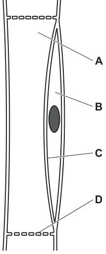
Working
Mature sieve tube elements have lost most of their organelles, including mitochondria, so they cannot generate their own ATP. Cotransport (active loading) of sucrose into the sieve tube element at the source requires ATP, and this ATP is supplied by mitochondria in the companion cell. The mitochondria for this cotransport are therefore located in the companion cell.
Answer
B
B
Background Concept
Phloem tissue is made of two cell types working as a functional unit: the sieve tube element and the companion cell, joined by plasmodesmata.
During differentiation, a sieve tube element loses its nucleus, ribosomes, vacuole, Golgi apparatus, and (essentially) its mitochondria. It retains only a thin layer of cytoplasm pressed against the cell wall, plus the sieve plates at each end through which phloem sap flows. Because it has so few organelles, a sieve tube element cannot make its own ATP, RNA or proteins.
The companion cell retains a full complement of organelles: a nucleus, many ribosomes, a dense cytoplasm, and very large numbers of mitochondria. The companion cell carries out the metabolism for both cells and supplies the sieve tube element with ATP, proteins and other molecules through the plasmodesmata.
Loading of sucrose (and other assimilates) into the sieve tube element at a source is an active process: a proton pump in the companion cell plasma membrane uses ATP to pump H⁺ out, building a proton gradient. H⁺ then re-enters down its gradient through a co-transporter, carrying sucrose with it. This is cotransport (symport), and it requires ATP — so the mitochondria powering it must be the abundant mitochondria of the companion cell, not the nearly organelle-free sieve tube element.
Understanding the Question
The diagram shows a sieve tube element on the left (with sieve plates at top and bottom) and a companion cell on the right. The labels point to:
- A: sieve plate
- B: the companion cell (its dense, organelle-rich cytoplasm around the nucleus)
- C: a region in the companion cell area
- D: the thin layer of cytoplasm inside the sieve tube element itself
The question asks where the mitochondria are located that supply the energy (ATP) for the cotransport step of phloem loading.
The command word is implicit (choose one option) and the biological demand is: which cell — and therefore which labelled region — actually contains the mitochondria capable of producing this ATP?
Approach
- Recall that sieve tube elements lose their mitochondria at maturity — so any structure inside the sieve tube (option D) cannot be the answer.
- Recognise that the companion cell retains a full set of organelles, including numerous mitochondria, and supplies ATP to the sieve tube.
- Choose the label that points to the companion cell.
Step-by-Step Reasoning
- The energy source for the proton pump (and therefore the cotransporter that loads sucrose) is ATP.
- ATP in these cells is generated by oxidative phosphorylation in mitochondria.
- Mature sieve tube elements have lost (or greatly reduced) their mitochondria — they depend on the companion cell for ATP.
- The companion cell contains the many mitochondria needed to produce this ATP.
- Therefore the mitochondria for cotransport are in the companion cell — option B.
Why the others are wrong:
- A (sieve plate): this is a perforated end wall of the sieve tube element; it has no cytoplasm and no mitochondria.
- C (cytoplasm of the companion cell / a region within it that is not the organelle-rich area): the specific correct location is the organelle-rich companion cell, not a peripheral area.
- D (sieve tube element cytoplasm): the sieve tube element cytoplasm lacks (or contains very few) mitochondria, so it cannot be the source of ATP for cotransport.
Key Takeaways
- Sieve tube elements lose their organelles (including mitochondria) at maturity and are metabolically dependent on companion cells.
- Companion cells supply ATP, proteins and signalling molecules to the sieve tube element via plasmodesmata.
- Phloem loading of sucrose at a source uses a proton pump + H⁺/sucrose cotransporter; both steps ultimately depend on the ATP produced by mitochondria in the companion cell.
Common Mistakes
- Picking D (sieve tube cytoplasm) because it is where the cotransport mechanistically loads sucrose. The pump and cotransporter sit in the plasma membrane shared between the two cells, but the ATP powering them comes from companion-cell mitochondria, not from the sieve tube.
- Forgetting that the sieve plate (A) is a wall with pores, not a living compartment, and so cannot contain mitochondria.
- Confusing which cell is the source of ATP with which cell transports the sugar.
Things to Be Careful About
- A-level answers must use the precise term companion cell (not "helper cell" or "supporting cell").
- Mitochondria in this context are described as providing ATP for the proton pump / for cotransport — saying only "for energy" is too vague to score the mark.
- Remember that the cotransporter is on the plasma membrane, but the mitochondria that supply its ATP are in the cytoplasm of the companion cell — the two are different things.
The diagram shows a phloem sieve tube element and a companion cell that are involved in translocation of sucrose.
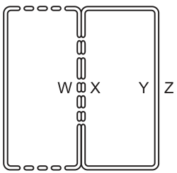
Which process correctly describes the translocation of sucrose through these cells?
Options
A Sucrose moves from Y to Z by active transport.
B Protons move from Y to Z by active transport.
C Protons move from Z to Y by diffusion.
D Protons move from X to W by diffusion.
Working
Loading of sucrose into the phloem at a source depends on an ATP-powered proton pump in the plasma membrane of the companion cell / sieve tube element. The pump uses ATP to move H⁺ ions out of the cytoplasm into the cell wall (apoplast), against their concentration gradient — this is active transport.
In the diagram, Y is the cytoplasm of the sieve tube element and Z is the cell wall of the sieve tube element, so the movement of protons from Y to Z corresponds to the action of the proton pump.
- A — Sucrose enters the phloem by co-transport with protons, not by direct active transport, and it is loaded into the cytoplasm, not into the cell wall. ✗
- B — Protons moving from cytoplasm (Y) to cell wall (Z) is exactly what the proton pump does (active transport). ✓
- C — Protons do move from the cell wall back into the cytoplasm, but only through a co-transporter carrying sucrose with them (facilitated diffusion / co-transport), and the locations cited are reversed relative to the proton-pump direction. ✗
- D — The direction X → W is the right direction for the proton pump, but the pump uses ATP, so the mechanism is active transport, not diffusion. ✗
Answer
B
B
Background Concept
Phloem transports organic solutes — mainly sucrose — from "sources" (e.g. photosynthesising leaves) to "sinks" (e.g. roots, fruits, growing tips). The current model for how sucrose enters the phloem at a source is the mass-flow / pressure-flow model, and a key part of the entry step is the proton-pump / co-transport mechanism operating in the companion cell and the sieve tube element at the source end.
The steps in phloem loading at a source are:
- An ATP-powered proton pump in the plasma membrane of the companion cell (or sieve tube element) actively transports H⁺ ions out of the cytoplasm into the cell wall (apoplast). This builds a high H⁺ concentration in the apoplast and a membrane potential that is negative inside the cell.
- H⁺ then flows back into the cytoplasm down its electrochemical gradient through a co-transporter (symporter) that, in doing so, drags a sucrose molecule with it. Because the H⁺ is moving down its gradient, this step is facilitated diffusion; the energy "stored" in the proton gradient powers the uphill movement of sucrose.
- The loaded sucrose moves from the companion cell into the sieve tube element through plasmodesmata, raising the solute concentration (and so lowering the water potential) inside the sieve tube.
- Water enters the sieve tube by osmosis from the neighbouring xylem, generating a high hydrostatic pressure that drives mass flow towards the sink.
It is therefore essential to remember that the first step — moving protons out of the cytoplasm into the cell wall — is active transport using ATP.
Understanding the Question
The question shows a schematic of a companion cell on the left and a sieve tube element on the right, connected by plasmodesmata. The letters label four regions:
- W — the cell wall of the companion cell (apoplast side)
- X — the cytoplasm of the companion cell
- Y — the cytoplasm of the sieve tube element
- Z — the cell wall of the sieve tube element (apoplast side)
The four options each describe a movement between two of these regions and name a mechanism. To pick the right one, the candidate has to (a) match the correct biological process to the diagram and (b) name the correct mechanism.
The command word is essentially "identify which statement is correct".
Approach
First, identify which membrane the proton pump sits in: it sits in the plasma membrane of the companion cell (and the sieve tube element), pumping H⁺ from the cytoplasm (X or Y) outward into the cell wall (W or Z). Then check each option:
- Does it describe the right direction? (Cytoplasm → cell wall, not the other way.)
- Does it name the right mechanism? (Active transport, because ATP is used.)
Any option that reverses the direction, names the wrong molecule, or uses "diffusion" where the cell is doing work is wrong.
Step-by-Step Reasoning
- Option A: Sucrose moves from Y to Z by active transport. Sucrose is the molecule being loaded, but it does not leave the cytoplasm by active transport — it is co-transported back into the cytoplasm (or moves through plasmodesmata from companion cell to sieve tube). It is not pumped out into the cell wall. ✗
- Option B: Protons move from Y to Z by active transport. Y is the sieve tube cytoplasm and Z is the sieve tube cell wall. The proton pump does exactly this — it uses ATP to pump H⁺ out of the cytoplasm into the apoplast, against the proton gradient. The direction is cytoplasm → wall, the molecule is the proton, and the mechanism is active transport. ✓
- Option C: Protons move from Z to Y by diffusion. Protons do flow back from the apoplast into the cytoplasm, but this happens through a specific co-transporter that also moves sucrose — it is co-transport, not free diffusion. Even so, the marking point at issue is which step the question is really testing; this option misrepresents the mechanism. ✗
- Option D: Protons move from X to W by diffusion. The direction (X = companion cell cytoplasm → W = companion cell wall) is the right direction for the proton pump, but the mechanism is wrong: this step requires ATP and is therefore active transport, not simple diffusion. ✗
Hence the only option that gets both the direction and the mechanism correct is B.
Key Takeaways
- Loading sucrose into the phloem at a source uses a proton pump that actively transports H⁺ out of the cytoplasm into the cell wall (apoplast).
- The H⁺ then re-enters the cytoplasm by co-transport with sucrose, using the energy stored in the proton gradient (this is not free diffusion — it is a specific symporter).
- Mass flow is driven by the resulting high solute concentration → low water potential → osmotic water entry → high hydrostatic pressure inside the sieve tube.
- When asked about phloem loading, distinguish carefully between direction (out into the apoplast, not back in) and mechanism (active transport using ATP, not diffusion).
Common Mistakes
- Confusing the direction of the proton pump: candidates often think protons move into the cell by active transport. In fact, the pump moves them out of the cytoplasm into the cell wall.
- Labelling the co-transport step as "diffusion". The H⁺ does move down its gradient, but it must do so through a sucrose co-transporter; describing it as plain diffusion is not credited.
- Confusing the role of sucrose. Sucrose is co-transported into the cytoplasm; it is not directly pumped by ATP, nor does it leave the cytoplasm into the cell wall.
- Misreading the diagram, treating W, X, Y and Z as four separate cells instead of alternating wall and cytoplasm regions.
Things to Be Careful About
- "Active transport" must always be the answer when the cell is moving something against its concentration gradient, even if a gradient already exists.
- Read every option to the end: an option with the right direction but the wrong mechanism (option D) is still wrong.
- Keep the membrane biology straight: proton pumps and co-transporters are proteins embedded in the plasma membrane — they are not free-floating in the cytoplasm or in the cell wall.
Which types of molecules are cotransported into companion cells?
Options
A monomers and disaccharides
B monomers and polysaccharides
C polymers and disaccharides
D polymers and monosaccharides
Working
Companion cells load sugars into the phloem by cotransport with protons (H+). A proton pump on the plasma membrane of the companion cell uses ATP to pump H+ out, creating an electrochemical gradient. H+ then flows back into the companion cell down this gradient through a cotransporter protein, carrying a sugar molecule with it. The sugars loaded are small, soluble carbohydrates — monosaccharides (e.g. glucose) and disaccharides (e.g. sucrose). Polymers such as starch are too large to be cotransported.
Answer
A
A
Background Concept
Phloem translocation moves assimilates (the products of photosynthesis) from a source (e.g. a photosynthesising leaf) to a sink (e.g. a root, fruit or growing tip). The conducting tubes of the phloem are the sieve tube elements, which are living but lack a nucleus and most organelles. Each sieve tube element is supported by a companion cell, joined to it by numerous plasmodesmata. The companion cell carries out the metabolic work (including active loading of sugars) that the sieve tube element cannot do on its own.
Sugars produced in the leaf are small and soluble. Monosaccharides (e.g. glucose) and disaccharides (e.g. sucrose — the main transport sugar in most plants) are both small enough to be loaded into the companion cell. Polysaccharides such as starch are large, insoluble storage molecules; they are not transported in the phloem and so cannot be the substrates for cotransport into companion cells.
Understanding the Question
The question asks which categories of molecule are moved into companion cells by cotransport (also called symport). This is a one-mark MCQ from Paper 1, so the answer is a single letter. The options pair the size category "monomers/polymers" with "disaccharides/monosaccharides". The biology narrows the answer to small, soluble sugars.
Approach
Recall the phloem-loading mechanism: H+ is pumped out of the companion cell by an ATP-driven proton pump; H+ re-enters through a cotransporter, dragging a sugar with it. The sugars must fit through the cotransporter — so they must be small. Eliminate the options that include polymers (too large) and the option that excludes disaccharides (sucrose is the principal transported sugar).
Step-by-Step Reasoning
- Proton pump on the companion cell membrane uses ATP to pump H+ out, building an electrochemical gradient.
- Cotransporter (symporter) allows H+ to flow back in down its gradient, and uses that energy to bring a sugar molecule in with it.
- The transported sugars in the phloem are the soluble end-products of photosynthesis: glucose (a monosaccharide / monomer) and sucrose (a disaccharide). Sucrose is the dominant transport sugar in most plants.
- Polymers (polysaccharides such as starch) are not transported in the phloem and are too large for the cotransporter, so any option containing "polymers" is wrong — that eliminates B, C and D.
- The remaining option, A — monomers and disaccharides — correctly matches the biology: monosaccharides (monomers) and disaccharides are cotransported into companion cells.
Key Takeaways
- Phloem loading uses H+/sucrose cotransporters on the companion cell plasma membrane, powered by an ATP-driven proton pump.
- The substrates of this cotransport are small, soluble sugars: monosaccharides (monomers) such as glucose, and disaccharides such as sucrose.
- Polysaccharides (polymers) are not transported in the phloem and cannot be cotransported.
Common Mistakes
- Confusing the direction of transport — students sometimes say sugars are cotransported out of the companion cell, but the active loading step is into the companion cell (then into the sieve tube element).
- Choosing an option with "polymers" because starch is a plant carbohydrate, forgetting that starch is an insoluble storage molecule and not a transport sugar.
- Confusing phloem loading with xylem transport; the cotransporter is a feature of the phloem (companion cell), not the xylem.
Things to Be Careful About
- The mark-scheme wording is "monomers and disaccharides". "Monomers" here means monosaccharides (e.g. glucose), not amino-acid monomers.
- Sucrose is technically a disaccharide of glucose and fructose, so it is a small, soluble molecule capable of being cotransported — do not reject it as "too big".
The diagram shows a section through the human heart.
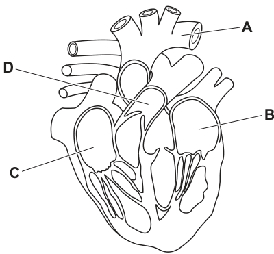
Which label is correct?
Options
A pulmonary artery
B left ventricle
C right atrium
D aorta
Working
Label C points to the chamber on the anatomical right side of the heart, above the right ventricle and receiving the superior and inferior venae cavae — this is the right atrium.
The other labels are misidentified by the options:
- A is the aorta (not the pulmonary artery).
- B is the left ventricle (correctly named).
- D is the pulmonary artery (not the aorta).
Answer
C
C
Background Concept
The human heart has four chambers: two atria (upper, thin-walled, receiving chambers) and two ventricles (lower, thick-walled, pumping chambers). In an anatomical (anterior) view of the heart drawn as if you are facing the person:
- The subject's right side of the heart appears on the left of the diagram.
- The subject's left side of the heart appears on the right of the diagram.
Key positional landmarks:
- The right atrium sits on the viewer's left, above the right ventricle, and receives the superior and inferior venae cavae.
- The right ventricle lies below the right atrium and forms most of the anterior surface of the heart; it leads into the pulmonary artery.
- The left ventricle is on the viewer's right, has the thickest muscular wall, and leads into the aorta.
- The aorta is the large arching vessel at the top, on the viewer's right.
- The pulmonary artery leaves the right ventricle and passes to the viewer's left, behind the aorta.
Understanding the Question
The diagram shows a section through the human heart with four labels (A, B, C, D). The question asks which label correctly identifies the structure it points to. The options give a name for each label; only one of those names actually matches the structure at that label. The mark scheme confirms the correct answer is C, which corresponds to the right atrium.
Approach
- Locate each label in the diagram and identify the structure at that position using the great vessels and the relative positions of the chambers.
- Check the option associated with that label and decide whether the name matches the structure.
- Select the label whose option correctly names the structure.
Step-by-Step Reasoning
- Label A sits at the top of the heart, on the viewer's right, arching over the other vessels. This is the aorta. The option offered for A is pulmonary artery, which is incorrect (the aorta carries oxygenated blood from the left ventricle, while the pulmonary artery carries deoxygenated blood from the right ventricle).
- Label B points to the chamber on the viewer's right, with a thick muscular wall and an apex pointing downward. This is the left ventricle, and the option given for B is left ventricle — so this label is correctly named. (However, the question asks which label is correct, and only one is the intended answer.)
- Label C points to the chamber on the viewer's left, above the right ventricle, where the venae cavae enter. This is the right atrium. The option for C is right atrium, so this is the correct match.
- Label D points to the vessel on the viewer's left, exiting the right ventricle and passing behind the aorta. This is the pulmonary artery, but the option offered for D is aorta — incorrect.
Therefore, the label whose option correctly identifies the structure is C (right atrium).
Key Takeaways
- Always orientate a heart diagram from the subject's perspective: the anatomical right is on the viewer's left.
- Use the great vessels as landmarks: the aorta arches on the viewer's right, the pulmonary artery runs on the viewer's left.
- The right atrium lies above the right ventricle on the viewer's left and receives the venae cavae; the right ventricle forms the front of the heart and leads into the pulmonary artery.
Common Mistakes
- Confusing right and left because the diagram is mirrored relative to the viewer.
- Mistaking the thick-walled right side of the heart for the left ventricle (the right ventricle is anterior and partly forms the apex, but its wall is much thinner than the left ventricle's).
- Swapping the aorta and pulmonary artery: the aorta arches to the viewer's right and the pulmonary artery runs to the viewer's left.
Things to Be Careful About
- "Right" and "left" in heart anatomy always refer to the subject, not the viewer.
- The right atrium and right ventricle are adjacent on the same side of the heart; distinguish them by position (atrium above, ventricle below) and by which vessels enter or leave them (venae cavae into the atrium; pulmonary artery out of the ventricle).
The graph shows how the volume of the left ventricle changes during one cardiac cycle.
Which point on the graph represents the start of atrial systole?
Options
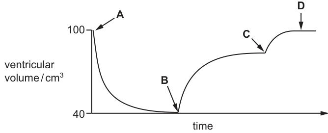
Working
During the cardiac cycle the left ventricle fills and empties. On a graph of ventricular volume against time:
- Point A () — end of ventricular filling; the ventricle is at its maximum volume.
- The steep fall from A to B — ventricular systole; the ventricle contracts and blood is ejected into the aorta.
- Point B () — end of ventricular systole; minimum ventricular volume.
- The slow rise from B to C — ventricular diastole; blood returns passively from the pulmonary veins through the open atrioventricular (bicuspid) valve.
- Point C — the curve changes slope and a small additional rise begins. This is the extra volume added by active contraction of the atria pushing blood into the ventricle.
- Point D () — end of atrial systole; the ventricle is full again.
The start of atrial systole is therefore the point at which the small extra rise begins: C.
Answer
C
C
Background Concept
The cardiac cycle is one complete sequence of contraction (systole) and relaxation (diastole) of the atria and ventricles. It produces the characteristic pressure and volume changes that drive blood through the heart. Three key events must be kept in order:
- Atrial systole — both atria contract and push a small extra volume of blood into the already-mostly-full ventricles. This contributes about the last of ventricular filling and produces a small, brisk rise in ventricular volume.
- Ventricular systole — both ventricles contract from the base upwards, closing the atrioventricular (AV) valves, opening the semilunar valves, and ejecting blood into the pulmonary artery and aorta. Ventricular volume falls rapidly.
- Ventricular diastole — the ventricles relax. The semilunar valves close; once ventricular pressure falls below atrial pressure the AV valves open and blood flows passively from atria into ventricles. This is the slow-filling phase that returns the ventricle to about of its full volume before atrial systole adds the rest.
A graph of left ventricular volume against time is the easiest way to read these phases. Volume is maximal at the end of filling and minimal just after ejection; the rate of change of volume gives the phases away.
Understanding the Question
The graph plots left ventricular volume on the y-axis (from to ) against time on the x-axis. Four labelled points (A, B, C, D) sit on the curve. The question asks specifically for the start of atrial systole — i.e. the moment the atria begin to contract and push the last portion of blood into the ventricle. The challenge is to recognise that atrial systole produces a small, additional rise in volume on top of the slow passive filling — not the steepest feature of the trace.
Approach
Match each segment of the curve to a phase of the cardiac cycle using the rate of change of volume:
- Steep fall → ventricular ejection (ventricular systole).
- Slow rise → passive filling of the ventricle once the AV valve opens (early ventricular diastole).
- Small, additional rise on top of the slow rise → active filling driven by atrial contraction (atrial systole).
- Flat plateau at → the ventricle is full and waiting for the next systole.
The start of atrial systole is therefore the inflection where the slow rise changes into the small extra rise.
Step-by-Step Reasoning
- Point A sits at the start of the trace at . The ventricle is full. This is the end of atrial systole / the start of ventricular systole, not the start of atrial systole.
- From A to B the curve falls steeply. A fall in ventricular volume means blood is leaving the ventricle faster than it is entering, so the AV valve must be closed and the semilunar valve open. This is ventricular systole (ejection).
- Point B is the minimum volume (). Ejection has just finished; the ventricle has emptied as much as it can. This is the end of ventricular systole.
- From B to C the curve rises slowly. A slow rise in volume with no abrupt step means the AV valve is open and blood is flowing passively from the atria (which are relaxed and acting as reservoirs) into the ventricle. This is ventricular diastole (passive filling). About of the final volume is regained during this phase.
- At point C the curve shows a clear change of slope: the rise suddenly becomes slightly steeper for a short section. That extra volume — typically the remaining of filling — is delivered by contraction of the atria, i.e. atrial systole. So C is the start of atrial systole.
- From C to D the curve continues to rise briefly and then plateaus at . Point D marks the end of atrial systole, with the ventricle full and the AV valves about to close as ventricular systole begins.
Hence the start of atrial systole is C.
Key Takeaways
- On a left-ventricular volume trace, steep fall = ventricular systole, slow rise = passive filling (ventricular diastole), and the small additional rise = atrial systole.
- The start of atrial systole is the inflection where passive filling hands over to active atrial filling — the kink in the curve, not its highest or lowest point.
- Reading any cardiac cycle graph is a routine exam skill: work from the shape of the curve to the heart-valve state, then to the named phase.
Common Mistakes
- Choosing A because it is at maximum volume. A is the end of filling / the moment the ventricle is about to eject, not the start of atrial systole.
- Choosing B because it is the lowest point. B is the end of ventricular systole (ejection finished).
- Choosing D because it is the end of the small extra rise. D is the end of atrial systole; C is the start.
- Forgetting that atrial systole is only a small contribution to filling. If you remember this, the small extra rise (between C and D) is unmistakable and you can locate C as its start.
Things to Be Careful About
- "Systole" means contraction and "diastole" means relaxation; this applies to both atria and ventricles, and the two events are staggered. Always say which chamber you mean.
- The order is: atrial systole → ventricular systole → ventricular diastole (with atrial diastole running through most of ventricular systole and ventricular diastole). The trace begins at maximum volume, so the first event the trace shows is ventricular systole, not atrial systole — that is why A is not the answer.
- Don't confuse the AV valve closing (start of ventricular systole) with atrial systole — they are different events, separated by a small gap in time corresponding to the plateau at at the end of the trace.
Which statement is correct?
Options
A In a red blood cell, can combine with haemoglobin to form haemoglobinic acid.
B Carbonic anhydrase is an enzyme that catalyses the reaction between and .
C At the lungs, carbon dioxide is released when carbonic acid and water react together.
D The greater the concentration of in the blood, the higher the affinity of haemoglobin for oxygen.
Working
- A: CO₂ does combine with haemoglobin, but the product is carbaminohaemoglobin (), not haemoglobinic acid.
- B: Carbonic anhydrase catalyses — this is correct.
- C: At the lungs, the direction of the reaction is reversed; carbonic acid decomposes into and (it does not react with water to release CO₂).
- D: The Bohr effect means that higher lowers haemoglobin's affinity for (curve shifts right), not raises it.
Answer
B
B
Background Concept
Carbon dioxide is carried in the blood in three main forms: dissolved in plasma (~5%), as hydrogencarbonate ions (, ~85%), and bound to haemoglobin as carbaminohaemoglobin (, ~10%). Most of the is produced inside red blood cells by the reaction:
This reaction is slow unless catalysed. Carbonic anhydrase, an enzyme inside red blood cells, greatly speeds up the forward (and reverse) reaction. The produced is buffered by haemoglobin itself, forming haemoglobinic acid (HHb), which is one reason haemoglobin is such an effective blood buffer. The diffuses out of the red cell into the plasma; the chloride shift maintains electrical balance.
At the lungs, the partial pressure gradient reverses and the entire sequence runs backwards: re-enters the red cell, recombines with to form , which carbonic anhydrase splits into and , and the diffuses out to be exhaled.
The Bohr effect describes how increased (and the resulting fall in pH) decreases haemoglobin's affinity for , shifting the oxygen dissociation curve to the right. This is physiologically valuable: actively respiring tissues produce more and so offload more readily, while at the lungs, where is low, the curve shifts back left and haemoglobin loads efficiently.
Understanding the Question
This is a multiple-choice question asking you to pick the single correct statement about transport in the blood. Each option tests a different piece of the same topic:
- A tests whether you know the direct product of binding to haemoglobin.
- B tests the definition of the enzyme carbonic anhydrase.
- C tests the direction of the reaction at the lungs.
- D tests the Bohr effect.
The mark scheme expects you to identify which of these four statements is fully correct; the other three each contain a specific, identifiable error.
Approach
Work through each option in turn, checking it against the textbook picture of transport, and reject any that contain an error. Pick the one that is wholly correct.
Step-by-Step Reasoning
Option A — CO₂ + haemoglobin → haemoglobinic acid
CO₂ does combine with the globin (amino) part of haemoglobin, but the product is carbaminohaemoglobin (), not haemoglobinic acid. "Haemoglobinic acid" (HHb) is the form haemoglobin takes when it buffers from the dissociation of carbonic acid — it is not a –haemoglobin adduct. So A is wrong.
Option B — Carbonic anhydrase catalyses CO₂ + H₂O
This is exactly the textbook role of the enzyme. It speeds up the otherwise slow hydration of to form (which then dissociates to and ). The reaction is reversible, so the same enzyme catalyses the release of at the lungs. B is correct.
Option C — At the lungs, and react to release
At the lungs the reaction runs in reverse: decomposes (catalysed by carbonic anhydrase) into and , releasing to be breathed out. It is not a reaction of carbonic acid with water. So C is wrong.
Option D — Higher → higher affinity of Hb for
This is the opposite of the truth. The Bohr effect states that increased (and the it produces) decreases haemoglobin's affinity for , shifting the oxygen dissociation curve to the right. D is wrong.
Key Takeaways
- Carbonic anhydrase catalyses the reversible hydration of : .
- bound directly to haemoglobin forms carbaminohaemoglobin, not haemoglobinic acid. HHb is haemoglobin that has bound an ion.
- At the lungs, the carbonic-anhydrase reaction runs in reverse, releasing for exhalation.
- The Bohr effect: more / lower pH → lower affinity of Hb for (curve shifts right).
Common Mistakes
- Confusing carbaminohaemoglobin (CO₂ bound to globin) with haemoglobinic acid (H⁺ bound to Hb) — they are not the same thing.
- Forgetting that the direction of the carbonic anhydrase reaction reverses at the lungs.
- Reversing the Bohr effect — saying that high increases affinity, which would defeat the purpose of the mechanism (tissues need offloaded where is high).
Things to Be Careful About
- " combining with haemoglobin" is a vague phrase — in exam answers, always specify the exact product (, carbaminohaemoglobin).
- "Haemoglobinic acid" can refer to HHb (Hb + H⁺), which is a buffer, not a carrier.
- "Reaction between carbonic acid and water" is a mis-statement of the lung-side process; correct wording is "carbonic acid dissociates/decomposes into and ".
Which row is correct for an artery?
Options
| inner layer | middle layer | outer layer | |
|---|---|---|---|
| A | smooth layer of endodermis cells | collagen, elastic fibres and smooth muscle | collagen only |
| B | smooth layer of endodermis cells | elastic fibres and smooth muscle only | collagen and elastic fibres |
| C | smooth layer of squamous cells | collagen, elastic fibres and smooth muscle | collagen and elastic fibres |
| D | smooth layer of squamous cells | elastic fibres and smooth muscle only | collagen only |
Working
The wall of an artery has three layers:
- Inner layer (tunica intima): a smooth single layer of squamous endothelial cells — not "endodermis", which is a plant root tissue.
- Middle layer (tunica media): collagen, elastic fibres and smooth muscle (thick in arteries to withstand the high blood pressure pulse).
- Outer layer (tunica externa/adventitia): mainly collagen with some elastic fibres.
Checking the options:
- A: "endodermis" is wrong (plant tissue); outer layer omits elastic fibres. ✗
- B: "endodermis" is wrong; middle layer omits collagen. ✗
- C: squamous endothelium ✓, middle layer with collagen, elastic fibres and smooth muscle ✓, outer layer with collagen and elastic fibres ✓. ✓
- D: outer layer omits elastic fibres; middle layer omits collagen. ✗
Answer
C
C
Background Concept
Blood vessels (arteries, veins and capillaries) are built from three concentric layers, each with a characteristic composition that matches its function. From the lumen outwards these are:
- Tunica intima (innermost layer): a single layer of flattened squamous endothelial cells sitting on a thin basement membrane. Its job is to provide a smooth, non-thrombogenic surface that minimises friction with the flowing blood.
- Tunica media (middle layer): smooth muscle together with elastic and collagen fibres. In arteries this layer is the thickest because it must resist and absorb the high pressure pulse from ventricular systole; the elastic fibres recoil, smoothing the pulse, while the smooth muscle regulates vessel diameter (vasoconstriction/vasodilation).
- Tunica externa (outer layer, tunica adventitia): a tough connective-tissue coat of collagen fibres with some elastic fibres, anchoring the vessel to surrounding tissues and preventing over-stretching.
It is also worth noting that endothelium is a squamous epithelium lining the inside of blood and lymphatic vessels. "Endodermis" is something completely different — it is a layer of cells in plant roots that controls the movement of water and minerals into the vascular cylinder. Confusing the two is a common trap in MCQs.
Understanding the Question
This MCQ asks you to pick the row that correctly describes the composition of an artery's three wall layers. The mark scheme is testing two things at once: (1) whether you can name the correct cell type in the inner layer (endothelium, not endodermis), and (2) whether you remember the full composition of the middle and outer layers — most wrong options omit one of the two fibre types (collagen or elastic).
The command word is implicit ("which row is correct") — you must select the single best option.
Approach
Read each option in three passes: (i) is the inner layer correctly named? (ii) does the middle layer include all three components (collagen, elastic fibres, smooth muscle)? (iii) is the outer layer complete (collagen and elastic fibres)? Only the row that passes all three tests can be correct.
Step-by-Step Reasoning
- Option A uses "endodermis" in the inner layer — this is a plant tissue, so A is eliminated immediately. (Even ignoring that, the outer layer is given as "collagen only", which is incomplete.)
- Option B also uses "endodermis", and additionally lists the middle layer as "elastic fibres and smooth muscle only" — omitting collagen. Eliminated.
- Option C describes the inner layer as a "smooth layer of squamous cells" — this matches endothelium, a simple squamous epithelium. The middle layer includes collagen, elastic fibres and smooth muscle (the full arterial tunica media). The outer layer includes collagen and elastic fibres (a typical tunica adventitia). All three layers are correctly described. ✓
- Option D has the inner layer right (squamous) but its middle layer omits collagen and its outer layer omits elastic fibres, so it is incomplete. Eliminated.
Therefore the only fully correct row is C.
Key Takeaways
- The three arterial layers are, from lumen outwards: tunica intima (squamous endothelium), tunica media (smooth muscle + collagen + elastic fibres), tunica externa (collagen + elastic fibres).
- The high-pressure arterial pulse is accommodated by the thick, elastic tunica media — the structure–function link at the heart of this question.
- "Endothelium" (vessel lining) and "endodermis" (plant root layer) are unrelated and a common distractor pair.
Common Mistakes
- Writing "endodermis" instead of "endothelium" — endodermis is a plant tissue; the correct term for the simple squamous lining of a blood vessel is endothelium.
- Omitting one of the fibre types in the middle or outer layer (e.g. giving the middle layer as "elastic fibres and smooth muscle only", forgetting collagen). All three components (collagen, elastic fibres, smooth muscle) belong in the tunica media of an artery.
- Confusing the relative thickness of layers between arteries, veins and capillaries — arteries have the thickest tunica media, not the thinnest.
Things to Be Careful About
- Use endothelium (or "squamous epithelium") — never endodermis — for the inner layer of a blood vessel.
- An MCQ option can be wrong in more than one place; you only need to find one error to eliminate it, but you must then check that the surviving option is also fully correct, not just less wrong than the others.
- The arterial tunica externa does contain some elastic fibres as well as collagen; "collagen only" is therefore too restrictive.
Which row shows the features of the gas exchange surface that increase diffusion of carbon dioxide and oxygen?
Options
| concentration gradient | diffusion distance | |
|---|---|---|
| A | steep | long |
| B | steep | short |
| C | shallow | long |
| D | shallow | short |
Working
Fick's law states that the rate of diffusion is proportional to the concentration gradient and inversely proportional to the diffusion distance. A gas exchange surface therefore needs a steep concentration gradient and a short diffusion distance to maximise diffusion of and . This matches row B.
Answer
B
B
Background Concept
Gas exchange surfaces (e.g. alveoli in mammals, the spongy mesophyll in leaves) are specialised to maximise the rate of diffusion of respiratory gases. Their features follow from Fick's law of diffusion:
To increase diffusion, an exchange surface should therefore have a large surface area, a steep (large) concentration gradient, and a thin (short) diffusion distance. Blood flow (or ventilation) maintains the steep gradient continuously.
Understanding the Question
This MCQ tests two of Fick's-law variables side by side: the concentration gradient and the diffusion distance. The candidate must recognise which combination of values for these two variables would increase diffusion.
Approach
Apply Fick's law: a steep gradient and a short distance both push the rate of diffusion up. Any other combination either flattens the gradient (slowing diffusion) or lengthens the diffusion path (also slowing it).
Step-by-Step Reasoning
- A steep concentration gradient means a large difference in gas partial pressures between the alveolar air and the blood. This drives more into the blood and more out per unit time. A shallow gradient would slow diffusion.
- A short diffusion distance means the gases only have to cross a very thin barrier (the alveolar epithelium plus the capillary endothelium — together about 1 µm thick). A long distance would slow diffusion.
- Row A (steep, long): the long distance cancels out the benefit of the steep gradient.
- Row B (steep, short): both variables favour rapid diffusion ✔
- Row C (shallow, long): both variables slow diffusion.
- Row D (shallow, short): the short distance helps, but the shallow gradient limits the rate.
Key Takeaways
- Fick's law links diffusion rate to surface area, concentration gradient and diffusion distance.
- Gas exchange surfaces combine a steep gradient (maintained by ventilation and blood flow), a short distance (thin epithelium) and a large surface area (millions of alveoli) to maximise diffusion.
Common Mistakes
- Confusing the direction of the relationship: some students think a shallow gradient or a long distance would help. Fick's law shows the opposite — both slow diffusion.
- Mixing this up with osmosis, where the driving force is a water potential gradient rather than a concentration gradient.
Things to Be Careful About
- The question asks for features that increase diffusion — be sure to read the stem carefully so you do not select the option that would decrease it.
- Remember that Fick's law is about diffusion distance, not diffusion time; a short distance means gases cross faster.
The electron micrograph shows some of the airways in the gaseous exchange system of an insect and the respiring body cells that surround them.
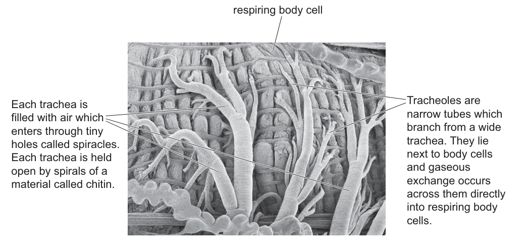
Which statements describe correct differences between the insect gas exchange system shown in the electron micrograph and the human gas exchange system?
1 Gas exchange occurs through the walls of the airways directly into respiring body cells in insects but this does not occur in humans.
2 There are spirals of chitin in the walls of a trachea in insects to hold it open but not in humans.
3 There is more than one trachea in the gas exchange system of the insect but only one in humans.
Options
A 1, 2 and 3
B 1 and 2 only
C 2 and 3 only
D 3 only
Working
Statement 1: In insects, tracheoles branch from tracheae and lie directly next to respiring body cells, so gases diffuse straight across the tracheole wall into the cells. In humans, gas exchange occurs in the alveoli and oxygen is carried by the blood to respiring cells — not directly from an airway wall. Statement 1 is correct.
Statement 2: The micrograph and its annotation confirm that insect tracheae are held open by spirals of chitin (taenidia). Humans do not have chitin; the trachea and bronchi are kept open by C-shaped rings of cartilage. Statement 2 is correct.
Statement 3: Insects have a network of many tracheae running throughout the body. Humans have only one trachea (which then branches into two bronchi). Statement 3 is correct.
All three statements are correct differences.
Answer
A
A
Background Concept
Animals need a gas exchange system to supply O₂ for aerobic respiration and to remove CO₂. The system must bring air close enough to every respiring cell that gases can diffuse across the short distance involved. Two very different solutions to this problem are seen in insects and in mammals (including humans):
-
Insects use a tracheal system. Air enters through small pores called spiracles on the body surface and flows into a network of tracheae (wide tubes). Each trachea branches repeatedly into finer tracheoles, which are very narrow tubes that lie directly alongside individual respiring body cells. The walls of tracheae are reinforced with spirals of chitin (taenidia) which keep the tubes from collapsing. Because gases diffuse from the tracheoles straight into the cells, insects do not need a blood circulatory system to transport O₂.
-
Humans use lungs. Air passes down a single trachea (windpipe), which branches into two bronchi, then into many bronchioles, ending in millions of tiny alveoli where gas exchange occurs. The trachea and bronchi are kept open by C-shaped rings of cartilage (not chitin). O₂ diffuses from alveoli into blood capillaries, is carried by the circulatory system (bound to haemoglobin in red blood cells) to respiring body cells, and CO₂ is returned the same way.
Understanding the Question
The electron micrograph (Fig. 34.1) shows an insect's tracheal system: thick, branching tracheae reinforced with visible spiral thickenings, and finer tracheoles running between the body cells. The candidate must decide which of the three numbered statements correctly describe differences between this insect system and the human gas exchange system. The question is an MCQ, so the candidate must identify the option (A, B, C or D) that lists only the correct statements.
Approach
Work through each statement in turn, comparing the structure/function it describes with what is true of both systems. A statement is "correct" if it is a genuine, accurate difference; reject any that are wrong, vague, or describe something present in both systems.
Step-by-Step Reasoning
Statement 1 – Direct delivery of gases to respiring cells. The annotation on the micrograph explicitly says tracheoles are narrow tubes that "lie next to body cells and gaseous exchange occurs across them directly into respiring body cells." In humans, however, the airway (trachea, bronchi, bronchioles) does not contact body cells at all; the alveoli are surrounded by capillaries, and gases must be carried by the blood to reach the respiring cells of the body. So this is a genuine, correct difference. ✓
Statement 2 – Spirals of chitin holding airways open. The micrograph's left-hand label states that each trachea "is held open by spirals of a material called chitin." In humans the trachea and bronchi are kept open by C-shaped cartilage rings — not spirals, and not chitin. This is a real structural difference. ✓
Statement 3 – Number of tracheae. Insects have a network of many tracheae distributed throughout the body. Humans have a single trachea (which then bifurcates into two main bronchi, not multiple tracheae). The statement is correct. ✓
All three statements describe accurate differences, so the correct answer is A (1, 2 and 3).
Key Takeaways
- Insects use a tracheal system in which tracheoles deliver air directly to respiring cells; humans rely on lungs plus a blood circulatory system.
- Chitin spirals (taenidia) are characteristic of insect tracheae; cartilage rings are characteristic of the human trachea and bronchi.
- Comparative questions often hinge on small but precise structural/functional details — read each statement carefully and verify it against both systems.
Common Mistakes
- Confusing cartilage (human) with chitin (insect) and assuming the airway supports are "the same thing" — they are chemically and structurally different.
- Assuming humans have "many tracheae" because the airways branch — the trachea itself is a single tube; it is the bronchi and bronchioles that branch.
- Selecting an answer because the statements sound true without checking each one individually — losing a single mark because statement 2 or 3 was wrong changes the option from A to B, C or D.
Things to Be Careful About
- The micrograph shows spirals on tracheae, not complete rings — a deliberate distinction from the C-shaped cartilage rings of the human trachea.
- "Gas exchange occurs directly into respiring body cells" only applies to the tracheoles, not to the larger tracheae. The statement is correct as written, but be aware of where exactly gas exchange happens in each system.
- In humans the bronchi are reinforced by cartilage, not chitin — do not accept chitin as a feature of any human tissue.
Which row is correct for the wall of the trachea and the wall of the bronchus?
Options
| cartilage | smooth muscle | goblet cells | |
|---|---|---|---|
| A | ✓ | ✓ | ✓ |
| B | ✓ | ✓ | ✗ |
| C | ✓ | ✗ | ✓ |
| D | ✗ | ✓ | ✗ |
key
✓ = present
✗ = not present
Working
The walls of both the trachea and the bronchi contain:
- Cartilage — C-shaped rings in the trachea and irregular plates in the bronchi keep the airways open.
- Smooth muscle — allows bronchoconstriction and bronchodilation to regulate airflow.
- Goblet cells — secrete mucus that traps inhaled pathogens and dust particles.
All three tissues are present in the walls of both the trachea and the bronchus.
Answer
A
A
Background Concept
The trachea (windpipe) branches into two primary bronchi, one entering each lung. The bronchi then branch repeatedly into smaller bronchioles, which finally end in the alveoli where gas exchange occurs. The trachea and the larger bronchi share a common wall structure because both need to remain patent (open) while also being able to regulate the volume of air passing through.
The three key tissues in the walls of the trachea and bronchi are:
- Cartilage — in the trachea this forms C-shaped (incomplete) rings with the open part facing the oesophagus, allowing food to pass down the oesophagus without being obstructed. In the bronchi, the cartilage is in the form of irregular plates rather than complete rings. Its function is to hold the airway open and prevent it from collapsing during breathing.
- Smooth muscle — the trachealis muscle (in the trachea) and smooth muscle in the bronchi can contract (bronchoconstriction) or relax (bronchodilation) to alter the diameter of the airway and so regulate the flow of air to the alveoli. This is also the tissue that constricts during an asthma attack.
- Goblet cells — these are specialised epithelial cells that secrete mucus onto the inner surface of the airway. The mucus traps inhaled dust, microbes and other particles. Ciliated epithelial cells then beat the mucus (and trapped material) upwards towards the pharynx, where it is swallowed.
All three of these tissues are present in the walls of both the trachea and the main bronchi, so the correct row must have a tick in all three columns.
Understanding the Question
The question presents a simple table with three tissues (cartilage, smooth muscle, goblet cells) and four rows, each indicating which tissues are present (✓) or not present (✗). The candidate must identify which row correctly describes the wall of the trachea AND the wall of the bronchus — i.e. the same combination must apply to both structures.
The command word here is implicit ("Which row is correct"), so a single letter is required. The marking scheme confirms A as the correct answer.
Approach
Recall the histology of the tracheal and bronchial walls. Both tubes have the same general layered wall (mucosa → submucosa → cartilage/smooth muscle layer → adventitia), so all three tissues in the table are present in both.
Step-by-Step Reasoning
- Cartilage in trachea and bronchus: The trachea has C-shaped hyaline cartilage rings; bronchi have irregular cartilage plates. Cartilage is present in BOTH — tick.
- Smooth muscle in trachea and bronchus: The trachealis muscle (the open part of the C-ring) and smooth muscle in the bronchi contract to constrict the airway. Smooth muscle is present in BOTH — tick.
- Goblet cells in trachea and bronchus: The pseudostratified ciliated epithelium lining both tubes contains mucus-secreting goblet cells. Goblet cells are present in BOTH — tick.
- Row A shows ✓ ✓ ✓, which matches the wall composition of both structures. Rows B, C and D each claim at least one of these tissues is absent, which is incorrect.
Key Takeaways
- The walls of the trachea and the (main) bronchi have the same three key tissue components: cartilage, smooth muscle and goblet cells.
- Cartilage keeps the airway open; smooth muscle regulates its diameter; goblet cells (with ciliated epithelium) form the mucociliary escalator that traps and removes inhaled debris and pathogens.
- As the bronchi branch into smaller bronchioles, cartilage disappears first, and goblet cells disappear later, leaving only smooth muscle in the terminal bronchioles — a common point of confusion.
Common Mistakes
- Choosing B, C or D because the candidate wrongly believes that goblet cells (or smooth muscle, or cartilage) are absent from one of the two tubes. Both tubes have the same three tissues.
- Confusing the trachea with the oesophagus (which has no cartilage) or with the bronchioles (which lose cartilage and goblet cells, retaining only smooth muscle).
- Thinking goblet cells are confined to the trachea, when they are also abundant in the bronchi.
Things to Be Careful About
- Goblet cells are restricted to the conducting zone (trachea, bronchi, larger bronchioles). They are absent from the smaller bronchioles and the alveoli, where the wall is extremely thin to permit gas exchange.
- Cartilage disappears at the level of the bronchioles (no cartilage rings or plates beyond the bronchi).
- The question tests recall of the trachea AND the bronchus together — make sure the chosen row is true for BOTH, not just one.
A bacterial pathogen produces a protein that acts as a toxin. This toxin is harmful to humans.
Scientists are developing monoclonal antibodies that can be used to detect the presence of the toxin in the body so that early treatment can be given.
Which statements describe steps in the development of these monoclonal antibodies?
1 The toxin protein is injected into a mouse and triggers mitosis of specific B-lymphocytes.
2 Antibodies are collected from the spleen of the mouse and fused with myeloma cells.
3 A hybridoma cell produces many antibodies with a variety of different variable regions.
Options
A 1 and 2
B 1 only
C 2 and 3
D 3 only
Working
- Statement 1: The toxin (antigen) is injected into a mouse, stimulating specific B-lymphocytes to undergo mitosis and produce antibody. ✓
- Statement 2: It is B-lymphocytes (not antibodies) that are removed from the spleen and fused with myeloma cells. ✗
- Statement 3: A single hybridoma produces identical antibodies with the same variable region; the key feature of monoclonal antibodies is specificity to one epitope, not variety. ✗
Answer
B
B
Background Concept
Monoclonal antibodies are identical antibodies produced by a single clone of B-lymphocytes, all directed against one specific epitope on one antigen. They are made using the hybridoma technique:
- An antigen is injected into a mouse (or other mammal) to stimulate an immune response.
- Specific B-lymphocytes in the mouse's spleen respond by dividing (mitosis) and producing antibodies against that antigen. However, these B-lymphocytes cannot divide indefinitely in culture.
- B-lymphocytes are extracted from the spleen and fused with myeloma cells (cancerous plasma cells) using a fusogen such as polyethylene glycol (PEG). Myeloma cells divide indefinitely but cannot produce a specific antibody on their own.
- The fused cell — a hybridoma — combines two properties: it divides indefinitely (from the myeloma) and produces a specific antibody (from the B-lymphocyte).
- Individual hybridomas are separated and screened so that only those producing the desired antibody are kept. Each hybridoma clone produces a single, pure type of antibody — hence "monoclonal".
Understanding the Question
The stem describes a scenario where a bacterial toxin is the target, and we are asked which statements correctly describe steps in producing monoclonal antibodies that will detect that toxin. The answer depends on recalling the exact sequence of the hybridoma method, particularly which cell type is harvested from the spleen and what a hybridoma actually produces.
The command word is "describe steps" — each statement must be a true, accurate description of one stage in the procedure.
Approach
Go through each statement and check it against the standard hybridoma procedure:
- Does it identify the correct cell or molecule at each step?
- Does it correctly describe what a hybridoma produces?
Reject any statement that swaps a cell for the molecule it secretes, or that contradicts the "monoclonal" (one type) nature of the product.
Step-by-Step Reasoning
Statement 1 — TRUE: Injecting the toxin (acting as an antigen) into the mouse provokes an immune response. Specific B-lymphocytes whose surface receptors recognise epitopes on the toxin are activated, proliferate by mitosis in the spleen, and begin secreting anti-toxin antibodies. This is the correct first step.
Statement 2 — FALSE: It is B-lymphocytes (whole cells) that are harvested from the spleen, not the antibodies themselves. The B-lymphocytes are then fused with myeloma cells to form hybridomas. Antibodies are the secreted product, not the cells used in fusion. The mark scheme rejects this because the correct cell — the B-lymphocyte — has been replaced by the wrong entity.
Statement 3 — FALSE: A hybridoma is the product of fusing ONE B-lymphocyte (producing ONE specific antibody) with ONE myeloma cell. Because all antibodies from that hybridoma originate from a single B-lymphocyte, they all have the same variable region and the same specificity. Producing antibodies "with a variety of different variable regions" describes a polyclonal response, not a monoclonal one. This directly contradicts the meaning of "monoclonal".
Only statement 1 is correct, so the answer is B (1 only).
Key Takeaways
- The starting material for the hybridoma fusion is the B-lymphocyte, not the antibody.
- A single hybridoma clone produces a single, specific antibody — that is the entire point of being "monoclonal".
- "Monoclonal" = one clone = identical variable regions; "polyclonal" = many clones = many variable regions.
- The myeloma contributes immortality; the B-lymphocyte contributes antibody specificity.
Common Mistakes
- Confusing the antibody (the secreted product) with the B-lymphocyte (the cell that makes it) — this traps students into accepting statement 2.
- Thinking a hybridoma produces a mixture of antibodies because it is a "fused" cell — fusion combines properties of the two parent cells but does not mix the antibody specificities; the antibody still comes only from the B-lymphocyte partner.
- Forgetting that monoclonal antibodies must be screened/selected for the desired specificity from a mixture of hybridomas, which is why a single hybridoma is described as producing just one type of antibody.
Things to Be Careful About
- Read the wording precisely: "antibodies are collected from the spleen" should immediately raise suspicion — antibodies circulate in blood/lymph, while B-lymphocytes reside in the spleen.
- "Variety of different variable regions" is the giveaway phrasing of a polyclonal mixture, even though it is attached to a hybridoma in statement 3.
- Do not be misled by the term "monoclonal" itself — it refers to the cells being a single clone, which by definition yields identical antibodies.
Which statements are correct for penicillin?
1 It is harmful to prokaryotic cells.
2 It disrupts cell wall synthesis.
3 It becomes less effective with regular use.
Options
A 1, 2 and 3
B 1 and 2 only
C 1 and 3 only
D 2 and 3 only
Working
- It is harmful to prokaryotic cells. Penicillin kills bacteria (prokaryotes) by interfering with their cell wall synthesis; it does not harm eukaryotic cells in the same way because their cells are not surrounded by a peptidoglycan wall. ✓ Correct.
- It disrupts cell wall synthesis. Penicillin inhibits transpeptidase enzymes that cross-link peptidoglycan chains in the bacterial cell wall, weakening the wall so the cell lyses. ✓ Correct.
- It becomes less effective with regular use. Repeated or incomplete courses select for resistant bacteria, so penicillin (and other antibiotics) become less effective over time. ✓ Correct.
All three statements are correct, so the answer is A.
Answer
A
A
Background Concept
Penicillin is an antibiotic — a chemical produced originally by the fungus Penicillium that kills or inhibits the growth of bacteria. Antibiotics are selective: they exploit features found in prokaryotic (bacterial) cells but absent from eukaryotic (plant, animal, fungal) cells, so they can be taken by humans with relatively few side effects.
The key feature of bacteria that penicillin targets is the peptidoglycan cell wall. Peptidoglycan is a mesh of long sugar–peptide chains cross-linked by short peptide bridges; the enzyme that catalyses this cross-linking is called transpeptidase (also known as a penicillin-binding protein). Penicillin binds irreversibly to transpeptidase, preventing cross-linking, so the growing wall is weakened. The bacterium then takes up water by osmosis and lyses (bursts) because it cannot resist the internal turgor pressure.
Antibiotic resistance arises when bacteria mutate (or acquire genes) that allow them to survive the drug. Common mechanisms include producing an enzyme (e.g. β-lactamase) that breaks down penicillin, modifying the target so the drug cannot bind, or pumping the drug out. The more penicillin (or any antibiotic) is used, the more opportunity there is for resistant strains to spread, and so the drug becomes less effective over time.
Understanding the Question
This is a multiple-choice question that presents three statements about penicillin and asks the candidate to identify the option that includes all of the correct ones (and no incorrect ones). The distractors are combinations of one or two correct statements only.
The command word is implicit ("Which statements are correct…") and the task is to evaluate each statement independently and then select the option that lists every correct one.
Approach
Go through each statement and decide whether it is true or false using your knowledge of:
- the selective toxicity of antibiotics (prokaryotes vs eukaryotes),
- the mechanism of action of penicillin (inhibition of cell wall synthesis),
- and the evolution of antibiotic resistance with regular use.
If all three are true, the answer is A (1, 2 and 3).
Step-by-Step Reasoning
Statement 1 — "It is harmful to prokaryotic cells."
True. Penicillin kills bacteria by attacking the peptidoglycan wall, which is unique to prokaryotes. Human (eukaryotic) cells lack a cell wall made of peptidoglycan, so they are not directly harmed. Statement 1 is correct.
Statement 2 — "It disrupts cell wall synthesis."
True. As described above, penicillin inhibits transpeptidase, the enzyme that cross-links the peptidoglycan strands of the bacterial cell wall. This is the textbook mechanism of penicillin and is the definition of the drug's action. Statement 2 is correct.
Statement 3 — "It becomes less effective with regular use."
True. The more frequently penicillin is used, the more selection pressure is placed on bacterial populations. Bacteria that carry resistance mutations or resistance genes survive and reproduce, so the population of resistant bacteria increases. The drug then becomes less effective, and this is precisely why doctors stress completing courses of antibiotics and avoiding unnecessary prescriptions. Statement 3 is correct.
All three statements are correct, so the option containing all of them is A.
Key Takeaways
- Penicillin is selectively toxic: it kills prokaryotic (bacterial) cells because they have a peptidoglycan cell wall that eukaryotic cells do not.
- Its mechanism is to inhibit cell wall synthesis (specifically, it blocks transpeptidase-mediated cross-linking of peptidoglycan), leading to lysis.
- Antibiotics (including penicillin) become less effective over time with regular/over-use because resistant strains are selected for and spread.
- Antibiotics do not affect viruses, because viruses have no cell wall, no cell machinery of their own, and are not cells at all.
Common Mistakes
- Choosing B (1 and 2 only) — usually because a student forgets that antibiotic resistance exists or thinks resistance is a separate issue from the drug itself. Resistance is precisely what makes a drug become "less effective with regular use".
- Choosing C (1 and 3 only) — usually because a student confuses the mechanism, perhaps thinking penicillin damages the cell membrane or stops protein synthesis (which is what other antibiotics, e.g. tetracyclines, do).
- Choosing D (2 and 3 only) — usually because a student thinks of penicillin as broadly "antibiotic" and forgets that prokaryotes, not eukaryotes, are the target.
- Thinking that any antibiotic is harmful to human cells. Penicillin is selectively toxic to bacteria; it is famously safe for humans precisely because we lack peptidoglycan walls.
Things to Be Careful About
- The phrase "becomes less effective with regular use" refers to antibiotic resistance, not to the body developing tolerance to the drug. The drug itself does not change; the bacteria do.
- "Harmful to prokaryotic cells" is the correct framing — the question does not say "harmful to all cells".
- Do not confuse the cell-wall mechanism of penicillin with the ribosome-inhibiting action of other antibiotic classes (e.g. streptomycin, tetracycline, chloramphenicol) or with the membrane-disrupting action of others (e.g. polymyxins). The mark scheme is testing your recall of the penicillin-specific mechanism.
- For the multiple-choice format, check that the option you choose contains every correct statement and no incorrect ones — partial combinations are common distractors.
Why is passive immunity effective for only a short time?
Options
A Antibodies are rapidly broken down.
B Antigens are rapidly broken down.
C Memory cells soon die.
D Phagocytes soon die.
Working
Passive immunity involves receiving ready-made antibodies (e.g. across the placenta, in breast milk, or by injection of antiserum). The recipient's own immune system is not activated, so no memory cells are produced. The protection lasts only as long as these donated antibodies remain in the circulation, and antibodies are proteins that are gradually broken down and replaced.
Answer
A
A
Background Concept
Passive immunity is protection conferred by receiving ready-made antibodies from another source rather than producing them in response to an antigen. Examples include antibodies crossing the placenta from mother to fetus, antibodies passed in breast milk, and antiserum injected after exposure to a pathogen (e.g. rabies, tetanus).
Because the recipient's lymphocytes are not stimulated by the antigen itself, no memory cells are formed. The protection therefore ends as soon as the donated antibodies are degraded by normal protein turnover. Antibodies are globular proteins with a finite plasma half-life (typically days to a few weeks), and they are progressively broken down by proteases and cleared.
This contrasts with active immunity, where the individual's own B-lymphocytes respond to an antigen, producing antibodies AND memory cells, giving long-lasting (often lifelong) protection.
Understanding the Question
The question asks why passive immunity is short-lived. We need to pick the option that correctly explains the limited duration. The candidate must recognise that passively acquired antibodies, not the recipient's own immune machinery, are the source of protection, and that the bottleneck is the lifespan of those antibodies.
Approach
Identify the molecular basis of passive protection (antibodies), and recall that antibodies are proteins that are continuously broken down. Eliminate options that do not describe what actually limits passive immunity.
Step-by-Step Reasoning
- A — Antibodies are rapidly broken down. ✓ Correct. Antibodies are proteins with a limited half-life in the blood. As they are catabolised, their concentration falls below the protective threshold and immunity wanes.
- B — Antigens are rapidly broken down. ✗ Passive immunity does not depend on persistence of antigens. The antigen may already be cleared; the donated antibodies are what continue to neutralise any new exposure, and it is THEIR degradation that ends protection.
- C — Memory cells soon die. ✗ Passive immunity does not generate memory cells at all in the recipient. The reason it is short-lived is not that memory cells die, but that no memory cells were ever produced. This is the key feature distinguishing passive from active immunity.
- D — Phagocytes soon die. ✗ Phagocytes (neutrophils, macrophages) provide innate, non-specific defence by engulfing pathogens. They are not the basis of the specific, humoral protection supplied by passively transferred antibodies.
Key Takeaways
- Passive immunity = borrowed antibodies; active immunity = self-generated antibodies + memory cells.
- The duration of passive immunity is limited by the half-life of the donated antibodies (typically weeks).
- No memory cells are formed in passive immunity, so there is no long-term immunological "record" of the encounter.
Common Mistakes
- Confusing passive with active immunity and choosing "memory cells die" — passive immunity never produces memory cells in the first place.
- Choosing "antigens are broken down" — antigens are the trigger, not the protective agent; the protective agent is the antibody.
- Choosing "phagocytes die" — phagocytosis is innate and unrelated to the antibody-mediated neutralisation that passive immunity provides.
Things to Be Careful About
- "Rapidly" in option A is relative — antibodies are degraded over days to weeks, but compared with the years of memory-cell-based protection from active immunity, this is rapid.
- Do not assume passive immunity is "weaker" than active — it can act faster (useful for post-exposure prophylaxis), it is just shorter-lasting.
Which row is correct for the control or prevention methods for each disease?
Options
| TB | malaria | cholera | |
|---|---|---|---|
| A | vaccination | chlorination of water | vaccination |
| B | chlorination of water | contact tracing | destruction of the vector |
| C | contact tracing | destruction of the vector | chlorination of water |
| D | destruction of the vector | vaccination | contact tracing |
Working
For each disease, the prevention method must match its route of transmission:
- TB is spread by airborne droplets → controlled by contact tracing (and vaccination).
- Malaria is spread by the Anopheles mosquito (vector) → controlled by destruction of the vector (e.g. insecticide, removing breeding sites).
- Cholera is spread by contaminated water → controlled by chlorination of water (and improved sanitation).
Only row C pairs each disease with a method that matches its transmission route.
Answer
C
C
Background Concept
Control and prevention of an infectious disease must be matched to how the pathogen leaves one host and reaches the next (its mode of transmission). For the three diseases in this question:
- Tuberculosis (TB) — caused by the bacterium Mycobacterium tuberculosis. It is spread in airborne droplet nuclei released when an infected person coughs or sneezes. The main public-health controls are BCG vaccination, contact tracing of close contacts of an infected person, and isolation/treatment of cases.
- Malaria — caused by protozoan parasites of the genus Plasmodium. It is vector-borne: transmitted by the bite of an infected female Anopheles mosquito. Control therefore targets the mosquito: destruction of the vector (insecticide spraying, draining breeding sites), insecticide-treated bed nets, and prophylactic antimalarial drugs. (There is no widely used vaccine that gives reliable protection, so vaccination is not the standard control.)
- Cholera — caused by the bacterium Vibrio cholerae. It is a water-borne disease, transmitted by the faecal–oral route through contaminated drinking water. Control focuses on the water supply: chlorination of water, sewage treatment, and good personal hygiene. (A vaccine exists but is not the principal preventive measure.)
Understanding the Question
The question is a single-best-answer multiple choice. Each row offers one prevention method for TB, one for malaria, and one for cholera, and the candidate must pick the row in which every method is appropriate for the disease listed in that column. The candidate therefore needs to know, for each of the three diseases, both its route of transmission and the public-health intervention that breaks that route.
Approach
For each option, check the TB method first, then the malaria method, then the cholera method, and reject the row as soon as a mismatch is found.
Step-by-Step Reasoning
- Option A — TB: vaccination (acceptable), malaria: chlorination of water (incorrect — malaria is not water-borne; chlorination does nothing to mosquitoes), cholera: vaccination (not the main preventive measure). Reject.
- Option B — TB: chlorination of water (incorrect — TB is airborne, not water-borne), malaria: contact tracing (not a main control — the infection is mosquito-borne), cholera: destruction of the vector (incorrect — cholera is water-borne, not vector-borne). Reject.
- Option C — TB: contact tracing ✓, malaria: destruction of the vector ✓, cholera: chlorination of water ✓. All three match the transmission routes. Accept.
- Option D — TB: destruction of the vector (incorrect — TB has no insect vector), malaria: vaccination (not an established control — there is no widely used effective vaccine), cholera: contact tracing (not the principal control — cholera is water-borne). Reject.
Key Takeaways
- The right public-health intervention always follows the mode of transmission: airborne → vaccination/contact tracing; vector-borne → attack the vector; water-borne → clean the water supply.
- A single mismatch in any column is enough to reject the row, even if the other two pairings are correct.
Common Mistakes
- Confusing cholera (water-borne) with malaria (vector-borne) and applying the wrong intervention to each.
- Assuming an effective vaccine exists for malaria — for AS-level purposes, malaria is controlled by attacking the Anopheles vector, not by vaccination.
- Picking a row because one or two of the pairings look right and not checking the third.
Things to Be Careful About
- Read every cell of the table — the mark is for the whole row, not for any single pairing.
- Do not assume that a method which controls one disease (e.g. vaccination) controls all three; prevention must match transmission.
- Be precise about transmission routes: TB is airborne droplets (not food or water); malaria is mosquito-borne (not person-to-person); cholera is contaminated water (not airborne and not vector-borne).
Peptidoglycan is stained purple by the chemical crystal violet.
Which cells would stain purple in the presence of crystal violet?
Options
A palisade mesophyll cells
B Vibrio cholerae cells
C Plasmodium falciparum cells
D endothelial cells
Working
Peptidoglycan is a component of the cell wall of bacteria (prokaryotes). Only Vibrio cholerae is a bacterium; the other options are eukaryotic cells.
- A — palisade mesophyll cells: plant cells with cellulose cell walls (no peptidoglycan).
- B — Vibrio cholerae: bacterium with a peptidoglycan cell wall — would take up crystal violet. ✓
- C — Plasmodium falciparum: eukaryotic protozoan parasite (no peptidoglycan).
- D — endothelial cells: animal cells, no cell wall at all.
Answer
B
B
Background Concept
Cell walls differ markedly between the major groups of organisms. Plant cells have walls made of cellulose, fungal cell walls contain chitin, and animal cells have no cell wall at all. Bacterial (prokaryotic) cells, by contrast, have a cell wall built around a mesh-like polymer called peptidoglycan (also called murein), which consists of alternating sugars (NAG and NAM) cross-linked by short peptide chains.
Crystal violet is a basic (cationic) dye that binds to negatively charged components of the cell, including the peptidoglycan layer of bacteria. This is the basis of the Gram stain: crystal violet is applied first, then iodine (a mordant that fixes the dye), then alcohol (a decoloriser), then safranin (a counterstain). Bacteria with a thick peptidoglycan layer retain the crystal violet–iodine complex and appear purple (Gram-positive); those with a thin peptidoglycan layer and an outer lipopolysaccharide membrane lose the dye during decolorisation and pick up the pink safranin instead (Gram-negative).
Understanding the Question
The stem establishes that peptidoglycan is stained purple by crystal violet. The question then asks which of the listed cells would stain purple. So you need to pick the option whose cells actually contain peptidoglycan. This is a "contain peptidoglycan?" question dressed up as a staining question.
Approach
Go through each option and decide whether the organism is:
- a bacterium (has peptidoglycan), or
- a eukaryote with a different / no cell wall component.
The only bacterium in the list wins.
Step-by-Step Reasoning
- Palisade mesophyll cells (A) — these are leaf photosynthetic cells in plants. Their cell walls are made of cellulose. Cellulose does not bind crystal violet in the same way, and plants have no peptidoglycan. ✗
- Vibrio cholerae (B) — Vibrio cholerae is a Gram-negative bacterium, the causative agent of cholera. Like all true bacteria, it has a peptidoglycan cell wall and therefore takes up crystal violet. ✓
- Plasmodium falciparum (C) — this is the protozoan parasite that causes malaria. It is a eukaryote with a plasma membrane surrounded by a parasitophorous vacuole inside host cells; it has no peptidoglycan cell wall. ✗
- Endothelial cells (D) — these line blood vessels and are animal cells. Animal cells have no cell wall, so there is no peptidoglycan to stain. ✗
The only option containing peptidoglycan is Vibrio cholerae, so the answer is B.
Key Takeaways
- Peptidoglycan is the diagnostic cell-wall molecule of bacteria (prokaryotes).
- Crystal violet (Gram stain) highlights peptidoglycan — it is the foundation of bacterial classification into Gram-positive and Gram-negative.
- Plant cells (cellulose), animal cells (no wall) and protozoan parasites (no peptidoglycan wall) do not stain with crystal violet because they lack peptidoglycan.
Common Mistakes
- Confusing Vibrio cholerae with a eukaryote because it is described as a "pathogen" alongside Plasmodium and HIV — but it is unambiguously a bacterium.
- Thinking that because V. cholerae is Gram-negative it cannot take up crystal violet at all. In the Gram procedure the dye is added first, so even Gram-negative cells initially bind crystal violet through their (thinner) peptidoglycan layer; they only lose it during decolorisation. The question only requires that peptidoglycan is present to be stained.
Things to Be Careful About
- Read the stem literally: it states peptidoglycan is stained purple by crystal violet, so the criterion is presence of peptidoglycan, not the final colour after a full Gram procedure.
- Vibrio is curved-rod shaped and has a single polar flagellum — it is a prokaryote, not a eukaryote.
- Cell-wall composition is a reliable way to separate the major taxonomic groups at A-level.
Your score so far
Answer a question to start scoring
Your marks add up here as you work through the paper.