Biology 9700/23 — May/June 2024
Cambridge AS Level · AS Level Structured Questions · worked solutions for every part, with the mark scheme
Topics Enzymes · Cell Structure · Infectious Diseases · Immunity · Biological Molecules · Transport in Mammals · +5 more
The Zika virus is a pathogen that can infect human cells.
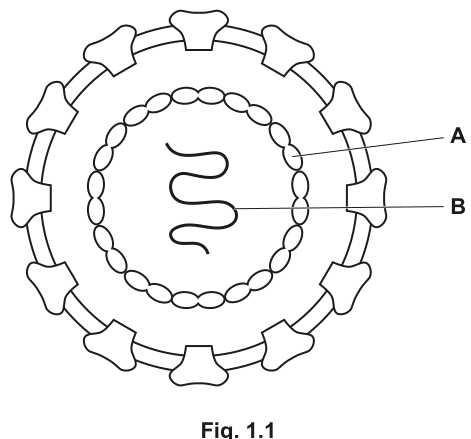
Fig. 1.1 is a drawing of the structure of a Zika virus.
Answer
capsid (or capsomere / protein coat).
capsid
Background Concept
A virus is a non-cellular infectious particle. Outside a host cell it is inert; it only replicates inside a living cell. The basic architecture of a virus consists of a nucleic-acid core (DNA or RNA) surrounded by a protein coat. The protein coat is built from many identical protein subunits that assemble into a geometric shell which protects the genetic material and helps the virus attach to and enter host cells.
The protein coat has a specific terminology:
- Capsid – the whole protein shell enclosing the nucleic acid.
- Capsomere (or capsomer) – one of the individual protein subunits that make up the capsid.
- Protein coat – an informal alternative term for the capsid.
In some viruses (e.g. HIV, influenza, Zika) an additional envelope of host-derived membrane studded with viral glycoprotein spikes surrounds the capsid.
Understanding the Question
Part (a)(i) shows Fig. 1.1, a labelled drawing of Zika virus. The arrow labelled A points to one of the protein subunits embedded in the outer envelope of the virion. You are told that A is made of protein and asked to give its name.
Approach
Identify A as one of the small protein projections on the virus surface and use the standard CIE terminology for that structural component.
Step-by-Step Reasoning
- The structure labelled A is a discrete protein unit sitting on (or projecting from) the viral envelope.
- An individual protein subunit of a viral coat is called a capsomere (or capsomer). The whole shell of such units is the capsid.
- The marking scheme accepts any of: capsid, capsomere, protein coat.
Key Takeaways
- Capsid = the protein shell of a virus.
- Capsomere = one protein subunit of the capsid.
- These terms apply to viruses whether they have an envelope (e.g. Zika, HIV) or not (e.g. adenovirus).
Common Mistakes
- Calling the protein subunit a "spike" or "receptor" – these are functional descriptions, not the structural name.
- Confusing the envelope with the capsid. The envelope is a membrane layer; the capsid is the protein shell beneath.
Things to Be Careful About
In an enveloped virus the outer ring in Fig. 1.1 represents the envelope with embedded glycoproteins, while the inner ring of repeating units represents the capsid. Read the label arrow carefully — A points to a protein subunit, not to the membrane layer.
In Fig. 1.1, structure B is a single-stranded molecule.
Suggest the name of structure B.
structure B = ______
Answer
RNA.
RNA
Background Concept
Viruses carry their genetic information as either DNA or RNA. Zika virus belongs to the Flaviviridae family, whose members have a single-stranded positive-sense RNA genome. Other well-known RNA viruses include HIV (a retrovirus), influenza and rhinovirus; DNA viruses include adenovirus and herpesvirus.
Understanding the Question
Part (a)(ii) tells you that B is a single-stranded molecule located at the centre of the virion (i.e. inside the capsid). You must suggest its identity.
Approach
Recall that the molecule at the core of a virus is its genetic material — DNA or RNA. Zika virus is an RNA virus, so the answer is RNA, but the mark scheme accepts either DNA or RNA here because the question only says "single-stranded".
Step-by-Step Reasoning
- The centre of a virion houses the genome.
- Viruses have either DNA or RNA as their genetic material; "single-stranded" is true of many of both types.
- For Zika virus specifically the genome is single-stranded RNA.
Key Takeaways
- A virus's central molecule is its nucleic-acid genome (DNA or RNA).
- Zika is an RNA virus (+ssRNA), so the correct, specific answer is RNA.
Common Mistakes
- Writing "mRNA", "tRNA" or "rRNA" – these are specific cellular RNA species, not the answer the mark scheme wants, and they are explicitly rejected.
- Writing "dsRNA" or "dsDNA" – the question states the molecule is single-stranded, so double-stranded answers are rejected.
- Writing "viral" on its own is ignored.
Things to Be Careful About
The question says "suggest", so even DNA is accepted — but giving RNA is the more biologically accurate answer.
Fig. 1.2 is a transmission electron micrograph of human kidney cells infected with Zika viruses.
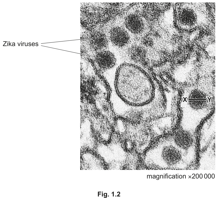
Calculate the actual diameter of a Zika virus using the line X–Y in Fig. 1.2.
Show your working.
Give your answer in nanometres (nm).
diameter = ______
Working
Answer
diameter = 50 nm
50 nm
Background Concept
The relationship between the size of an object as seen in a micrograph (image size) and its real-world size (actual size) is:
Rearranging:
Unit handling is critical. . Because the answer is required in nanometres and the magnification is large, working in mm and converting at the end (or converting the image size to nm first) is the safest approach.
Understanding the Question
Fig. 1.2 is a TEM of Zika viruses inside a human kidney cell. The line X–Y is drawn across the diameter of one virus particle. The magnification is given as . You must measure the length of X–Y on the printed micrograph, divide by the magnification and express the answer in nm.
Approach
- Measure (or estimate) the length of the line X–Y on the printed figure — in this type of question it is typically around .
- Apply the formula .
- Convert to nanometres.
- Quote the final answer with the correct unit.
Step-by-Step Reasoning
- Measure X–Y: the mark scheme accepts , , , or as the final answer. The corresponding image sizes are , , , and respectively. Most candidates measure and obtain .
- Substitute into the formula:
- Convert mm to nm: .
- The diameter of a Zika virus is therefore about , which is biologically sensible (flaviviruses are typically – across).
Key Takeaways
- is the workhorse formula.
- Always convert all lengths to the same unit before dividing.
- Viruses are tens of nanometres across — far below the resolution of a light microscope, which is why TEM is needed.
Common Mistakes
- Dividing magnification by image size instead of the other way round.
- Forgetting to convert mm to nm, so leaving the answer as instead of .
- Writing but then inverting the substitution.
- Quoting the answer with the wrong unit (µm instead of nm).
Things to Be Careful About
- Show the formula AND the substitution to earn the working mark; a bare final answer with no working earns only the final-answer mark.
- The mark scheme uses error carried forward (ecf): if you measure the line incorrectly but apply the formula correctly, you can still earn the working mark.
A magnification of cannot be achieved by a light microscope. The resolution of a transmission electron microscope is also higher than a light microscope.
Describe what is meant by the resolution of a microscope.
Answer
The ability to distinguish between two separate points (as distinct objects).
ability to distinguish between two points
Background Concept
Two properties of a microscope must not be confused:
- Magnification — how many times larger an image appears compared with the real specimen.
- Resolution — the ability of the microscope to show two close objects as two separate objects rather than as one blurred object. It is the minimum distance between two points at which they can still be seen as distinct.
A microscope can have a high magnification but a poor resolution — and vice versa. The human eye can resolve about . A light microscope, because it uses visible-light wavelengths, resolves down to about . A transmission electron microscope, using electrons of much shorter wavelength, resolves down to about — far better, which is why it can image viruses.
Understanding the Question
The stem tells you that the magnification used in Fig. 1.2 is beyond a light microscope's range and that the TEM has a higher resolution than a light microscope. You must define "resolution".
Approach
State the CIE definition: resolution is the ability to distinguish between two points (or two objects) as separate.
Step-by-Step Reasoning
- The mark scheme credits "ability to distinguish between two points" or any clear equivalent wording (AW).
- Just saying "clarity" or "sharpness" is not enough — those are everyday words, not the technical definition.
- Mentioning magnification alone does not score, because magnification and resolution are different properties.
Key Takeaways
- Resolution = minimum distance at which two points can be seen as separate.
- Higher resolution lets you see finer detail, regardless of magnification.
- TEM beats light microscope on resolution because electrons have much shorter wavelengths than visible light.
Common Mistakes
- Confusing resolution with magnification.
- Saying "how much an image is enlarged" — that is the definition of magnification, not resolution.
- Saying "how clear an image is" — too vague; the mark scheme requires reference to distinguishing between two points.
Things to Be Careful About
This is a one-mark, single-sentence definition question. Keep the answer short, technical and to the point.
The vector for Zika virus is the mosquito Aedes aegypti. The mosquito feeds on the blood of an infected person and transmits the virus to another person when it feeds again.
Describe the similarities and differences between the transmission of Zika virus disease and the transmission of malaria.
Answer
- Similarity 1: Both diseases are transmitted by vectors (mosquitoes).
- Similarity 2: Both are transmitted by mosquitoes when the vector feeds on the blood of an infected person.
- Difference: Malaria is transmitted by Anopheles mosquitoes, whereas Zika is transmitted by Aedes aegypti.
Similarities: both are vector-borne, both transmitted by mosquitoes, both blood-borne. Difference: malaria is transmitted by Anopheles, not Aedes.
Background Concept
Many infectious diseases are vector-borne — the pathogen is carried from one host to another by another organism, typically an arthropod. For malaria and Zika, that vector is a mosquito, and the mosquito becomes infected when it takes a blood meal.
- Malaria is caused by Plasmodium (mainly P. falciparum). The vector is a female Anopheles mosquito. The parasite enters the mosquito when it feeds on an infected human, undergoes sexual reproduction in the mosquito gut, and is transmitted when the mosquito later feeds on another person.
- Zika virus disease is caused by Zika virus (a flavivirus). The vector is Aedes aegypti (also Aedes albopictus). The mosquito picks up the virus in a blood meal from an infected person and transmits it when it feeds on another.
Both pathogens therefore share the same basic route: infected human blood → vector mosquito → new human host via mosquito bite.
Understanding the Question
You must describe both the similarities and the differences between the transmission of Zika virus disease and that of malaria. The stem gives you the Zika facts (vector = Aedes aegypti; feeds on infected blood; transmits on next feed); the mark scheme wants you to compare these with malaria.
Approach
Identify (1) one difference that is specific and correct (the genus of the mosquito vector) and (2) up to two shared features (vector-borne, mosquito-borne, blood-borne). Avoid incorrect statements about malaria, which would cap you at 1 mark.
Step-by-Step Reasoning
- Required difference (1 mark): Malaria is transmitted by Anopheles (not Aedes).
- Possible shared points (max 2 marks):
- Both are transmitted by vectors.
- Both are transmitted by mosquitoes.
- Both are blood-borne — the mosquito picks up the pathogen by feeding on infected blood, and transmits it while feeding again.
Any two of these three similarities, combined with the correct genus difference, scores 3.
Key Takeaways
- Both Zika and malaria are mosquito-borne vector diseases — but the mosquito genera are different (Aedes vs Anopheles).
- Be precise with genus names — italicised, capital first letter, lower-case species.
Common Mistakes
- Writing "mosquito" without specifying the genus — that just restates one of the similarities and does not earn the difference mark.
- Stating that malaria is transmitted by Aedes — the mark scheme caps the question at 1 mark if you give incorrect context for malaria.
- Confusing the pathogen: malaria is caused by Plasmodium (a protoctist), Zika by a virus — but this question asks about transmission, not the pathogen.
Things to Be Careful About
Use the command word — "describe" — so the response should describe the comparison, not just list bullet points with no link words. Italicise genus names.
Zika virus vaccines have been developed by scientists.
One of the vaccines contains small proteins from the Zika virus.
Explain how giving this vaccine to a person can lead to the development of long-term immunity against Zika virus disease.
Answer
- The small viral proteins in the vaccine act as (non-self / foreign) antigens.
- The antigens stimulate a primary immune response: macrophages present the antigen, and helper T-lymphocytes activate B-lymphocytes (clonal selection).
- Selected B-lymphocytes undergo clonal expansion (mitosis) to produce a clone of plasma cells (which secrete antibodies) and memory (B- and T-) lymphocytes.
- The memory lymphocytes remain in the circulation for a long time (long-lived). On later exposure to Zika virus, a secondary immune response occurs: there are more specific lymphocytes, they respond faster and produce a higher concentration of specific antibody more quickly, so the person does not develop symptoms of Zika virus disease.
This confers (artificial) active immunity.
Antigens in the vaccine trigger a primary response generating long-lived memory lymphocytes, which mount a faster, stronger secondary response on later exposure to Zika virus.
Background Concept
Active immunity is immunity generated by the body's own immune system in response to antigens; it is long-lasting because it produces memory lymphocytes. Vaccination is artificial active immunity — the antigens are delivered by injection rather than by a natural infection, but the response is the same as if the pathogen itself had been encountered.
The primary immune response has these stages:
- Entry of antigen (here: viral proteins) into the body.
- Phagocytosis by macrophages, which also present antigen on their surface.
- Activation of helper T-lymphocytes by the antigen-presenting macrophage.
- Activation / clonal selection of B-lymphocytes whose surface receptors match the antigen.
- Clonal expansion by mitosis, producing a clone of plasma cells (short-lived, antibody-secreting) and memory cells (long-lived).
- Antibody secretion by plasma cells, which neutralises / agglutinates the antigen.
On re-exposure to the same antigen, memory cells trigger a secondary immune response: faster, stronger, with higher antibody titres, often before symptoms develop. This is the basis of long-term vaccine protection.
Understanding the Question
The vaccine contains small proteins from Zika virus. You must explain how giving the vaccine leads to long-term immunity — i.e. trace the chain from antigen recognition to memory-cell formation and explain why future exposure does not cause disease.
Approach
Build the answer in the order of the primary response, then add the secondary-response step that explains the long-term aspect. The mark scheme caps you at 3 marks if you fail to mention memory cells / immunological memory — so this term is essential.
Step-by-Step Reasoning
Mark-scheme points to hit:
- Proteins are non-self / foreign antigens.
- The antigens stimulate a primary immune response.
- T-lymphocytes and B-lymphocytes bind to / recognise the antigen (with appropriate receptors); this is clonal selection / activation.
- Clonal expansion — selected lymphocytes divide by mitosis to form a clone.
- Memory (B- and T-) lymphocytes are formed.
- Memory cells remain in the circulation for a long time / are long-lived.
- Secondary response is faster and stronger (more antibody produced quickly; person does not develop symptoms).
A clean answer needs an explicit memory cell term; without it the cap is 3 marks.
Key Takeaways
- Vaccines deliver antigens, not the live pathogen — so they cannot cause disease but can still trigger immune memory.
- Long-term protection is conferred by memory lymphocytes, not by circulating antibody (which is short-lived).
- The secondary response is faster because the body already has a population of specific memory cells ready to expand.
Common Mistakes
- Saying the vaccine "gives antibodies" — this would be passive immunity and is biologically wrong for most modern vaccines (which contain antigens, not pre-formed antibodies).
- Describing only the primary response and forgetting memory-cell formation — caps the mark at 3.
- Saying "white blood cells fight the virus" without naming lymphocytes or specifying their roles.
- Confusing T-lymphocytes (cell-mediated) with B-lymphocytes (humoral / antibody-mediated).
Things to Be Careful About
- Use the precise term memory lymphocytes (not just "memory").
- Note that both memory B-cells and memory T-cells are produced.
- "Long-term" must be justified by the longevity of the memory cells, not by re-stating that they are formed.
Explain how a vaccination programme may limit the spread of Zika virus disease through a population.
Answer
- If a large proportion of the population is vaccinated and therefore immune to Zika virus (herd immunity), there are fewer non-immune people for the virus to infect.
- Unvaccinated / non-immune individuals are therefore less likely to come into contact with an infected person, so the transmission cycle is broken and the spread of Zika virus disease through the population is limited.
Herd immunity: a large proportion of the population is immune, so the virus cannot easily reach non-immune individuals and the transmission cycle is broken.
Background Concept
A vaccination programme does not only protect the individuals who are vaccinated. Once a sufficient proportion of the population is immune to a contagious disease, the pathogen can no longer find enough susceptible hosts to maintain its transmission cycle. This indirect protection of unvaccinated individuals is called herd immunity (or population immunity).
The proportion of the population that must be immune to achieve herd immunity depends on the pathogen's basic reproduction number — the average number of new cases generated by one infected individual in a fully susceptible population. The higher is, the higher the vaccination coverage needed. For measles (–) about % coverage is required; for less contagious diseases the threshold is lower.
Understanding the Question
Part (d)(ii) asks you to explain, at the population level, how a Zika vaccination programme limits the spread of the disease.
Approach
Anchor the answer in herd immunity — the technical term the mark scheme rewards. Then add a mechanism: how does herd immunity limit transmission? Because the pathogen runs out of susceptible hosts.
Step-by-Step Reasoning
Mark-scheme points:
- Herd immunity (or equivalently "a large proportion of the population is immune").
- Detail / mechanism: non-immune / unvaccinated people are less likely to come into contact with an infected person (or vice versa); this breaks the transmission cycle.
A two-mark answer needs both points. Possible alternatives the mark scheme also accepts:
- High percentage coverage achievable.
- If is high, a greater percentage of the population must be vaccinated.
Key Takeaways
- Population-level protection from vaccination depends on coverage.
- Herd immunity protects those who cannot be vaccinated (e.g. immunocompromised individuals, very young infants).
- The threshold for herd immunity rises with .
Common Mistakes
- Re-explaining the individual-level immunity (from part (d)(i)) instead of addressing the population level.
- Stating that vaccinated people "can't catch the disease so it can't spread" without referencing herd immunity or the proportion of immune individuals.
- Writing vaguely about "killing the virus" — the virus spreads between people, so the relevant quantity is the chain of transmission, not the pathogen itself.
Things to Be Careful About
This is a different question from (d)(i): here the focus is the population, not the individual. Make sure the wording makes that clear — mention "the population" and "unvaccinated / non-immune people" explicitly.
Collagen is a fibrous protein that is found in many tissues in animals.
Describe the structure of a collagen molecule and the structure of a collagen fibre.
collagen molecule
collagen fibre
Answer
Collagen molecule
- Three polypeptide chains (α-chains) wound around each other to form a triple helix.
- The chains are tightly wound / coiled.
- Hydrogen bonds form between the polypeptides, holding the triple helix together.
- Every third amino acid in each polypeptide is glycine (the smallest amino acid), allowing the chains to pack closely.
Collagen fibre
5. Molecules are arranged in parallel.
6. Molecules are staggered / their ends are not aligned (offset by about one-quarter of their length).
7. Covalent cross-links form between adjacent molecules (e.g. between lysine/hydroxylysine residues), giving the fibre tensile strength.
(Maximum of 4 marks from the molecule section and 4 from the fibre section; total of 5 marks available, so points are taken from both.)
Three helical polypeptide chains wound into a triple helix, cross-linked into staggered parallel arrays to form the fibre.
Background Concept
Collagen is the most abundant protein in mammals and the classic example of a fibrous protein. A single collagen molecule (tropocollagen) is built from three polypeptide α-chains wound together into a right-handed triple helix. Each α-chain is itself a left-handed helix, and the three chains are held together by hydrogen bonds. The sequence of these α-chains is unusual: roughly every third residue is glycine (Gly-X-Y, where X is often proline and Y is often hydroxyproline). Glycine is the only amino acid small enough to fit into the crowded interior of the triple helix where the three chains meet.
Many collagen molecules then assemble into the higher-order collagen fibre. Molecules lie parallel to one another, but their ends are staggered by about a quarter of their length. This staggered arrangement is what creates the characteristic banding pattern of collagen fibrils seen under the electron microscope. Adjacent molecules are joined by covalent cross-links (formed between lysine and hydroxylysine side chains, often via the action of lysyl oxidase), and these cross-links give collagen fibres their enormous tensile strength.
Understanding the Question
Part (a) is a 5-mark "describe" question with two clearly labelled sub-headings: collagen molecule and collagen fibre. The mark scheme warns the candidate NOT to mix molecule features into the fibre section (or vice versa) — there is a cap of 4 marks in each. So the answer must give some molecule points and some fibre points, each in the correct place.
Approach
The strategy is to give the standard structural bullet points in two clear blocks:
- For the molecule: chain number, helical arrangement, hydrogen bonding, the glycine regularity.
- For the fibre: parallel arrangement, staggering of ends, covalent cross-links.
These are the canonical features examiners test on and the ones the mark scheme credits. The "AVP" mark in the mark scheme is a safety net for less common but valid points (e.g. detail of hydrogen bonding, glycosylation).
Step-by-Step Reasoning
- Three polypeptides / triple helix — this is the defining quaternary feature of collagen; credit it first.
- Tightly wound — the chains wrap very closely around each other (unlike the loose coils in, say, keratin).
- Hydrogen bonds between the chains stabilise the triple helix. This is intramolecular, distinguishing it from the covalent cross-links of the fibre.
- Every third amino acid is glycine — this is the molecular-level reason the chains can pack so tightly.
- Parallel arrangement of molecules in the fibre — a fibre-level feature.
- Staggered ends — molecules overlap by about 67 nm (D-period), giving tensile strength along the fibre.
- Covalent cross-links between molecules — these are the strongest stabilising interactions of the fibre and are what give collagen its mechanical strength. They form between the side chains of lysine/hydroxylysine residues on adjacent molecules.
Key Takeaways
- A collagen molecule = three helical polypeptides + H-bonds + glycine regularity (quaternary structure).
- A collagen fibre = many molecules in parallel, staggered, with covalent cross-links between them.
- The molecule is held by hydrogen bonds; the fibre is held by covalent bonds. This two-tier stabilisation is what makes collagen both flexible and incredibly strong.
Common Mistakes
- Putting molecule features in the fibre section (or vice versa) — capped at 1 mark.
- Calling collagen "quaternary structure" without naming the triple helix, or vice versa.
- Describing the molecules themselves as "fibrils" or "fibres" — the molecule is the tropocollagen unit; the fibre is the macroscopic assembly.
- Saying hydrogen bonds hold the fibre together — they hold the molecule together; the fibre uses covalent cross-links.
- Forgetting glycine / every third residue.
Things to Be Careful About
- Use the exact CIE terminology: "polypeptide", "triple helix", "glycine", "cross-links", "staggered".
- Keep the molecule/fibre distinction crisp — label your bullet points if necessary.
- Don't write more than ~7 points; only 5 are needed.
Some amino acids in collagen can be modified to improve the stability of the protein. For example, the amino acid lysine can be modified to form hydroxylysine.
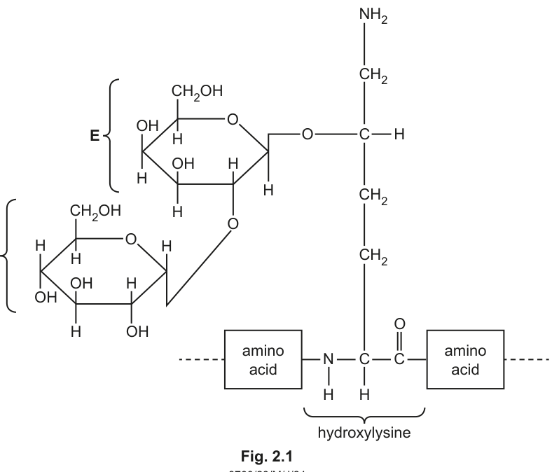
Fig. 2.1 shows a disaccharide bonded to the amino acid hydroxylysine in a collagen molecule. The disaccharide is made from two monosaccharides, which are indicated by the labels D and E in Fig. 2.1.
Answer
α-glucose (alpha-glucose).
α-glucose
Background Concept
Monosaccharides can be drawn as Haworth projections, where the ring oxygen sits at the back-right of the hexagon and the carbons are numbered clockwise from the anomeric carbon (C1) next to the ring oxygen. The position of the –OH group on C1 determines whether the sugar is the α- or β-anomer: –OH below the plane of the ring = α; –OH above the plane = β. This distinction matters because the two anomers form different glycosidic bonds (α-1,4 in starch/glycogen; β-1,4 in cellulose).
Collagen is normally glycosylated on hydroxylysine residues by the disaccharide glucose-galactose (or sometimes galactose-galactose), and the link to hydroxylysine is through the α-anomer of glucose.
Understanding the Question
Part (b)(i) asks the candidate to identify sugar D in Fig. 2.1. D is the sugar that is directly bonded to the hydroxyl oxygen of hydroxylysine's side chain. We need to read the ring structure carefully: it is a six-membered ring (a hexose / pyranose), and we need to decide whether the –OH on C1 is up or down.
Approach
- Identify the ring oxygen in D — it is at the back-right of the hexagon.
- Number the carbons clockwise from C1 (the anomeric carbon, just to the right of the ring oxygen).
- Look at the –OH on C1 — in D it sits below the plane of the ring.
- Below = α-anomer.
- The other features (–CH₂OH on C5, –OH orientations on C2, C3, C4) match glucose.
So D is α-glucose.
Step-by-Step Reasoning
- D is a hexose (six-membered ring with five carbons and one oxygen).
- The –CH₂OH group points up from C5 — consistent with D-series (glucose), not L-series.
- The –OH groups on C2, C3 and C4 match the pattern of glucose.
- The –OH on C1 (the anomeric carbon) points down (below the plane of the ring).
- Down = α-configuration → α-glucose.
Key Takeaways
- In Haworth projections: –OH below the ring on C1 = α; –OH above = β.
- The linkage of the glucose unit to hydroxylysine uses the α-anomer.
- Collagen's carbohydrate attachments occur on hydroxylysine, distinguishing it from most other proteins.
Common Mistakes
- Writing "glucose" without specifying α — the mark scheme explicitly requires "α-glucose" or "alpha-glucose".
- Confusing α and β (often guessed from memory of starch = α; cellulose = β) without reading the figure.
- Calling it "galactose" — galactose is sugar E in the figure, not D.
Things to Be Careful About
- Always give the full "α-glucose" — the alpha is the precise term the mark scheme credits.
- Read C1 carefully; on a printed figure the –OH can be small and easy to miss.
On Fig. 2.1, label the glycosidic bond with the letter G.
Write your answer on Fig. 2.1.
Answer
The candidate draws an arrow / line labeled G pointing to the C–O–C bridge (the covalent linkage formed by condensation between C1 of sugar D and a hydroxyl group of sugar E).
G (label drawn on the C–O–C bond between sugars D and E in Fig. 2.1)
Background Concept
A glycosidic bond is the covalent linkage formed when the –OH group on the anomeric carbon (C1) of one sugar condenses with the –OH of another sugar, releasing a molecule of water. On a structural diagram it appears as a single C–O–C bridge between the two rings.
Understanding the Question
Part (b)(ii) instructs the candidate to label the glycosidic bond with the letter G directly on Fig. 2.1. The bond in question is the oxygen bridge joining the two sugar rings (D and E).
Approach
- Locate the oxygen atom that sits between the two sugar rings (not the ring oxygens inside each sugar, and not the oxygen linking sugar D to hydroxylysine).
- Draw a label G with a clear leader line to that bridging oxygen (or to the whole C–O–C linkage).
Step-by-Step Reasoning
- Sugar E is on the upper ring; sugar D is on the lower ring.
- The two rings are connected by a single oxygen atom.
- That oxygen is the glycosidic linkage and is the one to label G.
- The oxygen that bonds sugar D to hydroxylysine is not the glycosidic bond being labelled — it is an O-glycosidic linkage to the protein, but the inter-sugar bond is the one asked for here.
Key Takeaways
- A glycosidic bond = the C–O–C bridge between two monosaccharide units.
- Distinguish ring oxygens (inside each sugar) from the bridging oxygen (between sugars).
Common Mistakes
- Labelling the ring oxygen of one of the sugars.
- Labelling the oxygen that connects sugar D to the hydroxylysine side chain — that is an O-linked glycosidic bond to protein, not the disaccharide's internal glycosidic bond.
Things to Be Careful About
- Use a single, clear leader line from the letter G to the bridging oxygen; do not write the letter on top of a structure.
On Fig. 2.1, draw a circle around the R group of hydroxylysine and label the circle with the letter R.
Write your answer on Fig. 2.1.
Answer
The candidate circles the side chain (i.e. the four-carbon chain with the terminal and the –OH on the second carbon from the end) attached to the α-carbon of hydroxylysine, and labels the circle with the letter R.
R (circle drawn around the side chain of hydroxylysine, labelled R)
Background Concept
An amino acid has a central α-carbon bonded to four groups: an α-amino group (), an α-carboxyl group (), a hydrogen atom, and an R group (side chain). The R group is what makes each amino acid chemically unique. In a polypeptide chain, the α-amino and α-carboxyl groups are used to form peptide bonds; the R group is left dangling off the backbone and is what determines the amino acid's character (size, charge, ability to form H-bonds, etc.).
Hydroxylysine is a post-translationally modified form of lysine: a hydroxyl group (–OH) has been added to the side chain, making the residue able to form cross-links and to carry glycosidic attachments such as the disaccharide in Fig. 2.1.
Understanding the Question
Part (b)(iii) asks the candidate to circle and label the R group of hydroxylysine in Fig. 2.1. The R group is everything attached to the α-carbon that is not the α-amino, α-carboxyl or α-hydrogen — i.e. the four-carbon side chain with the terminal –NH₂ and the –OH on one of the carbons.
Approach
- Find the α-carbon of hydroxylysine in Fig. 2.1 (the carbon labelled in the centre of the structure, just above the 'amino acid' boxes).
- Trace the side chain that goes upwards from the α-carbon: (the order of –OH along the chain follows the figure).
- Draw a circle around this entire side chain.
- Label the circle with the letter R.
Step-by-Step Reasoning
- The α-carbon has four bonds: one to the α-amino (–NH, going into the left 'amino acid' box), one to the α-carboxyl carbon (going into the right 'amino acid' box), one to an –H, and one to the side chain going up.
- The side chain going up is: with an –OH on one of the middle carbons.
- That whole side chain is the R group.
- The C=O group (carbonyl of the next peptide bond) is part of the backbone, not the R group — do not include it.
- The –NH attached to the α-carbon (going to the previous amino acid) is part of the backbone — do not include it.
Key Takeaways
- R group = side chain = the variable part of the amino acid.
- Hydroxylysine is lysine with an extra –OH on its side chain, and that –OH can be glycosylated or used to form cross-links.
- Don't confuse R group with the whole amino acid — the R group is only the side chain.
Common Mistakes
- Circling the whole amino acid or the whole polypeptide backbone instead of just the side chain.
- Including the α-amino or α-carboxyl group in the circle.
- Forgetting to label the circle R (just circling without the letter loses the mark).
Things to Be Careful About
- The circle must enclose the entire side chain, including the terminal group.
- Add the letter R close to the circle with a clear connection so the examiner can see which circle it labels.
Osteogenesis imperfecta is a disease that results from a deficiency in collagen.
Suggest how a named tissue or structure is affected in a person who has this disease.
Answer
- Bones are weakened / brittle and fracture easily.
(Equivalent acceptable answers: tendons/ligaments — joints become unstable and dislocate easily; skin — becomes thin, fragile and tears easily; cartilage — becomes deformed; blood vessels — become fragile and may rupture; teeth — become brittle.)
- Correct named tissue or structure (e.g. bones / tendons / ligaments / skin / cartilage / blood vessels).
- (The tissue is) weaker / deformed / easily broken — or a description of how its function is impaired.
Bones are weakened and fracture easily (collagen deficiency in osteogenesis imperfecta).
Background Concept
Collagen is the dominant structural protein of most connective tissues: bone matrix, tendons, ligaments, skin (dermis), cartilage, the walls of blood vessels, the dentine of teeth, and the sclera and cornea of the eye. In each of these locations it provides tensile strength — the ability to resist stretching and tearing.
Osteogenesis imperfecta (OI), sometimes called "brittle bone disease", is most often caused by mutations in the COL1A1 or COL1A2 genes, which encode the two α-chains of type I collagen (the main collagen of bone, tendon, ligament, skin and sclera). The mutations produce defective, reduced or abnormal collagen, which weakens every tissue that depends on type I collagen.
Understanding the Question
Part (c) is a 2-mark "suggest" question. The candidate must (1) name a tissue or structure that contains collagen and (2) explain how it is affected. The mark scheme accepts any reasonable collagen-containing tissue, with the second mark for saying it is weakened, deformed, fragile, easily broken, or for describing a specific functional loss.
Approach
- Pick a tissue you know is rich in collagen. Bones are the most obvious and score reliably.
- State how the tissue changes: weaker / brittle / easily broken.
A candidate who knows that OI = "brittle bone disease" can earn both marks from bones alone.
Step-by-Step Reasoning
- Collagen gives bone matrix its tensile strength; without functional collagen, the bone matrix cannot hold the mineral hydroxyapatite together under load.
- Result: bones fracture easily, often with minimal trauma. In severe (type II) OI, fractures occur in utero or at birth.
- Equivalent reasoning for other tissues:
- Tendons/ligaments: collagen transmits muscle force to bone and holds joints together; defective collagen → joints are loose, ligaments tear, joints dislocate.
- Skin: dermis is mostly type I collagen; thin, fragile skin that bruises and tears easily.
- Sclera (white of the eye): abnormally thin sclera lets the underlying choroid show through, giving a blue sclera — a classic OI sign.
- Blood vessels: weakened vessel walls; bruising and risk of vessel rupture.
- Teeth (dentin): dentin is largely type I collagen; brittle teeth that break.
Key Takeaways
- OI is a multi-system disease because type I collagen is widespread.
- The mark scheme accepts a wide range of tissues — pick one you can confidently describe.
- "Suggest" questions reward sound biological reasoning, not just one "correct" answer.
Common Mistakes
- Naming a tissue without saying how it is affected — only 1 mark.
- Naming a tissue that doesn't actually contain much collagen (e.g. skeletal muscle — the connective-tissue sheath has collagen, but the contractile tissue itself does not; an answer mentioning muscle weakness without specifying collagen-rich connective tissue is vague).
- Saying "muscles are weak" — this is incorrect; OI affects connective tissues, not the muscle fibres themselves.
Things to Be Careful About
- Pair the tissue with its specific symptom — the symptom must be plausible for that tissue (e.g. bone → fracture; skin → tears; sclera → blue).
- "Suggest" means you can pick an example; you do not have to list every tissue affected.
Arteries, capillaries and veins are three types of blood vessel.
Table 3.1 shows some features of these three types of blood vessel.
Complete Table 3.1 by using a tick () if the feature is present in the blood vessel and a cross () if the feature is absent from the blood vessel.
Put a tick () or a cross () in every box.
Table 3.1
| feature | artery | capillary | vein |
|---|---|---|---|
| smooth muscle | |||
| endothelium | |||
| tunica media |
Answer
| feature | artery | capillary | vein |
|---|---|---|---|
| smooth muscle | ✓ | × | ✓ |
| endothelium | ✓ | ✓ | ✓ |
| tunica media | ✓ | × | ✓ |
smooth muscle: artery ✓, capillary ×, vein ✓; endothelium: artery ✓, capillary ✓, vein ✓; tunica media: artery ✓, capillary ×, vein ✓
Background Concept
The wall of every blood vessel is built from up to three concentric layers (tunics):
- Tunica externa (adventitia) – outer layer of collagen-rich connective tissue that anchors the vessel and resists over-stretching.
- Tunica media – middle layer containing smooth muscle (which can contract or relax to alter vessel diameter) and elastic fibres. It is thick in arteries and thin in veins.
- Tunica intima / endothelium – innermost single layer of squamous endothelial cells in direct contact with the blood. It provides a smooth, non-thrombogenic surface in every vessel.
Arteries experience the high pressure generated by ventricular systole, so they have all three layers with a thick tunica media rich in smooth muscle and elastic fibres – this allows the wall to stretch and recoil (the elastic rebound that helps maintain blood pressure during diastole).
Veins carry blood back to the heart at low pressure. They contain all three layers but the tunica media is much thinner; their distinctive feature is internal valves that prevent backflow.
Capillaries are exchange vessels only one cell thick. Their wall is just endothelium (plus the basement membrane) – there is no smooth muscle and no recognisable tunica media. This minimal barrier is what allows rapid diffusion of O₂, CO₂, dissolved solutes and water between blood and tissue fluid.
Understanding the Question
The question presents a table with three features as rows (smooth muscle, endothelium, tunica media) and three vessel types as columns (artery, capillary, vein). Every cell must be filled with ✓ (feature present) or × (feature absent). One mark is awarded for each fully correct row, giving a maximum of 3 marks.
Approach
Recall the layer composition of each vessel type and translate each layer into a ✓ or × for the three rows given. Endothelium is the one feature common to every blood vessel; smooth muscle and a tunica media are only present where the wall is more than one cell thick.
Step-by-Step Reasoning
- Smooth muscle row – smooth muscle is found in the tunica media of arteries (thick) and veins (thin) but is absent from the single-cell-thick capillary wall. → artery ✓, capillary ×, vein ✓.
- Endothelium row – all three vessel types are lined by endothelium because every blood-contacting surface needs a smooth, non-thrombogenic lining. → artery ✓, capillary ✓, vein ✓.
- Tunica media row – present in arteries and veins, but capillaries do not have a distinct tunica media. → artery ✓, capillary ×, vein ✓.
Key Takeaways
- Endothelium is universal: it lines arteries, capillaries and veins alike.
- Smooth muscle and a recognisable tunica media are absent only from capillaries.
- The single-cell thickness of capillary walls is the structural basis of rapid gas and solute exchange.
Common Mistakes
- Putting a tick in the capillary column for smooth muscle or tunica media – capillaries are too small to need a contractile/elastic layer.
- Putting a cross for endothelium in the capillary column – even capillaries have a continuous endothelial lining.
- Confusing 'tunica intima' (the whole inner layer) with 'endothelium' (just the cell layer). For this syllabus, when the row says 'endothelium', the answer is whether endothelial cells are present – and they always are.
Things to Be Careful About
- Read the column headings carefully; do not assume the row order implies which feature is 'more important'.
- The mark scheme also has a column-based fallback: if a candidate scores 0 or 1 by rows, the examiner will check columns and award up to 2 marks for correct columns. The intended path, however, is to score each row right.
Fig. 3.1 shows a transmission electron micrograph of part of an alveolus and part of the adjacent capillary.
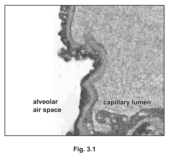
Draw two labelled arrows on Fig. 3.1 to show the direction of movement of oxygen and the direction of movement of carbon dioxide during gas exchange in the lungs.
Answer
- One arrow labelled O₂ drawn from the alveolar air space across the thin fused barrier into the capillary lumen.
- A second arrow labelled CO₂ drawn from the capillary lumen across the thin barrier into the alveolar air space.
O₂ arrow from alveolar air space to capillary lumen; CO₂ arrow from capillary lumen to alveolar air space
Background Concept
Gas exchange between the alveoli and the pulmonary capillary blood is by simple diffusion down partial pressure (concentration) gradients across a very thin barrier. In the lungs:
- The alveolar air has a high partial pressure of O₂ (about ) and a low partial pressure of CO₂ (about ), because inhaled air is continuously refreshed by ventilation.
- Deoxygenated blood arriving from the pulmonary artery has a low pO₂ (about ) and a high pCO₂ (about ), because the systemic tissues have unloaded CO₂ into it and removed its O₂.
Oxygen therefore diffuses from alveolus → blood and carbon dioxide diffuses from blood → alveolus.
Understanding the Question
Fig. 3.1 is a transmission electron micrograph of the alveolar–capillary interface. The alveolar air space is on the left, the capillary lumen on the right, and they are separated by the very thin barrier through which gas exchange occurs. The candidate must draw two labelled arrows on the figure to indicate the directions in which O₂ and CO₂ net-move during gas exchange in the lungs. One mark is awarded for correctly directed, labelled arrows for both gases.
Approach
Set up the partial pressure gradient in your head, then translate it into arrow direction across the thin barrier shown in the micrograph. The arrow tail should start in the higher-pO₂ (or higher-pCO₂) compartment and the head should end in the lower-pO₂ (or lower-pCO₂) compartment.
Step-by-Step Reasoning
- Decide the direction for O₂. Alveolar pO₂ > capillary pO₂, so O₂ diffuses from the alveolar air space into the capillary lumen. The arrow starts in the alveolar air space, crosses the fused basement-membrane region, and ends in the capillary lumen. Label the arrow O₂.
- Decide the direction for CO₂. Capillary pCO₂ > alveolar pCO₂, so CO₂ diffuses from the capillary lumen into the alveolar air space. The arrow starts in the capillary lumen, crosses the same barrier, and ends in the alveolar air space. Label the arrow CO₂.
- The two arrows should point in opposite directions, both crossing the thin barrier, with the labels clearly placed at the heads (or alongside) the arrows.
Key Takeaways
- Net diffusion is always down a partial pressure (concentration) gradient.
- In the lungs, O₂ and CO₂ move in opposite directions across the same barrier because their gradients point opposite ways.
- The very thin fused barrier seen in the TEM is what makes this diffusion rapid enough to equilibrate the blood in the ~0.75 s it spends in the pulmonary capillary.
Common Mistakes
- Drawing both arrows in the same direction – this would imply the same gas or the same gradient for both, which is wrong.
- Forgetting the labels – the mark scheme requires the arrows to be identified as O₂ and CO₂.
- Putting the arrowheads at the wrong ends – the head indicates where the gas is going, not where it comes from.
- Drawing arrows that miss the thin barrier (e.g. crossing through the cytoplasm of the endothelium) – they should cross the diffusion pathway shown.
Things to Be Careful About
- The image is a TEM, so the barrier is at most a few µm thick – the arrows must be drawn across this barrier, not on either side of it.
- The labels 'alveolar air space' and 'capillary lumen' are already printed on the figure; the candidate's job is to add the two arrows with their gas labels.
Suggest and explain how a steep oxygen concentration gradient is maintained in the lungs.
Answer
Any four of:
- Blood arriving at the alveolar capillaries is deoxygenated / has a low partial pressure of O₂, so the gradient from alveolus into blood is steep.
- O₂ diffusing into the blood is immediately taken up by haemoglobin to form oxyhaemoglobin, keeping the plasma pO₂ low and the gradient steep.
- Newly oxygenated blood is constantly carried away from the alveoli in the pulmonary veins, so the blood side of the gradient is continually renewed.
- The lungs contain a large, dense network of capillaries around each alveolus, giving a very large surface area for gas exchange and ensuring every red blood cell passes close to alveolar air.
- Ventilation (breathing in and out) continuously replaces alveolar air with fresh inhaled air, keeping the alveolar pO₂ high.
- A valid additional point – e.g. that ventilation maintains a large concentration difference between the alveolar space and the blood.
See working
Background Concept
The rate of diffusion of a gas across a respiratory surface is described by Fick's law:
For O₂ to diffuse from alveolus to blood at a high rate, three things are needed:
- A large surface area.
- A short diffusion distance.
- A large concentration (partial pressure) difference between the two sides.
The first two are anatomical (millions of alveoli, each wrapped in capillaries, with a barrier only about thick). The third – the steep partial pressure gradient – is maintained by two continuous processes:
- Ventilation keeps the alveolar air high in O₂ by replacing used air with inhaled air.
- Perfusion (blood flow) keeps the capillary blood low in O₂ by constantly bringing fresh deoxygenated blood from the pulmonary artery and removing oxygenated blood via the pulmonary vein. Haemoglobin rapidly mops up O₂ the moment it enters the blood, so plasma pO₂ stays low and the gradient stays steep.
Understanding the Question
The question asks the candidate to suggest and explain how a steep oxygen concentration gradient is maintained in the lungs. The command word 'suggest' means the candidate should propose ideas that go beyond pure recall; 'explain' means each suggestion must be justified. Four marks are available, drawn from a pool of six creditable ideas.
Approach
Think about what would happen if either side of the gradient were allowed to equilibrate. The answer must keep the alveolar pO₂ high (ventilation) and the capillary pO₂ low (continuous blood flow + haemoglobin uptake + a large capillary surface area). Cover both 'sides' of the gradient for full marks.
Step-by-Step Reasoning
- Deoxygenated blood arrives at the alveolar capillary. Blood returning from the body via the pulmonary artery has had most of its O₂ unloaded at the tissues, so the plasma pO₂ is low. As soon as it reaches the alveoli, there is an immediate, steep gradient for O₂ to enter. (Mark 1)
- Haemoglobin takes up the O₂. As O₂ diffuses into the plasma, it is rapidly bound by haemoglobin inside red blood cells, forming oxyhaemoglobin. This keeps the plasma pO₂ low, so the gradient from alveolus to plasma remains steep. (Mark 2)
- Blood flow constantly removes the oxygenated blood. The pulmonary circulation is continuous; freshly oxygenated blood is carried away in the pulmonary vein, so the blood side of the barrier is continually being renewed with deoxygenated blood. (Mark 3)
- A large capillary network surrounds the alveoli. Each alveolus is wrapped in a dense mesh of pulmonary capillaries, providing a huge surface area and ensuring that every red blood cell passes very close to the alveolar air. (Mark 4)
- Ventilation continuously refreshes the alveolar air. Inhalation brings in fresh atmospheric air (pO₂ ≈ ), and exhalation removes air that has been depleted of O₂. This keeps the alveolar pO₂ high, so the gradient is not allowed to equilibrate. (Mark 5 if needed)
- Acceptable additional valid point – e.g. that the combination of ventilation and perfusion maintains a large concentration difference between the alveolar space and the blood. (Mark 6 if needed)
Any four of these ideas earn the four marks.
Key Takeaways
- A diffusion gradient is maintained by continuously supplying one side and continuously removing the other.
- Ventilation keeps alveolar pO₂ high; blood flow + haemoglobin keep capillary pO₂ low.
- Fick's law ties together surface area, thickness and concentration difference – this question is about the last of these.
Common Mistakes
- Vague answers that just say 'breathing brings in oxygen' without explaining why this keeps the gradient steep (i.e. because it stops the alveolar pO₂ from falling towards equilibrium with the blood).
- Mentioning features of the gas exchange surface (large SA, thin barrier, moist) – these help Fick's law but they do not maintain the gradient; the question is specifically about the gradient.
- Confusing ventilation with perfusion – ventilation moves air, perfusion moves blood; both are needed but they do different jobs.
- Saying haemoglobin 'carries oxygen away' as if the blood is the gradient – haemoglobin is important because it keeps the plasma pO₂ low, not because it moves the blood.
Things to Be Careful About
- The question is not asking why gas exchange is efficient in general (Fick's law); it is asking specifically what maintains the concentration gradient for O₂.
- 'Deoxygenated' and 'low pO₂' are both acceptable ways of saying the same thing on the mark scheme; the precise term is the safer one.
- The word 'constantly' or 'continually' is part of the mark scheme wording – the gradient is only maintained because the renewal is continuous, not occasional.
The passage outlines the roles of blood vessels associated with the heart.
Complete the passage by using the most appropriate scientific terms.
The ______ carries blood to the left atrium. After passing from the left atrium to the left ventricle, blood is pumped into the aorta. The aorta is one of two large arteries that carry blood away from the ventricles of the heart. Blood that leaves the heart to enter these arteries must pass through the ______ valves.
Oxygenated blood is supplied to the cardiac muscle cells through the ______ arteries.
Answer
- The pulmonary vein carries blood to the left atrium.
- Blood that leaves the heart to enter these arteries must pass through the semilunar valves.
- Oxygenated blood is supplied to the cardiac muscle cells through the coronary arteries.
pulmonary vein; semilunar; coronary
Background Concept
The heart is served by a recognisable set of named vessels and valves:
- Pulmonary vein – returns oxygenated blood from the lungs to the left atrium. (Despite being a 'vein', it carries oxygenated blood – the names artery/vein refer to direction of flow, not oxygen content.)
- Aorta – the largest artery, carrying oxygenated blood from the left ventricle to the systemic circulation. Its companion on the right side of the heart is the pulmonary artery, which carries deoxygenated blood from the right ventricle to the lungs.
- Semilunar valves – the two valves at the exits of the ventricles, the aortic valve (at the entrance to the aorta) and the pulmonary valve (at the entrance to the pulmonary artery). They are called 'semilunar' because each of their three cusps is shaped like a half-moon. They prevent backflow of blood from the arteries into the ventricles during diastole.
- Coronary arteries – the first branches of the aorta, arising just above the aortic valve. They run over the surface of the heart in the coronary sulcus and supply the cardiac muscle (myocardium) with oxygenated blood. Blockage of these arteries causes myocardial infarction (a 'heart attack').
Understanding the Question
The passage has three blanks, each requiring the most appropriate scientific term. The context of each blank is given in the surrounding sentence, so the answer is not simply a free recall of any heart-related vessel – it must fit the description.
- Blank 1: a vessel that 'carries blood to the left atrium' – i.e. the vessel entering the left atrium.
- Blank 2: valves that blood must pass through to 'enter these arteries' from the ventricles – i.e. the valves at the ventricular outflow tracts.
- Blank 3: arteries that supply oxygenated blood to the cardiac muscle cells themselves.
Approach
Translate each blank into a structural description and pick the matching technical term from the heart's vocabulary. The mark scheme is strict – the answer must be the most appropriate term, so a near-synonym is unlikely to earn the mark.
Step-by-Step Reasoning
- 'Carries blood to the left atrium' – blood enters the left atrium from the lungs, and the only vessel doing this is the pulmonary vein (there are usually four pulmonary veins, two from each lung, but they are collectively referred to as 'the pulmonary vein' in this syllabus). (Mark 1)
- 'Blood that leaves the heart to enter these arteries must pass through the ____ valves' – the passage identifies 'these arteries' as the aorta and the pulmonary artery (the two large arteries leaving the ventricles). The valves at the entrances to these arteries are the aortic and pulmonary valves, both of which are semilunar valves. The mark scheme accepts either 'semilunar' (preferred) or 'aortic and pulmonary' as alternative wording, but the single-word answer 'semilunar' is the most appropriate scientific term. (Mark 2)
- 'Oxygenated blood is supplied to the cardiac muscle cells through the ____ arteries' – the myocardium has its own dedicated blood supply via the coronary arteries, which branch from the aorta just above the aortic valve. (Mark 3)
Key Takeaways
- Arteries leave the heart; veins enter it – this rule identifies the pulmonary vein as the only vein carrying oxygenated blood.
- The semilunar valves (aortic and pulmonary) sit at the exits of the ventricles; the atrioventricular valves (bicuspid and tricuspid) sit between the atria and ventricles.
- The myocardium is supplied by the coronary arteries, not by blood inside the chambers – the chamber walls are too thick to be nourished by diffusion from within.
Common Mistakes
- Writing 'pulmonary artery' for blank 1 – the pulmonary artery carries blood away from the heart to the lungs, not to the left atrium.
- Writing 'atrioventricular' or 'bicuspid' for blank 2 – these valves sit between atria and ventricles, not at the exits of the ventricles into the arteries.
- Writing 'aorta' for blank 3 – the aorta supplies the systemic circulation, not the heart muscle itself.
- Lower-case or imprecise wording – 'semi-lunar' or 'semi lunar' may be marked wrong on a strict mark scheme; the single-word form is 'semilunar'.
Things to Be Careful About
- The marks here are for the most appropriate scientific term. A near-miss (e.g. 'vena cava' for the pulmonary vein) will not earn the mark.
- Don't be confused by the fact that the pulmonary vein carries oxygenated blood – it is still a vein because it carries blood towards the heart.
- The coronary arteries deliver oxygenated blood to the cardiac muscle; the coronary veins drain deoxygenated blood from it back into the right atrium via the coronary sinus.
Lignin and suberin are polymers that are present in plant tissues.
Describe and explain the roles of lignin and suberin in the transport of water through the roots and stem of a plant.
Answer
- Lignin and suberin are both hydrophobic / waterproof polymers, so they act as barriers to the movement of water.
- Suberin is deposited in the Casparian strip in the walls of endodermal cells in the root.
- This forces water to move from the apoplast pathway to the symplast pathway (through the cytoplasm) at the endodermis, which allows the plant to control which solutes enter the xylem.
- Lignin is deposited in the cell walls of xylem vessel elements, where it waterproofs the walls (so water does not leak sideways out of the xylem) and provides mechanical strength so the vessels do not collapse inwards under the tension of the water column.
See working.
Background Concept
Plants move water from the soil to the atmosphere through the xylem, driven largely by the cohesion–tension mechanism: evaporation from the leaves pulls on a continuous column of water held together by hydrogen bonding, and this tension is transmitted down through the xylem to the roots. For this system to work, two physical conditions must be met: (1) the water must follow a controlled path into the xylem, and (2) the xylem vessels themselves must not leak or collapse under the negative pressure inside them. Two hydrophobic polymers — lignin and suberin — make both of these conditions possible.
Suberin is a waxy, hydrophobic polymer deposited in the cell walls of the root endodermis. In the radial and transverse walls of endodermal cells it forms a continuous band called the Casparian strip. Because suberin is waterproof, it blocks the apoplast pathway — the route in which water moves through the interconnected network of cell walls and intercellular spaces without ever crossing a plasma membrane. To get past the Casparian strip, water and dissolved substances must enter the cytoplasm of an endodermal cell and continue through the symplast pathway (cytoplasm connected by plasmodesmata). This means they have to cross at least one plasma membrane, which is a control point: the membrane determines which ions and molecules are allowed through, and it helps to keep pathogens and unwanted solutes out of the transpiration stream.
Lignin is a complex phenolic polymer laid down in the secondary cell walls of xylem vessel elements (and other supporting cells such as sclerenchyma fibres). Lignified walls are both waterproof and mechanically very strong. Waterproofing prevents water inside the xylem from leaking sideways out into the surrounding tissue. Strength is critical because the water column inside the vessels is under substantial negative pressure (tension) generated by evaporation from the leaves; without stiff, lignified walls the vessels would collapse inwards under this tension and the continuous water column would break.
Together, suberin in the root and lignin in the xylem allow the plant to control what enters the transpiration stream and to keep that stream physically intact as it is pulled up the plant.
Understanding the Question
The question asks you to describe and explain the roles of two polymers — lignin and suberin — in water transport through the roots and stem. The command words tell you to give both the function (describe) and the biological reason (explain). You must cover both polymers; an answer that deals with only one of them is capped at 3 marks. The mark scheme also allows a maximum of 3 marks for either lignin or suberin, so a balanced answer (one or two points for each, plus a shared introductory point about hydrophobicity) is the safest way to reach the full 4 marks.
Approach
- Open with the property that both polymers share: they are hydrophobic / waterproof.
- Handle suberin: state its location (Casparian strip of the endodermis), describe what this does to the route water takes (apoplast → symplast), and explain why that matters (control of solutes entering the xylem).
- Handle lignin: state its location (walls of xylem vessel elements), describe what it does to those walls (waterproofs them), and explain why this matters (prevents water leaking out and prevents vessel collapse under tension).
Step-by-Step Reasoning
- Shared property (1 mark). Lignin and suberin are both hydrophobic / waterproof polymers. This single fact is the foundation for every role they play in water transport — wherever one of these polymers is deposited, water can no longer move freely through that wall.
- Suberin location (1 mark). Suberin is deposited in the Casparian strip — a band of waterproof material in the radial and transverse walls of the endodermal cells of the root.
- Suberin function — pathway switch (1 mark). Because the apoplast pathway is blocked at the Casparian strip, water and dissolved substances must enter the cytoplasm of an endodermal cell and continue via the symplast pathway to reach the stele and the xylem.
- Suberin function — solute control (1 mark). This forced symplastic step is a control point: only solutes that the plasma membrane of the endodermal cell allows to pass can enter the xylem, and pathogens / unwanted solutes are largely excluded from the transpiration stream.
- Lignin location (1 mark). Lignin is deposited in the secondary cell walls of xylem vessel elements.
- Lignin function — waterproofing (1 mark). The lignified walls are impermeable to water, so water inside the xylem cannot leak out sideways into surrounding tissues.
- Lignin function — mechanical support (1 mark). Lignin adds great mechanical strength to the walls, which is essential because the water column inside xylem vessels is under strong tension (negative pressure) created by evaporation from the leaves. Without reinforcement, the thin vessel walls would collapse inwards and the continuous water column would break.
- Combined consequence (1 mark, AVP). Together, suberin and lignin make the cohesion–tension mechanism of water transport physically possible: suberin controls what enters the stream, and lignin keeps the stream intact as it is pulled up the plant.
Key Takeaways
- Both lignin and suberin are hydrophobic polymers, but they play different roles: suberin is a control barrier in the root, while lignin is a waterproofing and structural barrier in the xylem.
- The Casparian strip of suberin forces water from the apoplast pathway into the symplast pathway at the endodermis, giving the plant a checkpoint for what enters the xylem.
- Lignin in xylem walls is essential because xylem vessels carry water under tension; lignified walls prevent both sideways leakage and inward collapse.
- These two polymers together make the cohesion–tension mechanism of water transport work.
Common Mistakes
- Confusing suberin with cutin: suberin is in the Casparian strip of the root endodermis, cutin is in the leaf cuticle.
- Saying that suberin "blocks water" without explaining that this forces water into the symplast — the pathway switch is the key biological point.
- Claiming lignin "prevents the xylem from bursting" — the mark scheme explicitly rejects this; the risk is inward collapse under tension, not outward bursting.
- Describing lignin as merely "structural" without linking it to waterproofing the vessel walls.
- Omitting either lignin or suberin entirely — the mark scheme caps each at 3 marks, so to reach 4 you need at least one point from each.
- Using vague language like "in the root" or "in the stem" instead of the precise terms (Casparian strip, endodermis, xylem vessel elements).
Things to Be Careful About
- Use the exact terms apoplast, symplast, Casparian strip, endodermis, xylem vessel element. Vague wording will not earn credit.
- Be specific that lignin is in the walls of xylem vessels, not the lumen.
- Do not say suberin "lets water through" — it blocks the apoplastic route, forcing water to take the symplast route.
- Both polymers are hydrophobic, but their roles are different; do not collapse them into a single undifferentiated answer.
- The mark scheme allows "xylem / vessel elements" as the location of lignin — you do not need to say "tracheids" or "vessel elements" precisely, but you must name the xylem.
The enzyme laccase catalyses the formation of lignin in plants.
Fig. 4.1 is a diagram of the mode of action of laccase.
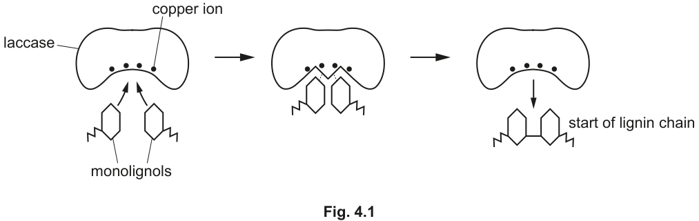
Describe and explain the mode of action of laccase when catalysing the formation of lignin.
Answer
- Laccase works by the induced-fit mechanism: in stage 1 of Fig. 4.1 the shape of the active site is not initially fully complementary to the monolignols.
- As the monolignols bind, the active site changes shape to become fully complementary to them, forming an enzyme–substrate complex (stage 2).
- Within the complex, laccase lowers the activation energy of the reaction joining the two monolignols together to form the start of a lignin chain (stage 3).
- After the product (start of lignin chain) leaves, the active site returns to its original shape and the enzyme is available to be re-used.
- AVP: the copper ion at the active site is a cofactor required for laccase activity.
See working.
Background Concept
Enzymes are globular proteins that act as biological catalysts — they speed up metabolic reactions without being consumed. Each enzyme has a region called the active site, a three-dimensional pocket whose shape and chemical properties are specific to the substrate(s) it binds. The substrate is the molecule the enzyme acts on; in this question the substrates are two monolignol molecules and the product is the start of a lignin chain.
Two classical models describe how substrates fit into the active site:
- The lock-and-key model proposes that the active site has a rigid, perfectly complementary shape to the substrate from the outset.
- The induced-fit model proposes that the active site is not initially a perfect fit. When the substrate begins to bind, the active site changes shape (the enzyme slightly adjusts its tertiary structure around the binding region) so that it becomes fully complementary and wraps closely around the substrate. This is the accepted model for most enzymes, and laccase is one of them.
The moment of catalysis is the enzyme–substrate (ES) complex — the transient state in which the substrate(s) is/are bound to the active site. Within this complex, the enzyme lowers the activation energy of the reaction, i.e. the minimum energy input required to convert substrate into product. Activation energy is lowered by mechanisms such as:
- holding the substrates in the correct orientation to react with each other,
- stressing or distorting bonds in the substrates so they break more easily,
- providing a chemical environment (e.g. acidic / basic amino acid side chains, or metal-ion cofactors) that stabilises the transition state.
The products are then released, the active site returns to its original shape, and the enzyme is available to catalyse another reaction. Some enzymes also require a cofactor — a non-protein component such as a metal ion — for activity. Laccase contains a copper ion at its active site, and this copper acts as a cofactor.
Understanding the Question
This part asks you to describe and explain the mode of action of the enzyme laccase, which catalyses the joining of monolignols into the start of a lignin chain. Fig. 4.1 shows the process in three stages: (1) two monolignol substrates approach the active site, which contains a copper ion; (2) the monolignols bind to the active site; (3) the joined product — the start of a lignin chain — is released.
The command words require both a description of what happens at each stage and an explanation of the biology behind it (induced fit, ES complex, activation energy). The mark scheme also notes that you can earn a maximum of 4 out of 5 marks if you do not refer specifically to laccase and monolignols — so use the names from the figure, not just "the enzyme" and "the substrate".
Approach
- Identify the model of action (induced fit) and link it to stage 1 → stage 2 of the figure.
- Name the transient state formed (ES complex) and the consequence of forming it (activation energy lowered → reaction occurs → stage 3).
- End with the re-use step: products leave, active site returns to original shape, enzyme available again.
- Add a specific AVP point — the most obvious one from the figure is that the copper ion is a cofactor.
Step-by-Step Reasoning
- Identify the model — induced fit (1 mark). Laccase works by the induced-fit mechanism: in stage 1 the shape of the active site is not yet fully complementary to the monolignols.
- Active-site change on binding (1 mark). As the monolignols enter and begin to bind, the active site changes shape so that it becomes fully complementary to them.
- Formation of the ES complex (1 mark). With the active site now fully complementary, an enzyme–substrate complex is formed — the monolignols are held in the correct orientation within the active site (stage 2 of the figure).
- Lowering of activation energy (1 mark). Within this ES complex, laccase lowers the activation energy of the reaction. Concretely, the two monolignols are held close together in the correct orientation for the bonding step, and the copper ion cofactor provides the chemical environment that makes the reaction possible. (Stage 3 of the figure: the monolignols are joined into the start of a lignin chain.)
- Re-use of the enzyme (1 mark). After the product leaves, the active site returns to its original shape, and laccase is available to catalyse further reactions — the enzyme is not used up in the process.
- AVP — cofactor (1 mark). The copper ion at the active site acts as a cofactor, a non-protein component that is required for laccase's catalytic activity. (Other valid AVP points include: the monolignols are held in place by temporary hydrogen bonds and other weak interactions with R groups in the active site; or, laccase is specific to monolignols and only catalyses this particular joining reaction.)
Key Takeaways
- Laccase uses induced fit, not lock-and-key: the active site is not a perfect fit until the substrate binds.
- The enzyme–substrate complex is the moment of catalysis — it is within this complex that the activation energy is lowered and the substrates are joined.
- The active site returns to its original shape after the product leaves, so the enzyme is re-usable.
- Some enzymes, including laccase, require a cofactor (here, a copper ion) for activity.
Common Mistakes
- Saying the active site has a rigid shape that fits perfectly from the start (this is lock-and-key, not induced fit).
- Failing to name the substrates and product — the mark scheme deducts up to one mark if laccase and/or monolignols are not specifically named.
- Stating that the enzyme is "used up" or "consumed" by the reaction. Enzymes are catalysts and are regenerated.
- Saying the enzyme "provides energy" for the reaction. The enzyme does not supply energy; it lowers the energy barrier so the reaction can proceed at the temperature of the cell.
- Describing only what the figure shows (e.g. "the substrates bind, then the product is released") without explaining the biology behind it. The command words are "describe AND explain" — both are required for full marks.
- Introducing molecules that are not in the figure (e.g. ATP, NADH) — the only cofactor the figure shows is the copper ion.
Things to Be Careful About
- Refer explicitly to Fig. 4.1 by quoting what is happening at each stage.
- Use the exact terms: induced fit, active site, enzyme–substrate complex, activation energy, cofactor, re-use.
- Note the mark scheme's rule: max 4 if no reference to laccase / monolignols — using the specific names from the figure is part of the credit.
- Do not contradict the figure: the substrates are two separate monolignols, the product is the joined "start of lignin chain", and the copper ion is at the active site — your answer should be consistent with this.
Cyclin-dependent kinases (CDKs) are enzymes that regulate the cell cycle.
Cell signalling by ligands causes the activation of CDKs in target cells.
Outline the main stages in the process of cell signalling by ligands that can cause specific responses in target cells.
Answer
- Cells secrete ligands, which travel to target cells (e.g. via the blood / circulatory system) ;
- Ligands bind to specific / complementary receptors on (target) cells ;
- (Binding) sets off reactions within the (target) cell / triggers secondary messengers (e.g. cyclic AMP) / sets off an enzyme cascade .
See working
Background Concept
Cell signalling is the process by which cells communicate with each other. A signalling cell releases a chemical messenger (the ligand), which travels to a target cell and binds to a specific receptor. This binding triggers a cascade of intracellular events that produce a specific response. The signal is often amplified through secondary messengers (e.g. cyclic AMP) or enzyme cascades. In the context of this question, the ligands activate CDKs in target cells.
Understanding the Question
The question asks for an outline of the main stages in cell signalling by ligands. The context is that ligands activate CDKs in target cells, but the focus is on the general process. The command word "outline" means a brief description of the main stages, not a detailed explanation. The part is worth 2 marks, so two distinct stages are required.
Approach
The main stages of cell signalling by ligands are:
- Secretion of the ligand by the signalling cell (or its transport in the blood).
- Binding of the ligand to a specific receptor on the target cell.
- Triggering of an intracellular response (secondary messengers or enzyme cascade).
For 2 marks, the candidate should provide any 2 of these 3 key stages; including all 3 in a concise list is the safest approach.
Step-by-Step Reasoning
- Signalling cells produce and release ligands. These are typically hormones or other signalling molecules. The ligands may travel through the bloodstream or other transport mechanisms to reach the target cells.
- The ligands bind to specific receptors on the target cells. The receptor has a binding site that is complementary to the ligand, ensuring specificity. Only cells with the correct receptor will respond to the signal.
- The binding of the ligand to the receptor triggers a series of reactions within the target cell. This can involve secondary messengers (such as cyclic AMP) or enzyme cascades that amplify the signal and lead to the cellular response. In this case, the response is the activation of CDKs.
Key Takeaways
- Cell signalling involves ligand secretion, transport, receptor binding, and intracellular response.
- The specificity of receptor-ligand binding determines which cells respond.
- The signal is often amplified through secondary messengers or enzyme cascades.
- In the context of this question, the ligands activate CDKs in target cells.
Common Mistakes
- Failing to mention the specificity of receptor-ligand binding.
- Confusing the roles of ligands and receptors (e.g. saying the receptor is released by the cell).
- Not mentioning the intracellular response (e.g. secondary messengers or enzyme cascade).
- Describing the process in too much detail when only an outline is required.
- Omitting the role of the bloodstream in transporting the ligand to the target cell.
Things to Be Careful About
- The question asks for stages, so the answer should be presented as a clear sequence.
- "Outline" suggests a brief description, not a paragraph of explanation.
- The mark scheme accepts either "cells secrete ligands" or "transport of ligands through blood" as one marking point, so the secretion step can be implied by mentioning transport.
The activity of CDKs is reduced by CDK inhibitors. Many of these inhibitors occur naturally in cells.
Fig. 5.1 is a diagram of a CDK inhibitor binding to a CDK molecule.
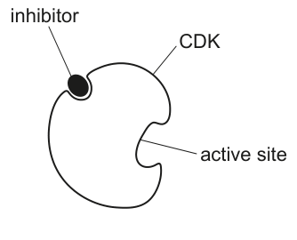
State and explain how the CDK inhibitor in Fig. 5.1 prevents the activity of the CDK molecule.
Answer
- (Non-competitive) inhibitor binds to the allosteric site / a site other than the active site ;
- (Binding causes) a change in shape of the active site ;
- The active site is no longer complementary to the substrate, so the substrate cannot bind .
See working
Background Concept
Enzyme inhibitors are molecules that reduce the activity of enzymes. There are two main types:
- Competitive inhibitors: bind to the active site, directly competing with the substrate.
- Non-competitive inhibitors: bind to a site other than the active site (the allosteric site), causing a conformational change in the enzyme.
The allosteric site is a distinct regulatory site on the enzyme. When an inhibitor binds to the allosteric site, it changes the three-dimensional shape of the enzyme, including the active site. As a result, the substrate can no longer bind effectively to the active site, and the enzyme's activity is reduced. Importantly, non-competitive inhibition cannot be overcome by increasing the substrate concentration.
Understanding the Question
The question shows Fig. 5.1, which depicts a CDK enzyme with an inhibitor binding to a region that is clearly distinct from the active site. The question asks to state and explain how this CDK inhibitor prevents the activity of the CDK. The part is worth 2 marks.
Approach
From the diagram:
- The inhibitor binds to a site on the CDK that is different from the active site — this is the allosteric site.
- The inhibitor is therefore a non-competitive inhibitor.
- The mechanism involves binding to the allosteric site, which causes a conformational change that affects the active site.
The candidate should state:
- The type of inhibition (non-competitive) and where the inhibitor binds (allosteric site).
- How this affects the active site (conformational change) and prevents substrate binding (active site no longer complementary to the substrate).
Step-by-Step Reasoning
- Fig. 5.1 shows the inhibitor binding to a region of the CDK that is distinct from the active site. This region is the allosteric site.
- Because the inhibitor binds to the allosteric site, it is a non-competitive inhibitor.
- The binding of the inhibitor to the allosteric site induces a conformational change in the enzyme, altering the three-dimensional shape of the active site.
- With the active site shape changed, it is no longer complementary to the substrate. The substrate cannot bind effectively to the active site.
- As a result, the CDK cannot catalyse its reaction, and its activity is reduced or stopped.
Key Takeaways
- Non-competitive inhibitors bind to the allosteric site, not the active site.
- Binding to the allosteric site causes a conformational change that distorts the active site.
- The substrate can no longer bind because the active site is no longer complementary.
- This type of inhibition is not affected by substrate concentration.
Common Mistakes
- Confusing non-competitive with competitive inhibition (saying the inhibitor blocks the active site directly).
- Saying the inhibitor "blocks" the active site (it binds elsewhere, not at the active site).
- Not mentioning the conformational change in the active site.
- Saying the active site is "destroyed" (it only changes shape; it is not destroyed).
- Failing to identify the inhibition as non-competitive.
Things to Be Careful About
- The diagram clearly shows the inhibitor binding AWAY from the active site.
- The key concept is the conformational change in the active site, not the active site being directly blocked.
- Do not describe the inhibitor as "covering" or "filling" the active site.
Table 5.1 lists three different CDKs, their roles in the cell cycle and molecules that inhibit them.
Table 5.1
| name of CDK | role of CDK in the cell cycle | CDK inhibitor |
|---|---|---|
| CDK1 | regulates cell progression from to mitosis | RO-3306 |
| CDK2 | regulates the processes of the S phase | p21Cip1 |
| CDK4 | regulates cell progression from to the S phase | palbociclib |
With reference to Table 5.1, state which CDK inhibitor is likely to result in a cell containing one chromatid per chromosome. Explain your answer.
inhibitor = ______
explanation
Answer
Inhibitor: Palbociclib (alternative: p21Cip1)
- Palbociclib inhibits CDK4, which regulates the transition from to S phase. The cell cannot enter S phase, so DNA replication does not occur and sister chromatids are not formed.
- (Alternative) p21Cip1 inhibits CDK2, which regulates S phase. DNA replication cannot occur / be completed, so sister chromatids are not formed.
- Therefore each chromosome remains with one chromatid .
Palbociclib (or p21Cip1)
Background Concept
The cell cycle consists of several phases:
- phase: cell growth and preparation for DNA replication.
- S phase: DNA replication; each chromosome is duplicated to form two sister chromatids joined at the centromere.
- phase: further growth and preparation for mitosis.
- Mitosis: nuclear division.
- Cytokinesis: cell division.
Before S phase, each chromosome consists of a single DNA molecule (one chromatid). After S phase, each chromosome has two sister chromatids.
CDKs (cyclin-dependent kinases) regulate progression through the cell cycle. Different CDKs are responsible for different transitions:
- CDK4: to S phase transition.
- CDK2: regulation of S phase processes.
- CDK1: to mitosis transition.
Understanding the Question
The question asks which CDK inhibitor would result in a cell with one chromatid per chromosome. The mark scheme uses Table 5.1, which lists each CDK, its role in the cell cycle, and a specific inhibitor. We need to consider the role of each CDK and determine which one, if inhibited, would prevent the formation of sister chromatids.
Approach
To answer, we identify:
- The phase in which sister chromatids are formed (S phase).
- The CDKs that regulate entry into or progression through S phase (CDK4 and CDK2).
- The corresponding inhibitors (Palbociclib for CDK4; p21Cip1 for CDK2).
- The effect of these inhibitors: the cell cannot complete S phase, so DNA replication does not produce sister chromatids.
Step-by-Step Reasoning
- Sister chromatids are formed during S phase, when DNA is replicated.
- CDK4 regulates the transition from into S phase. If CDK4 is inhibited by Palbociclib, the cell cannot enter S phase at all. DNA replication does not occur, so sister chromatids are not formed. Each chromosome therefore remains with a single chromatid.
- Alternatively, CDK2 regulates the S phase itself. If CDK2 is inhibited by p21Cip1, DNA replication cannot proceed normally. Sister chromatids are not formed (or are not completed). Each chromosome therefore remains with a single chromatid.
- In contrast, RO-3306 inhibits CDK1 ( to mitosis). The cell would still pass through S phase, so each chromosome would have two chromatids. This does not fit the question.
- The correct inhibitor is Palbociclib (or p21Cip1).
Key Takeaways
- Sister chromatids are produced by DNA replication in S phase.
- Inhibiting CDK4 (Palbociclib) prevents entry into S phase.
- Inhibiting CDK2 (p21Cip1) prevents DNA replication during S phase.
- Either way, the cell will have chromosomes with one chromatid each.
Common Mistakes
- Choosing RO-3306 (CDK1 inhibitor), which would stop the cell in after S phase, giving two chromatids per chromosome.
- Confusing the order of the cell cycle phases.
- Not explaining WHY the chosen inhibitor leads to one chromatid per chromosome (the explanation must refer to S phase / DNA replication).
- Naming an inhibitor without linking it to the relevant CDK.
Things to Be Careful About
- Both Palbociclib and p21Cip1 are accepted answers.
- The explanation must mention S phase and DNA replication / sister chromatid formation.
- A cell with one chromatid per chromosome is in (or arrested before completing S phase).
With reference to Table 5.1, state which CDK inhibitor is likely to result in a cell with:
• a relatively high concentration of mitochondria
• two chromatids per chromosome.
Explain your answer.
inhibitor = ______
explanation
Answer
Inhibitor: RO-3306
- RO-3306 inhibits CDK1, which regulates the transition from to mitosis, so the cell cycle stops in (after S phase).
- Mitochondria divide in and , so by the time the cell is arrested in , mitochondria have already divided / increased in number, giving a relatively high concentration of mitochondria.
- DNA replication occurred in S phase (which precedes ), so each chromosome has two chromatids (sister chromatids) .
RO-3306
Background Concept
The cell cycle has several phases, and specific events happen in each:
- phase: cell growth; organelle replication, including mitochondrial division, begins.
- S phase: DNA replication; each chromosome is duplicated to form two sister chromatids.
- phase: continued growth, including further mitochondrial division.
- Mitosis: nuclear division.
- Cytokinesis: cell division.
The order is therefore: → S → → mitosis → cytokinesis.
CDK1 regulates the transition from to mitosis. If CDK1 is inhibited, the cell is arrested in .
Understanding the Question
The question asks which inhibitor would result in a cell with BOTH:
- A relatively high concentration of mitochondria.
- Two chromatids per chromosome.
This means the cell must be stopped AFTER both mitochondrial division and DNA replication, but BEFORE mitosis.
Approach
We need to identify the inhibitor that arrests the cell in :
- Mitochondrial division occurs in and — a cell in has divided mitochondria.
- DNA replication occurs in S phase — a cell in has replicated DNA, so chromosomes have two chromatids.
- Mitosis has not yet started — the cell is in , not mitosis.
Only the CDK1 inhibitor (RO-3306) stops the cell in .
Step-by-Step Reasoning
- RO-3306 inhibits CDK1, which regulates the transition from to mitosis. The cell is therefore arrested in .
- Mitochondrial division occurs in and . Because the cell is stopped in , the mitochondria have already divided / increased in number, giving a relatively high mitochondrial concentration.
- DNA replication occurs in S phase, which precedes . By the time the cell reaches , DNA replication is complete and each chromosome consists of two sister chromatids.
- The cell cannot enter mitosis because CDK1 is inhibited, so the chromosomes remain with two chromatids each.
- The other inhibitors would not work: Palbociclib (CDK4 inhibitor) would stop the cell in (before S phase), and p21Cip1 (CDK2 inhibitor) would stop the cell in S phase. In both cases, the chromosomes would not have two chromatids.
- The correct inhibitor is RO-3306.
Key Takeaways
- RO-3306 inhibits CDK1, arresting the cell in .
- After S phase, each chromosome has two chromatids.
- Mitochondrial division occurs in and , so a cell in has divided mitochondria.
- A cell in is the only cell-cycle state with both features described.
Common Mistakes
- Choosing Palbociclib or p21Cip1, which would stop the cell before S phase is complete, so chromosomes would not have two chromatids.
- Not understanding when mitochondria divide.
- Confusing the order of events in the cell cycle (e.g. placing mitochondrial division in S phase or in mitosis).
- Failing to link the arrest point to both the chromosome state and the mitochondrial state.
Things to Be Careful About
- The cell must be stopped AFTER both S phase (for two chromatids) and the mitochondrial division window (/ for high mitochondria), but BEFORE mitosis.
- Only RO-3306 fits this description.
- Mitochondrial division is independent of mitosis; it happens during interphase.
Scientists have developed CDK inhibitors that are synthetic.
Explain why CDK inhibitors can be used to treat cancerous tumours.
Answer
- CDK inhibitors stop the cell cycle before mitosis / cytokinesis / cell division ;
- This prevents uncontrolled cell division (the cause of increasing tumour size) .
See working
Background Concept
Cancer is a disease of uncontrolled cell division. In healthy cells, the cell cycle is tightly regulated by checkpoints and by CDKs (cyclin-dependent kinases). In cancer cells, these regulatory mechanisms often fail, and the cells divide continuously, forming a mass of cells called a tumour.
CDKs are enzymes that drive the cell cycle forward. By inhibiting CDKs, the cell cycle can be stopped at specific checkpoints, preventing the cell from dividing.
Several synthetic CDK inhibitors (such as Palbociclib and ribociclib) are already used in the clinic, particularly for certain types of breast cancer.
Understanding the Question
The question asks why CDK inhibitors can be used to treat cancerous tumours. The context is that scientists have developed synthetic CDK inhibitors. The part is worth 2 marks, so two distinct points are required.
Approach
We need to link the mechanism of CDK inhibitors to the biology of cancer:
- CDK inhibitors stop the cell cycle (specifically, before mitosis / cell division).
- Cancer is caused by uncontrolled cell division; stopping cell division prevents tumour growth.
Step-by-Step Reasoning
- CDK inhibitors bind to CDK molecules and reduce their activity, as explained in part (b). This arrests the cell cycle at a specific checkpoint (e.g. before mitosis).
- Because the cell cannot complete mitosis, cytokinesis or cell division, it cannot divide.
- Cancerous tumours grow because cancer cells undergo uncontrolled, repeated cell division. If cell division is prevented, the tumour cannot increase in size. The tumour may even shrink as cells die naturally without being replaced.
- Therefore, CDK inhibitors can be used to treat cancerous tumours by stopping uncontrolled cell division.
Key Takeaways
- Cancer is caused by uncontrolled cell division.
- CDK inhibitors stop the cell cycle before mitosis / cytokinesis / cell division.
- Preventing cell division slows or halts tumour growth.
- Some CDK inhibitors are already approved as cancer therapies.
Common Mistakes
- Saying CDK inhibitors "kill cancer cells" — they actually stop division; they do not necessarily kill the cell directly.
- Not mentioning uncontrolled cell division as the cause of tumour growth.
- Not linking CDK inhibition to cell cycle arrest.
- Confusing the role of CDK inhibitors with other cancer treatments (e.g. chemotherapy, which has a different mechanism).
Things to Be Careful About
- The question is about WHY CDK inhibitors can be used, not HOW they work at the molecular level.
- The connection to uncontrolled cell division is the key point.
- Do not overstate the effect: CDK inhibitors stop cell division, they do not always kill the cancer cells.
DNA and RNA are polynucleotides.
Describe three ways in which the structure of messenger RNA (mRNA) differs from the structure of DNA.
In each of your answers, include information about the structure of mRNA and the structure of DNA.
1
2
3
Answer
-
mRNA is single-stranded (one polynucleotide chain) whereas DNA is double-stranded (two polynucleotide chains) forming a double helix.
-
mRNA contains the sugar ribose whereas DNA contains the sugar deoxyribose.
-
mRNA contains the base uracil (in place of thymine) along with A, C and G, whereas DNA contains thymine along with A, C and G.
Any three of: single- vs double-stranded / unpaired vs paired bases / ribose vs deoxyribose / uracil vs thymine / shorter vs longer polynucleotide.
Background Concept
Both DNA (deoxyribonucleic acid) and RNA (ribonucleic acid) are polynucleotides — long chains of monomers called nucleotides. Each nucleotide consists of three components joined together:
- a pentose sugar (ribose in RNA, deoxyribose in DNA — the only difference being the loss of an oxygen atom on the 2′ carbon of deoxyribose),
- a phosphate group (attached to the 5′ carbon of the sugar), and
- a nitrogenous base (either a purine with two fused rings, or a pyrimidine with one ring).
The two families of nitrogenous bases are:
- Purines (double-ring): adenine (A) and guanine (G).
- Pyrimidines (single-ring): cytosine (C), thymine (T, only in DNA) and uracil (U, only in RNA).
DNA normally exists as a double helix of two antiparallel polynucleotide strands held together by complementary base pairing (A=T with two hydrogen bonds; C≡G with three hydrogen bonds). RNA, including messenger RNA, is normally single-stranded and considerably shorter, because it is transcribed from a single gene rather than the whole genome.
Understanding the Question
Part (a) is a "describe" question asking for three structural differences between mRNA and DNA. The marking scheme is strict: each difference must be correctly stated for both mRNA and DNA — a one-sided statement is not credited. The candidate chooses any three from the table of acceptable comparisons, and the easiest, safest three are usually strandedness, the sugar, and the bases.
Approach
- Decide on the three best comparisons to give.
- For each one, write the mRNA feature and the corresponding DNA feature so the comparison is paired.
- Use precise biological terminology (ribose / deoxyribose; single-stranded / double-stranded; uracil / thymine) because the technical term is the marking point.
Step-by-Step Reasoning
Comparison 1 — strandedness.
- mRNA: a transcript of one gene, so it is a single polynucleotide chain (single-stranded).
- DNA: a chromosome is two polynucleotide chains wound into a double helix (double-stranded).
- This is a definitional difference and is hard to get wrong.
Comparison 2 — the pentose sugar.
- mRNA contains ribose (with a 2′-OH group).
- DNA contains deoxyribose (with a 2′-H instead — hence "deoxy").
- Pairing the two sugar names is what earns the mark; mentioning only "ribose" without "deoxyribose" is not credited.
Comparison 3 — the bases.
- mRNA contains uracil (which base-pairs with adenine in the same way thymine does in DNA), along with A, C and G.
- DNA contains thymine (along with A, C and G) and has no uracil.
- Other acceptable comparisons would have been the length (mRNA is shorter / has fewer nucleotides than DNA) or the absence of base pairing in mRNA compared to the strict A–T / C–G base pairing in DNA.
Key Takeaways
- mRNA and DNA differ in: strandedness, sugar, the thymine/uracil swap, and the length/scale of the molecule.
- Comparative questions in CIE Biology always reward paired statements — write the feature for both molecules in the same sentence.
- Memorise the chemical name of the sugar (ribose vs deoxyribose) — vague answers such as "different sugar" do not score.
Common Mistakes
- Writing only the mRNA feature (e.g. "mRNA is single-stranded") and not the matching DNA feature — the mark scheme explicitly requires both sides.
- Confusing ribose and deoxyribose; saying mRNA has "deoxyribose" or DNA has "ribose".
- Listing "RNA has uracil" without also stating that DNA has thymine.
- Trying to score with a non-structural difference (such as "mRNA is made in the nucleus" or "DNA is found in chromosomes") — these are not structural features and are not on the mark scheme.
- Stating "mRNA is shorter" without the matching "DNA is longer".
Things to Be Careful About
- The question asks for structural differences. Functional differences (e.g. mRNA carries the code to the ribosome) are not what is being asked and would not score here.
- Always use the full technical name of the sugar — "ribose" and "deoxyribose" — not "sugar in RNA" or "sugar in DNA".
- Do not confuse the bases: A, C and G occur in both mRNA and DNA; only T (DNA) and U (RNA) are unique to one of them.
Scientists have synthesised four synthetic bases, Z, P, S and B. The base pairings of the synthetic bases are shown in Fig. 6.1.
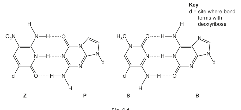
Answer
P and B
P and B
Background Concept
Nitrogenous bases in nucleic acids come in two structural families, distinguished by the number of fused rings in the base:
- Purines have two fused rings (a six-membered ring joined to a five-membered ring). In DNA and RNA the purines are adenine (A) and guanine (G).
- Pyrimidines have one ring only. The pyrimidines are cytosine (C), thymine (T, DNA only) and uracil (U, RNA only).
A useful rule of thumb: purines pair with pyrimidines in the DNA double helix so that the helix has a uniform width — two rings on one strand plus one ring on the other.
Understanding the Question
The candidate is shown Fig. 6.1, in which the four synthetic bases Z, P, S and B are drawn with their full chemical structures. Each base has a clear ring count, so the question reduces to "which of these bases are double-ringed?"
Approach
Look at each structure and count the rings. The key in the figure ("d = site where bond forms with deoxyribose") simply tells you which atom joins to the sugar and is not relevant to identifying the purines. Bases with two fused rings = purines; bases with one ring = pyrimidines.
Step-by-Step Reasoning
- Z: a single six-membered ring (with NO₂ and NH groups) → pyrimidine (one ring).
- P: a six-membered ring fused to a five-membered ring → purine (two rings).
- S: a single six-membered ring (with H₃C–N and C=O) → pyrimidine (one ring).
- B: a six-membered ring fused to a five-membered ring → purine (two rings).
Therefore the two purine synthetic bases are P and B.
Key Takeaways
- Purines = two rings; pyrimidines = one ring. This is the only structural distinction needed to identify them.
- The functional groups drawn inside the rings (amino, carbonyl, nitro, methyl) are characteristic of particular natural bases, but the question does not ask you to name the bases — only to identify which ones are purines.
Common Mistakes
- Reading the structures too quickly and miscounting rings. Both P and B clearly have a five-membered ring fused onto a six-membered ring; Z and S do not.
- Confusing "purine" with "pyrimidine" by spelling — make sure you know which is which.
- Listing one of the letters twice (e.g. "P and P") instead of giving two different letters.
Things to Be Careful About
- The question awards 1 mark for two correct letters. A single correct letter would not score; both must be given.
- Spelling the letters exactly as shown in the figure (capital P, capital B) is what the mark scheme accepts.
State and explain which DNA base pair is most similar to the synthetic base pairs in Fig. 6.1.
Answer
C and G — the C–G base pair is held together by three hydrogen bonds, the same as the synthetic Z–P and S–B base pairs in Fig. 6.1.
C and G, because they are joined by three hydrogen bonds (the same number as the synthetic base pairs).
Background Concept
In a DNA double helix, complementary bases pair through specific patterns of hydrogen bonding:
- Adenine (A) pairs with Thymine (T) through two hydrogen bonds.
- Guanine (G) pairs with Cytosine (C) through three hydrogen bonds.
The number of hydrogen bonds is one of the most reliably memorised features of DNA structure, and it is what determines the strength of the interaction between the two strands. The Z–P and S–B synthetic base pairs in Fig. 6.1 are both shown to be held together by three hydrogen bonds (the dotted lines in the diagram represent H-bonds).
Understanding the Question
The candidate is asked which natural DNA base pair is most similar to the synthetic pairs in Fig. 6.1, and to give a reason. The mark scheme awards one mark for naming the pair and one mark for the explanation (three hydrogen bonds).
Approach
- Count the H-bonds shown in each synthetic pair in the figure (Z–P and S–B).
- Recall the H-bond count for the two natural DNA base pairs.
- Match the natural pair to the synthetic pair with the same number of H-bonds.
Step-by-Step Reasoning
- In Fig. 6.1, both the Z–P pair and the S–B pair are drawn with three dotted lines between the bases, each representing a hydrogen bond.
- In real DNA, the A–T pair has two hydrogen bonds; the C–G pair has three hydrogen bonds.
- The natural base pair with three hydrogen bonds is therefore C and G, and this is the pair most similar to the synthetic ones.
Key Takeaways
- A–T = 2 H-bonds; C–G = 3 H-bonds. This is a high-frequency, low-marks-cost fact to learn.
- When two structures are compared in a question, the explanation is what carries the second mark — naming the right pair is not enough on its own.
- Hydrogen bonds in nucleic acids are not visible in the usual simplified diagrams, so reading the dotted lines carefully is essential.
Common Mistakes
- Naming "A and T" — this pair has only two H-bonds and is less similar to the synthetic pairs.
- Naming the correct pair but giving a wrong reason (e.g. "because they are both purines" or "because they have similar shapes"). The H-bond number is the mark-scheme reason.
- Naming the correct pair but not mentioning hydrogen bonds at all — the second mark is for the explanation.
- Confusing C–G with C–U; uracil is found in RNA, not DNA, and pairs with adenine.
Things to Be Careful About
- The explanation mark is for three hydrogen bonds, not for "strong" or "weak" or "similar shape". The mark scheme is specific about this.
- The synthetic bases in Fig. 6.1 are an invented system — do not try to interpret the functional groups as if they were the natural bases. The only feature that matters here is the number of H-bonds shown.