Biology 9700/22 — May/June 2024
Cambridge AS Level · AS Level Structured Questions · worked solutions for every part, with the mark scheme
Topics Cell Structure · Transport in Mammals · Nucleic Acids and Protein Synthesis · Biological Molecules · Immunity · Enzymes · +4 more
Smooth muscle is a tissue composed of smooth muscle cells. The cells contain cytoplasm packed with proteins that are involved in contraction and relaxation.
Smooth muscle is present in the airways of the gas exchange system.
Explain how smooth muscle cells in the walls of the bronchioles contribute to the function of these airways.
Answer
- Smooth muscle cells contract and relax.
- This decreases and increases the diameter of the bronchiole lumen, controlling the flow/volume of air to the alveoli.
Contraction and relaxation change the lumen diameter of the bronchioles, regulating air flow to the alveoli.
Background Concept
The gas exchange system consists of the trachea, two bronchi, and a branching network of bronchioles ending in alveoli. The walls of the trachea and bronchi are supported by C-shaped rings of cartilage (which hold them open), while the bronchioles lack cartilage and instead have smooth muscle in their walls. This makes the smaller airways distensible and able to actively change their internal diameter.
Smooth muscle is involuntary (not under conscious control) and is made of elongated, tapered (fusiform) cells. When the cells contract, they pull on the wall of the airway, narrowing the lumen; when they relax, the lumen widens again. This is distinct from elastic tissue recoil, which is a passive stretching and springing back.
Understanding the Question
The stem has told us that smooth muscle cells contain proteins involved in contraction and relaxation. Part (a) asks specifically about the bronchioles (the small airways, not the trachea or main bronchi). The question is a "describe/explain" asking how contraction and relaxation in these particular cells supports the function of the airways. The command word "explain" means each point must carry the consequence (why the contraction matters), not just the observation.
Approach
Start with what smooth muscle physically does (contract and relax), then link this to the consequence for the airway wall (change in lumen diameter), and finally tie this to the function of gas exchange (controlling the volume of air reaching the alveoli).
Step-by-Step Reasoning
- Marking point 1: Smooth muscle cells both contract and relax. This must be in the context of the bronchioles — not the trachea or main bronchi (which are stiffened by cartilage).
- Marking point 2: As a result, the diameter (lumen size) of the bronchiole can be changed/controlled. Contraction narrows the lumen (constricts); relaxation widens it (dilates).
- Marking point 3: The physiological consequence is that the flow (or volume) of air into and out of the alveoli is regulated. This matches air demand — for example, bronchioles dilate during exercise.
The mark scheme explicitly rejects describing this as "elastic fibres" or "elastic tissue", and rejects stretch/expand/recoil (these describe elastic behaviour, not active smooth muscle action). Vasoconstriction/vasodilation terms also do not earn credit because they refer to blood vessels.
Key Takeaways
- Smooth muscle in the bronchioles is the active, living component that regulates airway diameter.
- Cartilage (trachea/bronchi) keeps airways open; smooth muscle (bronchioles) actively changes their diameter.
- The function is regulation of air flow to match the body's needs.
Common Mistakes
- Describing the action as "elastic" or "stretching/recoil" — this is passive and does not earn credit.
- Putting the answer in the context of the trachea or bronchi rather than the bronchioles.
- Vague statements like "lets air in" or "allows breathing" without linking to the lumen diameter.
Things to Be Careful About
- The command word is "explain", so each point must include the consequence, not just an observation.
- Use the words "contract" and "relax" together (both must appear).
- Mention "diameter" or "lumen size" explicitly rather than just "size".
When viewed in longitudinal section (LS), smooth muscle cells are elongated and taper at both ends. This is known as a fusiform shape. Each cell has a central nucleus, which also appears elongated.
Fig. 1.1 is a diagram of a smooth muscle cell to show the fusiform shape.
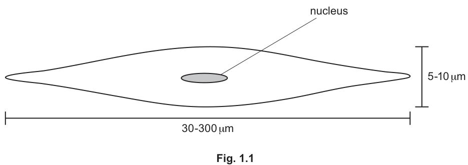
A student used a microscope fitted with a calibrated eyepiece graticule to estimate that the length of one smooth muscle cell was micrometres ().
Name the type of microscope slide that the student used to calibrate the eyepiece graticule.
Answer
Stage micrometer (slide).
Stage micrometer
Background Concept
An eyepiece graticule is a small glass disc, etched with a scale, that fits into the eyepiece of a light microscope. It appears superimposed on the specimen when the user looks down the microscope. However, the divisions on the graticule are arbitrary — they correspond to a different actual length depending on which objective lens is being used. To convert graticule divisions into real units (µm or mm), the eyepiece graticule must be calibrated for each objective.
Understanding the Question
The stem says the student used a microscope fitted with a calibrated eyepiece graticule and used it to estimate the length of a smooth muscle cell as 250 µm. The question (b)(i) asks what type of microscope slide was used in the calibration step. The calibration step requires a scale of known length to be placed on the stage so its actual length can be matched to the graticule divisions.
Approach
Recall the standard CIE terminology: the slide placed on the stage of the microscope that carries a scale of known length (typically 1 mm divided into 100 units of 10 µm each) is called a stage micrometer.
Step-by-Step Reasoning
- A stage micrometer is a specially etched slide with a precisely known scale (commonly 1 mm in 100 divisions of 10 µm each).
- It is placed on the stage and used to calibrate the eyepiece graticule at each magnification by aligning the two scales and counting how many eyepiece divisions correspond to one stage micrometer division.
- Once calibrated, the eyepiece graticule can be used to measure the specimen (here, the smooth muscle cell), giving the 250 µm result.
The term "stage micrometer" is the precise CIE term; "graticule" alone would refer to the eyepiece disc and is not credit-worthy here. "Micrometre slide", "calibration slide" or similar vague terms are also not credited.
Key Takeaways
- Calibration of an eyepiece graticule requires a scale of known length on the stage.
- The apparatus for this is called a stage micrometer.
- Calibration must be repeated for each objective lens.
Common Mistakes
- Saying "graticule" — this is the eyepiece component, not the stage slide.
- Saying "ruler" or "scale" — not the correct scientific term.
Things to Be Careful About
- The wording must be "stage micrometer" (or "stage micrometer scale" / "stage micrometer slide"), exactly.
The smallest object the student can see without the use of a microscope is in length.
Explain whether the student would be able to see a cell of length without the use of a microscope.
Answer
Yes. The smallest object the student can see without a microscope is 0.2 mm = 200 µm. The cell is 250 µm (= 0.25 mm) long, which is longer than 200 µm (by 50 µm / 0.05 mm), so the cell would be visible to the naked eye.
Yes — 250 µm (0.25 mm) is longer than 200 µm (0.2 mm), so the cell is visible to the naked eye.
Background Concept
The unaided human eye has a resolving power of about 0.1–0.2 mm — that is, the smallest objects we can distinguish as separate are roughly 100–200 µm across. The question gives the student's own practical limit as 0.2 mm = 200 µm.
Units:
So:
Understanding the Question
The stem gives us a cell length of 250 µm (from the calibrated eyepiece graticule in part (b)). The question tells us the smallest object the student can resolve unaided is 0.2 mm. We must decide whether a 250 µm cell is large enough for the student to see without a microscope, and justify the answer with the numbers and units.
Approach
Convert one of the values so both are in the same units, then compare numerically.
Step-by-Step Reasoning
- Convert 0.2 mm into µm: 0.2 mm × 1000 = 200 µm.
- Compare: 250 µm (cell) vs 200 µm (smallest visible object).
- Since 250 µm > 200 µm, the cell is longer than the student's unaided resolution limit, so it can be seen without a microscope.
The mark scheme accepts several equivalent justifications: that 250 µm = 0.25 mm and 0.2 mm < 0.25 mm; or that the cell is 50 µm (0.05 mm) longer than the resolution limit; or a standard-form comparison (2 × 10⁻⁴ m vs 2.5 × 10⁻⁴ m). The numerical values and the units must both appear.
Key Takeaways
- The unaided eye can resolve objects down to roughly 0.1–0.2 mm (100–200 µm).
- Always convert to common units before comparing two lengths.
- 1 mm = 1000 µm is the key conversion here.
Common Mistakes
- Saying "yes" without converting units or without quoting numbers.
- Confusing which way the inequality goes (250 µm is bigger, not smaller, than 200 µm).
- Quoting a number without units — the mark scheme explicitly requires the units.
Things to Be Careful About
- Both a numerical value AND the units must be present to earn the mark.
- The answer must include "yes" with the qualifying comparison; a bare "yes" or "no" is not credited.
Fig. 1.2 is a photomicrograph of smooth muscle tissue in the wall of the intestines. A capillary is visible in addition to smooth muscle cells.
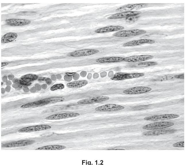
Outline the features that help to identify the blood vessel in Fig. 1.2 as a capillary.
Answer
- The wall of the vessel is one cell thick (a single layer of endothelial cells).
- Red blood cells pass through in single file.
- The red blood cells appear similar in diameter to the lumen of the vessel.
Wall one cell thick; red blood cells in single file; RBC diameter similar to capillary lumen.
Background Concept
Capillaries are the smallest blood vessels (typically 5–10 µm in diameter — comparable to the size of a single red blood cell). Their wall consists of a single layer of flattened endothelial cells, with no muscle or elastic tissue, no collagen layer, and no outer tunica. This is much thinner than the walls of arterioles, arteries or veins, all of which have multiple layers.
Because the lumen is so narrow, only one red blood cell at a time can pass through, giving the characteristic "single file" appearance seen in histological sections and micrographs.
Understanding the Question
Part (c)(i) shows a photomicrograph (Fig. 1.2) of smooth muscle tissue with one vessel visible. The student is asked to outline the features that allow that vessel to be identified as a capillary — i.e., the distinguishing visible features in the image.
Approach
Recall the structural hallmarks of a capillary and check which are visible in the micrograph:
- Wall is a single layer of cells (one cell thick).
- Lumen is narrow — about the same diameter as a red blood cell.
- Red blood cells travel in single file because only one fits at a time.
- No muscle or elastic layer is visible around the vessel.
Step-by-Step Reasoning
- The vessel in the centre of the field shows a row of pale, rounded red blood cells in a narrow channel. The cells are arranged one after another — this is the "single file" appearance.
- Comparing the diameter of a red blood cell to the width of the channel, they are roughly the same — the lumen is about as wide as one RBC.
- The wall cannot be resolved as multiple layers — there is no visible smooth muscle or connective tissue coat around the vessel, consistent with a one-cell-thick endothelium.
The mark scheme also accepts features like: red blood cells deforming slightly to squeeze through (showing flexibility) — this is visible in some micrographs where the RBCs appear biconcave/curved.
Key Takeaways
- Capillaries are the smallest vessels; their lumen is barely wider than an RBC.
- One-cell-thick wall is a defining structural feature of capillaries.
- Single-file RBC flow is the most easily recognised identifying feature in a micrograph.
Common Mistakes
- Saying "capillaries are thin" without specifying "one cell thick".
- Confusing the identifying features with those of arteries or veins (thick muscular wall, multiple layers).
- Saying "the wall has one cell" rather than "the wall is one cell thick" — both work, but the precise form is clearer.
Things to Be Careful About
- The features must be visible in the image (or implied by what's visible) — do not credit features you would only see at much higher magnification.
Answer
- Capillaries supply oxygen and nutrients (e.g. glucose, amino acids) to smooth muscle cells and remove carbon dioxide and other waste products.
- The wall is one cell thick, giving a short diffusion distance between the blood and the muscle cells.
- Endothelial pores (gaps between the endothelial cells) allow efficient passage of substances (and phagocytes) between the blood and the surrounding tissue fluid.
- The narrow lumen (≈ 5–10 µm) means the capillary can pass between cells, so every smooth muscle cell is close to a capillary supply.
Thin one-cell wall → short diffusion distance; pores → efficient exchange; narrow lumen → all cells reached.
Background Concept
Capillaries are the site of exchange between blood and the surrounding tissues. Their structure is uniquely suited to this role:
- The wall is a single layer of squamous (flattened) endothelial cells, often only 1 µm thick in places.
- There are small gaps (pores/fenestrations) between adjacent endothelial cells, and sometimes through the cells themselves.
- The lumen is very narrow (5–10 µm), only just wide enough for a red blood cell.
Fick's law tells us that the rate of diffusion is inversely proportional to the distance, and proportional to the surface area and the concentration gradient. So a thin wall maximises the rate of exchange, and a large total surface area (achieved by vast numbers of capillaries in a network) does the same.
Understanding the Question
Part (c) has introduced Fig. 1.2, a photomicrograph showing a capillary running through smooth muscle tissue. The smooth muscle cells are highly active (continuous contraction and relaxation) and therefore have a high demand for oxygen and nutrients, and a high production of waste products (CO₂, lactate). Part (c)(ii) asks the candidate to explain how the structure of a capillary relates to its function in supplying (and draining) such a tissue.
The command word "explain" means each point must pair a structural feature with a functional consequence — saying "thin wall" alone is not enough; it must be tied to "short diffusion distance" or similar.
Approach
Go through the key structural features of a capillary one at a time and, for each, state the functional consequence for exchange with smooth muscle cells.
Step-by-Step Reasoning
- Function stated first: a capillary supplies oxygen, glucose, amino acids and other nutrients to smooth muscle cells and removes carbon dioxide and waste products (e.g. lactate from respiration).
- Thin wall → short diffusion distance: the wall is one cell thick (≈ 1 µm). According to Fick's law, this shortens the diffusion path between the blood and the muscle cells, increasing the rate of exchange.
- Endothelial pores → efficient passage of substances and cells: gaps between adjacent endothelial cells (pores/fenestrations) allow rapid movement of substances between the plasma and the tissue fluid bathing the muscle cells. They also allow phagocytes (neutrophils, monocytes, macrophages) to squeeze out of the blood to reach any site of damage or infection in the tissue.
- Narrow lumen (5–10 µm) → cells are close to a blood supply: because the capillary is so narrow, it can branch through the tissue between the muscle cells, so no smooth muscle cell is far from a capillary. The narrow lumen also slows the flow of blood, giving more time for exchange.
The mark scheme accepts slightly different framings — for example, "endothelial pores allow formation of tissue fluid around the cells" (since tissue fluid forms by filtration of plasma through these pores). It explicitly does not credit "blood leaves to make tissue fluid" — the blood stays inside the capillary; only fluid and small solutes pass out.
Key Takeaways
- Capillary structure is the classic example of "form follows function" in biology.
- The three features to learn are: thin wall, pores, narrow lumen.
- Each must be tied to a functional consequence when you answer an "explain" question.
- The narrow lumen slowing blood flow is what gives red blood cells time to off-load oxygen.
Common Mistakes
- Stating "the wall is thin" without specifying "one cell thick" and without linking it to a short diffusion distance.
- Saying "blood leaves the capillary to make tissue fluid" — blood stays inside; fluid leaves.
- Naming only the substances without linking to a structural feature (or vice versa).
- Confusing capillaries with veins/arteries (which have thick muscular walls and would prevent exchange).
Things to Be Careful About
- Each marking point pairs STRUCTURE with FUNCTION; do not separate them.
- "Oxygen in / carbon dioxide out" is acceptable shorthand; the mark scheme rejects "oxygen out, carbon dioxide in".
- Plasma proteins do NOT pass through capillary pores (they are too large), so "plasma proteins into tissue fluid" is not credited.
Caldesmon is a large protein with a number of binding sites to attach to other proteins.
Caldesmon exists in two different forms, H-caldesmon and L-caldesmon.
H-caldesmon helps to regulate contraction and relaxation in smooth muscle cells.
L-caldesmon is found in some non-muscle cells, where it also acts as a regulatory protein.
- Caldesmon is coded for by a gene known as CALD1.
- CALD1 has 17 exons.
- The primary structure of H-caldesmon has a repeating sequence in the middle of the amino acid chain that is not present in L-caldesmon.
Researchers have discovered that a gene mutation is not the cause of the two different forms of caldesmon.
Explain what is meant by a gene mutation.
Answer
A gene mutation is a change in the sequence of base pairs (bases/nucleotides) in a DNA molecule, which results in the coding of an altered/different polypeptide.
A change in the base-pair sequence of a DNA molecule that codes for a different polypeptide.
Background Concept
A gene is a sequence of DNA bases (A, T, C, G) that codes for a polypeptide. The order of the bases determines the order of amino acids in the polypeptide, via the genetic code (read as triplets/codons during translation).
A mutation is a permanent alteration in the DNA sequence of a gene. It can take several forms: a substitution (one base replaced by another), a deletion (one or more bases removed), or an insertion (one or more bases added). Because the code is read in non-overlapping triplets, deletions and insertions cause a frameshift, which alters every downstream codon; a substitution usually alters only one codon.
Understanding the Question
The stem of part (d) introduces caldesmon, a protein coded for by the gene CALD1, and notes that two different forms of this protein exist. Part (d)(i) asks the student to define a gene mutation. This is a definition-style "explain what is meant by" question, which the mark scheme typically marks as two points: the cause (the DNA change) and the consequence (an altered product).
Approach
Build the definition in two parts:
- State what changes at the molecular level.
- State the consequence for the polypeptide.
Step-by-Step Reasoning
- A gene mutation is a change in the sequence of bases (nucleotides / base pairs) along the DNA molecule that makes up the gene. The mark scheme accepts "change in the DNA base sequence" or "change in the DNA nucleotide sequence". It explicitly does NOT credit "change in RNA base sequence" — the change must be in DNA.
- Because the base sequence of the gene determines the order of amino acids in the polypeptide, a change in the base sequence can result in an altered/different polypeptide. The mark scheme accepts "protein / amino acid sequence / primary structure" as alternatives to "polypeptide".
The mark scheme does NOT credit naming types of mutation (substitution, deletion, insertion) — these are examples, not the definition.
Key Takeaways
- A mutation is at the DNA level, not at the RNA or protein level.
- It is a permanent change in base sequence.
- The consequence (and the second mark) is that the polypeptide coded for may be different.
Common Mistakes
- Saying "a change in the bases" without specifying that they are in DNA.
- Saying "a change in amino acids" — this is the consequence, not the definition; the change must be in the DNA.
- Saying "a change in a chromosome" — too vague; the change is specifically in the base sequence.
Things to Be Careful About
- The two marking points must both appear: the change AND the consequence for the polypeptide.
- Use the precise CIE word "polypeptide" (or accepted equivalent "protein / amino acid sequence / primary structure").
Researchers now know that the two different forms of caldesmon are the result of events occurring directly after transcription of DNA. Changes occur to the primary transcript that is formed by DNA transcription.
Suggest how the smooth muscle cells and non-muscle cells can produce different forms of caldesmon from the same primary transcript.
Answer
- The primary transcript is processed by RNA splicing: introns (non-coding sequences) are removed and exons (coding sequences) joined together.
- Smooth muscle cells and non-muscle cells splice the primary transcript differently (alternative splicing), so different combinations of exons are joined — producing different mature mRNAs and therefore different polypeptides (H-caldesmon and L-caldesmon).
Alternative splicing of the same primary transcript produces different mature mRNAs in the two cell types.
Background Concept
When a protein-coding gene is transcribed, the RNA polymerase first produces a primary transcript (pre-mRNA). This transcript contains both:
- Exons — sequences that code for amino acids (the "expressed" sequences).
- Introns — non-coding sequences that lie between exons.
Before the mRNA leaves the nucleus, the introns are removed and the exons are joined together by a process called RNA splicing. A single gene can therefore give rise to different mature mRNAs in different cell types because different combinations of exons may be retained. This is called alternative splicing and is the molecular basis for producing multiple protein isoforms (such as H- and L-caldesmon) from a single gene.
Understanding the Question
The stem of part (d) tells us that caldesmon exists as two isoforms (H-caldesmon in smooth muscle, L-caldesmon in non-muscle cells) but that the gene mutation is NOT the cause. It also tells us that the difference arises from events occurring directly after transcription, involving the primary transcript. Part (d)(ii) asks the student to suggest how two different polypeptides can be made from the same primary transcript.
Approach
Recall that post-transcriptional processing in eukaryotes includes RNA splicing (intron removal and exon joining). Apply the concept of alternative splicing: the same primary transcript can be spliced differently in different cell types, giving different mature mRNAs.
Step-by-Step Reasoning
- The primary transcript contains exons (coding) and introns (non-coding).
- Splicing removes the introns and joins the exons.
- In smooth muscle cells, one combination of exons is joined (e.g. including the middle exons that code for the repeating sequence unique to H-caldesmon).
- In non-muscle cells, a different combination of exons is joined (e.g. the middle exons coding for the repeating sequence are excluded), giving L-caldesmon.
- The two mature mRNAs are different, so the two polypeptides translated from them are different.
The mark scheme accepts any combination of: "RNA splicing", "alternative splicing" (do not say "DNA splicing" — the splicing happens to the primary RNA transcript), "removal of introns", "exons joined differently / in a different combination", "(so the) mRNA formed is different". It explicitly rejects "different number of introns removed / some introns remain" — the number of introns is fixed; it is which exons are joined that varies. It also rejects "mutation occurring during splicing" — the stem has told us there is no gene mutation.
Key Takeaways
- Alternative splicing is the main mechanism by which one gene can produce several different protein isoforms.
- The cell type determines which exons are retained in the mature mRNA.
- The process occurs to the primary transcript, before translation — i.e., at the post-transcriptional stage.
Common Mistakes
- Saying "DNA splicing" — splicing happens to RNA, not DNA.
- Saying "mutations during splicing" — the question stem rules out mutations.
- Saying "different introns removed" — the introns are the same; what differs is which exons are joined.
- Confusing introns with exons (introns are removed, exons are kept and joined).
Things to Be Careful About
- The splicing is to RNA, not DNA.
- The different forms come from different combinations of exons, not different numbers of introns.
Suggest how the two different forms of caldesmon can still have similar functions, even though they have a different primary structure.
Answer
The two forms still have a similar tertiary structure / similar binding-site shape, because the amino acids that differ (the middle repeating sequence) are not part of the binding sites used to attach to the partner proteins.
Similar tertiary structure / binding-site shape, because the differing amino acids are not part of the binding sites.
Background Concept
A protein's function depends on its three-dimensional shape (tertiary structure), which in turn is determined by the sequence of amino acids (primary structure) and the way the R-groups interact (hydrogen bonds, ionic bonds, disulfide bridges, hydrophobic interactions).
Caldesmon is described as a large protein with multiple binding sites that attach to other proteins. These binding sites are discrete regions of the polypeptide where the local 3D shape is complementary to the partner protein (analogous to an enzyme's active site being complementary to its substrate).
Understanding the Question
The stem tells us that the primary structures of H-caldesmon and L-caldesmon differ (H has a middle repeating sequence that L lacks). Yet both proteins act as regulatory proteins that bind to other proteins. The question asks how the two forms can still have similar functions despite this difference in primary structure.
Approach
Think about what determines function: it is the 3D shape of the binding sites. A change in primary structure does not abolish function unless that change affects the binding sites themselves.
Step-by-Step Reasoning
- The two forms differ only in the middle repeating sequence.
- The binding sites (where caldesmon attaches to its partner proteins) are located elsewhere in the molecule and have the same amino acid sequence in both forms.
- Because the binding-site amino acid sequence is unchanged, R-group interactions in those regions are unchanged, so the tertiary structure (3D shape) of the binding sites is preserved.
- Therefore both forms can still bind to the same partner proteins and carry out a similar regulatory role.
The mark scheme explicitly forbids referring to "active sites" (the language of enzymes) but allows "binding sites". It accepts the idea that "removed amino acids are not part of the binding site" or that "R-group interactions (still) the same/similar".
Key Takeaways
- Protein function depends on tertiary structure at binding/active sites, not on primary structure globally.
- A change in primary structure outside the binding site need not change function.
- This is why a single gene can produce several protein isoforms with similar (or partially different) functions.
Common Mistakes
- Saying "active site" — caldesmon does not catalyse a reaction; it has binding sites, not an active site.
- Saying "the primary structure is the same" — the question stem says they are different.
- Saying "they have the same function" — the question implies they may have somewhat different functions; the mark is for explaining how similar function is preserved.
Things to Be Careful About
- Use "binding sites" (plural is acceptable), not "active site".
- The explanation must locate the difference outside the binding site.
Alveolar macrophages are cells of the immune system that remain in the alveolar region of the gas exchange system. The macrophages protect against infection caused by pathogens that have been inhaled.
Alveolar macrophages have the same cell structures as typical animal cells.
Complete Table 2.1 to name the cell structures that match the functions stated.
Do not use abbreviations.
Table 2.1
| cell structure | function |
|---|---|
| manufactures ribosomal subunits from proteins and ribosomal RNA | |
| synthesises triglycerides and other lipids | |
| pair of | organise microtubules of the cell cytoskeleton |
Answer
| cell structure | function |
|---|---|
| nucleolus | manufactures ribosomal subunits from proteins and ribosomal RNA |
| smooth endoplasmic reticulum | synthesises triglycerides and other lipids |
| pair of centrioles | organise microtubules of the cell cytoskeleton |
nucleolus; smooth endoplasmic reticulum; pair of centrioles
Background Concept
Eukaryotic cells contain many membrane-bound and non-membrane-bound organelles, each with a specific function. Three that are particularly easy to confuse are the nucleolus, the smooth endoplasmic reticulum (SER) and the centrioles — partly because each is small, often hidden in diagrams, or bears some resemblance to other structures.
- Nucleolus — a dense, roughly spherical body inside the nucleus. It is not a separate organelle surrounded by a membrane; it is a region of the nucleus where ribosomal RNA (rRNA) is transcribed and combined with proteins imported from the cytoplasm to assemble the large and small ribosomal subunits. These subunits are then exported through nuclear pores to the cytoplasm.
- Smooth endoplasmic reticulum (SER) — a network of flattened sacs and tubules continuous with the rough ER but lacking ribosomes on its cytoplasmic surface. It synthesises lipids (including triglycerides, phospholipids and steroids), metabolises carbohydrates and detoxifies some drugs and poisons. By contrast, rough ER is studded with ribosomes and is the site of synthesis of proteins destined for secretion or for membranes.
- Centrioles — a pair of short, hollow cylinders made of microtubules. The pair is arranged at right angles to each other (perpendicular) and sits in the centrosome near the nucleus. They organise the microtubules of the cytoskeleton and nucleate the spindle fibres that separate chromosomes during mitosis. Animal cells have centrioles; higher plant cells do not.
Understanding the Question
Part (a)(i) supplies a blank three-row table in which each row gives a function and asks for the cell structure that performs it. The instructions explicitly forbid abbreviations, so "ER" alone would not be accepted. The candidate must recall the full name of each organelle that performs the given function in a typical animal cell (here an alveolar macrophage).
Approach
Read each function and decide which organelle performs it, then write the full name of that organelle in the left-hand column.
Step-by-Step Reasoning
- "Manufactures ribosomal subunits from proteins and ribosomal RNA" → This is the definition of the nucleolus (the site of rRNA transcription and ribosomal subunit assembly, found inside the nucleus).
- "Synthesises triglycerides and other lipids" → This is the role of the smooth endoplasmic reticulum. Lipid synthesis is the textbook lipid-related function of the SER. The word "triglycerides" is the giveaway — triglycerides are lipids, and SER (not RER) makes them.
- "Organise microtubules of the cell cytoskeleton" → A pair of centrioles acts as the microtubule-organising centre (MTOC) in animal cells, nucleating the cytoskeletal microtubules and the mitotic spindle.
Key Takeaways
- Nucleolus ≠ nucleus. The nucleus stores DNA; the nucleolus makes ribosomes inside it.
- Smooth ER vs Rough ER is decided by the absence or presence of surface ribosomes, and hence by function (lipid synthesis vs protein synthesis).
- Centrioles always come as a pair of perpendicular cylinders — never a single centriole.
Common Mistakes
- Writing "nucleus" instead of "nucleolus" — confuses the container with its internal compartment.
- Writing "endoplasmic reticulum" without specifying "smooth" — loses the mark because RER also fits the description.
- Writing "centrosome" instead of "centrioles" — the centrosome is the surrounding matrix; the centrioles are the pair of cylinders inside it.
- Using abbreviations such as "ER" or omitting the word "pair" before centrioles.
Things to Be Careful About
The mark scheme rejects "endoplastic" (misspelling) and "centrosomes". Always spell out organelle names in full and use the precise term.
Fig. 2.1 is a diagram of an alveolar macrophage showing:
- some of the cell structures that would be visible using an electron microscope
- a newly formed phagocytic vacuole (phagosome) containing two cells of Mycobacterium tuberculosis.
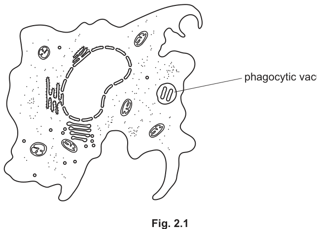
The cell structures with the functions described in Table 2.1 are not shown in Fig. 2.1.
Complete Fig. 2.1 by drawing and labelling the cell structures described in Table 2.1.
Answer
Three structures must be added to Fig. 2.1 and labelled:
- Nucleolus — a small, roughly spherical/circular structure drawn inside the nucleus. A leader line should be drawn from it to the label "nucleolus".
- Smooth endoplasmic reticulum (SER) — at least one membrane-bound tubular or curved sac drawn without dots on its surface (no ribosomes). A leader line to the label "smooth endoplasmic reticulum" or "smooth ER".
- Pair of centrioles — two short cylinders (or pairs of short lines) drawn at right angles to each other (not parallel), located in the cytoplasm near (but outside) the nucleus. A leader line to the label "centrioles".
Three labelled additions to Fig. 2.1: nucleolus inside the nucleus, smooth ER without ribosomes, and a pair of centrioles at right angles in the cytoplasm.
Background Concept
This part of the question tests whether a candidate can recognise and reproduce cellular ultrastructure on a diagram, not merely name it. Many organelles look superficially similar at the EM level, so drawings must include the distinguishing features listed in the mark scheme:
- The nucleolus is recognisable only because it is inside the nucleus and is a smaller dense body within it.
- The smooth ER is recognisable because it is a membrane-bound tubular or flattened sac without ribosomes (no dots on its cytoplasmic face). If ribosomes (dots) are drawn on the membrane, the structure looks like rough ER and the mark is lost.
- Centrioles are recognisable as two short cylinders perpendicular to each other. They must be in the cytoplasm, not inside the nucleus (which would suggest a nucleolus or chromatin), and they must not be parallel (which would simply look like a piece of ER).
Understanding the Question
Fig. 2.1 shows an alveolar macrophage with its nucleus, mitochondria, rough ER and the phagosome already drawn. The three structures from Table 2.1 (nucleolus, SER, pair of centrioles) are deliberately not shown. The candidate must add them to the figure and label them with leader lines.
Approach
Decide where on the figure each structure will be drawn so that it is in a sensible position and obviously distinct:
- Nucleolus → inside the existing nucleus.
- Smooth ER → somewhere in the cytoplasm, drawn as smooth curved or tubular sacs with no dots.
- Centrioles → in the cytoplasm near the nucleus, drawn as two short cylinders at right angles.
Draw each, then add a label connected by a clear leader line.
Step-by-Step Reasoning
- Nucleolus. Place a small circle inside the boundary of the nucleus. Label it "nucleolus" with a leader line.
- Smooth ER. Draw at least one curved/tubular membrane-bound sac in the cytoplasm. Do not put dots on it — that would make it look like rough ER and lose the mark. Label it "smooth endoplasmic reticulum" or "smooth ER".
- Centrioles. Draw two short hollow cylinders (or two pairs of short lines) at right angles to each other in the cytoplasm, not inside the nucleus. Label it "centrioles".
According to the mark scheme: "all three correct = 2 marks; one or two correct = 1 mark; all three drawn correctly but no labels = 1 mark". So a single attempt that labels nothing but draws all three correctly still earns 1 mark — but only labelling all three correctly (with at least some attempt at distinguishing detail) earns the full 2 marks.
Key Takeaways
- Drawings of organelles must include the features that distinguish them from similar-looking organelles.
- Labels need leader lines that end on the structure, not in empty space nearby.
- Recognising what isn't on a printed figure is part of the skill — read the stem to know what to add.
Common Mistakes
- Drawing the nucleolus outside the nucleus — the nucleolus is inside the nucleus; placing it in the cytoplasm is wrong.
- Putting dots on the smooth ER membrane so it resembles rough ER.
- Drawing centrioles as parallel lines or as a single structure rather than a perpendicular pair.
- Drawing centrioles inside the nucleus (rejected by the mark scheme).
- Forgetting the labels altogether.
Things to Be Careful About
- Use single lines or a simple shape for each structure; do not attempt to draw fine ultrastructural detail.
- Keep the drawings clearly distinguishable from each other and from the organelles already present in Fig. 2.1 (rough ER, mitochondria, nucleus).
- Add leader lines and labels for full marks.
Tuberculosis (TB) can be prevented if the bacterial cells that have reached the alveoli are rapidly destroyed. Alveolar macrophages can detect the presence of M. tuberculosis in the alveolar space and can carry out phagocytosis to form phagocytic vacuoles, such as the one shown in Fig. 2.1.
Outline the sequence of events that leads to the formation of a phagocytic vacuole after detection of the bacterial cells by an alveolar macrophage.
Answer
- The (alveolar) macrophage / phagocyte detects the bacterial cells by chemotaxis / chemotactic response and the (bacterial) antigens bind to receptors on the macrophage cell surface membrane (allowing recognition before engulfment).
- Endocytosis occurs: the macrophage cell surface membrane extends pseudopodia that surround / engulf the bacterial cells, and the membrane pinches off to form a (membrane-bound) phagocytic vacuole / vesicle containing the bacteria.
Chemotaxis/antigens bind to receptors on the macrophage; the cell surface membrane surrounds the bacteria (pseudopodia) and pinches off to form a phagocytic vacuole.
Background Concept
Phagocytosis is a form of endocytosis in which specialised cells (phagocytes — macrophages and neutrophils) engulf solid particles such as bacteria, dead cells or debris. The process involves:
- Detection / attraction — phagocytes are attracted to the site of infection by chemicals released by pathogens or by damaged host cells (chemotaxis). Receptors on the phagocyte cell surface membrane bind to antigens (often after opsonisation — coating of the pathogen by antibodies or complement proteins).
- Engulfment — the phagocyte extends finger-like cytoplasmic projections called pseudopodia around the particle. The cell surface membrane surrounds the particle and fuses behind it.
- Vesicle formation — the membrane pinches off internally to release a membrane-bound phagocytic vacuole (phagosome) inside the cytoplasm, enclosing the engulfed material.
- Digestion — lysosomes fuse with the phagosome and release hydrolytic enzymes to destroy the contents.
Understanding the Question
The question asks the candidate to outline the sequence of events leading to the formation of the phagocytic vacuole after the macrophage has detected the bacteria. So the focus is on engulfment plus one earlier (recognition) or later (vesicle release) event. The two marks can be earned by: (1) describing endocytosis / engulfment (mandatory), and (2) describing a before- or after-event (chemotaxis, receptor binding, opsonisation, or membrane fusion).
Approach
State the engulfment step (pseudopodia / membrane surrounds / pinches off) for one mark, then add either a recognition event (chemotaxis / antigen–receptor binding) or the vesicle pinch-off event for the second mark.
Step-by-Step Reasoning
- Mark 1 — endocytosis. "Endocytosis occurs: the cell surface membrane (pseudopodia) surrounds / engulfs the bacterial cells." This is the core of phagocytosis.
- Mark 2 — a recognisable before- or after-event. Either before — chemotaxis / antigen binding to surface receptors / opsonisation by antibodies; or after — the surrounding membrane fuses and pinches off internally to release the phagocytic vacuole into the cytoplasm.
Either combination is acceptable; the mark scheme allows the second mark for a "before" or "after" event.
Key Takeaways
- Phagocytosis is endocytosis of a solid particle by a phagocyte.
- The cell surface membrane is the active component — it surrounds the particle and pinches off internally.
- Recognition (chemotaxis / antigen binding / opsonisation) precedes engulfment; lysosomal fusion follows it.
Common Mistakes
- Describing only the recognition event and not the engulfment — loses the core "endocytosis" mark.
- Calling pseudopodia "cilia" or "flagella" — pseudopodia are the cytoplasmic projections of phagocytes, not motile organelles.
- Saying the bacteria enter the cell — it is the macrophage that actively engulfs them.
- Describing events that happen after the phagosome has formed (e.g. lysosome fusion, digestion) — these are outside the scope of this question.
Things to Be Careful About
- "Pseudopodia surround / form a ring around / engulf" the bacteria is the credit-worthy phrasing.
- The mark scheme ignores binding "to the macrophage" alone (it requires binding to a receptor or "membrane" of the macrophage).
Name the cell structures that fuse with the phagocytic vacuole and release hydrolytic enzymes to destroy the bacterial cells.
Answer
Lysosomes (lysosomal vesicles).
Lysosomes
Background Concept
After a phagocytic vacuole (phagosome) forms, lysosomes fuse with it. Lysosomes are small, spherical organelles bounded by a single membrane that contain hydrolytic (digestive) enzymes such as lysozyme, proteases, lipases and nucleases, active at the low internal pH (~pH 4.5–5.0). When a lysosome fuses with a phagosome, the combined structure is called a phagolysosome, and the hydrolytic enzymes digest the engulfed material — in this case the M. tuberculosis bacterial cells.
Understanding the Question
The question asks for the name of the cell structures that fuse with the phagocytic vacuole and release hydrolytic enzymes. This is a one-mark recall item.
Approach
Recall that the organelle responsible for intracellular digestion of engulfed material is the lysosome.
Step-by-Step Reasoning
- Lysosomes contain hydrolytic enzymes.
- Lysosomes fuse with phagosomes to form phagolysosomes, in which the engulfed bacteria are digested.
The mark scheme accepts "lysosomes", "lysosome" or "lysosomal vesicles".
Key Takeaways
- Lysosomes are the digestive organelles of the cell.
- They fuse with phagosomes to form phagolysosomes, enabling destruction of pathogens such as M. tuberculosis.
Common Mistakes
- Writing "phagosome" or "vacuole" — these are the structures formed by engulfment, not the ones that fuse with it.
- Writing "Golgi apparatus" — the Golgi packages hydrolytic enzymes into lysosomes but does not itself fuse with the phagosome.
- Writing "ribosome" or "mitochondrion" — these have unrelated functions.
Things to Be Careful About
The mark scheme accepts "lysosomal vesicles" as well, so if the term "lysosome" is not to hand, "lysosomal vesicle" is acceptable.
Research has shown that vaccination programmes are cost effective and are very helpful in the prevention and control of TB. The programmes may be aimed at particular groups of people that are at a high risk of getting the disease, or they may be aimed at an entire population because the country has a high number of cases of TB.
The Bacillus Calmette-Guérin (BCG) vaccine is freeze-dried and contains live, weakened (attenuated) Mycobacterium bovis.
Apart from being cost effective, suggest and explain the advantages of using the BCG vaccine for the prevention and control of TB.
Answer
Any four of:
- The vaccine contains live (weakened/attenuated) M. bovis, so the bacterial cells are still able to replicate inside the body — this increases the amount of (non-self / foreign) antigen to which the immune system is exposed.
- The high antigen load therefore stimulates a strong (primary) immune response in which (T- and B-) memory cells are produced, giving long-lasting immunity.
- The memory cells persist, so if the person is later infected with Mycobacterium tuberculosis, the immune system mounts a rapid secondary response that destroys the bacteria before disease develops.
- The vaccine provides (artificial) active immunity, in which the body makes its own antibodies and memory cells.
- Because of this strong, lasting response, boosters are not required to maintain protective immunity.
- Because the bacteria are weakened / attenuated, the vaccine does not cause the disease / TB itself.
- Because the vaccine is freeze-dried, it is easy to transport, store and deliver (it does not require a cold chain) — a logistical advantage, particularly in low-resource settings.
- (AVP) Effective against both M. tuberculosis and M. bovis; only one dose is needed; provides herd immunity; few/mild side effects so uptake is high.
Any four advantages (see working): live cells replicate → high antigen load → strong primary response → memory cells / long-lasting immunity → secondary response on infection → no boosters needed; attenuated so does not cause TB; freeze-dried so easy to store/transport; only one dose; provides herd immunity.
Background Concept
A vaccine trains the adaptive immune system to recognise a pathogen without causing the disease, so that if the real pathogen is encountered later, a rapid secondary response destroys it before symptoms develop. Vaccines come in several forms:
- Live attenuated vaccines contain weakened but still-replicating versions of the pathogen (e.g. BCG for TB; MMR for measles, mumps, rubella). They mimic a real infection closely because the attenuated pathogen still multiplies inside the body.
- Inactivated / killed vaccines contain pathogens that have been killed.
- Subunit / toxoid vaccines contain only specific antigens or inactivated toxins.
The BCG vaccine is a live attenuated preparation of Mycobacterium bovis (the cattle form of TB, related to but not identical with human M. tuberculosis). It is freeze-dried for stability during storage and transport.
Immune response logic:
- Primary response — first exposure to an antigen; slow, dominated by IgM; produces memory cells.
- Secondary response — later exposure to the same antigen; faster, stronger, dominated by IgG; memory cells respond.
Vaccines generate the primary response, so that on real infection the secondary response can act immediately.
Understanding the Question
The stem has told the candidate two specific properties of the BCG vaccine: it is live and attenuated, and it is freeze-dried. The question asks the candidate to suggest and explain advantages of using this vaccine, beyond the cost-effectiveness already mentioned. The candidate must therefore exploit the live, attenuated and freeze-dried properties to construct a list of immunological or logistical advantages. Up to four marks are available.
Approach
List the consequences of each property:
- Live → can replicate → more antigen.
- More antigen / longer exposure → stronger primary response → more memory cells.
- Memory cells → faster, larger secondary response on real infection.
- Attenuated (weakened) → does not cause disease.
- Freeze-dried → stable, easy to store and transport.
Turn each consequence into a credit-worthy point by stating both the property and its effect on immunity or logistics.
Step-by-Step Reasoning
- Live → replicates. A live vaccine replicates inside the host, presenting a continuously increasing dose of antigen over time.
- High antigen load → strong primary response. A larger and more prolonged antigen exposure drives proliferation of more lymphocytes and hence more memory cells.
- Memory cells → secondary response on infection. When the real pathogen is encountered, memory cells respond rapidly — a hallmark of vaccine-conferred protection.
- Active immunity. The body produces its own antibodies and memory cells, in contrast to passive immunity (where ready-made antibodies are given).
- Boosters not needed. The strong primary response is itself enough; repeated doses are not required to maintain immunity.
- Attenuated → no disease. The weakened bacterium cannot cause TB in a healthy host, so the vaccine is safe.
- Freeze-dried → stable logistics. No cold chain required; can be transported to remote clinics; long shelf life.
- AVP candidates include: only one dose needed; herd immunity reduces transmission in the population; few side effects so uptake is high; cross-protection against M. bovis as well.
The mark scheme lists eight possible points and credits any four. Each point must be both suggested (stated) and explained (linked to a property or consequence) to earn a mark — pure statements like "it is safe" without the reasoning "because the bacteria are attenuated" would lose the mark.
Key Takeaways
- Live attenuated vaccines give stronger, longer-lasting immunity than killed/subunit vaccines, because the pathogen replicates and provides sustained antigen exposure.
- The "cost-effective" benefit already given in the stem is distinct from these immunological advantages — a candidate who only restates cost-effectiveness scores nothing here.
- Each advantage must be linked to a specific property of the vaccine (live, attenuated, freeze-dried) to earn the mark.
Common Mistakes
- Repeating "cost-effective" — already credited in the stem.
- Vague statements like "it is effective" without explaining why.
- Saying it gives "natural immunity" — vaccination is artificial active immunity (rejected by the mark scheme).
- Stating that the vaccine "doesn't show symptoms" — the mark scheme ignores this; the required phrase is that the vaccine "does not cause the disease" because it is attenuated.
- Confusing primary and secondary responses.
Things to Be Careful About
- Each suggested advantage must be explained, not merely listed.
- "Booster" is a single word but must be linked to a reason: "boosters not needed because a strong primary response is sufficient".
- "Long-lasting immunity" is only acceptable if linked to memory cells; on its own it is too vague.
Lysozyme is an antibacterial enzyme that was discovered in 1921 by Alexander Fleming, the scientist who discovered penicillin.
Lysozyme catalyses the hydrolysis of glycosidic bonds present in peptidoglycan molecules to form smaller products, NAG (N-acetylglucosamine) and NAM (N-acetylmuramic acid).
Before the induced fit hypothesis was proposed in 1958, scientists believed that the lock and key hypothesis explained how lysozyme catalyses the hydrolysis of peptidoglycan to its products.
Draw labelled and annotated diagrams in the space provided to show how the lock and key hypothesis was used to explain the mechanism of action of lysozyme on peptidoglycan.
Answer
See diagram.
Background Concept
Lysozyme is a globular, antibacterial enzyme. Its substrate is peptidoglycan (also called murein), the structural polysaccharide that forms the bacterial cell wall. Peptidoglycan is a polymer of two alternating sugar derivatives, N-acetylglucosamine (NAG) and N-acetylmuramic acid (NAM), joined by glycosidic bonds. Lysozyme hydrolyses these glycosidic bonds, breaking the polymer into individual NAG and NAM units and so weakening the cell wall until the bacterium bursts (lysis) under its own internal (turgor) pressure.
Enzymes work because they have an active site — a small region of the protein whose three-dimensional shape is complementary to the shape of the substrate. Two historical models describe this:
- Lock-and-key hypothesis (Fischer, 1894): the active site is already exactly the right shape for the substrate, and the substrate fits into it like a key into a lock. No shape change is implied.
- Induced-fit hypothesis (Koshland, 1958): the active site is approximately the right shape, but it moulds itself around the substrate on binding, putting catalytic amino acids into the precise geometry needed for the reaction.
The question (part a) specifically asks for the older, lock-and-key view.
Understanding the Question
You are asked to produce a series of labelled, annotated diagrams that explain how the lock-and-key hypothesis accounts for lysozyme catalysing the hydrolysis of peptidoglycan into NAG and NAM. The marking points are all about what is drawn and labelled, not written:
- the lysozyme and its active site (the 'lock')
- the peptidoglycan substrate (the 'key') shown with a complementary shape
- an enzyme-substrate (ES) complex where the substrate sits in the active site
- the two products, NAG and NAM, drawn leaving the same unchanged active site after the glycosidic bond is broken
Up to 2 marks can be awarded even without a specific example, but to reach 3 you must use the names (lysozyme, peptidoglycan, NAG, NAM or murein). Without any labels at all, only 1 mark is available.
Approach
Plan three small, sequential drawings in a vertical column: (1) enzyme alone with empty active site, (2) enzyme + substrate forming the ES complex, (3) enzyme with the two products leaving. Use clear shapes (e.g. a notched lysozyme outline and a peptidoglycan shape that fits the notch), label everything in pencil-rule lines, and use annotation arrows to point at the active site, the substrate, the complex, and the products. Because the lock-and-key model says the active site does not change shape, the lysozyme in drawings 1 and 3 should look identical.
Step-by-Step Reasoning
-
Drawing 1 – lysozyme before binding. Draw the outline of the lysozyme molecule (an irregular blob) and cut a notch into one face of it. Label the molecule 'lysozyme' and the notch 'active site'. This represents the rigid, pre-formed shape that will accept the substrate.
-
Drawing 2 – enzyme–substrate complex. Draw a second lysozyme with the same notch, and inside the notch draw a peptidoglycan segment whose shape exactly complements the notch (e.g. a zigzag or T-shape that interlocks). Label the substrate 'peptidoglycan' and add a label or annotation 'enzyme–substrate complex'. The perfect, unchanging fit is the visual signature of the lock-and-key model — no distortion of either shape.
-
Drawing 3 – products leaving. Draw the lysozyme again, identically, with the same empty active site. On either side, draw the two hydrolysis products labelled NAG and NAM, having been released. The unchanged active site shows that the enzyme is a true catalyst and is available to bind another peptidoglycan molecule. An optional annotation showing a water molecule being added to the cleaved bond (the hydrolysis step) can earn the AVP mark.
Each drawing carries its own labels, and an additional general annotation can identify lysozyme as the 'lock' and peptidoglycan as the 'key', reinforcing the hypothesis.
Key Takeaways
- The lock-and-key hypothesis explains enzyme specificity as a rigid, pre-formed geometric fit between active site and substrate.
- For lysozyme, that fit is between the enzyme and peptidoglycan; the products of the catalysed reaction are NAG and NAM.
- A diagram-based answer must contain the specific biological labels (lysozyme, peptidoglycan, NAG, NAM, active site) to reach the top mark band; unlabelled shapes score at most 1 mark.
Common Mistakes
- Drawing the active site reshaping itself around the substrate — that is induced fit, not lock-and-key, and would only earn a single mark (the ES-complex mark) under the scheme.
- Drawing only the enzyme without a complementary substrate shape, or drawing a substrate that does not actually fit the active site.
- Forgetting to label the products NAG and NAM, or omitting the words 'lysozyme' and 'peptidoglycan'.
- Not showing that the active site is unchanged after the reaction, missing the point that lysozyme is a reusable catalyst.
Things to Be Careful About
- The mark scheme explicitly caps marks at 2 if no specific biological example is named, and at 1 if there are no labels at all, so always add at least the minimum labels (lysozyme, peptidoglycan, NAG, NAM).
- Use neat, ruled label lines with the label text written horizontally, not at an angle, in an exam-standard biological drawing.
- Keep the lysozyme outline the same shape in all three drawings to emphasise that the enzyme is unchanged by the reaction.
Lysozyme and penicillin can be described as antibacterial agents.
Compare lysozyme and penicillin to show the similarities and differences between these two antibacterial agents.
Answer
Similarities (both lysozyme and penicillin):
- Both cause cell lysis / bursting of the bacterium.
- Both act on the peptidoglycan (murein) cell wall, weakening it so it can no longer withstand the cell's turgor pressure.
Differences:
- Lysozyme is a protein / enzyme / biological catalyst; penicillin is a (secondary metabolite) antibiotic and an enzyme inhibitor (not a catalyst), containing a β-lactam ring.
- Lysozyme hydrolyses the glycosidic bonds in peptidoglycan, releasing the sugars NAG and NAM; penicillin inhibits transpeptidase(s), preventing the formation of peptide / cross-bridges between peptidoglycan chains.
- Lysozyme can act at any stage of the bacterial life cycle; penicillin is only effective against bacteria that are actively growing / synthesising new cell wall.
- Lysozyme is a (large) globular protein, so it is denatured by heat / extreme pH; penicillin is a smaller, more heat-stable molecule and is not denatured in the same way.
Both lysozyme and penicillin weaken the peptidoglycan bacterial cell wall causing lysis, but lysozyme is an enzyme that hydrolyses glycosidic bonds (releasing NAG and NAM) and acts at any growth stage, whereas penicillin is a non-catalytic antibiotic (β-lactam) that inhibits transpeptidase, prevents cross-bridge formation, and only acts on growing cells.
Background Concept
The bacterial cell wall is a single, enormous macromolecule called peptidoglycan (or murein). Its backbone is a chain of alternating N-acetylglucosamine (NAG) and N-acetylmuramic acid (NAM) sugars linked by glycosidic bonds. Short peptide chains attached to the NAM units are then cross-linked to peptides on neighbouring chains, forming peptide (cross-) bridges that give the wall its strength.
Because the inside of a bacterium is hypertonic to fresh water, water enters by osmosis and pushes the membrane outwards. The peptidoglycan wall resists this turgor pressure and prevents the cell from bursting (lysing). Anything that weakens the wall allows the cell to lyse and die.
Two antibacterial agents that exploit this vulnerability are:
- Lysozyme — a small, globular, naturally occurring enzyme found in tears, saliva and egg white. It is a true catalyst that hydrolyses the glycosidic bonds between NAG and NAM, breaking the backbone of peptidoglycan.
- Penicillin — a secondary metabolite produced by the fungus Penicillium. It is not an enzyme; it is a small molecule containing a reactive β-lactam (four-membered) ring. The β-lactam ring binds irreversibly to the active site of transpeptidase, the enzyme that forms the peptide cross-bridges between peptidoglycan chains. By inhibiting transpeptidase, penicillin stops new cross-bridges being made, so the wall is left with gaps and cannot grow. Penicillin only works on cells that are actively making new wall; in dormant or non-dividing bacteria it has little effect. Lysozyme, by contrast, can attack the existing wall at any stage.
Understanding the Question
The command word is compare, so the answer must contain both similarities and differences to reach the maximum mark. The mark scheme states explicitly that candidates must attempt similarities and differences; an answer that gives only one or the other is capped. The question sets up the two agents as antibacterial and asks the candidate to make explicit parallel points.
Approach
Build the answer in two columns, mentally or on the page: one side for 'similar' and one for 'different', and reach for the most mark-worthy comparison points first. A good strategy is to start with the shared outcome (cell lysis caused by weakened cell wall) and then move into the different mechanisms (catalytic hydrolysis vs enzyme inhibition), the different chemical natures (protein/enzyme vs small antibiotic), and the different conditions of action (any growth stage vs only during growth). Use the technical names — glycosidic bonds, transpeptidase, peptide cross-bridges, β-lactam — because the mark scheme rewards these specific terms.
Step-by-Step Reasoning
-
Similarity – shared outcome. Both lysozyme and penicillin cause the bacterial cell to lyse (burst), because both weaken the peptidoglycan cell wall so it can no longer resist the cell's turgor pressure. This is the headline similarity and is the easiest mark to pick up.
-
Similarity – shared target. Both act on the peptidoglycan (murein) cell wall; this is the structural detail that links the two agents and explains why bacteria are vulnerable to both.
-
Difference – chemical nature. Lysozyme is a protein / enzyme / biological catalyst, whereas penicillin is an antibiotic and an enzyme inhibitor, not a catalyst. Penicillin is a much smaller molecule built around a β-lactam ring; this earns the 'AVP' mark and clearly separates the two.
-
Difference – mechanism on the wall. Lysozyme hydrolyses the glycosidic bonds of peptidoglycan, releasing the products NAG and NAM. Penicillin inhibits the enzyme transpeptidase, which is responsible for forming the peptide (cross-) bridges that link adjacent peptidoglycan chains. (A common error is to say penicillin 'breaks' the cross-bridges; the mark scheme rejects this — penicillin prevents them from being formed in the first place.)
-
Difference – when they work. Lysozyme can act on bacteria at any stage of their life cycle, because the wall is always there to be hydrolysed. Penicillin only works on bacteria that are actively growing / synthesising new cell wall, since transpeptidase is needed only when new bridges are being laid down. In non-dividing bacteria the existing wall is unaffected.
-
Optional difference – stability / size. Lysozyme, being a protein, is denatured by high temperature or extreme pH, whereas penicillin's small-molecule structure is not denatured in the same way. Lysozyme is also much larger than penicillin, and lysozyme is a globular protein while penicillin contains a β-lactam (four-membered) ring — both are valid AVP differences.
Key Takeaways
- Both lysozyme and penicillin are antibacterial because they target the peptidoglycan cell wall and lead to lysis, but the molecular nature (enzyme vs small-molecule inhibitor) and the biochemical step (hydrolysis of glycosidic bonds vs inhibition of transpeptidase) are different.
- 'Compare' means give both similarities and differences; an answer that ignores one side cannot reach the top band.
- Precise terms (peptidoglycan, glycosidic bonds, transpeptidase, peptide cross-bridges, β-lactam) are what gain marks, not vague paraphrases such as 'attacks the wall'.
Common Mistakes
- Saying penicillin 'kills' or 'destroys' bacteria on its own — the mark scheme ignores this and only credits lysis as the shared outcome.
- Saying penicillin 'breaks' the peptide cross-bridges. Penicillin prevents them being formed; existing bridges are not cleaved. The mark scheme specifically rejects 'breaks'.
- Confusing the substrate: some candidates say lysozyme hydrolyses the peptide bonds. It hydrolyses the glycosidic bonds between the sugars; the peptide side-chains are unaffected by lysozyme.
- Treating penicillin as if it were an enzyme (it is an enzyme inhibitor, not a catalyst).
- Giving only similarities or only differences — capped marks under the scheme.
Things to Be Careful About
- The comparison must be balanced: at least one similarity and at least one difference, ideally more, to access the full 3 marks.
- Use the precise vocabulary: peptidoglycan (or murein, which is accepted), glycosidic bond, transpeptidase, peptide / cross-bridge, β-lactam.
- Note that penicillin is described in the mark scheme as an antibiotic / enzyme inhibitor / β-lactam; do not call it a 'drug' alone — that is too imprecise for a credit-worthy mark.
- Lysozyme acts at all stages of the cell cycle; penicillin only on growing cells. This timing difference is a high-value, often-missed credit point.
In the mesophyll tissue of leaves, products of photosynthesis can be used to synthesise organic compounds, such as the polysaccharide cellulose and some amino acids.
A source of nitrogen for amino acid synthesis can be provided by nitrate ions that have been taken up in the roots and transported to the leaves.
Answer
- Cellulose is a polymer of -glucose monomers linked by 1,4 glycosidic bonds.
- Each -glucose is rotated by 180° relative to the adjacent -glucose.
- This produces a long, straight (linear), unbranched chain.
Cellulose is a straight-chain, unbranched polymer of -glucose monomers joined by 1,4 glycosidic bonds, with each glucose rotated 180° relative to its neighbour.
Background Concept
Cellulose is the most abundant organic polymer on Earth and the main structural component of plant cell walls. Like starch and glycogen, it is a polysaccharide built from glucose monomers, but the specific geometry of the bonds between its monomers gives cellulose very different properties from the other two glucose polymers.
Glucose exists in two isomeric forms, -glucose and -glucose. The difference is the orientation of the –H and –OH groups attached to carbon 1. In -glucose, the –OH on C1 sits below the ring (in Haworth projection); in -glucose, the –OH on C1 sits above the ring. This small difference has enormous consequences for the polymers these molecules form.
Understanding the Question
The question uses the command word 'Describe' so a clear, factual account of the structure of cellulose is required, with the specific features that distinguish it from other glucose polymers. The stem links cellulose to photosynthesis in the mesophyll, reminding you that cellulose is the structural polysaccharide built from the products of photosynthesis. Three marks are available, so expect to make three distinct points.
Approach
Recall the four key features that examiners look for: the monomer, the type of glycosidic bond, the alternating orientation of monomers, and the overall shape of the chain. Make any three (or all four) of these points to access all three marks.
Step-by-Step Reasoning
- The monomer is -glucose (NOT -glucose — this is the critical distinction from starch and glycogen). The mark scheme states that a maximum of 1 mark is awarded if no monomer is stated, so always name the monomer.
- Monomers are joined by 1,4 glycosidic bonds. The bond forms between carbon 1 of one -glucose and carbon 4 of the next, releasing a molecule of water. The 1,4 means only these carbons are involved in the bond — there is no 1,6 branching.
- Because of the geometry of the 1,4 bond with -glucose, each successive monomer is rotated by 180° relative to its neighbour. This allows long, straight chains to form, and the –OH groups on alternating carbons project on opposite sides of the chain. In contrast, -glucose produces a helical chain (as in starch and glycogen).
- The result is a long, straight (linear) and unbranched chain. The straight chains hydrogen-bond together in parallel to form microfibrils, which give plant cell walls their high tensile strength.
Key Takeaways
- Cellulose is made of -glucose linked by 1,4 glycosidic bonds.
- The 180° rotation between adjacent monomers produces straight, unbranched chains.
- This is fundamentally different from the helical, branched structure of starch and glycogen, which is built from -glucose.
Common Mistakes
- Writing -glucose or just 'glucose' without specifying — this is explicitly rejected by the mark scheme and loses the mark.
- Stating that cellulose has 1,4 AND 1,6 bonds (this is true of glycogen, not cellulose) — the mark scheme rejects 1,6.
- Describing cellulose as 'branched' — it is unbranched.
- Calling the bonds 'glucosidic' instead of 'glycosidic' — both spellings are accepted but 'glycosidic' is standard.
Things to Be Careful About
- The term 'monomer' (or 'subunit' / 'unit') must be used — do not just say 'made of glucose'.
- A clearly drawn diagram showing alternating upright/inverted glucoses, joined by 1,4 bonds, can be credited for any of the descriptive points; draw it if you have time.
Studies of nitrate uptake and nitrate metabolism help to provide information to scientists who are investigating ways to increase the yield of crop plants.
The first step of nitrate metabolism in leaf cells is the reduction of nitrate to nitrite, catalysed by the enzyme nitrate reductase. The activity of the enzyme can be studied by detecting the presence of nitrite formed.
Researchers have found that adding nitrate to leaf tissue results in an increase in messenger RNA (mRNA) molecules of the gene NR, which codes for nitrate reductase.
State one benefit to leaf cells of an increase in mRNA molecules of gene NR after the addition of nitrate.
Answer
More nitrate reductase enzyme is synthesised, so the rate of reduction of nitrate to nitrite increases, increasing the supply of nitrogen for amino acid synthesis.
More nitrate reductase is produced (by translation), increasing the rate of reduction of nitrate to nitrite and so the supply of nitrite for amino acid synthesis.
Background Concept
Gene expression flows from DNA → mRNA → protein. The quantity of an enzyme a cell produces is determined by how much mRNA for that enzyme is available to be translated by ribosomes. Cells can therefore control which enzymes are present (and in what quantity) by regulating transcription of the relevant gene — only producing the mRNA (and hence the enzyme) when the substrate of that enzyme is available.
In this question, the gene NR codes for the enzyme nitrate reductase, which catalyses the first step of leaf nitrate metabolism (NO₃⁻ → NO₂⁻). The supply of nitrite is the rate-limiting step that determines how fast nitrogen can be fed into amino acid synthesis in the mesophyll.
Understanding the Question
A single mark is available. The question asks for one benefit to the leaf cell of having more NR mRNA after nitrate is added. Focus on what the EXTRA mRNA allows the cell to do, not on how the mRNA is made.
Approach
Connect 'more mRNA' → 'more translation' → 'more enzyme' → 'faster reduction of nitrate to nitrite' → 'more nitrogen for amino acid synthesis'. One focused sentence is enough; pick the most biologically meaningful link.
Step-by-Step Reasoning
- The mark scheme credits several equivalent points: increased rate of translation of the enzyme, increased enzyme synthesis/concentration, the idea that the enzyme is only produced when needed (metabolic efficiency), and downstream effects (more nitrite, more amino acid synthesis).
- A complete answer would link the mRNA to the enzyme to the metabolic outcome. The strongest single point is: 'More nitrate reductase enzyme is synthesised, so the rate of reduction of nitrate to nitrite increases.'
- The question is worth only 1 mark, so do not write an essay — one focused sentence is sufficient.
Key Takeaways
- More mRNA for a gene generally means more of the corresponding protein.
- Cells regulate gene expression to make enzymes only when their substrate is available — an example of metabolic efficiency (inducible enzyme systems).
Common Mistakes
- Describing what happens at transcription ('more transcription occurs') rather than at translation — the benefit of mRNA is realised at translation.
- Writing about general growth or protein synthesis rather than nitrate reductase specifically.
- Failing to link the benefit to the metabolic outcome (nitrogen supply for amino acids).
Things to Be Careful About
- The mRNA increase is the CAUSE; the benefit is the CONSEQUENCE. Make sure the answer states the benefit (what the cell gains), not the cause (mRNA is transcribed).
One method used to detect the presence of nitrite formed from the reduction of nitrate in leaf tissue involves:
- using an inhibitor to prevent nitrite from taking part in further reactions in the leaf tissue
- immersing the leaf tissue in a solution containing a colourless test reagent.
The nitrite from the leaf tissue enters the surrounding solution, changing the colour of the solution to magenta (red-purple).
Suggest why using a colorimeter can improve this method to detect the presence of nitrite.
Answer
- A colorimeter measures absorbance (or percentage transmission) numerically, so the result is objective and not a subjective judgement of colour by eye.
- Different intensities of magenta correspond to different concentrations of nitrite, so a calibration curve can be used to convert absorbance into an actual concentration of nitrite (and hence the rate of nitrate uptake).
A colorimeter produces objective, numerical absorbance values; with a calibration curve this gives the actual concentration of nitrite rather than a subjective judgement of colour intensity.
Background Concept
Colorimetry is a quantitative analytical technique. A colorimeter shines light of a specific wavelength (chosen to be maximally absorbed by the coloured product) through a sample and measures how much light is transmitted (or absorbed) by the solution. The more coloured the solution, the less light passes through and the higher the absorbance. A calibration curve, made from standard solutions of known concentration, lets you convert an absorbance reading into the actual concentration of the substance in an unknown sample.
In this experiment, nitrite reacts with the colourless reagent (Griess-type reagent) to produce a magenta (red-purple) colour. The intensity of this colour is proportional to the amount of nitrite present, which in turn reflects the activity of nitrate reductase (and hence the rate of nitrate metabolism).
Understanding the Question
Two marks are available. The command word 'suggest' means you should think about WHY a colorimeter is better than the alternative (visual judgement of colour) in this specific method. You are being asked to evaluate the technique, not just to describe it.
Approach
Think about the limitations of judging colour by eye, and how a colorimeter overcomes each one. The two most important advantages are: (1) the measurement is objective/numerical rather than subjective, and (2) the data can be used quantitatively (with a calibration curve) to find the actual concentration of nitrite.
Step-by-Step Reasoning
- Visual judgement: 'Is the solution magenta? How magenta? Very magenta? Slightly magenta?' — these judgements are subjective, vary between observers, and give only a yes/no or a rough 'more'/'less' answer.
- Colorimeter: gives a numerical absorbance (or percentage transmission) value. This is objective, repeatable between observers and instruments, and far more sensitive than the eye — small differences in colour intensity that would be invisible to a person become measurable differences in absorbance.
- Because absorbance is proportional to concentration (Beer–Lambert law), a calibration curve (absorbance vs. known nitrite concentration) lets the experimenter convert the absorbance reading into an actual concentration of nitrite, and therefore a true rate of nitrate metabolism. Without a colorimeter, only a relative comparison could be made.
- A colorimeter can also detect very low concentrations (faintly coloured samples) that the eye would miss entirely.
Key Takeaways
- Colorimetry converts a qualitative colour change into a quantitative, numerical measurement.
- The key advantages are objectivity (no observer bias), sensitivity (detects very small colour differences), and the ability to use a calibration curve to find actual concentrations.
Common Mistakes
- Vague answers like 'it is more accurate' or 'it gives better results' — the mark scheme wants the REASON it is more accurate (numerical values, not subjective judgement).
- Confusing 'transmission' and 'absorbance' — they are inversely related, and colorimeters can measure either.
- Forgetting to mention the calibration curve, which is the key to producing actual concentration values.
- Saying 'measures colour' rather than 'measures absorbance' — absorbance is the technical term.
Things to Be Careful About
- The mark scheme explicitly accepts 'improves accuracy' but better answers explain WHY (no subjective judgement, numerical values, calibration curve).
- A common CIE rule: a single statement such as 'gives a numerical value' is only worth 1 mark — you also need the consequence (it can be used to find concentration, OR it removes subjectivity).
In the leaf, transport of amino acids from mesophyll cells to companion cells involves using a number of different membrane transport proteins called amino acid transporters.
There is evidence that amino acids can move from the apoplast into the cytoplasm of a companion cell using the same transport mechanism that is used for sucrose transport.
Outline and explain the sequence of events that occurs, which allows amino acids to be transported from the apoplast into the cytoplasm of a companion cell.
Answer
- Protons () are actively transported out of the companion cell, into the apoplast / cell wall, using ATP (from aerobic respiration).
- This builds up a high concentration of protons in the apoplast, creating an electrochemical (proton) gradient between the apoplast and the cytoplasm of the companion cell.
- Protons diffuse back into the companion cell down this electrochemical gradient, through a cotransporter (facilitated diffusion).
- As protons re-enter the cell, amino acids are cotransported with them, allowing amino acids to be moved into the companion cell against their concentration gradient.
Protons are actively pumped out (using ATP) to build a gradient; protons then diffuse back in through a cotransporter, pulling amino acids with them against their concentration gradient.
Background Concept
Plants move assimilates (sugars, amino acids) from 'source' tissues (e.g. mesophyll, where they are made) to 'sink' tissues (e.g. roots, fruits, growing tips) through the phloem. To load sugars and amino acids into the phloem against a concentration gradient, plant cells use an elegant indirect active-transport mechanism: the proton pump.
The proton pump is a membrane protein (a -ATPase) that uses the energy from ATP hydrolysis to pump protons () out of the cell against their concentration gradient. This builds up a high concentration of protons in the apoplast (cell wall space) and a negative membrane potential inside the cell. The combined electrochemical gradient (higher outside, lower inside, with a more negative potential inside) is then used to drive the uptake of other solutes by COTRANSPORT — protons diffuse back into the cell through a cotransporter, and the energy released pulls another molecule (e.g. sucrose or amino acid) in against its own gradient.
The same mechanism is used for both sucrose and amino acid loading into companion cells, which is why the question states that 'amino acids can move from the apoplast into the cytoplasm of a companion cell using the same transport mechanism that is used for sucrose transport'.
Understanding the Question
Four marks are available. The command words 'outline AND explain' mean: state each step of the mechanism in sequence (outline) AND give the reason why each step is needed (explain). A good answer will describe the cycle: pump out, gradient, diffuse back, cotransport — and explain the role of ATP and the concentration gradient.
Approach
Visualise the process as a four-step cycle:
- Protons are pumped out (active transport, ATP used).
- A proton gradient builds up in the apoplast.
- Protons diffuse back into the companion cell down the gradient (facilitated diffusion through a cotransporter).
- As protons re-enter, amino acids are pulled in with them (cotransport) — this is how amino acids move against their own gradient.
Step-by-Step Reasoning
Step 1 — Active extrusion of protons:
- The companion cell's plasma membrane contains a proton pump (a -ATPase) that hydrolyses ATP and uses the released energy to move from the cytoplasm into the apoplast / cell wall.
- ATP is provided by the many mitochondria in companion cells (aerobic respiration).
Step 2 — A proton gradient is established: - This pumping creates a high concentration (and lower pH) in the apoplast, and a relatively low concentration inside the cell. The membrane potential is also more negative inside.
- This is an electrochemical gradient — both a concentration gradient AND a voltage gradient driving back into the cell.
Step 3 — Protons return by facilitated diffusion: - ions diffuse back into the cytoplasm of the companion cell, down their electrochemical gradient, through a specific transport protein (a cotransporter / symporter).
- This is facilitated diffusion (no ATP needed for the proton movement itself, but the energy stored in the gradient comes from the ATP used in step 1).
Step 4 — Cotransport of amino acids: - The same cotransporter has a binding site for an amino acid. As the proton moves into the cell, the protein undergoes a conformational change that pulls an amino acid molecule in with it.
- The energy released by the proton moving down its gradient is used to move the amino acid AGAINST its own concentration gradient — into the cell.
- The result is that amino acids accumulate in the companion cell at a higher concentration than in the apoplast.
Key Takeaways
- The proton pump creates a proton gradient using ATP (primary active transport).
- The energy stored in this gradient is then used to drive the uptake of other solutes (cotransport — a form of secondary active transport).
- Cotransport explains how plant cells can load large quantities of sugars and amino acids into the phloem against steep concentration gradients.
Common Mistakes
- Saying 'amino acids are actively transported' without explaining the mechanism — active transport is the OUT pump, not the cotransport step.
- Confusing the direction of proton movement — protons go OUT first, then BACK IN. Many candidates describe only one direction.
- Stating that amino acids 'use' the proton gradient without saying HOW (cotransport, against their own gradient).
- Writing 'diffusion' alone for the inward movement of protons — the mark scheme requires 'facilitated diffusion' or reference to a transport protein.
Things to Be Careful About
- A maximum of 3 marks is awarded if there is no mention of amino acids — every step must be tied to what happens to the amino acid.
- 'Protons', 'hydrogen ions' and '' are all accepted — pick whichever you find easiest to write.
- The question asks for the sequence of events, so use clear, ordered steps and time-marker words like 'first', 'this creates', 'then', 'as a result'.
Suggest why amino acid transporters are not needed to move amino acids from the companion cell into a phloem sieve tube element.
Answer
Plasmodesmata connect the companion cell to the phloem sieve tube element, so amino acids can move between them by diffusion down their concentration gradient, without the need for transporter proteins.
Plasmodesmata connect the companion cell to the sieve tube element, so amino acids can move between them by diffusion down their concentration gradient, without a transporter.
Background Concept
Plant cells are connected to one another by plasmodesmata — narrow cytoplasmic channels that pass through the cell walls, linking the cytoplasm of adjacent cells into a continuous network called the symplast. Small molecules (and even some larger ones) can move from cell to cell through plasmodesmata without having to cross a plasma membrane.
Companion cells and phloem sieve tube elements are intimately connected by many plasmodesmata, which is essential because the sieve tube element has lost most of its organelles (including the nucleus) during development and depends on the companion cell to keep it alive and to load assimilates.
Understanding the Question
One mark is available. The question asks why amino acid transporters are NOT needed at the companion cell → sieve tube interface. The context (amino acids moving from mesophyll to phloem) is provided by the parent stem.
Approach
Think about what alternative route exists between the two cells that does not require crossing a membrane. The answer is: plasmodesmata, which form a symplastic (cytoplasm-to-cytoplasm) bridge.
Step-by-Step Reasoning
- The companion cell and the phloem sieve tube element are connected by plasmodesmata, which are direct cytoplasmic channels between the two cells.
- Through these plasmodesmata, amino acids can move by simple diffusion from the companion cell (where they are at high concentration after the cotransport step in c.i) into the sieve tube element (where they are at lower concentration).
- Because no membrane is crossed, no transport protein is required — the amino acids simply diffuse down their concentration gradient through the plasmodesmata.
The mark scheme also accepts these alternative one-mark answers:
- 'Movement is down the concentration/diffusion gradient.'
- 'Because there is a higher concentration in the companion cell' (or the opposite stated for the sieve tube sap).
Key Takeaways
- Plasmodesmata are the cytoplasmic bridges that allow direct cell-to-cell movement in plants.
- They make membrane transporters unnecessary between cells that are symplastically connected.
- This is one of the key structural features that makes the phloem loading system so efficient: the active loading step (proton pump + cotransport) occurs across the companion cell membrane, and the short symplastic step from companion cell to sieve tube is passive.
Common Mistakes
- Suggesting that no transport is needed because 'the membrane is permeable' — this is wrong; the plasma membrane is selectively permeable, but the route here is not across the membrane at all (it is through plasmodesmata).
- Confusing the apoplast–symplast distinction; the question is about the symplastic route.
- Saying 'because the cells are joined' without naming plasmodesmata or the mechanism (diffusion down a gradient).
Things to Be Careful About
- Either naming plasmodesmata OR stating that movement is down the concentration gradient is sufficient for the mark — but combining both is the strongest answer.
To investigate nitrate uptake, roots can be cut and removed (excised) and placed in a buffered solution containing nitrate ions. The root tissue can be analysed to determine the quantity of nitrate taken up over a set time period.
Excised roots of the crop plant maize, Zea mays, were placed in three different concentrations of nitrate solution: , and .
The solutions were maintained at and were aerated to provide a continuous supply of oxygen to the root tissue.
Nitrate () uptake by the root tissue was determined each hour for five hours.
The results are shown in Fig. 4.1.
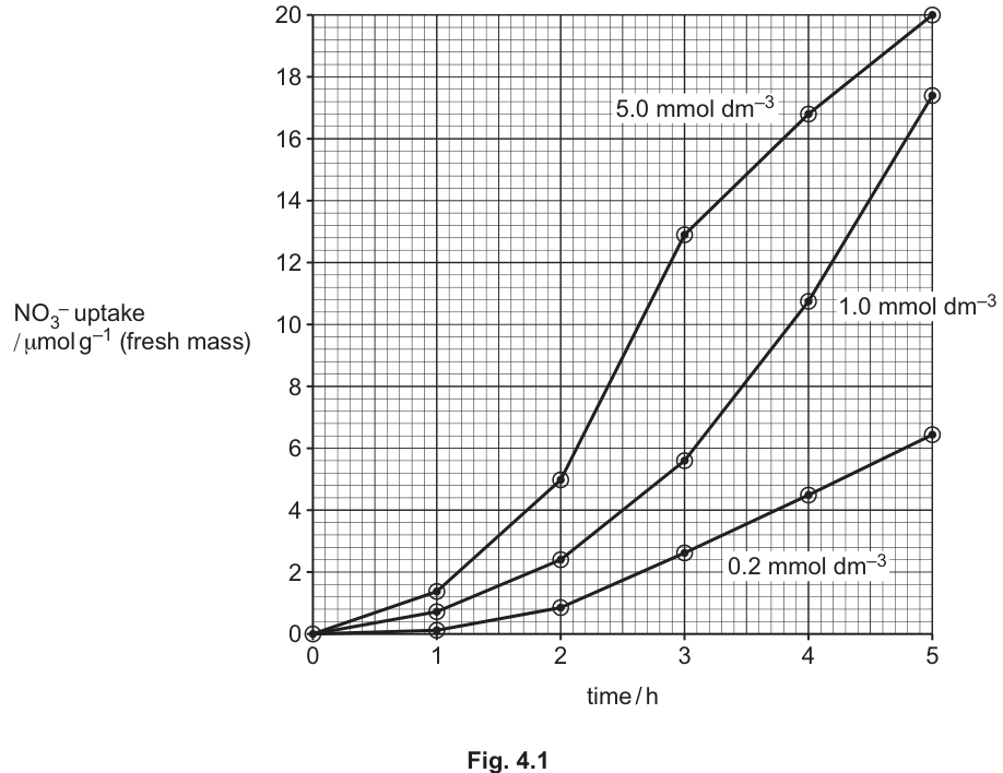
Fig. 4.1 shows that the rate of nitrate uptake is very low initially and then increases for all three concentrations of nitrate solution tested.
Describe the differences in the rates of nitrate uptake for nitrate solution compared with nitrate solution, between and
Answer
- Between 2 h and 5 h, the rate of nitrate uptake at is approximately constant (the curve is nearly linear), whereas the rate at is higher overall but is not constant — it decreases over this interval.
- The rate of uptake at is much higher (steeper gradient, e.g. rising from about to ) than at (e.g. rising from about to ).
Between 2 h and 5 h, the rate at is approximately constant, while the rate at is higher overall but decreases over time.
Background Concept
When reading a rate from a line graph, the rate of change is the gradient of the line (Δy / Δx). A straight, steeply rising line indicates a high constant rate; a curve that is becoming less steep indicates a rate that is decreasing over time. The y-axis here is the cumulative uptake (µmol g⁻¹) and the x-axis is time (h), so the gradient at any point is the rate of uptake at that moment (µmol g⁻¹ h⁻¹).
The graph in Fig. 4.1 shows the cumulative nitrate uptake by excised maize roots at three external concentrations. The data is for cumulative uptake (it keeps rising), so the RATE of uptake at any time is the slope of the curve at that time, not the y-value itself.
Understanding the Question
The question asks for a description of the differences between two curves (5.0 and 0.2 mmol dm⁻³) over a specific time window (between 2 h and 5 h). It is a 'describe' question, so you need to use values and trends from the graph to make comparative statements, and you need at least one piece of supporting data with units.
Approach
Look at the shape of each curve between 2 h and 5 h:
- 0.2 mmol dm⁻³: nearly a straight line (the rate is roughly constant) — a small, steady increase from about 1 to 6.5 µmol g⁻¹.
- 5.0 mmol dm⁻³: a curve that is steepest early in the interval and then flattens slightly — the rate is NOT constant, it is higher overall, and it decreases slightly between 2 h and 5 h. The cumulative uptake rises from about 5 to 20 µmol g⁻¹.
Then make the comparison: 5.0 is higher (faster) overall, and its rate is not constant, whereas 0.2 is approximately constant.
Step-by-Step Reasoning
- The 0.2 mmol dm⁻³ curve between 2 h and 5 h: starts at ~1 µmol g⁻¹, ends at ~6.5 µmol g⁻¹, change ≈ 5.5 µmol g⁻¹ over 3 h, giving a mean rate of ~1.8 µmol g⁻¹ h⁻¹. The curve is almost linear → the rate is approximately constant.
- The 5.0 mmol dm⁻³ curve between 2 h and 5 h: starts at ~5 µmol g⁻¹, ends at ~20 µmol g⁻¹, change ≈ 15 µmol g⁻¹ over 3 h, mean rate ≈ 5 µmol g⁻¹ h⁻¹. But the curve flattens: at 2 h the rate is about 8 µmol g⁻¹ h⁻¹ (5→13 between 2 and 3 h), but between 4 and 5 h the rate is only about 3 µmol g⁻¹ h⁻¹ (17→20). So the rate is decreasing — not constant.
- Comparative statement 1: 5.0 has a higher (steeper, faster) rate of uptake than 0.2 throughout the interval.
- Comparative statement 2: the 0.2 curve is approximately linear (constant rate), whereas the 5.0 curve is not constant — its rate decreases over the interval.
Key Takeaways
- Read rate from the gradient of a cumulative-quantity graph.
- A 'describe the difference' question often requires both a trend description and a comparative magnitude, with at least one piece of supporting data.
- Be alert to non-linear curves: 'not constant' is a valid description, even if the curve is still rising.
Common Mistakes
- Saying only '5.0 is faster' without describing the shape of the curves — you need to address whether the rate is constant or changing.
- Reading the rate from the wrong interval — the question specifies 2 h to 5 h.
- Confusing cumulative uptake with rate of uptake — the y-axis is cumulative, so the rate is the GRADIENT, not the y-value.
- Failing to quote any values from the graph — without numbers, your description is just an impression.
Things to Be Careful About
- The mark scheme awards marks for both: (1) constant vs. non-constant rate, and (2) 5.0 higher than 0.2. Aim to make both points.
- Numbers should be quoted with units (e.g. ~5 µmol g⁻¹ at 2 h for the 5.0 curve) for maximum credit.
- 'Steep' and 'steeper' are accepted shorthand for 'higher rate' / 'higher rate of uptake'.
The nitrate uptake of excised maize roots was investigated under different conditions.
Table 4.1 shows details and results for a control experiment and four modified experiments, 1, 2, 3 and 4. The same concentration of nitrate solution was used throughout for all the experiments. All the results were taken after a set time period.
Table 4.1
| experiment | temperature / | aeration | additional substances present in nitrate solution | nitrate uptake / (fresh mass) |
|---|---|---|---|---|
| 1 | 30 | no | nitrogen gas bubbled through instead of oxygen | 0.4 |
| 2 | 3 | yes | none | 0.6 |
| 3 | 30 | yes | protein synthesis inhibitor | 1.4 |
| 4 | 30 | yes | antibacterial compound | 10.0 |
| control | 30 | yes | none | 10.9 |
The results for experiments 1, 2, 3 and 4 in Table 4.1 can be compared to the results for the control experiment.
Discuss how comparing each of the results with the control provides information about:
- how nitrate ions are taken up by the root cells
- the factors affecting the uptake of nitrate ions.
Answer
- Comparing experiments 1, 2 and 3 with the control () shows nitrate uptake is much reduced without oxygen (), at low temperature (), and when protein synthesis is inhibited (). This indicates that nitrate uptake is mainly by active transport, requiring ATP from aerobic respiration, transport (carrier) proteins, and working at a suitable temperature.
- Experiment 1 vs control: vs — without O₂, aerobic respiration stops, so no ATP is produced for active transport.
- Experiment 2 vs control: vs — at , respiratory enzymes work more slowly, so less ATP is made, and carrier proteins move more slowly.
- Experiment 3 vs control: vs — without protein synthesis, transport (carrier) proteins cannot be made, so uptake is much reduced.
- A small amount of uptake still occurs in experiment 1 (), suggesting that some (facilitated) diffusion of nitrate also takes place.
- Experiment 4 () is very similar to the control (), so bacteria associated with the roots have little effect on nitrate uptake.
Nitrate uptake is mainly by active transport (needs O₂, ATP, carrier proteins, and a suitable temperature); a small component is by (facilitated) diffusion. Bacteria on the roots have little effect.
Background Concept
A controlled experiment varies ONE variable at a time while keeping all others constant. By comparing each modified experiment with the control (in which only the standard conditions are used), the experimenter can attribute any difference in the result to the variable that was changed.
The control here: 30 °C, aerated (oxygen), no inhibitors, no antibacterial agent, 10.9 µmol g⁻¹ nitrate uptake.
The four modified experiments test the following variables:
- 1: nitrogen gas instead of oxygen (tests the effect of oxygen / aerobic respiration).
- 2: 3 °C instead of 30 °C (tests the effect of temperature).
- 3: protein synthesis inhibitor added (tests whether newly made proteins are needed).
- 4: antibacterial compound added (tests whether bacteria on the roots are responsible for the uptake).
This is a classic experimental design pattern: the variable changed in each experiment is the variable the experimenter is testing.
Understanding the Question
Four marks are available. The command word 'Discuss' means you must do more than just describe the results — you must INTERPRET them, i.e. explain what the comparisons tell us about the mechanism of nitrate uptake. The mark scheme requires: (1) the active-transport conclusion, (2) at least one experiment-vs-control comparison, (3) at least one piece of comparative data with units, and (4) at least one discussion point explaining the biology behind a result.
Approach
Take each experiment in turn, compare its result with the control, and draw the conclusion about the mechanism that this comparison supports. Group the conclusions into a coherent narrative: 'nitrate uptake is mainly by active transport; it requires ATP, transport proteins, and is temperature-dependent; bacteria play a minimal role; a small component may be by diffusion.'
Step-by-Step Reasoning
Comparison 1 — control vs. experiment 1 (no oxygen):
- Control: 10.9 µmol g⁻¹. Experiment 1: 0.4 µmol g⁻¹. Difference: 10.5 µmol g⁻¹ lower without O₂.
- Interpretation: without O₂, aerobic respiration cannot occur, so little ATP is made. The huge drop in uptake suggests that uptake needs ATP → it is largely an active process.
- Small residual uptake (0.4) suggests a tiny contribution from (facilitated) diffusion.
Comparison 2 — control vs. experiment 2 (3 °C):
- Control: 10.9 µmol g⁻¹. Experiment 2: 0.6 µmol g⁻¹. Difference: 10.3 µmol g⁻¹ lower at 3 °C.
- Interpretation: at low temperature, respiratory enzymes work slowly (Q₁₀ effect), so less ATP is produced for active transport. Also, the kinetic energy of nitrate ions is reduced and the carrier proteins move more slowly in the less fluid membrane, slowing both diffusion and active transport.
Comparison 3 — control vs. experiment 3 (protein synthesis inhibitor):
- Control: 10.9 µmol g⁻¹. Experiment 3: 1.4 µmol g⁻¹. Difference: 9.5 µmol g⁻¹ lower when protein synthesis is blocked.
- Interpretation: uptake depends on the continuous synthesis of new carrier / transport proteins (which turn over in the membrane and must be replaced).
Comparison 4 — control vs. experiment 4 (antibacterial compound):
- Control: 10.9 µmol g⁻¹. Experiment 4: 10.0 µmol g⁻¹. Difference: only 0.9 µmol g⁻¹ lower without bacteria.
- Interpretation: bacteria associated with the roots have very little effect on nitrate uptake. (The small difference could be due to bacteria themselves taking up a small amount of nitrate, or a slight effect on the experiment.)
Overall conclusion:
- Nitrate uptake is mainly an active process (requires O₂, ATP, carrier proteins, and is temperature-sensitive).
- A small proportion may be by (facilitated) diffusion (since some uptake still occurs at 3 °C or without O₂).
Key Takeaways
- The experimental design here is a 'controlled comparison' — each experiment tests one variable.
- The most powerful general conclusion is that the uptake mechanism is mainly active transport, because it needs energy (ATP) from aerobic respiration.
- The need for protein synthesis (Experiment 3) confirms that membrane transport proteins are required and that they turn over.
- A small residual uptake in the absence of energy (Experiment 1) is the signature of facilitated diffusion contributing a small amount.
Common Mistakes
- Failing to quote the actual numbers from Table 4.1 with units — the mark scheme requires at least one comparative result with units.
- Confusing 'temperature' and 'enzyme activity' — the correct explanation links low temperature to slower enzyme activity, hence less ATP.
- Saying 'diffusion' instead of 'facilitated diffusion' for the small residual uptake — the membrane is not freely permeable to a charged ion like nitrate.
- Saying that bacteria 'help' uptake (slightly higher control than Experiment 4) without saying that the effect is very small.
- Suggesting that the protein-synthesis inhibitor proves 'enzymes' are needed without specifying that the relevant proteins are transport/carrier proteins in the membrane.
- Vague answers such as 'proteins are needed' or 'oxygen is needed' without the comparative data.
- Forgetting to state the conclusion that uptake is by active transport.
Things to Be Careful About
- One mark is reserved for COMPARATIVE DATA with units — quote at least one number with µmol g⁻¹ in your answer.
- The mark scheme allows a maximum of 3 discussion points (mp4–11) for 1 mark; the other 3 marks are: 1 for the active transport conclusion, 1 for an experiment-vs-control comparison, 1 for the data.
- Avoid blanket statements like 'energy is needed' without specifying the source (aerobic respiration) and the use (active transport).
During interphase of the cell cycle, individual chromosomes cannot be seen within the nucleus. The genetic material is termed chromatin during this stage.
Changes occur to chromatin during mitosis so that chromosomes become visible.
State what happens to chromatin so that individual chromosomes can be seen during mitosis.
Answer
The chromatin coils (supercoils) and condenses, becoming shorter and fatter so that individual chromosomes become visible.
The chromatin condenses (supercoils), becoming shorter and fatter.
Background Concept
During interphase, the cell's DNA exists as chromatin — long, thin, unwound DNA–protein fibres dispersed throughout the nucleus. Because the fibres are extremely fine and diffuse, individual chromosomes cannot be distinguished under a light microscope, even though the DNA is organised. Before mitosis begins, this DNA–protein complex must be packaged into discrete, transportable units. The change that allows visualisation is a massive physical compaction of the chromatin fibre.
Understanding the Question
The question is a one-mark "state what happens" item. The candidate simply has to give the change in chromatin that makes chromosomes individually visible at the start of mitosis. The mark scheme accepts any of: coiling / supercoiling / condensation / becomes more compact, or the descriptive alternative "becomes shorter and fatter".
Approach
Recall that during prophase, chromatin fibres coil repeatedly around histone proteins to form the compact chromatids of a metaphase chromosome. A short, clear statement covering the idea of compaction is enough.
Step-by-Step Reasoning
- In interphase, DNA is wrapped around histone octamers to form nucleosomes, but the nucleosome chain is loosely extended (like beads on a string and a 30 nm fibre).
- As the cell enters prophase, this fibre undergoes further coiling and supercoiling into loops and higher-order structures.
- The result is that the long, thin chromatin becomes shorter and fatter, so each chromosome is now thick enough to absorb light and be seen as a discrete structure in the microscope.
Key Takeaways
- Chromatin and chromosome are the same genetic material in different states of packaging.
- Condensation (supercoiling) is what makes chromosomes visible during mitosis.
Common Mistakes
- Saying the DNA "doubles" or "replicates" — replication occurs in S phase of interphase, before any condensation.
- Saying the chromatin "disappears" or "breaks up" — it remains continuous, just packaged differently.
- Mentioning the nuclear envelope breaking down — that is a separate event at the end of prophase, not what makes chromosomes visible.
Things to Be Careful About
The mark scheme accepts several equivalent answers (condenses, supercoils, coils, becomes more compact, becomes shorter and fatter). Any one of these is sufficient for the single mark.
Fig. 5.1 is a transmission electron micrograph of two human chromosomes at metaphase of mitosis.
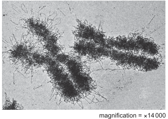
Describe the structure of chromosomes at metaphase, such as the two chromosomes shown in Fig. 5.1.
Answer
- Each chromosome consists of two sister chromatids that are genetically identical.
- The two chromatids are held together at a centromere.
- Each chromatid contains a single DNA molecule (so the whole chromosome contains two DNA molecules).
- The DNA is associated with histone proteins (basic proteins) that package it.
- The ends of the chromatids are protected by telomeres (repeating non-coding DNA sequences).
Two sister (genetically identical) chromatids joined at a centromere; each chromatid contains one DNA molecule wrapped around histone proteins; telomeres at the ends.
Background Concept
A metaphase chromosome is a fully condensed, duplicated chromosome in its most compact form. Each one is built from one continuous DNA double helix (replicated in S phase to give two identical copies), with that DNA wrapped around histone proteins to form nucleosomes, and the nucleosome chain further coiled and supercoiled. The two identical copies of the DNA molecule — the sister chromatids — remain physically joined at a specialised region called the centromere until they are separated in anaphase. The ends of the DNA molecule are capped by telomeres, short repeating non-coding sequences that protect the chromosome from degradation and from end-to-end fusions.
Understanding the Question
The question shows a transmission electron micrograph of two human chromosomes at metaphase (×14 000). The fuzzy "frayed" material around the dense central axis is chromatin that has been less tightly condensed and projects outwards as loops. The candidate is asked to describe the structure of such a chromosome, and up to four marking points are available from a list of standard structural features.
The mark scheme explicitly rejects reference to "homologous chromosomes" here — the two chromosomes in Fig. 5.1 are not necessarily a homologous pair; they are just two separate chromosomes caught at metaphase. The term to use is sister chromatids.
Approach
Work through the standard anatomy of a metaphase chromosome, from the most prominent visible feature outwards:
- Two chromatids joined at a centromere — the most obvious structure.
- Each chromatid is one DNA molecule — the molecular basis of "identical".
- DNA is packaged with histones — the protein component.
- The ends of the DNA are the telomeres — often forgotten but credited here.
Step-by-Step Reasoning
- Two sister chromatids: A metaphase chromosome always appears as an X- or H-shaped structure because the DNA was replicated in S phase. The two resulting copies are called sister chromatids. They carry the same alleles in the same order, i.e. they are genetically identical. The mark scheme rejects "homologous" — homologous chromosomes are a pair of similar chromosomes (one from each parent), not the two halves of a single replicated chromosome.
- Centromere: This is the constricted, often pale-staining region where the two sister chromatids are held together. It is also the site where kinetochore proteins assemble and where spindle microtubules attach during mitosis.
- One DNA molecule per chromatid: Each sister chromatid contains a single, long, linear DNA double helix. A whole metaphase chromosome therefore contains two DNA molecules. The mark scheme allows "2 chromatids and 2 DNA molecules" as an alternative phrasing.
- Histone proteins: The DNA is not naked; it is wrapped around histone octamers to form nucleosomes, and these are further folded into higher-order structures. Histones are basic (positively charged) proteins that bind the negatively charged phosphate backbone of DNA.
- Telomeres: The two ends of each linear DNA molecule carry telomeres — short, repeating, non-coding nucleotide sequences (e.g. TTAGGG in humans) that protect the ends from enzymatic degradation and prevent chromosome ends from being joined to one another.
- AVP — chromosomes are highly/most condensed: A valid extra point — at metaphase, chromosomes reach their maximum degree of compaction, which is why they are so clearly visible in the TEM and can be moved without tangling.
Key Takeaways
- A metaphase chromosome = 2 sister chromatids + 1 centromere + 2 DNA molecules + histone-packaged chromatin + telomere-capped ends.
- "Sister chromatids" ≠ "homologous chromosomes" — sister chromatids are identical copies of one chromosome; homologues are a maternal–paternal pair.
- Each chromatid is one DNA molecule, so the two chromatids together give two DNA molecules per replicated chromosome.
Common Mistakes
- Calling the two chromatids "homologous chromosomes" — explicitly rejected by the mark scheme.
- Saying a chromosome has "two strands of DNA" or "many DNA molecules" — each chromatid is one DNA molecule.
- Forgetting the histones, or calling them "histone enzymes" — they are structural proteins, not enzymes.
- Ignoring the telomeres, even though the mark scheme credits them.
- Describing only the appearance of the TEM (e.g. "the chromosomes are dark and fuzzy") without giving the named structural features.
Things to Be Careful About
- Use the precise term sister chromatids, not just "two chromatids".
- The TEM shows chromatin loops extending from the central axis; these are still chromatin, just less tightly packaged, and do not need to be described — the mark scheme rewards only the four standard structural points (or a fifth AVP).
- The magnification (×14 000) is given but not needed for the description; it would only be needed if a size calculation were asked.
Some people who are infected with HIV have HIV/AIDS.
Answer
Human Immunodeficiency Virus
Human Immunodeficiency Virus
Background Concept
HIV is a retrovirus that targets cells of the human immune system, particularly the CD4⁺ T-helper lymphocytes. By destroying these cells, the virus progressively weakens the host's adaptive immune response, eventually leading to AIDS (Acquired Immune Deficiency Syndrome), where the individual becomes vulnerable to a wide range of opportunistic infections. Knowing the full name of common pathogen abbreviations is essential because it immediately identifies the type of organism and the system it attacks — 'virus' here distinguishes HIV from the bacterial or protoctist pathogens responsible for TB, cholera and malaria respectively.
Understanding the Question
This part simply asks for the expansion of the abbreviation HIV, awarded 1 mark for the full name. The question stem reminds you that HIV/AIDS is an infectious disease, so the answer must refer to the pathogen itself, not the disease.
Approach
No analysis is required — write out each word of the abbreviation in full and in the correct order.
Step-by-Step Reasoning
- H → Human
- I → Immunodeficiency
- V → Virus
Combined: Human Immunodeficiency Virus.
Key Takeaways
The three-word expansion identifies the pathogen type (virus), what it damages (the immune system, specifically the deficiency component) and its host (human). Memorising these standard abbreviations (HIV, AIDS, TB, ART, CD4) is foundational for the infectious diseases topic.
Common Mistakes
- Writing 'Human Immuno-Deficiency Virus' with a hyphen — not wrong, but the cleanest mark-scheme wording is three separate words.
- Confusing HIV with the disease AIDS (Acquired Immune Deficiency Syndrome) rather than the pathogen.
- Using lowercase inconsistently; capitalising each word is conventional.
Things to Be Careful About
Cambridge does not require a definite article. Just write 'Human Immunodeficiency Virus' — that is the full and complete term.
Following transmission of HIV, early diagnosis of infection and treatment with anti-retroviral therapy (ART) helps to control the spread of the pathogen and prevent HIV/AIDS.
Suggest why treating people who have developed HIV/AIDS with ART may help to reduce the number of overall deaths from infectious diseases, such as cholera, TB and malaria.
Answer
- ART helps to maintain/increase the number of T-lymphocytes (T-helper cells), preventing their destruction by HIV.
- This maintains/strengthens the immune system, so the patient has a higher chance of recovery from infectious (opportunistic) diseases such as cholera, TB and malaria.
ART preserves T-lymphocytes → strengthens immune system → improves recovery from opportunistic infections
Background Concept
HIV infects and destroys CD4⁺ T-helper lymphocytes, the cells that coordinate the adaptive immune response. As T-helper cell numbers fall, the body becomes progressively less able to mount effective responses against new infections — this is why HIV/AIDS patients develop so-called 'opportunistic' infections (TB, pneumonia, chronic diarrhoea). Anti-retroviral therapy (ART) inhibits viral replication, slowing or halting the destruction of T-helper cells and allowing the immune system to recover to some degree.
Understanding the Question
The question stem links HIV/AIDS with other infectious diseases (cholera, TB, malaria). It then asks why treating HIV/AIDS patients with ART might reduce the overall number of deaths from those other diseases. The 2 marks require two distinct, creditable points about how ART affects immunity.
Approach
Build a causal chain from the drug's action to the population-level outcome:
ART → preserves T-helper cells → strengthens immune system → better recovery from other infections → fewer deaths.
Each mark is earned for one logical link in this chain, so the candidate should articulate at least two distinct links explicitly.
Step-by-Step Reasoning
- T-lymphocyte preservation: ART suppresses HIV replication, so fewer T-helper cells are killed. The patient retains more CD4⁺ T-cells. (Mark-scheme point: 'helps to increase/maintain number of T-lymphocytes / T-helper cells'.)
- Stronger immune response: with more T-helper cells, B-cell, cytotoxic T-cell and macrophage activity is better coordinated. The immune system as a whole is stronger. (Mark-scheme point: 'maintains/improves strength of immune system / immune response'.)
- Better recovery from opportunistic infections: a competent immune system can fight off pathogens that an HIV-weakened body could not. The patient is therefore more likely to survive a concurrent infection such as TB, cholera or malaria. (Mark-scheme point: 'increases chance of recovery from infectious/opportunistic disease'.)
Any two of these distinct points earn the two marks.
Key Takeaways
- ART benefits extend beyond HIV itself to other infectious diseases because it preserves the very cells (T-helpers) needed to fight them.
- The question tests whether you can transfer an immunological principle from one disease to a related clinical scenario.
- Mark-scheme wording for credit includes 'opportunistic disease', 'T-helper cells' and 'immune system'.
Common Mistakes
- Saying ART 'cures' HIV — it controls but does not cure.
- Saying ART directly kills cholera/TB/malaria pathogens — it does not; it works by restoring immunity.
- Mentioning only a reduction in 'viral load' — this is accepted (AVP) but on its own does not explain reduced deaths; the link to immunity is required.
- Stating 'more white blood cells' generically — the mark scheme wants T-lymphocytes / T-helper cells specifically.
- Confusing the cause-and-effect direction (saying ART causes the other infections).
Things to Be Careful About
The mark-scheme guidance states 'max 1 if no mention of benefit of ART'. So at least one mark must explicitly reference what ART does. Vague answers about 'people getting better' will not score both marks.
Studies suggest that people who are infected with HIV may be at a higher risk of heart disease. One cause of heart disease is the narrowing of the lumen of one or both of the main coronary arteries.
A coronary artery bypass graft (bypass graft) is a surgical operation that uses healthy blood vessels to divert blood around diseased sections of coronary arteries. The main choice of blood vessel to use for a bypass graft is known as the internal thoracic artery.
Fig. 6.1 is a diagram of an external view of the heart to show a double bypass graft.
The blood vessels used in the bypass graft shown in Fig. 6.1 are the great saphenous vein from the leg and the internal thoracic artery.
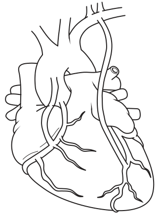
Draw a cross (X) on Fig. 6.1 to show an area of the right coronary artery that has been bypassed.
Answer
Place a clear cross (X) on the right coronary artery on the left-hand side of Fig. 6.1 (i.e. the anatomical right side of the heart), positioned ABOVE the junction where the graft joins the coronary artery. The X must NOT be placed at the junction between the bypass graft (great saphenous vein) and the coronary artery itself.
X placed on the right coronary artery, above the graft junction, on the anatomical right (left of figure).
Background Concept
The heart is supplied by two coronary arteries that arise from the aorta just above the aortic valve:
- The left coronary artery runs down the left side of the heart (appearing on the right of an anterior view).
- The right coronary artery runs down the right side of the heart (appearing on the left of an anterior view).
When a coronary artery is narrowed by atherosclerotic plaque, blood flow to the heart muscle is restricted. Surgeons use a healthy vessel (a 'graft') to bypass the blocked segment, restoring flow to the tissue beyond.
Understanding the Question
Fig. 6.1 shows the heart from the front (anterior view) with a double bypass graft in place. The candidate must mark a cross on the part of the right coronary artery that the graft has bypassed.
A critical orientational point: in an anterior view of the heart, the heart's own right side appears on the LEFT of the page as the viewer sees it (mirror-image view). So the right coronary artery is on the left side of the figure.
Approach
- Locate the right coronary artery on Fig. 6.1 — it runs along the right atrioventricular groove, which appears on the left side of the diagram.
- Identify the junction where the bypass graft joins onto the coronary artery (the distal anastomosis).
- Place the X above (proximal to) this junction, on the coronary artery itself, in the region that the graft is bypassing.
Step-by-Step Reasoning
- The right coronary artery is the vessel descending along the AV groove on the anatomical right of the heart.
- In Fig. 6.1, the right coronary artery is the vessel on the left-hand side of the diagram.
- The bypass graft (the great saphenous vein, shown running up from the lower part of the figure) joins onto the right coronary artery at a junction.
- The X should be placed on the right coronary artery between its origin (at the aorta) and that junction — that is the diseased segment that has been bypassed.
- It must not be placed at the actual junction itself, because that is the surgical join, not the diseased segment.
Key Takeaways
- Anterior heart view = mirror image: anatomical right appears on viewer's left.
- A bypass graft diverts blood around the diseased segment, which lies between the aorta and the distal anastomosis.
- Anatomical identification of vessels on a heart diagram is a recurring Paper 2 skill.
Common Mistakes
- Marking the X on the right side of the figure (which is the left coronary artery, not the right).
- Placing the X at the junction between the graft and the native artery (the mark scheme explicitly rejects this).
- Placing the X on the graft itself rather than on the diseased native artery.
- Confusing the great saphenous vein (a low-pressure vessel used as a graft) with the right coronary artery.
Things to Be Careful About
The mark scheme is strict: 'X is positioned above the join on the coronary artery on the left-hand side of Fig. 6.1 / right heart' earns the mark. Wrong vessel, wrong side, or placement on the junction all fail.
After surgery, the wall of the great saphenous vein becomes thicker.
Suggest and explain why it is important for the wall of the vein to become thicker after surgery.
Answer
- In its new arterial position the great saphenous vein is exposed to much higher blood pressure than it normally experiences in the leg, so the wall must thicken to withstand this higher pressure (and is less likely to burst).
- More smooth muscle and elastic fibres develop in the tunica media (and more collagen in the tunica externa/adventitia), giving the grafted vessel the strength and recoil needed to handle the higher pressure and maintain rapid onward blood flow.
Thicker wall withstands higher arterial blood pressure; more smooth muscle/elastic tissue develops in tunica media.
Background Concept
Blood vessels are classified by the structure of their walls, which reflects the pressure they normally experience:
- Arteries: thick tunica media rich in smooth muscle and elastic fibres, plus a tough outer tunica externa (adventitia) of collagen. This lets them withstand the high, pulsatile pressure generated by ventricular systole and recoil during diastole to maintain flow.
- Veins: thin walls with much less smooth muscle and elastic tissue, because venous blood is at low pressure. Forward flow relies on skeletal muscle contraction and one-way valves rather than arterial recoil.
When a vein (the great saphenous vein) is grafted into the coronary circulation, it is suddenly exposed to arterial pressures. Its wall must adapt to avoid bursting.
Understanding the Question
This part asks the candidate to suggest AND explain why the wall of the great saphenous vein must thicken once it is functioning as a bypass graft in the arterial system. Two marks are awarded — one for the functional reason (higher pressure) and one for the structural adaptation (more muscle/elastic tissue).
Approach
Make the structure–function link explicit: the vessel must handle higher pressure → therefore needs more structural tissue (smooth muscle / elastic fibres / thicker wall layers).
Step-by-Step Reasoning
- Functional demand: in its native position the great saphenous vein carries low-pressure blood back to the heart. Once it is connected to the coronary artery it is exposed to high, pulsatile arterial pressure. A normal thin venous wall would be likely to burst.
- Structural adaptation: in response, the wall thickens. Specifically, the tunica media gains more smooth muscle and elastic fibres (giving it strength to withstand pressure and recoil to push blood onward), and the tunica externa gains more collagen. With these reinforcements the grafted vessel can survive long term in the arterial circulation.
- Linking the two: a thicker wall + more elastic fibres = a vessel that can withstand higher pressure without bursting AND that can recoil to maintain the rapid, pulsatile flow required in the coronary circulation.
Key Takeaways
- Veins and arteries are structurally different because of the different pressures they carry.
- When a vessel is moved from a low-pressure to a high-pressure environment, its wall remodels (smooth muscle / elastic tissue proliferate) to handle the new load.
- This is a classic structure–function application in a clinical context.
Common Mistakes
- Saying the wall thickens 'because more blood flows through it' — pressure, not volume, is the issue.
- Saying the wall thickens 'to push blood back to the heart' — irrelevant in a grafted arterial position.
- Saying the vein would 'collapse' if it did not thicken — the mark scheme rejects this; the real risk is bursting under high pressure.
- Saying 'more blood' without specifying 'higher pressure'.
- Naming only one wall layer without linking to the functional reason.
Things to Be Careful About
The mark scheme lists two clearly distinguishable marking routes (pressure-resistance AND vessel durability) and explicitly accepts the structural detail of more smooth muscle / elastic fibres / collagen. At least one mark must combine the higher-pressure observation with the consequence (bursting or reduced durability). The other mark must name the wall layer and tissue type that increase.