Biology 9700/21 — May/June 2024
Cambridge AS Level · AS Level Structured Questions · worked solutions for every part, with the mark scheme
Topics Cell Structure · Nucleic Acids and Protein Synthesis · Immunity · Cell Membranes and Transport · Biological Molecules · Transport in Mammals · +4 more
Fig. 1.1 is a diagram showing part of a cell surface membrane of an animal cell.
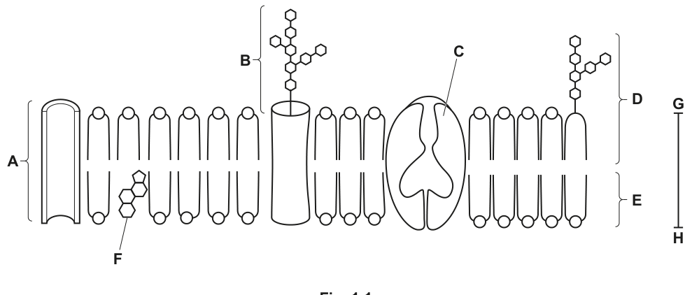
State the approximate thickness of the membrane as shown by the line G–H.
.....................................................................................................................................
Answer
(any value in the range – is accepted, but the unit must be shown).
7 nm
Background Concept
The fluid-mosaic model describes the cell surface membrane as a phospholipid bilayer in which proteins float. A typical cell surface membrane is approximately () thick, although the value varies from about to depending on cell type and on which components (cholesterol, glycolipids, integral proteins) are particularly abundant.
The membrane is so thin that it is below the resolution of the light microscope and can only be visualised with the transmission electron microscope (TEM). For scale, , so is roughly the diameter of a small globular protein — about times thinner than a millimetre.
Understanding the Question
In Fig. 1.1 the bracket – on the right-hand side spans the full vertical thickness of the membrane, from the outer surface (top) to the inner surface (bottom). The command word is "state" — a single value with the correct unit, no explanation required.
Approach
Recall the standard textbook thickness of a cell surface membrane, write it with the correct unit (nanometres), and check that it lies inside the mark-scheme range.
Step-by-Step Reasoning
- The membrane shown in Fig. 1.1 is a phospholipid bilayer drawn at very high magnification so that its two layers are clearly visible.
- The – bracket measures the entire bilayer, top to bottom.
- The standard textbook value is .
- The mark scheme accepts any value in the range –, so candidates who write, say, or also score the mark.
- The unit must be given. Writing the number alone, or with the wrong unit (, or ), would lose the mark because the unit is explicitly required.
Key Takeaways
- A cell surface membrane is approximately thick.
- Always include the unit when asked to state a measurement.
- Useful conversions: .
Common Mistakes
- Writing the value without a unit (e.g. just "7") — the mark scheme insists the unit is shown.
- Writing the value in micrometres by mistake — that is times too large.
- Writing it in Ångströms — the mark scheme specifically asks for nanometres.
Things to Be Careful About
- The question asks for the thickness of the membrane as a whole, not the length of a phospholipid tail or the diameter of a single protein. Stay with the bilayer as one object.
- If you cannot recall the precise value, a range of – is accepted, so do not leave the answer blank.
Complete Table 1.1 to show:
• the names and functions of the components of the cell surface membrane
• the letters of the labels in Fig. 1.1 that identify each component.
Table 1.1
| component | function | letter on Fig. 1.1 |
|---|---|---|
| channel protein | ||
| phospholipid | ||
| receptor for cell signalling | ||
| F |
Answer
| component | function | letter on Fig. 1.1 |
|---|---|---|
| channel protein | facilitated diffusion (of ions / water / polar / water-soluble substances) | A |
| phospholipid | forms a bilayer; barrier to water-soluble substances / allows diffusion of fat-soluble substances / provides fluidity | E |
| glycoprotein (or glycolipid) | receptor for cell signalling | B (or D) |
| cholesterol | gives mechanical stability / regulates fluidity | F |
See working — channel protein A; phospholipid E; receptor = glycoprotein at B (or glycolipid at D); F = cholesterol (stability / fluidity).
Background Concept
The fluid-mosaic model has the following key molecular components, each with a recognisable appearance in a diagram such as Fig. 1.1:
- Phospholipid bilayer — two rows of phospholipids arranged tail-to-tail. The hydrophilic phosphate heads face the aqueous environments on either side; the hydrophobic fatty-acid tails meet in the middle. The bilayer is the structural backbone and the main selective barrier.
- Channel proteins — integral proteins that form a water-filled pore through the bilayer, allowing specific ions or small polar molecules to cross by facilitated diffusion down their concentration gradient.
- Carrier proteins — integral proteins that bind a specific molecule on one side, change shape, and release it on the other; used in both facilitated diffusion and active transport.
- Glycoproteins and glycolipids — proteins or lipids with a short carbohydrate chain on the outer surface. They act as cell-signalling receptors and as cell-recognition markers (e.g. ABO blood-group antigens).
- Cholesterol — small lipid molecules wedged between phospholipid tails in animal-cell membranes. At warm temperatures they reduce fluidity and stiffen the membrane; at cool temperatures they prevent the fatty-acid tails from packing too closely and so maintain fluidity.
Understanding the Question
The candidate is given a partly completed table with three columns: component, function and letter on Fig. 1.1. Two rows have the component already given; one row has the function already given; one row has the letter already given. The candidate must fill in the missing cells so that, in each row, the name, function and letter all agree.
The mark scheme notes that the answer is marked row by row, with one mark for each row where all three entries are consistent. As a fallback, a candidate who gets columns rather than rows correct can still score up to two marks column-wise.
Approach
For each row, look at the part already given, use Fig. 1.1 to identify which structure has that label, and complete the row so that name, function and letter are all consistent with the same structure.
Step-by-Step Reasoning
Row 1 — channel protein (component already given)
- A channel protein forms an open pore, so it allows ions / water / small polar / water-soluble molecules to pass by facilitated diffusion down their concentration gradient (no ATP required).
- In Fig. 1.1 the tall cylindrical protein spanning the bilayer at the far left of the diagram is labelled A.
Row 2 — phospholipid (component already given)
- A phospholipid's main job is to form the bilayer and act as the barrier to water-soluble / polar substances / ions, while still allowing small non-polar (fat-soluble) substances to diffuse across. The bilayer also provides the membrane's basic fluidity and stability.
- The bracket E on the right-hand side of Fig. 1.1 points to a single phospholipid in the lower leaflet (note that the upper layer of heads is labelled D and the whole bilayer thickness is G–H).
Row 3 — function already given: receptor for cell signalling
- The cell-signalling receptor on the outer surface is a glycoprotein (a protein bearing a short branched carbohydrate chain). Glycolipids (carbohydrate on a phospholipid head) also act as receptors, and "glycolipid" is accepted as an alternative.
- In Fig. 1.1 the long carbohydrate chain attached to a protein on the upper (outer) surface is labelled B. A similar carbohydrate chain attached to a phospholipid head in the upper layer is labelled D — accept either for this row, but B is the more usual answer because the carbohydrate is clearly on a protein.
Row 4 — letter already given: F
- F points to a small hexagonal/ring-shaped molecule sitting among the phospholipid tails in the hydrophobic core. This is cholesterol.
- Cholesterol's function: it gives mechanical stability to the membrane and regulates its fluidity — at higher temperatures it decreases fluidity, while at lower temperatures it prevents the fatty-acid tails from packing too closely and so keeps the membrane from solidifying.
Key Takeaways
- The fluid-mosaic diagram has a small set of iconic features; you should be able to read each one at sight.
- Glycoprotein vs glycolipid: same short carbohydrate chain, but anchored to a protein (glycoprotein) or a phospholipid head (glycolipid).
- Channel vs carrier: a channel is a continuous open pore; a carrier binds its substrate and changes shape. In a typical diagram, channels are drawn as open cylinders, carriers as a closed blob with a clear shape-change illustrated.
- Cholesterol has a bidirectional effect on fluidity: it buffers fluidity against temperature change.
Common Mistakes
- Writing "phospholipid" but giving only a vague function ("forms the membrane") without specifying bilayer, barrier role or fluidity. The mark scheme requires a role within the bilayer.
- Putting B and D in the wrong row. B is the glycoprotein (carbohydrate on a protein), D is the glycolipid (carbohydrate on the upper phospholipid head). Both can act as signalling receptors, but the mark scheme prefers B for the standard answer.
- Confusing F (cholesterol, in the hydrophobic core) with one of the phospholipid labels. F has a distinctive shape — a small ring of three fused hexagons — not a phospholipid with a head and two tails.
- For cholesterol, writing only "stability" without any reference to fluidity. The mark scheme rewards stability and a fluidity role, or a temperature-dependent effect.
Things to Be Careful About
- "Glycolipid" is offered as an alternative to "glycoprotein" for the receptor row — pick the one that matches the letter you are using.
- The mark scheme's column-fallback means a candidate who gets every component right but the wrong letter on a row still scores nothing for that row; the letter has to be correct in the same row.
- The lipid labelled D is in the outer leaflet, so it is unambiguously a glycolipid; the lipid labelled E is in the inner leaflet and is just a phospholipid (no carbohydrate shown).
Fig. 1.2 is a drawing of a transmission electron micrograph (TEM) of a cell from the palisade mesophyll of a leaf.
The drawing does not show all of the organelles visible in a transmission electron micrograph.
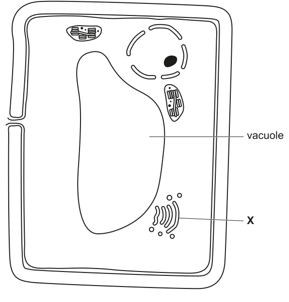
Complete Fig. 1.2 by drawing and labelling:
• a mitochondrion
• rough endoplasmic reticulum
• smooth endoplasmic reticulum.
Your drawings should show the detail that can be seen in a transmission electron micrograph.
Working
Draw the three organelles in the empty cytoplasm of Fig. 1.2 — anywhere clear of the cell wall, the central vacuole, the nucleus, the chloroplasts and the existing Golgi (X). Use a sharp pencil, continuous clear lines, and a straight label line that ends precisely on the organelle it identifies.
Mitochondrion — an oval about the same size as the chloroplasts already shown. Outer membrane drawn as a smooth continuous curve; inner membrane as a second parallel curve just inside it, with at least one infolding (crista) projecting into the matrix. No ribosomes on the outside. Label: mitochondrion.
Rough endoplasmic reticulum (RER) — at least one flattened cisterna drawn as two close parallel lines with closed, rounded ends, like a deflated balloon. Stud the outer (cytoplasmic) face with small solid dots (ribosomes). Optionally several cisternae stacked loosely. Label: RER or rough endoplasmic reticulum.
Smooth endoplasmic reticulum (SER) — at least one branching, tubular network drawn as a single curving line that splits and rejoins, with no ribosomes anywhere on its surface. Place it near the RER so the eye can compare. Label: SER or smooth endoplasmic reticulum.
See diagram — mitochondrion (with crista), RER (with ribosomes on outside), SER (tube, no ribosomes), all drawn in 2-D and clearly labelled in the cytoplasm.
Background Concept
Three endomembrane-system organelles appear in plant cells and have distinctive appearances in a transmission electron micrograph (TEM):
- Mitochondrion — a double-membrane organelle about the size of a bacterium. The outer membrane is smooth; the inner membrane is thrown into folds called cristae that project into the matrix. Cristae are the TEM-visible feature that confirms the structure is a mitochondrion rather than a vesicle or a plastid.
- Rough endoplasmic reticulum (RER) — a network of flattened membrane sacs (cisternae) whose cytoplasmic face is studded with ribosomes, giving the membrane a "rough" appearance in the TEM. RER is the site of synthesis of membrane-bound and secretory proteins.
- Smooth endoplasmic reticulum (SER) — a network of branching tubules (not flattened sacs) with no ribosomes. SER is the site of lipid and steroid synthesis, and (in some cell types) detoxification or Ca²⁺ storage.
The only visual difference between RER and SER at TEM level is the presence of ribosomes on the cytoplasmic face of the membrane, and the fact that RER is typically drawn as flattened sacs while SER is drawn as smooth tubes.
Understanding the Question
Fig. 1.2 is a biological drawing of a palisade mesophyll cell. It already shows the cell wall, a large central vacuole, a nucleus with nucleolus, several chloroplasts and a Golgi body (X). The candidate must add three more organelles to the empty cytoplasm: a mitochondrion, RER and SER.
The mark scheme requires:
- Each organelle drawn in the cytoplasm and clearly labelled (max 2 marks if drawn correctly but not labelled, or labelled incorrectly).
- Mitochondrion: two membranes and at least one crista.
- RER: at least one cisterna (two close lines) with ribosomes on the outside; ribosomes inside the membrane are rejected.
- SER: at least one tube with no ribosomes.
- 3-D drawings of any organelle are rejected — flat 2-D outlines are required.
- The abbreviations "RER" and "SER" are accepted as labels.
- The RER or SER may be drawn attached to the nuclear envelope.
Approach
- Find an empty region of cytoplasm in Fig. 1.2 — somewhere clear of the vacuole, nucleus, chloroplasts and the existing Golgi (X).
- Draw the mitochondrion first, then the RER, then the SER, in distinct areas so that none of them is hidden behind another structure.
- Add a clean, ruled label line to each one.
Step-by-Step Reasoning
Mitochondrion
- Draw an oval roughly the size of one of the chloroplasts already on the page.
- Sketch the outer membrane as a smooth, continuous curve.
- Inside, draw the inner membrane as a second parallel curve, leaving a thin intermembrane space.
- Add at least one crista: a finger-like infolding of the inner membrane that projects into the matrix.
- Do not add any external ribosomes (mitochondria have their own ribosomes in the matrix, but at this scale and for this mark scheme they are not part of the feature being tested).
- Add a label "mitochondrion".
Rough endoplasmic reticulum
- Draw a flattened cisterna: two parallel lines a short distance apart, with closed, rounded ends, like a deflated balloon. (The membrane appears in the TEM as two dark lines because the section cuts through both leaflets of the bilayer; the candidate should mimic that.)
- Stud the outer (cytoplasmic) face of these two lines with small solid dots — these are the ribosomes. They should sit on the surface, not be embedded in the membrane or floating in the lumen.
- Adding several cisternae stacked loosely improves recognition; one cisterna is the minimum to score.
- The RER can also be drawn continuous with the nuclear envelope, which reflects the in-cell reality.
- Add a label "rough endoplasmic reticulum" (or "RER").
Smooth endoplasmic reticulum
- Draw a branching, tubular network: a single line that splits and re-forms curves, with no parallel partner.
- Make sure the line has no ribosomes anywhere on it. This is the key visual contrast with the RER.
- Place it near the RER so the eye can compare the two.
- Add a label "smooth endoplasmic reticulum" (or "SER").
Drawing conventions to follow
- Use a sharp HB pencil and continuous, clean lines — no sketchy or broken outlines.
- Keep drawings two-dimensional — no 3-D perspective or shading, as the mark scheme rejects 3-D.
- Use a ruler for label lines; the line should end exactly on the organelle, with no arrowhead.
- Labels should be written horizontally in lower case (a capital first letter is acceptable).
Key Takeaways
- The TEM feature that distinguishes RER from SER is the presence of ribosomes on the cytoplasmic face. This is the only feature the examiner needs to see in a small drawing.
- A crista is the diagnostic TEM feature of a mitochondrion — without it, the organelle could be confused with a vesicle or an amyloplast.
- Drawings on a TEM-style question should be flat, 2-D and unlabelled-by-anything-but-text. No shading, no 3-D perspective.
Common Mistakes
- Drawing ribosomes on the inside of the RER membrane (i.e. inside the cisterna). The mark scheme rejects this because ribosomes on the cytoplasmic face synthesise secretory and membrane proteins; ribosomes inside the lumen would be incorrect.
- Drawing the SER as flattened sacs rather than tubes. The textbook/electron-microscope convention is that SER is tubular and RER is cisternal, and the mark scheme explicitly mentions a tube for SER.
- Drawing the mitochondrion with a single membrane, or with no cristae. Without a crista, the organelle does not score.
- Putting a label line through the cell wall, the vacuole or another organelle, or using a label that is hard to read. The mark scheme allows only two of the three marks if labels are missing or unclear.
- Drawing in 3-D (e.g. shading or perspective). The mark scheme rejects 3-D drawings of any of the three organelles.
Things to Be Careful About
- Make sure the RER and SER are clearly separated in the drawing. If they overlap so much that the eye cannot tell which is which, the SER mark (no ribosomes) may be lost.
- The mitochondrion should be roughly the size of a chloroplast, not enormous and not tiny — proportions matter for a recognisable drawing.
- "RER" and "SER" abbreviations are accepted by the mark scheme, so time-pressed candidates can save writing by using the abbreviations.
- Drawings are scored strictly on observable features, so a correctly drawn but unlabelled organelle can still earn a mark, but the ceiling drops to two marks out of three.
Identify the organelle labelled X and state one function of this organelle.
name .................................................................................................................................
function ..............................................................................................................................
Answer
Name: Golgi body (apparatus / complex / dictyosome)
Function (any one of):
- modifies / processes proteins (e.g. by glycosylation);
- packages proteins into Golgi vesicles for secretion.
Golgi body; modifies / packages proteins into Golgi vesicles.
Background Concept
The Golgi apparatus (also called the Golgi body, Golgi complex, or — in plant cells, where it often appears as a set of discrete stacks — dictyosome) is a stack of flattened membrane sacs (cisternae) that receives proteins and lipids from the rough endoplasmic reticulum, modifies them, and dispatches them in membrane-bound vesicles to other destinations.
In a TEM, a Golgi stack looks like a small pile of smooth, curved cisternae with small round vesicles budding off the edges — exactly the appearance of the organelle labelled X in Fig. 1.2.
The Golgi is the cell's "post office": proteins arrive on its cis (forming) face in vesicles from the RER, are passed through the stack, and are modified along the way — for example by glycosylation (addition of sugar groups), by trimming of polypeptides, or by assembly into multi-subunit complexes. On the trans (maturing) face, finished products are packaged into vesicles for secretion, for delivery to lysosomes, or for insertion into the plasma membrane.
Understanding the Question
In Fig. 1.2 the organelle labelled X is a small stack of curved, smooth cisternae with associated small round vesicles — a textbook picture of a Golgi body. The candidate must give its name and one function.
The command word is "identify … and state" — one mark for the name, one mark for the function.
Approach
- Look at the shape: a stack of curved, smooth cisternae (no ribosomes, no cristae) with small round vesicles budding off the rims.
- Match it to the organelle with that appearance — the Golgi body.
- Pick one standard function that can be stated in a short sentence.
Step-by-Step Reasoning
Name
- The shape is diagnostic: a stack of smooth flattened sacs with vesicles, with no ribosomes on the surface. This is the Golgi body (also accepted: Golgi apparatus, Golgi complex, or, in plant cells, dictyosome).
- A mitochondrion would be a single oval with cristae — not this shape.
- Smooth ER would be a branching tubular network with no stack structure — not this shape either.
Function
- The Golgi's main jobs are:
- Modifying proteins (and lipids) — for example, adding or trimming sugar chains (glycosylation), adding phosphate groups, or cutting polypeptides.
- Packaging finished products into vesicles that bud off the trans face and travel to the plasma membrane (for secretion), to lysosomes, or back to the ER.
- Forming lysosomes (in animal cells) by packaging hydrolytic enzymes.
- Producing carbohydrates for the plant cell wall (in plant cells).
- Any one of these is acceptable. The mark scheme's preferred answers are "modifies / processes proteins (or polypeptides or lipids)" or "packaging of proteins into Golgi vesicles".
Key Takeaways
- The Golgi's appearance — a stack of smooth curved cisternae with budding vesicles — is unmistakable in a TEM.
- "Modify and package" is a useful one-line summary of its function.
- "Transports" alone, without a destination, is too vague and is ignored by the mark scheme.
Common Mistakes
- Writing "Golgi body" with the wrong function — e.g. "makes proteins" (that is the ribosome) or "makes ATP" (that is the mitochondrion).
- Writing "transports proteins" without saying where or how. The mark scheme ignores "transports" on its own.
- Mis-identifying X as smooth ER or as a mitochondrion. The stack of cisternae with budding vesicles is the giveaway for Golgi, not the membrane-less tube of SER or the cristae of a mitochondrion.
- Spelling "Golgi" correctly but giving only a vague function such as "processes things" — the mark scheme wants a specific modification or a specific packaging role.
Things to Be Careful About
- The plant-cell Golgi is also called a dictyosome. Either name is accepted.
- The mark scheme gives an AVP (any valid point) option, so a candidate who writes "forms lysosomes" or "synthesises polysaccharides for the cell wall" can also score, even though these are not the textbook first answer.
- The function mark is awarded independently of the name mark — but a clearly wrong function for the organelle named will not earn the function mark.
Water is the main component of blood.
Explain how the properties of water make it suitable as the main component of blood.
Answer
- Water is a good / universal solvent, so it dissolves many substances (e.g. ions and polar molecules such as glucose, urea, amino acids and hormones) for transport in the blood.
- Water has a high specific heat capacity, so a large amount of heat energy is required to change its temperature.
- This means the temperature of the blood remains (fairly) constant, so heat is dispersed evenly throughout the body without a large change in blood temperature.
See working
Background Concept
Water is the most abundant substance in living organisms, and in blood plasma it makes up about 90-95% of the volume. The unique properties of water arise from its polar structure (slight positive charges on the H atoms and a slight negative charge on the O atom) and the hydrogen bonds that form between water molecules. The properties most relevant to this question are:
- Solvent action: water dissolves polar and ionic substances because of its polar nature.
- High specific heat capacity: a large amount of heat energy is needed to raise the temperature of 1 g of water by 1 °C, because much of the energy is used to break hydrogen bonds rather than increase the kinetic energy of the molecules.
These properties determine the suitability of water as the main component of blood.
Understanding the Question
The question asks you to link specific properties of water to its role as the main component of blood. The mark scheme rewards any three of: (1) water is a good solvent, (2) an example of a transported substance (with a correct reason), (3) high specific heat capacity, (4) constant blood temperature — this last point must be linked to (3). You need three mark-worthy points.
Approach
Think about the FUNCTIONS of blood — it transports substances (nutrients, gases, wastes, hormones, heat) and helps regulate body temperature. Then match each function to a property of water.
Step-by-Step Reasoning
- Solvent role (mark 1): Blood transports many dissolved substances. Because water is polar, it dissolves polar molecules (such as glucose, urea, amino acids) and ions (such as Na⁺, Cl⁻, HCO₃⁻). Without this property, blood could not transport nutrients to cells or carry waste products away.
- Specific heat capacity (mark 3): A high specific heat capacity means a lot of heat energy is needed to change water's temperature. This buffers the blood against sudden temperature changes caused by metabolism in active tissues (e.g. during exercise).
- Temperature regulation (mark 4, linked to mark 3): Because water can absorb or release a lot of heat with little change in its own temperature, the temperature of the blood remains fairly constant, and heat is dispersed evenly throughout the body. This is essential because enzyme-controlled metabolic reactions are temperature-sensitive.
- Example of transport (mark 2): Pick a specific example, e.g. urea is transported in solution to the kidneys for excretion, or glucose is transported from the small intestine / liver to respiring cells.
Key Takeaways
- Water's polarity makes it a good solvent for ions and polar molecules.
- Water's high specific heat capacity makes it a temperature buffer.
- Blood's role as a transport and temperature-regulating medium depends directly on these water properties.
Common Mistakes
- Stating 'water is a good solvent' without explaining what this means for blood.
- Stating 'high specific heat capacity' without linking it to body temperature regulation.
- Vague answers like 'water helps with transport' without specifying a property.
- Confusing the role of water with the role of red blood cells (which transport O₂ bound to haemoglobin — this is not a water property).
Things to Be Careful About
- 'Temperature of water is constant' is ignored by the mark scheme — you need to link it to blood / body temperature.
- Make sure each property you state is linked to a function in the blood or body.
- The temperature regulation mark (mark 4) must be linked to the high specific heat capacity mark (mark 3) — you cannot get mark 4 without having stated mark 3.
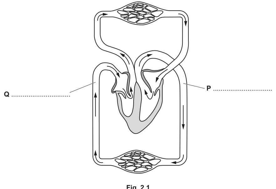
Fig. 2.1 is a diagram of the circulation in a mammal.
Answer
- P = aorta
- Q = vena cava
P = aorta; Q = vena cava
Background Concept
The mammalian heart has four chambers: right atrium, right ventricle, left atrium, left ventricle. The pulmonary artery carries deoxygenated blood from the right ventricle to the lungs, and the pulmonary vein returns oxygenated blood from the lungs to the left atrium. The aorta carries oxygenated blood from the left ventricle to the rest of the body (systemic circulation), and the vena cava returns deoxygenated blood from the body to the right atrium.
When a heart is drawn in a textbook, the right side of the body is conventionally shown on the LEFT of the diagram (as if you are looking at the person facing you). So the right ventricle is on the left of the diagram, and the left ventricle is on the right of the diagram.
Understanding the Question
The question shows a diagram of the double circulation with two labels: P and Q. P is on the right side of the diagram (corresponding to the left side of the body), and Q is on the left side of the diagram (corresponding to the right side of the body). You need to identify each vessel by name.
Approach
Trace the direction of blood flow in the diagram. The vessel on the right of the diagram leaves the heart and goes to the body — this must be the AORTA (left ventricle → body). The vessel on the left of the diagram enters the heart from the body — this must be the VENA CAVA (body → right atrium).
Step-by-Step Reasoning
- The diagram shows the pulmonary circulation (heart ↔ lungs) at the top and the systemic circulation (heart ↔ body) at the bottom/sides.
- P is on the right of the diagram, connected to the systemic circulation, with blood leaving the heart in this direction. This is the AORTA — the largest artery in the body, which branches from the left ventricle to deliver oxygenated blood at high pressure to the systemic circulation.
- Q is on the left of the diagram, connected to the systemic circulation, with blood entering the heart in this direction. This is the VENA CAVA — which returns deoxygenated blood from the body to the right atrium. It can be either the superior vena cava (from above the diaphragm) or the inferior vena cava (from below), but the mark scheme does not require this distinction.
Key Takeaways
- Aorta = left ventricle → body, oxygenated, high pressure.
- Vena cava = body → right atrium, deoxygenated, low pressure.
- Pulmonary artery = right ventricle → lungs, deoxygenated.
- Pulmonary vein = lungs → left atrium, oxygenated.
- Arteries and veins are defined by direction of flow (away from / towards the heart), not by oxygen content.
Common Mistakes
- Confusing the pulmonary artery with the aorta (because of the 'artery = oxygenated' misconception).
- Naming P as the pulmonary artery because the right side of the heart is sometimes (incorrectly) thought of as 'right' of the body.
- Writing 'superior vena cava' or 'inferior vena cava' instead of 'vena cava' — both are accepted, but the mark scheme ignores the distinction.
Things to Be Careful About
- Arteries and veins are defined by direction of flow (away from / towards the heart), not by oxygen content.
- The diagram convention: right of body = left of diagram.
Answer
- P (aorta): transports / delivers oxygenated blood at high pressure from the left ventricle to the organs / body / respiring tissues of the systemic circulation.
- Q (vena cava): returns deoxygenated blood at low pressure from the systemic circulation to the right atrium of the heart.
See working
Background Concept
The function of any blood vessel is determined by where it comes from, where it goes to, what it carries, and the pressure at which it carries it.
- Arteries (including the aorta) have thick, muscular, elastic walls to withstand and maintain high blood pressure. They generally carry blood away from the heart.
- Veins (including the vena cava) have thinner walls and lower pressure; they often have valves to prevent backflow. They generally carry blood back to the heart.
Understanding the Question
The question asks you to describe the function of P (aorta) and Q (vena cava) — i.e. what each vessel does. Two marks are available, one per vessel.
Approach
For each vessel, state: (a) what is being transported (oxygenated/deoxygenated OR at high/low pressure) AND (b) where it comes from and goes to. The mark scheme accepts either the oxygen content OR the pressure, plus the destination, as one complete mark.
Step-by-Step Reasoning
- Aorta (P): The aorta is the largest artery in the body. It leaves the LEFT VENTRICLE and carries OXYGENATED BLOOD at HIGH PRESSURE to the organs and respiring tissues of the systemic circulation. The high pressure is generated by the contraction of the thick left ventricular wall.
- Vena cava (Q): The vena cava is the largest vein in the body. It carries DEOXYGENATED BLOOD at LOW PRESSURE from the organs of the systemic circulation back to the RIGHT ATRIUM of the heart. (There are actually two venae cavae — superior and inferior — but the question is asking about the vessel in general.)
Key Takeaways
- Aorta = oxygenated blood, high pressure, left ventricle → body.
- Vena cava = deoxygenated blood, low pressure, body → right atrium.
- Function follows structure: thick-walled arteries carry blood at high pressure; thin-walled veins carry blood at low pressure.
Common Mistakes
- Saying 'the aorta carries blood to the body' without specifying OXYGENATED or HIGH PRESSURE.
- Confusing the destinations (e.g. saying the aorta goes to the lungs).
- Saying the vena cava carries blood to the LEFT atrium (this is the pulmonary vein).
Things to Be Careful About
- Be specific about which chamber of the heart blood enters or exits.
- Aorta = left ventricle; vena cava = right atrium.
- Either oxygen content or pressure is accepted by the mark scheme — but stating both makes the answer clearer and harder to mis-mark.
Answer
- Closed: blood is contained within blood vessels (arteries, arterioles, capillaries, venules, veins) and does not flow freely in tissue spaces.
- Double: blood passes through the heart twice during one complete circuit of the body — once in the pulmonary circulation (heart → lungs → heart) and once in the systemic circulation (heart → body → heart).
See working
Background Concept
The mammalian circulatory system is described as CLOSED and DOUBLE. These two terms are independent and refer to different features:
- Closed: blood is always contained inside blood vessels and never leaves them to flow freely in body cavities. This is in contrast to an OPEN circulation (found in insects), where blood (haemolymph) flows directly over the tissues in an open body cavity.
- Double: blood passes through the heart twice during one complete circuit of the body. The two passes are the pulmonary circulation (right side of heart → lungs → left side of heart) and the systemic circulation (left side of heart → body → right side of heart). A single circulation (found in fish) has blood passing through the heart only once per circuit.
Understanding the Question
The question asks you to explain WHY mammalian circulation is described as both 'closed' and 'double'. You need to give one mark-worthy point for each term.
Approach
For each adjective, state a defining feature of the mammalian circulation that matches the adjective.
Step-by-Step Reasoning
- Closed: The blood is contained within the network of blood vessels (arteries → arterioles → capillaries → venules → veins). The blood never directly bathes the body tissues; exchange happens only by diffusion across the thin walls of the capillaries. The blood cells and plasma remain inside the vessels at all times.
- Double: In one complete circuit of the body, blood passes through the heart twice. The first pass is the pulmonary circulation (deoxygenated blood from the right ventricle → lungs → oxygenated blood returns to the left atrium), and the second pass is the systemic circulation (oxygenated blood from the left ventricle → body → deoxygenated blood returns to the right atrium). The two circuits operate in series.
Key Takeaways
- Closed = blood stays inside blood vessels.
- Double = blood goes through the heart twice per complete circuit.
- Pulmonary circulation: heart ↔ lungs.
- Systemic circulation: heart ↔ rest of body.
- The two circuits are in series, not in parallel.
Common Mistakes
- Saying 'blood goes through the heart twice' without explaining what this means (e.g. not naming the two circuits).
- Saying 'blood is closed' (which makes no sense).
- Confusing the two circuits (e.g. saying the pulmonary circulation goes to the body).
- Stating only that the heart has 'two sides' without explaining that blood passes through the heart TWICE per circuit.
Things to Be Careful About
- The pulmonary and systemic circulations are in series, not in parallel — blood must go through one before the other.
- The mark scheme accepts the alternative wording 'pulmonary circulation and systemic circulation' as a description of 'double'.
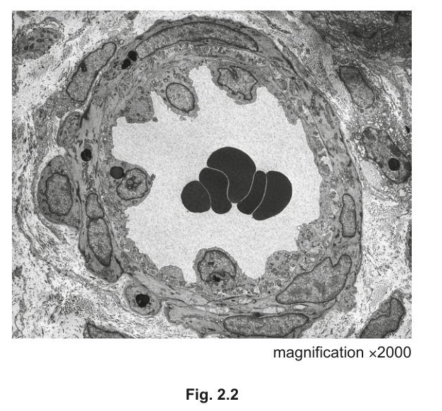
Fig. 2.2 is a transmission electron micrograph of a cross-section of an arteriole. Blood flows from muscular arteries through arterioles into capillary networks.
The lining of the arteriole is folded because the arteriole has constricted. This constriction causes the blood pressure to decrease from in the muscular artery to at the end of the arteriole.
Explain why it is important that the pressure of blood decreases as it passes through arterioles.
Answer
- Capillary walls are only one (endothelial) cell thick, so they cannot withstand high pressure — blood at the high arterial pressure () would burst or damage the capillaries.
- By the time the blood reaches the capillaries the pressure is low (), so blood flows slowly. This allows sufficient time for the exchange / diffusion of substances (e.g. oxygen, carbon dioxide, nutrients, waste products) between the blood and the tissues.
See working
Background Concept
Blood pressure is the force that blood exerts on the walls of the vessels. Pressure is highest in the arteries (especially the aorta) and decreases as blood moves through arterioles, capillaries, venules, and veins. The values given in the question — in the muscular artery and at the end of the arteriole — illustrate this large pressure drop.
The walls of capillaries are very thin (one cell thick, formed of endothelial cells) to allow rapid exchange of substances between the blood and the surrounding tissues by diffusion and through pores.
Understanding the Question
The question gives the pressure values in the muscular artery and at the end of the arteriole, and asks why the decrease in pressure is important. You need two mark-worthy points.
Approach
Think about what would happen if capillaries received blood at the same high pressure as arteries, and what the capillaries need in order to function correctly.
Step-by-Step Reasoning
- Capillary structure cannot withstand high pressure: Capillaries have very thin walls — only ONE (endothelial) cell thick — to allow rapid exchange of substances. If blood at high pressure (e.g. ) were to enter the capillaries, the thin walls would burst or be damaged. By the time the blood reaches the capillaries, the pressure has dropped to , which the thin walls can safely withstand.
- Slow flow for exchange: At low pressure, blood flows slowly through the capillaries. This slow flow gives sufficient time for the exchange / diffusion of substances (oxygen, carbon dioxide, glucose, urea, etc.) between the blood and the tissues. High pressure would push blood through too quickly for efficient exchange.
- (Optional credit) At the venous end of the capillaries, the pressure is low enough to allow reabsorption of tissue fluid back into the blood (by osmosis, due to the plasma proteins remaining in the blood). If pressure were still high, fluid would continue to be forced out and tissue fluid would build up (oedema).
Key Takeaways
- Capillaries are one cell thick — they cannot withstand high pressure.
- Slow blood flow through capillaries is necessary for efficient exchange of substances.
- Arterioles act as a 'pressure buffer' to protect the capillaries.
Common Mistakes
- Saying 'capillaries are thin because they have to be' without linking it to why high pressure would damage them.
- Not mentioning exchange / diffusion.
- Saying capillaries have 'cell walls' (which they do not — this is a common error that the mark scheme explicitly REJECTS).
- Giving only one point when two are needed.
Things to Be Careful About
- Capillaries are composed of ONE layer of endothelial cells, not plant-style 'cell walls'.
- The link between low pressure and slow flow is important — the rate of flow, not just the pressure, is what allows exchange.
- Pressure drop is not just 'to slow the blood' but also 'to protect the thin capillary walls'.
Compare the structure of a muscular artery with the structure of the arteriole shown in Fig. 2.2.
Answer
Similarities (features present in BOTH):
- Both have an endothelium / tunica intima lining the lumen.
- Both have a tunica media containing smooth muscle in the wall.
Differences (muscular artery compared to arteriole):
- The muscular artery has a thicker wall (especially relative to the size of its lumen).
- The muscular artery has a wider lumen.
- The muscular artery has more (layers of) smooth muscle / a thicker tunica media.
- The muscular artery has more elastic fibres / an elastic lamina (which is absent or not visible in the arteriole).
- The muscular artery has more collagen fibres in its wall.
See working
Background Concept
Blood vessels have three main layers:
- Tunica intima (endothelium): the inner layer, made of a single layer of thin squamous endothelial cells, in direct contact with the blood.
- Tunica media: the middle layer, made of smooth muscle and elastic fibres. The amount varies with the function of the vessel.
- Tunica externa (adventitia): the outer layer, made mainly of collagen fibres.
The proportions of these layers vary with function. Muscular arteries have a thick tunica media with lots of smooth muscle to regulate blood flow by vasoconstriction / vasodilation, and lots of elastic fibres to withstand and maintain the high blood pressure generated by the heart. Arterioles are smaller vessels that lead into capillary networks; they have proportionally less smooth muscle and elastic tissue than muscular arteries but still regulate flow into capillary beds.
Understanding the Question
The question shows a TEM of an arteriole in cross-section (Fig. 2.2) and asks you to compare its structure with that of a muscular artery. You need to identify both similarities and differences. Three marks are available — the mark scheme accepts any three from a combined list of similar and different features.
Approach
First, list the structural features common to BOTH vessels. Then list the differences — always making it clear which is which (the mark scheme accepts ORA, or reverse argument, for the arteriole). Do not describe functions — only structures earn marks here.
Step-by-Step Reasoning
Similarities (features in BOTH):
- Both have an endothelium (tunica intima) lining the lumen — a single layer of squamous epithelial cells in direct contact with the blood.
- Both have a tunica media containing smooth muscle in the wall.
Differences (muscular artery vs arteriole):
3. The muscular artery has a thicker wall (in absolute terms, and especially relative to the size of its lumen). The mark scheme explicitly REJECTS vague statements like 'the artery is bigger' or 'thicker' without specifying the structure.
4. The muscular artery has a wider lumen (in absolute terms).
5. The muscular artery has more (layers of) smooth muscle / a thicker tunica media.
6. The muscular artery has more elastic fibres / a clear elastic lamina (which is absent or not visible in the arteriole).
7. The muscular artery has more collagen fibres (mainly in the tunica externa / adventitia).
Key Takeaways
- Both muscular arteries and arterioles have an endothelium and a smooth muscle layer.
- Muscular arteries are larger, with a relatively thicker wall, more smooth muscle, more elastic fibres, and more collagen fibres.
- These structural differences reflect the muscular artery's role in withstanding and regulating the high blood pressure of the systemic circulation.
Common Mistakes
- Comparing 'size' without specifying the relative thickness of wall to lumen.
- Saying the artery is 'bigger' or 'thicker' without specifying in what way (wall vs lumen).
- Describing FUNCTIONS instead of structures (e.g. 'the artery carries blood at high pressure') — these do not earn marks.
- Saying capillaries have 'cell walls' (which they do not — this is a common error that the mark scheme explicitly REJECTS).
- Forgetting the similarities (only listing differences).
Things to Be Careful About
- The mark scheme rejects vague statements like 'arteries are bigger' without specifying the structure (wall thickness, lumen size, smooth muscle content, etc.).
- The ORA (or reverse argument) is allowed for the differences — you can describe the arteriole as 'smaller lumen', 'less smooth muscle', 'fewer elastic fibres', etc.
- Functions do not earn marks here; only structural descriptions do.
- The TEM shows the arteriole in a constricted state (lining is folded), so the wall appears even thicker relative to the lumen than it would in a relaxed arteriole.
A class of students was studying the features of some human pathogens. One of the students constructed a flow chart to identify four different human pathogens. The student used information about the structure and mode of transmission of each of these pathogens.
Fig. 3.1 shows the partially completed flow chart.
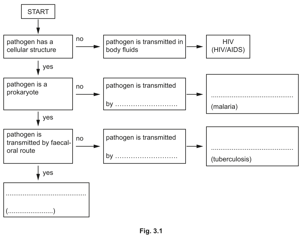
Complete the flow chart in Fig. 3.1 by identifying:
• the modes of transmission
• the scientific names of the pathogens
• the name of one of the diseases.
Answer
- Transmission of malaria: (insect) vector / Anopheles / (a) mosquito
- Scientific name for malaria pathogen: Plasmodium falciparum (or P. ovale / P. malariae / P. vivax)
- Scientific name for TB pathogen: Mycobacterium tuberculosis (or M. bovis)
- Transmission of TB: airborne droplets / droplet infection / aerosol (infection)
- Blank box (pathogen transmitted by faecal–oral route): Vibrio cholerae and cholera
Vector/Anopheles; Plasmodium (spp.); Mycobacterium tuberculosis; airborne droplets; Vibrio cholerae and cholera
Background Concept
Four human pathogens are highlighted in the AS Biology syllabus: HIV (a virus causing HIV/AIDS), Plasmodium species (protoctist causing malaria), Mycobacterium tuberculosis / M. bovis (prokaryote causing tuberculosis) and Vibrio cholerae (prokaryote causing cholera). Each has a characteristic structure and mode of transmission:
- HIV is acellular (it is a virus, made only of an RNA core, capsid and envelope) and is transmitted in body fluids such as blood, semen and vaginal secretions.
- Plasmodium is a eukaryotic protoctist; it is transmitted by the bite of an infected female Anopheles mosquito acting as a vector.
- Mycobacterium tuberculosis is a prokaryote transmitted by airborne droplets/aerosols when an infected person coughs or sneezes.
- Vibrio cholerae is a prokaryote transmitted by the faecal–oral route, usually through contaminated drinking water.
Understanding the Question
The flow chart in Fig. 3.1 starts with a binary choice: does the pathogen have a cellular structure? HIV does not, so it is the 'no' branch. The remaining three pathogens do have a cellular structure, so the chart next asks whether the pathogen is a prokaryote. The malaria pathogen is not a prokaryote (eukaryote), while TB and cholera bacteria are. The chart then asks whether the pathogen is transmitted by the faecal–oral route: cholera is, TB is not (TB is droplet-borne). The five blanks in the diagram therefore need filling with: (i) malaria's mode of transmission, (ii) malaria's scientific name, (iii) TB's mode of transmission, (iv) TB's scientific name, and (v) the pathogen and disease transmitted by the faecal–oral route.
Approach
Use your knowledge of the scientific (binomial) name (genus and species) and the mode of transmission of each pathogen. Remember that scientific names are written in italics with a capitalised genus and lower-case species. Match each pathogen to the branch of the flow chart on which it sits.
Step-by-Step Reasoning
- The blank at the top of the flow chart labelled 'transmitted by … (malaria)' sits on the branch of the chart for a cellular, non-prokaryote pathogen. Malaria is spread by the bite of an infected mosquito: credit the vector / Anopheles / mosquito.
- The same disease box asks for the pathogen's scientific name: Plasmodium (one of falciparum, ovale, malariae or vivax is acceptable).
- The 'transmitted by … (tuberculosis)' blank sits on the prokaryote / not faecal-oral branch. M. tuberculosis spreads by airborne droplets (coughs/sneezes).
- The TB disease box asks for the pathogen's scientific name: Mycobacterium tuberculosis (or M. bovis for cattle-borne disease).
- The final blank sits on the prokaryote / yes faecal-oral branch. The pathogen and disease are Vibrio cholerae and cholera.
Key Takeaways
- A pathogen's structure (acellular virus, prokaryote, eukaryote) and mode of transmission are the two key features used in a dichotomous identification key.
- Scientific names are binomial (Genus species), italicised, with a capital genus.
- Recognise the four core AS pathogens: HIV, Plasmodium, Mycobacterium and Vibrio cholerae.
Common Mistakes
- Writing the malaria pathogen as just Plasmodium malaria (this is the disease, not the species) or omitting the italics.
- Writing the TB mode of transmission as 'air' or 'water droplets' — the mark scheme rejects unqualified 'air droplets' and 'water droplets from coughing/sneezing'.
- Naming cholera incorrectly as a virus or as Cholera vibrio (the genus/species order is reversed).
- Confusing the order in the flow chart: malaria is on the non-prokaryote branch, TB is on the prokaryote but not faecal-oral branch.
Things to Be Careful About
- Use the scientific name (Vibrio cholerae), not the common name (comma bacillus).
- 'Droplet infection', 'aerosol infection' and 'airborne droplets' are all accepted — but not 'air droplets' on its own.
- Mycobacterium bovis is acceptable for TB but, if used, an alternative transmission route (e.g. unpasteurised milk, contaminated meat) must be given.
HIV has a nucleic acid core of RNA. The virus also contains the enzyme reverse transcriptase.
After HIV enters T-lymphocytes, reverse transcriptase catalyses the formation of DNA using activated DNA nucleotides with the viral RNA as a template.
Some drugs, such as tenofovir, have been developed to inhibit the action of reverse transcriptase.
The structure of tenofovir is similar to the structure of deoxyribose adenosine monophosphate, as shown in Fig. 3.2.
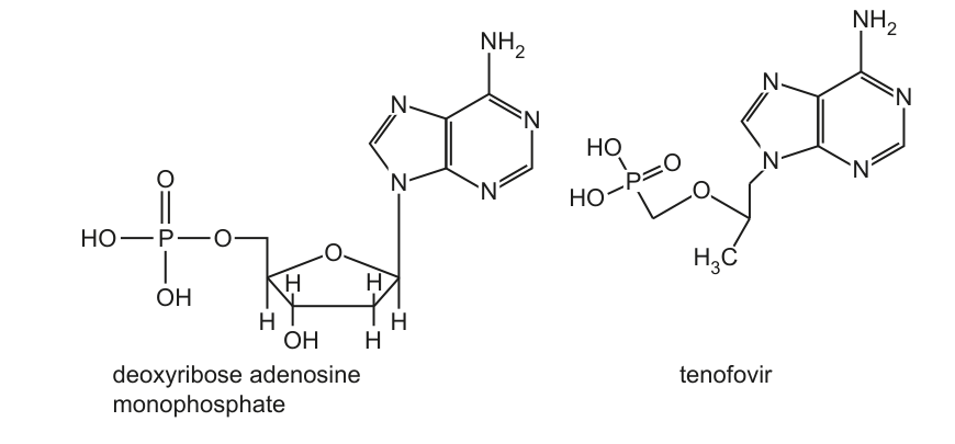
After tenofovir is absorbed into cells it is phosphorylated twice and can be used by reverse transcriptase in the synthesis of DNA.
When a tenofovir molecule is added to the DNA strand being synthesised, the process stops.
Suggest the mechanism of action of tenofovir to prevent the synthesis of DNA by reverse transcriptase. Use the information in Fig. 3.2 in your answer.
Answer
Any two of:
-
Tenofovir competes with (activated/phosphorylated) adenine nucleotide / dATP for the active site of reverse transcriptase (it acts as a competitive inhibitor). (1 mark)
-
Tenofovir is incorporated into the growing DNA strand by forming a phosphodiester bond with the previous nucleotide, but no further nucleotides can be added because tenofovir lacks a 3´-OH group / lacks the (deoxy)ribose ring needed to form the next phosphodiester bond, so DNA synthesis stops. (1 mark)
Tenofovir competes with dATP for the active site of reverse transcriptase and, once incorporated, lacks a 3´-OH group so the next phosphodiester bond cannot form, terminating DNA synthesis.
Background Concept
HIV is a retrovirus: its genetic material is single-stranded RNA. Once inside a host T-lymphocyte, the viral enzyme reverse transcriptase synthesises a complementary DNA (cDNA) strand using the viral RNA as a template, requiring the four DNA nucleotides (dATP, dTTP, dGTP, dCTP). Each new nucleotide is added to the 3´-OH of the previous sugar, forming a phosphodiester bond. Drugs that interfere with this process are nucleoside/nucleotide reverse-transcriptase inhibitors (NRTIs).
Understanding the Question
Fig. 3.2 compares the structure of deoxyribose adenosine monophosphate (dAMP) with tenofovir. They share an adenine base and a phosphate-containing group, but differ in the sugar component: dAMP has a deoxyribose ring (with a 3´-OH) whereas tenofovir has an acyclic sugar-like chain (no 3´-OH, no pentose ring) and a phosphonate group rather than a phosphate. The question asks for the mechanism of action — i.e. how tenofovir prevents DNA synthesis.
Approach
A complete mechanism has two parts: (i) how tenofovir binds to the enzyme, and (ii) why, once bound, it terminates the growing DNA strand. Use Fig. 3.2 to justify both ideas structurally.
Step-by-Step Reasoning
- Because tenofovir's adenine and overall shape resemble dAMP, it is recognised as a substrate analogue and binds to the active site of reverse transcriptase in place of dATP. This is competitive inhibition at the active site of reverse transcriptase.
- After phosphorylation inside the cell, tenofovir is added to the growing DNA strand by forming a phosphodiester bond (the OH on the previous nucleotide attacks the phosphorus of tenofovir).
- Looking at Fig. 3.2, the sugar ring of dAMP provides a 3´-OH that is needed to attack the next incoming nucleotide's phosphate. Tenofovir's acyclic chain has no 3´-OH and no (deoxy)ribose, so no further phosphodiester bond can form and the DNA chain is terminated — chain elongation stops.
Key Takeaways
- Many antiviral drugs are nucleotide analogues: they mimic natural nucleotides to be incorporated by viral polymerases.
- Termination occurs because the analogue lacks the 3´-OH needed for the next phosphodiester bond.
- This is a chain-terminating mechanism distinct from non-nucleoside reverse-transcriptase inhibitors (NNRTIs) that bind an allosteric site.
Common Mistakes
- Saying tenofovir 'changes the shape of the active site' (this is non-competitive inhibition language, not warranted here).
- Vague statements such as 'it stops DNA being made' without identifying the structural reason (no 3´-OH / no deoxyribose).
- Rejecting 'sugar' on its own — the mark scheme requires '(deoxy)ribose' or 'pentose'.
Things to Be Careful About
- The mark scheme rewards any two of three points: (a) competition for the active site, (b) phosphodiester bond formed but no further reaction, (c) absence of 3´-OH/(deoxy)ribose.
- Use the precise term phosphodiester bond (not 'bond' or 'phosphate bond').
- Note the prefix 'deoxy' is important; do not just write 'ribose'.
Pre-exposure prophylaxis (PrEP) is the use of therapeutic drugs to prevent the replication of HIV in the body following infection. The drugs are taken by people who are at risk of becoming infected. Tenofovir is one of these therapeutic drugs.
In 2016, the United Nations (UN) set a global target of 3 million PrEP users by 2020.
Table 3.1 shows the number of people across the world who received a therapeutic drug for PrEP in each of the years between 2012 and 2019.
Table 3.1
| year | number of people who received PrEP |
|---|---|
| 2012 | 10000 |
| 2013 | 15000 |
| 2014 | 27500 |
| 2015 | 57500 |
| 2016 | 95000 |
| 2017 | 145000 |
| 2018 | 340000 |
| 2019 | 605000 |
Calculate the percentage of people who received PrEP in 2019 as a percentage of the target set by the UN in 2016.
Give your answer to the nearest whole number.
...................................................... %
Working
Answer
20 %
20 %
Background Concept
A percentage expresses one quantity as a proportion of 100 of another. Here we are asked what fraction of the UN's 2020 target (3 million PrEP users) was actually reached by 2019 (605 000 users).
Understanding the Question
The data are given in Table 3.1. The 2019 value is 605 000 and the UN target is 3 000 000. We must give the answer to the nearest whole number — the mark scheme specifically rejects answers given to decimal places (e.g. 20.2 %).
Approach
Use the standard percentage formula:
Step-by-Step Reasoning
- Read the 2019 figure: 605 000.
- Identify the UN 2020 target: 3 000 000.
- Substitute: .
- Round to the nearest whole number: 20 %.
Key Takeaways
- Percentage calculations require the part to be divided by the whole, then multiplied by 100.
- Always check whether the question requires rounding and to what precision.
- Be alert to data-table questions where the target value is in the question text rather than in the table.
Common Mistakes
- Giving 20.2 % (or similar) — the mark scheme rejects decimal-place answers.
- Using 2018's value (340 000) by mistake.
- Dividing 3 000 000 by 605 000 and quoting 496 % — the wrong way round.
Things to Be Careful About
- The question says 'percentage of people who received PrEP in 2019 as a percentage of the target' — so 2019 is the part and 3 000 000 is the whole.
- Round to the nearest whole number, not to one decimal place.
PrEP does not prevent transmission of HIV.
State and explain how health authorities can reduce the transmission of HIV.
Answer
Any four from the following paired points (a statement and its explanation):
- Supply condoms / femidoms / dental dams for protection during sex. They form a barrier to transmission of HIV during sexual intercourse.
- Provide needle-exchange schemes for intravenous drug users. This decreases the risk of sharing contaminated equipment.
- Use new / sterilised needles and syringes for medical procedures. This decreases the risk of transmission from contaminated blood.
- Test pregnant women for HIV and provide powdered milk to HIV-positive women. This prevents HIV-positive women passing HIV to the baby in breast milk.
- Prevent HIV-positive people from donating blood / screen donated blood / heat-treat donated blood. This prevents recipients receiving HIV-infected blood during transfusions or surgery.
- Carry out contact tracing. This locates people who may be undiagnosed / HIV-positive so that they can be offered a test.
- Provide named antiretroviral drugs to people living with HIV (e.g. tenofovir, to pregnant women with HIV). This prevents HIV spreading through the body / infecting more T-lymphocytes (lowering viral load reduces onward transmission).
- Provide education / information about HIV transmission and treatments. This raises awareness of ways to reduce infection.
Multiple paired measures: supply condoms (barrier during sex); needle-exchange schemes (less sharing of equipment); screen blood / prevent HIV+ donors (less transfusion transmission); test pregnant women and provide powdered milk (less MTCT); contact tracing (find undiagnosed cases); provide antiretrovirals (lower viral load).
Background Concept
HIV is transmitted via exchange of certain body fluids — blood, semen, vaginal secretions and breast milk. The main routes are unprotected sexual intercourse, contaminated needles (intravenous drug use, unsafe medical injections), contaminated blood products, and mother-to-child transmission (in utero, at birth, or via breast milk). Public-health measures that interrupt any of these routes reduce transmission. The question stresses that PrEP alone does not prevent transmission, so additional behavioural and medical interventions are needed.
Understanding the Question
The question asks candidates to state measures that health authorities can use and explain how each reduces HIV transmission. The mark scheme pairs each measure with its explanation and awards one mark per statement and one mark per explanation. Crucially, the mark scheme caps marks at 3 unless the response includes at least one statement and at least one explanation — so a list of bare statements, or a list of bare explanations, will earn a maximum of 3 marks even if more than four points are made.
Approach
Think of HIV transmission routes and pick a measure for each:
- Sexual route → condoms / femidoms / dental dams.
- Needle route → needle exchange, sterile injecting equipment.
- Blood-transfusion route → screening donated blood, excluding HIV-positive donors, heat-treating blood products.
- Mother-to-child route → test pregnant women, give antiretrovirals, provide powdered milk.
- Case-finding → contact tracing and HIV testing so that infected people can be diagnosed early.
- Reducing infectiousness → supply antiretrovirals (e.g. tenofovir) to HIV-positive people, lowering viral load so they are less likely to transmit.
- Behavioural → public-health education about safer sex and transmission routes.
For each, give both the action (statement) and the reason (explanation).
Step-by-Step Reasoning
- Condoms are physical barriers that stop the exchange of semen, vaginal secretions and blood during intercourse, so they directly block the sexual route.
- Needle-exchange schemes give injecting drug users sterile equipment, so contaminated needles are not shared, removing a key blood-borne route.
- Screening donated blood (and excluding HIV-positive donors) prevents any HIV present in donated blood from reaching a recipient during surgery or transfusion.
- Testing pregnant women identifies those who are HIV-positive so they can be given antiretrovirals during pregnancy and powdered milk instead of breast milk, preventing mother-to-child transmission.
- Contact tracing finds the sexual and needle-sharing contacts of newly diagnosed individuals so they too can be tested, breaking chains of silent transmission.
- Supplying antiretrovirals to HIV-positive people suppresses viral replication, lowering viral load so the patient is less infectious to others (treatment-as-prevention).
- Public education raises awareness of how HIV is spread and how it can be avoided, leading to behaviour change such as condom use or fewer sexual partners.
Key Takeaways
- HIV control depends on interrupting every transmission route, not just one.
- Public-health responses combine biomedical (condoms, antiretrovirals, blood screening) and behavioural/educational (information, contact tracing) measures.
- PrEP is for uninfected at-risk individuals, not a transmission-blocker on its own — so it must be combined with other measures.
- 'Statement + explanation' is a recurring Cambridge command structure; marks are often lost by giving only one half of the pair.
Common Mistakes
- Writing only statements and no explanations (capped at 3 marks even if many statements are made).
- Vague phrases like 'use clean needles' — the mark scheme rejects 'clean needles' and rewards 'new / sterilised needles' or 'needle-exchange schemes'.
- Mentioning 'a vaccine' — no effective HIV vaccine exists.
- Saying 'abstain from sex' as a measure without explaining how authorities could implement it.
- Listing individual behaviour changes (which a person can do) rather than what health authorities can do.
Things to Be Careful About
- Match each statement to a specific explanation of how it cuts transmission; partial credit is given for the explanation even if the statement is weak, but you need at least one of each to access the fourth mark.
- Use precise terms: 'condoms' (not 'protection'), 'needle-exchange schemes' (not 'clean needles'), 'antiretrovirals' (not 'drugs' alone).
- The mark scheme accepts 'screen donated blood' or 'heat-treat donated blood' as alternatives to 'prevent HIV-positive people from being donors' — pick whichever is most precise for the explanation given.
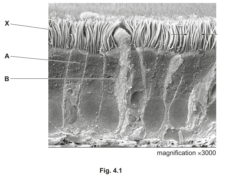
Fig. 4.1 is a scanning electron micrograph showing the tissue that lines the bronchi in the gas exchange system.
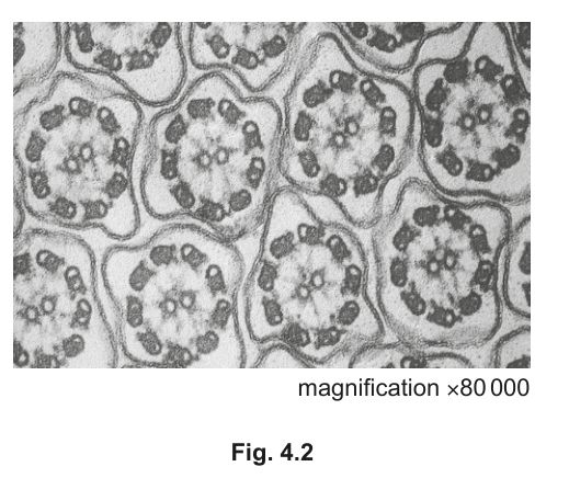
Fig. 4.2 is a transmission electron micrograph of a horizontal section made at the position indicated by the two arrows in Fig. 4.1.
Name the cells labelled A and B in Fig. 4.1.
A ........................................................................................................................................
B ........................................................................................................................................
Answer
A – Ciliated epithelial cell
B – Goblet cell
A: ciliated epithelial cell; B: goblet cell
Background Concept
The gas exchange system (trachea, bronchi and larger bronchioles) is lined by a pseudostratified ciliated columnar epithelium. Within this epithelium sit two principal cell types that work together as the mucociliary escalator. Ciliated epithelial cells bear many motile cilia on their apical (free) surface. Goblet cells are mucus-secreting cells interspersed among the ciliated cells; their apical cytoplasm is packed with mucin-containing secretory vesicles that bulge the membrane outwards, giving them a characteristic 'goblet' or wine-glass shape.
Understanding the Question
This part is a straight identification. Fig. 4.1 is a scanning electron micrograph (SEM) of the lining of a bronchus, magnified ×3000. The surface bristles are cilia (label X). Label A points to a tall cell whose apex bears these cilia; label B points to a shorter, rounded cell that bulges above its neighbours and has no cilia on its surface. You must name each cell type using the correct biological term.
Approach
Use the visible morphology of each cell together with your knowledge of which two cell types populate the respiratory epithelium.
Step-by-Step Reasoning
- Cell A is column-shaped and its apical surface carries many of the hair-like cilia. This is the classic appearance of a ciliated epithelial cell of the respiratory lining.
- Cell B has a rounded, bulging apical surface and no cilia visible on top. This is typical of a goblet cell, whose apical cytoplasm is swollen with mucin vesicles ready for secretion.
- The mark scheme requires the exact terms 'ciliated epithelial cell' and 'goblet cell' (singular cell, not the tissue). 'Ciliated epithelium' is rejected because the question asks for a cell, not a tissue.
Key Takeaways
The bronchial epithelium is dominated by ciliated cells and goblet cells; you should know both by name and by their appearance in an SEM.
Common Mistakes
- Writing 'ciliated epithelium' for A — the mark scheme rejects this because it names a tissue, not a cell.
- Calling B a 'mucus cell', 'gland cell' or 'secretory cell' — the specific term 'goblet cell' is required.
Things to Be Careful About
Use the singular 'cell'. Note that goblet cells in the SEM look smooth and rounded because their cilia-free apex is full of secretory vesicles, in contrast to the cilia-covered ciliated cells.
Describe how the tissue shown in Fig. 4.1 is adapted to its function in the gas exchange system.
Answer
- Goblet cells (B) secrete / release mucus (mucin) onto the epithelial surface.
- The mucus covers the ciliated epithelium and traps (named) particles such as dust, bacteria or smoke particles / acts as a barrier that prevents (named) pathogens reaching the epithelium or the alveoli (gas exchange surface).
- The cilia (on the ciliated cells) beat to move the mucus, with the trapped material, upwards towards the mouth / throat / pharynx and away from the alveoli / lungs / gas exchange surface.
Goblet cells secrete mucus that traps particles/pathogens; cilia move the mucus (and trapped material) upwards away from the lungs.
Background Concept
The mucociliary escalator is the principal defence of the conducting airways. Goblet cells secrete mucin glycoproteins which, mixed with water, salts and antibacterial proteins, form a sticky layer of mucus over the epithelium. This mucus traps inhaled particles (dust, smoke, microorganisms, pollen). Ciliated cells then beat their cilia in a coordinated metachronal rhythm so that the mucus sheet, with everything trapped in it, is propelled up the airways towards the pharynx and swallowed.
Understanding the Question
The question asks for a description of how the tissue seen in Fig. 4.1 is adapted to its function. The tissue is the ciliated pseudostratified columnar epithelium of the bronchi, and its function in the gas exchange system is to keep the airways clean by trapping and removing inhaled debris. Three marks require three linked points, covering both cell types and the way they cooperate.
Approach
Structure your answer in three clearly separated points: (1) what goblet cells do, (2) what the mucus does, and (3) what the cilia do. This order mirrors the sequence of events — secretion, entrapment, removal.
Step-by-Step Reasoning
- Goblet cells are full of mucin-containing secretory vesicles. They release their contents by exocytosis onto the apical surface of the epithelium. The mucin swells with water to form the mucus layer that sits on top of the cilia.
- The mucus is sticky and covers the ciliated epithelium. Inhaled particles (dust, microbes, smoke particles) stick to it, so the mucus acts as a physical barrier preventing pathogens and debris from reaching the delicate gas exchange surface of the alveoli.
- The cilia on the ciliated epithelial cells beat in a coordinated, wave-like pattern (power stroke followed by recovery stroke). The tips of the cilia grip the mucus and propel it, with its trapped contents, upwards along the airway towards the pharynx, where it is swallowed and destroyed in the stomach. The direction must be specified (upwards / towards the mouth / away from the alveoli / away from the lungs / away from the gas exchange surface).
Key Takeaways
The mucociliary escalator is a clear structure–function example: goblet cells provide the trap, cilia move the trap. Together they keep the gas exchange surface clean.
Common Mistakes
- 'Cilia filter the air' — cilia do not filter; they move the mucus. The mucus itself does the filtering.
- 'Mucus moves out of the lungs' unqualified — this does not specify the direction of ciliary beating; the mark scheme requires 'towards the mouth/throat/pharynx' or 'away from the alveoli/lungs/gas exchange surface'.
- 'To be swallowed' on its own — not credited without the direction of ciliary beating.
- Failing to mention the role of goblet cells at all — at least one mark is tied to the mucus-secreting function.
Things to Be Careful About
Always give the direction of mucus movement. The mark scheme explicitly rejects vague phrasings such as 'out of the lungs', 'out of the gas exchange system' or 'out of the respiratory system' as the destination for ciliary beating. Equally, do not give the function of mucus to cilia or the function of cilia to mucus.
The structures labelled X in Fig. 4.1 have a characteristic internal appearance, as seen in Fig. 4.2.
Describe the internal appearance of the structures labelled X.
Answer
- The cilia show a 9 + 2 pattern / arrangement / structure.
- They are composed of microtubules: the outer 9 are pairs (doublets) of microtubules and the central 2 are single microtubules.
- AVP, e.g. dynein 'arms' on the outer doublets / the structure is enclosed by the cell surface membrane.
9+2 arrangement of microtubules (9 outer doublets, 2 central singlets).
Background Concept
A motile cilium has a highly conserved internal ultrastructure — the axoneme — that is only resolvable by electron microscopy. In transverse section the axoneme shows nine outer doublets of microtubules arranged in a ring around two central single microtubules. This is the '9 + 2' pattern. Each outer doublet bears dynein arms, which hydrolyse ATP and slide adjacent doublets past one another, producing the bending motion that drives ciliary beating. Radial spokes, nexin links and a central sheath hold the structure together; the whole axoneme is enclosed by an extension of the plasma membrane.
Understanding the Question
Fig. 4.2 is a TEM (×80 000) of a horizontal section cut at the position of the arrows in Fig. 4.1 — that is, a cross-section through the cilia (X) on top of the ciliated cells. Two marks are available; you must describe the internal appearance of one cilium as seen in this TEM.
Approach
Look at any one cilium cross-section in Fig. 4.2. Count the dark structures around the outside and in the middle; identify what they are made of; and note any extra detail that is visible (the dynein arms or the surrounding membrane).
Step-by-Step Reasoning
- Each cilium shows a ring of nine paired dark structures around its edge with two lighter single structures in the middle. This is the canonical 9 + 2 microtubule pattern of a motile cilium.
- All of these structures are microtubules — hollow cylinders made of tubulin protein. The '9' are doublets (two fused microtubules), the '2' are single microtubules.
- On closer inspection, the outer doublets carry short dynein 'arm' projections, which are the motor proteins responsible for ciliary beating. The whole axoneme is bounded by a thin line — the plasma membrane (see part (b)(ii)). Any one of these additional correct observations earns the AVP mark.
Key Takeaways
The motile cilium has a 9 + 2 microtubule axoneme with dynein arms, all wrapped in an extension of the plasma membrane.
Common Mistakes
- Writing only 'microtubules' without the 9 + 2 pattern — the 9 + 2 is the specific feature worth a mark.
- Writing '9 + 2 arrangement of cilia' instead of '9 + 2 arrangement of microtubules' — the doublets themselves are microtubules.
- Confusing this with the unrelated structure of a bacterial flagellum.
Things to Be Careful About
You are describing a transverse (cross) section through one cilium, not its length. Refer to microtubules, not 'fibres' or 'filaments'.
Answer
Each cilium (structure X) is surrounded by the cell (surface) membrane, so its contents lie inside the cell / cytoplasm.
Each cilium is surrounded by the cell surface membrane, so its contents lie within the cell.
Background Concept
A cilium grows outwards from the cell as an extension of the apical plasma membrane. The plasma membrane is continuous with, and an extension of, the cell's surface membrane, so anything inside that membrane is by definition inside the cell — i.e. intracellular. (Contrast: a secreted mucus droplet lying in the airway lumen would be outside this membrane, and therefore extracellular.)
Understanding the Question
Part (b)(ii) asks you to use Fig. 4.2 as visual evidence that the cilia (X) are intracellular. Only one mark is available, and the mark scheme is specific: you must refer to the cell (surface) membrane that you can see wrapping each cilium in the TEM.
Approach
Look at Fig. 4.2 again. Notice that every 'ring' of microtubules has a thin dark line around it. Identify that line as the cell's plasma membrane and use it as evidence that the cilium lies inside that membrane.
Step-by-Step Reasoning
- In the TEM, each cilium cross-section is enclosed by a thin dark boundary. That boundary is the cell's plasma membrane, which extends up and around the cilium as the cilium projects from the cell surface.
- Because the microtubules of the axoneme lie inside this membrane, they are inside the cell — or, more precisely, inside an extension of the cell.
- Therefore the cilium is an intracellular structure: a surface projection of the cell, not a free-floating extracellular filament.
Key Takeaways
A cilium is intracellular because the plasma membrane wraps around it; the TEM makes this directly visible.
Common Mistakes
- Saying 'the microtubules are inside the cell' without mentioning the membrane — the membrane observation is the specific evidence the mark scheme requires.
- Writing 'inside the cell body' — true but imprecise; the mark scheme wants the membrane observation.
- Confusing 'intracellular' with 'within the nucleus' — a cilium is intracellular but lies in the cytoplasm, not inside the nucleus.
Things to Be Careful About
'Intracellular' means inside the cell (i.e. enclosed by the plasma membrane). Cilia project above the apical surface but remain wrapped in plasma membrane, so they remain part of the cell.
Stem cells are found in the lining of the bronchi.
Describe the function of centrioles and explain how they are involved in the cell cycle of a stem cell.
Answer
- Centrioles organise microtubules (they are microtubule-organising centres, MTOCs).
- They form the spindle / spindle fibres that separate the chromosomes during mitosis.
- Before mitosis, each centriole replicates / duplicates to form two centrioles (a pair).
- This replication occurs during S phase / G2 phase of the cell cycle.
- The two centriole pairs then move to opposite poles of the cell during prophase.
- Centrioles lengthen / shorten the spindle fibres (microtubules) to separate the sister chromatids.
Centrioles organise microtubules to form the spindle; they replicate in S/G2 phase, move to opposite poles in prophase, and lengthen/shorten spindle fibres to separate chromatids.
Background Concept
Centrioles are small, barrel-shaped organelles built from microtubules. They usually exist in pairs at right angles to one another, forming a centrosome — the main microtubule-organising centre (MTOC) of an animal cell. Their job is to nucleate and anchor microtubules, and during mitosis they organise the spindle that separates sister chromatids. Because the bronchial epithelium is constantly shed and renewed, the stem cells in its lining are actively cycling, and the centriole cycle runs continuously in them.
Understanding the Question
This is a 4-mark 'describe and explain' question with two demands: (1) describe the function of centrioles, and (2) explain how they participate in the cell cycle of a stem cell. The mark scheme supplies six creditable points; only four are needed for full marks, but at least one of the function points (mp1 or mp2) must appear, otherwise the answer is capped at 3 marks.
Approach
Plan a logical sequence that moves from function (microtubule organisation, spindle formation) through the cell-cycle timing (replication in S/G2, separation in prophase) to the mechanical action during mitosis (lengthening and shortening spindle fibres). This order matches the chronological order of events and lets each statement build on the previous one.
Step-by-Step Reasoning
- Function: Centrioles are MTOCs. They nucleate microtubules by anchoring their minus ends, allowing the microtubules to grow outwards as plus ends. Without this organising role, microtubules could not be assembled into a coherent spindle.
- Function → spindle: The organised microtubules form the mitotic spindle, the apparatus whose kinetochore microtubules attach to chromosomes and pull sister chromatids apart.
- Cell-cycle timing, S/G2: Before each mitosis, the centriole must be duplicated so that each daughter cell inherits a centrosome. Replication begins during S phase (alongside DNA replication) and is complete by G2 phase. By the start of mitosis, the cell contains two centrosomes — one will become each pole's spindle organiser.
- Cell-cycle timing, prophase: As mitosis begins, the two centrosomes migrate to opposite ends of the cell, establishing the two spindle poles. (The mark scheme explicitly rejects 'poles of the nucleus' — the centrosomes move to the poles of the cell, not to the nuclear envelope.)
- Mechanical action: Once the spindle is set up, microtubule dynamics lengthen and shorten spindle fibres. During anaphase, kinetochore microtubules shorten (depolymerise) at their poles to pull sister chromatids apart, while polar microtubules lengthen to push the poles further apart. The centrioles/centrosomes anchor and direct this dynamic instability. (The mark scheme rejects 'contract'; centrioles lengthen/shorten microtubules — they do not contract them.)
Key Takeaways
Centrioles duplicate once per cell cycle in S/G2 phase, separate to opposite poles during prophase, and then organise the spindle fibres whose dynamic length changes separate the chromatids. In stem cells of the bronchial lining, this cycle runs continuously to replace shed epithelial cells.
Common Mistakes
- 'Centrioles contract the spindle fibres' — rejected; centrioles lengthen/shorten microtubules, they do not contract them.
- 'Centrioles move to the poles of the nucleus' — rejected; the cell, not the nucleus, has spindle poles.
- 'Centrioles replicate during mitosis' — they replicate before mitosis (in S/G2), not during.
- 'Centrioles produce the chromosomes' — they have nothing to do with chromosome formation; they organise the spindle that separates already-replicated chromosomes.
- Giving only the function (microtubule organisation) and omitting the cell-cycle stages — the question explicitly asks for both, and you are capped at 3 marks without a function point.
Things to Be Careful About
A cap of 3 marks applies if neither mp1 nor mp2 is earned — so always include at least one function point (centrioles organise microtubules / form the spindle). Use precise phase names: replication in 'S phase or G2 phase', movement to poles in 'prophase'. Use 'centrosome' if you mean the pair of centrioles, and 'centriole' if you mean a single barrel. Distinguish between the duplication event (S/G2) and the separation event (prophase).
The pressure of water vapour inside and outside leaves can be measured. The difference between these pressures is known as the leaf vapour pressure deficit (LVPD).
LVPD is one of the factors that influences the rate of transpiration.
Scientists measured the effect of changing the LVPD on the rate of transpiration in several species of flowering plant that live in a variety of different habitats. Two of these species were:
• Nerium oleander, a species that is adapted to grow in hot, dry conditions
• Helianthus annuus, a species that is not adapted for survival in hot, dry conditions.
Fig. 5.1 shows the effect of increasing the LVPD on the transpiration rates of the two species.
All other factors were kept constant.
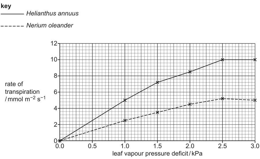
Answer
- At an LVPD of both species have a transpiration rate of .
- Between and , the transpiration rate of both species increases; H. annuus has a steeper gradient / higher rate of increase than N. oleander.
- H. annuus has a higher transpiration rate than N. oleander at every LVPD, e.g. at H. annuus is while N. oleander is .
- Above , the rate for H. annuus remains constant at , whereas the rate for N. oleander decreases slightly to at .
Both species show no transpiration at LVPD = 0 kPa and rise until 2.5 kPa; H. annuus always has the higher rate and steeper gradient, plateaus at 10 mmol m⁻² s⁻¹ above 2.5 kPa, while N. oleander declines slightly to ≈5 mmol m⁻² s⁻¹ at 3.0 kPa.
Background Concept
Transpiration is the loss of water vapour from a plant, mainly through open stomata. The rate depends on a vapour-pressure (or water-potential) gradient between the inside of the leaf and the surrounding air, on temperature, on wind, and on the resistance offered by the leaf surface. The leaf vapour pressure deficit (LVPD) is the difference between the saturation vapour pressure inside the leaf and the actual vapour pressure in the outside air; the larger this gradient, the faster water diffuses out of the leaf.
Plants from dry, hot habitats (xerophytes), such as Nerium oleander, possess structural adaptations that reduce transpiration: a thick waxy cuticle, multiple epidermal layers (a hypodermis), stomata sunken in pits/crypts and surrounded by hairs. These features raise the resistance to water-vapour diffusion and so reduce the rate of water loss compared with mesophytes such as Helianthus annuus (sunflower).
Understanding the Question
You are given a graph of transpiration rate against LVPD for two species under identical conditions, and you must compare the two curves. The mark scheme awards marks for: (i) a shared trend, (ii) the difference in rate at any LVPD, (iii) the difference in gradient, and (iv) what happens between and . It also requires a comparative data quote that uses units from both axes and includes figures from both species.
Approach
Read the two curves carefully. Identify the points they share (origin, rising trend, the LVPD where they stop rising). Note where they differ (gradient up to , absolute rate at any chosen LVPD, behaviour above ). Then pick a clear, easy-to-read pair of x and y values to quote with units from each species — e.g. at LVPD .
Step-by-Step Reasoning
- Origin. Both lines pass through : when there is no vapour-pressure difference between leaf and air there is no driving force for evaporation, so no transpiration. (Mark point 1.)
- Shared rising phase. Between and both curves rise: a larger LVPD steepens the diffusion gradient and water vapour escapes faster. (Mark point 2.)
- Different absolute rates. At any LVPD , H. annuus lies above N. oleander. For example at , H. annuus and N. oleander . (Mark points 4 and 6.)
- Different gradients. H. annuus rises more steeply than N. oleander over – — its rate of increase is higher. (Mark point 5.)
- Behaviour above . H. annuus's rate plateaus at ; N. oleander's falls slightly to about at . The plateau for H. annuus is set by stomatal control; the decline for N. oleander suggests stronger stomatal closure as the leaf protects itself from desiccation. (Mark point 3.)
Key Takeaways
- Xerophytes lose water more slowly than mesophytes at any given LVPD.
- Comparing curves means quoting values with units, not just describing shapes.
- Above a critical LVPD, both species regulate water loss by stomatal closure (the rate stops rising).
Common Mistakes
- Giving only one species' values — the mark scheme demands figures from both axes and both species in any data quote.
- Saying "the rate is higher" without quoting numbers or an LVPD.
- Forgetting units ( and ).
- Implying that N. oleander's rate keeps rising — it actually dips slightly at the highest LVPD.
Things to Be Careful About
- "Higher rate of transpiration" alone scores no mark unless supported by a comparative data quote.
- Avoid "steeper" as a standalone point: state what steeper means (greater rate of increase / greater gradient).
- A descriptive phrase such as "H. annuus transpires more" is insufficient — pair it with units and numbers.
Fig. 5.2 shows part of a plant of N. oleander.
Fig. 5.3 shows a cross-section of part of an oleander leaf.
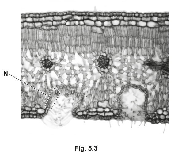
Fig. 5.4 is a drawing of a high-power view of region N on Fig. 5.3.
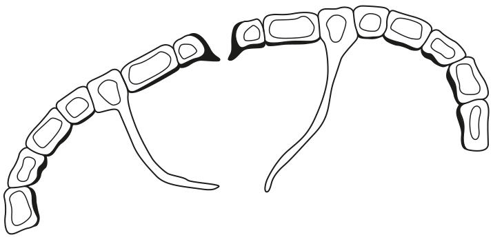
State and explain two adaptations shown by the leaves of N. oleander that are visible in Fig. 5.3 and Fig. 5.4.
one adaptation visible in Fig. 5.3 ..............................................................................................
explanation ...............................................................................................................................
one adaptation visible in Fig. 5.4 ..............................................................................................
explanation ...............................................................................................................................
Answer
Adaptation visible in Fig. 5.3 — Stomata are located in pits / depressions / crypts (sunken stomata) on the lower surface of the leaf.
Explanation — The pit traps a layer of still, humid air around the stomatal pore. This reduces the diffusion / water-potential gradient between the inside of the leaf and the air outside, so the rate of transpiration (diffusion of water vapour) is reduced.
Adaptation visible in Fig. 5.4 — Epidermal hairs / trichomes fill the stomatal crypt and surround the stomata.
Explanation — The hairs trap a layer of still, moist air in the crypt and reduce air movement (wind) across the stomatal pore. This creates a humid atmosphere and lowers the diffusion gradient for water vapour, decreasing the rate of transpiration.
Fig. 5.3: stomata in crypts trap humid air → reduces diffusion gradient. Fig. 5.4: epidermal hairs/trichomes trap still moist air → reduces transpiration.
Background Concept
A xerophyte is a plant adapted to dry conditions. To survive it must limit water loss while still allowing enough uptake for photosynthesis. Two universal anatomical strategies are:
- Increasing resistance to diffusion of water vapour by lengthening the diffusion pathway (multiple epidermal layers, sunken stomata) or by adding a waterproof barrier (thick waxy cuticle).
- Reducing the water-vapour gradient between the inside of the leaf and the air just outside the stomatal pore. This is achieved by trapping a layer of still, humid air next to the pore — which both reduces wind and accumulates water vapour so the gradient is shallower.
- Nerium oleander* (oleander) is a Mediterranean xerophyte whose leaves show almost all of these features.
Understanding the Question
You must give two adaptations, each linked to a specific figure:
- one that is visible in Fig. 5.3 (the low-power cross-section of the whole leaf);
- one that is visible in Fig. 5.4 (the high-power drawing of region N, the stomatal crypt).
Each adaptation needs an explanation of how it reduces water loss. Each pair (adaptation + explanation) is worth 2 marks, for 4 marks total.
Approach
Look first at Fig. 5.3 to spot large-scale features of the whole leaf cross-section (multiple cell layers at the surface, thick cuticle, distribution of stomata). Then look at Fig. 5.4 to spot features only visible in the crypt itself (hairs, position of stomata deep in the cavity). For each one, give a single-sentence structure statement followed by a single-sentence function statement that mentions the diffusion gradient of water vapour.
Step-by-Step Reasoning
Fig. 5.3 — the cross-section shows:
- Three or more layers of thick-walled epidermal cells (epidermis + hypodermis) at upper and lower surfaces.
- A thick waxy cuticle on the outside.
- No stomata on the upper surface; stomata only on the lower surface, set inside deep invaginations of the epidermis (stomatal crypts).
The feature most easily tied to Fig. 5.3 as a whole is the sunken stomata / stomatal crypts. Pits are a whole-leaf feature visible at this magnification, so this is the cleanest answer.
Explanation. Water vapour that diffuses out of the stomatal pore accumulates in the still air trapped inside the pit. Because the air just outside the pore is humid, the diffusion (water-potential / vapour-pressure) gradient between the leaf interior and the air in the pit is small, so the rate of diffusion out of the leaf falls. Reduced air movement in the pit reinforces this effect.
Fig. 5.4 — the high-power drawing shows:
- Hairs (trichomes) projecting from the epidermal cells lining the crypt.
- The two guard cells of a stoma at the base of the crypt.
The clearest adaptation is the epidermal hairs. They are only visible when you zoom in on the crypt, which is why they belong with Fig. 5.4 rather than Fig. 5.3.
Explanation. Hairs break up air movement across the stomatal pore and trap a layer of still, moist air inside the crypt. The local humidity rises, the diffusion gradient for water vapour becomes shallower, and the rate of transpiration falls.
Key Takeaways
- Sunken stomata and trichomes are classic xerophyte features: they reduce the gradient for water-vapour diffusion, not the pathway length.
- When asked to "state and explain", the mark scheme always wants a structural observation and a physiological reason — never one without the other.
- Different features are visible at different magnifications: pick the adaptation that genuinely belongs to the figure named.
Common Mistakes
- Giving a feature that is actually visible in the other figure (e.g. trichomes as the Fig. 5.3 adaptation) — the mark scheme only allows ecf for the explanation, not for swapping the figures.
- Describing a feature without saying why it reduces water loss (no explanation = no second mark).
- Writing "reduces transpiration" without mentioning the diffusion / water-potential gradient.
- Citing the thick cuticle under Fig. 5.4 — the cuticle is a surface feature and is properly attributed to the whole cross-section (Fig. 5.3).
Things to Be Careful About
- "Sunken stomata" or "stomata in pits/crypts" are all acceptable; "stomata at the bottom of a hole" is too vague.
- For hairs, use trichomes or epidermal hairs; "root hairs" is wrong (different structure, different plant part).
- The explanation must specifically connect the structure to reduced water loss via a reduced vapour-pressure / diffusion gradient, not just "helps the plant survive in dry places".
Antibodies are produced by plasma cells.
Fig. 6.1 shows antigens bound to antigen-binding sites of an antibody molecule.
Explain how the structure of an antigen-binding site makes it specific to a particular antigen, as shown in Fig. 6.1.
Answer
- The (variable region / antigen-binding site) has a tertiary structure that is complementary in shape to the antigen ;
- The variable region has a specific sequence / primary structure of amino acids ;
- Different amino acids have different R-groups / side chains, which produce different tertiary structures, so each binding site has a unique shape that fits only its specific antigen.
Two marking points from: complementary shape; specific amino-acid sequence; different R-groups giving different shapes.
Background Concept
An antibody (immunoglobulin) is a Y-shaped protein made of four polypeptide chains: two identical heavy chains and two identical light chains, held together by disulfide bonds. Each chain has a constant region (the stem and lower arms of the Y) and a variable region (the tips of the two arms). The variable region is where the antigen binds, and because each antibody can bind only one specific antigen, the variable region must be uniquely shaped for that antigen. The shape of any protein is determined by its amino-acid sequence (primary structure), because different amino acids have different R-groups / side chains that fold in different ways.
Understanding the Question
Part (a)(i) asks you to explain the link between the structure of the antigen-binding site and its specificity. The word "explain" means you must give the reason, not just describe. You need to connect the shape of the binding site to the underlying amino-acid sequence and R-group chemistry that produces that shape. Fig. 6.1 shows two antigens bound at the tips of the Y — those tips are the variable regions.
Approach
Reach for the structure–function relationship: shape comes from sequence, sequence comes from differing R-groups. Combine these to produce a complementary fit between binding site and antigen (an extension of the lock-and-key / induced-fit principle you met in enzymes).
Step-by-Step Reasoning
- The variable regions at the tips of the Y carry the antigen-binding sites, and Fig. 6.1 shows them in direct contact with the antigen.
- The first marking point is that the binding site has a tertiary structure whose shape is complementary to the antigen — just as an enzyme active site is complementary to its substrate. "Tertiary" or "quaternary" structure is acceptable wording.
- The second marking point is the underlying reason: the variable region has a specific amino-acid sequence (primary structure), and because different amino acids have different R-groups, they interact differently during folding.
- Hence different sequences fold into different tertiary structures, producing binding sites of unique shape that fit only their specific antigen — the molecular basis of antibody specificity.
Key Takeaways
- Antibody specificity arises from the tertiary/quaternary structure of the variable region.
- The shape is ultimately dictated by the primary sequence of amino acids, and each amino acid contributes its R-group to that final shape.
- The principle is the same as enzyme–substrate specificity: complementary shape.
Common Mistakes
- Saying the antibody "is the right shape" without saying complementary — "shape" on its own is too vague; the examiner needs complementary.
- Confusing constant and variable regions — only the variable region at the tips binds antigen.
- Saying "the antigen has a specific shape that fits the antibody" — this is reverse; it is the binding site that is complementary to the antigen.
Things to Be Careful About
- "Explain" needs a reason, not just a description — pair every observation with a mechanism.
- Acceptable alternative wording per the mark scheme includes reference to tertiary or quaternary structure, and to epitopes.
State the function of the hinge region of the antibody shown in Fig. 6.1.
.....................................................................................................................................
Answer
The hinge region allows flexibility so that the two variable regions can move / bind antigens at different angles.
Allows flexibility / movement of the variable regions for binding to antigens.
Background Concept
The hinge region is the short flexible section of polypeptide between the Fab arms and the Fc stem of an antibody. It contains proline-rich sequences that make it less rigid than the surrounding globular domains, so the two arms of the Y can swivel independently. This is what allows a single antibody to bind two antigens that may be spaced differently on the surface of a pathogen.
Understanding the Question
The question simply asks for the function of the hinge region labelled in Fig. 6.1. One mark — one short, focused point.
Approach
Think about what the hinge does mechanically: it permits movement between the two binding arms and the stem.
Step-by-Step Reasoning
- The Y-shape of the antibody in Fig. 6.1 has two antigen-binding tips that need to contact antigens on the surface of a pathogen.
- Antigens on a real pathogen surface are not always the same distance apart — the spacing varies.
- The hinge region gives the two Fab arms flexibility so they can move independently and bind antigens that are at different distances or angles from each other.
Key Takeaways
- Hinge region = flexibility of the antibody arms.
- This flexibility maximises the chance that both binding sites can engage their antigens simultaneously.
Common Mistakes
- Saying "holds the two chains together" — that is the disulfide bonds, not the hinge.
- Saying "helps bind to the antigen" — too vague; the marks want explicit mention of flexibility / movement.
Things to Be Careful About
- The mark scheme rewards "allows flexibility for binding" or "allows variable regions to move"; do not invent a function that is not on the mark scheme.
Antibodies can bind to membrane receptors on cells of the immune system, such as macrophages.
Suggest an advantage of antibodies binding to receptors on macrophages.
Answer
Antibodies bound to antigens can attach to receptors on the macrophage, making it easier for the macrophage to engulf the antibody-antigen complex / opsonisation — this facilitates destruction of the pathogen marked by the antibody.
Easier for the macrophage to engulf antibodies that have bound antigens / opsonisation of pathogens.
Background Concept
Macrophages are phagocytic cells that engulf and digest pathogens. They have Fc receptors on their plasma membrane that recognise the constant (Fc) region of antibodies. When antibodies coat a pathogen, the Fc regions stick outwards; when a macrophage's Fc receptors grab these Fc regions, the entire antibody-pathogen complex is pulled into the macrophage for destruction. This is called opsonisation — the antibody acts as an opsonin, "marking" the pathogen for phagocytosis.
Understanding the Question
The question is a "suggest" type — you are asked for a reasonable biological advantage. The mark scheme accepts opsonisation or "easier for the macrophage to engulf antibody-bound antigens". The word suggests allows reasonable reasoning rather than demanding recall of a single fact.
Approach
Think about what antibodies do when they bind to a pathogen, and what happens when a macrophage meets that antibody-coated pathogen.
Step-by-Step Reasoning
- Antibodies have two Fab arms that bind antigens and an Fc stem (constant region).
- When antibodies coat a pathogen, their Fc regions project outward from the pathogen surface.
- Macrophages have Fc receptors on their membranes that bind these exposed Fc regions.
- This attachment "flags" or opsonises the pathogen, making it much easier for the macrophage to recognise, engulf and destroy it. Without opsonisation, the macrophage might not recognise the pathogen as readily.
Key Takeaways
- Antibody Fc regions bind to Fc receptors on macrophages.
- This is opsonisation, and it greatly enhances phagocytic destruction of pathogens.
- The antibody is therefore both a neutraliser and a signal to other immune cells.
Common Mistakes
- Writing "stimulates phagocytosis of the pathogen" without mentioning that the antibody–antigen complex is what is engulfed — the mark scheme explicitly ignores unqualified "stimulates phagocytosis".
- Saying antibodies "kill" pathogens — antibodies do not kill; they mark pathogens for destruction by phagocytes or activate complement.
- Confusing this with B-cell receptor binding, which is a different receptor–antibody interaction.
Things to Be Careful About
- Make clear it is the antibody–antigen complex that is engulfed, not the pathogen on its own.
- "Opsonisation" is the technical term that the examiner will recognise immediately.
It is estimated that the immune system of each person can make enough antibodies to bind to over different antigens.
When plasma cells make antibody molecules they combine the polypeptides produced by the expression of genes for heavy chains and the genes for light chains.
Research has shown that producing this very large number of antibodies is only possible by modifying the primary transcripts of the genes that code for heavy chains and the genes that code for light chains.
Suggest how this modification of the primary transcripts occurs in plasma cells.
Answer
- Introns are removed from the primary transcript ;
- (After intron removal) exons are joined together in different, sequences / combinations — this is alternative splicing ;
- Not all of the exons are used (in making each polypeptide) ;
- Different mRNAs are produced from the same gene, leading to different polypeptide sequences and therefore different antibody variable regions.
Alternative splicing — introns are removed and exons are joined in different combinations so different mRNAs (and so different antibody variable regions) are produced.
Background Concept
Eukaryotic genes contain coding regions called exons and non-coding regions called introns. When a gene is transcribed, the initial product — the primary transcript (pre-mRNA) — contains both exons and introns. Before the mRNA leaves the nucleus, introns are cut out and exons are spliced together to form the mature mRNA that is translated.
Crucially, the same gene can produce different mature mRNAs by alternative splicing — different combinations of exons are joined together, or some exons are left out altogether. This means one gene can code for many different polypeptide variants. This is a major source of antibody diversity in B and plasma cells.
Understanding the Question
Part (b) describes the fact that the immune system can produce antibodies against more than different antigens, yet this relies on a relatively small number of genes (heavy-chain and light-chain genes). The question asks how modification of the primary transcripts in plasma cells allows such diversity. The key word is primary transcript, which signals that the modification occurs at the RNA-processing stage, not by mutation of DNA.
Approach
Reach for alternative splicing: the same gene can produce different mature mRNAs because its primary transcript can be spliced in different ways — different introns removed and different combinations of exons retained. Each different mature mRNA is translated into a polypeptide with a different amino-acid sequence in the variable region, generating a different antibody specificity.
Step-by-Step Reasoning
- Marking point 1 — introns are removed from the primary transcript. This is a basic, required step.
- Marking point 2 — the remaining exons are joined together in different sequences/combinations; this is alternative splicing. The mark scheme explicitly accepts "alternative splicing" as a phrase.
- Marking point 3 — not all exons are used; some are skipped in some transcripts and used in others, multiplying the possible mature mRNAs.
- The combined effect: the same heavy-chain or light-chain gene produces many different mature mRNAs → many different polypeptide variants → many different variable-region shapes → antibody diversity covering far more antigens than there are antibody genes.
Key Takeaways
- Antibody diversity is generated at the RNA-processing level, not by changes to DNA sequence.
- Alternative splicing (and, elsewhere in immunity, V(D)J recombination) lets a small genome produce vast numbers of different antibody variable regions.
- The same gene can yield many polypeptide products — a general principle that goes well beyond antibodies.
Common Mistakes
- Talking about "genes being rearranged" or "DNA recombination" — that is V(D)J recombination, a different (and earlier) mechanism, and is not a modification of the primary transcript.
- Describing transcription or translation in detail — the question is about modification of the primary transcript, which means post-transcriptional processing.
- Saying "capping and poly-A tails" — the mark scheme explicitly ignores this.
- Confusing introns and exons (exons are kept, introns are removed).
Things to Be Careful About
- The phrase "primary transcript" is the key; do not answer with DNA-level mechanisms.
- Use the exact terms intron, exon, and alternative splicing — these are the technical words the mark scheme rewards.
- Two marks only — pick two distinct, mark-worthy points and state each clearly.