Biology 9700/12 — May/June 2024
Cambridge AS Level · Multiple Choice (AS Level) · answer key with instant marking and worked solutions
Topics Cell Structure · Biological Molecules · Cell Membranes and Transport · Transport in Plants · Gas Exchange · Enzymes · +5 more
Tap an option under each question to check it — your score builds as you go.
A prokaryotic cell which is in diameter is magnified times in an electron micrograph.
What is the diameter of the cell in the electron micrograph?
Options
A
B
C
D
Working
Convert to mm:
Answer
C
C
Background Concept
Magnification is defined as how many times larger an image is compared to the real object. The key relationship is:
which can be rearranged to:
This rearrangement is what is used whenever the actual (real) size and the magnification are known and the image size needs to be found. Units must be consistent, so it is essential to be confident converting between mm, µm and nm ().
Prokaryotic cells (e.g. bacteria) are typically only about in diameter — far below the resolution of a light microscope, which is why an electron microscope is required to resolve them. The high magnification available with electron microscopy ( or more) is what makes such small structures visible in micrographs.
Understanding the Question
The question states:
- Actual (real) diameter of the prokaryotic cell
- Magnification of the electron micrograph
- Required: the diameter of the cell as it appears on the micrograph, expressed in mm.
The four answer options are all written in the form , so only the power of 10 needs to be determined — a unit conversion is the key step.
Approach
- Use to find the image size in µm.
- Convert µm to mm by dividing by 1000.
- Express the result in standard form to match the answer options.
Step-by-Step Reasoning
Step 1 — Calculate the image size in µm.
Step 2 — Convert µm to mm.
Since :
Step 3 — Express in standard form.
This matches option C.
(Quick check of the distractors: option A would correspond to , i.e. forgetting to convert from µm; option B is , i.e. dividing 50,000 by 10,000 instead of 1,000; option D is , i.e. failing to convert at all.)
Key Takeaways
- The magnification formula can be rearranged to find any one of the three quantities (image size, actual size, magnification).
- Unit conversion is a frequent source of error: always convert to a consistent unit before substituting into the equation, and then convert the final answer to the unit the question asks for.
- A prokaryote of diameter, magnified , gives an image across — small enough to fit in a micrograph frame.
Common Mistakes
- Forgetting the unit conversion, so giving an answer of (i.e. ), which does not match any option and would also leave the answer in the wrong unit.
- Dividing by 10,000 instead of 1,000 when converting µm to mm, giving (option B).
- Confusing the direction of the formula and dividing actual size by magnification, which would give an image size of (option A) — the inverse of what is asked.
- Adding instead of multiplying, e.g. — a surprisingly common error under exam pressure.
Things to Be Careful About
- The prefix µ (micro) means and m (milli) means ; converting between them is a factor of , not 10 or 1,000,000.
- Express the final answer in the same form as the options (here, standard form with one non-zero digit before the decimal point) to make comparison straightforward.
- This question is a multiple choice — if a quick sanity-check gives an answer that does not appear, the unit conversion is the most likely place to re-examine.
The diagram shows a plant cell with some labelled structures.
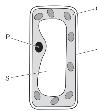
Which labelled structures are bound by a double membrane?
Options
A P and Q
B P and S
C R and Q
D R and S
Working
- P = nucleus: surrounded by the nuclear envelope, a double membrane pierced by nuclear pores.
- Q = chloroplast: surrounded by a double membrane (outer and inner envelope), with thylakoids inside.
- R = mitochondrion: (also a double-membrane organelle in standard biology).
- S = large central vacuole: bounded by a single membrane called the tonoplast.
The only answer choice that correctly identifies the double-membrane-bound structures P and Q is A.
Answer
A
A
Background Concept
Eukaryotic organelles are compartments inside the cell, each bounded by one or more membranes. The number of membranes an organelle has is a key identifying feature and reflects its evolutionary origin:
- Nucleus — bounded by the nuclear envelope, a double membrane studded with nuclear pores that control the exchange of molecules (e.g. mRNA) between the nucleus and cytoplasm.
- Chloroplast — bounded by a double membrane (outer and inner envelope). Inside, the thylakoid membrane system carries out the light-dependent reactions of photosynthesis.
- Mitochondrion — bounded by a double membrane: a smooth outer membrane and a highly folded inner membrane (cristae) where the electron transport chain operates.
- Vacuole (large central vacuole of plant cells) — bounded by a single membrane called the tonoplast, which controls the movement of solutes between the vacuolar sap and the cytoplasm.
The double-membrane nature of the nucleus, chloroplast and mitochondrion is often explained by the endosymbiotic theory, which proposes that chloroplasts and mitochondria originated from engulfed prokaryotes.
Understanding the Question
The diagram shows a plant cell with four labelled structures: P (nucleus), Q (chloroplast), R (mitochondrion), and S (large central vacuole). The command is a multiple-choice question asking which of these are bound by a double membrane. We must match each label to the correct organelle and apply knowledge of its membrane structure, then select the option whose pair both satisfy the "double membrane" criterion.
Approach
- Identify each labelled structure from the diagram.
- Recall the membrane structure of each organelle.
- Eliminate options that include S (the vacuole, which is single-membrane).
- From the remaining options, identify the pair that consists of two double-membrane organelles.
Step-by-Step Reasoning
- P (nucleus) → double membrane (nuclear envelope) ✓
- Q (chloroplast) → double membrane (envelope) ✓
- R (mitochondrion) → double membrane in standard biology
- S (vacuole) → single membrane (tonoplast) ✗
Any option containing S is immediately wrong, because the vacuole is bounded by only one membrane. This eliminates options B (P and S) and D (R and S). We are left with A (P and Q) and C (R and Q).
Both A and C list one organelle that is unambiguously double-membrane bound (Q, the chloroplast). The mark scheme identifies A (P and Q) as the correct answer, recognising the nucleus (P) and the chloroplast (Q) as the two double-membrane-bound structures in the option list.
Key Takeaways
- The nucleus and chloroplast are bounded by a double membrane.
- The large central vacuole in plant cells is bounded by a single membrane (the tonoplast).
- Membrane number is a defining feature of an organelle and a common MCQ discriminator.
Common Mistakes
- Confusing the vacuole's tonoplast with a double membrane. The tonoplast is a single lipid bilayer; do not credit it as double.
- Forgetting that the chloroplast has two envelope membranes in addition to the internal thylakoid system. The envelope alone is a double membrane.
- Treating the nuclear envelope as a single thick membrane. It is two membranes separated by a perinuclear space, joined at nuclear pores.
Things to Be Careful About
- When a "double membrane" question lists four organelles, always cross off any organelle that is single-membrane first (here, the vacuole). This narrows the options quickly.
- Note that the mark scheme answer for this question is A (P and Q). In standard A-level biology, mitochondria also have a double membrane, but the options provided restrict the correct choice to A.
- Use precise terminology: nuclear envelope (not just "membrane around the nucleus"), tonoplast (for the vacuolar membrane), and envelope (for the double membrane of the chloroplast).
Which size of ribosome is found in mitochondria and typical prokaryotic cells?
Options
A 50S
B 60S
C 70S
D 80S
Working
Ribosomes are described by their sedimentation coefficient (S, Svedberg units). Prokaryotic cells have 70S ribosomes (composed of a 50S large subunit and a 30S small subunit), while eukaryotic cytoplasmic ribosomes are 80S (composed of a 60S large subunit and a 40S small subunit). Mitochondria (and chloroplasts) contain 70S ribosomes because these organelles are thought to have originated from free-living prokaryotes engulfed by an ancestral eukaryotic cell (endosymbiosis), so they retain prokaryote-like ribosomes.
Answer
C
C
Background Concept
Ribosomes are the site of protein synthesis in all cells. They are made of ribosomal RNA (rRNA) and protein, organised into a large and a small subunit. They are described by their sedimentation coefficient in Svedberg units (S), which is a measure of how fast they sediment in a centrifuge — it depends on mass, shape and density, not just size.
Two main classes of ribosome are relevant at A-level:
- 70S ribosomes — found in prokaryotes (bacteria and archaea). They consist of a 50S large subunit and a 30S small subunit.
- 80S ribosomes — found in the cytoplasm of eukaryotic cells. They consist of a 60S large subunit and a 40S small subunit.
Mitochondria (and chloroplasts in plants) are unusual: although they sit inside eukaryotic cells, they contain 70S ribosomes, the same as prokaryotes. This is one of the key pieces of evidence for the endosymbiotic theory, which proposes that mitochondria and chloroplasts evolved from free-living prokaryotes that were engulfed by an ancestral eukaryotic cell and became permanent residents. They kept their own DNA and their own prokaryote-type ribosomes.
Understanding the Question
The question asks which single ribosome size is found in two locations: mitochondria AND typical prokaryotic cells. The two locations are the clue — mitochondria and prokaryotes both have 70S ribosomes, whereas the eukaryotic cytoplasm has 80S. The "S" values 50S and 60S in the options refer to large-subunit sizes, not whole ribosomes, so they are distractors.
Approach
Match the location to the known ribosome size:
| Location | Ribosome |
|---|---|
| Prokaryotic cell | 70S |
| Mitochondrial matrix | 70S |
| Eukaryotic cytoplasm | 80S |
Both required locations (mitochondria and prokaryotes) point to 70S.
Step-by-Step Reasoning
- A is 50S — this is only the large subunit of the prokaryotic 70S ribosome, not a whole ribosome. Reject.
- B is 60S — this is only the large subunit of the eukaryotic 80S ribosome, not a whole ribosome. Reject.
- C is 70S — the whole ribosome of prokaryotes and of mitochondria (and chloroplasts). Correct.
- D is 80S — the whole ribosome of the eukaryotic cytoplasm. Reject.
Key Takeaways
- 70S = prokaryotes, mitochondria, chloroplasts.
- 80S = eukaryotic cytoplasm.
- The presence of 70S ribosomes in mitochondria is a key piece of evidence for the endosymbiotic origin of these organelles.
- 50S and 60S are subunits, not whole ribosomes, so they are not valid answers to questions about "ribosome size".
Common Mistakes
- Choosing 80S because mitochondria are inside "eukaryotic" cells — but mitochondrial ribosomes are an exception, inherited from their prokaryotic ancestors.
- Confusing subunit sizes (50S, 60S) with whole-ribosome sizes (70S, 80S).
- Choosing 70S for chloroplasts in plant questions is also correct; the rule generalises to all endosymbiont-derived organelles.
Things to Be Careful About
- The Svedberg unit is not additive across subunits (50S + 30S ≠ 80S), because sedimentation depends on shape as well as mass. This is why 50S + 30S gives 70S, not 80S.
- "Typical prokaryotic cell" rules out unusual organelles or viruses — the question is asking about the standard bacterial ribosome.
- Read the stem carefully: it asks for the size found in BOTH mitochondria AND prokaryotes — a single shared answer, which is 70S.
Which row about typical prokaryotic cells and typical animal cells is correct?
Options
| lysosomes present for the break down of old organelles | ATP is produced by the cell | |
|---|---|---|
| A | ✓ | ✗ |
| B | ✓ | ✓ |
| C | ✗ | ✓ |
| D | ✗ | ✗ |
key
✓ = correct for typical prokaryotic cells and typical animal cells
✗ = not correct for both cells but correct for either typical prokaryotic cells or typical animal cells
Working
Evaluate each statement against the key:
- Lysosomes present for the breakdown of old organelles
- Animal cells: ✓ (lysosomes contain hydrolytic enzymes that digest worn-out organelles)
- Prokaryotic cells: ✗ (prokaryotes have no membrane-bound organelles, so no lysosomes)
- Therefore true for animal cells only → ✗ under the key
- ATP is produced by the cell
- Animal cells: ✓ (ATP made by aerobic respiration in mitochondria)
- Prokaryotic cells: ✓ (ATP made by aerobic respiration; enzymes located on the plasma membrane since there are no mitochondria)
- Therefore true for both cell types → ✓ under the key
The row ✗, ✓ is C.
Answer
C
C
Background Concept
All cells fall into one of two broad categories: prokaryotic (e.g. bacteria) and eukaryotic (e.g. animal, plant, fungal and protist cells). The defining distinction is that eukaryotic cells contain membrane-bound organelles (a true nucleus enclosed by a nuclear envelope, mitochondria, endoplasmic reticulum, Golgi apparatus, lysosomes in animals, etc.), whereas prokaryotic cells do not. Instead, their DNA lies free in the cytoplasm in a region called the nucleoid, and their respiratory enzymes are located on the infolded plasma membrane (mesosome-like invaginations).
Two consequences of this are central to the question:
- Lysosomes are membrane-bound sacs of hydrolytic (digestive) enzymes. They exist only in eukaryotic cells — in particular, animal cells use them to break down worn-out organelles and engulfed material. Prokaryotes simply have no equivalent organelle.
- ATP production is a universal requirement of all living cells. Both prokaryotes and animal cells respire (prokaryotes on the plasma membrane, animal cells in mitochondria), and both therefore make ATP. The site differs but the process occurs in both.
Understanding the Question
This is a multiple-choice question that uses a small comparison table. The command word is 'is correct'. Crucially, the key below the table re-defines the usual meaning of the ticks and crosses:
- ✓ means the statement is true for both typical prokaryotic cells and typical animal cells.
- ✗ means the statement is true for only one of the two cell types (not both).
So each statement must be tested against both cell types before the row can be classified.
Approach
For each statement, ask two questions:
- Is the statement true for a typical animal cell?
- Is the statement true for a typical prokaryotic cell?
If yes to both → ✓. If yes to only one → ✗. Then pick the row that matches.
Step-by-Step Reasoning
Statement 1: Lysosomes present for the breakdown of old organelles
- Animal cell: Yes — lysosomes are characteristic animal-cell organelles. They contain hydrolytic enzymes (proteases, lipases, nucleases, glycosidases) that operate at the low internal pH (~4.5–5) and digest worn-out organelles delivered to them by autophagy.
- Prokaryotic cell: No — prokaryotes lack all membrane-bound organelles, including lysosomes. They cannot isolate digestive enzymes inside a vesicle because they have no vesicle system.
- Conclusion: true for animal cells only → ✗ under the key.
Statement 2: ATP is produced by the cell
- Animal cell: Yes — ATP is generated by aerobic respiration in the mitochondria (and additionally by glycolysis in the cytoplasm, which alone produces a small amount of ATP).
- Prokaryotic cell: Yes — prokaryotes also respire and produce ATP. Because they have no mitochondria, the electron-transport chain and ATP synthase are embedded in the plasma membrane, which folds inwards to increase the surface area available.
- Conclusion: true for both cell types → ✓ under the key.
The row containing ✗ (lysosomes) and ✓ (ATP production) is row C.
Key Takeaways
- The fundamental prokaryote/eukaryote difference is the presence or absence of membrane-bound organelles; this is why lysosomes exist only in animal (eukaryotic) cells.
- ATP synthesis is universal — every living cell must make ATP — but the location differs (plasma membrane in prokaryotes; mitochondria in animal cells).
- Always read the key of any comparison question carefully; the meaning of ✓ and ✗ here is non-standard, and misreading it would change the answer.
Common Mistakes
- Marking the lysosome row as ✓ because lysosomes are 'a real structure'. The key demands it be true for both cell types, and prokaryotes lack lysosomes.
- Marking the ATP row as ✗ on the mistaken belief that prokaryotes 'do not have mitochondria, so do not respire'. Respiration does occur in prokaryotes — only the location of the respiratory enzymes is different.
- Reading ✓ and ✗ in their ordinary everyday sense rather than using the key's specific definitions.
Things to Be Careful About
- The question is about typical cells — there are some atypical exceptions in real biology (e.g. some bacteria do have protein-bounded microcompartments, and red blood cells lose their organelles), but the question uses the textbook 'typical' picture.
- Apply the key before reading the options: determine the correct ✓/✗ pattern first, then match it to a row.
- Use precise CIE terminology — say 'membrane-bound organelle' rather than 'membrane' or 'vesicle' when explaining why prokaryotes lack lysosomes.
Which row is correct for the structures present in typical plant cells and typical animal cells?
Options
| cell structure | plant cell | animal cell | |
|---|---|---|---|
| A | plasmodesmata | present | present |
| B | Golgi body | present | not present |
| C | centriole | not present | present |
| D | tonoplast | not present | not present |
Working
Centrioles are small cylindrical organelles made of microtubules that organise the spindle during cell division in animal cells. Higher plant cells lack centrioles; spindle fibres are instead organised from microtubule-organising centres without centrioles.
Checking the other options:
- Plasmodesmata: present only in plant cells (channels through cell walls), not in animal cells → A is wrong.
- Golgi body: present in both plant and animal cells (modifies, sorts and packages proteins/lipids) → B is wrong.
- Tonoplast: the membrane surrounding the large central vacuole, present in plant cells but absent in animal cells → D is wrong.
Answer
C
C
Background Concept
Typical plant and animal cells share many organelles (nucleus, mitochondria, ribosomes, endoplasmic reticulum, Golgi body, plasma membrane), but they also have distinctive structures that reflect their different ways of life. Plant cells possess a rigid cellulose cell wall, a large permanent central vacuole bounded by a membrane called the tonoplast, and plasmodesmata — cytoplasmic channels that pass through the cell wall to link adjacent plant cells. Animal cells lack a cell wall, a large central vacuole, and plasmodesmata.
Centrioles are barrel-shaped structures made of triplets of microtubules (a 9 + 0 arrangement). In animal cells they sit within the centrosome and nucleate microtubule organisation, including formation of the spindle during mitosis. Higher plant cells do not have centrioles, yet they still form a spindle using microtubule-organising centres at the cell poles — a centriole is therefore not strictly necessary for cell division in plants.
The Golgi body (Golgi apparatus) is universal in eukaryotes; it receives proteins from the rough endoplasmic reticulum, modifies them (e.g. by glycosylation), and packages them into vesicles for secretion or delivery to other organelles. It is present in both plant and animal cells.
Understanding the Question
This is a multiple-choice question (Paper 1 style) testing knowledge of which structures are present in typical plant cells and which in typical animal cells. The mark scheme gives the correct row as C: centriole is not present in the typical plant cell but is present in the typical animal cell.
The command word is implicit in the structure of a "which row is correct" MCQ — pick the single row where both entries (plant and animal) are stated correctly.
Approach
Go through each row and decide whether the structure is really present in the plant cell and/or animal cell as stated, using knowledge of cell ultrastructure.
Step-by-Step Reasoning
- Row A — plasmodesmata: Plasmodesmata are cell-wall channels unique to plant cells. They are absent in animal cells (which have no cell wall to span). The table claims they are present in both — incorrect.
- Row B — Golgi body: The Golgi body is found in both plant and animal cells, where it modifies and packages proteins and lipids. The table says it is not present in animal cells — incorrect.
- Row C — centriole: Typical higher plant cells lack centrioles; animal cells possess a pair of centrioles inside the centrosome. The table correctly states "not present" for plant and "present" for animal — correct.
- Row D — tonoplast: The tonoplast is the membrane enclosing the large central vacuole of a plant cell. Plant cells have it; animal cells (which lack a large central vacuole) do not. The table says "not present" for both — incorrect.
Key Takeaways
- Plant-specific structures: cell wall, large central vacuole (with tonoplast), plasmodesmata, chloroplasts.
- Animal-specific structures: centrioles (within the centrosome), lysosomes (more prominent than in plants).
- Shared eukaryotic structures: nucleus, mitochondria, ribosomes, ER, Golgi body, plasma membrane.
- The presence or absence of centrioles in plants is a common MCQ trap — remember that plant mitosis still occurs but without centrioles.
Common Mistakes
- Confusing the cell wall with a plasma membrane and assuming "Golgi absent in animal cells" because plants have cell walls — both kingdoms have a Golgi body.
- Thinking centrioles are essential for mitosis, leading to the wrong belief that plants must have them too.
- Believing plasmodesmata are a type of gap junction found in both kingdoms; they are plant-specific.
- Confusing the tonoplast with the plasma membrane.
Things to Be Careful About
- "Typical" is the operative word — a few lower plant groups (some algae, mosses) and certain animal cells in specific phases may differ, but the syllabus is concerned with the standard comparison.
- The question does not ask about frequency or abundance (e.g. lysosomes are not exclusive to animal cells but are more characteristic of them) — answer strictly on presence/absence as stated.
Which row is correct for cellulose?
Options
| rotation of alternate monomers by | shape of molecule | hydrogen bonds between molecules | |
|---|---|---|---|
| A | ✓ | branched | ✗ |
| B | ✗ | branched | ✓ |
| C | ✗ | unbranched | ✗ |
| D | ✓ | unbranched | ✓ |
key
✓ = present
✗ = not present
Working
Cellulose is a polysaccharide of β-glucose monomers linked by 1,4-glycosidic bonds.
- Because each β-glucose is rotated 180° relative to its neighbour, the chain runs straight and is unbranched.
- The –OH groups projecting from adjacent chains form hydrogen bonds between molecules, holding them together in microfibrils.
This matches: rotation by 180° = ✓, unbranched = ✓, hydrogen bonds between molecules = ✓.
Answer
D
D
Background Concept
Cellulose is the main structural polysaccharide of plant cell walls. It is built from β-glucose monomers joined by 1,4-glycosidic bonds. The β-glucose monomer is the mirror-image isomer of α-glucose: the –OH group on carbon 1 projects upwards, not downwards. Because of this, every alternate glucose unit must be rotated by 180° for the 1,4-link to form, and the result is a long, straight, unbranched chain. Many parallel cellulose chains are then held together laterally by hydrogen bonds between the –OH groups on adjacent molecules, producing strong microfibrils that give plant cell walls their tensile strength.
Understanding the Question
The question asks the candidate to identify which row in the table correctly describes cellulose for three structural features:
- Whether alternate monomers are rotated by 180°
- Whether the molecule is branched or unbranched
- Whether hydrogen bonds form between cellulose molecules
The mark scheme confirms D as the correct row.
Approach
Recall the three defining structural features of cellulose and check each row systematically against them.
Step-by-Step Reasoning
- Rotation of alternate monomers by 180°: This IS a feature of cellulose because β-glucose units must alternate orientation to form the 1,4-glycosidic bond in a straight chain. So this is ✓ (present). Starch (amylose) and glycogen do not have this 180° rotation.
- Shape of molecule — branched or unbranched: Cellulose is a straight, unbranched chain. The 180° rotation of alternate β-glucose units prevents branching. Amylopectin and glycogen are branched; cellulose and amylose are not.
- Hydrogen bonds between molecules: Adjacent cellulose chains align and are cross-linked by hydrogen bonds between –OH groups, producing microfibrils. So this is ✓ (present). In starch and glycogen, molecules are coiled and the predominant intermolecular interaction is via α-1,4 and α-1,6 bonds, not extensive inter-chain hydrogen bonding.
Putting these together: rotation 180° = ✓, unbranched = ✓, hydrogen bonds = ✓. Only row D shows this combination.
Key Takeaways
- Cellulose is a polymer of β-glucose with 1,4-glycosidic bonds.
- Alternate β-glucose units are rotated 180°, producing a straight unbranched chain.
- Hydrogen bonds between parallel cellulose chains form microfibrils, giving plant cell walls high tensile strength.
- These features distinguish cellulose from α-glucose-based storage polysaccharides (starch, glycogen), which are helical and often branched.
Common Mistakes
- Confusing cellulose (β-glucose, unbranched, hydrogen-bonded microfibrils) with amylopectin/glycogen (α-glucose, branched via 1,6 links) — selecting a "branched" option.
- Forgetting that the rotation of 180° is specifically a cellulose feature (it arises from β-glucose geometry) and choosing a row that omits it.
- Assuming hydrogen bonds are only intramolecular and so rejecting D — in cellulose the hydrogen bonds are extensively intermolecular.
Things to Be Careful About
- The 180° rotation is a consequence of using β-glucose, not an arbitrary design feature; it is essential for producing the straight chain that can pack tightly and hydrogen-bond to neighbours.
- "Unbranched" is critical — even a single branch would disrupt the regular hydrogen-bonding pattern and reduce structural strength.
- The mark scheme often rejects answers that confuse cellulose with starch (amylose vs amylopectin distinctions).
Which statements about peptide bond formation are correct?
1 The bond formation occurs between a carbon of one amino acid and a nitrogen of the next amino acid after the amino acids detach from tRNA.
2 The bond formation occurs at the ribosome while the amino acids are still attached to tRNA, and is a hydrolysis reaction.
3 The bond formation is important for growth of an organism and when the bond forms, a water molecule is removed.
Options
A 1 and 3
B 2 and 3
C 2 only
D 3 only
Working
- Statement 1 is wrong: peptide bond formation occurs at the ribosome while amino acids are still attached to their tRNAs, not after they have detached.
- Statement 2 is wrong: although peptide bond formation does occur at the ribosome while the amino acids remain attached to tRNA, it is a condensation (dehydration) reaction, not a hydrolysis reaction.
- Statement 3 is correct: peptide bond formation is essential for protein synthesis required for growth, and it is a condensation reaction in which a water molecule is removed.
Only statement 3 is correct.
Answer
D
D
Background Concept
Amino acids are joined together by peptide bonds to form polypeptides (which then fold into proteins). Each amino acid has a central carbon atom (the -carbon) bonded to an amino group (), a carboxyl group (), a hydrogen atom, and a variable R-group. When two amino acids react, the carboxyl group of one loses an and the amino group of the other loses an ; these combine to release a water molecule, and the remaining carbon and nitrogen are joined by a peptide (covalent) bond.
Because water is released, this is a condensation (or dehydration) reaction — the opposite of hydrolysis. Peptide bond formation during translation occurs on the ribosome: the ribosome's peptidyl transferase activity catalyses the bond between amino acids while they are still attached to tRNA molecules in the P and A sites.
Understanding the Question
This is a multiple-choice question asking which of three statements about peptide bond formation is/are correct. The candidate must assess each statement independently and then identify the option that lists only the correct ones. The answer is worth 1 mark.
Approach
Test each statement against three pieces of core knowledge:
- Where peptide bonds form (ribosome) and when relative to tRNA attachment (while still attached).
- What type of reaction forms a peptide bond (condensation, not hydrolysis).
- Why it matters for the organism (proteins are required for growth and repair) and that water is released.
Step-by-Step Reasoning
- Statement 1 claims the bond forms after amino acids detach from tRNA, and links a carbon of one amino acid to a nitrogen of the next. The C-to-N linkage is correct (it is a C–N bond), but the timing is wrong: peptide bond formation occurs at the ribosome while the amino acids are still attached to tRNAs. Statement 1 is therefore incorrect.
- Statement 2 correctly locates peptide bond formation at the ribosome and during attachment to tRNA, but calls it a hydrolysis reaction. Hydrolysis would break bonds by adding water; peptide bond formation makes a bond and releases water — it is a condensation. Statement 2 is therefore incorrect.
- Statement 3 correctly identifies two facts: peptide bond formation is essential for the growth of an organism (proteins make up much of new tissue), and a water molecule is released when the bond forms (a condensation reaction). Both parts are correct, so statement 3 is correct.
Only statement 3 is correct, which corresponds to option D.
Key Takeaways
- A peptide bond is a C–N covalent bond between the carboxyl carbon of one amino acid and the amino nitrogen of the next.
- It is formed by a condensation reaction (water released), not hydrolysis.
- It is catalysed at the ribosome by peptidyl transferase while amino acids are still attached to tRNAs.
- Protein synthesis (and hence peptide bond formation) is essential for growth, repair and enzymes of an organism.
Common Mistakes
- Confusing condensation with hydrolysis — the most common error here. Condensation removes water to form a bond; hydrolysis adds water to break one.
- Thinking amino acids are released from tRNA before the peptide bond forms — they remain bound to tRNA during condensation.
- Stating that the bond forms between two carbons or two nitrogens — it is specifically carbon to nitrogen.
Things to Be Careful About
- The "and" in each statement links two claims; both must be true for the statement to be credited.
- Remember the direction of water movement: formation = water released; breakdown (e.g. digestion) = water added.
- "Growth" in this context refers to the synthesis of new proteins needed for increases in cell number/size, not to cell division per se.
The diagram shows naturally occurring D-glucose and a form of glucose that can be synthesised in the laboratory, known as L-glucose.
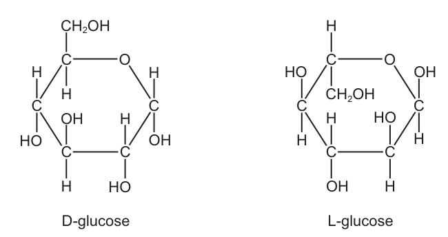
The enzyme glucose oxidase catalyses the oxidation of D-glucose. The enzyme cannot catalyse the oxidation of L-glucose.
Which statement about L-glucose explains this?
Options
A L-glucose does not fit into the active site of glucose oxidase.
B L-glucose has a different structural formula to D-glucose.
C L-glucose is a synthetic sugar.
D L-glucose is the mirror image of D-glucose.
Working
Glucose oxidase is specific to D-glucose. Enzymes are specific because the substrate must be the correct 3D shape to fit into the active site. L-glucose is the mirror image of D-glucose, so its 3D shape does not fit the active site, and it cannot be oxidised.
Answer
A
A
Background Concept
Enzymes are biological catalysts that are highly specific: each enzyme acts on only one (or a very small range of) substrate(s). This specificity arises from the precise three-dimensional shape of the enzyme's active site — a pocket or groove formed by the folding of the protein. According to the lock-and-key hypothesis, only a substrate whose shape is complementary to the active site can bind and be converted to product. The induced-fit hypothesis refines this: the active site moulds itself around the substrate on binding, but the substrate must still be the correct overall shape to be recognised in the first place.
Glucose exists as stereoisomers — molecules with the same structural (molecular and connectivity) formula but different spatial arrangements of atoms. D-glucose and L-glucose are enantiomers (non-superimposable mirror images), as shown in the Haworth projection in Fig. 8.1. Every hydroxyl (-OH) and hydrogen group around the ring sits in the opposite position in L-glucose compared to D-glucose.
Understanding the Question
The question gives you two pieces of information:
- Glucose oxidase catalyses the oxidation of D-glucose but not L-glucose.
- L-glucose is the mirror image of D-glucose.
It then asks you to choose the statement that explains why L-glucose is not catalysed. The command word is effectively explain, so the answer must give a reason for the lack of activity, not just describe the molecule.
Approach
The key is to link the property of L-glucose (its mirror-image shape) to the mechanism of enzyme specificity (the shape of the active site). The correct answer must therefore mention the active site and the idea of fitting/binding, not merely state a property of the molecule in isolation.
Step-by-Step Reasoning
- Option A: "L-glucose does not fit into the active site of glucose oxidase." This directly gives the mechanism: the L-glucose molecule, being the mirror image, has the wrong 3D shape to slot into the active site of the enzyme, so no enzyme–substrate complex forms and no reaction occurs. ✓
- Option B: "L-glucose has a different structural formula to D-glucose." False. D- and L-glucose are stereoisomers, so they have the same structural (molecular) formula — only the spatial arrangement of atoms differs. Even if the formula did differ, the answer still would not explain the mechanism.
- Option C: "L-glucose is a synthetic sugar." Irrelevant. Whether a molecule is natural or laboratory-made has no bearing on whether an enzyme can act on it. Enzymes recognise shape, not origin.
- Option D: "L-glucose is the mirror image of D-glucose." This is true (and is essentially the context given in the question stem), but it is only a description. It does not state why this prevents the reaction. To score, the answer must connect the mirror-image shape to the active site — which is exactly what option A does.
Key Takeaways
- Enzyme specificity is due to the precise 3D shape of the active site being complementary to the substrate's shape.
- D- and L-glucose are enantiomers — same molecular formula, mirror-image 3D arrangement of atoms.
- A correct "explain" answer must state the mechanism (no fit → no binding → no catalysis), not just a property of the substrate.
Common Mistakes
- Choosing D because it is the only statement that is biologically true. The question asks for an explanation, not a description — so D is incomplete.
- Choosing B because students confuse "structural formula" with "spatial arrangement". The structural formula of an isomer is the same; what differs is the 3D configuration.
- Choosing C because students think "natural = enzyme works, synthetic = doesn't". This is not a biological principle — enzyme activity depends purely on molecular shape, not on how the molecule was obtained.
Things to Be Careful About
- Read the command word: explain demands a mechanism, not a label.
- "Structural formula" refers to which atoms are bonded to which, not the 3D arrangement. D- and L-glucose share the same structural formula; they differ in stereochemistry.
- The lock-and-key and induced-fit models both predict that a mirror-image substrate will not bind productively — you do not need to choose between them here.
- The mark scheme phrasing is tight: a candidate who writes "it has a different shape" without mentioning the active site often does not earn the mark, because the explanation is the link between shape and the enzyme's pocket.
Tests for biological molecules were carried out on three solutions. Each solution contained only one type of biological molecule.
The observations were as follows.
| solution | test | observation |
|---|---|---|
| 1 | Benedict’s test | blue to orange |
| 2 | Benedict’s test after acid hydrolysis | blue to red |
| 3 | biuret test | blue to purple |
Which solutions would contain either sucrose or amylase?
Options
A 1, 2 and 3
B 1 and 3 only
C 2 and 3 only
D 2 only
Working
- Solution 1: Benedict's test directly positive (blue → orange) means a reducing sugar (e.g. glucose or maltose). Sucrose is non-reducing, and amylase is a protein — so solution 1 is neither.
- Solution 2: Benedict's test only positive after acid hydrolysis (blue → red) means a non-reducing sugar that has been broken down into reducing sugars. This matches sucrose.
- Solution 3: Biuret test positive (blue → purple) means protein is present. This matches amylase (an enzyme, which is a protein).
Therefore solutions 2 and 3 contain either sucrose or amylase.
Answer
C
C
Background Concept
Benedict's reagent detects reducing sugars. A reducing sugar has a free aldehyde (–CHO) or ketone group that can reduce Cu²⁺ (blue) in Benedict's reagent to Cu⁺ (brick-red/orange precipitate of Cu₂O). Glucose, fructose, maltose and lactose are reducing sugars. Sucrose is non-reducing because its glycosidic bond joins the two anomeric carbons (C1 of glucose to C2 of fructose), leaving no free group that can open to an aldehyde. Boiling sucrose with dilute hydrochloric acid (acid hydrolysis) splits this bond, releasing glucose and fructose — both reducing — so a subsequent Benedict's test will then be positive.
The biuret test detects peptide bonds. Cu²⁺ ions in biuret reagent form a violet/purple complex with the –CO–NH– group of proteins (and peptides of three or more amino acids). Any protein — including enzymes such as amylase — gives a positive biuret.
Understanding the Question
We are given the results of two tests on three single-component solutions and asked which of them could contain sucrose (a non-reducing disaccharide) or amylase (a protein/enzyme). The key biological reasoning is to match each test result to the type of molecule that would give it.
Approach
For each solution, identify what kind of molecule the observation implies, then check whether that matches sucrose (non-reducing sugar) or amylase (protein).
Step-by-Step Reasoning
- Solution 1 (Benedict's, no hydrolysis): blue → orange. A direct positive Benedict's test indicates a reducing sugar. Sucrose does not give a positive result without prior hydrolysis, and amylase is a protein — neither fits. So solution 1 is something like glucose or maltose. Excluded.
- Solution 2 (Benedict's after acid hydrolysis): blue → red. Without hydrolysis, this solution would not reduce Benedict's reagent (it would remain blue); after acid hydrolysis it gives a strong red precipitate. This is the textbook signature of a non-reducing sugar such as sucrose. Included.
- Solution 3 (biuret): blue → purple. A positive biuret test indicates protein (or long peptide). Amylase is an enzyme — and enzymes are proteins — so amylase fits. Sucrose is a carbohydrate, not a protein, so sucrose does not fit here on its own; but the question only requires each solution to contain either sucrose or amylase. Amylase accounts for solution 3. Included.
Therefore solutions 2 and 3 only — answer C.
Key Takeaways
- A direct positive Benedict's test → reducing sugar; a positive test only after acid hydrolysis → non-reducing sugar (typically sucrose).
- The biuret test (blue → purple) is the standard test for protein/peptides and is therefore positive for any enzyme.
- Each test result narrows the molecule to a class, so interpreting colour-change data is at the heart of "tests for biological molecules" questions.
Common Mistakes
- Confusing the order of the Benedict's test for non-reducing sugars: the acid hydrolysis must come before neutralisation and before adding Benedict's reagent, otherwise the acid interferes with the test.
- Saying a solution "contains protein" when only the biuret test has been performed — biuret detects peptide bonds, so the conclusion must specifically be "protein or polypeptide".
- Choosing option B (1 and 3) by forgetting that sucrose is non-reducing and so would not give a direct positive Benedict's test in solution 1.
Things to Be Careful About
- Orange vs red in Benedict's test both indicate a positive result; the precise shade depends on concentration, not the identity of the sugar.
- "Blue to blue" would indicate a negative Benedict's test, not a failed experiment — important when interpreting tables of results.
- The question states each solution contains only one type of biological molecule, so do not try to invoke mixtures (e.g. solution 1 cannot be both a reducing sugar and a protein).
Which row describes the expected effect on and when a competitive reversible inhibitor is added to an enzyme-catalysed reaction?
Options
| effect on | substrate concentration at | |
|---|---|---|
| A | no change | increases |
| B | no change | no change |
| C | decreases | increases |
| D | decreases | no change |
Working
A competitive inhibitor binds reversibly to the active site, competing with the substrate. Because the inhibition is reversible and competition-based, raising the substrate concentration can out-compete the inhibitor, so the reaction can still reach the same maximum rate → is unchanged. However, more substrate is now required to reach half of , so the substrate concentration at increases.
Answer
A
A
Background Concept
Two parameters describe the kinetics of an enzyme-catalysed reaction in the Michaelis–Menten model:
- : the maximum rate, reached when every active site is saturated with substrate.
- (the Michaelis constant): the substrate concentration at which the reaction rate is . A larger means the enzyme has a lower apparent affinity for its substrate — more substrate is needed to push the reaction halfway to its maximum rate.
A competitive inhibitor is a molecule shaped like the natural substrate. It binds reversibly to the active site, directly competing with the substrate. The defining feature of competitive inhibition is therefore that the effect of the inhibitor can be overcome by adding more substrate, because substrate and inhibitor are in direct competition for the same site.
By contrast, a non-competitive inhibitor binds to a site other than the active site (an allosteric site) and changes the shape/activity of the enzyme; this cannot be overcome by raising substrate concentration.
Understanding the Question
This is a multiple-choice question asking how and change when a competitive reversible inhibitor is added to an enzyme-catalysed reaction. The table offers four combinations: each of the two parameters can either stay the same, increase, or decrease. The correct answer is the pair of changes that uniquely characterises competitive inhibition.
The key words are competitive and reversible — together they dictate the kinetic signature.
Approach
Recall the standard Michaelis–Menten fingerprint for a competitive inhibitor:
- : no change
- : increases
This is the textbook distinguishing signature; memorise it together with the non-competitive fingerprint ( decreases, unchanged) so the two can never be confused.
Step-by-Step Reasoning
- The inhibitor binds to the active site, so it directly blocks substrate binding — but only when it is occupying the site.
- Because the inhibitor is reversible and in direct competition with substrate, raising shifts the equilibrium so that substrate molecules occupy the active sites more often than the inhibitor does.
- At sufficiently high , essentially every active site is occupied by substrate and the inhibitor is effectively out-competed. The reaction can therefore still reach the same as in the uninhibited case → is unchanged.
- At any given (non-saturating) , however, a fraction of the enzyme molecules is bound to inhibitor instead of substrate. To compensate, a higher substrate concentration is needed to drive the rate up to → the apparent increases.
- The correct row is therefore: no change; substrate concentration at increases → option A.
Key Takeaways
- Competitive reversible inhibitor: unchanged, increased.
- Non-competitive inhibitor: decreased, unchanged.
- The "reversible" wording matters: if the inhibitor were irreversible (e.g. it covalently modified the active site), would fall because increasing could not recover the lost active sites.
- is a measure of apparent affinity, not of binding strength in the absence of inhibitor.
Common Mistakes
- Picking C ( decreases, increases): this is a common mix-up with non-competitive inhibition, or with the (incorrect) idea that "the enzyme is being slowed down so its maximum rate must fall". In fact, the maximum rate is recovered at high .
- Picking D ( decreases, unchanged): this is the non-competitive signature — wrong here.
- Picking B ( unchanged, unchanged): the inhibitor would have no measurable effect, which contradicts the fact that some active sites are being blocked.
- Forgetting the difference between a competitive and a non-competitive inhibitor when revising the kinetic plots — a common cause of lost marks.
Things to Be Careful About
- Always check whether the question specifies competitive or non-competitive — the kinetic fingerprints are mirror images of each other and the answers change accordingly.
- The word reversible in the stem is a deliberate cue: it confirms that increasing can out-compete the inhibitor, preserving .
- A Lineweaver–Burk plot of competitive inhibition shows the lines intersecting on the y-axis (same ) but with different x-intercepts (different ); this is a useful visual way to remember the result.
The graph shows the effect of substrate concentration on the rates of reaction of three enzymes, X, Y, and Z.
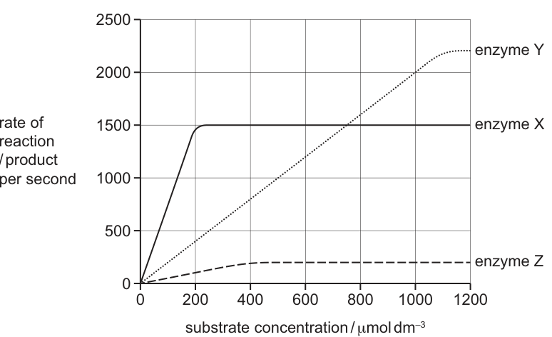
What is the correct order of affinity of these enzymes for their substrates, starting with the enzyme with the highest affinity?
Options
A X Y Z
B X Z Y
C Y X Z
D Z X Y
Working
Enzyme affinity for substrate is inversely related to the Michaelis–Menten constant, : the lower the , the higher the affinity.
= the substrate concentration at which the rate of reaction is half of .
Reading the graph:
- Enzyme X: , so half . Curve X reaches 750 at a very low substrate concentration (≈ 100 ). Lowest → highest affinity.
- Enzyme Z: , so half . Curve Z reaches 125 at ≈ 200 . Intermediate → intermediate affinity.
- Enzyme Y: still rising at 1200 ; to reach a hypothetical half- would require a much higher substrate concentration. Highest → lowest affinity.
Order of affinity (highest → lowest): X → Z → Y.
Answer
B
B
Background Concept
Enzyme kinetics for many enzymes follows the Michaelis–Menten model, which describes how the rate of an enzyme-catalysed reaction varies with substrate concentration. Two key parameters come from this model:
- — the maximum rate achieved when the enzyme is fully saturated with substrate (every active site is occupied). Beyond , adding more substrate cannot increase the rate because the enzyme, not the substrate, becomes the limiting factor.
- — the Michaelis–Menten constant, defined as the substrate concentration at which the reaction rate is half of . It is a measure of the affinity of the enzyme for its substrate: a low means high affinity (the enzyme needs only a little substrate to work at half its maximum rate), and a high means low affinity.
The curve shape is hyperbolic: a steep initial rise as substrate binds the active site, then a plateau as saturation is approached.
Understanding the Question
The graph plots rate of reaction against substrate concentration for three enzymes, X, Y and Z. We are asked to rank them by affinity for their substrates, from highest to lowest. The correct answer is the option whose order corresponds to the order of increasing (i.e. the order in which the curves reach half their respective ).
Approach
- Identify the plateau (or the projected plateau) for each curve to determine .
- Calculate half of each .
- Read off the substrate concentration at which each curve crosses its own half- — that is the .
- Rank the values from smallest to largest; reverse this to get the affinity order from highest to lowest.
A common pitfall is to look at the steepness of the initial slope, or at itself, and confuse those with affinity. Affinity is specifically tied to , not to .
Step-by-Step Reasoning
Enzyme X (solid line). It plateaus at . Half of is . Looking at the curve, it crosses 750 at a substrate concentration of roughly 100 . This is the lowest of the three, so X has the highest affinity for its substrate.
Enzyme Z (dashed line). It plateaus at . Half of is . The curve crosses 125 at about 200 . This is an intermediate , so Z has intermediate affinity.
Enzyme Y (dotted line). It has not reached a plateau within the range shown — at 1200 it is still climbing, near 2200. Even estimating a generous of ~2400, half that is ~1200, which the curve does not reach until ~800 and beyond. Y therefore has a much larger than X or Z, and so the lowest affinity.
Ranking: X (highest affinity) > Z > Y (lowest affinity). This matches option B: X → Z → Y.
Key Takeaways
- Affinity is read from , not from or the slope of the curve.
- is the [S] at half ; low = high affinity.
- When differs between enzymes, you must compare each curve against its own half-, not against an arbitrary horizontal line on the y-axis.
- An enzyme that has not yet plateaued still has a definable (high) — you simply have to estimate where half of the eventual would be reached.
Common Mistakes
- Ranking by steepness of the initial slope. The slope reflects the combination of and , not affinity alone. Two enzymes with very different values can have similar slopes at low [S] but very different affinities.
- Ranking by . A higher plateau does not mean higher affinity; it generally means more active enzyme or a faster catalytic step.
- Comparing rates at a single [S]. At one chosen substrate concentration, the highest rate belongs to the enzyme that happens to be closest to its there, which conflates and .
- Forgetting to halve . Using itself instead of half misreads the for every curve.
Things to Be Careful About
- Read each curve against its own — never compare X and Z on the same y-value when is different.
- For enzyme Y, the absence of a visible plateau means you must estimate half of an extrapolated ; a wide reading is acceptable as long as it is clearly larger than the other two values.
- The question asks for affinity order from highest to lowest — once you have ranked from lowest to highest, present the list in that same order, not reversed.
Which row correctly identifies the weak and strong bonds in the tertiary and quaternary structure of a typical protein?
Options
| disulfide | hydrogen | hydrophobic | ionic | |
|---|---|---|---|---|
| A | strong | strong | weak | weak |
| B | strong | weak | weak | weak |
| C | weak | weak | strong | strong |
| D | weak | weak | weak | strong |
Working
In the tertiary and quaternary structure of a protein:
- Disulfide bonds (S–S) are covalent and therefore strong.
- Hydrogen bonds, hydrophobic interactions and ionic bonds are all non-covalent and therefore weak (intermolecular forces).
The only option that classifies disulfide as strong and hydrogen, hydrophobic and ionic as weak is B.
Answer
B
B
Background Concept
A protein's three-dimensional shape is held in place by several types of bonds. In secondary structure (α-helix and β-pleated sheet) the polypeptide chain is held by hydrogen bonds between the C=O and N–H groups of the backbone. In tertiary structure the chain is folded into a 3D shape, and in quaternary structure several polypeptide subunits are held together. Both tertiary and quaternary structures are maintained by the same four bond types:
- Disulfide bonds (S–S) – covalent bonds formed between the –SH groups of two cysteine side chains. These are true covalent bonds and are therefore strong.
- Hydrogen bonds – weak attractions between a slightly δ⁺ hydrogen and a slightly δ⁻ atom (usually O or N). These are weak.
- Ionic (electrostatic) bonds – attractions between positively and negatively charged R groups (e.g. –COO⁻ and –NH₃⁺). These are weak compared with covalent bonds (though stronger than hydrogen bonds in some textbooks' qualitative ordering, they are still classed as weak relative to disulfide).
- Hydrophobic interactions – non-polar R groups cluster together away from water; these are weak, entropy-driven interactions, not true bonds.
So in summary, the only strong bond among these four is the disulfide bond; the other three are all weak.
Understanding the Question
The question is a multiple-choice item that presents a table of the four bond types found in tertiary and quaternary structure and asks which row correctly labels each as strong or weak. The mark scheme confirms that the row with disulfide = strong and the other three = weak is correct.
Approach
Identify the chemical nature of each bond:
- Is the bond covalent or non-covalent?
- Covalent bonds are strong; non-covalent interactions are weak.
- Match the answer to the option that reflects this.
Step-by-Step Reasoning
- Disulfide (S–S): This is a covalent bond. Covalent bonds involve sharing of electron pairs and require significant energy to break. → strong.
- Hydrogen bond: A dipole–dipole attraction between a δ⁺ H and a δ⁻ O/N. Easily broken by heat or pH change. → weak.
- Hydrophobic interaction: Non-polar side chains associate to exclude water; this is an entropic effect, not a true bond. → weak.
- Ionic bond: Attraction between fully charged groups; while stronger than H-bonds in isolation, it is still non-covalent and easily disrupted by pH or salt changes. → weak.
Comparing with the options:
- A: has hydrogen as strong → wrong.
- B: disulfide strong, the rest weak → correct.
- C: has disulfide as weak → wrong.
- D: has disulfide as weak → wrong.
Key Takeaways
- The only strong bond in tertiary and quaternary structure is the disulfide (covalent) bond.
- Hydrogen bonds, ionic bonds and hydrophobic interactions are all classified as weak (non-covalent) interactions.
- The same four bond types operate in both tertiary and quaternary structure.
Common Mistakes
- Treating ionic bonds as "strong" because they are stronger than hydrogen bonds in everyday chemistry — in protein structure, ionic bonds are still classified as weak relative to covalent disulfide bonds.
- Confusing secondary structure (held only by hydrogen bonds) with tertiary structure (held by all four bond types).
- Forgetting that hydrophobic interactions are not true chemical bonds at all.
Things to Be Careful About
- The classification is qualitative: strong vs weak, not a quantitative ranking within the weak group.
- "Strong" in this context means covalent (S–S); everything else is non-covalent and therefore weak.
Which row correctly describes haemoglobin?
Options
| A | four polypeptide chains, each containing a haem group | iron ions can associate with oxygen, forming oxyhaemoglobin | in each chain, hydrophobic R-groups of amino acids point towards the centre of the molecule | at 50% saturation, two oxygen molecules are transported by the molecule |
| B | polypeptide chains interact to produce a globular chain | each chain contains a haem group of amino acids surrounding an iron ion | consists of two identical alpha chains and two identical beta chains | each chain can transport an oxygen molecule |
| C | polypeptide chains interact to produce an almost spherical molecule | an iron ion is present within each haem group | quaternary structure has two alpha chains and two beta chains | each molecule can transport a total of four oxygen atoms |
| D | polypeptide chains produce a loose helical shape, which folds to form a spherical molecule | iron ions in the molecule can bind reversibly with oxygen | in each chain, hydrophobic R-groups of amino acids surround the iron ion | each molecule can transport a total of eight oxygen atoms |
Working
Haemoglobin is a globular, conjugated protein with quaternary structure: four polypeptide chains (2α + 2β), each carrying one haem group. Each haem group is a porphyrin ring with a central Fe²⁺ ion that binds one O₂ reversibly, giving a maximum of four O₂ molecules per haemoglobin molecule. In any globular protein, hydrophobic R-groups are orientated towards the interior of the molecule (away from the aqueous cytosol), while hydrophilic R-groups face the outside.
Checking each row:
- A — all four statements are correct:
- four chains, each with a haem group ✓
- Fe²⁺ associates reversibly with O₂ to form oxyhaemoglobin ✓
- hydrophobic R-groups point inwards, towards the centre of the globular molecule ✓
- 4 binding sites × 50% saturation = 2 O₂ molecules ✓
- B — statement 2 is wrong: the haem group is a porphyrin ring (not amino acids) with an Fe²⁺ at its centre; amino acids surround the haem group, not the other way round.
- C — statement 4 is wrong: haemoglobin transports four oxygen molecules (O₂), not four oxygen atoms.
- D — statements 3 and 4 are wrong: hydrophobic R-groups point to the interior of the whole protein, not specifically around the iron ion; the molecule transports four O₂ molecules, conventionally described as such rather than as eight oxygen atoms.
Answer
A
A
Background Concept
Haemoglobin is the oxygen-carrying pigment in red blood cells. It is a conjugated globular protein, meaning it has a non-protein (prosthetic) group attached to a protein. Its quaternary structure consists of four polypeptide chains — two α (alpha) chains and two β (beta) chains — folded together into an almost spherical shape. Each chain carries one haem group: a flat porphyrin ring with a central iron (Fe²⁺) ion. Each Fe²⁺ can bind one O₂ molecule reversibly, so one haemoglobin molecule transports a maximum of four O₂ molecules (i.e. eight oxygen atoms in total, although we always say "four oxygen molecules" in biology).
Globular proteins, including haemoglobin, are soluble because their hydrophilic (polar) R-groups project outwards into the surrounding water, while their hydrophobic (non-polar) R-groups are tucked into the interior, away from water. This is what drives the folding of the polypeptide chain and is the same principle that gives enzymes their specific 3-D shape.
Percentage saturation describes the proportion of binding sites occupied by O₂. Because haemoglobin has four sites, 50% saturation means two of the four sites are filled — i.e. two O₂ molecules are being carried.
Understanding the Question
This is a multiple-choice question with four options (A–D), each containing four statements about haemoglobin. The candidate must decide which whole row is correct, meaning every statement in the chosen row must be biologically accurate. The marking scheme rewards the single row in which all four statements are correct.
To succeed, the student must be able to:
- describe haemoglobin's quaternary structure accurately (4 chains, 2α + 2β, each with a haem group);
- explain what a haem group is and what it contains (porphyrin + Fe²⁺, not amino acids);
- explain the orientation of hydrophobic vs hydrophilic R-groups in a globular protein;
- use the correct terminology — "oxygen molecules" (O₂), not "oxygen atoms" (O);
- relate percentage saturation to the number of O₂ molecules carried.
Approach
The strategy is to evaluate every one of the sixteen statements in turn, eliminating any option that contains a single incorrect statement. Look first for the most obviously flawed statement in each option, because identifying one error in B, C, or D is enough to rule that option out.
Key traps to watch for:
- "Haem group of amino acids" — the haem group is not made of amino acids; it is a porphyrin ring with an iron ion. This rules out B.
- "Four oxygen atoms" — haemoglobin carries four O₂ molecules, not four oxygen atoms. This rules out C.
- "Hydrophobic R-groups surround the iron ion" — hydrophobic R-groups cluster in the interior of the whole protein, not specifically around the iron. This rules out D (statement 3).
- "Eight oxygen atoms" — technically true (4 O₂ × 2 atoms), but the accepted description is four oxygen molecules. The standard wording is what the mark scheme credits; this also rules out D (statement 4).
Once B, C, and D are eliminated, A remains. A is also internally consistent: 4 chains × 1 haem = 4 Fe²⁺, 4 × 50% = 2 O₂, and hydrophobic R-groups correctly point inwards in a globular protein.
Step-by-Step Reasoning
Option A
- "four polypeptide chains, each containing a haem group" — TRUE. Haemoglobin has quaternary structure: 2α + 2β chains, each with one haem group.
- "iron ions can associate with oxygen, forming oxyhaemoglobin" — TRUE. The word "associate" is important — binding is reversible (not a permanent chemical reaction), and the resulting complex is oxyhaemoglobin.
- "in each chain, hydrophobic R-groups of amino acids point towards the centre of the molecule" — TRUE. This is the standard description of how R-groups are arranged in a soluble globular protein: hydrophobic in, hydrophilic out.
- "at 50% saturation, two oxygen molecules are transported by the molecule" — TRUE. 4 binding sites × 50% = 2 O₂ molecules bound.
All four statements in A are correct, so A is the answer.
Option B
- "each chain contains a haem group of amino acids surrounding an iron ion" — FALSE. A haem group is a porphyrin ring (a flat organic ring system, derived from porphyrin), not a group of amino acids. The amino acids of the globin chain lie around the haem group, not the other way round. This single error rules out B.
Option C
- "each molecule can transport a total of four oxygen atoms" — FALSE in standard biology terminology. Haemoglobin carries four oxygen molecules (O₂). Each O₂ has two oxygen atoms, so the molecule actually carries eight oxygen atoms in total, but we describe it as four oxygen molecules. The wording "four oxygen atoms" is incorrect and rules out C.
Option D
- "in each chain, hydrophobic R-groups of amino acids surround the iron ion" — FALSE. Hydrophobic R-groups are distributed throughout the interior of the whole globular protein; they do not specifically ring the iron ion. The haem group itself is a non-amino-acid structure that holds the iron.
- "each molecule can transport a total of eight oxygen atoms" — FALSE for the same reason as in C. The accepted description is four oxygen molecules.
- Two errors in D — this option is ruled out.
Only A is left with all four statements correct.
Key Takeaways
- Haemoglobin is a conjugated globular protein with quaternary structure: four chains (2α, 2β), each carrying one haem group (porphyrin ring + Fe²⁺).
- Each Fe²⁺ binds one O₂ reversibly, so a haemoglobin molecule carries up to four O₂ molecules.
- In soluble globular proteins, hydrophobic R-groups point inwards and hydrophilic R-groups point outwards — this is a core principle of protein folding.
- Use the precise term: "oxygen molecules" (O₂), not "oxygen atoms."
- A haem group is not made of amino acids; it is a porphyrin ring with an iron ion at its centre, and the surrounding protein is what holds it in place.
Common Mistakes
- Saying "haem group of amino acids" — the haem group is a non-protein prosthetic group (porphyrin + Fe²⁺); the amino acids lie around the haem, not the other way round.
- Confusing oxygen molecules with oxygen atoms — haemoglobin carries 4 O₂ (4 oxygen molecules, 8 oxygen atoms). In exam answers, always say "oxygen molecules."
- Placing hydrophobic R-groups on the outside — this would make the protein insoluble. Hydrophobic R-groups go to the centre, away from water.
- Thinking haemoglobin is fibrous — it is globular (compact, roughly spherical, soluble); collagen is the classic fibrous example.
- Forgetting that the four chains are two α and two β — they are not four identical chains.
Things to Be Careful About
- The verb matters: iron ions associate (reversibly bind) with O₂. "React with" or "combine with" suggests an irreversible chemical change and is less accurate.
- "50% saturation" describes the proportion of binding sites occupied, not the proportion of haemoglobin molecules in the blood that are fully saturated.
- A row in this style of question is only correct if every statement is correct; one error eliminates the whole row.
- When the mark scheme wording in this question says "associate with oxygen, forming oxyhaemoglobin" (A) versus "bind reversibly with oxygen" (D), both convey the same idea, but A is the option the mark scheme credits overall because the other statements in A are also correct.
Which process always takes place without the involvement of energy from ATP?
Options
A active transport
B endocytosis
C exocytosis
D facilitated diffusion
Working
Active transport, endocytosis and exocytosis all require metabolic energy (ATP) because they move substances against a concentration gradient or transport large quantities of material in bulk. Facilitated diffusion moves substances down their concentration gradient through channel or carrier proteins, relying only on the kinetic energy of the particles — it does not use ATP.
Answer
D
D
Background Concept
Movement of substances across cell membranes falls into two broad categories:
- Passive transport — driven by the kinetic energy of the particles themselves, with substances moving down their concentration gradient (or, in the case of osmosis, down a water potential gradient). No ATP is required. The three passive processes are diffusion (through the phospholipid bilayer), facilitated diffusion (through channel or carrier proteins) and osmosis (passive movement of water). Channel and carrier proteins simply provide a route; they do not actively pump the substance.
- Active transport — uses metabolic energy (ATP) to move substances against their concentration gradient, through specific carrier proteins.
- Bulk transport (endocytosis and exocytosis) — uses ATP to move large quantities of material (or very large molecules) in vesicles formed from, or fused with, the cell-surface membrane.
Understanding the Question
The command word is implicit but the question asks for the process that always takes place without ATP. The word always is important: the answer must be a process that is exclusively ATP-independent under all circumstances, not just sometimes.
Approach
Classify each option as active or passive:
- A. Active transport — definitionally ATP-dependent (carrier protein, against gradient). Eliminated.
- B. Endocytosis — bulk uptake of materials in vesicles. Requires ATP for vesicle formation and cytoskeletal movement. Eliminated.
- C. Exocytosis — bulk secretion of materials in vesicles. Requires ATP for vesicle trafficking and membrane fusion. Eliminated.
- D. Facilitated diffusion — passive, down the concentration gradient, via channel or carrier proteins. ATP is never required. ✓
Step-by-Step Reasoning
Because facilitated diffusion uses the inherent kinetic energy of the particles, the energy for movement comes from the concentration gradient itself. The proteins merely lower the activation energy by providing a hydrophilic route through the hydrophobic bilayer. ATP is not hydrolysed, and so the process is independent of cellular respiration. This is true regardless of the substance being transported (e.g. glucose, ions, amino acids via carriers or channels).
Key Takeaways
- Active transport, endocytosis and exocytosis all require ATP.
- Diffusion, facilitated diffusion and osmosis do not require ATP — they use the kinetic energy of the particles.
- The word always in the question rules out any process that sometimes needs ATP (which is why facilitated diffusion is a safer answer than simply saying "diffusion", which is not an option).
Common Mistakes
- Choosing A because it "carries things across the membrane" — confusing active transport with any transport involving a protein.
- Choosing B or C because they are types of transport, without recalling that bulk transport always consumes ATP for vesicle dynamics.
- Confusing facilitated diffusion (passive, down gradient) with active transport (against gradient, needs ATP) because both involve membrane proteins.
Things to Be Careful About
- The precise CIE wording is "facilitated diffusion" (not just "diffusion"), and the term "ATP" is preferred over loose synonyms such as "energy" or "metabolic energy".
- Note that energy from ATP is distinct from the kinetic energy of particles — the question is testing this distinction.
The diagram shows the entry of molecule X into a cell.
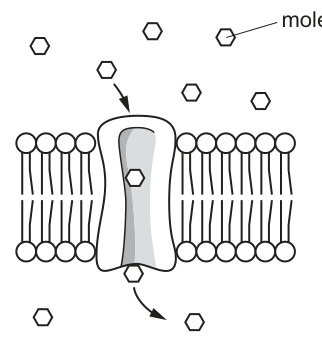
Which row shows a property of molecule X and the effect of the concentration of ATP in the cytoplasm on the rate of entry of molecule X?
Options
| property of molecule X | concentration of ATP in the cytoplasm | |
|---|---|---|
| A | non-polar | affects rate of entry of molecule X |
| B | non-polar | has no effect on rate of entry of molecule X |
| C | polar | affects rate of entry of molecule X |
| D | polar | has no effect on rate of entry of molecule X |
Working
The diagram shows molecule X passing through a transport protein in the phospholipid bilayer, moving from a region of higher concentration (outside) to lower concentration (inside).
- A transport protein is required because the molecule cannot cross the hydrophobic phospholipid bilayer directly — this means molecule X is polar.
- Movement down the concentration gradient through a protein is facilitated diffusion, which does not require ATP from the cytoplasm. Therefore the rate of entry is not affected by ATP concentration.
Answer
D
D
Background Concept
The cell-surface membrane is a phospholipid bilayer. The phospholipid "tails" (fatty acid chains) are hydrophobic, so:
- Non-polar / lipid-soluble molecules (e.g. O₂, CO₂, steroid hormones) dissolve in the bilayer and cross by simple diffusion, with no protein and no energy needed.
- Polar / charged molecules (e.g. glucose, amino acids, ions) cannot pass through the hydrophobic core and require a transport protein. They cross by either facilitated diffusion (down the concentration gradient, no ATP) or active transport (against the concentration gradient, requires ATP).
The key distinction between the two protein-mediated processes is therefore the direction of movement relative to the concentration gradient, and the dependence on ATP.
Understanding the Question
The diagram (Fig. 15.1) shows hexagon-shaped molecules of X outside the cell, with arrows indicating net movement of X through a membrane-spanning transport protein into the cytoplasm where the concentration of X is lower. The question asks for two pieces of information:
- Whether molecule X is polar or non-polar.
- Whether the cytoplasmic ATP concentration affects the rate of entry.
Each option pairs a property of X with an ATP effect, so both must be correct for the option to be the answer.
Approach
- Look at the direction of movement: if it is down the concentration gradient, it is facilitated diffusion; if up, it is active transport.
- Use the type of transport protein involved to infer the polarity of X.
- Decide whether ATP is needed and therefore whether changing ATP concentration would affect the rate.
Step-by-Step Reasoning
-
Identify the transport process. The diagram shows more molecules of X outside than inside, and X is moving from outside to inside — i.e. down its concentration gradient. Movement down a gradient through a protein is facilitated diffusion.
-
Infer the polarity of X. Facilitated diffusion via a transport protein is needed precisely because X cannot cross the hydrophobic phospholipid bilayer unaided. This is characteristic of polar (or charged) molecules. Non-polar molecules would diffuse straight through the bilayer without a protein, so options A and B (which say X is non-polar) are inconsistent with the diagram.
-
Decide the role of ATP. Facilitated diffusion uses only the kinetic energy of the molecules and the binding/release of X by the protein — no ATP is hydrolysed. Changing the cytoplasmic ATP concentration therefore has no effect on the rate of entry of X.
-
Match to the options. The only option with both correct statements (polar; ATP has no effect) is D.
Key Takeaways
- Non-polar molecules cross the membrane by simple diffusion (no protein, no ATP).
- Polar molecules require a transport protein, crossing either by facilitated diffusion (down the gradient, no ATP) or active transport (against the gradient, ATP-dependent).
- Looking at the direction of the concentration gradient in any membrane-transport diagram is the fastest way to distinguish facilitated diffusion from active transport.
Common Mistakes
- Choosing C — assuming that any transport via a protein must use ATP. The presence of a protein indicates only that the molecule is polar, not that the process is active transport.
- Choosing A or B — judging polarity by the shape of the hexagon in the diagram. Polarity is not visible; it must be inferred from whether a protein is required.
- Confusing facilitated diffusion and active transport. Both use proteins, but only active transport moves substances against the gradient and consumes ATP.
Things to Be Careful About
- Always read both the concentration gradient direction and the type of protein involved from the diagram before deciding between facilitated diffusion and active transport.
- "Non-polar" molecules do not typically need a channel/carrier protein; if the diagram shows a protein in use, the molecule is almost certainly polar.
- ATP dependence is a property of the process, not of the molecule. The same polar molecule could, in a different scenario, be actively transported, in which case ATP concentration would matter — the answer here depends on the specific mechanism shown.
The electron micrograph shows some human blood cells.
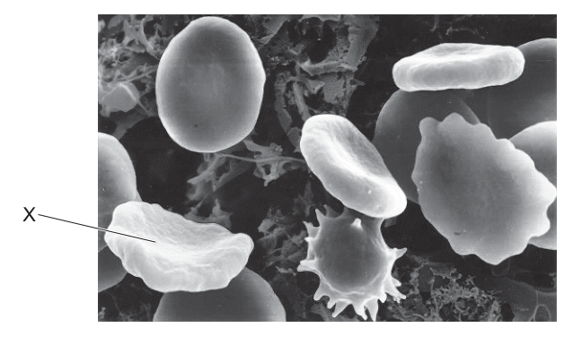
Which row correctly shows the net movement of water by osmosis and the water potential of the cytoplasm of cell X compared with the solution surrounding the cells?
Options
| net movement of water by osmosis | water potential of cytoplasm of cell X compared with the solution | |
|---|---|---|
| A | into the cell | higher |
| B | into the cell | lower |
| C | out of the cell | higher |
| D | out of the cell | lower |
Working
Cell X is crenated (shrivelled with a spiky, irregular surface). Crenation occurs when a red blood cell loses water, so the net movement of water by osmosis is OUT of the cell.
Water moves by osmosis from a region of higher water potential to a region of lower water potential. Since water left the cell, the cytoplasm of cell X must have had a higher water potential than the surrounding solution (the solution is more concentrated, i.e. hypertonic to the cell).
Answer
C
C
Background Concept
Osmosis is the net movement of water molecules across a partially permeable membrane from a region of higher water potential () to a region of lower water potential (). Pure water has a water potential of (the highest possible value); adding solutes lowers the water potential, making it more negative. A solution is described as:
- hypertonic to a cell if it has a lower (more negative) water potential than the cell cytoplasm — so water leaves the cell by osmosis;
- hypotonic to a cell if it has a higher (less negative) water potential — so water enters the cell by osmosis;
- isotonic if the water potentials are equal — no net movement of water.
Red blood cells (erythrocytes) are useful indicators of osmotic conditions because their flexible membranes allow visible shape changes:
- In a hypotonic solution the cell swells and may burst (haemolysis).
- In an isotonic solution the cell retains its normal biconcave disc shape.
- In a hypertonic solution the cell shrinks and the membrane develops spiky projections — this shrivelled, spiky appearance is called crenation.
Understanding the Question
The electron micrograph (Fig. 16.1) shows several red blood cells in a solution. Cell X has a shrivelled, spiky outline — it is crenated. The question asks two linked things:
- The direction of the net movement of water by osmosis across the membrane of cell X.
- How the water potential of the cytoplasm of cell X compares with that of the surrounding solution.
The command word is implicit: you must select the row where BOTH statements are correct.
Approach
The visual cue (crenation) is the key. Crenation means the cell has lost water to the solution, so:
- Step 1: Direction of water movement → out of the cell.
- Step 2: Apply the rule "water moves from high to low " → the cytoplasm of cell X has a higher water potential than the surrounding solution.
- Step 3: Match these two conclusions to the correct row in the table.
Step-by-Step Reasoning
Step 1 — Read the image. Cell X is clearly crenated. Unlike the other red cells in the micrograph (which look relatively smooth and disc-shaped, except one that is also visibly crenated in the middle of the image), cell X has an irregular, shrivelled outline with spike-like projections. This appearance is the diagnostic feature of a red blood cell that has lost water to a hypertonic external solution.
Step 2 — Net direction of water movement. Because cell X has lost volume, water has moved out of the cell. So the net movement of water by osmosis is out of the cell.
- This immediately rules out options A and B (both of which claim water moves into the cell).
Step 3 — Compare water potentials. Osmosis moves water from higher to lower . Since water moved out of cell X, the cytoplasm of cell X must have had the higher water potential; the surrounding solution must have had the lower (more negative) water potential, making it hypertonic to the cell.
- Option D says "out of the cell" (correct direction) but "lower" water potential for the cytoplasm (incorrect — this would mean water should move INTO the cell, not out). So D is internally inconsistent.
- Option C says "out of the cell" AND "higher" water potential for the cytoplasm — both statements are correct and consistent.
Step 4 — Final selection. Row C is the only one with both correct and consistent statements, so the answer is C.
Key Takeaways
- Crenation of a red blood cell = water has left the cell = surrounding solution is hypertonic.
- The direction of osmosis is always from a region of higher water potential to a region of lower water potential.
- For a cell that has lost water: cytoplasm is higher than surrounding solution (the solution is more negative).
- Always check that both columns of a two-part MCQ are individually correct AND mutually consistent before choosing the answer.
Common Mistakes
- Confusing crenation with the normal red-cell shape. If a student mistakenly thinks cell X is a healthy biconcave disc, they may wrongly select "into the cell".
- Mixing up "higher" and "lower" water potential. Students often think that a concentrated solution (which causes crenation) has a "higher" water potential because it has more solutes. In fact, more solute → lower (more negative) water potential. The mnemonic: solute lowers .
- Selecting option D because the direction of water movement is right but forgetting that the cytoplasm must be the source (higher ) when water is leaving the cell.
Things to Be Careful About
- Water potential values are negative (or zero for pure water). When you say one region has a "higher" water potential, you mean it is less negative — closer to zero. Don't be tempted to write "positive water potential" for the cytoplasm.
- The question is a two-part MCQ. Both columns must be correct, AND the two statements must logically agree with each other (the direction of osmosis must match the water-potential comparison). Cross-check before finalising your answer.
- The crenated appearance is the diagnostic clue — always look carefully at the labelled cell rather than assuming it looks like the textbook "normal" red blood cell.
A red indicator solution was mixed with agar and the resulting solid was cut into small cylindrical blocks. The blocks were placed in an acid which turns the indicator yellow and all other variables were kept constant. The dimensions of the blocks are shown.
block 1: height , diameter
block 2: height , diameter
block 3: height , diameter
The formula for calculating the surface area of a cylinder is . The formula for calculating the volume of a cylinder is .
Which row shows the correct surface area (SA) to volume (V) ratio for each block and the time taken for the block to turn yellow?
Options
| block 1 SA to V ratio | block 1 time to turn yellow/ mins | block 2 SA to V ratio | block 2 time to turn yellow/ mins | block 3 SA to V ratio | block 3 time to turn yellow/ mins | |
|---|---|---|---|---|---|---|
| A | 0.75 : 1.0 | 4 | 1.5 : 1.0 | 5 | 2.0 : 1.0 | 11 |
| B | 0.75 : 1.0 | 11 | 1.5 : 1.0 | 5 | 2.0 : 1.0 | 4 |
| C | 1.33 : 1.0 | 4 | 0.67 : 1.0 | 5 | 0.5 : 1.0 | 11 |
| D | 1.33 : 1.0 | 11 | 0.67 : 1.0 | 5 | 0.5 : 1.0 | 4 |
Working
For a cylinder:
Block 1 (r = 3 mm, h = 3 mm):
Block 2 (r = 6 mm, h = 6 mm):
Block 3 (r = 8 mm, h = 8 mm):
A higher SA:V ratio allows faster diffusion. Block 1 has the largest SA:V, so it turns yellow fastest (4 min). Block 3 has the smallest SA:V, so it takes the longest (11 min). Block 2 is intermediate (5 min).
Answer
C
C
Background Concept
Every cell (and every multicellular organism) exchanges substances — oxygen, carbon dioxide, nutrients, waste — with its surroundings across its outer surface. The rate at which a substance can reach every part of an organism depends on two things: how fast it diffuses through the tissue, and the surface area available for it to cross. The amount of tissue that needs to be supplied, on the other hand, is proportional to the volume. So the critical ratio is surface area : volume (SA:V).
For any regular shape, as the object gets bigger, its volume grows faster than its surface area (volume scales with the cube of the linear dimension, surface area with the square). This means larger objects have a smaller SA:V ratio. Since diffusion can only bring material in across the surface, a small SA:V means the centre of the object is far from any surface and is supplied slowly.
For a cylinder:
Dividing gives the convenient form:
Understanding the Question
The question simulates cells of different sizes using agar cylinders containing a pH indicator. When the agar is placed in acid, the acid diffuses in from the outside and turns the indicator yellow. The block that turns yellow fastest is the one with the largest SA:V — diffusion reaches its centre quickly. The block that takes longest is the one with the smallest SA:V — diffusion has a relatively long way to go to the centre.
We must:
- Calculate the SA:V for each of the three cylinders using the given dimensions.
- Match the SA:V to the correct time, recognising that SA:V and diffusion time are inversely related.
Approach
Use the simplified formula SA/V = 2/r + 2/h for each block, then use the biological principle that higher SA:V = faster diffusion = shorter time to turn yellow, and rank the times accordingly.
Step-by-Step Reasoning
Step 1 — Block 1 (radius = 3 mm, height = 3 mm):
Step 2 — Block 2 (radius = 6 mm, height = 6 mm):
Step 3 — Block 3 (radius = 8 mm, height = 8 mm):
Step 4 — Match to times. Block 1 has the largest SA:V (1.33), so acid reaches its centre fastest → shortest time = 4 min. Block 3 has the smallest SA:V (0.50), so diffusion is slowest → longest time = 11 min. Block 2 is intermediate → 5 min.
This matches row C: 1.33 / 4 min; 0.67 / 5 min; 0.50 / 11 min.
Key Takeaways
- The SA:V ratio of a cylinder simplifies to 2/r + 2/h, which lets you rank sizes without computing full areas and volumes.
- A higher SA:V means a substance diffuses into the object faster — the centre is reached sooner.
- This is the same principle that limits how large single cells (or organisms without a circulatory system) can be: beyond a certain size, diffusion alone cannot supply the interior fast enough.
Common Mistakes
- Calculating full SA and V values and dividing them — this gives the right answer but invites arithmetic errors. The shortcut 2/r + 2/h is faster and less error-prone.
- Forgetting that the time is inversely related to SA:V. A larger SA:V means a shorter time. Candidates who reverse this pick option B or D.
- Confusing radius and diameter. The question gives the diameter (6, 12, 16 mm), so the radii are 3, 6, 8 mm. Using diameters instead doubles the ratios and leads to a wrong row.
- Reading the options carelessly — row A and row C have the same SA:V ratios (just reordered) so it is easy to pick the wrong one if you do not check the time column too.
Things to Be Careful About
- Always halve the diameter to get the radius before using r in the formula.
- The two formula paths (full SA ÷ V, or the shortcut 2/r + 2/h) must give the same ratio; the shortcut is just algebraically equivalent.
- The trend in time must be monotonic with the SA:V trend: 1.33 → 4 min (shortest), 0.67 → 5 min, 0.50 → 11 min (longest). Any answer that pairs the shortest time with the smallest SA:V is wrong on biological grounds alone, even before checking the numbers.
Which metabolic processes will be very active in a cell that has just completed cytokinesis?
1 ATP formation
2 DNA replication
3 protein synthesis
Options
A 1, 2 and 3
B 1 and 3 only
C 2 only
D 3 only
Working
Cytokinesis completes the M phase; the daughter cell then enters G1 of interphase. In G1 the cell is metabolically active but DNA is not yet being replicated (replication occurs in S phase).
- 1 ATP formation – respiration is continuous, so ATP production is active. ✔
- 2 DNA replication – does not occur until S phase, which comes after G1. ✘
- 3 Protein synthesis – the cell must grow in G1, producing enzymes and structural proteins; transcription and translation are active. ✔
Answer
B
B
Background Concept
The cell cycle has two major parts: interphase (G1, S, G2) and mitosis (M phase) followed by cytokinesis. Each daughter cell produced by cytokinesis enters a new cycle beginning with G1. During G1 the cell grows, performs normal metabolism, and prepares the machinery and nucleotides needed for DNA synthesis, but the DNA itself is not yet replicated. DNA replication is confined to S phase. Throughout all of interphase (and indeed throughout the life of the cell), respiration occurs continuously to supply ATP, and transcription/translation operate to replace proteins and to produce the proteins required for the next stages of the cycle.
Understanding the Question
A cell that has just completed cytokinesis is at the very start of G1. The question asks which of the three listed processes are very active at this point. We have to decide for each of the three options whether it is happening strongly immediately after cytokinesis.
Approach
Place cytokinesis on the cell-cycle timeline, identify the phase the cell is now in, then test each numbered process against the activities characteristic of that phase.
Step-by-Step Reasoning
- Locate cytokinesis on the cell cycle. Mitosis (prophase, metaphase, anaphase, telophase) ends with cytokinesis. The new daughter cell then re-enters interphase, starting at G1.
- Test 1 — ATP formation. ATP is made continuously by respiration because every living cell requires a constant supply of energy to maintain ion gradients, carry out biosynthesis, and power motor proteins. Therefore ATP formation is very active in G1. ✔
- Test 2 — DNA replication. Replication is restricted to S phase, which is downstream of G1. In early G1 the DNA is not being replicated; instead, the cell is synthesising nucleotides, histones and the replication enzymes in preparation. ✘
- Test 3 — protein synthesis. G1 is a growth phase. The cell must produce new proteins — enzymes for metabolism and DNA replication, structural proteins, and regulatory proteins such as cyclins — so transcription and translation are very active. ✔
- Combine the answers. Only 1 and 3 are correct, which matches option B.
Key Takeaways
- Cytokinesis produces two daughter cells that each start a new cell cycle at G1.
- In G1 the cell grows and prepares for DNA replication, but does not yet replicate DNA.
- DNA replication occurs only in S phase, not in G1, G2, mitosis or cytokinesis.
- Respiration (ATP formation) and protein synthesis run throughout interphase, including G1.
Common Mistakes
- Choosing A (1, 2 and 3) because protein synthesis is active — but forgetting that DNA replication does not start until S phase, not immediately after cytokinesis.
- Choosing C (2 only) by confusing cytokinesis with the start of S phase, or assuming that the cell must immediately copy its DNA.
- Choosing D (3 only) and overlooking that ATP production by respiration is a continuous, very active process in every living cell, not just dividing cells.
Things to Be Careful About
- "Just completed cytokinesis" specifically means early G1, not S phase and not "any time in interphase".
- "Metabolic processes" refers to the cell's own biochemical activity, not the events of mitosis itself.
- ATP formation is treated as a metabolic process that is always on; do not assume it is restricted to specific phases.
- "Very active" is the wording — both ATP formation and protein synthesis are sustained, high-rate processes during G1, whereas DNA replication is essentially zero at this point.
The diagram shows a typical mitotic cell cycle and the point in the cell cycle that has been reached by each of four cells, V, W, X and Y.
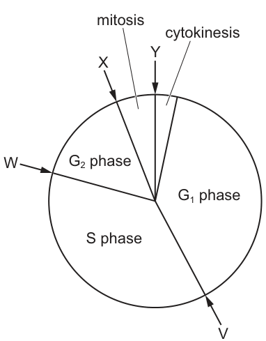
Which row correctly identifies the cells that match the two descriptions?
Options
| DNA replication is complete but the cell has not yet reached its maximum size | preparation for microtubule formation is nearly complete but chromosomes have not yet condensed | |
|---|---|---|
| A | V | X |
| B | W | Y |
| C | V | Y |
| D | W | X |
Working
Description 1: "DNA replication is complete but the cell has not yet reached its maximum size."
- DNA replication occurs during S phase and is complete by the end of S phase.
- The cell continues to grow and synthesise proteins during G2 phase, only reaching its maximum size at the end of G2.
- Cell W is positioned in the middle of G2 phase, so DNA replication is complete but the cell is still growing.
- ➜ W matches description 1.
Description 2: "Preparation for microtubule formation is nearly complete but chromosomes have not yet condensed."
- During G2, the centrosomes duplicate and microtubules are organised in preparation for the mitotic spindle.
- Chromosome condensation begins at the start of mitosis (prophase).
- Cell X is at the very beginning of mitosis, where microtubule preparation is essentially complete but chromosomes have not yet condensed.
- ➜ X matches description 2.
Answer
D
D
Background Concept
The mitotic cell cycle is a continuous, ordered sequence of events that prepares a cell to divide into two genetically identical daughter cells. It is conventionally divided into:
- G1 phase – cell grows, synthesises proteins and organelles, carries out normal metabolism.
- S phase – DNA replication occurs; each chromosome is copied to form two sister chromatids joined at the centromere.
- G2 phase – cell continues to grow, produces additional proteins, and prepares for mitosis. Crucially, the centrosomes (which organise microtubules) duplicate, and tubulin is synthesised so that the mitotic spindle can be built.
- Mitosis (M phase) – nuclear division, comprising prophase, metaphase, anaphase and telophase. Chromosomes condense, align, separate and decondense.
- Cytokinesis – division of the cytoplasm to give two daughter cells.
A useful way to think about G2 is: DNA is already duplicated, but the cell is still finishing its growth and assembling the machinery (centrosomes, tubulin, spindle components) needed for mitosis. Chromosome condensation is the visual hallmark that mitosis has begun (prophase).
Understanding the Question
The question shows a circular cell-cycle diagram with four labelled cells:
- V – early S phase
- W – middle of G2 phase
- X – beginning of mitosis (early prophase)
- Y – end of mitosis / start of cytokinesis
It then gives two descriptions and asks which row correctly assigns a cell to each description. The first description concerns completion of DNA replication versus cell size; the second concerns microtubule preparation versus chromosome condensation.
Approach
For each description, identify the cell-cycle phase in which the described state exists, then read off the diagram which labelled cell lies in that phase.
- DNA replication completes at the end of S phase; the cell then grows through G2. So the description matches the G2 phase.
- Microtubule (spindle) preparation is largely a G2 activity, while chromosome condensation is a mitotic (prophase) event. The transition point — where microtubule prep is essentially done but condensation has not yet happened — corresponds to the G2/M boundary or very early prophase.
Step-by-Step Reasoning
Description 1 – DNA replicated, cell not yet at maximum size:
- DNA replication is finished by the end of S phase. The cell now contains 4C DNA content (each chromosome has two chromatids).
- Maximum cell size is reached at the end of G2, just before mitosis.
- A cell "halfway" through G2 therefore satisfies the description: replication is complete, growth is ongoing.
- Looking at the diagram, W is positioned in the middle of G2 → W is the match.
- V is in S phase (DNA still being replicated), so V fails the first condition. X and Y are already in/after mitosis, so they are past the growth phase entirely.
Description 2 – microtubule prep nearly complete, chromosomes not yet condensed:
- Centriole/centrosome duplication finishes in G2; tubulin is stockpiled; microtubule organising centres (MTOCs) are positioned at opposite poles.
- Chromosome condensation is the defining event of prophase (start of mitosis) and is not a G2 event.
- The cell must therefore be at the G2/M transition or in very early prophase, where the spindle components are essentially ready but the chromatin is still dispersed (chromosomes have not yet condensed).
- X is labelled at the beginning of mitosis → X is the match.
- W (mid-G2) would still be actively preparing microtubules, so the prep is not yet "nearly complete". Y is past mitosis — condensation has long since occurred.
Row D (W for description 1, X for description 2) is therefore correct.
Key Takeaways
- G1 = growth; S = DNA replication; G2 = further growth + preparation for mitosis (centrosome duplication, tubulin synthesis).
- Mitosis begins with chromosome condensation in prophase; this is the first visible mitotic event.
- The cell does not reach maximum size until the end of G2; once mitosis begins, the cell is already at peak size.
- Microtubule/spindle preparation occurs in G2, before chromosome condensation, so a cell just entering mitosis is the stage where "spindle prep is nearly complete but chromosomes are not yet condensed".
Common Mistakes
- Confusing V (S phase) with the cell that has completed DNA replication — DNA replication is still in progress in S phase, not complete.
- Selecting Y for description 2 because it is "near the end of the cycle" — by cytokinesis the chromosomes have long since condensed, separated and decondensed.
- Assuming G2 cells have already begun condensing chromosomes — condensation is a mitotic, not an interphase, event.
- Thinking microtubule formation only happens during mitosis — the preparation (centrosome duplication, tubulin synthesis) is a G2 activity; the spindle is then assembled in prophase and metaphase.
Things to Be Careful About
- "Maximum size" is reached at the end of G2, not the start, so a mid-G2 cell (W) correctly matches "not yet at maximum size".
- "Preparation for microtubule formation" refers to the G2 work (centrosomes, tubulin); the actual spindle microtubules polymerise in prophase and metaphase. Reading the wording carefully is essential.
- The cell-cycle diagram is circular, so reading the position of each arrow relative to the phase boundaries (not just "near the top") is what determines the answer.
The graph shows the mean length of the spindle fibres during mitosis.
Which region of the graph shows when all the centromeres have detached from the spindle fibres?
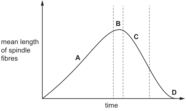
Options
A A
B B
C C
D D
Working
During mitosis the spindle fibres change in length as follows:
- Region A (length increasing): prophase → metaphase — spindle fibres are being assembled and elongating as they attach to centromeres and align chromosomes at the equator.
- Region B (peak): metaphase — maximum spindle length, with all centromeres still attached to spindle fibres via their kinetochores.
- Region C (length decreasing): anaphase — spindle fibres shorten, pulling sister chromatids towards opposite poles. The centromeres are still attached throughout anaphase.
- Region D (level off at low value): telophase/end of mitosis — the spindle apparatus breaks down. Once the spindle depolymerises, the centromeres are no longer attached to spindle fibres.
The centromeres detach from the spindle fibres when the spindle itself disintegrates, which corresponds to the region where spindle fibre length has fallen to a low, stable value — region D.
Answer
D
D
Background Concept
Mitosis is the division of a nucleus into two genetically identical daughter nuclei. Its accuracy depends on the spindle apparatus — a structure made of microtubules (spindle fibres) that physically separates the sister chromatids of each chromosome.
Key structural points to keep in mind:
- Each chromosome at the start of mitosis consists of two sister chromatids joined at a centromere (a constricted region of DNA-protein that forms the kinetochore — the actual attachment site for spindle microtubules).
- Spindle fibres emanate from the two centrosomes (poles) at opposite ends of the cell.
- Kinetochore microtubules attach to the kinetochore at the centromere; polar/interpolar microtubules overlap in the middle and push the poles apart.
The four key stages and what happens to the spindle and centromeres:
- Prophase: spindle fibres begin to assemble and grow; they reach out and attach to kinetochores on the centromeres.
- Metaphase: chromosomes are aligned at the cell equator; spindle length is at its maximum because the poles have been pushed apart by polar microtubule elongation; all centromeres are firmly attached.
- Anaphase: centromeres split, and the kinetochore microtubules shorten, pulling sister chromatids (now individual chromosomes) to opposite poles. Throughout anaphase the centromeres remain attached to shrinking spindle fibres.
- Telophase: chromatids reach the poles, the spindle apparatus depolymerises (the microtubules break down into tubulin subunits), nuclear envelopes reform, and cytokinesis follows. Only when the spindle breaks down do the centromeres finally lose their attachment to spindle fibres.
Understanding the Question
The graph plots the mean length of spindle fibres on the y-axis against time on the x-axis, with four labelled regions:
- A: rising portion
- B: peak
- C: falling portion
- D: low, level portion (the tail)
The question asks which region corresponds to the moment when all centromeres have detached from the spindle fibres. The key biological event we are looking for is therefore the breakdown of the spindle apparatus at the end of mitosis, not the moment chromatids separate (that happens during the falling phase, when centromeres are still attached).
The command word is implicit "which" — a single choice, not an explanation — so the answer is a letter.
Approach
Link each region of the graph to a stage of mitosis by recalling what the spindle apparatus is doing at each stage. Then identify the stage at which centromere–spindle attachment is finally lost (the spindle disassembles), and pick the corresponding region on the graph.
Step-by-Step Reasoning
-
Region A — spindle fibres are growing.
This is prophase/prometaphase, when the spindle is being built. Length increases as microtubules polymerise and as the two poles move apart. Centromeres are being captured by kinetochore microtubules, not detached. -
Region B — spindle at its maximum length.
This is metaphase: chromosomes are aligned at the equator, the cell is at its widest pole-to-pole, and every centromere is held under tension by attached kinetochore fibres. All centromeres are still attached — the opposite of what the question asks. -
Region C — spindle fibres shortening.
This is anaphase. Kinetochore microtubules depolymerise and shorten, drawing the separated chromatids towards the poles. Although the length is decreasing rapidly, the centromeres are still attached throughout anaphase — that is precisely how the chromatids are being moved. So C is not the answer either. -
Region D — spindle length at a low, stable value.
This is telophase / the end of mitosis. The spindle apparatus has largely depolymerised, the microtubules have broken down into tubulin subunits, and the centromeres are no longer attached to any spindle fibres. This is the only region in which "all the centromeres have detached from the spindle fibres" is true.
Therefore the correct answer is D.
Key Takeaways
- The graph of mean spindle length is a "rise – peak – fall – plateau" curve that maps cleanly onto prophase → metaphase → anaphase → telophase.
- Centromeres remain attached to spindle fibres from prometaphase all the way through anaphase — the shortening of fibres in anaphase is what moves the chromatids, not a sign of detachment.
- Centromeres only detach from spindle fibres when the spindle apparatus itself breaks down in telophase, corresponding to the low, level tail (region D) of the graph.
Common Mistakes
- Choosing C because the curve is falling. A falling curve suggests "detachment" intuitively, but in anaphase the centromeres are still very much attached — the chromatids are being pulled precisely because of that attachment.
- Choosing B because the spindle is at its biggest. At maximum length, every centromere is still firmly attached, not detached.
- Confusing centromere splitting (anaphase) with centromere detachment (telophase). The centromere splits so the two chromatids can separate, but each new centromere remains attached to a spindle fibre until the spindle breaks down.
Things to Be Careful About
- "Centromere detachment" ≠ "centromere splitting." The centromere splits in anaphase; it detaches from spindle fibres in telophase.
- Read the graph by its shape, not just its trend: the low, flat tail (D) signals a completed process — the spindle is gone.
- Keep the four mitotic stages and the four spindle-length regions paired in your memory: A = prophase, B = metaphase, C = anaphase, D = telophase. This pairing answers many spindle-related questions on this topic.
The mRNA codons ACU, ACC, ACA and ACG all code for the same amino acid, threonine.
Which anticodons could specify an amino acid other than threonine?
1 UCA
2 ACC
3 UGU
4 UGC
Options
A 1, 3 and 4
B 1 and 2
C 2 and 3
D 3 and 4 only
Working
The threonine codons are ACU, ACC, ACA and ACG. The tRNA anticodon must be complementary to the codon (A–U, C–G).
Complementary anticodons for the threonine codons:
- ACU → UGA
- ACC → UGG
- ACA → UGU
- ACG → UGC
Checking each option:
- UCA — not complementary to any threonine codon → would carry a different amino acid ✓
- ACC — not complementary to any threonine codon → would carry a different amino acid ✓
- UGU — complementary to ACA (threonine) → would carry threonine ✗
- UGC — complementary to ACG (threonine) → would carry threonine ✗
Only 1 and 2 specify an amino acid other than threonine.
Answer
B
B
Background Concept
The genetic code is read in triplets of bases (codons) on mRNA, with each codon specifying one amino acid. The code is degenerate — several different codons can code for the same amino acid. Threonine, for example, is specified by four codons: ACU, ACC, ACA and ACG.
During translation, a tRNA molecule carries a specific amino acid and has a three-base anticodon that base-pairs with the mRNA codon. The base-pairing rules are the same as in DNA–RNA hybrid pairing: adenine (A) pairs with uracil (U), and cytosine (C) pairs with guanine (G).
If a tRNA's anticodon is complementary to one of the threonine codons, that tRNA will carry threonine. If the anticodon is NOT complementary to any threonine codon, the tRNA must carry a different amino acid.
Understanding the Question
The question gives four threonine codons and four candidate anticodons. It asks which of those anticodons could be carried by a tRNA that delivers an amino acid other than threonine — i.e. which anticodons are NOT complementary to any threonine codon.
The command word is implicit but the logic is: find the anticodons that would NOT pair with ACU, ACC, ACA or ACG.
Approach
- For each threonine codon, work out its complementary anticodon (A↔U, C↔G).
- Compare the four candidate anticodons against these complements.
- Any candidate that is NOT among the four complements must belong to a tRNA carrying a different amino acid.
Step-by-Step Reasoning
Step 1 — write the complementary anticodon for each threonine codon:
| mRNA codon | Complementary anticodon |
|---|---|
| ACU | UGA |
| ACC | UGG |
| ACA | UGU |
| ACG | UGC |
Step 2 — check each option:
- Option 1 (UCA): The complement of UCA is AGU, which is not one of the threonine codons. So this anticodon is NOT complementary to any threonine codon → the tRNA carries a different amino acid. ✓
- Option 2 (ACC): The complement of ACC is UGG, which is not one of the threonine codons. So this anticodon is NOT complementary to any threonine codon → the tRNA carries a different amino acid. ✓ (Note: ACC happens to BE a threonine codon, but as an anticodon it would not pair with any threonine codon.)
- Option 3 (UGU): The complement of UGU is ACA — a threonine codon. So this tRNA carries threonine. ✗
- Option 4 (UGC): The complement of UGC is ACG — a threonine codon. So this tRNA carries threonine. ✗
Step 3 — combine: Options 1 and 2 specify an amino acid other than threonine. The correct answer is B (1 and 2).
Key Takeaways
- Anticodons are complementary (not identical) to their codons: A pairs with U, C pairs with G.
- Because the genetic code is degenerate, multiple codons can specify one amino acid — and a tRNA whose anticodon matches ANY of those codons will deliver that amino acid.
- An anticodon that does not complement any of the codons for amino acid X must belong to a tRNA that carries a different amino acid.
- A common trap: an option that looks like a codon for the same amino acid (e.g. ACC, which is itself a threonine codon) is NOT a valid anticodon for threonine — it has to be the complement.
Common Mistakes
- Treating an anticodon as identical to the codon instead of complementary. This is the single most common error and would lead students to pick options 3 and 4 (which are the complements of threonine codons).
- Confusing which strand is which: the codon is on mRNA, the anticodon is on tRNA. They pair by base complementarity, not by sequence identity.
- Forgetting that the code is degenerate: students sometimes think there is only one codon per amino acid, and ignore the other three threonine codons.
Things to Be Careful About
- Read each option carefully — option 2 (ACC) is itself a threonine codon, but as an anticodon it is invalid for threonine because it is not complementary to any threonine codon.
- Use the correct base-pairing rules: in RNA, A pairs with U (not T), and C pairs with G.
- When the mark scheme offers alternatives, choose the option that lists only the genuinely non-threonine anticodons; "3 and 4 only" (option D) is a distractor that looks attractive because UGU and UGC are clearly related to the threonine codons, but they actually code for threonine.
Which bond formation does DNA polymerase catalyse?
Options
A hydrogen bonds between bases
B hydrogen bonds between nucleotides
C phosphodiester bonds between bases
D phosphodiester bonds between nucleotides
DNA polymerase joins adjacent nucleotides by forming phosphodiester bonds between the 3′-OH of one nucleotide's deoxyribose sugar and the 5′-phosphate group of the next nucleotide, building the sugar–phosphate backbone of the new DNA strand. Hydrogen bonds between complementary bases form spontaneously by base pairing and are not catalysed by DNA polymerase.
Answer
D
D
Background Concept
DNA is a polymer of deoxyribonucleotides. Each nucleotide has three components: a deoxyribose sugar, a phosphate group attached to the 5′ carbon of the sugar, and a nitrogenous base (A, T, G or C) attached to the 1′ carbon. Nucleotides are linked into a single strand by phosphodiester bonds — covalent bonds that form between the 3′-OH of the sugar of one nucleotide and the 5′-phosphate of the next. The two antiparallel strands of the double helix are held together by hydrogen bonds between complementary base pairs (A=T, two H-bonds; G≡C, three H-bonds).
During DNA replication, the new strand is built one nucleotide at a time. DNA polymerase is the enzyme that assembles the new strand, but it does two chemically distinct jobs and it is important not to confuse them:
- It catalyses the formation of the phosphodiester bond between an incoming 5′-dNTP and the free 3′-OH of the growing strand, releasing pyrophosphate. This is the covalent joining of nucleotides into the sugar–phosphate backbone.
- It does not form the hydrogen bonds between complementary bases. These hydrogen bonds form spontaneously once the correct base is positioned in the active site, governed by base-pairing rules; no enzyme is required to make them.
Understanding the Question
This is a multiple-choice question asking specifically which type of bond DNA polymerase catalyses. Each option combines one of two bond types (phosphodiester vs hydrogen) with one of two locations (between bases vs between nucleotides), so the candidate must identify both the bond type and the components being joined.
Approach
Recall the precise substrate and chemistry of the DNA polymerase reaction: it joins a free nucleotide to the end of a growing nucleotide chain, building the backbone — i.e. phosphodiester bond between nucleotides.
Step-by-Step Reasoning
- Eliminate hydrogen-bond options (A and B). Hydrogen bonds between complementary bases form by base-pairing rules and do not require catalysis; they form and break during strand separation and re-annealing. DNA polymerase does not catalyse hydrogen-bond formation, so A and B are wrong.
- Eliminate option C. "Phosphodiester bonds between bases" is chemically meaningless — phosphodiester bonds are part of the sugar–phosphate backbone, not the base-pairing interface. Bases are joined only by hydrogen bonds to their complementary partner on the opposite strand. So C is wrong.
- Confirm D. DNA polymerase adds dNTPs to the free 3′-OH of the growing strand, forming a phosphodiester bond between adjacent nucleotides. This is the only option that correctly names both the bond and the components being joined.
Key Takeaways
- DNA polymerase catalyses phosphodiester bond formation between nucleotides (the covalent sugar–phosphate backbone linkage).
- Hydrogen bonds between complementary bases form by base pairing and do not require an enzyme.
- The sugar–phosphate backbone is built by repeated phosphodiester bonds; the two strands of the double helix are held together by hydrogen bonds between bases.
Common Mistakes
- Choosing A because students associate "bond formation" loosely with DNA and forget that hydrogen bonds form without an enzyme.
- Choosing B for the same reason, or because they misremember hydrogen bonds as being "between nucleotides" rather than "between bases".
- Choosing C by confusing the location of the phosphodiester bond (sugar–phosphate backbone) with the location of hydrogen bonds (between bases).
Things to Be Careful About
- Read the wording precisely: "between bases" vs "between nucleotides" is a deliberate discriminator.
- Phosphodiester bonds always link the 3′ carbon of one sugar to the 5′ phosphate of the next sugar — never directly between bases.
- The complementary base pairing (A–T, G–C) is governed by hydrogen bonds, which is a separate structural feature from the covalent backbone.
In eukaryotes, the RNA molecules formed during transcription are modified by the removal of non-coding sequences. This is followed by the joining together of coding sequences to form mRNA.
What are the coding sequences also called?
Options
A codons
B exons
C introns
D primary transcripts
Working
In eukaryotes, the primary RNA transcript contains both coding and non-coding sequences. The non-coding sequences (introns) are removed and the coding sequences are joined together to form mature mRNA. The coding sequences are called exons.
Answer
B
B
Background Concept
In eukaryotic cells, transcription produces a primary RNA transcript (pre-mRNA) that contains both coding and non-coding regions. The non-coding regions, called introns (int-ervening sequences), must be removed before the mRNA can be translated. The coding regions, called exons (ex-pressed sequences), are the parts that are kept and joined together. This editing process is called splicing and is carried out by a ribonucleoprotein complex called the spliceosome. Once splicing is complete, the mature mRNA exits the nucleus and is translated at the ribosomes.
It is worth noting that the word "introns" contains the letters "in-tr-ons" (non-coding, removed), and "exons" contains "ex-ons" (expressed, coding). This etymology helps with recall.
Understanding the Question
The stem describes the standard process of eukaryotic mRNA maturation: non-coding sequences are removed and coding sequences are joined. The question simply asks for the name given to the coding sequences. The command word is "what are", so the answer is a single term. Note that "codons" refers to the three-base triplets within the mRNA that code for amino acids, not to the longer coding regions of a gene. "Primary transcripts" refers to the unmodified initial RNA product, not the retained segments.
Approach
Recall the splicing vocabulary:
- Exons = coding regions (kept)
- Introns = non-coding regions (removed)
Match this to the description in the stem.
Step-by-Step Reasoning
- The question describes two operations: (i) removal of non-coding sequences, and (ii) joining of coding sequences.
- The non-coding sequences removed are introns (intervening sequences).
- Therefore, the coding sequences that remain and are joined together must be the opposite — exons (expressed sequences).
- Eliminating the other options:
- A (codons) — these are triplets of bases within mature mRNA that each specify an amino acid; they are a sub-feature of the mRNA, not a name for the coding regions of the gene.
- C (introns) — these are the non-coding sequences, which by the question's wording have been removed.
- D (primary transcripts) — this is the name of the initial RNA copy before any modification, not a term for the coding segments within it.
- The correct answer is B (exons).
Key Takeaways
- In eukaryotic mRNA processing, introns are spliced out and exons are joined together.
- Splicing is a post-transcriptional modification that occurs in the nucleus before mRNA is exported to the cytoplasm for translation.
- Genes in eukaryotes are often described as "split genes" because they contain both introns and exons.
Common Mistakes
- Confusing exons and introns. A quick way to keep them straight: introns are intervening (removed, stay in the nucleus); exons are expressed (kept, leave the nucleus).
- Choosing "codons" because it sounds like "coding". Codons are three-base sequences on mature mRNA, not regions of a gene.
- Choosing "primary transcripts", which describes the unmodified RNA before splicing has occurred.
Things to Be Careful About
- The term "exon" applies to the region of the gene/transcript that is retained; it does not mean the entire mature mRNA is itself "an exon".
- Some genes have alternative splicing patterns where different combinations of exons are joined, producing different protein variants from a single gene — but in every case the joined units are still called exons.
Which row correctly identifies sinks for sucrose transported by mass flow in plants?
Options
| root storage organ | growing leaf bud | growing shoot tip | |
|---|---|---|---|
| A | ✓ | ✓ | ✓ |
| B | ✓ | ✗ | ✓ |
| C | ✗ | ✓ | ✗ |
| D | ✗ | ✗ | ✓ |
key
✓ = sink
✗ = not a sink
Working
In phloem transport, a sink is any region that uses or stores sucrose (it does not photosynthesise enough to meet its own needs, or stores assimilates).
- Root storage organ — stores sucrose (e.g. carrots, sugar beet); a sink. ✓
- Growing leaf bud — young, non-photosynthetic, needs sucrose for respiration and cell division; a sink. ✓
- Growing shoot tip — meristematic, needs sucrose for respiration and growth; a sink. ✓
All three are sinks, so row A is correct.
Answer
A
A
Background Concept
In vascular plants, sucrose (the main transport sugar) is moved through the phloem from sources to sinks by mass flow.
- A source is a region that produces more sucrose than it needs — typically a mature, photosynthesising leaf. Other sources include storage organs during mobilisation (e.g. a germinating seed's cotyledons, or a taproot at the start of the growing season).
- A sink is a region that uses or stores sucrose and cannot supply its own needs.
Common sinks include:
- Growing (meristematic) regions — root tips, shoot tips, young leaves, developing buds.
- Storage organs — roots (e.g. carrot, sugar beet), stems (e.g. sugarcane), fruits, seeds.
- Reproductive structures — flowers, fruits, developing seeds.
The direction of transport can actually change with the season: a taproot is a sink in summer (it stores sucrose made in the leaves) but becomes a source in spring (it releases sucrose to fuel the new growing shoot). The general rule, however, is that any organ which is not photosynthetically self-sufficient — or which actively accumulates assimilates — is acting as a sink.
Understanding the Question
The stem asks us to identify which row of the table correctly labels all three structures — root storage organ, growing leaf bud, and growing shoot tip — as sinks (✓) or not sinks (✗). Only one row is fully correct.
Approach
Ask the same question of each structure: Is this organ net-consuming or net-storing sucrose, rather than producing it? If yes, it is a sink. Work through all three, then match to the answer row.
Step-by-Step Reasoning
- Root storage organ (e.g. carrot, sugar beet): Its function is to accumulate sucrose translocated from photosynthesising leaves. By definition this is a sink. ✓
- Growing leaf bud: The bud is enclosed, not yet expanded, and has little/no photosynthetic capacity. It needs an incoming supply of sucrose to support rapid cell division and growth. This is a sink. ✓
- Growing shoot tip: The apical meristem is highly metabolically active (high respiration, new cell production) and is non-photosynthetic. It is a classic sink. ✓
All three are sinks, which matches row A (✓, ✓, ✓).
Key Takeaways
- Source = net producer of sucrose (mature photosynthesising leaf, mobilising storage organ).
- Sink = net consumer or storer of sucrose (meristems, young leaves, roots, fruits, storage organs, seeds).
- A structure's role as source or sink can change with developmental stage or season, but for an actively growing plant, growing tips/buds and storage roots are all sinks.
Common Mistakes
- Treating a growing leaf bud as a source because it will eventually become a photosynthesising leaf — at this stage it is small, enclosed, and entirely dependent on imported sucrose, so it is a sink.
- Confusing xylem (transports water and mineral ions from roots to shoots) with phloem (transports assimilates like sucrose from sources to sinks). The question is about phloem mass flow.
- Forgetting that a "storage" organ is, by definition, accumulating — therefore a sink.
Things to Be Careful About
- Watch the seasonal caveat: a root storage organ is a sink in summer and a source in spring, so context matters. The question's phrasing ("sinks for sucrose transported by mass flow") is the typical summer/end-of-growing-season scenario in which all three are sinks.
- The mark scheme rewards the candidate identifying all three correctly; missing one leads to the wrong row (B, C or D).
The diagram shows a transverse section through a transport tissue in a plant.
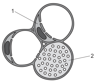
Which row correctly identifies cell 1 and cell 2?
Options
| cell 1 | cell 2 | |
|---|---|---|
| A | companion cell | phloem sieve tube element |
| B | companion cell | xylem vessel element |
| C | phloem sieve tube element | phloem sieve tube element |
| D | phloem sieve tube element | xylem vessel element |
Working
In a transverse section of plant transport tissue:
- A companion cell is small with dense cytoplasm and a prominent nucleus, sitting alongside a sieve tube element.
- A phloem sieve tube element is a larger cell with a thin wall and a sieve plate — the sieve plate appears as a row of pores (the small circles) crossing the lumen.
- A xylem vessel element is a large, empty (no cytoplasm at maturity) cell with a thick, lignified wall.
Cell 1 is small with dense contents next to a larger cell → companion cell.
Cell 2 shows a sieve plate (rows of pores) across the lumen → phloem sieve tube element.
Answer
A
A
Background Concept
Plants have two main transport tissues: xylem and phloem, each made of different cell types that are easy to tell apart in a transverse section (TS).
- Xylem vessels are dead at maturity. They have no cytoplasm or nucleus, and their walls are thickened with lignin. In TS they appear as large, empty cells with thick walls; in surface view the lignified wall often shows pits (small circular thin regions where water can pass sideways between vessels).
- Phloem sieve tube elements are living but enucleate at maturity. They are connected end-to-end by sieve plates — modified end walls perforated by pores through which assimilates (e.g. sucrose) flow. In TS, a sieve plate is seen as a row of small circular pores crossing the lumen of the cell. Each sieve tube element is paired with a companion cell — a small cell with dense cytoplasm and a nucleus that supplies the sieve tube element with ATP and metabolites (since the sieve tube element itself lacks many organelles).
Understanding the Question
We are shown a TS of plant vascular tissue and asked to identify two cells, labelled 1 and 2, from four options. The command word is "identifies", so we need to match the visible features to the correct cell type. The image shows:
- Two large empty cells with thick walls (top-right and bottom-left) — these are xylem vessel elements.
- A small dark, dense cell labelled 1.
- A cell with rows of small circles inside it, labelled 2.
Approach
The key diagnostic features to use are:
- Size and contents — companion cells are small and dense (lots of cytoplasm/nucleus); sieve tube elements are larger but still relatively thin-walled.
- Internal structures — sieve plates appear as pores in a sieve tube element; xylem vessel elements look empty in the lumen.
- Wall thickness — xylem walls are thickly lignified; phloem walls are thin (cellulose only).
Step-by-Step Reasoning
- Cell 1 is drawn as a small, darkly shaded cell pressed against a larger cell. The dark shading represents the dense cytoplasm and prominent nucleus typical of a companion cell. This rules out options C and D, which label cell 1 as a sieve tube element (sieve tube elements are larger and not typically densely shaded).
- Cell 2 is a larger cell whose lumen is filled with rows of small circles. These circles are the pores of a sieve plate viewed in TS — diagnostic of a phloem sieve tube element. A xylem vessel element in TS would have a thick wall but an empty lumen (no pores inside).
- Therefore cell 1 = companion cell, and cell 2 = phloem sieve tube element. This is option A.
- Option B is wrong because it identifies cell 2 as a xylem vessel element, but xylem vessels do not contain sieve-plate pores — the dots inside cell 2 are not pits in a lignified wall (pits appear in the wall, not in the lumen).
- Option C is wrong because both cells cannot be sieve tube elements; cell 1 is clearly small and dense like a companion cell.
- Option D is wrong for both reasons.
Key Takeaways
- Companion cells = small, dense, nucleated, sit next to sieve tube elements.
- Sieve tube elements = larger, thin-walled, joined by sieve plates (pores) — pores are visible in TS as rows of circles inside the cell.
- Xylem vessel elements = large, empty (dead at maturity), thick lignified walls.
- Distinguishing xylem from phloem in TS relies on wall thickness, cell contents (lignified empty vs. thin-walled living), and the presence of sieve plates.
Common Mistakes
- Confusing pits in a xylem wall (which appear in the wall, not the lumen) with sieve plate pores (which appear in the lumen of a sieve tube element).
- Identifying a small dense cell as a young xylem vessel rather than a companion cell — remember companion cells are always paired with sieve tube elements.
- Calling every thick-walled cell xylem: phloem fibres can also have thick walls, but they lack a lumen and are not conducting cells.
Things to Be Careful About
- In TS, a sieve plate is recognised by pores in a row crossing the cell lumen, not by wall thickness.
- The "empty-looking" cells in this diagram with thick walls are xylem vessels — do not mistake them for phloem.
- Companion cells are not conducting cells; they support the sieve tube element metabolically. The question's wording ("cell 1") is generic — it does not mean cell 1 is a conducting cell.
Which statement correctly describes the movement of solutes in the symplast pathway?
Options
A Cell surface membranes regulate the selective absorption of solutes into the symplast pathway.
B Plasmodesmata control the movement of solutes from the symplast pathway to the apoplast pathway.
C The symplast pathway transports dissolved mineral ions from the soil that cannot be transported by the apoplast pathway.
D The movement of solutes through plasmodesmata in the symplast pathway is prevented in the endodermis by suberin.
Working
In the symplast pathway, solutes move from one living cell to the next through the cytoplasm, connected by plasmodesmata. To enter a cell, a solute must cross the selectively permeable cell surface membrane, so the membrane controls which solutes enter the symplast.
A — Correct. The cell surface membrane is selectively permeable and so controls which solutes enter the symplast pathway.
B — Incorrect. Plasmodesmata connect the cytoplasm of adjacent cells within the symplast; they do not act as a gateway from symplast to apoplast.
C — Incorrect. Both pathways can carry dissolved mineral ions; neither is restricted to ions that the other cannot transport.
D — Incorrect. Suberin (in the Casparian strip) blocks the apoplast pathway through the endodermis; it does not block the symplast pathway. Indeed, the Casparian strip forces water and ions into the symplast by blocking the apoplast route.
Answer
A
A
Background Concept
Water and dissolved solutes move from the soil into a plant root through two parallel pathways:
- Apoplast pathway — the continuous network of cell walls and intercellular spaces. Movement here is by mass flow and diffusion through the porous cell-wall material, without crossing any membrane.
- Symplast pathway — the continuous cytoplasm of living cells, connected from one cell to the next by plasmodesmata (cytoplasmic channels through cell walls). Solutes travelling by this route must cross the cell surface membrane to enter the first cell.
At the endodermis of the root, a waxy band of suberin — the Casparian strip — runs through the cell walls. Suberin is impermeable to water and solutes, so the apoplast pathway is blocked at this point. Water and minerals that were travelling in the apoplast are forced to cross the endodermal cell's plasma membrane and continue via the symplast (or are actively pumped into the symplast by carrier proteins). This gives the plant control over which substances enter the vascular tissue: anything entering the xylem has had to cross at least one selectively permeable membrane.
Understanding the Question
The question gives four statements and asks which correctly describes the movement of solutes in the symplast pathway. This requires knowledge of: (i) how solutes enter and travel within the symplast, (ii) the role of plasmodesmata, (iii) the role of the cell surface membrane, and (iv) the effect of the Casparian strip in the endodermis.
Approach
For each option, decide whether the claim is biologically correct:
- Does the statement accurately describe a feature of the symplast pathway?
- Does it correctly locate the action at the cell surface membrane, plasmodesmata, or Casparian strip?
- Discard any option that mixes up apoplast and symplast features.
Step-by-Step Reasoning
Option A — Cell surface membranes regulate the selective absorption of solutes into the symplast pathway.
A solute entering the symplast must first cross the plasma membrane of a root cell. The plasma membrane is selectively permeable: it allows some ions and molecules through (via channel and carrier proteins) and excludes others. So the cell surface membrane is the gatekeeper for entry into the symplast. ✔ Correct.
Option B — Plasmodesmata control the movement of solutes from the symplast pathway to the apoplast pathway.
Plasmodesmata connect the cytoplasm of neighbouring cells — they are the bridges within the symplast, not a gateway out of it into cell walls. They do not switch solutes from symplast to apoplast. ✘ Incorrect.
Option C — The symplast pathway transports dissolved mineral ions from the soil that cannot be transported by the apoplast pathway.
Both pathways can carry dissolved mineral ions. The apoplast actually carries most ions through the cortex until the Casparian strip stops them; only at the endodermis must they switch to the symplast. There is no category of mineral ion that only the symplast can carry. ✘ Incorrect.
Option D — The movement of solutes through plasmodesmata in the symplast pathway is prevented in the endodermis by suberin.
Suberin is deposited in the Casparian strip in the radial and transverse walls of endodermal cells, blocking the apoplast. Plasmodesmata pass through cell walls, not blocked by suberin in the same way, and the symplast actually continues through the endodermis — in fact, the Casparian strip is what funnels solutes into the symplast at that point. ✘ Incorrect.
Only option A is consistent with the biology of the symplast pathway.
Key Takeaways
- The symplast is the cytoplasm of cells connected by plasmodesmata; the apoplast is the cell-wall continuum.
- Entry into the symplast requires crossing a selectively permeable cell surface membrane — this is what gives the plant control over uptake.
- The Casparian strip of suberin in the endodermis blocks the apoplast, not the symplast; it forces water and ions into the symplast and so acts as a selective checkpoint before the xylem.
- Plasmodesmata mediate cell-to-cell symplastic movement; they are not exits into the apoplast.
Common Mistakes
- Saying that plasmodesmata switch solutes between apoplast and symplast — they only connect the cytoplasm of adjacent cells.
- Believing the Casparian strip blocks the symplast — it blocks the apoplast; this actually enhances symplastic flow through the endodermis.
- Thinking the symplast carries substances the apoplast cannot — both pathways overlap in the range of solutes they can carry; the difference is where each is interrupted.
- Forgetting that entering the symplast requires crossing a membrane, and so underestimating the role of the cell surface membrane in selective uptake.
Things to Be Careful About
- Keep the two pathways clearly separated: apoplast = cell walls (no membrane crossing); symplast = cytoplasm via plasmodesmata (membrane crossed at entry).
- The Casparian strip is in the endodermis, not the epidermis or pericycle.
- "Selective" in option A refers to the membrane allowing some solutes through and excluding others — not to active transport specifically (though active loading is one way selectivity is achieved).
Which statement helps to explain why water molecules are forced to move through xylem vessel elements as a consequence of transpiration?
Options
A Water molecules form hydrogen bonds with cellulose in the walls of xylem vessel elements in a process known as adhesion.
B Water molecules form hydrogen bonds with neighbouring water molecules in a process known as cohesion.
C Water molecules form ionic bonds with dissolved mineral ions, which helps to keep the water molecules together in a continuous column.
D Water has a high latent heat of vaporisation and this prevents the evaporation of water in the xylem vessel elements.
Working
Transpiration pulls water up the xylem by the cohesion-tension mechanism:
- Evaporation of water from mesophyll cell walls in the leaf lowers the water potential in those cells.
- Water moves out of the nearest xylem vessel by osmosis, generating a tension (negative pressure) in the xylem sap.
- Because each water molecule is held to its neighbours by hydrogen bonds, the pull on the top of the column is transmitted all the way down to the roots — the water column does not break.
Evaluating the options:
- A is not the mechanism that forces water through; adhesion of water to the cellulose wall helps water adhere to vessel walls but does not transmit the transpirational pull along the column.
- B correctly identifies the hydrogen bonding between neighbouring water molecules (cohesion) that transmits the pull from the leaf down to the roots.
- C is incorrect; water does not form ionic bonds with dissolved mineral ions to keep the column intact.
- D is incorrect; a high latent heat of vaporisation affects the energy cost of evaporation, not the mechanism pulling water through the xylem.
Answer
B
B
Background Concept
Water is pulled from roots to leaves in the xylem by a passive mechanism called the cohesion-tension theory. It relies on two unusual properties of water that arise from its hydrogen bonding:
- Cohesion — water molecules stick to other water molecules via hydrogen bonds between the slightly positive H of one molecule and the slightly negative O of another.
- Adhesion — water molecules also stick to other surfaces (such as the cellulose of xylem walls) by hydrogen bonding.
Because of cohesion, the water inside a xylem vessel behaves like a single continuous thread. When water evaporates from the wet cell walls of spongy mesophyll in the leaf, the meniscus retreats into the nearest xylem vessel. The curvature of this meniscus generates a very large negative pressure (tension) that pulls the entire water column upwards and ultimately draws more water in from the soil through the roots. Adhesion keeps the water film clinging to the walls of the narrow vessels, which prevents the column from collapsing away from the wall, but it is the cohesion between water molecules that transmits the transpirational pull down through the column.
Understanding the Question
The question asks which statement explains why water is forced to move through the xylem as a consequence of transpiration. The command is "helps to explain", so we need the option that describes the actual pull-through mechanism driven by water loss from the leaves. Adhesion, ionic interactions, and latent heat are real water properties, but the mark scheme credits only the one that is the recognised driver of the transpiration stream.
Approach
- Recall the cohesion-tension theory: evaporation → tension in leaf xylem → cohesive hydrogen bonds transmit that tension down the column → water is pulled up from the roots.
- Scan each option for the property that actually transmits the upward pull along the water column.
- Eliminate options that describe true properties of water but are not the mechanism that drives bulk flow in the xylem.
Step-by-Step Reasoning
- Option A — Adhesion to cellulose. Adhesion is real and important (it helps water climb narrow capillary tubes and keeps the column pressed against the vessel wall), but it does not by itself pull water through the xylem. Adhesion is a wall-effect, not a column-effect. Not the answer.
- Option B — Cohesion between water molecules. This is the heart of cohesion-tension. Hydrogen bonds between water molecules mean that as the topmost molecules are pulled by evaporation from the leaf, they pull their neighbours, and so on down the whole column. This is the property that forces water to move through the xylem as a consequence of transpiration. ✓
- Option C — Ionic bonds with mineral ions. Mineral ions are dissolved in xylem sap, but they do not form ionic bonds to water molecules in any way that holds the column together. Water–ion interactions are weak and actually lower water potential (drawing water in osmotically at the root, a separate issue). Not the answer.
- Option D — High latent heat of vaporisation. This property means a lot of energy is required to evaporate water, so it cools leaves efficiently and slows evaporation. It does not, however, exert a pulling force on the water inside the xylem. Not the answer.
The mark scheme therefore credits B.
Key Takeaways
- Transpiration stream = evaporation + cohesion + tension.
- Cohesion (water–water hydrogen bonds) is what allows the column to be pulled as a single unit.
- Adhesion (water–wall hydrogen bonds) is a supporting effect, not the driving pull.
- Many water properties (high latent heat, surface tension, solvent ability) are biologically important, but only cohesion explains the bulk upward flow in the xylem.
Common Mistakes
- Picking A because adhesion is a true property and is often mentioned together with cohesion in the cohesion-tension theory. The question, however, asks what forces water through — that is the pull transmitted along the column, which is cohesion.
- Picking C because xylem sap does contain mineral ions, so it sounds plausible. Water does not form ionic bonds with these ions in a way that holds the column together.
- Picking D because "latent heat of vaporisation" sounds related to evaporation. It controls the rate of evaporation, not the force pulling water through the xylem.
Things to Be Careful About
- Distinguish cohesion (water–water) from adhesion (water–other surface) — the prefixes are confusingly similar.
- The question tests the mechanism of the transpiration pull, not other consequences of water's hydrogen bonding.
- CIE mark schemes typically require the property described in the option to match the biological mechanism; a scientifically true but irrelevant statement is still wrong here.
The diagram shows the internal structure of the mammalian heart.
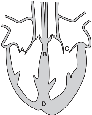
Which letter identifies the location of the atrioventricular node?
Options
A A
B B
C C
D D
Working
The atrioventricular (AV) node is a small cluster of specialised cardiac muscle cells located in the interatrial septum, close to where the atria meet the ventricles (at the base of the right atrium, near the opening of the coronary sinus). On the diagram, label B points to this region of the septum between the atria and ventricles.
- A — wall of the right atrium (this is muscle tissue of the atrial wall, not the AVN).
- B — septum between the atria and ventricles (correct site of the AVN).
- C — wall of the left atrium (atrial muscle, not the AVN).
- D — apex of the heart (tip of the left ventricle, no part of the conduction system here).
Answer
B
B
Background Concept
The mammalian heart contains a specialised conduction system that coordinates the contractions of the atria and ventricles. The two key nodes are:
- Sinoatrial node (SAN): located in the wall of the right atrium, near the entry of the superior vena cava. It sets the basic rhythm of the heart (the pacemaker).
- Atrioventricular node (AVN): located in the interatrial septum, just above the junction where the atria meet the ventricles (close to the tricuspid valve and the opening of the coronary sinus).
The AVN receives the wave of electrical excitation from the SAN after it has spread across the atria, introduces a short delay (so that the atria finish contracting and emptying before the ventricles contract), and then passes the impulse down the Bundle of His and Purkyne fibres to the ventricular muscle.
Understanding the Question
The question shows a simplified longitudinal (front-view) section of a mammalian heart with four labelled positions: A (right atrial wall), B (septum between atria and ventricles), C (left atrial wall) and D (apex). The candidate must identify which letter marks the location of the AVN.
The command word is essentially "identify" — a single correct letter is required.
Approach
Recall the anatomical position of the AVN: it sits in the lower part of the interatrial septum, near the atrioventricular valves. Then match that position to the closest letter on the diagram. The septal region between the atria and the ventricles is letter B, so B is the answer.
Step-by-Step Reasoning
- The AVN is a small mass of nodal tissue in the interatrial septum, near the junction of the right atrium and the ventricles. It is not in the bulk atrial wall (so A and C are wrong), nor at the apex of the heart (so D is wrong — the apex is left ventricular muscle).
- The only label pointing to the interatrial/atrial–ventricular septal region is B.
- Therefore B correctly identifies the location of the atrioventricular node.
Key Takeaways
- SAN = in the wall of the right atrium (pacemaker).
- AVN = in the interatrial septum, just above the AV valves (delays the impulse before passing it to the ventricles).
- Bundle of His and Purkyne fibres then carry the impulse through the interventricular septum and around the ventricles.
- Being able to map each part of the conduction system onto a heart diagram is a standard Paper 1 skill.
Common Mistakes
- Confusing the SAN with the AVN: the SAN is in the atrial wall (A or C region), not the septum.
- Choosing D (the apex) because it "looks like the middle of the heart" — the conduction system does not include the apex.
- Picking the interventricular septum (lower, between the two ventricles) instead of the interatrial/atrial–ventricular septal region where the AVN actually sits.
Things to Be Careful About
- On a simple front-view diagram, "septum" can be ambiguous: make sure you distinguish the interatrial septum (upper, between the two atria) from the interventricular septum (lower, between the two ventricles). The AVN is in the lower part of the interatrial region, near the AV valves — best described in this question as the septum between the atria and ventricles.
- The AVN is part of the right side of the heart (it lies in the right side of the interatrial septum), but the label on the diagram is in the central septal region, which is the closest point shown.
The diagram shows pressure changes in the left side of the heart during the cardiac cycle.
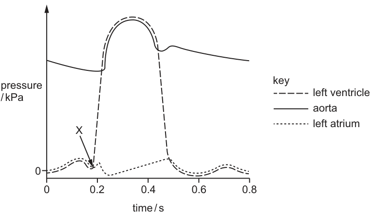
What happens in the heart at X?
Options
A The atrioventricular valves close.
B The atrioventricular valves open.
C The semilunar valves close.
D The semilunar valves open.
Working
At point X, the left ventricular pressure is rising rapidly and has just exceeded the left atrial pressure. The atrioventricular (bicuspid) valve closes to prevent backflow of blood from the ventricle into the atrium. This marks the start of ventricular systole and produces the first heart sound ("lub").
Answer
A
A
Background Concept
The cardiac cycle describes the sequence of pressure and volume changes in the heart during one complete heartbeat. The left side of the heart (left atrium and left ventricle) pumps oxygenated blood into the aorta, which then carries it to the systemic circulation. The cycle has two main phases:
- Diastole — the heart muscle relaxes and fills with blood; the atrioventricular (AV) valves are open and the semilunar (aortic) valve is closed.
- Systole — the heart muscle contracts; the AV valves close at the start (when ventricular pressure exceeds atrial pressure) and the semilunar valve opens once ventricular pressure exceeds aortic pressure.
Two pairs of valves ensure one-way flow:
- The atrioventricular valves (bicuspid/mitral on the left, tricuspid on the right) sit between atria and ventricles.
- The semilunar valves (aortic on the left, pulmonary on the right) sit between the ventricles and the great arteries.
A valve closes whenever the pressure downstream of it exceeds the pressure upstream — the higher pressure pushes the valve cusps shut.
Understanding the Question
The graph shows three pressure traces through one cardiac cycle: left ventricle (dashed), aorta (solid) and left atrium (dotted). Point X is placed on the steeply rising portion of the left ventricular trace, at the moment ventricular pressure has just overtaken atrial pressure. The question asks which valve event coincides with this point.
Approach
Identify which two pressure traces cross at point X, then decide which valve sits between those two chambers and whether it must be opening or closing at the crossover. A valve closes when upstream pressure exceeds downstream pressure and opens when downstream pressure exceeds upstream pressure.
Step-by-Step Reasoning
-
Locate point X on the graph. It lies on the left ventricular (dashed) trace where it is rising sharply and crosses above the left atrial (dotted) trace, at about 0.15–0.2 s into the cycle.
-
Identify the two chambers involved. Left ventricle and left atrium. The valve between them is the bicuspid (left atrioventricular) valve.
-
Determine the direction of the pressure difference. Just before X, atrial pressure > ventricular pressure, so the AV valve is open and blood is flowing from atrium to ventricle (late diastole / atrial systole). Just after X, ventricular pressure > atrial pressure, so the valve must shut to stop blood flowing back into the atrium.
-
Match to the answer. The event is the closure of the atrioventricular valve, producing the first heart sound and marking the start of ventricular systole. This is option A.
-
Eliminate the distractors.
- B is wrong: the AV valve was already open before X and now closes.
- C is wrong: the semilunar (aortic) valve closes at the end of systole, when aortic pressure rises above the falling ventricular pressure (around 0.5 s on this graph), not at X.
- D is wrong: the semilunar valve opens later, once the rising ventricular pressure exceeds the aortic pressure (the next crossover on the graph, around 0.2 s after X).
Key Takeaways
- A valve closes when the pressure on its downstream side exceeds the pressure on its upstream side.
- In the cardiac cycle, the AV valve closes at the start of ventricular systole (ventricular pressure overtakes atrial pressure), and the semilunar valve opens shortly afterwards (ventricular pressure overtakes aortic pressure).
- Pressure traces for the left atrium, left ventricle and aorta are a standard way to visualise and time the events of the cardiac cycle.
Common Mistakes
- Confusing the two crossovers on the graph: the first crossover (ventricular > atrial) is the AV valve closing; the second crossover (ventricular > aortic, slightly later) is the semilunar valve opening.
- Saying the AV valve "opens" at X because blood is being pushed towards it — the valve is actually being shut by the rising ventricular pressure.
- Confusing the aortic and left atrial traces; remember the aorta sits at a much higher pressure throughout most of the cycle.
Things to Be Careful About
- Read the key carefully: the dashed line is the left ventricle, not the aorta. The aorta is the solid line and sits high on the y-axis throughout most of the cycle.
- The "lub" (first heart sound) corresponds to AV valve closure at X; the "dub" (second heart sound) corresponds to semilunar valve closure later in the cycle.
- X is at the start of systole, not at the end — ventricular pressure is rising sharply, not falling.
Which components of blood are present in tissue fluid?
Options
| phagocytes | some proteins | sodium ions | |
|---|---|---|---|
| A | ✓ | ✓ | ✓ |
| B | ✓ | ✗ | ✗ |
| C | ✗ | ✓ | ✓ |
| D | ✗ | ✓ | ✗ |
key
✓ = present
✗ = not present
Working
Tissue fluid is formed by filtration of blood plasma through the capillary walls. To answer, check each component:
- Phagocytes: These white blood cells can squeeze between the endothelial cells of capillary walls (diapedesis) to enter tissue fluid to fight pathogens. Present in tissue fluid (✓).
- Some proteins: Most plasma proteins are too large to pass through, but a small number of smaller proteins can pass through the gaps between capillary endothelial cells. Present in tissue fluid (✓).
- Sodium ions: These are small dissolved ions and pass freely through the capillary wall along with water by ultrafiltration. Present in tissue fluid (✓).
All three components are present, so the correct option is A.
Answer
A
A
Background Concept
Tissue fluid surrounds the cells of body tissues and is the medium through which substances are exchanged between blood and cells. It is formed from blood plasma by ultrafiltration at the arterial end of a capillary:
- A high hydrostatic pressure (from the pumping of the heart) pushes fluid out of the capillary through tiny gaps between the endothelial cells of the capillary wall.
- Large plasma proteins (e.g. most albumins and globulins) are too big to pass through these gaps, so they remain in the blood and generate an oncotic (osmotic) pressure that draws water back into the capillary at the venous end.
- Most dissolved solutes — small ions such as , , ; glucose; urea; oxygen and carbon dioxide — pass freely out of the capillary with the water.
- White blood cells (phagocytes) can deform and squeeze between the endothelial cells in a process called diapedesis, allowing them to leave the blood and enter the tissue fluid to engulf pathogens.
The tissue fluid that is not reabsorbed at the venous end drains into the lymphatic system and eventually returns to the blood.
Understanding the Question
The question presents a table of three blood components (phagocytes, some proteins, sodium ions) and asks which of them are also present in tissue fluid. Each component is ticked (✓) or crossed (✗) in the four answer options; the correct option is the one whose pattern matches the truth for tissue fluid composition.
Approach
Go through each component in turn and decide whether it is present in tissue fluid, using knowledge of capillary wall permeability and the behaviour of cells, large molecules, and small ions. Then match the resulting pattern (✓, ✓, ✓) to the correct option.
Step-by-Step Reasoning
-
Phagocytes (✓): Although phagocytes are whole cells, they are not confined to the blood. When they detect an infection in surrounding tissue, they undergo diapedesis — they flatten and squeeze between adjacent capillary endothelial cells to enter the tissue fluid and engulf pathogens by phagocytosis. Therefore phagocytes are present in tissue fluid.
-
Some proteins (✓): The capillary endothelium is not a perfect barrier. While most plasma proteins are too large to escape, a small fraction of smaller proteins do pass through into the tissue fluid. This is why "some proteins" rather than "all proteins" is the correct wording. The vast majority of plasma protein stays in the blood, which is what creates the oncotic pressure that helps draw water back into the capillary at the venous end.
-
Sodium ions (✓): ions are small and water-soluble, and pass freely through the capillary wall along with water during ultrafiltration. Tissue fluid contains essentially the same small-ion composition as plasma, so sodium ions are definitely present.
All three components are present, so the correct option is the row with ✓ in every column: A.
Key Takeaways
- Tissue fluid is plasma that has been ultrafiltered through capillary walls, so it has a similar composition to plasma except it lacks most plasma proteins and lacks blood cells (with the exception of phagocytes that can escape by diapedesis).
- Capillary permeability is size-selective: small ions and water pass freely, most proteins do not, and whole cells normally do not — but phagocytes are an exception because they actively migrate out.
- The retention of most plasma proteins in the blood creates the colloid osmotic (oncotic) pressure that returns water to the capillary at the venous end and prevents the tissue fluid from accumulating excessively.
Common Mistakes
- Thinking that no blood cells are in tissue fluid: This is a common error. Phagocytes are an exception and are routinely present in tissue fluid, especially during infection. Choosing an option that crosses out phagocytes (B, C, D) loses the mark.
- Thinking that no proteins are in tissue fluid: It is true that most plasma proteins remain in the blood, but some smaller proteins do pass through. "No proteins" is too absolute, so options that cross out proteins (B, D) are wrong.
- Confusing tissue fluid with lymph: Lymph is what tissue fluid becomes after it enters lymphatic vessels, but the two are essentially the same fluid at the moment of formation; the question is about what tissue fluid contains.
Things to Be Careful About
- Read the table column by column and compare with the correct pattern — candidates who misread the column order or tick marks often select the wrong row.
- The phrasing "some proteins" is important: a candidate who writes "proteins are not present in tissue fluid" in an essay answer would be marked wrong because the textbook position is that small proteins do escape; the capillary is leaky to a limited extent.
- The question concerns what is present in tissue fluid, not what is present only in blood — be careful not to over-think the answer by trying to find a component that is unique to blood.
In the lungs, movement of dissolved carbon dioxide out of the capillaries occurs in one of two ways:
● by diffusion through the endothelial cells of the capillaries
● by leakage through pores in the endothelial cells of the capillaries.
What is the minimum number of times that a carbon dioxide molecule that has been transported to the lungs in a red blood cell must cross a cell surface membrane to reach an air space in an alveolus?
Options
A 2
B 3
C 4
D 5
Working
To get from a red blood cell to the air in an alveolus, a CO₂ molecule must cross the following membranes:
- Red blood cell membrane — CO₂ must leave the red blood cell. 1 membrane crossed.
- Capillary endothelium — to minimise membrane crossings, CO₂ passes through a pore between endothelial cells. 0 additional membrane crossings.
- Alveolar epithelium (pneumocyte) — CO₂ diffuses through the type I pneumocyte. This requires crossing both the basal and the apical plasma membrane. 2 membrane crossings.
Minimum total = 1 + 0 + 2 = 3 membrane crossings.
Answer
B
B
Background Concept
At the gas-exchange surface, three cellular layers separate the air in an alveolus from the blood inside a pulmonary capillary:
- the type I pneumocyte (squamous alveolar epithelial cell) lining the alveolus;
- a very thin layer of tissue fluid and basement membrane;
- the capillary endothelial cell.
A small, lipid-soluble molecule such as CO₂ can cross a plasma membrane by simple diffusion, so passing through a cell means crossing two membranes (one to enter, one to leave). Alternatively, CO₂ can squeeze through the pores / clefts between adjacent endothelial or epithelial cells without crossing any membrane at all. The question asks for the minimum number of membrane crossings, so the candidate route uses pores where they are available.
Understanding the Question
The question defines a specific scenario: a CO₂ molecule has been carried to the lungs inside a red blood cell. The question asks the smallest possible number of plasma-membrane (cell-surface-membrane) crossings required to reach the alveolar air space. The candidate must identify every membrane in the path and choose the route that minimises the count.
Approach
List each barrier in order, decide whether the CO₂ passes through a cell (2 membrane crossings) or between cells through a pore (0 membrane crossings), and sum the minimum totals.
Step-by-Step Reasoning
-
Red blood cell membrane. CO₂ is generated inside respiring tissue cells and carried in the blood mostly as HCO₃⁻, but at the lungs some is also free in the cytoplasm of the red blood cell. Whatever the form, the CO₂ (or HCO₃⁻ converted back to CO₂ by carbonic anhydrase) must leave the red blood cell, crossing its plasma membrane. +1 membrane.
-
Capillary endothelium. The question states explicitly that movement out of the capillary is either by diffusion through the endothelial cell (2 membranes) or by leakage through pores (0 membranes). To minimise, choose the pores. +0 membranes.
-
Alveolar epithelium. The same logic applies to the thin squamous pneumocyte lining the alveolus. To minimise, the CO₂ diffuses through the type I pneumocyte, entering at its basal surface and exiting at its apical surface. +2 membranes.
-
Total minimum = 1 + 0 + 2 = 3.
Therefore the answer is B (3).
Key Takeaways
- A substance crossing through a cell must cross the plasma membrane twice (entry and exit).
- A substance passing between cells through a pore or cleft crosses no membranes.
- The alveolar–capillary barrier consists of the red blood cell, capillary endothelium, and alveolar epithelium — three cell layers — giving a minimum of 1 + 0 + 2 = 3 membrane crossings for a gas moving out of a red blood cell into alveolar air.
- A small lipid-soluble gas like CO₂ is well suited to this route because it diffuses rapidly across phospholipid bilayers.
Common Mistakes
- Forgetting the red blood cell membrane and answering 2 (treating the path as endothelium + alveolus only).
- Forgetting that diffusing through a cell means 2 crossings, not 1 — leading to answers of 2 (if the alveolar cell is counted once) or 4 (if every cell is counted as 1 crossing).
- Assuming CO₂ must cross both endothelium and alveolus through cells, ignoring the option of pores in the capillary endothelium, giving 5.
Things to Be Careful About
- The question is about cell surface (plasma) membranes only — basement membranes and tissue fluid are not counted.
- The wording "movement of dissolved carbon dioxide out of the capillaries occurs in one of two ways" applies specifically to the capillary wall; the same options exist at the alveolar wall, and the minimum-pathway argument must be applied independently to each barrier.
- Choose the route that minimises crossings; this is the route through capillary pores, not through the endothelial cell.
What maintains the steep concentration gradients needed for successful gas exchange in the lungs?
1 Air flow in the alveoli is in the opposite direction to blood flow in the capillaries.
2 Blood arrives in the lungs with a lower oxygen concentration and a higher carbon dioxide concentration than the air in the alveoli.
3 Blood is constantly flowing through and out of the lungs, bringing a fresh supply of red blood cells.
Options
A 1, 2 and 3
B 1 and 2 only
C 1 and 3 only
D 2 and 3 only
Working
Statement 1 is wrong because human lungs do not use counter-current exchange. Air flow in the alveoli and blood flow in the surrounding capillaries are not in opposite directions; instead, alveolar ventilation is essentially a uniform (tidal) pool, with air entering and leaving via the same airways. (True counter-current exchange is found in fish gills, not mammalian lungs.)
Statement 2 is correct: deoxygenated blood reaching the pulmonary capillaries has a lower partial pressure and a higher partial pressure than the air in the alveoli, so a steep diffusion gradient exists for both gases across the alveolar–capillary membrane.
Statement 3 is correct: blood flows continuously through the pulmonary circulation, so deoxygenated blood is constantly delivered and oxygenated blood is constantly removed. This prevents the alveolar and blood gas partial pressures from equilibrating, sustaining the gradient.
Only statements 2 and 3 are correct.
Answer
D
D
Background Concept
Gas exchange across a respiratory surface (the alveolar epithelium in mammals) occurs by diffusion, which requires a partial-pressure (concentration) gradient for each gas. Fick's law tells us that the rate of diffusion is proportional to the surface area, the concentration gradient, and a diffusion constant, and inversely proportional to the thickness of the membrane. For the lungs to load oxygen onto haemoglobin and unload carbon dioxide efficiently, the gradient for (alveolus → blood) and for (blood → alveolus) must be kept as steep as possible.
Two processes maintain these gradients:
- Ventilation – fresh atmospheric air (high , low ) is repeatedly moved into the alveoli by breathing, refreshing the alveolar gas.
- Perfusion – deoxygenated blood (low , high ) is continuously pumped through the pulmonary capillaries by the right ventricle, replacing blood that has equilibrated with alveolar gas.
It is important to know that mammalian lungs do NOT use counter-current exchange. In human lungs, inspired and expired air mix in the alveoli (a form of uniform/tidal ventilation), and capillary blood flows more or less perpendicularly to the airway. Counter-current exchange is characteristic of fish gills, where water flows over the lamellae in the opposite direction to blood inside them, allowing extraction of up to ~80% of dissolved oxygen. In mammals, only about 25% of the oxygen in inspired air is taken up because of the less efficient (but energetically cheaper) tidal system.
Understanding the Question
The question asks which of the three statements correctly describes what maintains the steep concentration gradients in the lungs. It is a multi-statement MCQ: candidates must evaluate each statement independently and then pick the option listing only the correct ones.
- Statement 1 invokes the idea of counter-current flow.
- Statement 2 describes the partial-pressure difference between arriving blood and alveolar air.
- Statement 3 describes continuous blood flow (perfusion).
Approach
For each statement, ask: is this an accurate description of what happens in human lungs, AND does it actually help maintain the concentration gradient?
- Statement 1: although the idea (opposing flows → maintained gradient) is biologically valid in general, it does not describe the human lung accurately. Reject.
- Statement 2: yes, this is exactly why diffusion proceeds — there is a difference in partial pressure between the alveolar air and the incoming blood. Accept.
- Statement 3: yes, without continuous flow, blood would quickly equilibrate with alveolar gas and the gradient would collapse. Accept.
Step-by-Step Reasoning
-
Evaluate statement 1. Human alveolar ventilation is tidal: air enters through the trachea/bronchi/bronchioles during inspiration and leaves by the same route during expiration. It does not flow in one direction across the gas-exchange surface as it would in a fish gill. The pulmonary capillaries form a dense mesh around each alveolus, and blood passes through them in a roughly perpendicular direction — this is sometimes called a cross-current arrangement, not a true counter-current. Therefore statement 1 is incorrect.
-
Evaluate statement 2. Deoxygenated blood returning from the systemic tissues has and . Alveolar air has and . Because alveolar is higher than blood , diffuses into the blood; because blood is higher than alveolar , diffuses out. This statement is correct.
-
Evaluate statement 3. The entire cardiac output (~5 L min⁻¹ at rest) passes through the lungs. Each red blood cell spends roughly 0.75 s in the pulmonary capillaries, offloads , loads , and is carried away. New deoxygenated erythrocytes continually replace those that have equilibrated, so the gradient between alveolar gas and incoming blood is constantly re-established. This statement is correct.
-
Combine the correct statements. Only 2 and 3 are correct → option D.
Key Takeaways
- Mammalian lungs rely on ventilation and perfusion to maintain diffusion gradients, not on counter-current exchange.
- Counter-current exchange is a feature of fish gills, not human lungs.
- The gradient is sustained because (a) alveolar gas has different partial pressures from arriving blood and (b) blood is continually replaced.
Common Mistakes
- Confusing mammalian lungs with fish gills. Many students assume any opposing flow must be counter-current and pick option A or B. The hallmark of true counter-current exchange is that the two fluids flow in exactly opposite directions along a long, thin interface; this is not what happens in alveoli.
- Thinking breathing alone is enough. Ventilation refreshes alveolar gas, but without continuous blood flow the gradient would still collapse as blood equilibrates. Likewise, blood flow alone without ventilation would deplete alveolar and saturate it with . Both are needed.
- Misreading partial pressures. Remember that "lower oxygen concentration" refers to blood plasma and erythrocytes compared with alveolar air, not to a different body compartment.
Things to Be Careful About
- The wording of statement 1 is precise: it says "air flow in the alveoli is in the opposite direction to blood flow in the capillaries." In the alveoli, air does not really have a directional flow in the same sense as water over a fish gill — it is essentially a stirred pool. The statement is therefore factually wrong, not just a simplification.
- In some textbooks, alveolar capillary blood flow is loosely described as "counter-current"; the CIE mark scheme, however, treats this as incorrect for human lungs. Stick to the precise description: tidal ventilation, with capillaries forming a network around each alveolus.
- The question is worth only 1 mark but tests a concept-rich distinction; read every option carefully before committing.
Where is cartilage tissue always found in the human gas exchange system?
Options
A in the trachea only
B in the bronchi only
C in the bronchioles and trachea
D in the bronchi and trachea
Working
Cartilage keeps the larger conducting airways patent (open) and prevents their collapse during breathing. In the gas exchange system:
- The trachea contains C-shaped rings of hyaline cartilage.
- The bronchi (left and right primary bronchi, and their branches down to about the 11th–12th generation) also contain irregular plates of hyaline cartilage in their walls.
- The bronchioles do NOT contain cartilage; their walls are dominated by smooth muscle and elastic tissue instead.
Therefore cartilage is always present in the bronchi and trachea, but absent from the bronchioles.
Answer
D
D
Background Concept
The human gas exchange (respiratory) system is a branching tree of conducting airways that lead from the outside air down to the gas-exchange surface in the alveoli. The wall composition changes progressively as the airways divide and become narrower:
- Trachea: a large flexible tube supported by C-shaped rings of hyaline cartilage (open at the back, where the oesophagus sits). It is lined with ciliated epithelium and goblet cells.
- Bronchi: the two main branches off the trachea (left and right primary bronchi) and their subsequent branches within the lungs. Their walls also contain plates of hyaline cartilage, together with smooth muscle, elastic fibres, ciliated epithelium and goblet cells.
- Bronchioles: smaller airways (diameter < 1 mm) that branch from the bronchi. Their walls have no cartilage; instead they contain relatively more smooth muscle and elastic tissue, which constricts or dilates the airway. Cartilage disappears once the airway diameter falls below about 1 mm.
The functional reason cartilage is present in the trachea and bronchi is mechanical: it holds these airways permanently open so that air can flow freely to and from the lungs despite the pressure changes that occur during breathing. In the smaller bronchioles, cartilage would be obstructive and unnecessary — the smooth muscle allows fine control of airflow instead.
Understanding the Question
This is a one-mark multiple-choice item. The command word "always" is the key: the question is asking where cartilage is unfailingly present in the gas exchange system, not just where it is sometimes or occasionally found. We must select the option that names every airway type whose wall always contains cartilage and excludes any airway that lacks it.
Approach
Recall the three named levels — trachea, bronchi, bronchioles — and decide for each whether cartilage is a definite (always) component of its wall:
- Trachea → yes, C-shaped hyaline cartilage rings.
- Bronchi → yes, hyaline cartilage plates.
- Bronchioles → no cartilage.
The option that correctly includes the trachea and bronchi, and excludes the bronchioles, is the answer.
Step-by-Step Reasoning
- Option A (trachea only): incomplete. Cartilage is also present in the bronchi, so this option names too few locations.
- Option B (bronchi only): incomplete. The trachea is also supported by cartilage, so this option also names too few locations.
- Option C (bronchioles and trachea): incorrect. The bronchioles do not contain cartilage — they have smooth muscle instead. Including the bronchioles makes this option wrong.
- Option D (bronchi and trachea): correct. Cartilage is consistently present in both the trachea and the bronchi, and is absent from the bronchioles, exactly matching the distribution required by "always".
Key Takeaways
- Cartilage in the respiratory system is found in the trachea and bronchi only.
- Bronchioles lack cartilage but have abundant smooth muscle.
- The function of cartilage is to keep the larger conducting airways open (patent) during the pressure changes of breathing.
Common Mistakes
- Selecting C by confusing bronchioles with bronchi. The terminology is similar, but bronchioles are the smaller, distal airways and are the level at which cartilage is lost.
- Selecting A or B because the candidate remembers "cartilage is in the trachea" (or "in the bronchi") but forgets it is also present at the other named level.
- Thinking cartilage is found throughout the entire airway tree, including down to the alveoli — it is not.
Things to Be Careful About
- The word "always" in the stem matters: any airway in which cartilage is sometimes present but sometimes absent (e.g. very small distal bronchi where cartilage plates become sparse) would not be a safe answer. The trachea and main/medium bronchi consistently contain cartilage, so they are the only reliable answers.
- Distinguish carefully between bronchi (with cartilage) and bronchioles (without cartilage). The two words look alike and are easy to swap in a hurried MCQ.
Scientists compared the density of goblet cells in the lungs and the density of mucus in the lungs of three groups of people:
● people who do not smoke and do not have lung disease
● people who smoke tobacco but do not have lung disease
● people who smoke tobacco and have lung disease.
The results are shown in the table.
| group | goblet cell density / cells per | mucus density / arbitrary units |
|---|---|---|
| non-smokers | 19 | 6 |
| smokers who do not have lung disease | 54 | 26 |
| smokers with lung disease | 37 | 15 |
What is indicated by these data?
1 There is a positive correlation (relationship) between density of goblet cells and density of mucus.
2 Lung disease results in an increase in goblet cell density.
3 There is an association between tobacco smoking and an increase in mucus density.
Options
A 1, 2 and 3
B 1 and 2 only
C 1 and 3 only
D 2 and 3 only
Working
Test each statement against the data:
Statement 1 — positive correlation between goblet cell density and mucus density
- Non-smokers → smokers without disease: 19 → 54 cells/mm² and 6 → 26 units (both rise)
- Smokers without disease → smokers with disease: 54 → 37 cells/mm² and 26 → 15 units (both fall)
- Both variables change in the same direction throughout → positive correlation. ✓
Statement 2 — lung disease results in an increase in goblet cell density
- Compare the two smoker groups (the only difference is lung disease):
- Smokers without disease: 54 cells/mm²
- Smokers with disease: 37 cells/mm²
- Goblet cell density is lower, not higher, in the disease group. ✗
Statement 3 — association between tobacco smoking and an increase in mucus density
- Non-smokers: 6 units
- Smokers without disease: 26 units
- Smokers with disease: 15 units
- Both smoker groups have higher mucus density than non-smokers → smoking is associated with increased mucus. ✓
Statements 1 and 3 are supported; statement 2 is contradicted by the data.
Answer
C
C
Background Concept
Goblet cells are specialised secretory epithelial cells scattered among the ciliated cells that line the trachea, bronchi and larger bronchioles. They synthesise and release mucus, which traps inhaled dust, microbes and chemical irritants. The overlying carpet of cilia then beats in a coordinated fashion to move the mucus layer upwards towards the pharynx — the so-called mucociliary escalator. The system is a key non-specific defence of the gas exchange surface.
Tobacco smoke is a powerful irritant. The tar fraction and many of its component chemicals (polycyclic aromatic hydrocarbons, phenols, etc.) provoke inflammation of the airway epithelium. The epithelium responds by undergoing goblet cell hyperplasia (more goblet cells per unit area) and by secreting more mucus per cell, so both goblet cell density and mucus density rise in smokers.
Two terms that must be kept distinct in this kind of question:
- Correlation — a statistical statement that two variables tend to vary together. Positive correlation means they move in the same direction (both rise or both fall); it does not imply that one causes the other.
- Association — a general term meaning the variables are not independent. A claim of association is weaker than a claim of cause and effect.
When a question asks what effect a single variable has, the comparison must be made between groups that differ only in that variable. Comparing non-smokers to smokers with disease, for example, confounds the effect of smoking with the effect of disease.
Understanding the Question
The table gives three paired values of goblet cell density and mucus density:
| group | goblet cells / | mucus / a.u. |
|---|---|---|
| non-smokers | 19 | 6 |
| smokers, no disease | 54 | 26 |
| smokers, with disease | 37 | 15 |
The question asks which of three statements the data support. The statement to mark is the option that lists exactly the supported statements.
Approach
For each statement:
- Identify what claim is being made.
- Choose the rows of the table that test that claim (matching groups that differ only in the relevant variable).
- Read the values off the table and decide whether the claim holds.
For a correlation claim, check the direction of the changes as you move through the table — both variables moving the same way is positive correlation. For an effect claim, isolate the variable by comparing the two groups that differ only in it.
Step-by-Step Reasoning
Statement 1: positive correlation between goblet cell density and mucus density.
Read the pairs in order: (19, 6), (54, 26), (37, 15). As we move from the first pair to the second, both values rise. As we move from the second to the third, both values fall. Two variables that change in the same direction throughout define a positive correlation. ✓
Statement 2: lung disease results in an increase in goblet cell density.
To isolate the effect of lung disease, we must compare the two smoker groups (the only difference between them is the presence of lung disease). Goblet cell density goes from 54 cells/mm² in smokers without disease to 37 cells/mm² in smokers with disease — a decrease, not an increase. The data therefore contradict this statement. ✗
Statement 3: there is an association between tobacco smoking and an increase in mucus density.
Compare the non-smoker row with each of the smoker rows. Non-smokers: 6 a.u. Smokers without disease: 26 a.u. Smokers with disease: 15 a.u. Both smoker values are higher than the non-smoker value, so smoking is associated with raised mucus density. ✓
Only statements 1 and 3 are supported, so the correct combination is 1 and 3 only → option C.
Key Takeaways
- A positive correlation exists when two variables move in the same direction across the data set; correlation is not causation.
- To judge the effect of one variable, compare groups that are identical except for that variable — here, the two smoker rows for lung disease, and the non-smoker vs smoker rows for smoking.
- Tobacco smoke causes goblet cell hyperplasia and increased mucus secretion as part of the airway's defensive response to inhaled irritants.
- In established chronic obstructive lung disease, the damaged epithelium can show squamous metaplasia — the normal pseudostratified ciliated epithelium with goblet cells is replaced by a stratified squamous epithelium that lacks goblet cells — which is why goblet cell density in smokers with disease can be lower than in healthy smokers.
Common Mistakes
- Reading the table upside-down or mixing up the rows, so that the comparison for statement 2 is wrongly done against the non-smoker group rather than against the other smoker group.
- Thinking a positive correlation requires the values to be perfectly proportional, and rejecting statement 1 because the rise from row 1 to row 2 is not the same size as the fall from row 2 to row 3.
- Concluding statement 2 is true because goblet cells are higher in smokers with disease (37) than in non-smokers (19) — this rise is due to smoking, not to lung disease.
- Confusing association (a relationship) with causation (one variable producing the other). Statement 3 is correctly phrased as an association.
Things to Be Careful About
- "Positive" correlation refers to the direction of the relationship, not its strength or its significance.
- When assessing the effect of lung disease, the non-smoker group must not be used — it differs in two variables (smoking and disease) and cannot isolate either.
- Watch for the precise wording of each statement; a single word such as "increase" vs "decrease" or "association" vs "causation" can flip whether the statement is supported by the data.
- The values in the table are quoted to different precisions, but the comparisons are clear from the integer values and do not require any calculation.
Which disease does Mycobacterium bovis cause?
Options
A cholera
B HIV/AIDS
C malaria
D tuberculosis
Working
Mycobacterium bovis is a species of bacterium in the genus Mycobacterium. It is the causative agent of tuberculosis (TB), particularly bovine tuberculosis, and is closely related to Mycobacterium tuberculosis, the primary cause of human TB. The other options are caused by unrelated pathogens: cholera by Vibrio cholerae, HIV/AIDS by HIV (a virus), and malaria by Plasmodium species.
Answer
D
D
Background Concept
Infectious diseases are caused by pathogens — microorganisms such as bacteria, viruses, fungi and protoctists. Each major disease is associated with one (or a few closely related) causative organisms, and the species name is part of the required knowledge for CIE Biology. The four diseases emphasised at AS Level are cholera, malaria, tuberculosis (TB) and HIV/AIDS, each caused by a very different type of pathogen.
- Cholera — caused by the bacterium Vibrio cholerae.
- Malaria — caused by protoctist parasites of the genus Plasmodium (e.g. Plasmodium falciparum), transmitted by the female Anopheles mosquito.
- HIV/AIDS — caused by the Human Immunodeficiency Virus (HIV), a retrovirus.
- Tuberculosis (TB) — caused by bacteria of the genus Mycobacterium. Mycobacterium tuberculosis is the main human pathogen, while Mycobacterium bovis causes TB in cattle and can be transmitted to humans (especially through unpasteurised milk), producing the same disease.
All Mycobacterium species are slow-growing, acid-fast, rod-shaped bacteria with a waxy, mycolic-acid-rich cell wall that makes them resistant to many disinfectants and to the host immune system.
Understanding the Question
The question is a straight identification task: given the species Mycobacterium bovis, which of the four listed diseases does it cause? The four options cover exactly the four diseases named in the syllabus, so a candidate is being tested on the link between the bacterial species and the disease name.
The command word is implicit ("Which disease…") and demands only the letter of the correct option.
Approach
Match the genus/species given in the stem to the disease list:
- Mycobacterium → tuberculosis. ✓
- Vibrio → cholera. (Option A)
- HIV (a virus) → HIV/AIDS. (Option B)
- Plasmodium → malaria. (Option C)
Only option D fits.
Step-by-Step Reasoning
- The stem names Mycobacterium bovis. The genus Mycobacterium is the diagnostic clue — all members of this genus are associated with tuberculosis.
- M. bovis specifically causes bovine TB, and cross-infection to humans produces tuberculosis. It is a key reason for pasteurising milk and for the test-and-slaughter cattle TB control programmes in many countries.
- The other three options are caused by completely different organisms and must be rejected.
Key Takeaways
- Mycobacterium tuberculosis and Mycobacterium bovis both cause tuberculosis; M. bovis is the zoonotic (cattle-to-human) form.
- Each of the four AS diseases has a characteristic pathogen: Vibrio cholerae (cholera), Plasmodium spp. (malaria), HIV (HIV/AIDS), Mycobacterium spp. (TB).
Common Mistakes
- Confusing Mycobacterium with another bacterial genus and linking it to cholera (which is Vibrio cholerae).
- Assuming any bacterium must be a "throat/chest" infection and choosing the most familiar-sounding option, without checking the species name.
Things to Be Careful About
- Note the spelling Mycobacterium (not Mycobactrium or Micobacterium).
- Recognise that bovis in the species name signals cattle — useful for remembering that M. bovis is the bovine form of TB, transmissible to humans through unpasteurised milk.
An antibiotic inhibits the formation of cross-links between the molecules that form cell walls in bacteria.
Which statements explain why bacteria are killed by the antibiotic?
1 The bacterial cell is destroyed by osmotic lysis.
2 Cellulose molecules cannot form hydrogen bonds.
3 The cell wall is no longer partially permeable.
Options
A 1 and 2 only
B 2 and 3 only
C 1 only
D 2 only
Working
- Statement 1: The bacterial cell wall is made of peptidoglycan, in which polysaccharide chains are joined by peptide cross-links. If cross-links cannot form, the wall loses its tensile strength and cannot resist the high internal water potential of the bacterial cytoplasm. Water therefore enters by osmosis, the cell swells, and the weakened wall ruptures — i.e. osmotic lysis. Correct.
- Statement 2: Bacterial cell walls are made of peptidoglycan (murein), not cellulose. Furthermore, cellulose molecules do form hydrogen bonds between adjacent β-glucose chains, which is precisely what gives plant cell walls their strength. Incorrect.
- Statement 3: Cell walls are fully permeable to small solutes and water — the cell (plasma) membrane is the partially permeable barrier. This statement misrepresents the role of the cell wall. Incorrect.
Only statement 1 is correct.
Answer
C
C
Background Concept
Bacteria are prokaryotes, and almost all of them (apart from a few archaea-related exceptions) build a rigid cell wall out of a unique polymer called peptidoglycan (also known as murein). Peptidoglycan is a mesh of long polysaccharide chains made of alternating N-acetylglucosamine (NAG) and N-acetylmuramic acid (NAM) sugars, cross-linked by short peptide bridges between the amino-acid side chains attached to the NAM sugars. It is these peptide cross-links that give the wall its tensile strength.
Because the bacterial cytoplasm contains a high concentration of dissolved solutes, the cytoplasm has a more negative water potential than the surrounding medium. Water therefore tends to move into the cell by osmosis. In an intact bacterium this inward osmotic pressure is balanced by the mechanical strength of the peptidoglycan wall — without that wall the cell would swell and burst. The cell wall's job is structural support against osmotic turgor, not selective permeability: it is the plasma (cell surface) membrane, a phospholipid bilayer with embedded proteins, that is the partially permeable barrier controlling what enters and leaves the cytoplasm.
Many antibiotics exploit this vulnerability. Penicillin and related β-lactam drugs work by inhibiting transpeptidase (DD-peptidase), the enzyme that catalyses the peptide cross-links between peptidoglycan strands. With no cross-links, the wall becomes a weak, loose net. Osmotic lysis follows.
Understanding the Question
This is a multiple-choice question (MCQ) testing whether you can identify the correct biological explanation for why an antibiotic that blocks cell-wall cross-linking kills bacteria. You are given three statements and asked which combination is correct. Only one option is right; the distractor statements mix up cellulose with peptidoglycan and conflate the wall with the membrane.
Approach
Evaluate each statement independently using core knowledge:
- Will the bacterium undergo osmotic lysis if its wall is weakened? (Yes — recall the role of peptidoglycan.)
- Is the cell-wall molecule cellulose? (No — bacterial walls are peptidoglycan, not cellulose, and cellulose does hydrogen-bond.)
- Is the cell wall partially permeable? (No — it is fully permeable; the plasma membrane is partially permeable.)
Step-by-Step Reasoning
Statement 1 — "The bacterial cell is destroyed by osmotic lysis." This is exactly the textbook mechanism for penicillin-type antibiotics. With the peptide cross-links absent, the peptidoglycan mesh cannot resist the inward osmotic gradient. Water rushes in, the cell swells, and the weakened wall bursts. Statement 1 is correct.
Statement 2 — "Cellulose molecules cannot form hydrogen bonds." Two independent errors:
- Bacterial cell walls are made of peptidoglycan, not cellulose. Cellulose is found in plant cell walls (and is made by some algae, oomycetes and a few bacteria, but never as the structural wall component of disease-causing bacteria such as those targeted by penicillin).
- Cellulose is held together by extensive intra- and intermolecular hydrogen bonds between the –OH groups of adjacent β-1,4-glucose chains. Without these H-bonds plant cell walls would not be rigid. So the claim that cellulose cannot form hydrogen bonds is biologically false.
Statement 2 is incorrect.
Statement 3 — "The cell wall is no longer partially permeable." A second conceptual error. The cell wall is freely permeable to water, ions and small molecules; it never acted as a partially permeable barrier in the first place. Selective permeability is the function of the plasma membrane, a phospholipid bilayer. Even an intact, fully cross-linked wall is not "partially permeable" in the membrane-physiology sense. Statement 3 is incorrect.
Because only statement 1 is true, the correct option is the one that credits statement 1 alone: C — 1 only.
Key Takeaways
- Bacterial cell walls are made of peptidoglycan cross-linked by short peptides; cellulose is irrelevant to them.
- The cell wall's job is structural — to resist the high internal osmotic pressure of the cytoplasm.
- Antibiotics such as penicillin kill bacteria by weakening the wall so the cell undergoes osmotic lysis.
- The partially permeable barrier in any cell is the plasma membrane, not the cell wall.
Common Mistakes
- Confusing peptidoglycan with cellulose because both are described as "structural carbohydrates". They are chemically and biologically different.
- Believing cellulose is held together by covalent bonds only and "cannot" hydrogen-bond — in fact the H-bond network between β-glucose chains is what makes plant walls strong.
- Attributing selective permeability to the cell wall rather than to the plasma membrane. The wall is a porous molecular sieve.
- Selecting option D because statement 1 "sounds right" but students doubt themselves and drop it — but statement 1 is the central mechanism.
Things to Be Careful About
- The "molecules that form cell walls in bacteria" referred to in the stem are the peptidoglycan monomers (NAG-NAM), not cellulose.
- "Partially permeable" is a term strictly applied to membranes in CIE Biology. Do not transfer it to walls.
- Be alert to MCQs where two statements are wrong for different reasons (here, statement 2 fails on chemistry and identity; statement 3 fails on terminology and function). Check both independently.
Scientists investigated the effect of increasing concentrations of an antibiotic on the development of antibiotic resistance in bacteria.
The scientists grew four groups of bacteria and added a different concentration of antibiotic to each group. The number of resistant bacteria and the total population of bacteria were measured at intervals for 24 hours for each group.
The graphs show the results.
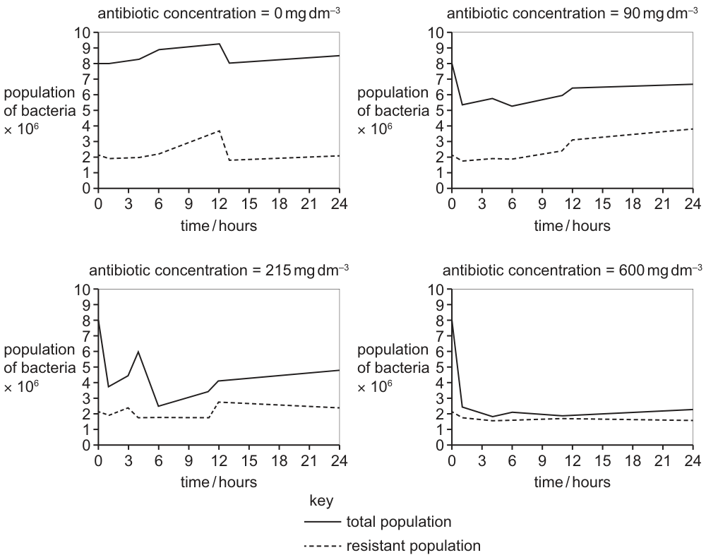
Which statements are correct conclusions that can be made from the results of this investigation?
1 Increasing the concentration of antibiotic decreases the population of non-resistant bacteria at the end of 24 hours.
2 The proportion of antibiotic-resistant bacteria increases with increasing concentrations of antibiotics.
3 Increasing the concentration of antibiotic always increases the number of resistant bacteria.
Options
A 1, 2 and 3
B 1 and 2 only
C 1 and 3 only
D 2 and 3 only
Working
Statement 1 – Non-resistant population at 24 h (read off each panel; non-resistant = total − resistant):
| total | resistant | non-resistant | |
|---|---|---|---|
| 0 | ~8.5 | ~2.0 | ~6.5 |
| 90 | ~7.0 | ~3.5 | ~3.5 |
| 215 | ~5.0 | ~2.5 | ~2.5 |
| 600 | ~2.0 | ~1.5 | ~0.5 |
The non-resistant count falls as antibiotic concentration rises → Statement 1 is correct.
Statement 2 – Proportion resistant at 24 h (resistant / total):
| proportion resistant | |
|---|---|
| 0 | ~24% |
| 90 | ~50% |
| 215 | ~50% |
| 600 | ~75% |
The fraction of resistant cells rises as antibiotic concentration rises → Statement 2 is correct.
Statement 3 – Does the resistant number always rise with concentration?
Resistant counts: 0 mg → ~2.0 × 10⁶; 90 mg → ~3.5 × 10⁶; 215 mg → ~2.5 × 10⁶; 600 mg → ~1.5 × 10⁶.
The resistant number rises then falls — it does not always increase. The word "always" makes the statement too strong → Statement 3 is incorrect.
Answer
B
B
Background Concept
Antibiotics are chemicals that kill bacteria (bactericidal) or stop them dividing (bacteriostatic). Within any large bacterial population a few individuals will, by random mutation, carry alleles that confer resistance to a given antibiotic. When the population is exposed to that antibiotic, the susceptible cells die or stop replicating while the resistant ones continue to grow, so over time the proportion of resistant cells in the population rises — this is the basic mechanism behind the spread of antibiotic resistance. Importantly, the absolute number of resistant cells depends on both the size of the surviving population and their growth rate, so it does not necessarily increase monotonically with antibiotic dose.
The graphs distinguish two quantities on each panel:
- Total population (solid line) — every cell, resistant or not.
- Resistant population (dashed line) — only the cells that can grow in the presence of that antibiotic concentration.
The difference (total − resistant) is the non-resistant population, the part being killed or inhibited by the drug.
Understanding the Question
The question gives four panels at 0, 90, 215 and 600 mg dm⁻³ of antibiotic and asks which of three statements can be supported by the data. This is a data-interpretation question, not a knowledge question: each statement must be checked directly against what the graphs show, paying close attention to qualifiers such as "proportion", "always" and "at the end of 24 hours".
The command word is implicit ("which statements are correct conclusions") — every statement must be warranted by the evidence, not merely plausible.
Approach
For each statement:
- Identify the quantity the statement refers to (non-resistant number, proportion resistant, or absolute resistant number).
- Read the relevant value from each of the four panels.
- Compare across panels to see whether the trend described in the statement actually holds.
- Watch for absolute versus proportional language — the same data can support one and contradict the other.
Step-by-Step Reasoning
Statement 1 — non-resistant population at 24 h
Subtracting the dashed (resistant) value from the solid (total) value at for each panel:
- 0 mg dm⁻³:
- 90 mg dm⁻³:
- 215 mg dm⁻³:
- 600 mg dm⁻³:
The non-resistant count falls monotonically as antibiotic concentration rises. Statement 1 is supported.
Statement 2 — proportion resistant at 24 h
Proportion resistant = resistant / total at 24 h:
- 0 mg dm⁻³: (24%)
- 90 mg dm⁻³: (50%)
- 215 mg dm⁻³: (50%)
- 600 mg dm⁻³: (75%)
The proportion of resistant cells rises (or stays high) as concentration rises, even where the absolute number does not. Statement 2 is supported.
Statement 3 — does the resistant number always increase?
Absolute resistant counts at 24 h: (all ). The number goes up, then down. Because the trend is not monotonic, the word "always" is wrong. Statement 3 is not supported.
Statements 1 and 2 are correct → answer B.
Key Takeaways
- Always distinguish absolute numbers from proportions: the same dataset can show a falling absolute count alongside a rising proportion.
- Watch for absolute qualifiers such as "always", "never" and "only" in conclusion questions — they require the trend to hold across the entire dataset, not just most of it.
- Non-resistant count = total − resistant, so a small gap between the two lines at the right-hand end of a panel signals heavy selection by the antibiotic.
- This is exactly the evolutionary principle that underpins antibiotic stewardship: higher doses select more strongly for resistance (rising proportion) even while the total population shrinks.
Common Mistakes
- Confusing the two lines. The solid line is the total population, the dashed line is only the resistant subpopulation. Many candidates mistakenly take the dashed line as "sensitive" bacteria.
- Ignoring the word "always" in statement 3 and ticking it because the resistant number is generally higher at intermediate doses.
- Misreading proportions as absolute numbers (e.g. concluding that "more resistant bacteria at 600 mg" is true when in fact their number is lowest there, only their share of the total is highest).
- Choosing D (2 and 3) because statement 1 requires the extra step of subtracting to get non-resistant cells — a step that is easy to skip.
Things to Be Careful About
- The values quoted above are read off the printed gridlines and are approximate. You do not need an exact number — what matters is the trend across the four panels.
- Be careful with units: the y-axis is in cells, so the values themselves are read as , etc., and only the trend is being assessed, not the precise count.
- "At the end of 24 hours" in statement 1 fixes the time point — do not be distracted by earlier peaks or troughs on the graphs.
- For Paper-1 multiple-choice conclusion questions, only tick a statement if the data unambiguously support it; a single counter-example is enough to reject a universal claim.
What is the correct sequence of events in a primary immune response?
Options
A T-lymphocyte activation B-lymphocyte selection plasma cell release
B antigen presentation by macrophages cytokines released by T-helper cells B-lymphocyte differentiation
C antigen presentation by neutrophils T-memory cell activation B-lymphocyte selection
D T-memory cell activation B-memory cell activation antibody production
Working
The primary immune response begins when a pathogen enters the body. Macrophages engulf pathogen by phagocytosis and present antigens on their surface. T-helper cells recognise these presented antigens and release cytokines. These cytokines stimulate B-lymphocytes to differentiate into plasma cells, which then produce antibodies.
Option A is wrong because B-lymphocyte selection is not triggered by T-lymphocyte activation alone — antigen presentation is needed first.
Option C is wrong because antigen presentation is performed by macrophages (not neutrophils) and T-memory cells are formed during the primary response but mediate the secondary response.
Option D is wrong because memory cells are produced after the primary response; their activation describes the secondary response.
Answer
B
B
Background Concept
The primary immune response is the body's first encounter with a specific antigen. It involves coordinated action of several types of white blood cell:
- Macrophages are phagocytic cells that engulf pathogens at the site of infection. After digesting the pathogen, they display fragments of the antigen on their cell surface, acting as antigen-presenting cells (APCs).
- T-helper cells (a type of T-lymphocyte) recognise the antigen presented by macrophages. Once activated, they release signalling molecules called cytokines.
- B-lymphocytes are stimulated by these cytokines to undergo clonal selection and differentiation, multiplying and becoming plasma cells that secrete antibodies, plus memory cells that remain for the secondary response.
The secondary response is faster and stronger because memory cells are already present and can be rapidly reactivated on re-exposure to the same antigen.
Understanding the Question
This is a multiple-choice question asking the candidate to identify the biologically correct chronological sequence of three key events in a primary immune response. The options present alternative orderings and substitutions of immune cell types; only one chain matches the actual mechanism.
Approach
Recall the canonical sequence: phagocytosis/antigen presentation by macrophages → T-helper cell activation with cytokine release → B-lymphocyte clonal selection and differentiation into plasma cells. Then test each option against this sequence.
Step-by-Step Reasoning
- A pathogen enters the body, e.g. through a cut.
- Macrophages engulf the pathogen by phagocytosis and present antigen fragments on their surface — this is the only correct starting point among the options.
- T-helper cells recognise the presented antigen and become activated. They release cytokines, the chemical messengers that stimulate other lymphocytes.
- Cytokines trigger B-lymphocyte differentiation (clonal selection) into plasma cells, which secrete antibodies specific to the antigen.
Option A: T-lymphocyte activation cannot precede antigen presentation — T-cells need an APC to be activated. ❌
Option C: Neutrophils are phagocytic but are not professional antigen-presenting cells in the way macrophages are; T-memory cells are produced during the primary response but mediate the secondary response, not the primary. ❌
Option D: Memory cells are produced as a result of the primary response; their activation occurs during the secondary response. ❌
Option B: Matches the correct sequence precisely — macrophages → T-helper cytokines → B-cell differentiation. ✓
Key Takeaways
- Antigen presentation is done by macrophages (and dendritic cells), not neutrophils.
- T-helper cells must be activated before they can release cytokines to stimulate B-cells.
- B-lymphocyte differentiation into plasma cells occurs in response to T-helper cytokine signalling.
- Memory cell activation is a feature of the secondary, not the primary, immune response.
Common Mistakes
- Choosing C because neutrophils are phagocytes; confusing them with macrophages, which are the professional antigen-presenting cells in adaptive immunity.
- Choosing D because memory cells are involved in immunity generally, missing that they are formed during the primary response and activated during the secondary response.
- Confusing B-lymphocyte selection with B-lymphocyte activation/differentiation — selection occurs after T-helper signalling in the standard model taught at AS Level.
Things to Be Careful About
- Ensure the answer specifically refers to the primary response; memory cell activity belongs to the secondary response.
- Be precise about which cells present antigen: macrophages (and B-cells to some extent), not neutrophils.
- The order matters: antigen presentation must precede T-helper activation, which must precede B-cell differentiation.
Which statement about the properties of the antigen-binding sites in different antibody molecules is correct?
Options
A They are located on the light chains only.
B They have a hinge region to give flexibility for different antigens.
C They have binding sites for receptors on phagocytes.
D They have variable amino acid sequences for different antigens.
Working
Antibodies (immunoglobulins) are Y-shaped molecules made of two heavy chains and two light chains, linked by disulfide bonds. Each chain has a constant region and a variable region at its N-terminus.
The antigen-binding site is formed by the variable regions of BOTH a heavy chain and a light chain coming together. Different antibodies have different amino acid sequences in these variable regions, and this is what allows each antibody to bind a specific antigen.
- A is wrong: antigen-binding sites are formed by the variable regions of BOTH heavy and light chains, not the light chains alone.
- B is wrong: the hinge region lies between the Fab and Fc regions and gives the whole antibody flexibility, but it is not part of the antigen-binding site.
- C is wrong: phagocyte receptors bind to the Fc region (constant region of the heavy chains), not the antigen-binding site.
- D is correct: the variable regions of different antibodies have different amino acid sequences, producing antigen-binding sites with different specificities.
Answer
D
D
Background Concept
An antibody (immunoglobulin) is a Y-shaped glycoprotein made of four polypeptide chains: two identical heavy chains and two identical light chains, held together by disulfide bonds. Each chain has a constant region (the C-terminal portion, which is the same in all antibodies of the same class) and a variable region (the N-terminal portion, which differs between antibodies).
The two tips of the Y form the two antigen-binding sites. Each antigen-binding site is built from the variable region of one heavy chain paired with the variable region of one light chain — so both chain types contribute. The stem of the Y is the Fc region (constant regions of the two heavy chains), which binds to receptors on immune cells such as phagocytes and complement proteins. A flexible hinge region between the Fab arms and the Fc stem lets the two antigen-binding arms move independently to grab antigens at different spacings.
The huge diversity of antigen-binding specificities comes from the fact that the variable regions of different antibody molecules have different amino acid sequences. This variation is generated by genetic recombination (V(D)J recombination) and other mechanisms during B-lymphocyte development, producing a vast repertoire of antibodies before any antigen is ever encountered.
Understanding the Question
This is a multiple-choice question testing detailed knowledge of antibody structure. The command is implicit: pick the statement that correctly describes a property of the antigen-binding sites themselves. Each option targets a different part of the antibody molecule, so you must locate the antigen-binding site precisely and recall what it is and is not.
Approach
Read each option and check whether the statement describes something that is true of the antigen-binding site specifically. The antigen-binding site is at the N-terminal tips of the Fab arms and is built from the variable regions of a heavy and a light chain. Anything that refers to the Fc region, the hinge, or only one chain type is wrong.
Step-by-Step Reasoning
- Option A — light chains only: Incorrect. The antigen-binding site is formed by the variable regions of BOTH a heavy chain and a light chain. The light chain alone does not constitute the binding site.
- Option B — hinge region gives flexibility for different antigens: Incorrect. The hinge region (between Fab and Fc) does give flexibility, but it is not part of the antigen-binding site, and its role is to allow the antibody to bind antigens at varying spacings, not to give different specificities.
- Option C — binding sites for receptors on phagocytes: Incorrect. Phagocyte receptors (Fc receptors) bind the Fc region of the heavy chains, which is the constant stem of the Y — not the antigen-binding sites at the tips.
- Option D — variable amino acid sequences for different antigens: Correct. The variable regions of different antibody molecules have different amino acid sequences, generating the different shapes and chemistries of the antigen-binding site that allow each antibody to recognise a specific antigen.
Key Takeaways
- The antigen-binding site is formed by the variable regions of ONE heavy chain and ONE light chain.
- The variable region's amino acid sequence differs between antibody molecules, giving each antibody its specificity.
- The hinge region is structural (flexibility), not part of the antigen-binding site.
- The Fc region (constant heavy-chain region) binds phagocyte receptors and complement, not the antigen-binding site.
Common Mistakes
- Confusing the antigen-binding site with the Fc region — students sometimes think the antibody's "sticky end" for cells is the same as the antigen-binding end. They are at opposite ends of the Y.
- Saying the light chain alone forms the binding site — both heavy and light chain variable regions contribute.
- Attributing specificity to the hinge region — the hinge gives flexibility, not specificity.
Things to Be Careful About
- The question asks specifically about the antigen-binding sites, not the antibody as a whole. Statements that are true of the whole antibody (e.g. "it has a hinge region") but are not properties of the binding site itself are still wrong.
- "Variable" here refers to the variable region/amino acid sequence of the antibody, not to whether the binding site changes after antigen exposure.
The diagram shows a stage in monoclonal antibody production.
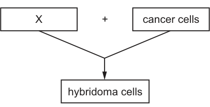
What is represented by X?
Options
A T-lymphocytes
B B-lymphocytes
C antigens
D antibodies
Answer
Monoclonal antibodies are produced by fusing B-lymphocytes (which make a single specific antibody) with cancer (myeloma) cells, which divide uncontrollably. The resulting hybridoma cells combine these two properties: they produce one specific antibody and they divide indefinitely.
X must therefore be the antibody-producing cell, i.e. B-lymphocytes.
B
B
Background Concept
Monoclonal antibodies are identical antibodies that all recognise the same epitope (a single, specific binding site on an antigen). To produce them in useful quantities, scientists exploit two very different cell types:
- B-lymphocytes make antibody, but only one specific kind each, and they cannot divide indefinitely in culture.
- Myeloma (cancer) cells divide uncontrollably (they are 'immortal' in culture) but make the wrong antibody, or none at all.
A hybridoma is a cell made by fusing a B-lymphocyte with a myeloma cell. It inherits the B-lymphocyte's ability to make one specific antibody, and the myeloma cell's ability to divide without limit. Each hybridoma is a clone, so all of the antibody it produces is identical — monoclonal.
Understanding the Question
The diagram is a simple flow chart: a box labelled X is added to cancer cells and the result is hybridoma cells. The question asks what cell type X is, given that it combines with a cancer cell to make a hybridoma. This is the standard hybridoma production step from the immunity topic of the AS syllabus.
Approach
Apply the definition of a hybridoma: it is formed by fusing a B-lymphocyte with a cancer (myeloma) cell. Whichever cell is fused with the cancer cell to give the hybridoma must be the B-lymphocyte.
Step-by-Step Reasoning
- Hybridoma = B-lymphocyte + myeloma (cancer) cell. Therefore, in the diagram, cancer cells = myeloma cells (given) and the other input, X, must be the B-lymphocyte.
- Check the distractors:
- A. T-lymphocytes — involved in cell-mediated immunity (and helping B-cells), not the source of secreted antibody in this process. Not fused with myeloma cells to make hybridomas.
- C. Antigens — these are the molecules (e.g. on a pathogen) that stimulate antibody production; they are not cells and cannot be fused.
- D. Antibodies — these are Y-shaped proteins secreted by B-cells/plasma cells, not cells themselves. They are the product of the hybridoma, not an ingredient in making it.
- Only B-lymphocytes fit the role of being a cell fused with cancer cells to produce hybridomas.
Key Takeaways
- A hybridoma = B-lymphocyte fused with a myeloma (cancer) cell.
- B-lymphocytes supply antibody specificity; cancer cells supply the ability to divide indefinitely.
- Monoclonal = every antibody from a single hybridoma clone recognises the same epitope.
Common Mistakes
- Choosing T-lymphocytes because they are also involved in the immune response — but T-cells are not fused to form hybridomas in the monoclonal antibody process.
- Choosing antibodies because the technique is called monoclonal antibody production — but antibodies are the output of the hybridoma, not one of the two fused cells.
- Choosing antigens, which are non-cellular molecules and could not be fused with a cell to make a hybrid cell line.
Things to Be Careful About
- The word 'cancer cells' in the diagram refers specifically to myeloma cells (a cancer of B-lymphocytes), not any cancer cell. The fusion partner is always a myeloma line.
- 'X' is a cell, so the answer must be a cell type. Only B-lymphocytes (and T-lymphocytes) among the options are cells, and only B-lymphocytes fit the role.
Your score so far
Answer a question to start scoring
Your marks add up here as you work through the paper.