Biology 9700/11 — May/June 2024
Cambridge AS Level · Multiple Choice (AS Level) · answer key with instant marking and worked solutions
Topics Biological Molecules · Cell Structure · Enzymes · Nucleic Acids and Protein Synthesis · The Mitotic Cell Cycle · Transport in Mammals · +5 more
Tap an option under each question to check it — your score builds as you go.
The diagram shows a stage micrometer scale viewed with an eyepiece graticule, using a magnification of .
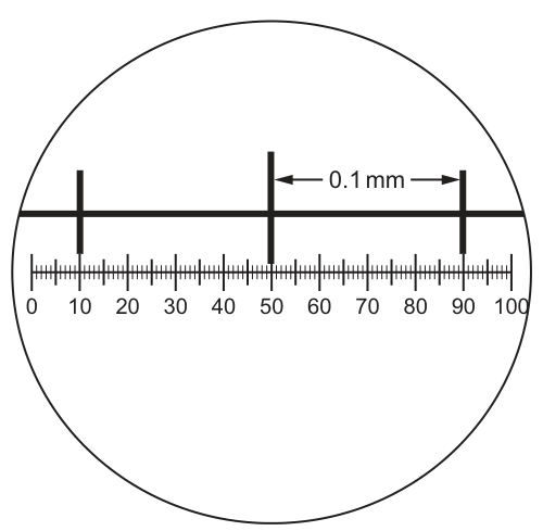
Using the same magnification, a chloroplast is measured as 4 eyepiece graticule divisions long.
How long is the chloroplast?
Options
A
B
C
D
Working
The stage micrometer shows that aligns with eyepiece graticule divisions (from 50 to 90).
Length of 1 eyepiece division:
The chloroplast is eyepiece graticule divisions long:
Answer
A
A
Background Concept
A stage micrometer is a slide that carries a scale of known length (here graduated so that each large division = ). An eyepiece graticule is a small glass disc, ruled with arbitrary divisions, that sits inside the eyepiece of a microscope. Because the graticule's divisions are not labelled with real units, they must be calibrated against the stage micrometer at each magnification you intend to use. Once calibrated, the stage micrometer is removed and any specimen on the slide can be measured directly in real units by counting graticule divisions.
Two important consequences follow:
- The calibration is only valid at the magnification used to perform it. If the objective is changed, the eyepiece graticule must be re-calibrated with the stage micrometer at the new magnification.
- The stated magnification is already "baked into" the calibration, so once a calibration exists for , you do not divide by the magnification again — doing so double-counts it and is a very common trap.
Understanding the Question
The figure shows both scales superimposed in the field of view at :
- The upper (stage micrometer) scale shows that the interval between two of its large marks is .
- That interval is exactly spanned by the eyepiece graticule from division to division , i.e. 40 graticule divisions.
The chloroplast on the (real) slide measures 4 eyepiece graticule divisions at the same magnification. The question asks for the chloroplast's actual length, in micrometres.
The command word here is the implicit "calculate" — the candidate must convert graticule divisions to a real length, so a numerical answer is required.
Approach
- Use the graticule/stage-micrometer alignment to find how many real millimetres (then µm) one graticule division represents.
- Multiply by the number of graticule divisions the chloroplast spans (4).
- Ignore the magnification — it has already been used to produce the alignment shown, and re-using it would give a result off by a factor of 200.
Step-by-Step Reasoning
Step 1 — Calibrate one graticule division.
From the figure, eyepiece divisions span on the stage micrometer, so:
Step 2 — Convert mm to µm.
, therefore:
Step 3 — Apply to the chloroplast (4 divisions).
In standard form, .
Step 4 — Sanity check against biology and the options.
A typical higher-plant chloroplast is about long, so is realistic. This matches option A. Options C and D (sub-micrometre lengths) are smaller than a ribosome, and option B () is the size of a small plant cell, not a chloroplast.
Key Takeaways
- An eyepiece graticule is calibrated against a stage micrometer at the magnification you will use; the calibration is the conversion factor from graticule divisions to real length.
- Once the calibration is done at , the magnification value is not re-applied to the measured specimen — it is already incorporated.
- The two essential unit conversions to know cold: and ? — actually , and the working units you should be most fluent in for microscopy are mm ↔ µm ↔ nm.
- After any microscope-size calculation, do a quick biological check: the answer should be the right order of magnitude for the organelle/cell in question.
Common Mistakes
- Dividing the image size by the magnification (). This is the formula for a printed/photographed image with a scale bar, not for an eyepiece graticule. Doing it here gives — close to option D () and wrong by a factor of 200. The CIE mark scheme will not award credit for this approach in graticule problems.
- Reading the wrong number of graticule divisions that align with . The figure specifically shows the alignment between 50 and 90, which is 40 divisions (not 50, not 100, not the full scale).
- Forgetting to convert mm to µm, so reporting instead of . Either is dimensionally correct but µm is the conventional unit for cellular structures and matches the options.
- Choosing option B () by treating the calibration as graticule division and multiplying by 4. This ignores that the spans 40 divisions, not 1.
Things to Be Careful About
- Always read the alignment between two matching features of the two scales (here the two long stage-micrometer marks), and count the graticule divisions that fall strictly between them (here 50 → 90 = 40 divisions).
- Write the final answer in the same unit as the answer options (µm here). If you obtain , express it as to match option A exactly.
- The magnification stated in the question is deliberately a red herring in graticule questions: if the alignment you have already comes from that magnification, the magnification value plays no further role in the calculation.
- For Paper 3 / Paper 5 microscope work, this calibration is the foundation of every drawing and every size measurement. Practise it until you can do the conversion without reaching for a calculator.
Which range of cell diameters is typical for prokaryotic cells?
Options
A to
B to
C to
D to
Working
Typical prokaryotic cells (e.g. bacteria such as Escherichia coli) have diameters in the range of about to .
Converting the options to a common unit:
- A: to = to — too small (virus/macromolecule range)
- B: to = to — correct
- C: to — typical eukaryotic cell range
- D: to — far too large
Answer
B
B
Background Concept
Prokaryotic cells (bacteria and archaea) are the simplest and smallest cellular organisms. Unlike eukaryotic cells, they lack a true membrane-bound nucleus and other membrane-bound organelles. A typical bacterium such as Escherichia coli is rod-shaped, about 1 µm wide and 2 µm long, and most prokaryotes fall within a diameter range of roughly to . By contrast, typical eukaryotic cells range from about to in diameter.
Key unit relationships:
Understanding the Question
This is a multiple-choice question asking which of the four given size ranges corresponds to a typical prokaryotic cell diameter. The candidate must know the standard size of prokaryotes and be able to convert between nanometres and micrometres to evaluate the options.
Approach
Recall the typical size of a bacterium, then convert all four options into the same units so they can be compared directly against the expected range of about to .
Step-by-Step Reasoning
- Option A spans to (i.e. to ). This is in the size range of viruses and large macromolecules, not whole cells.
- Option B spans to = to . This matches the typical diameter of prokaryotic cells perfectly.
- Option C spans to , which is the range of typical eukaryotic cells (e.g. many animal and plant cells).
- Option D spans to ( to ), which is far larger than any typical cell and is in the range visible to the naked eye.
Therefore, B is the correct answer.
Key Takeaways
- Prokaryotic cells typically have diameters of about to .
- Eukaryotic cells are typically to in diameter — about 10× larger.
- Viruses and macromolecules sit in the nanometre range ( to a few hundred nm).
- Always convert to consistent units (µm vs nm) when comparing size ranges.
Common Mistakes
- Confusing viral size (tens to hundreds of nm) with cellular size (≥ 1 µm).
- Misreading the exponent and confusing with .
- Selecting C because it "looks big enough" without converting the units to check.
Things to Be Careful About
- Read the units in each option carefully — options A and B are in nanometres while C and D are in micrometres.
- Remember that , so .
- The typical diameter of a bacterium such as E. coli is a useful benchmark to remember.
The diagram shows a typical animal cell.
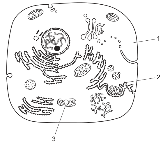
Where would nucleic acids be found?
Options
A 1, 2 and 3
B 1 and 2 only
C 1 and 3 only
D 2 and 3 only
Working
Nucleic acids (DNA and RNA) are found in all three labelled structures:
- 1 — Nucleus: contains DNA (as chromatin) and is the site where RNA (mRNA, tRNA, rRNA) is transcribed, so both DNA and RNA are present.
- 2 — Mitochondrion: contains its own circular mitochondrial DNA and mitochondrial ribosomes (rRNA), so both DNA and RNA are present.
- 3 — Ribosome: composed of rRNA and protein, and is the site where mRNA is translated, so RNA is present.
Therefore nucleic acids are found in 1, 2 and 3.
Answer
A
A
Background Concept
Nucleic acids are macromolecules built from nucleotide monomers and exist in two main forms in cells: DNA (deoxyribonucleic acid) and RNA (ribonucleic acid). They are not confined to a single compartment — different types of nucleic acid are distributed across several organelles, each performing a distinct role in storing or expressing genetic information.
Key locations of nucleic acids in a eukaryotic cell:
- Nucleus: the main store of cellular DNA, packaged with histone proteins as chromatin. The nucleolus within the nucleus is where ribosomal RNA (rRNA) is transcribed and ribosomal subunits are assembled. The nucleus is also the site of transcription of all types of RNA (mRNA, tRNA, rRNA).
- Mitochondrion: contains a small amount of its own mitochondrial DNA (mtDNA) — a circular molecule resembling bacterial DNA, evidence for the endosymbiotic origin of mitochondria. Mitochondria also have their own ribosomes (containing mitochondrial rRNA) and so carry out a limited form of protein synthesis.
- Ribosome: built from rRNA molecules combined with ribosomal proteins. It is the site of translation, where mRNA is decoded to synthesise polypeptides, so rRNA and (transiently) mRNA and tRNA are all present.
A useful rule of thumb: anywhere protein synthesis occurs, or where genetic information is stored or used, nucleic acids will be present.
Understanding the Question
The question shows a diagram of a typical animal cell with three labelled structures: 1 points to the nucleus (with chromatin and a nucleolus visible inside it), 2 points to a mitochondrion, and 3 points to a ribosome (shown attached to the rough endoplasmic reticulum). The task is to select which of these structures contain nucleic acids.
The command word is implicit here — the question offers four combinations of the three labels, so the candidate must decide whether each organelle contains nucleic acids and then match the answer to the correct option.
Approach
Work through each label systematically and decide whether nucleic acids (DNA and/or RNA) are present there. If nucleic acids are present in all three, the answer is "1, 2 and 3".
Step-by-Step Reasoning
-
Label 1 — Nucleus. DNA is the genetic material of the cell and is stored in the nucleus as chromatin (DNA wrapped around histone proteins). The nucleolus is rich in rRNA, and the nucleoplasm contains newly transcribed mRNA and tRNA. Nucleic acids: present (DNA + RNA).
-
Label 2 — Mitochondrion. Mitochondria are semi-autonomous organelles that contain their own circular DNA and their own ribosomes, enabling them to transcribe and translate a small number of their own proteins. Nucleic acids: present (mtDNA + mitochondrial rRNA/mRNA/tRNA).
-
Label 3 — Ribosome. Ribosomes are themselves roughly two-thirds rRNA by mass; the catalytic activity that forms peptide bonds is carried out by the rRNA of the large subunit (a ribozyme). The mRNA being translated and the tRNAs delivering amino acids also pass through the ribosome. Nucleic acids: present (rRNA, and transiently mRNA and tRNA).
Because all three structures contain nucleic acids, the correct combination is "1, 2 and 3", which matches option A.
The distractors test common misconceptions:
- B (1 and 2 only): the misconception that ribosomes are "just protein" or that RNA isn't really a nucleic acid.
- C (1 and 3 only): the misconception that mitochondria have no DNA of their own.
- D (2 and 3 only): the misconception that the nucleus does not contain nucleic acids — a clear error.
Key Takeaways
- DNA is found in the nucleus and in mitochondria (and in chloroplasts in plant cells).
- RNA is found in the nucleus, in ribosomes, and in mitochondria — in fact, all sites of transcription and translation.
- Ribosomes are not pure protein; they are ribonucleoprotein particles, and their rRNA performs the catalytic (peptidyl transferase) activity.
- A cell cannot carry out protein synthesis without RNA at the site of translation.
Common Mistakes
- Thinking ribosomes contain only protein. Ribosomes are about 60% rRNA and 40% protein; the rRNA is the catalytically active component.
- Forgetting that mitochondria have their own DNA. Mitochondrial DNA encodes some of the proteins of the electron transport chain and is replicated inside the organelle.
- Confusing the nucleolus with the whole nucleus. The nucleolus itself does not contain DNA directly, but it lies within the nucleus, which does. The question labels the whole nucleus, so DNA is included.
- Treating RNA as somehow "not a nucleic acid". RNA is a nucleic acid, so any structure containing RNA answers "yes" to the question.
Things to Be Careful About
- The question asks where nucleic acids would be found — both DNA and RNA count, so the answer requires considering both types.
- Always check the labels carefully: label 3 in this diagram is the ribosome, not the rough ER itself. Ribosomes are the structures containing nucleic acids, not the membrane to which they are attached.
- Do not assume that only the "headquarters" of the cell (the nucleus) contains genetic material; the endosymbiotic organelles retain their own.
Which structure is found in a typical bacterial cell?
Options
A intron
B telomere
C template strand
D capsid
Working
- A bacterial cell is prokaryotic, with a single circular DNA molecule but no nucleus or linear chromosomes.
- A template strand is the strand of DNA that is transcribed to produce mRNA. Bacteria transcribe their DNA, so they must have a template strand.
- Introns are non-coding sequences typical of eukaryotic genes; bacteria rarely have them.
- Telomeres cap the ends of linear eukaryotic chromosomes; bacterial DNA is circular and has no telomeres.
- A capsid is the protein coat of a virus, not a cellular structure.
Answer
C
C
Background Concept
Bacteria are prokaryotes. Their defining features include:
- A single circular DNA molecule located in the nucleoid (not enclosed by a membrane).
- No membrane-bound organelles.
- Cell wall made of peptidoglycan.
- 70S ribosomes.
- Sometimes plasmids (small circular pieces of DNA carrying accessory genes).
Eukaryotic cells (animals, plants, fungi, protists) have:
- Linear DNA packaged into chromosomes inside a nucleus.
- Telomeres at the ends of each linear chromosome to protect them from degradation.
- Split genes with introns (non-coding sequences) and exons (coding sequences).
- 80S ribosomes and membrane-bound organelles.
Viruses are non-cellular. They consist of nucleic acid (DNA or RNA) enclosed in a protein coat called a capsid, and sometimes a lipid envelope. They have no ribosomes, cytoplasm, or metabolism of their own, and are not considered living cells.
Understanding the Question
The question asks which of the four structures is found inside a typical bacterial cell. The command word is implicit here — you must recognise that only one option is compatible with prokaryotic cellular organisation, and the other three either belong to eukaryotes or to viruses (which are not cells at all).
Approach
Eliminate each wrong option by linking the structure to the cell type it belongs to, then confirm the remaining option is present in bacteria. This is a process of elimination based on the prokaryote vs eukaryote vs virus distinction.
Step-by-Step Reasoning
Option A — intron. Introns are non-coding sequences within eukaryotic genes that are spliced out of pre-mRNA before translation. Prokaryotic genes are typically not split; the mRNA is translated as soon as it is transcribed (coupled transcription–translation). Bacteria rarely have introns. Reject.
Option B — telomere. Telomeres are repetitive nucleotide sequences (e.g. TTAGGG in humans) at the ends of linear eukaryotic chromosomes. They prevent chromosome ends from being recognised as DNA damage and are shortened each round of replication. Bacterial DNA is circular, so it has no ends and therefore no telomeres. Reject.
Option C — template strand. The template strand (also called the antisense or non-coding strand) is the strand of DNA that RNA polymerase reads during transcription to synthesise a complementary mRNA molecule. Bacteria carry out transcription using their single DNA molecule, so they must have a template strand. Accept.
Option D — capsid. A capsid is the protein shell that surrounds the nucleic acid of a virus. Bacteria are cellular organisms, not viruses, and they do not possess a capsid. Reject.
The only structure compatible with a typical bacterial cell is the template strand, so the answer is C.
Key Takeaways
- Prokaryotes and eukaryotes differ in chromosome structure: circular vs linear.
- Telomeres, split genes (introns) and a nuclear membrane are eukaryotic features.
- A capsid belongs to viruses, not cells.
- The template strand is a universal feature of any cell (or virus) that uses DNA-directed transcription — including bacteria.
Common Mistakes
- Confusing the bacterial chromosome (circular DNA) with eukaryotic linear chromosomes and assuming telomeres are universal — they are not.
- Choosing capsid because bacteria are small and "virus-like" — bacteria are cells; viruses are non-cellular and lack cytoplasm, ribosomes and metabolism.
- Forgetting that bacteria still carry out transcription, so they require a template strand of DNA.
Things to Be Careful About
- Read the stem carefully: it asks what is found in a typical bacterial cell. Options belonging to eukaryotes or to viruses must be ruled out.
- "Template strand" is a feature of any DNA-based genetic system, not a specifically eukaryotic one — it is therefore the safest and most accurate answer for a prokaryote.
What is the maximum number of hydrogen bonds that can form between a single water molecule and other water molecules?
Options
A 1
B 2
C 3
D 4
Working
A water molecule (H₂O) has two hydrogen atoms and one oxygen atom with two lone pairs of electrons.
Each hydrogen atom can act as a hydrogen bond donor, forming 1 hydrogen bond (2 in total).
Each lone pair on the oxygen can act as a hydrogen bond acceptor, forming 1 hydrogen bond (2 in total).
Maximum number of hydrogen bonds = 2 (from H atoms) + 2 (from O lone pairs) = 4.
Answer
D
D
Background Concept
A water molecule consists of one oxygen atom covalently bonded to two hydrogen atoms. The oxygen is highly electronegative and pulls electron density away from the hydrogens, leaving the O with a partial negative charge (δ⁻) and the H atoms with partial positive charges (δ⁺). The oxygen also carries two lone pairs of non-bonding electrons.
A hydrogen bond is an intermolecular attraction that forms when:
- a δ⁺ hydrogen on one molecule (the donor) is electrostatically attracted to a lone pair on a strongly electronegative atom (typically O, N or F) on a neighbouring molecule (the acceptor).
Each H can donate one hydrogen bond, and each lone pair on an O can accept one hydrogen bond.
Understanding the Question
The question asks for the maximum number of hydrogen bonds that one water molecule can participate in with other water molecules. We must consider both the donor sites (the H atoms) and the acceptor sites (the lone pairs on the O).
The command word is "maximum", so we assume every donor and acceptor site is simultaneously involved in a hydrogen bond — i.e. water in its optimal hydrogen-bonded arrangement (such as in ice).
Approach
Count the donor hydrogens and the acceptor lone pairs on a single water molecule; add them to get the total maximum number of hydrogen bonds per molecule.
Step-by-Step Reasoning
- A water molecule has 2 hydrogen atoms, each able to donate 1 hydrogen bond → 2 hydrogen bonds from the H side.
- The oxygen atom has 2 lone pairs of electrons, each able to accept 1 hydrogen bond → 2 hydrogen bonds from the O side.
- Maximum total = 2 + 2 = 4 hydrogen bonds per water molecule.
This is exactly what is observed in the structure of ice, where each H₂O is tetrahedrally hydrogen-bonded to four neighbours. The other options fail because:
- A (1) only accounts for a single donor or acceptor site.
- B (2) ignores either the second H or the second lone pair.
- C (3) misses one of the four available sites.
Key Takeaways
- Water is a polar molecule with 2 H-bond donor sites and 2 H-bond acceptor sites, giving a maximum of 4 hydrogen bonds per molecule.
- This 4-bond capacity underpins many of water's unusual properties (high specific heat capacity, high boiling point, cohesion, ice being less dense than liquid water).
Common Mistakes
- Counting only the hydrogen atoms (2) and forgetting the lone pairs on oxygen — leads to answer B.
- Confusing the number of covalent bonds within a water molecule (2: O–H) with the number of hydrogen bonds between water molecules.
Things to Be Careful About
- The question specifies with other water molecules, so the H and lone pair sites on the same molecule are not counted twice.
- Hydrogen bonds are intermolecular, not covalent; the O–H bonds within a water molecule are polar covalent bonds and are not the same as hydrogen bonds.
The diagrams show the structure of four amino acids in aqueous solution.
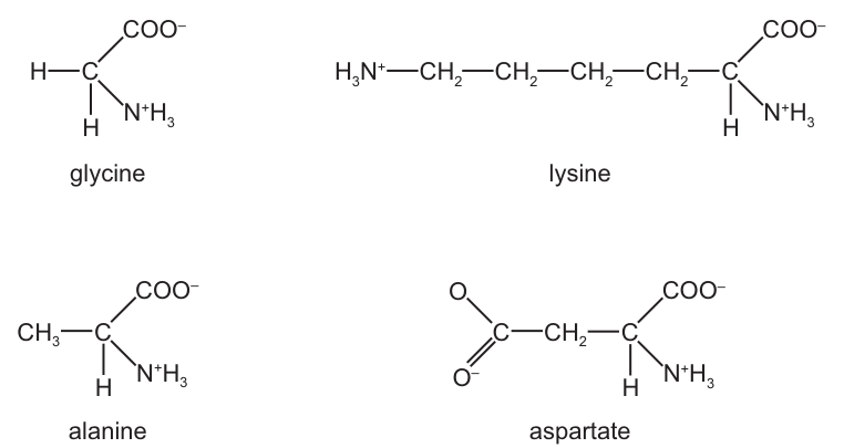
Which two structures have an overall charge?
Options
A alanine and aspartate
B alanine and glycine
C aspartate and lysine
D glycine and lysine
Working
At physiological (aqueous) pH, amino acids exist as zwitterions: the α-amino group is protonated (, +1) and the α-carboxyl group is deprotonated (, −1). A side chain (R group) that is itself ionised tips the molecule away from a net charge of zero.
Working out the net charge for each amino acid shown:
- Glycine (R = H): one (+1) + one (−1) → net = 0
- Alanine (R = ): one (+1) + one (−1) → net = 0
- Lysine (R = ): (+1) + side-chain (+1) + (−1) → net = +1
- Aspartate (R = ): (+1) + (−1) + side-chain (−1) → net = −1
Only lysine and aspartate carry an overall charge.
Answer
C
C
Background Concept
An amino acid has a central (α) carbon bonded to four groups: an amino group (), a carboxyl group (), a hydrogen atom and a variable R group (side chain). At physiological pH (~7.4) the molecule is ionised:
- The α-amino group gains a proton to become (charge +1).
- The α-carboxyl group loses a proton to become (charge −1).
Together these give the molecule a +1 and a −1, summing to a net charge of zero. This doubly-ionised, electrically neutral form is called a zwitterion.
For most amino acids the side chain (R group) is non-ionisable, so the zwitterion form has a net charge of zero. However, the side chains of a few amino acids carry their own ionisable groups. If the side chain adds a positive charge, the overall molecule becomes positive; if it adds a negative charge, the molecule becomes negative. The relevant examples here are:
- Lysine — side chain ends in an extra group (basic amino acid).
- Aspartate — side chain contains an extra group (acidic amino acid).
A simple way to determine the net charge of any amino acid in the diagram is to count the + signs and the − signs on the structure and add them up.
Understanding the Question
The question supplies the structures of four amino acids already drawn in their ionised (aqueous) forms. The task is to decide which two of them do NOT have their + and − charges balancing out — i.e. which two have a non-zero net charge.
The command word "have an overall charge" is decisive: it asks for a net (total) charge, not the presence of any charged group. Even glycine, which is a zwitterion, contains both a and a , but those two cancel.
Approach
For each of the four structures, tally the number of (or other +) groups and the number of (or other −) groups. Whichever amino acids do not have the + and − totals equal are the ones with an overall charge.
Step-by-Step Reasoning
-
Glycine — drawn as . One +1 and one −1 → net charge = 0. Although ionised, it is a neutral zwitterion.
-
Alanine — drawn as . The side chain is uncharged. One +1 and one −1 → net charge = 0. Again, a neutral zwitterion.
-
Lysine — drawn with a long side chain ending in a second group. So the structure carries two groups (+1 +1 = +2) and one group (−1). Net charge = +2 + (−1) = +1. Lysine has an overall positive charge.
-
Aspartate — drawn with a side chain containing a second group. The structure carries one group (+1) and two groups (−1 + −1 = −2). Net charge = +1 + (−2) = −1. Aspartate has an overall negative charge.
Only lysine and aspartate have a non-zero net charge, so the answer is C: aspartate and lysine.
Key Takeaways
- All amino acids drawn at physiological pH contain at least one and one , but only those whose side chains are themselves ionised end up with a net charge.
- Basic amino acids (e.g. lysine, arginine, histidine) carry a net positive charge because their side chain has an extra amino/imidazole group that is protonated.
- Acidic amino acids (e.g. aspartate, glutamate) carry a net negative charge because their side chain has an extra carboxyl group that is deprotonated.
- The shortcut: count the +'s and −'s in the structure; if they balance, the molecule is a zwitterion; if they do not, the molecule has an overall charge.
Common Mistakes
- Counting any ionised group as "having a charge" and picking glycine (which is a zwitterion, net charge 0) or alanine (also a zwitterion, net charge 0). Glycine and alanine do carry and groups but their net charge is zero, so this answer is rejected.
- Forgetting the second charged group in the side chain — lysine's R group is , not just a long hydrocarbon, and aspartate's R group is , not just . Treating the side chains as uncharged makes the net charge look zero for all four.
- Picking the two zwitterions (option B, glycine and alanine) because they "have charges drawn". The question asks for overall net charge, not the number of ionised groups.
Things to Be Careful About
- Read the question carefully: "overall charge" means the net charge after all + and − contributions are added, not the mere presence of any charged group.
- Memorise the three acidic amino acids (aspartate, glutamate) and the three basic amino acids (lysine, arginine, histidine) so you can recognise them from the side chain alone — this is faster and more reliable than counting signs on every structure you meet.
- In a zwitterion (net zero), the molecule is still drawn with and in CIE-style diagrams; do not mistake this for an uncharged molecule. Look at the side chain.
- The CIE convention is to write these side-chain charges in superscript form (e.g. , ) — be sure to render them correctly in any answer you write.
The table shows the number of carbon atoms and the number of carbon–carbon double bonds in molecules of three triglycerides and the fatty acids they contain.
The information is shown in the format ‘number of carbon atoms : number of carbon–carbon double bonds’.
| triglyceride | fatty acid 1 | fatty acid 2 | fatty acid 3 |
|---|---|---|---|
| 54 : 1 | 15 : 0 | 18 : 0 | 18 : 1 |
| 56 : 4 | 17 : 0 | X | 18 : 2 |
| 57 : 3 | 18 : 0 | 18 : 1 | 18 : 2 |
Which description matches fatty acid 2 of the 56 : 4 triglyceride, identified in the table as X?
Options
A saturated with 17 carbon atoms
B saturated with 18 carbon atoms
C unsaturated with 17 carbon atoms
D unsaturated with 18 carbon atoms
Working
A triglyceride is made from one glycerol (3 carbons) + 3 fatty acids. The carbon count in the triglyceride therefore equals 3 + (sum of the three fatty acid carbon numbers).
For the 56 : 4 triglyceride, fatty acid 1 = 17 : 0 and fatty acid 3 = 18 : 2.
Carbons in X:
Double bonds in X:
So X is 18 : 2 — a fatty acid with 18 carbons and 2 C=C double bonds, i.e. unsaturated.
Answer
D
D
Background Concept
A triglyceride is formed by the condensation of one glycerol molecule and three fatty acid molecules, joined by three ester bonds. Because three water molecules are removed during formation, the carbons of the three fatty acids are not lost — only the –OH from each fatty acid and an –H from each glycerol hydroxyl. So the total number of carbon atoms in a triglyceride equals:
Fatty acids are described as saturated (no C=C double bonds; only C–C single bonds) or unsaturated (one or more C=C double bonds). The notation n : m means n carbons and m C=C double bonds, so an 18:2 fatty acid is unsaturated (it has 2 double bonds).
Understanding the Question
We are given a table where each row shows a triglyceride (in total C : total C=C form) and the three fatty acids it contains. Two fatty acids in row 2 are known (17:0 and 18:2), and we must deduce the third, X, from the totals 56:4. We then choose the description (A–D) that fits X.
The command word is implicit: this is an MCQ, so we pick the option that correctly states whether X is saturated or unsaturated and how many carbons it has.
Approach
- Use the row totals to write two simple equations: one for the carbon count, one for the double-bond count.
- Solve each for the missing value of X.
- Read off whether X is saturated (0 double bonds) or unsaturated (≥1 double bond), and its carbon number.
- Match to the options.
Step-by-Step Reasoning
Carbons in X
The triglyceride has 56 carbons. Subtract the 3 glycerol carbons and the carbons of the two known fatty acids:
Double bonds in X
The triglyceride has 4 C=C bonds in total. Subtract the contributions of the two known fatty acids (0 and 2):
So X is 18 : 2.
Interpretation
- 18 carbons → option B or D.
- 2 C=C double bonds → unsaturated (saturated would have 0).
Therefore X is unsaturated with 18 carbon atoms → option D.
Cross-check with the other rows
- Row 1: 15 + 18 + 18 + 3 (glycerol) = 54 ✓; 0 + 0 + 1 = 1 ✓
- Row 3: 18 + 18 + 18 + 3 = 57 ✓; 0 + 1 + 2 = 3 ✓
This confirms the glycerol-inclusive counting is correct, validating our answer.
Key Takeaways
- The carbon count of a triglyceride includes the 3 carbons of glycerol.
- The number of C=C double bonds in a triglyceride equals the sum of the C=C double bonds in its three fatty acids (ester bond formation does not create or destroy C=C bonds).
- A fatty acid is saturated only when it has zero C=C double bonds; anything ≥ 1 is unsaturated.
Common Mistakes
- Forgetting the glycerol carbons. Students often do 56 − 17 − 18 = 21, getting 21 carbons (not in the options) and end up guessing. Always subtract 3 for glycerol.
- Confusing saturation with carbon number. A long-chain fatty acid can still be saturated; "saturated" describes the bonds, not the length.
- Ignoring the 18:2 in fatty acid 3. Some students assume fatty acid 2 has all 4 double bonds and pick something like "saturated with 18 C" incorrectly.
Things to Be Careful About
- The notation
n : malways means carbons : C=C double bonds — never carbons : hydrogens or anything else. - When a question gives a triglyceride carbon total, mentally partition it as 3 (glycerol) + three fatty-acid contributions before subtracting.
- The options are designed so that a sign or arithmetic slip pushes you to a closely-related but wrong answer (e.g. 17 carbons with 1 double bond would also look plausible) — keep both the carbon and the double-bond arithmetic separate and correct.
Collagen molecules are made up of three polypeptide chains interacting together. The individual polypeptide chains consist of a regular pattern of amino acids. Almost every third amino acid is glycine.
Which protein structures of collagen are described?
Options
A primary and secondary
B primary and quaternary
C secondary and tertiary
D tertiary and quaternary
Working
-
"The individual polypeptide chains consist of a regular pattern of amino acids. Almost every third amino acid is glycine."
- This refers to the primary structure — the specific sequence of amino acids joined by peptide bonds in a single polypeptide chain.
-
"Collagen molecules are made up of three polypeptide chains interacting together."
- This refers to the quaternary structure — the association of more than one polypeptide chain (subunit) to form the functional protein.
-
The structures described are therefore primary and quaternary.
Answer
B
B
Background Concept
Proteins have four levels of structure, each defined by what holds it together and what gives rise to it:
- Primary structure: the linear sequence of amino acids in a polypeptide chain, linked by peptide bonds formed in condensation reactions. It is determined by the gene that codes for the protein.
- Secondary structure: regular, repeated folding of a single polypeptide chain, most commonly an α-helix or β-pleated sheet, stabilised by hydrogen bonds between the C=O of one peptide bond and the N–H of another.
- Tertiary structure: the overall 3-D folding of a single polypeptide chain into its functional shape, stabilised by hydrogen bonds, ionic bonds, disulfide bridges and hydrophobic interactions between R groups.
- Quaternary structure: the association of two or more polypeptide chains (subunits) into a single functional protein molecule, held by the same types of R-group interactions as in tertiary structure.
Collagen is a fibrous protein made of three polypeptide (α) chains wound around each other to form a triple helix. Each α chain has a very unusual amino acid composition: roughly one residue in three is glycine, and many of the others are proline or hydroxyproline. Glycine is the only amino acid small enough to fit into the tight central axis where the three chains meet.
Understanding the Question
The stem gives two separate facts about collagen:
- The individual chains have a regular sequence in which every third amino acid is glycine.
- Three of these chains interact to form a collagen molecule.
The question asks which of the four protein-structure levels these two facts correspond to.
Approach
Match each fact to the correct level of structure by using the definitions above:
- A statement about the sequence of amino acids in a single chain = primary structure.
- A statement about multiple chains associating = quaternary structure.
The secondary and tertiary levels are not described in the stem: the stem never mentions hydrogen bonding within a chain (secondary) or the overall 3-D folding of a single chain (tertiary).
Step-by-Step Reasoning
-
"Individual polypeptide chains consist of a regular pattern of amino acids. Almost every third amino acid is glycine."
- This is a description of the order of amino acids along the chain. That is, by definition, the primary structure. The repeating Gly-X-Y motif is determined at the level of the gene and built by peptide bonds during translation.
-
"Collagen molecules are made up of three polypeptide chains interacting together."
- Three separate polypeptide chains coming together to form a single functional protein is the textbook definition of quaternary structure (e.g. haemoglobin with 4 chains, immunoglobulins with 4 chains, collagen with 3 chains).
-
The two structures named are therefore primary and quaternary, which is option B.
-
Why the other options are wrong:
- A (primary and secondary) — secondary structure is the regular H-bonded folding within a single chain (α-helix / β-sheet). The stem says nothing about H-bonded folding patterns.
- C (secondary and tertiary) — neither is described: no mention of H-bonded coiling, and no mention of a single chain folding into a 3-D shape.
- D (tertiary and quaternary) — the description of the amino acid sequence in each chain is primary, not tertiary.
Key Takeaways
- Primary structure = sequence of amino acids in one chain.
- Quaternary structure = how chains are assembled into the whole protein.
- Collagen is a classic example of a protein with quaternary structure (3 chains) and a striking primary structure (Gly-X-X repeat).
Common Mistakes
- Confusing the triple helix of collagen with secondary structure. The triple helix is quaternary because it is built from three separate chains; an α-helix or β-sheet (secondary) is folded within a single chain.
- Treating "regular pattern of amino acids" as if it implied secondary structure. A regular sequence is primary; a regular folding pattern is secondary.
- Forgetting that fibrous proteins such as collagen still have all four levels of structure, even though they are not globular.
Things to Be Careful About
- Read the question for what is actually being described. The stem separates the statements deliberately — one is about the sequence in a chain, the other is about chains coming together.
- Use the precise CIE definitions of primary, secondary, tertiary and quaternary structure. Marks are awarded for matching the definition to the feature, not for a vague impression that the answer "looks right".
The masses of the parts of haemoglobin are shown in the table.
| component | mass / Da |
|---|---|
| -globin chain | 15 126 |
| -globin chain | 15 868 |
| haem group | 617 |
There are 1000 Da in 1 kDa.
What is the mass of a haemoglobin molecule in kDa?
Options
A 31.6
B 33.5
C 62.6
D 64.5
Working
Haemoglobin is a quaternary protein made of 2 α-globin chains, 2 β-globin chains and 4 haem groups.
Converting to kDa:
Answer
D
D
Background Concept
Haemoglobin is a globular, conjugated protein — it has a quaternary structure made up of four polypeptide chains (two identical α-globin chains and two identical β-globin chains), each of which is wrapped around a haem prosthetic group that contains an iron ion at its centre. The standard adult form (HbA) therefore contains 2 α + 2 β + 4 haem groups.
The masses of the individual components are measured in daltons (Da), where 1 Da is defined as 1/12 of the mass of a carbon-12 atom and is approximately equal to the mass of one hydrogen atom. For larger molecules like proteins, kilodaltons (kDa) are used; 1 kDa = 1000 Da.
Understanding the Question
This is a calculation question (an MCQ). The table gives the mass of each type of component, and the candidate must use knowledge of haemoglobin's structure to decide how many of each to add together, then convert from Da to kDa to compare with the options.
The four options differ in two ways:
- A (31.6) and B (33.5) are roughly half of the correct value — these would arise if a candidate only counted one α chain and one β chain (forgetting the quaternary structure).
- C (62.6) and D (64.5) include all four chains; the difference between them is whether the 4 haem groups have been included (each haem is 617 Da, so 4 of them add 2.468 kDa — about 2 kDa of difference).
Approach
- State the subunit composition of haemoglobin: .
- Multiply each mass by the appropriate number of copies and sum.
- Convert Da to kDa by dividing by 1000.
- Select the matching option.
Step-by-Step Reasoning
Subunit composition. A single haemoglobin molecule is a tetramer of two α-globin and two β-globin chains, each chain non-covalently bonded to one haem group. So there are 2 α chains, 2 β chains, and 4 haem groups per molecule.
Summing the masses:
- Da from the two α chains.
- Da from the two β chains.
- Da from the four haem groups.
- Total: Da.
Converting to kDa:
This matches option D.
Key Takeaways
- Haemoglobin has a quaternary structure: .
- The mass of a multi-subunit protein is the sum of the masses of all its constituent chains and prosthetic groups.
- 1 kDa = 1000 Da — divide by 1000 to convert.
Common Mistakes
- Forgetting the quaternary structure — adding only one α and one β chain gives 30.994 kDa, roughly option A (31.6) after rounding. This is the most common error.
- Forgetting the haem groups — adding just the four globin chains gives Da, which is 62.0 kDa, close to option C (62.6).
- Confusing the direction of conversion — multiplying by 1000 instead of dividing gives 64,456 kDa, which does not appear in the options but indicates the unit confusion.
Things to Be Careful About
- The mass of a haem group (617 Da) is small compared with the globin chains, but there are four of them, so they contribute ~2.5 kDa in total — never omit them when calculating total molecular mass.
- The answer should be quoted to one decimal place to match the precision of the options.
What is the correct order of locations in the cell for the production of an extracellular enzyme?
Options
A nucleus ribosome rough endoplasmic reticulum Golgi body
B ribosome nucleus Golgi body rough endoplasmic reticulum
C ribosome rough endoplasmic reticulum nucleus Golgi body
D rough endoplasmic reticulum nucleus Golgi body ribosome
Working
Extracellular enzymes are proteins, so they are made by the same pathway as any secreted protein:
- The gene encoding the enzyme is in the nucleus; transcription here produces mRNA.
- mRNA leaves the nucleus and binds to a ribosome, where translation begins. For a secreted protein, the ribosome docks onto the rough endoplasmic reticulum (RER).
- The growing polypeptide enters the RER lumen, where it folds and undergoes initial post-translational modification.
- Transport vesicles bud off the RER and carry the protein to the Golgi body, where it is further modified (e.g. glycosylation) and packaged into secretory vesicles for release by exocytosis.
So the correct order is nucleus ribosome rough endoplasmic reticulum Golgi body.
Answer
A
A
Background Concept
An extracellular enzyme is a protein that is synthesised inside the cell but then secreted to act outside the cell (e.g. digestive enzymes such as amylase, proteases and lipase, or extracellular hydrolases released by bacteria and fungi). Because it has to leave the cell, the enzyme must be:
- correctly folded,
- often glycosylated (carbohydrate added) to make it a stable glycoprotein,
- packaged into a membrane-bound vesicle that fuses with the plasma membrane, releasing the protein by exocytosis.
The organelles involved in this journey form a defined sequence known as the secretory pathway:
Each organelle has a specific job:
- Nucleus – holds the DNA; transcription produces a complementary mRNA copy of the gene.
- Ribosome – site of translation; reads the mRNA codons and assembles the amino acid chain. For secreted proteins the ribosome becomes attached to the rough ER, recognised by a signal sequence at the start of the polypeptide.
- Rough endoplasmic reticulum (RER) – studded with ribosomes; the new polypeptide enters the RER lumen where it folds, disulfide bonds form, and initial glycosylation takes place.
- Golgi body (Golgi apparatus) – a stack of flattened cisternae that further modifies proteins (e.g. trimming and adding sugars to make the mature glycoprotein), sorts them, and packages them into vesicles.
Understanding the Question
This is a multiple-choice question asking you to put the locations involved in producing an extracellular enzyme into the correct order. The command word "correct order" tells you that more than one organelle is involved, and the sequence matters. The four options each place the same four structures (nucleus, ribosome, rough ER, Golgi body) into a different sequence, so you have to identify the biologically correct flow of information and material through the cell.
Approach
The key is to remember that:
- Genetic information starts in the nucleus (DNA is transcribed to mRNA there).
- The mRNA is then translated by a ribosome.
- Because the protein is destined for secretion, the ribosome docks on the rough ER.
- The polypeptide is then handed, by transport vesicle, to the Golgi body for final modification and packaging.
So the production sequence is: nucleus → ribosome → rough ER → Golgi body.
Step-by-Step Reasoning
- Start with the gene in the nucleus. All proteins begin as a stretch of DNA; the first committed step of expression is transcription, which happens in the nucleus, producing a pre-mRNA that is processed and exported as mature mRNA.
- Move to the ribosome. Translation of the mRNA into a polypeptide chain occurs on a ribosome. For a secreted/extracellular protein, the very first amino acids form an N-terminal signal sequence that directs the ribosome to the rough ER membrane.
- Rough ER next. Once on the RER, the polypeptide is threaded into the lumen, folds into its 3D shape, has disulfide bonds formed, and receives its first carbohydrate (N-linked glycosylation).
- Golgi body last. Vesicles bud from the RER and fuse with the cis face of the Golgi. The protein moves through the Golgi cisternae, is further glycosylated, sorted, and packaged into secretory vesicles that bud from the trans face and travel to the plasma membrane for exocytosis.
Checking the options:
- A nucleus → ribosome → RER → Golgi body ✔ (matches the pathway above)
- B puts the nucleus in second place – impossible, the gene must be read in the nucleus first.
- C puts the nucleus in third place – again, transcription must precede translation.
- D starts with the RER – the RER has no DNA, so the protein cannot begin there; it must be specified by mRNA that originated in the nucleus.
Only A gives the biologically valid sequence.
Key Takeaways
- The secretory pathway for any extracellular protein is: nucleus → ribosome → rough ER → Golgi body → secretory vesicle → exocytosis.
- The nucleus comes first because it holds the gene; transcription must happen before translation.
- The rough ER comes after the ribosome because the ribosome docks on the RER (it does not float free for secreted proteins).
- The Golgi body is last in the production line because it is the final modification/sorting station before exocytosis.
Common Mistakes
- Putting the ribosome before the nucleus (as in B, C, D): the ribosome needs an mRNA, and the mRNA is made from DNA inside the nucleus, so the nucleus must come first.
- Placing the Golgi body before the rough ER: the RER is the first station of the endomembrane system; proteins must pass through it before reaching the Golgi.
- Confusing transcription and translation locations: thinking the ribosome is in the nucleus – it is not. The nuclear membrane separates transcription (nucleus) from translation (cytoplasm/ER).
- Treating the smooth ER and rough ER as interchangeable: the rough ER is defined by bound ribosomes and is the entry point of the secretory pathway; the smooth ER has different functions (lipid synthesis, detoxification).
Things to Be Careful About
- The question asks for the order of production, so the final exocytosis step is not needed in the answer – just the four organelles up to the Golgi.
- "Rough endoplasmic reticulum" is the exact term expected; "endoplasmic reticulum" alone is too vague because it could include the smooth ER.
- Remember that the mRNA, not the protein, is what moves from nucleus to ribosome – the protein itself never enters the nucleus.
- All extracellular enzymes are glycoproteins by the time they leave the Golgi, so the Golgi step is essential, not optional.
The graph shows the trend from an enzyme-catalysed reaction.
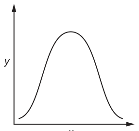
Which labels are correct for the x-axis and y-axis?
Options
| x-axis | y-axis | |
|---|---|---|
| A | rate of reaction | substrate concentration |
| B | enzyme concentration | temperature |
| C | pH | rate of reaction |
| D | substrate concentration | pH |
Working
The curve shown is bell-shaped: the y-value starts low, rises to a single peak, then falls back toward zero as x increases. This is the signature shape produced when the rate of an enzyme-catalysed reaction (y-axis) is plotted against pH (x-axis), because each enzyme has an optimum pH at which activity is maximal, with activity falling away on either side.
Checking the options:
- A — rate vs substrate concentration gives a hyperbolic curve that plateaus (not a bell shape).
- B — the axes are implausible (enzyme concentration is normally an independent variable, not temperature) and this pairing does not produce a bell shape.
- C — pH on the x-axis and rate of reaction on the y-axis gives exactly the bell-shaped curve shown. ✓
- D — pH on the y-axis would not produce a bell shape when plotted against substrate concentration.
Answer
C
C
Background Concept
Enzymes are biological catalysts whose activity is strongly influenced by the physical and chemical conditions of their environment. Two of the most important conditions tested at AS Level are temperature and pH, both of which alter the shape of the active site.
- Effect of temperature on rate: rate increases up to an optimum, then falls sharply because the enzyme is denatured (the tertiary structure and active site are irreversibly damaged by heat). The graph has a steep rise, a peak, then a steep fall to (or near) zero.
- Effect of pH on rate: each enzyme has an optimum pH at which the rate is maximal. On either side of this optimum the ionisation of amino acid side-chains lining the active site is altered, substrate binding is impaired, and the rate falls. The result is a roughly symmetrical bell-shaped curve, rising to a single peak and falling back towards zero at very low and very high pH values. Extreme pH can also denature the enzyme.
In contrast, the rate-versus-substrate-concentration curve is hyperbolic (rises steeply, then plateaus at when all active sites are saturated), and the rate-versus-enzyme-concentration curve is roughly linear at low enzyme concentrations (more enzyme, more product per unit time, until substrate becomes limiting).
Understanding the Question
The stem tells us we are looking at the trend from an enzyme-catalysed reaction, and the figure shows an x–y graph. The shape shown in Fig. 11.1 is bell-shaped: the y-value starts low, climbs to a single rounded peak, and then descends back toward zero as x continues to increase. We must decide which pair of labels (x-axis, y-axis) correctly produces this trend.
Approach
Match the curve shape to the well-known graphical signatures of the four common enzyme investigations. The bell shape is the fingerprint of the pH experiment, so the y-axis must be rate of reaction and the x-axis must be pH.
Step-by-Step Reasoning
- Identify the shape — symmetric, rises to one peak, falls to (near) zero. This is not the substrate-concentration hyperbolic plateau, not a straight line, and not a curve that simply rises.
- Eliminate A — rate (x) vs substrate concentration (y) would show a hyperbolic plateau as substrate saturates the active sites; this does not match the bell shape.
- Eliminate B — enzyme concentration (x) vs temperature (y) is an unusual pairing that, if anything, would not give a peak-and-fall shape; you would plot one variable against rate, not two non-rate variables against each other.
- Eliminate D — substrate concentration (x) vs pH (y) does not make biological sense as a standard investigation, and pH on the y-axis here would not give a symmetric bell shape over a range of substrate concentrations.
- Confirm C — pH on the x-axis, rate of reaction on the y-axis: at low pH the rate is low, it climbs to a maximum at the optimum pH, then falls again as the pH moves further from the optimum, producing exactly the bell-shaped curve drawn.
Key Takeaways
- A bell-shaped curve in an enzyme context almost always means rate of reaction vs pH (or, less commonly, vs temperature) at a fixed, otherwise optimal, set of conditions.
- The key diagnostic features are: a single peak (the optimum), a return toward zero on both sides, and symmetry (typical of pH; temperature curves usually fall more steeply above the optimum because of denaturation).
- Substrate concentration produces a plateau, not a peak; enzyme concentration produces a straight line through the origin (initially).
Common Mistakes
- Confusing the substrate-concentration curve with the pH curve — the former plateaus, the latter peaks and falls. Students who pick A usually remember "enzyme rate goes up then levels off" but mis-attribute it.
- Confusing the axes — the rate of reaction is always the dependent (y) variable in these standard AS experiments; only the independent variable (substrate concentration, enzyme concentration, pH or temperature) changes on the x-axis.
- Picking D because it "has pH in it" — pH must be the independent variable (x-axis) to generate the bell shape; pH on the y-axis does not.
Things to Be Careful About
- Always read the figure shape first before reading the axis labels — the shape itself tells you which pair of variables is involved.
- Remember that enzymes can be denatured at extreme pH (or temperature), which is why the curve falls back toward zero rather than just plateauing.
- The bell shape is also produced by plotting rate vs temperature, but temperature curves usually show a steeper drop above the optimum due to denaturation; a symmetric bell is the hallmark of pH in most textbook questions.
Which statements about the Michaelis–Menten constant () are correct?
- The higher the , the higher the enzyme affinity for the substrate.
- is a measure of the degree of enzyme affinity for the substrate.
- is defined as the substrate concentration at which the enzyme functions at half its maximum rate.
Options
A 1, 2 and 3
B 1 and 2 only
C 1 and 3 only
D 2 and 3 only
Working
- Statement 1 is wrong: and enzyme affinity are inversely related. A higher means the enzyme has lower affinity for its substrate (more substrate is required to reach ).
- Statement 2 is correct: is a measure of how strongly an enzyme binds its substrate — it is the standard quantitative indicator of enzyme affinity.
- Statement 3 is correct: is defined as the substrate concentration at which the reaction rate equals .
Only statements 2 and 3 are correct.
Answer
D
D
Background Concept
Enzyme-catalysed reactions follow Michaelis–Menten kinetics, in which the initial reaction rate () increases with substrate concentration until the enzyme becomes saturated, at which point the rate plateaus at the maximum velocity (). The substrate concentration at which the rate reaches half of is the Michaelis–Menten constant, , and it has units of concentration (e.g. mol dm).
The biological meaning of is tied to how tightly the enzyme binds its substrate:
- A low means only a small amount of substrate is needed to push the enzyme to half its maximum rate. This implies the ES complex is formed readily — the enzyme has high affinity for the substrate.
- A high means a lot of substrate is needed before the enzyme can work at half . The ES complex is harder to form — the enzyme has low affinity for the substrate.
So and affinity are inversely related: .
Understanding the Question
This is a multiple-choice question testing whether the candidate knows:
- The formal definition of (substrate concentration at ).
- The direction of the relationship between and enzyme–substrate affinity.
You must evaluate three statements and pick the option that lists only the correct ones.
Approach
Check each statement against the two facts above, marking it correct or wrong, then match the combination to the options A–D.
Step-by-Step Reasoning
Statement 1: "The higher the , the higher the enzyme affinity for the substrate."
This reverses the correct relationship. Higher ⇒ lower affinity, because more substrate is needed to saturate the enzyme. Statement 1 is false.
Statement 2: " is a measure of the degree of enzyme affinity for the substrate."
This is precisely how is used biologically — to compare how tightly different enzymes bind their substrates. Statement 2 is true.
Statement 3: " is defined as the substrate concentration at which the enzyme functions at half its maximum rate."
This is the textbook definition of on the Michaelis–Menten curve. Statement 3 is true.
Correct statements: 2 and 3 only, which matches Option D.
Key Takeaways
- = substrate concentration at .
- is inversely proportional to enzyme affinity: high = low affinity; low = high affinity.
- is the standard numerical way of comparing the binding strength of enzymes (or of the same enzyme under different conditions, e.g. with vs without a competitive inhibitor, which raises the apparent ).
Common Mistakes
- Reversing the affinity relationship and thinking "high means high affinity" — a very common error driven by intuitive (but wrong) reading.
- Confusing with : depends on enzyme concentration and catalytic rate, not on affinity.
- Treating as a measure of enzyme activity or speed — it is a measure of binding, not of how fast the enzyme works once saturated.
Things to Be Careful About
- Units: has units of concentration (typically mol dm); has units of rate (e.g. mol dm min).
- Read each statement carefully — option B ("1 and 2 only") is the trap for candidates who think statement 1 is correct because it pairs with affinity in any sentence.
- In Paper 1 style questions, "measure of…" wording is usually accepted as long as it conveys the correct direction or sense — but the direction must be correct, which is why statement 1 fails.
The diagram shows how nicotine is transported from the blood plasma into a cell using a type of cotransporter mechanism.
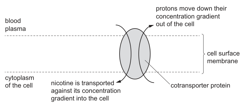
In the phloem tissue, there is a cotransporter mechanism that moves sucrose into the cytoplasm of a companion cell.
Which statement correctly describes a similarity between the cotransport of nicotine and the cotransport of sucrose?
Options
A The cotransporter proteins generate a proton gradient by moving protons out of the cell by active transport.
B The protons are transported through the cotransporter proteins by facilitated diffusion.
C The protons move through the cotransporter proteins in the opposite direction to the movement of nicotine and sucrose.
D The cotransporter proteins use energy from ATP to transport protons with nicotine and sucrose.
Working
In cotransport, the cotransporter protein does not generate the proton gradient — a separate proton pump (ATPase) uses ATP to actively pump protons and set up the electrochemical gradient. The cotransporter then allows protons to flow back down this pre-existing gradient by facilitated diffusion, and the energy released is used to drag another molecule (nicotine or sucrose) against its gradient.
- A is wrong because the cotransporter protein does not generate the proton gradient; the proton pump (a different protein) does this using ATP.
- C is wrong because, although protons and nicotine move in opposite directions (antiport), protons and sucrose move in the same direction through their cotransporter (symport) into the companion cell.
- D is wrong because the cotransporter protein itself does not use ATP directly; only the proton pump does.
- B is correct: in both cotransport systems, protons move through the cotransporter down their concentration gradient by facilitated diffusion.
Answer
B
B
Background Concept
Cotransport (also called secondary active transport) is a mechanism in which one substance is moved down its electrochemical gradient through a carrier protein, and the energy released is used to drag a second substance against its gradient through the same protein.
The proton-motive force that powers cotransport is set up by a proton pump (H⁺-ATPase) — a separate membrane protein that uses the hydrolysis of ATP to actively pump protons across the membrane, building up an electrochemical gradient. The cotransporter itself does not use ATP directly; it exploits the energy already stored in the proton gradient.
There are two forms of cotransport:
- Symport — both substances move in the same direction through the protein. Example: H⁺/sucrose cotransport into companion cells of phloem.
- Antiport — the two substances move in opposite directions. Example: H⁺/nicotine cotransport shown in Fig. 13.1 (protons out, nicotine in).
Because protons always move down their concentration gradient through the cotransporter, their movement is a form of facilitated diffusion (passive transport through a protein). It is the coupled second substance (nicotine or sucrose) that is being moved against its gradient — that is what makes the overall process secondary active transport.
Understanding the Question
The question shows Fig. 13.1, in which a cotransporter protein in a cell surface membrane moves:
- Protons down their concentration gradient out of the cell (cytoplasm → blood plasma)
- Nicotine against its concentration gradient into the cell (blood plasma → cytoplasm)
The question then asks for a statement that correctly describes a similarity between this nicotine cotransport and the H⁺/sucrose cotransport that loads sucrose into companion cells of phloem tissue. The four options each propose a different feature of the mechanism — the test is to identify which one is true of both.
The command word "correctly describes a similarity" means the answer must be a feature that both mechanisms share.
Approach
For each option, ask:
- Does the cotransporter itself generate the proton gradient, or does a separate pump do this?
- How do protons move through the cotransporter — passive or active?
- Do protons and the coupled substance move in the same direction or opposite directions in each case?
- Does the cotransporter protein directly hydrolyse ATP?
Comparing nicotine cotransport (Fig. 13.1: antiport) with sucrose cotransport (companion cell: symport) reveals that they share the mechanism (protons flowing passively down a pre-existing gradient through a carrier protein) but differ in the direction of the coupled molecule relative to the protons.
Step-by-Step Reasoning
Option A — cotransporter generates the proton gradient by active transport
The proton gradient is established by a proton pump (H⁺-ATPase), an entirely separate membrane protein that uses ATP. The cotransporter simply allows the protons to flow back down this gradient. The cotransporter itself performs no active transport; it facilitates diffusion. → Incorrect for both nicotine and sucrose cotransport.
Option B — protons are transported through the cotransporter by facilitated diffusion
In both systems, protons pass through the cotransporter down their electrochemical gradient, through a protein, without the cotransporter hydrolysing ATP. This is the textbook definition of facilitated diffusion. → Correct for both.
Option C — protons move opposite to nicotine and sucrose
- For nicotine: protons go out of the cell while nicotine comes in → opposite directions. ✓
- For sucrose in companion cells: protons and sucrose are both moved into the cell together (symport) → same direction. ✗
Because the statement must describe a similarity, and sucrose cotransport does not fit the "opposite direction" description, this option fails. → Incorrect.
Option D — cotransporter uses energy from ATP to transport protons with nicotine/sucrose
The cotransporter does not use ATP directly. The only protein that uses ATP in this system is the proton pump that establishes the gradient. The cotransporter is fuelled indirectly by the energy stored in that gradient (the proton-motive force). → Incorrect for both.
Therefore B is the only statement that is true of both cotransport systems.
Key Takeaways
- The proton pump (a separate, ATP-dependent protein) generates the proton gradient; the cotransporter simply allows protons to flow back down it by facilitated diffusion.
- Cotransport is secondary active transport: the coupled molecule (nicotine or sucrose) is moved against its gradient using energy released by protons moving with their gradient.
- Antiport (e.g. nicotine) ≠ symport (e.g. sucrose) — both are forms of cotransport, but the relative direction of the two molecules differs.
- A common exam trap is to attribute ATP use to the cotransporter rather than the proton pump.
Common Mistakes
- Mistake 1: Saying the cotransporter uses ATP. The cotransporter protein is not an ATPase; ATP is used by the separate proton pump. Candidates often confuse "active transport" (used to describe the overall process) with the protein directly responsible for ATP hydrolysis.
- Mistake 2: Assuming all cotransport is symport. Nicotine and nicotine-like compounds are often co-transported by antiport (H⁺ out, substrate in), while sugars and amino acids are typically co-transported by symport (H⁺ and substrate in together).
- Mistake 3: Saying the cotransporter "actively transports" protons. Protons move down their gradient through the cotransporter, so their movement through this protein is passive (facilitated diffusion).
Things to Be Careful About
- Always specify which protein uses ATP (the proton pump, not the cotransporter).
- Distinguish the direction of proton flow through the cotransporter from the direction of the coupled molecule — they are not always the same (antiport vs symport).
- Read the figure carefully: in Fig. 13.1 the arrows clearly show protons leaving the cell and nicotine entering — i.e. an antiport configuration.
- When a question asks for a "similarity", an option that is true for only one of the two systems cannot be the answer — even if biologically correct for that one system.
The diagram shows a simple metabolic pathway.
The letters W, X, Y and Z represent four different substances. At each step in the diagram the substrate undergoes a chemical reaction catalysed by an enzyme. The reaction produces the next substance in the pathway.
Which statements correctly describe the enzymes taking part in this metabolic pathway?
- They are all globular proteins.
- They all have the same tertiary structure.
- They all contain hydrogen atoms in their structure.
Options
A 1, 2 and 3
B 1 and 2 only
C 1 and 3 only
D 2 only
Working
Evaluate each statement:
-
They are all globular proteins — TRUE. All enzymes are globular proteins, with a specific 3D shape that creates an active site.
-
They all have the same tertiary structure — FALSE. The four enzymes catalyse four different reactions on four different substrates (W, X, Y, Z). Different substrates require different active sites, so the enzymes must have different tertiary structures (different amino acid sequences folded differently).
-
They all contain hydrogen atoms in their structure — TRUE. All proteins contain hydrogen atoms — in the amino acid R groups, in the peptide bonds, and in the polypeptide backbone.
Only statements 1 and 3 are correct.
Answer
C
C
Background Concept
Enzymes are biological catalysts — they speed up metabolic reactions without being used up. Every enzyme is a globular protein, meaning its polypeptide chain is folded into a roughly spherical 3D shape. This folding creates a specific pocket called the active site, where the substrate binds to form an enzyme–substrate (ES) complex.
The specificity of an enzyme for its substrate depends on the precise 3D shape of its active site, which is determined by the enzyme's tertiary structure. Different enzymes that catalyse different reactions have different tertiary structures (different amino acid sequences folded in different ways), giving them different active site shapes suited to their particular substrates.
Protein composition: All proteins, regardless of their structure or function, are built from amino acids linked by peptide bonds. Every amino acid contains hydrogen atoms — in the amino group (–NH₂), in the carboxyl group (–COOH), in the central α-carbon, and in the R group. So every protein, including every enzyme, contains hydrogen atoms in its structure.
Understanding the Question
The question presents a simple linear metabolic pathway:
At each step, a different enzyme catalyses the conversion of one substance into the next. Because W, X, Y, and Z are stated to be four different substances, each enzyme in the pathway must be catalysing a different chemical reaction on a different substrate.
We are asked to evaluate which of the three statements correctly describe all the enzymes in this pathway.
Approach
For each statement, ask: "Is this true for every enzyme in the pathway?" If the statement fails for even one enzyme, it is incorrect.
- Statement 1: Is it universally true that enzymes are globular proteins? (Check against the definition of an enzyme.)
- Statement 2: Do enzymes catalysing different reactions have the same tertiary structure? (Think about specificity and the active site.)
- Statement 3: Do all proteins contain hydrogen? (Think about the elemental composition of amino acids.)
Step-by-Step Reasoning
Statement 1: They are all globular proteins.
This is correct. By definition, all enzymes are globular proteins. Their polypeptide chains fold into compact, spherical shapes with active sites on the surface. This is true for every enzyme in the pathway.
Statement 2: They all have the same tertiary structure.
This is incorrect. Each enzyme in the pathway catalyses a different reaction on a different substrate. For an enzyme to recognise and bind its specific substrate, its active site must have a complementary shape to that substrate. Since the substrates (W, X, Y, Z) are all different, the active sites of the four enzymes must be different, which means their tertiary structures must be different. The same tertiary structure would imply the same active site, which would only work for one specific substrate.
Statement 3: They all contain hydrogen atoms in their structure.
This is correct. Every amino acid contains hydrogen atoms (in the amino group, carboxyl group, R group, and backbone). Even after peptide bond formation, hydrogen atoms remain in the polypeptide backbone and in the R groups. Therefore, every protein — including every enzyme — contains hydrogen in its structure.
Conclusion: Statements 1 and 3 are correct; statement 2 is incorrect. The answer is C (1 and 3 only).
Key Takeaways
- All enzymes are globular proteins — this is a defining feature.
- Enzymes are specific to their substrates because each has a unique tertiary structure that creates a unique active site shape.
- All proteins contain hydrogen atoms because hydrogen is a component of every amino acid and is retained after peptide bond formation.
- When evaluating statements about "all" members of a group, check whether the statement must be true universally — one counterexample invalidates it.
Common Mistakes
- Choosing B (1 and 2 only): This mistake comes from assuming that because all enzymes share the property of being globular proteins, they must also share the same tertiary structure. But "globular protein" is a category, not a specific shape — different globular proteins can have very different tertiary structures.
- Choosing A (all three): Same mistake as above, plus failing to recognise that enzyme specificity requires structural differences.
- Choosing D (2 only): Recognising that enzymes have different structures but forgetting that all proteins are globular and all contain hydrogen.
Things to Be Careful About
- "Same tertiary structure" is a very specific claim — it means identical 3D folding, not just "both are folded." Different enzymes have different amino acid sequences and therefore different tertiary structures.
- Always check the wording: "all", "each", and "every" mean the statement must be universally true for the claim to hold.
- Remember that even a single amino acid change can completely alter a protein's tertiary structure and function (e.g. the sickle cell mutation in haemoglobin).
- Hydrogen is present in virtually all biological molecules — proteins, carbohydrates, lipids, nucleic acids, and water all contain hydrogen. This is rarely a point of failure on its own, but it is worth confirming when evaluating such statements.
A student observed the effect of two different concentrations of salt solution on blood cells.
The student added each concentration of salt solution to one of two microscope slides, and then a small drop of fresh blood was added.
Each slide was viewed using the high power lens of a microscope and the student’s observations were recorded.
- slide 1: No red blood cells were visible.
- slide 2: The red blood cells were visible but looked slightly crinkled.
Which row correctly explains the results obtained?
Options
| slide 1 | slide 2 | |
|---|---|---|
| A | Swelling of the cells caused them all to burst. | The of the cell was more negative than the of the external solution. |
| B | The of the cell was more negative than the of the external solution. | The of the external solution was more negative than the of the cell. |
| C | The of the cell was less negative than the of the external solution. | There is a net movement of water out of the cell by osmosis. |
| D | The of the external solution was less negative than the of the cell. | The of the cell was very similar to, but slightly more negative than, the of the external solution. |
key
= water potential
Working
- Slide 1: cells burst → water moved INTO the cells by osmosis → the external solution had a higher (less negative) Ψ than the cell → the cell's Ψ was more negative than the external solution.
- Slide 2: cells slightly crinkled (crenated) → water moved OUT of the cells by osmosis → the external solution had a lower (more negative) Ψ than the cell → the external solution's Ψ was more negative than the cell's.
- Only option B states both Ψ relationships correctly.
Answer
B
B
Background Concept
Water potential (Ψ) is the tendency of a solution to lose water; it is measured in kilopascals (kPa). Pure water has Ψ = 0 kPa, and any dissolved solute makes Ψ more negative (lower). Water always moves by osmosis from a region of higher (less negative) water potential to a region of lower (more negative) water potential, across a partially permeable membrane.
A red blood cell has a cytoplasm with a Ψ of about −300 kPa. In solutions of different water potential it behaves as follows:
- In a solution of higher (less negative) Ψ (hypotonic): net water entry → cell swells and bursts (haemolysis). No whole cells are visible because the membrane ruptures and the haemoglobin disperses.
- In a solution of similar Ψ (isotonic): no net water movement → cells keep their normal biconcave shape.
- In a solution of lower (more negative) Ψ (hypertonic): net water loss → cell shrivels and the membrane becomes crinkled (crenation).
Understanding the Question
Two slides were prepared with salt solutions of different concentrations, each mixed with a drop of fresh blood, then examined under high power.
- Slide 1: no red blood cells visible → the cells have burst (haemolysed).
- Slide 2: red blood cells visible but slightly crinkled → the cells have lost a small amount of water and crenated.
We must match each observation to the correct relationship between the cell's Ψ and the external solution's Ψ.
Approach
Apply the rule "water moves from higher (less negative) Ψ to lower (more negative) Ψ" to each slide in turn, then read each option to find the one that states both Ψ relationships correctly.
Step-by-Step Reasoning
- Slide 1 – cells burst: For the cell to gain enough water to burst, the external solution must have had a higher (less negative) Ψ than the cell. Equivalently, Ψ(cell) was more negative than Ψ(external). Option B states this correctly; option D says the same thing in different words but is then wrong about slide 2.
- Slide 2 – cells crenated: For the cell to lose water and crinkle, the external solution must have had a lower (more negative) Ψ than the cell. Equivalently, Ψ(external) was more negative than Ψ(cell). Option B states this correctly.
- Evaluate the alternatives:
- A: Slide 1 description (swelling/bursting) is a correct observation but not a Ψ explanation; slide 2 statement is the wrong way round (if cell Ψ were more negative, water would enter, not cause crenation).
- C: Slide 1 is the wrong way round (less negative cell Ψ would cause water to leave, not burst the cells).
- D: Slide 1 is correct, but slide 2 is wrong (if the cell Ψ were "slightly more negative" than the external, water would still enter, not cause crenation).
- B: both statements match the required Ψ relationships.
- Therefore the correct row is B.
Key Takeaways
- Water moves from higher (less negative) Ψ to lower (more negative) Ψ.
- Burst red blood cells indicate a hypotonic external solution; crenated red blood cells indicate a hypertonic external solution.
- When asked to choose between equivalent phrasings, focus on whether the direction of the Ψ gradient matches the direction of net water movement.
Common Mistakes
- Confusing which way round the comparison is: a cell bursts when Ψ(external) > Ψ(cell) (less negative external), NOT when the cell's Ψ is less negative.
- Reading option D as equivalent to option B for slide 1 (it is) and then overlooking the incorrect slide 2 statement.
- Conflating observation ("cells burst") with explanation (the underlying Ψ relationship).
Things to Be Careful About
- Ψ values are negative numbers; "more negative" means a smaller (lower) number, e.g. −500 kPa is more negative than −300 kPa.
- The term "water potential" is interchangeable with Ψ in the CIE mark scheme, but the symbol and the unit (kPa) should be used in extended answers.
Which statement correctly describes facilitated diffusion?
Options
A The process only occurs using channel proteins that change shape and that use energy provided by the cell.
B The process occurs using channel proteins or carrier proteins that may or may not change shape.
C The process occurs using channel proteins or carrier proteins that use energy provided by the cell.
D The process only occurs using carrier proteins that create a gradient to move ions in opposite directions.
Working
Facilitated diffusion is a passive process: substances move down their concentration gradient through specific membrane proteins, so no ATP/energy from the cell is required.
Two types of protein can assist:
- Channel proteins form a continuous pore; they do not change shape.
- Carrier proteins bind the molecule and undergo a conformational change to move it across the membrane.
Therefore the proteins used may or may not change shape, and ATP is not used.
- A is wrong: claims energy is used and that channel proteins change shape.
- C is wrong: claims energy is used.
- D is wrong: claims only carrier proteins are used and that a gradient is created to move ions in opposite directions (that describes co-transport, not facilitated diffusion).
Answer
B
B
Background Concept
Facilitated diffusion is a form of passive transport across a cell membrane. "Passive" means the cell does not supply metabolic energy (no ATP) — the driving force is the concentration gradient of the substance itself, which carries the molecules from where they are more concentrated to where they are less concentrated. The "facilitated" part refers to the fact that molecules (especially charged ions or larger polar molecules) cannot cross the phospholipid bilayer directly and need help from a specific membrane protein.
Two kinds of transport protein can assist:
- Channel proteins form a water-filled pore through the bilayer. They are essentially static — ions diffuse through the open channel. They do not change shape (or only very subtly, e.g. a gate opening/closing) and they do not consume ATP.
- Carrier proteins bind the molecule on one side of the membrane, then change their three-dimensional shape (a conformational change) to release it on the other side. They also do not consume ATP.
So a statement about facilitated diffusion must include: passive (no energy), down a gradient, and that the protein involved is either a channel or a carrier — and only the carrier changes shape.
Understanding the Question
This is a single-best-answer multiple choice question asking which option correctly describes facilitated diffusion. The four options make competing claims about:
- Whether energy is used.
- Whether only one type of protein (or both) is used.
- Whether the proteins change shape.
- Whether a gradient is created to move ions in opposite directions.
The student must pick the option that is consistent with the textbook definition above.
Approach
Test each option against the defining features of facilitated diffusion (passive, down a gradient, uses channel or carrier proteins, carrier may change shape). Reject any option that wrongly claims ATP use, restricts the process to one protein type, or misdescribes the direction of ion movement.
Step-by-Step Reasoning
- Option A — claims the process only uses channel proteins that change shape and use energy. Two errors: facilitated diffusion does not use ATP, and channel proteins do not undergo the shape change that carriers do. Rejected.
- Option B — says the process uses channel proteins or carrier proteins that may or may not change shape. This is exactly right: channels generally do not change shape, carriers do, so within facilitated diffusion as a whole the protein involved may or may not change shape. Accepted.
- Option C — claims the proteins use energy. This makes the process active, which contradicts the definition of facilitated (passive) diffusion. Rejected.
- Option D — claims the process only uses carrier proteins that create a gradient to move ions in opposite directions. Two errors: facilitated diffusion uses channels and carriers, and "creating a gradient to move ions in opposite directions" describes co-transport (a form of active transport using a carrier and a proton/ion gradient set up by ATP-driven pumps), not facilitated diffusion. Rejected.
Only option B matches the definition.
Key Takeaways
- Facilitated diffusion = passive transport down a concentration gradient, mediated by a specific membrane protein (channel or carrier).
- It does not require ATP; that would make it active transport.
- Channel proteins form pores (no major shape change); carrier proteins bind the molecule and change shape.
- "Creating a gradient to move ions in opposite directions" describes co-transport, not facilitated diffusion.
Common Mistakes
- Confusing facilitated diffusion with active transport and so selecting an option that mentions ATP/energy.
- Believing that only carrier proteins are involved (channels also mediate facilitated diffusion, e.g. for ions).
- Misreading "may or may not change shape" as a vague phrase rather than as the precise summary it is (channels don't, carriers do).
- Picking D because it sounds technical, without recognising it describes co-transport.
Things to Be Careful About
- "Passive" and "no energy" are synonymous here — if ATP is mentioned, the process is not facilitated diffusion.
- Channels and carriers are both valid; do not restrict the answer to one type.
- Conformational change applies to carriers; channels merely open (often gated, but not by ATP).
- Co-transport / counter-transport uses carriers and a pre-existing ion gradient maintained by active transport — it is a different process from facilitated diffusion.
Which row is correct for stem cells?
Options
| can repair cells | can be involved in the formation of phagocytes | |
|---|---|---|
| A | yes | no |
| B | yes | yes |
| C | no | no |
| D | no | yes |
Working
Stem cells are undifferentiated cells that can divide and differentiate into specialised cell types.
- Can stem cells repair cells? No. Stem cells do not directly repair existing cells; they divide and differentiate to replace damaged or lost cells, but they do not themselves carry out a repair function on other cells.
- Can stem cells be involved in the formation of phagocytes? Yes. Haematopoietic (bone marrow) stem cells give rise to all blood cell types, including phagocytes such as neutrophils and monocytes.
So the correct row is no, yes → D.
Answer
D
D
Background Concept
Stem cells are unspecialised cells that can both self-renew (divide to produce more stem cells) and differentiate into a range of specialised cell types. In an adult, the most important reservoir of stem cells is the bone marrow, where haematopoietic stem cells continuously produce the cellular components of blood — red blood cells, platelets, and several classes of white blood cells, including the phagocytes (neutrophils, monocytes, macrophages).
A key distinction that the CIE mark scheme tests here is the difference between forming new cells and repairing existing cells. Stem cells do not patch or mend other cells; rather, they supply replacements by mitosis followed by differentiation. The mature differentiated cell that replaces a damaged one is what performs the "repair" of a tissue — the stem cell itself is upstream of that process.
Understanding the Question
The question offers four combinations of two statements about stem cells:
- Whether stem cells "can repair cells"
- Whether stem cells "can be involved in the formation of phagocytes"
You have to pick the row in which both statements are correctly assigned true/false. The mark scheme keys this as no, yes.
Approach
Take each statement and decide whether it correctly describes a property of stem cells:
- The first statement is treated as false because, strictly, stem cells do not repair cells directly — they generate new differentiated cells.
- The second statement is true because haematopoietic stem cells in bone marrow give rise to the white blood cell lineages that include the phagocytes (neutrophils, monocytes, macrophages).
Only one row says no / yes → option D.
Step-by-Step Reasoning
- What stem cells do: divide (by mitosis) to renew themselves and to produce progenitor cells that differentiate into one or more specialised cell types.
- Stem cells and "repair": Stem cells do not operate on other cells to fix them. They contribute to tissue maintenance by replacing cells that have been lost or damaged. The wording of the mark scheme treats the strict idea of "repairing cells" as something stem cells do not do. → "can repair cells" = no.
- Stem cells and phagocytes: Neutrophils and monocytes are short-lived and must be continuously produced. They originate from haematopoietic stem cells in the red bone marrow through the myeloid lineage. → "can be involved in the formation of phagocytes" = yes.
- Match to the table:
- A: yes / no ✗
- B: yes / yes ✗
- C: no / no ✗
- D: no / yes ✓
Key Takeaways
- Stem cells are undifferentiated, self-renewing cells that give rise to specialised cell types.
- They do not directly "repair" cells; they supply replacements by division and differentiation.
- Haematopoietic stem cells in bone marrow produce all blood cells, including the phagocytic white blood cells (neutrophils, monocytes, macrophages).
- For CIE-style MCQs, take care over the precise wording — "repair cells" and "replace cells" are not treated as synonyms by the mark scheme.
Common Mistakes
- Choosing B (yes / yes) because students correctly know stem cells form phagocytes but assume they also "repair cells" in a tissue-level sense. The CIE mark scheme distinguishes replacement (stem cell function) from repair of an existing cell.
- Choosing A (yes / no) by reversing the phagocyte statement — confusing stem cells with mature immune cells.
- Choosing C (no / no) by misreading the second column, perhaps thinking only fully differentiated white blood cells exist (ignoring the bone-marrow stem cell origin).
Things to Be Careful About
- Use the precise wording of the mark scheme. The official key treats "can repair cells" as not a property of stem cells.
- Remember the haematopoietic lineage: HSC → common myeloid progenitor → neutrophil / monocyte / macrophage (all phagocytes).
- Do not confuse embryonic stem cells (pluripotent) with adult tissue stem cells (multipotent). The question is general, but the phagocyte link is firmly adult / bone-marrow based.
Eukaryotic organisms grow and increase in size as a result of cell division. The diagram shows some stages of mitosis with one stage missing, labelled X.
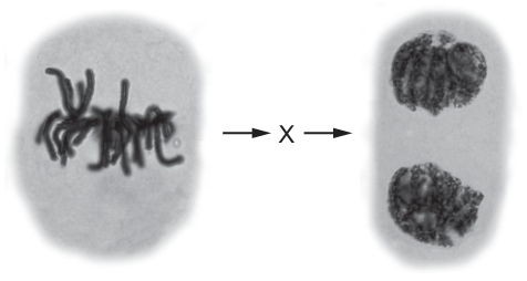
What happens in stage X?
Options
A Spindle fibres attach to the centromeres and the chromosomes line up along the equator.
B The chromatin coils and condenses and the nuclear membrane disintegrates.
C The chromosomes line up in pairs along the equator of the cell.
D Microtubules shorten so that spindle fibres contract and the centromeres divide.
Working
The diagram shows the sequence: prophase → X → telophase.
Stage X must therefore be a stage that occurs after prophase and before telophase. The order of mitosis is:
Evaluating the options:
- A describes metaphase (spindle fibres attach to centromeres, chromosomes align at the equator).
- B describes prophase (chromatin condenses, nuclear membrane disintegrates) — this is already shown before X.
- C describes chromosomes lining up in pairs — this is meiosis I, not mitosis, so it is wrong.
- D describes anaphase: microtubules shorten, spindle fibres contract, centromeres divide, and sister chromatids are pulled to opposite poles. This is the stage that leads directly into telophase, where two new nuclei form.
Answer
D
D
Background Concept
Mitosis is a continuous process, but biologists divide it into four named stages based on what the chromosomes and spindle are doing:
- Prophase — Chromatin (diffuse DNA + protein) coils and condenses into visible chromosomes. Each chromosome consists of two sister chromatids joined at a centromere. The nuclear membrane breaks down and the nucleolus disappears. Spindle fibres begin to form.
- Metaphase — Spindle fibres attach to the centromeres of the chromosomes. The chromosomes (still as pairs of sister chromatids) are pulled to the equator of the cell and line up along it.
- Anaphase — The centromeres divide. The spindle fibres shorten (microtubules depolymerise), pulling the now-separated sister chromatids (now individually chromosomes) to opposite poles of the cell.
- Telophase — Chromatids reach the poles. A new nuclear membrane forms around each set, the chromosomes decondense back to chromatin, and the nucleoli reappear. Cytokinesis then divides the cytoplasm.
In mitosis the chromosomes line up singly at the equator (not in pairs). Lining up in pairs (homologous pairs) only happens in meiosis I, which is why option C is wrong.
Understanding the Question
The figure shows two micrographs of mitosis stages with a missing stage X in between. The first micrograph shows condensed chromosomes scattered in the cell with no nuclear membrane — this is prophase. The last micrograph shows two distinct nuclei forming at opposite ends of the cell — this is telophase. The question asks what happens in the stage between them (X).
Approach
Identify the sequence (prophase → X → telophase) and determine which option describes a stage that occurs between prophase and telophase. The correct answer must describe a mitotic event (not a meiotic one) that fits the position in the sequence.
Step-by-Step Reasoning
- The cell is already past prophase (chromosomes condensed, nuclear membrane gone) and not yet at telophase (two nuclei forming). So X must be metaphase or anaphase.
- Option A (spindle fibres attach to centromeres; chromosomes line up at the equator) describes metaphase. This does occur between prophase and telophase, but the mark scheme treats X as the stage leading directly into telophase.
- Option B describes prophase again — this is already shown before X, so it cannot be the missing stage.
- Option C mentions chromosomes lining up in pairs along the equator. This is wrong for mitosis; homologous pairs only align in this way during meiosis I. In mitosis, chromosomes line up singly.
- Option D describes anaphase: microtubules shorten, spindle fibres contract, and the centromeres divide so that the sister chromatids are pulled apart to opposite poles. This is precisely the event that converts a single cell with chromosomes at the equator into the configuration seen in telophase — chromatids at opposite poles, ready for new nuclei to form around them.
Therefore X = anaphase, and the answer is D.
Key Takeaways
- The order of mitosis is prophase → metaphase → anaphase → telophase.
- Anaphase is defined by centromere division and shortening of spindle microtubules, which pulls sister chromatids to opposite poles.
- In mitosis, chromosomes align singly at the equator; alignment in pairs is a feature of meiosis I.
- Reading the position of a stage within the mitotic sequence is often the key to identifying it.
Common Mistakes
- Choosing A because metaphase also occurs between prophase and telophase — but the mark scheme credits D because anaphase is the stage that leads directly into the telophase image shown.
- Choosing C because "line up along the equator" sounds correct — but the word pairs makes this a description of meiosis I, not mitosis.
- Confusing prophase events (condensation, nuclear membrane breakdown) with later stages.
- Forgetting that in anaphase it is the centromeres that divide (not the chromatids themselves), separating the sister chromatids.
Things to Be Careful About
- Use the precise term centromere for the structure that divides in anaphase, not "chromosome" or "chromatid".
- Remember that spindle fibres are made of microtubules; saying "microtubules shorten" and "spindle fibres contract" are both acceptable descriptions of anaphase movement.
- Distinguish between mitosis (singly aligned chromosomes, sister chromatids separated) and meiosis (paired homologues in meiosis I, sister chromatids separated in meiosis II).
The graphs show various distance measurements taken from the start of metaphase of mitosis. The graphs are to scale when compared to one another.
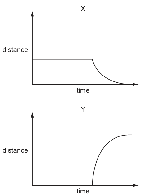
Which row correctly identifies the distance measurement for each graph?
Options
| X | Y | |
|---|---|---|
| A | distance between poles of spindle | distance of centromeres from poles of spindle |
| B | distance between poles of spindle | distance between sister chromatids |
| C | distance of centromeres from poles of spindle | distance between sister chromatids |
| D | distance of centromeres from poles of spindle | distance between poles of spindle |
Working
During metaphase the centromeres lie on the equator and sister chromatids are held together at the centromere, so:
- distance between sister chromatids ≈ 0
- distance from each centromere to the spindle pole = maximum
- distance between the two spindle poles is essentially fixed (spindle already formed)
At the onset of anaphase the centromeres split and the chromatids are pulled toward opposite poles, so the centromere-to-pole distance falls (toward zero) while the distance between the separating sister chromatids rises (then plateaus). The pole-to-pole distance stays roughly constant.
Graph X (high constant, then falls to 0) matches the centromere-to-pole distance. Graph Y (zero, then rises to a plateau) matches the distance between sister chromatids.
Answer
C
C
Background Concept
During mitosis, replicated chromosomes are moved to opposite ends of the cell by the spindle apparatus. The key structures to keep in mind are:
- Chromatid / sister chromatids: a chromosome that has been replicated consists of two identical sister chromatids joined at the centromere.
- Centromere: the constricted region that holds the two sister chromatids together and is the attachment point for spindle microtubules.
- Spindle poles: the two ends of the mitotic spindle (organised by centrosomes) from which microtubules radiate.
- Equator / metaphase plate: the mid-line of the spindle where chromosomes line up at metaphase.
The important sequence of events around metaphase and anaphase is:
- Metaphase: chromosomes (centromeres) are aligned at the equator, sister chromatids still joined.
- Anaphase onset: the centromeres split; each former sister chromatid is now an independent chromosome whose centromere is pulled toward a spindle pole by shortening kinetochore microtubules.
- Late anaphase / telophase: chromosomes reach the poles, nuclear envelopes reform.
Understanding the Question
We are given two distance-vs-time graphs, both starting at the beginning of metaphase. We must identify what each graph is measuring, choosing from three possible distance quantities: pole-to-pole distance, centromere-to-pole distance, and distance between sister chromatids.
- Graph X: distance is large and constant for a time, then decreases rapidly toward zero.
- Graph Y: distance is zero for a time, then increases rapidly and levels off at a high value.
Approach
For each candidate distance, decide what its value is at the start of metaphase and how it changes as anaphase proceeds, then match to the graph shape.
| Distance | At start of metaphase | Behaviour through anaphase |
|---|---|---|
| Pole to pole | Roughly constant (spindle is already formed and length stays similar) | Little change |
| Centromere to pole | Maximum (centromere at equator, far from pole) | Decreases as centromere is pulled to the pole |
| Between sister chromatids | Essentially zero (joined at centromere) | Increases as chromatids separate and travel to opposite poles |
Step-by-Step Reasoning
Graph X — high → zero:
This must be a distance that is initially large and shrinks. The only candidate that fits is the centromere-to-pole distance: at metaphase the centromere is on the equator (maximum distance from the pole), and during anaphase the kinetochore microtubules shorten, hauling the centromere toward the pole, so the distance falls — eventually approaching zero as the chromosome arrives at the pole.
Pole-to-pole distance is wrong here: it does not collapse to zero; the spindle maintains its length. Distance between sister chromatids is also wrong: it starts at zero, not at a high value.
Graph Y — zero → high plateau:
This must be a distance that starts at zero and grows. The only candidate is the distance between sister chromatids: at metaphase they are still attached at the centromere (so the distance between them is effectively zero), and at anaphase the centromere splits, so the two former sisters are pulled apart toward opposite poles. The distance increases rapidly and then levels off once the chromatids reach the poles (a maximum separation).
Pole-to-pole distance is wrong: it does not start at zero. Centromere-to-pole distance is wrong: it starts high, not at zero.
Both X and Y cannot be the same quantity (they have opposite shapes), which immediately rules out the distractor rows A and D that put the same quantity in both columns.
Row C is the only one that assigns a different, consistent quantity to each graph and matches the data.
Key Takeaways
- At metaphase, centromeres sit on the equator; sister chromatids are joined; the spindle length is set.
- At anaphase, sister chromatids separate and centromeres are pulled to the poles — so centromere-to-pole distance falls and inter-sister-chromatid distance rises.
- The inter-pole (spindle) distance is essentially constant and is the trick distractor in this style of question.
- Always check both graphs together: a good answer explains both shapes from the same biological event.
Common Mistakes
- Choosing A or D because the candidate remembers that "the spindle" is involved and confuses the centromere-to-pole distance with the pole-to-pole distance. The pole-to-pole distance does not change much, so it cannot produce a graph that drops to zero.
- Choosing B because the student thinks both graphs show distances that decrease or start high. Graph Y clearly starts at zero and increases, so it cannot be the centromere-to-pole distance.
- Forgetting that sister chromatids are joined at the centromere at metaphase, so the inter-sister-chromatid distance really does start at (near) zero.
Things to Be Careful About
- Read the y-axis values at the start of the graph: Graph X is high, Graph Y is zero — this single observation eliminates most options.
- "Distance between poles of the spindle" refers to the two centrosome ends of the mitotic spindle, not the distance between the two sister chromatids.
- The graphs being "to scale when compared to one another" means the final plateau of Y should correspond to roughly the same magnitude as the initial value of X (both reflect the cell's pole-to-equator/pole length) — a useful cross-check if you want to be sure.
- Do not confuse centromere (the joining region) with centrosome (the spindle pole organiser); the question concerns the centromere.
Which row is correct for the start of anaphase of mitosis?
Options
| form of DNA | DNA is associated with histone proteins | state of the cell surface membrane | |
|---|---|---|---|
| A | chromosomes | always | broken apart |
| B | chromosomes | sometimes | intact |
| C | separated sister chromatids | always | intact |
| D | separated sister chromatids | sometimes | broken apart |
Working
- At the start of anaphase, the centromeres divide and the sister chromatids are pulled apart, so each former chromatid is now an independent chromosome moving toward a pole. The DNA is therefore in the form of separated sister chromatids.
- DNA is associated with histone proteins at all times — whether as chromatin in interphase, as condensed chromosomes, or as the separated chromatids at anaphase. So this association is always present.
- The cell surface (plasma) membrane remains intact throughout mitosis. It is only involved in cytokinesis (cleavage furrow formation in animal cells) after mitosis is complete; it is not broken apart during anaphase.
Only row C satisfies all three conditions.
Answer
C
C
Background Concept
During mitosis a cell divides its already-replicated nucleus so that each daughter cell receives an identical set of chromosomes. The key stages are prophase, metaphase, anaphase and telophase, followed by cytokinesis (division of the cytoplasm).
A replicated chromosome consists of two sister chromatids joined at a centromere. Throughout interphase, prophase and metaphase, the two chromatids of each chromosome stay attached. The critical event that defines the start of anaphase is the splitting of the centromeres: from this point, the two sister chromatids separate and are pulled to opposite poles of the cell by the spindle fibres. Once separated, each former chromatid is regarded as a daughter chromosome in its own right.
DNA is packaged with histone proteins at all times in a eukaryotic cell. The DNA double helix is wound around histone octamers to form nucleosomes, which are then coiled and supercoiled into the higher-order structures that make up chromatin and, when condensed, visible chromosomes. This histone association is constitutive — it does not come and go during the cell cycle.
The cell surface (plasma) membrane is distinct from the nuclear envelope. The nuclear envelope breaks down during prometaphase, but the cell surface membrane stays intact throughout mitosis. In animal cells it is only during cytokinesis that a cleavage furrow forms, pinching the cytoplasm into two daughter cells; even then, the membrane is reorganised, not torn. In plant cells a cell plate forms across the middle of the cell.
Understanding the Question
The question presents a three-column table about (1) the form the DNA is in, (2) whether DNA is associated with histone proteins, and (3) the state of the cell surface membrane at the start of anaphase. The candidate must pick the row whose three entries are all correct for that exact moment in the cell cycle.
The command word is implicit but the task is a multiple-choice selection: pick the row that is correct.
Approach
- Determine what happens to the chromatids at the start of anaphase — this rules in or out "chromosomes" vs "separated sister chromatids".
- Apply the principle that DNA is always packaged with histones — this rules out "sometimes".
- Recall that the cell surface membrane is not disrupted during mitosis — this rules out "broken apart".
- Match these three conclusions to the row in the table.
Step-by-Step Reasoning
- Form of DNA at the start of anaphase. The defining event of anaphase is the separation of sister chromatids. Therefore the DNA is no longer in the form of intact, paired chromosomes — it is in the form of separated sister chromatids (now functioning as individual chromosomes heading for opposite poles). This eliminates rows A and B.
- DNA associated with histone proteins. Histone packaging is continuous throughout the cell cycle. Even when chromatin is most condensed (metaphase chromosomes), and even as the chromatids separate at anaphase, each DNA molecule remains wrapped around histone octamers. The association is always present, not sometimes. This eliminates row D (which says "sometimes").
- State of the cell surface membrane. The plasma membrane of the cell remains intact throughout mitosis. It does not fragment or break apart at anaphase. The membrane is only remodelled later, during cytokinesis. This also supports the "intact" column and rules out rows A and D.
- The only row with "separated sister chromatids", "always" and "intact" is row C.
Key Takeaways
- Anaphase begins when centromeres split and sister chromatids separate; from that moment, each chromatid behaves as a chromosome.
- DNA is associated with histones at all times in eukaryotic cells — this is a permanent feature of chromatin organisation.
- The cell surface (plasma) membrane stays intact throughout mitosis; it is involved in cytokinesis, not in the nuclear division stages.
- Be careful to distinguish the nuclear envelope (which breaks down) from the cell surface membrane (which does not).
Common Mistakes
- Confusing chromatids with chromosomes at anaphase. Many students think "chromosomes are still there, so the DNA is in chromosome form". The mark scheme expects you to recognise that once separated, each former chromatid is now itself a chromosome — but the more precise description in this context is "separated sister chromatids".
- Thinking histone association is intermittent. Histones are removed only transiently during very specific processes (such as DNA replication or transcription); the bulk DNA of the nucleus is always histone-associated. The word "sometimes" is therefore wrong.
- Conflating the nuclear envelope with the cell surface membrane. The nuclear envelope fragments in prometaphase, but this is internal. The cell surface (plasma) membrane is not broken during mitosis.
Things to Be Careful About
- "Always" vs "sometimes" for histone association: histone packaging is a continuous feature, so the correct word is "always".
- The plasma membrane is intact during anaphase; "broken apart" only describes events in certain forms of cytokinesis (and even then, the membrane is reorganised rather than torn).
- Read each column independently — the question rewards three correct statements in a single row, not just one or two.
The diagrams show the chemical structure of four bases.
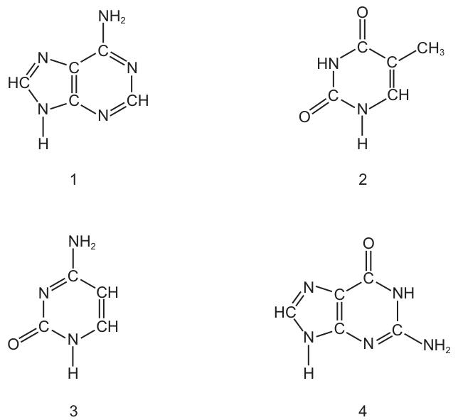
Which diagrams show thymine and cytosine?
Options
A 1 and 2
B 1 and 4
C 2 and 3
D 3 and 4
Working
Thymine and cytosine are both pyrimidines, so they each have a single ring.
- Structure 1 has a fused double ring with an –NH₂ group → a purine (adenine).
- Structure 2 has a single ring with two C=O groups and a –CH₃ group → thymine (the methyl group is unique to thymine among the DNA bases).
- Structure 3 has a single ring with one –NH₂ and one C=O → cytosine.
- Structure 4 has a fused double ring with one C=O and one –NH₂ → a purine (guanine).
Thymine = 2, cytosine = 3.
Answer
C
C
Background Concept
DNA contains four nitrogenous bases: two purines (adenine, A; guanine, G) and two pyrimidines (cytosine, C; thymine, T). Purines are built from a fused double ring (a six-membered ring joined to a five-membered ring), whereas pyrimidines have only a single six-membered ring. This structural difference is biologically important because in the DNA double helix a purine always pairs with a pyrimidine, giving the helix a uniform width.
Each base carries a characteristic set of functional groups, and these are the quickest way to tell them apart from a drawn structure:
- Adenine – purine; one –NH₂ group attached to the six-membered ring.
- Guanine – purine; one C=O and one –NH₂ group on the six-membered ring.
- Cytosine – pyrimidine; one –NH₂ group and one C=O group on the ring.
- Thymine – pyrimidine; two C=O groups and a –CH₃ (methyl) group. The methyl group is unique to thymine among the DNA bases (uracil, found in RNA, is the same as thymine but without the methyl group).
In base pairing, A pairs with T (two hydrogen bonds) and G pairs with C (three hydrogen bonds).
Understanding the Question
The question presents four unlabelled chemical structures and asks which two of them are thymine and cytosine. This is a pure recognition task: you must use the ring structure (single vs double) and the functional groups (–NH₂, C=O, –CH₃) to assign each diagram to a named base.
Approach
Two filters applied in sequence will identify thymine and cytosine efficiently:
- Ring count – thymine and cytosine are pyrimidines, so they must be single-ringed structures. This immediately rules out the two purines (the double-ringed structures).
- Functional group pattern – among the single-ringed structures, thymine is the one with two C=O groups and a methyl (–CH₃) group, whereas cytosine is the one with one –NH₂ and one C=O group.
Step-by-Step Reasoning
-
Structure 1 – Two fused rings, with a single –NH₂ group projecting from the six-membered ring. Two rings = purine; –NH₂ on a purine = adenine. Not thymine or cytosine.
-
Structure 2 – Single six-membered ring. Substituents: one C=O at the top, a second C=O on the left side, and a –CH₃ group on the right. Single ring = pyrimidine; two C=O plus a methyl = thymine. ✓
-
Structure 3 – Single six-membered ring. Substituents: one –NH₂ at the top and one C=O on the lower left. Single ring = pyrimidine; one amine and one carbonyl = cytosine. ✓
-
Structure 4 – Two fused rings, with a C=O group on the upper part of the six-membered ring and an –NH₂ group on the lower part. Two rings = purine; C=O + –NH₂ on a purine = guanine. Not thymine or cytosine.
So thymine is 2 and cytosine is 3, giving the pair "2 and 3".
Key Takeaways
- Purines (A, G) have two rings; pyrimidines (C, T, U) have one ring — this is the fastest first filter when identifying bases from structures.
- Thymine is the only DNA base that carries a methyl (–CH₃) group, and it has two C=O groups.
- Cytosine has exactly one –NH₂ and one C=O on its single ring.
- In DNA, A–T pair with two hydrogen bonds and G–C pair with three; recognising the bases is the first step towards understanding this pairing.
Common Mistakes
- Confusing adenine and guanine (both purines): remember that adenine has only an –NH₂ on the six-membered ring, whereas guanine has both a C=O and an –NH₂.
- Confusing cytosine and thymine (both pyrimidines): thymine has two C=O groups plus a methyl; cytosine has one C=O and one –NH₂. The methyl group is the giveaway for thymine.
- Counting rings incorrectly — make sure you see whether the diagram shows a fused five- and six-membered ring (purine) or only a six-membered ring (pyrimidine).
Things to Be Careful About
- In written skeletal formulae, only the heteroatoms (N, O) and the attached hydrogens (–NH₂, –NH, –OH) are usually drawn. Carbon and hydrogen atoms on the ring carbons are implied. Do not "see" a CH₃ where only a –CH₃ substituent label is shown — that is a methyl group, not a separate carbon of the ring.
- Be precise about which ring of a purine carries each substituent; an –NH₂ on the five-membered ring would not be adenine.
- Thymine is found mainly in DNA; in RNA it is replaced by uracil, which has the same ring as thymine but lacks the –CH₃ group. If you see a single ring with two C=O groups and no methyl, you are looking at uracil, not thymine.
The diagram shows the nucleotide sequence of a small section of the transcribed strand of a gene.
The table shows the amino acids coded for by 10 mRNA codons.
| mRNA codon | amino acid |
|---|---|
| AAG | Lys |
| ACG | Thr |
| CGG CGC CGU | Arg |
| CCG | Pro |
| GCC GCG | Ala |
| GGC | Gly |
| UGC | Cys |
What is the sequence of the four amino acids in the polypeptide translated from this small section of a gene?
Options
A Ala-Ala-Cys-Ala
B Ala-Arg-Gly-Ala
C Arg-Ala-Pro-Arg
D Arg-Arg-Thr-Arg
Working
The "transcribed strand" is the template strand of DNA. The mRNA is synthesised using complementary base pairing, with U replacing T in RNA.
DNA template: 5'-CGG GCC CCG CGG-3'
mRNA: 3'-GCC CGG GGC GCC-5'
Reading the mRNA as triplets:
Answer
B (Ala–Arg–Gly–Ala)
B
Background Concept
DNA is the cell's permanent store of genetic information, but proteins are assembled by ribosomes using messenger RNA (mRNA) as the working copy. Producing that working copy is called transcription, and reading it to build a polypeptide is translation.
During transcription, only one of the two DNA strands is read by RNA polymerase. This strand is called the template strand (sometimes the antisense strand, or — as here — the transcribed strand). Bases pair with their complements, with one important difference from DNA replication: RNA contains uracil (U) in place of thymine (T), so an A in the template DNA pairs with a U in the new mRNA:
- DNA A → mRNA U
- DNA T → mRNA A
- DNA G → mRNA C
- DNA C → mRNA G
Once the mRNA is made, the ribosome reads it in groups of three bases called codons. Each codon specifies one amino acid (or a stop signal). The codon–amino acid correspondence is known as the genetic code, and because 64 codons must specify only 20 amino acids, the code is degenerate — several codons can code for the same amino acid (e.g. CGG, CGC and CGU all specify arginine).
Understanding the Question
We are given a short nucleotide sequence that the question describes as belonging to "the transcribed strand of a gene." That wording tells us the sequence is the template strand of DNA, not yet the mRNA. We are also given a partial codon table listing 10 codons and their amino acids. The task is to work out which of four polypeptide sequences (options A–D) is produced by translating this segment.
The command word is "what is" — we identify the option whose amino acid sequence matches the translation.
Approach
- Recognise that the given sequence is the DNA template strand.
- Convert it to the equivalent mRNA by complementary base pairing, swapping T for U.
- Split the mRNA into triplets (codons).
- Look each codon up in the supplied table to find the amino acid.
- Compare the resulting four-amino-acid sequence with the four options.
Step-by-Step Reasoning
Step 1 — Convert DNA template to mRNA.
Base-by-base pairing of CGG GCC CCG CGG gives:
Note that C and G pair with C and G in both DNA and RNA, so most bases look "the same"; the only change is that any T in the DNA would have become an A in the mRNA (none appear here).
Step 2 — Read the mRNA as triplets (codons).
GCC | CGG | GGC | GCC
Step 3 — Use the codon table.
- GCC → the table lists GCC and GCG as coding for Ala.
- CGG → the table lists CGG, CGC and CGU as coding for Arg.
- GGC → the table lists GGC as coding for Gly.
- GCC → Ala again.
Step 4 — Assemble the polypeptide.
Ala – Arg – Gly – Ala
Step 5 — Match to the options.
- A: Ala-Ala-Cys-Ala ✗
- B: Ala-Arg-Gly-Ala ✓
- C: Arg-Ala-Pro-Arg ✗
- D: Arg-Arg-Thr-Arg ✗
The correct answer is B.
Key Takeaways
- "Transcribed strand" = the DNA template strand used to make mRNA.
- DNA → mRNA conversion uses complementary base pairing and U replaces T.
- mRNA is read as non-overlapping triplets (codons), each specifying one amino acid.
- The genetic code is degenerate — several different codons can code for the same amino acid, which is why a codon table is essential.
Common Mistakes
- Treating the given DNA sequence as if it were already mRNA. If you simply read CGG GCC CCG CGG as codons you obtain Arg-Ala-Pro-Arg, which is the distractor C. Always check whether the question shows you DNA (T present) or mRNA (U present) and convert accordingly.
- Forgetting the U-for-T swap. Even when only A/T/U letters appear, students sometimes leave Ts in place; here the sequences happen to contain no T so this trap is not triggered, but in other questions it is.
- Reading the wrong strand. Some textbooks use "transcribed strand" loosely; if you are unsure, look at whether complementary base pairing (option B) or direct translation (option C) matches the answer — biology tells you the former is correct.
- Misreading the codon table, e.g. confusing CGU (Arg) with GGU or treating "GCC GCG" as two separate entries rather than two codons for the same amino acid.
Things to Be Careful About
- The supplied codon table is partial — it lists only 10 codons. If a codon derived from the gene is not in the table, you cannot determine its amino acid from this question alone; in this question all four codons happen to be present.
- The direction of reading (5′ → 3′) of the mRNA matters biologically, but at this level the question is testing the base-pairing and codon-reading skills rather than strand polarity. The codons GCC CGG GGC GCC are obtained by simple complementarity of the given template, and they yield a polypeptide matching option B.
- Use the three-letter abbreviations in the table (Ala, Arg, Gly, etc.) and write the sequence in the order corresponding to the codons from the start of the mRNA — do not rearrange.
What does the process of translation require?
Options
A DNA, free nucleotide bases and mRNA
B DNA, mRNA, amino acids and RNA polymerase
C mRNA, ribosomes and RNA polymerase
D mRNA, ribosomes, amino acids and tRNA
Working
Translation assembles a polypeptide at a ribosome. The required components are:
- mRNA — carries the codon sequence copied from DNA.
- Ribosomes — the site where codons are read and peptide bonds are formed.
- tRNA — delivers the correct amino acid by base-pairing its anticodon with the mRNA codon.
- Amino acids — the monomers joined by peptide bonds to form the polypeptide.
DNA and free nucleotide bases are required for replication/transcription, not translation. RNA polymerase catalyses transcription, not translation.
Answer
D
D
Background Concept
Protein synthesis occurs in two stages: transcription (in the nucleus) and translation (in the cytoplasm at ribosomes).
- During transcription, RNA polymerase uses one strand of DNA as a template to synthesise a complementary mRNA molecule. Free ribonucleotide triphosphates (ATP, GTP, CTP, UTP) are added. The product is a single-stranded mRNA transcript.
- During translation, the mRNA is read by ribosomes, and tRNA molecules bring amino acids in the order specified by the mRNA codons. Amino acids are joined by peptide bonds to form a polypeptide chain.
The components required for translation are therefore:
- mRNA — the template that carries the codon sequence from the gene to the ribosome.
- Ribosomes — rRNA + protein complexes that read the codons and catalyse peptide bond formation.
- tRNA — small RNA molecules with an anticodon loop (that base-pairs with the mRNA codon) and a 3' acceptor end (to which the correct amino acid is attached by aminoacyl-tRNA synthetase).
- Amino acids — the building blocks of the polypeptide.
DNA and RNA polymerase are NOT required for translation. DNA is required to make mRNA in transcription, and RNA polymerase catalyses that step. Free nucleotide bases are required for nucleic acid synthesis (replication and transcription), not for translation.
Understanding the Question
This is a multiple-choice question testing recall of the molecular components of translation. The command word "require" means: which of these four things are essential for translation to occur? Only the four correct components should be selected together.
Approach
Recall the four essentials of translation (mRNA, ribosomes, tRNA, amino acids) and compare them to each option to find the one that lists exactly these four (and nothing extra or missing).
Step-by-Step Reasoning
- Option A (DNA, free nucleotide bases, mRNA): DNA is not used directly in translation — only the mRNA copy of it is. Free nucleotide bases are substrates for replication/transcription (nucleic acid synthesis), not for translation (which is protein synthesis). Incorrect.
- Option B (DNA, mRNA, amino acids, RNA polymerase): DNA is not a translation substrate. RNA polymerase is the enzyme that synthesises RNA from DNA during transcription, not translation. Incorrect.
- Option C (mRNA, ribosomes, RNA polymerase): RNA polymerase is not used in translation. Also, this option omits tRNA and amino acids, both of which are essential. Incorrect.
- Option D (mRNA, ribosomes, amino acids, tRNA): This is the complete and correct set of translation requirements. mRNA is the template, ribosomes are the site, tRNA delivers the amino acids, and amino acids are linked to form the polypeptide. Correct.
Key Takeaways
- Translation = mRNA + ribosomes + tRNA + amino acids (and ATP/GTP for energy).
- DNA, RNA polymerase and free nucleotide bases belong to transcription/replication, not translation.
- Distinguishing the two stages of protein synthesis is a high-frequency MCQ theme.
Common Mistakes
- Confusing transcription with translation: adding DNA or RNA polymerase to a translation answer is a classic error.
- Omitting tRNA: tRNA is essential because it physically carries each amino acid to the ribosome and decodes the mRNA codon via its anticodon.
- Confusing free nucleotide bases (DNA/RNA building blocks) with amino acids (protein building blocks) — translation uses amino acids, not nucleotides.
Things to Be Careful About
- Ribosomes are made of rRNA and protein; they are a structure required for translation, not an enzyme like RNA polymerase.
- tRNA is an RNA molecule but a distinct species from mRNA — both are required, and they have different roles.
- Energy (ATP/GTP) is also consumed in translation, but the question only lists the four main molecular components.
The electron micrograph shows a longitudinal section of part of the stem of a plant.
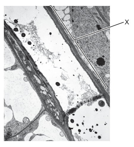
What is the name of the structure labelled X?
Options
A companion cell
B Casparian strip
C phloem sieve tube element
D xylem vessel element
Working
The label X points to a long, relatively thin-walled cell in a longitudinal section of a stem. The diagnostic feature visible is the perforated end wall (the sieve plate) — the cross-hatched/perforated wall joining this cell to the next element along the file. This is the hallmark of a phloem sieve tube element.
- A is incorrect: a companion cell is a small cell with dense cytoplasm lying alongside a sieve tube element, not the elongated element itself.
- B is incorrect: the Casparian strip is a band of suberin in the radial walls of endodermal cells in roots, not visible in a stem section.
- D is incorrect: a xylem vessel element has thick, lignified secondary walls (often with pits visible) and is empty of cytoplasm at maturity; no sieve plate is present.
Answer
C
C
Background Concept
Phloem is the plant tissue that translocates organic solutes (mainly sucrose) from sources (e.g. photosynthesising leaves) to sinks (e.g. roots, fruits, growing shoots). The conducting cells of phloem are sieve tube elements, joined end-to-end into sieve tubes. The end walls between successive sieve tube elements are modified into sieve plates, perforated by pores through which cytoplasm (and dissolved assimilates) can pass from one element to the next. At maturity, a sieve tube element has very little of its own cytoplasm — the nucleus and most organelles have degenerated — and is closely associated with one or more companion cells that carry out the metabolic roles the sieve tube element cannot.
Contrast this with xylem, which conducts water and mineral ions. Xylem vessel elements are dead at maturity, with thick lignified secondary cell walls (often leaving only small pits where the wall is absent), and they lack sieve plates. The Casparian strip, meanwhile, is a waterproof band of suberin in the radial and transverse walls of endodermal cells in roots; it would not be seen in a typical stem section.
Understanding the Question
The candidate is shown an electron micrograph of a longitudinal section through plant stem tissue, with one structure labelled X, and is asked to name it from four options. The image description states that X points to a sieve plate between two sieve tube elements, with the characteristic pores visible.
Approach
The key skill is to recognise the sieve plate — the perforated end wall — because this feature is unique to phloem sieve tube elements. The student should scan the labelled cell for an end wall that looks like a perforated plate rather than a smooth, thickened, lignified wall (xylem) or a small densely cytoplasmic cell alongside (companion cell). Once a sieve plate is recognised, the answer must be the phloem sieve tube element.
Step-by-Step Reasoning
- The labelled cell is elongated and runs along the long axis of the stem, consistent with a vascular-tissue conducting element seen in longitudinal section.
- The end wall visible on the cell is perforated — it has the cross-hatched/sieve appearance of a sieve plate with open pores connecting one element to the next.
- A sieve plate is diagnostic: only phloem sieve tube elements possess this structure. Therefore the labelled cell is a phloem sieve tube element.
- Checking the distractors:
- Companion cell (A): these are short cells with dense cytoplasm lying beside a sieve tube, not the long element bearing the sieve plate. Reject.
- Casparian strip (B): this is a suberised band in endodermal cell walls of roots, unrelated to a sieve plate. Reject.
- Xylem vessel element (D): xylem elements have thick, often lignified secondary walls and are empty of contents at maturity; they do not bear perforated end walls of the sieve-plate type. Reject.
- Only option C fits the observed structure.
Key Takeaways
- The sieve plate is the defining feature of a phloem sieve tube element; look for it when you need to identify phloem in micrographs.
- Sieve tube elements are living but have reduced cytoplasm at maturity; companion cells next to them provide metabolic support.
- Xylem vessel elements are dead at maturity with thick lignified walls and no sieve plates — easy to confuse in greyscale EM until you inspect the end wall.
- The Casparian strip is a feature of root endodermis, not stem vascular tissue.
Common Mistakes
- Choosing D (xylem vessel element) because both phloem and xylem appear as long tubes in longitudinal section. The deciding evidence is the end wall: perforated (sieve plate) → phloem; thick and complete (or with bordered pits) → xylem.
- Choosing A (companion cell) if the label appears to point near a small dense cell next to the sieve tube. Companion cells lie alongside the sieve tube element — they are not the element itself.
- Choosing B (Casparian strip) by guessing at unfamiliar terminology. The Casparian strip is restricted to the endodermis of roots and is not seen in stem vascular tissue.
Things to Be Careful About
- In greyscale electron micrographs, sieve plates can resemble xylem perforation plates. The distinction: sieve plates have many small pores in a phloem context and are at the end wall between two living elements; xylem perforation plates are at the ends of dead, lignified elements and the surrounding wall is conspicuously thick.
- Always use the end wall as the diagnostic feature for sieve tube elements; cytoplasm density and wall thickness alone are not reliable on their own.
- The question says "part of the stem" — this rules out features restricted to roots, such as the Casparian strip, without further work.
Which substance makes xylem vessel walls impermeable to water?
Options
A cellulose
B lignin
C suberin
D collagen
Working
Xylem vessels are dead, hollow tubes whose walls are reinforced and waterproofed by lignin, a hard, hydrophobic phenolic polymer. Lignin deposition makes the walls rigid (preventing collapse under the negative pressure of the transpiration pull) and impermeable to water, so that water within the lumen cannot leak sideways out of the vessel.
- A — cellulose is the main structural polysaccharide of plant cell walls but is hydrophilic and permeable to water.
- C — suberin waterproofs the Casparian strip in the endodermis and the walls of cork cells, not xylem vessels.
- D — collagen is a protein found in animal connective tissues, not in plant cell walls.
Answer
B
B
Background Concept
Xylem is the plant tissue responsible for transporting water and dissolved mineral ions from the roots up to the leaves. Mature xylem vessel elements are dead, elongated cells arranged end-to-end; their end walls have broken down to form continuous hollow tubes, and the lateral walls are reinforced with lignin.
Lignin is a complex, cross-linked phenolic polymer (built from monomers such as coniferyl alcohol). It is deposited within the cellulose–hemicellulose matrix of the secondary cell wall and has two crucial consequences for xylem function:
- It makes the wall rigid, so the vessel does not collapse inwards under the strong negative (tensile) pressure generated by transpiration.
- It makes the wall impermeable to water, so water inside the lumen is forced to flow longitudinally along the vessel rather than leaking out radially into surrounding tissues.
Understanding the Question
This is a multiple-choice item testing knowledge of the chemical composition of xylem vessel walls and which component is responsible for waterproofing them. The command word is implicit in the MCQ format — the candidate must select the correct substance from four options.
Approach
Identify the unique substance associated with xylem vessel walls and recall which of the four listed compounds is (a) present in xylem and (b) responsible for waterproofing. Then briefly rule out the other options by their actual roles in plant or animal biology.
Step-by-Step Reasoning
- Option B — lignin: this is THE substance deposited in xylem vessel (and tracheid) walls. Its hydrophobic, cross-linked structure renders the wall impermeable to water and gives it the mechanical strength needed to withstand the tensions of the cohesion–tension theory of water transport. Correct.
- Option A — cellulose: forms the bulk of plant cell walls (including xylem walls) but is a hydrophilic polysaccharide of β-1,4-linked glucose units. Cellulose itself is freely permeable to water, so it cannot be the waterproofing agent.
- Option C — suberin: is a waxy, hydrophobic substance, but it is found in the Casparian strip of the endodermis (controlling entry of water and ions into the vascular cylinder) and in cork (suberised) cells of the bark. It is not present in xylem vessel walls. CIE mark schemes treat suberin as the answer to endodermis/Casparian strip questions, not xylem questions.
- Option D — collagen: is a fibrous protein found in animal connective tissues (tendons, skin, bone matrix). It is not present in plants and so is irrelevant to xylem.
Key Takeaways
- Lignin waterproofs and strengthens xylem vessel walls.
- Suberin waterproofs endodermal cells (Casparian strip) and cork cells — often confused with lignin.
- Cellulose is the main wall polysaccharide but is hydrophilic, not waterproofing.
- Collagen is an animal protein, not a plant cell-wall component.
Common Mistakes
- Choosing C (suberin) because both lignin and suberin are hydrophobic plant wall substances — but suberin is restricted to the endodermis and periderm, not xylem.
- Choosing A (cellulose) by assuming that the most abundant wall component must be the waterproofing one — in fact, cellulose is hydrophilic.
- Choosing D (collagen) from mixing up plant and animal structural molecules.
Things to Be Careful About
- "Impermeable to water" is the key phrase: only lignin and suberin fit, and the question specifies xylem vessel walls, which uniquely contain lignin.
- Remember that xylem vessels are dead at maturity; their waterproofing is therefore a permanent feature of the wall, not a living membrane property.
- Distinguish lignin (xylem) from suberin (Casparian strip / cork) — these are the two classic "waterproof plant substance" answers and examiners regularly use them as paired distractors.
The following tissues carry an electrical impulse during the cardiac cycle.
- atrioventricular node
- muscle wall of atria
- Purkyne tissue
- sinoatrial node
In which order does the electrical impulse travel during the cardiac cycle?
Options
A 1 2 3 4
B 1 4 2 3
C 4 2 1 3
D 4 2 3 1
Working
The cardiac cycle is initiated by the sinoatrial node (SAN), which acts as the pacemaker. The electrical impulse then spreads across the muscle wall of the atria, causing atrial contraction. From there, the impulse reaches the atrioventricular node (AVN), which delays it briefly to allow the atria to empty, before passing down the Purkyne tissue to trigger ventricular contraction.
Matching this to the numbered list:
- 4 = sinoatrial node
- 2 = muscle wall of atria
- 1 = atrioventricular node
- 3 = Purkyne tissue
Answer
C
C
Background Concept
The mammalian heart is myogenic — it generates its own electrical impulses without needing external nervous stimulation. The rhythmic contraction of the heart during the cardiac cycle is coordinated by a specialised conduction system made up of three key components: the sinoatrial node (SAN), the atrioventricular node (AVN), and the bundle of His/Purkyne tissue (Purkyne fibres). Together they ensure that the atria contract before the ventricles, allowing efficient filling and ejection of blood.
The SAN sits in the wall of the right atrium and is the heart's natural pacemaker. It sets the resting heart rate by spontaneously depolarising at the highest frequency. Once an impulse is generated, it must travel in a precise order to coordinate contraction.
Understanding the Question
The question provides four structures and asks you to put them in the correct order in which the electrical impulse travels during one cardiac cycle. The list is:
- atrioventricular node (AVN)
- muscle wall of the atria
- Purkyne tissue
- sinoatrial node (SAN)
The command word is essentially "sequence" — you need to recall the path of the depolarisation wave.
Approach
Recall the canonical conduction pathway:
SAN → atrial muscle wall → AVN → bundle of His/Purkyne tissue → ventricular muscle.
Now match each step to the numbered item in the question.
Step-by-Step Reasoning
- Step 1 — Impulse generation (4, SAN): The SAN in the right atrium spontaneously depolarises and initiates the action potential. Because it has the fastest intrinsic rate, it overrides any other potential pacemaker and sets the rhythm for the entire heart.
- Step 2 — Atrial depolarisation (2, muscle wall of the atria): The impulse spreads across the atrial muscle wall via gap junctions, depolarising both atria almost simultaneously and causing them to contract. This pushes blood down into the ventricles.
- Step 3 — Atrioventricular delay (1, AVN): The impulse arrives at the AVN, located in the septum between the atria. The AVN delays conduction for about 0.1 s, ensuring the atria have fully emptied before the ventricles contract.
- Step 4 — Ventricular depolarisation (3, Purkyne tissue): The impulse then travels rapidly down the bundle of His and the Purkyne fibres, which spread the depolarisation throughout the ventricular muscle, triggering ventricular contraction and ejection of blood into the pulmonary artery and aorta.
Matching to the numbered options: 4 → 2 → 1 → 3, which is option C.
Key Takeaways
- The heart is myogenic; the SAN is the pacemaker.
- The order of conduction is: SAN → atrial muscle wall → AVN → Purkyne tissue → ventricular muscle.
- The AVN introduces a short delay that allows the atria to empty before the ventricles contract.
- Purkyne tissue rapidly distributes the impulse across the ventricles for a coordinated contraction.
Common Mistakes
- Reversing the SAN and AVN at the start of the sequence (e.g. starting with the AVN) — the SAN is always first because it initiates the impulse.
- Placing the Purkyne tissue before the AVN — the AVN must receive the impulse before it can be passed on to the ventricles via the Purkyne system.
- Confusing "muscle wall of atria" with "muscle wall of ventricles" — the atrial muscle is depolarised directly by the SAN, while the ventricular muscle is depolarised via the Purkyne tissue.
Things to Be Careful About
- Remember that the AVN is a delay, not a block — it slows the impulse but does not stop it; conduction still continues down to the Purkyne tissue.
- The Purkyne tissue does not start the impulse; it transmits it to the ventricles after the AVN has delayed it.
- Be precise with terminology: "Purkyne tissue/fibres" (or bundle of His/Purkyne fibres), not just "bundle of His" — the mark scheme recognises both.
The graph shows changes in the volume of the ventricles during a single cardiac cycle.
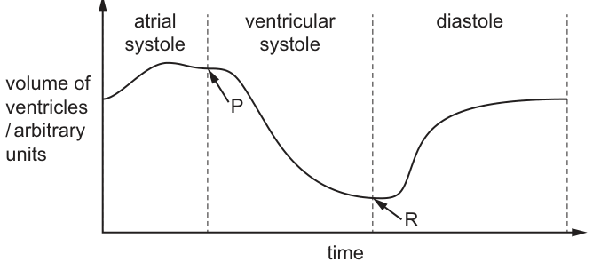
Which row is correct for the atrioventricular valve at P and for the semilunar valve at R?
Options
| atrioventricular valve at P | semilunar valve at R | |
|---|---|---|
| A | closes | closes |
| B | closes | opens |
| C | opens | closes |
| D | opens | opens |
Working
At point P, ventricular systole is beginning. As the ventricular muscle contracts, the pressure inside the ventricles rises above the pressure in the atria, forcing the atrioventricular (bicuspid/tricuspid) valves to close. This prevents backflow of blood into the atria.
At point R, ventricular systole is ending and diastole is beginning. As the ventricular muscle relaxes, the pressure inside the ventricles falls below the pressure in the aorta and pulmonary artery, so the semilunar (aortic and pulmonary) valves close. This prevents backflow of blood from the arteries into the ventricles. The rise in ventricular volume that follows R is due to the AV valves opening and blood flowing from atria to ventricles.
Answer
A
A
Background Concept
The heart contains four valves that ensure blood flows in one direction. The atrioventricular (AV) valves — the bicuspid (mitral) valve on the left and the tricuspid valve on the right — sit between the atria and ventricles. The semilunar valves — the aortic valve and the pulmonary valve — sit at the exits of the ventricles into the aorta and pulmonary artery respectively.
Valves open and close passively in response to pressure differences across them:
- An AV valve opens when atrial pressure > ventricular pressure, and closes when ventricular pressure > atrial pressure.
- A semilunar valve opens when ventricular pressure > arterial pressure, and closes when arterial pressure > ventricular pressure.
The cardiac cycle has three phases visible on a ventricular volume–time graph:
- Atrial systole — atria contract, pushing a small volume of blood into the ventricles (small rise in ventricular volume).
- Ventricular systole — ventricles contract; AV valves close, then semilunar valves open and blood is ejected (ventricular volume falls sharply).
- Diastole — ventricles relax; semilunar valves close, then AV valves open and blood flows passively from atria to ventricles (ventricular volume rises).
Understanding the Question
The graph plots ventricular volume against time and divides one cardiac cycle into the three phases. Point P is at the start of ventricular systole (just as the steep fall in volume begins) and Point R is at the end of ventricular systole (the minimum volume, just as the volume begins to rise again as the cycle enters diastole). The question asks which state the AV valve is in at P and which state the semilunar valve is in at R.
Approach
Determine the pressure change occurring in the ventricles at each labelled point, and then decide which valve responds to that pressure change.
- At P, ventricular contraction is just beginning → pressure rising inside the ventricles → which valve responds to rising ventricular pressure first? The AV valves, because they are on the atrial side and close as soon as ventricular pressure exceeds atrial pressure.
- At R, ventricular relaxation is just beginning → pressure falling inside the ventricles → which valve responds to falling ventricular pressure first? The semilunar valves, because they are on the arterial side and close as soon as arterial pressure exceeds ventricular pressure.
Step-by-Step Reasoning
- Identify point P. P is at the boundary between atrial systole and ventricular systole, where the ventricular volume curve begins its steep descent. The ventricles are starting to contract.
- Determine the pressure change at P. Contraction raises ventricular pressure sharply. Once it exceeds atrial pressure, blood would tend to flow backwards into the atria.
- Decide the AV valve state at P. The AV valve closes to prevent backflow into the atria. This produces the first heart sound ("lub").
- Identify point R. R is at the boundary between ventricular systole and diastole, at the minimum ventricular volume. The ventricles have just finished ejecting blood and are starting to relax.
- Determine the pressure change at R. Relaxation lowers ventricular pressure. Once it falls below the pressure in the aorta and pulmonary artery, blood in those arteries would tend to flow back into the ventricles.
- Decide the semilunar valve state at R. The semilunar valve closes to prevent backflow from the arteries. This produces the second heart sound ("dub").
- Confirm by reading the graph after R. The ventricular volume rises after R because blood is once again flowing into the ventricles from the atria — which can only happen because the AV valves are now open. So at R, just before this rise, the AV valves must still be closed and the semilunar valves are also closing (this is the isovolumetric relaxation phase), with the volume change shown next being driven by passive filling once AV valves open.
Key Takeaways
- Valves operate passively in response to pressure gradients, not by active contraction.
- The first heart sound is the AV valves closing at the start of ventricular systole.
- The second heart sound is the semilunar valves closing at the start of ventricular diastole.
- On a ventricular volume–time graph: a fall in volume = ventricular ejection (semilunar valves open); a rise in volume = ventricular filling (AV valves open); the two flat/transition points are where the relevant valve has just closed.
Common Mistakes
- Confusing which valve closes at which sound — many students mix up the "lub" (AV closing) and "dub" (semilunar closing).
- Saying the semilunar valve opens at P because the ventricles are contracting — in fact the semilunar valve opens only after the AV valve has closed and ventricular pressure has risen above arterial pressure; the very first event at P is the AV valve closing.
- Saying the AV valve opens at R because the volume is about to rise — the volume rise is delayed slightly; at R itself the semilunar valve is closing, and only afterwards (during isovolumetric relaxation) does the AV valve open.
Things to Be Careful About
- Read the graph carefully: P is at the top of the curve (just before the fall) and R is at the bottom (just before the rise). Do not confuse the two points.
- The two phases labelled on the graph (ventricular systole, diastole) describe the ventricles, not the atria — the AV valve is between them, so its state at these boundaries is the pivot point of the cycle.
- Valves never open and close simultaneously; there is a brief isovolumetric phase at both the start of systole and the start of diastole when all four valves are closed.
The graph shows how changes in oxygen concentration affect the percentage oxygen saturation of human haemoglobin and cat haemoglobin under normal physiological conditions. The partial pressure of carbon dioxide was kept constant at and the temperature was kept constant at .
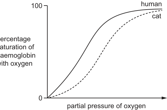
Which conclusion is supported by the graph?
Options
A The affinity of cat haemoglobin for oxygen is greater than the affinity of human haemoglobin for oxygen.
B At high partial pressures of oxygen, cat haemoglobin picks up oxygen more easily than human haemoglobin picks up oxygen.
C At low partial pressures of oxygen, oxygen is released more easily from cat haemoglobin than from human haemoglobin.
D The shift in the oxygen dissociation curve caused by the Bohr effect is larger for cat haemoglobin than for human haemoglobin.
Working
The cat curve lies to the right of the human curve. A rightward shift on an oxygen dissociation curve means lower affinity of haemoglobin for oxygen — at any given partial pressure of oxygen, cat haemoglobin is less saturated than human haemoglobin.
At low partial pressures of oxygen, the cat curve shows a lower percentage saturation than the human curve, so cat haemoglobin holds onto less oxygen and therefore releases oxygen more readily into respiring tissues.
Answer
C
C
Background Concept
An oxygen dissociation curve plots the percentage saturation of haemoglobin with oxygen against the partial pressure of oxygen (pO₂) at which the haemoglobin is exposed. The position of the curve reveals the affinity of haemoglobin for oxygen:
- A curve shifted to the left indicates higher affinity — haemoglobin becomes saturated at lower pO₂ and holds onto oxygen more tightly.
- A curve shifted to the right indicates lower affinity — haemoglobin needs a higher pO₂ to become saturated and releases oxygen more readily at any given pO₂.
The shape of the curve is sigmoidal because haemoglobin is a tetramer with four haem groups; binding of O₂ to one haem increases the affinity of the remaining haems (cooperative binding).
Understanding the Question
The graph presents two oxygen dissociation curves drawn under identical conditions (CO₂ at 5.0 kPa, temperature at 37 °C). The human curve is to the left of the cat curve. The question asks which statement is supported by this graph.
Approach
The key skill is translating curve position into a statement about oxygen behaviour. Remember the rule:
Apply this to each option using the graph.
Step-by-Step Reasoning
Option A — "The affinity of cat haemoglobin for oxygen is greater than the affinity of human haemoglobin for oxygen."
Incorrect. The cat curve is to the right, so cat haemoglobin has lower affinity, not greater.
Option B — "At high partial pressures of oxygen, cat haemoglobin picks up oxygen more easily than human haemoglobin."
Incorrect. At high pO₂ both curves plateau near 100 % saturation, but the human curve reaches saturation first. Cat haemoglobin has lower affinity, so it does not pick up O₂ more easily — if anything, it requires a higher pO₂ to reach the same saturation.
Option C — "At low partial pressures of oxygen, oxygen is released more easily from cat haemoglobin than from human haemoglobin."
Correct. At low pO₂, the cat curve sits below the human curve, meaning cat haemoglobin is less saturated. Lower saturation at low pO₂ is equivalent to releasing oxygen more easily — exactly what the rightward shift predicts. This also makes biological sense: cats are active predators with high metabolic rates, so their tissues need efficient oxygen unloading; a right-shifted curve delivers more O₂ to respiring cells.
Option D — "The shift in the oxygen dissociation curve caused by the Bohr effect is larger for cat haemoglobin than for human haemoglobin."
Incorrect. The Bohr effect describes the rightward shift that occurs when pCO₂ rises (or pH falls). The graph shows only one pCO₂ (5.0 kPa), so a Bohr shift cannot be measured or compared from this figure — a single curve per species cannot reveal a shift between conditions.
Key Takeaways
- Curve left = higher O₂ affinity; curve right = lower O₂ affinity.
- Lower affinity (right curve) means haemoglobin releases O₂ more readily at low pO₂ — useful in tissues with high metabolic demand.
- The Bohr effect requires comparing curves at different pCO₂; a single curve cannot show it.
- Reading affinity from a curve is a common exam skill: pick a pO₂ and compare percentage saturations.
Common Mistakes
- Confusing curve position with affinity direction. Students often think "right = right answer" without linking position to the underlying concept of affinity.
- Mixing up pickup and release. "Higher affinity" means better at picking up O₂ (in the lungs) and holding onto it — i.e. worse at releasing it. A common error is to say high affinity releases more O₂.
- Trying to read the Bohr effect from a single curve. The Bohr effect is a shift between two conditions; one curve per species gives no shift to compare.
- Ignoring the units/axes. Always read what the y-axis represents (percentage saturation) and what the x-axis represents (partial pressure of O₂) before drawing conclusions.
Things to Be Careful About
- "At low pO₂" and "at high pO₂" produce opposite conclusions; the right-hand side of a graph is not always the answer.
- Saturation approaching 100 % at high pO₂ is similar for both curves, so do not over-interpret the upper plateau.
- Biological context (active animal = right-shifted curve for tissue unloading) is supporting evidence but the answer must come from the graph itself.
- Keep terminology precise: use partial pressure of oxygen and percentage saturation of haemoglobin rather than vague terms like "amount of oxygen".
Which statement is correct about how oxygen combines with haemoglobin?
Options
A Combining all four oxygen molecules with haemoglobin does not affect the shape of haemoglobin.
B One oxygen molecule can combine with each haem group of the haemoglobin molecule.
C The first oxygen molecule to combine with haemoglobin does not affect the shape of haemoglobin.
D The third oxygen molecule to combine with haemoglobin makes it more difficult for the fourth oxygen molecule to combine with haemoglobin.
Working
Haemoglobin is a globular protein with four polypeptide chains, and each chain carries one haem group. The Fe²⁺ ion at the centre of each haem group can bind one O₂ molecule, so one haemoglobin molecule can carry up to four O₂ molecules. The four binding sites are equivalent; binding of O₂ at one site induces a conformational change in the globin chains that makes the remaining sites bind O₂ more readily (positive cooperativity).
Answer
B
B
Background Concept
Haemoglobin is the oxygen-carrying pigment in red blood cells. Each molecule is a globular protein made of four polypeptide chains (two α and two β chains in adult haemoglobin, HbA), and each chain carries one haem group. A haem group is a flat porphyrin ring with a central Fe²⁺ ion, and it is the Fe²⁺ that reversibly binds one O₂ molecule. Because there are four haem groups, one haemoglobin molecule can carry a maximum of four O₂ molecules, giving the molecule its cooperative, sigmoidal oxygen-binding behaviour.
Cooperative binding is the key idea. When the first O₂ binds to a haem group, the Fe²⁺ moves slightly into the plane of the porphyrin ring, and this small movement is transmitted through the globin chain. The whole tetramer shifts from the T (tense, low-affinity) state to the R (relaxed, high-affinity) state. As a result, the second O₂ binds more easily than the first, the third more easily than the second, and the fourth more easily than the third. Each successive binding therefore becomes progressively easier, not harder, and the binding curve is sigmoidal as a consequence.
Understanding the Question
This is a single-best-answer multiple choice item. The candidate is asked to identify which one of four statements correctly describes how O₂ combines with haemoglobin. Each option tests a slightly different aspect of haemoglobin structure or binding, so the distractors are designed to trap candidates who half-remember the cooperative-binding story.
Approach
Decide what haemoglobin's structure is, then check each statement against that structure and against the principle of positive cooperativity. Eliminate the option that is inconsistent with the known facts.
Step-by-Step Reasoning
- Option A claims that combining all four O₂ molecules does not change the shape of haemoglobin. This is wrong. Every O₂ that binds causes a conformational shift; the cumulative shift from T to R state is precisely what underlies cooperative binding.
- Option B states that one O₂ molecule can combine with each haem group of the haemoglobin molecule. This is correct: there are four haem groups, each with one Fe²⁺ that binds one O₂, so four O₂ molecules in total can be carried per haemoglobin molecule.
- Option C claims that the first O₂ does not change the shape of haemoglobin. This is wrong; the very first O₂ triggers the T→R transition that enables the remaining sites to bind more easily.
- Option D says the third O₂ makes it harder for the fourth to bind. This is the reverse of what actually happens. Positive cooperativity means each successive O₂ binds more easily than the previous one, so the third O₂ makes the fourth easier, not harder, to bind.
Only option B is fully consistent with the structure of haemoglobin and the mechanism of cooperative binding.
Key Takeaways
- Haemoglobin has four haem groups, each binding one O₂ at its central Fe²⁺; maximum carrying capacity is four O₂ per molecule.
- O₂ binding is cooperative: each successive O₂ binds more readily than the previous one because of a T→R conformational change.
- The sigmoidal shape of the oxygen dissociation curve is a direct consequence of this positive cooperativity.
Common Mistakes
- Confusing positive cooperativity with negative cooperativity: students sometimes assume that later O₂ molecules bind harder, the way substrate binds to an allosteric enzyme with multiple inhibitory sites. In haemoglobin, the opposite is true.
- Forgetting that haemoglobin has four haem groups and assuming it carries only one or two O₂ molecules.
- Believing that the first O₂ binds without any structural change; in reality, that first binding event is the trigger for the T→R transition.
Things to Be Careful About
- The conformational change is small but functionally critical: even a partial movement of Fe²⁺ into the porphyrin plane is amplified through the globin subunits to alter the affinity of the other three sites.
- Note that Fe²⁺ stays in the +2 oxidation state when O₂ binds; oxidation to Fe³⁺ produces methaemoglobin, which cannot bind O₂, and this is a pathological condition, not normal binding.
- "Easier" binding after each O₂ does not mean the binding becomes energy-free; O₂ binding to haemoglobin is always reversible and concentration-dependent, as the oxygen dissociation curve shows.
How are alveoli adapted to their function?
Options
A They contain squamous epithelium for a short diffusion distance.
B They contain goblet cells to produce mucus.
C They contain ciliated epithelium to move mucus.
D They contain cartilage to prevent alveolar collapse.
Working
Alveoli are adapted for gas exchange by providing a short diffusion distance between the air and the blood. This is achieved because the alveolar wall is only one cell thick and is lined by squamous (pavement) epithelium.
- B is incorrect — goblet cells are found in the trachea and bronchi, not in alveoli.
- C is incorrect — ciliated epithelium lines the trachea and bronchi to move mucus, not alveoli.
- D is incorrect — cartilage is present in the trachea and bronchi, not in alveoli.
Answer
A
A
Background Concept
Gas exchange in mammals occurs in the alveoli, which are tiny air sacs at the end of the branching airways (bronchioles) in the lungs. The function of the alveoli is to allow oxygen to diffuse from the alveolar air into the blood in the surrounding pulmonary capillaries, and carbon dioxide to diffuse in the opposite direction. For efficient gas exchange, Fick's law tells us that the rate of diffusion is increased by:
- a large surface area
- a short diffusion distance
- a steep concentration gradient (maintained by ventilation and blood flow)
Alveoli are structurally adapted to maximise each of these factors. They are numerous (millions per lung), giving a huge total surface area; they are extremely thin-walled; and they are surrounded by a dense capillary network that continually carries blood away (replacing CO₂-rich blood with O₂-poor blood) while breathing continually refreshes the alveolar air.
The alveolar wall itself is composed of:
- Squamous (pavement) epithelium — extremely thin, flattened cells, only one cell thick. This gives a very short diffusion distance (often cited as around 1 µm between alveolar air and red blood cell cytoplasm).
- Elastic fibres — allow the alveoli to stretch on inhalation and recoil on exhalation.
- Capillaries — endothelial cells also only one cell thick, so the diffusion path is epithelium + fused basement membrane + endothelium.
Squamous epithelium is also found lining the pulmonary capillaries, and together these two thin layers form the respiratory surface.
Understanding the Question
The question asks you to identify which of four statements correctly describes an adaptation of the alveoli to their function of gas exchange. The correct option must:
- Describe a real feature of alveoli (not of other parts of the respiratory system).
- Explain how that feature helps gas exchange.
This is a "relate structure to function" question, and the trap is to confuse the histology of alveoli with the histology of the conducting airways (trachea, bronchi, bronchioles), which have very different features.
Approach
First, recall the histology of the alveoli (the gas-exchange region) and contrast it with the conducting airways (trachea → bronchi → bronchioles). The conducting airways have goblet cells, cilia, mucus, and (in the trachea/bronchi) rings or plates of cartilage. Once these features are associated with the conducting airways, anything that describes them can be ruled out for alveoli. Then confirm which option correctly describes a feature of alveoli AND links it to gas exchange.
Step-by-Step Reasoning
-
Option A — "They contain squamous epithelium for a short diffusion distance."
Squamous epithelium is the correct tissue for alveoli. Its flat, thin cells reduce the diffusion distance to a minimum, which directly increases the rate of diffusion of O₂ and CO₂ across the respiratory surface. ✓ Correct. -
Option B — "They contain goblet cells to produce mucus."
Goblet cells are found in the trachea, bronchi and larger bronchioles, where they secrete mucus that traps inhaled particles and pathogens. Mucus is not needed in the alveoli; in fact it would impair gas exchange. Alveoli do not contain goblet cells. ✗ Wrong. -
Option C — "They contain ciliated epithelium to move mucus."
Ciliated epithelium also lines the trachea and bronchi. The cilia beat in a coordinated wave to move mucus (and the trapped debris) up the airway towards the pharynx, where it is swallowed. Alveoli have no cilia. ✗ Wrong. -
Option D — "They contain cartilage to prevent alveolar collapse."
Cartilage is found in the trachea and bronchi to keep these airways open. Alveoli do not contain cartilage. Their shape is maintained by the surrounding network of elastic fibres, which allows them to stretch and recoil with each breath. ✗ Wrong.
Key Takeaways
- The alveoli are the site of gas exchange, and their epithelium is squamous — only one cell thick — to minimise the diffusion distance.
- The conducting airways (trachea, bronchi) are lined with ciliated epithelium and goblet cells that produce and clear mucus, and are reinforced with cartilage to hold them open.
- A common exam trap: distractor options are always features of a different part of the gas exchange system. Recognising where each histological feature belongs is the key skill.
- The other alveolar adaptations worth memorising are: large total surface area (millions of alveoli), a rich capillary network, elastic fibres, and a moist surface (water dissolves gases before they can diffuse across the membrane).
Common Mistakes
- Confusing the histology of the alveoli (squamous epithelium, elastic fibres, capillaries) with that of the trachea/bronchi (ciliated epithelium, goblet cells, cartilage, smooth muscle, elastic fibres). All three wrong options here are real features — just of the wrong region.
- Picking an option because the function sounds right (e.g. "cartilage supports structures" or "mucus traps pathogens") without checking whether that feature is actually present in alveoli.
- Forgetting that alveoli must be thin: any mucus layer, cartilage, or thick epithelium would dramatically slow diffusion and defeat the purpose of the respiratory surface.
Things to Be Careful About
- Ciliated epithelium is not in alveoli — the cilia would interfere with the very thin, flat surface needed for gas exchange.
- Alveoli are kept open not by cartilage but by elastic fibres (and surfactant, which reduces surface tension).
- When a question asks "how is X adapted to its function?", make sure the structure AND the function both match the correct answer; mark schemes often require both elements to be present in the candidate's response.
The diagram shows oxygen diffusing from the space inside an alveolus into the blood through the gaseous exchange surface.
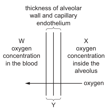
What would increase the rate of diffusion of oxygen from the alveolus to the blood?
Options
| increase | decrease | keep the same | |
|---|---|---|---|
| A | W | X | Y |
| B | W | Y | X |
| C | Y | X | W |
| D | X | Y | W |
Working
Fick's law of diffusion states:
The three variables in the figure are:
- W = oxygen concentration in the blood
- X = oxygen concentration inside the alveolus
- Y = thickness of the alveolar wall and capillary endothelium
To increase the rate of diffusion:
- X (alveolar O₂ concentration) should increase → this widens the concentration gradient driving diffusion from alveolus to blood.
- Y (thickness of exchange surface) should decrease → a shorter diffusion distance speeds up diffusion.
- W (blood O₂ concentration) should be kept the same → so the increased gradient produced by raising X is maintained.
This matches option D: X increase, Y decrease, W keep the same.
Answer
D
D
Background Concept
Gas exchange in the lungs depends on the simple diffusion of oxygen (and carbon dioxide) across the alveolar wall and the capillary endothelium. The rate at which a gas diffuses across a surface is described by Fick's law of diffusion:
where is the surface area, is the concentration gradient across the surface, and is the thickness of the surface. Anything that increases the surface area or the concentration gradient, or decreases the thickness, will increase the rate of diffusion.
In the alveoli, evolution has produced features that maximise diffusion rate: an enormous total surface area, an extremely thin (one-cell-thick) exchange surface, and a steep concentration gradient maintained by continuous ventilation (bringing fresh air) and continuous blood flow (carrying away oxygenated blood and bringing deoxygenated blood).
Understanding the Question
The figure labels three variables on a simple model of the alveolar gas exchange surface:
- W = oxygen concentration in the blood on the far side of the surface
- X = oxygen concentration inside the alveolus on the near side
- Y = thickness of the alveolar wall plus capillary endothelium (the diffusion distance)
The question asks which combination of changes to W, X and Y would increase the rate at which oxygen moves from the alveolus into the blood. The four options tabulate, for W, X and Y, whether each should be increased, decreased or kept the same.
Approach
Apply Fick's law term by term to the three variables. Decide, for each variable in turn, whether increasing, decreasing or leaving it unchanged produces the higher diffusion rate, and then match the resulting combination to one of the four answer options.
Step-by-Step Reasoning
Concentration inside the alveolus (X). Fick's law says the rate is proportional to the concentration gradient . Increasing X raises , so the gradient becomes steeper and diffusion is faster. → X should increase.
Thickness of the exchange surface (Y). The rate is inversely proportional to thickness. Halving the thickness roughly doubles the rate. → Y should decrease.
Concentration in the blood (W). The same gradient reasoning applies: lowering W would also increase the gradient and speed diffusion. However, the option that combines "increase X, decrease Y" with "decrease W" is not listed. The remaining sensible choice is to keep W the same, so the gain made by raising X is not offset by a rise in W. → W should be kept the same.
Combining these three decisions: X increase, Y decrease, W keep the same — option D.
Key Takeaways
- Fick's law links diffusion rate to surface area, concentration gradient and diffusion distance.
- A steeper concentration gradient and a thinner exchange surface both speed up gas exchange.
- In real lungs the gradient is maintained by ventilation (high alveolar ) and by perfusion carrying oxygenated blood away (low blood in the pulmonary artery).
Common Mistakes
- Choosing an option that increases W: raising the blood oxygen concentration would reduce the gradient from alveolus to blood, slowing diffusion — the opposite of what is asked.
- Choosing an option that increases Y: a thicker barrier is exactly what Fick's law tells us slows diffusion.
- Confusing the direction of the gradient: oxygen diffuses from alveolus to blood, so it is that matters, not .
Things to Be Careful About
- "Increase the rate" means each individual change must be in the direction that Fick's law predicts speeds diffusion.
- The mark scheme does not credit answers where W is decreased, even though this would technically also widen the gradient, because the question's correct answer is the one that combines "raise X, reduce Y, hold W constant" (option D).
- Read each option's table carefully — the columns are W, X, Y in a specific order, not always the same order across options.
Which tissues are present in a bronchus?
Options
| cartilage | ciliated epithelium | smooth muscle | |
|---|---|---|---|
| A | ✓ | ✓ | ✓ |
| B | ✓ | ✓ | ✗ |
| C | ✓ | ✗ | ✓ |
| D | ✗ | ✓ | ✓ |
key
✓ = present
✗ = not present
Working
A bronchus is a conducting airway in the lower respiratory tract. Its wall contains three key tissues:
- Cartilage — present as irregular (C-shaped / helical) plates that hold the airway open and prevent collapse during inhalation.
- Ciliated epithelium — a pseudostratified ciliated columnar epithelium that moves mucus (and trapped particles) upward away from the lungs.
- Smooth muscle — lies beneath the epithelium and contracts/relaxes to alter airway diameter (bronchoconstriction and bronchodilation).
All three are present, matching option A.
Answer
A
A
Background Concept
The trachea branches into two primary bronchi, one entering each lung. Each bronchus then branches repeatedly into smaller bronchi and finally into bronchioles. A bronchus is a conducting airway — it carries air to and from the gas-exchange surfaces (alveoli) but does not itself take part in gas exchange. Because it must remain patent (open) during breathing, withstand pressure changes, and clear inhaled debris, its wall is built from three functionally distinct tissues arranged in concentric layers.
Understanding the Question
The stem asks which of the three listed tissues — cartilage, ciliated epithelium, and smooth muscle — are all present in the wall of a bronchus. The options differ in which single tissue is omitted, so the question is essentially testing whether the student can recall the complete tissue composition of a bronchus and, importantly, distinguish a bronchus from a bronchiole (which has no cartilage and no ciliated epithelium in its terminal portions, and which relies mainly on smooth muscle).
Approach
Recall the three layers of a typical bronchus wall:
- Mucosa — pseudostratified ciliated columnar epithelium with goblet cells. The cilia beat in a coordinated wave to move the mucus layer (and trapped pathogens/dust) upward toward the pharynx (the mucociliary escalator).
- Submucosa — connective tissue with seromucous glands.
- Outer wall — contains C-shaped/helical plates of hyaline cartilage that hold the lumen open, and bands of smooth muscle (lying between the cartilage and the epithelium) that constrict or dilate the airway under autonomic control.
All three tissues (cartilage, ciliated epithelium, smooth muscle) are present, so option A is correct.
Step-by-Step Reasoning
- Option A (cartilage ✓, ciliated epithelium ✓, smooth muscle ✓): matches the structure of a bronchus — correct.
- Option B (cartilage ✓, ciliated epithelium ✓, smooth muscle ✗): smooth muscle is present in a bronchus, so this is wrong. (This combination would describe a trachea if smooth muscle were absent, but in reality the trachea also has smooth muscle in its posterior wall.)
- Option C (cartilage ✓, ciliated epithelium ✗, smooth muscle ✓): ciliated epithelium is definitely present in a bronchus (replaced by simple cuboidal/columnar only in the smaller bronchioles) — wrong.
- Option D (cartilage ✗, ciliated epithelium ✓, smooth muscle ✓): cartilage is one of the defining features of a bronchus; it is only lost in bronchioles — wrong.
Therefore A is the only option that lists all the tissues actually found in a bronchus wall.
Key Takeaways
- A bronchus wall contains: cartilage (C-shaped/helical plates), smooth muscle, and ciliated epithelium with goblet cells.
- A bronchiole is distinguished by the absence of cartilage (and the loss of cilia and goblet cells in the smallest, terminal bronchioles); it has only smooth muscle in appreciable amounts and relies on elastic recoil of surrounding lung tissue to stay open.
- The mucociliary escalator in the bronchi traps and removes inhaled particles — an important non-specific defence.
Common Mistakes
- Confusing a bronchus with a bronchiole. Students often forget that bronchioles have no cartilage and may have no cilia in the terminal branches, and consequently pick an option (often D) that omits cartilage.
- Thinking smooth muscle is restricted to bronchioles. Smooth muscle is, in fact, abundant in bronchioles, but it is also present in the bronchial wall between the cartilage plates and the epithelium — this is what allows bronchoconstriction in asthma.
- Confusing the trachea and bronchus. They are very similar in tissue composition; both have cartilage, ciliated epithelium, and smooth muscle, so the same answer applies.
Things to Be Careful About
- The question is about a bronchus, not a bronchiole — the distinction (cartilage present vs absent) is the single most commonly tested point at AS level.
- Remember that ciliated epithelium is replaced gradually as airways get smaller; the question's wording ("a bronchus") unambiguously points to cartilage-bearing airways.
- Don't be misled by the asymmetric structure of the trachea: the C-shaped cartilage has a posterior gap bridged by smooth muscle — this still means both tissues are present.
Steep concentration gradients must be maintained for efficient gaseous exchange to occur in the human lungs.
Which row correctly describes how steep concentration gradients can be maintained?
Options
| elastic fibres in the walls of the alveoli recoil when breathing out | continual supply of deoxygenated blood by the pulmonary artery | |
|---|---|---|
| A | ✓ | ✓ |
| B | ✗ | ✓ |
| C | ✓ | ✗ |
| D | ✗ | ✗ |
key
✓ = helps maintain
✗ = does not help maintain
Working
For efficient gas exchange in the alveoli, a steep concentration gradient for O₂ and CO₂ must be kept between the alveolar air and the blood in the surrounding capillaries.
Statement 1 — elastic fibres recoil on breathing out:
Elastic fibres in alveolar walls stretch during inspiration and recoil during expiration. This recoil helps expel air from the alveoli, so stale (O₂-depleted, CO₂-rich) air leaves the lungs and can be replaced by fresh atmospheric air on the next inhalation. This keeps alveolar O₂ high and alveolar CO₂ low, helping to maintain the concentration gradients. ✓
Statement 2 — continual supply of deoxygenated blood by the pulmonary artery:
The pulmonary artery delivers a continual flow of deoxygenated blood to the alveolar capillaries. This keeps the blood O₂ concentration low (steep gradient for O₂ into the blood) and its CO₂ concentration high (steep gradient for CO₂ out of the blood). At the same time, oxygenated blood is continuously removed by the pulmonary veins, preventing the gradient from equilibrating. ✓
Both statements help maintain steep concentration gradients, so the correct row is the one in which both are ticked.
Answer
A
A
Background Concept
Efficient gas exchange across the alveolar wall depends on diffusion of O₂ from alveolar air into the pulmonary capillary blood, and of CO₂ in the opposite direction. Fick's law tells us that the rate of diffusion is proportional to the concentration gradient between the two sides:
To keep the gradient steep (and therefore keep diffusion fast), two things must be true continuously:
- The alveolar air must keep a high O₂ partial pressure and a low CO₂ partial pressure — i.e. it must be constantly refreshed with atmospheric air.
- The capillary blood must keep a low O₂ partial pressure and a high CO₂ partial pressure — i.e. deoxygenated blood must continually arrive and oxygenated blood must continually leave.
Anything that disturbs either side of the gradient slows gas exchange. The human lung has several features that maintain these gradients: elastic fibres in alveolar walls, the action of breathing (ventilation), and the pulmonary circulation.
Understanding the Question
The question is an MCQ that asks you to judge two separate statements about how the lungs keep the concentration gradients across the alveolar wall steep. A tick (✓) means the statement helps maintain a steep gradient; a cross (✗) means it does not. You have to decide independently for each statement, then pick the row that correctly describes both. The four answer options combine the two judgements in every possible way, so the question is essentially two true/false decisions rolled into one.
The command word is implicit ("correctly describes") and the question tests understanding of the maintenance of concentration gradients in the alveolus — a sub-topic of the Gas Exchange section of the AS syllabus.
Approach
Decide, for each statement, whether the described feature actually helps keep the O₂ or CO₂ gradient steep between alveolar air and blood. If both do, the answer is the option in which both are ticked. If one does not, choose the option that reflects only the one(s) that are true.
Step-by-Step Reasoning
Statement 1: elastic fibres in alveolar walls recoil when breathing out.
- During inspiration the alveoli are stretched; their elastic fibres are loaded.
- During expiration these fibres recoil, passively squeezing alveolar air out through the bronchioles and trachea.
- This expulsion of alveolar air (which has had some of its O₂ removed and CO₂ added) is what allows the next breath of fresh atmospheric air to replace it.
- Without recoil, alveolar air would become progressively depleted in O₂ and enriched in CO₂, and the gradient would flatten.
- Therefore elastic fibre recoil does help maintain a steep concentration gradient. ✓
Statement 2: continual supply of deoxygenated blood by the pulmonary artery.
- The pulmonary artery is the only artery in the body that carries deoxygenated blood; it takes blood from the right ventricle to the lungs.
- A constant inflow of blood with low O₂ (and high CO₂) into the alveolar capillaries keeps the blood-side O₂ concentration low and CO₂ concentration high.
- This sustains the diffusion gradient for O₂ (from alveolus into blood) and for CO₂ (from blood into alveolus).
- If blood flow stopped, the blood would quickly equilibrate with alveolar air and diffusion would cease.
- Therefore the continual supply of deoxygenated blood does help maintain a steep concentration gradient. ✓
Since both statements are correct, the answer must be the row in which both are ticked — option A.
Key Takeaways
- Steep concentration gradients in the alveoli are maintained by ventilation (replacing alveolar air) and by perfusion (replacing capillary blood).
- Elastic fibres in alveolar walls recoil on expiration, helping to expel air and making room for fresh inhaled air — this keeps alveolar O₂ high and CO₂ low.
- The pulmonary artery delivers a continual flow of deoxygenated blood to the lungs — this keeps blood O₂ low and CO₂ high, sustaining the gradient.
- The pulmonary veins are equally important because they continually remove oxygenated blood, preventing equilibration.
Common Mistakes
- Rejecting the elastic-fibre statement because students confuse elastic fibres with the smooth muscle or with cartilage. Elastic fibres are responsible for the passive recoil that drives expiration; they are not the same as the smooth muscle that changes airway diameter.
- Rejecting the pulmonary-artery statement because students forget that this is the artery carrying deoxygenated blood, or because they confuse the pulmonary artery with the pulmonary vein (which carries oxygenated blood away from the lungs and so would not by itself help maintain the gradient in the same way).
- Stating that the blood is 'constantly moving' is too vague to score on a 'describe' question; you must specify what kind of blood (deoxygenated) and which vessel supplies it.
Things to Be Careful About
- The pulmonary artery carries deoxygenated blood — this is unusual for an artery and is the kind of detail examiners test.
- Maintenance of the gradient is two-sided: it is not just about getting O₂-rich air in, but also about ensuring O₂-poor blood is always at the exchange surface.
- The options are designed so that every combination of ticks/crosses is offered; check both columns carefully before selecting, as half the candidates will read only one of the two statements.
A scientist investigated the effect of an antibiotic on the treatment of cholera.
320 people with cholera were divided into two groups. One group was treated with the antibiotic while the other group was not given the antibiotic. Both groups were given fluids containing sugars and mineral salts (oral rehydration therapy).
The scientist recorded the number of days that each person had diarrhoea.
The table shows the results.
| treatment | mean time person had diarrhoea / days |
|---|---|
| antibiotic and oral rehydration therapy | 3.2 |
| oral rehydration therapy | 5.3 |
What is the percentage decrease in the mean time that a person had diarrhoea when they were treated with the antibiotic?
Options
A 39.6%
B 60.4%
C 165.6%
D 252.4%
Working
Answer
A
A
Background Concept
Cholera is a water-borne infectious disease caused by the bacterium Vibrio cholerae. It produces a toxin that causes the intestinal lining to secrete large volumes of water and electrolytes, leading to severe, watery diarrhoea. The standard treatment is oral rehydration therapy (ORT) — a solution of water, sugars and mineral salts that replaces the fluid and ions lost in the diarrhoea. Antibiotics can be given alongside ORT to reduce the bacterial load and shorten the duration of symptoms. To compare the effectiveness of two treatments quantitatively, biologists calculate the percentage change in a measured variable (here, the mean number of days of diarrhoea) between the two groups.
The percentage change formula is:
A decrease is reported when the new value is smaller than the original; the sign (or wording) tells the reader whether it is an increase or a decrease, but the magnitude is always expressed as a positive percentage.
Understanding the Question
The question gives a small results table with two mean values: 3.2 days of diarrhoea for the group that received antibiotic + ORT, and 5.3 days for the group that received ORT alone. The command word is "What is the percentage decrease… when they were treated with the antibiotic?" — so the ORT-only value (5.3) is the original/baseline and the antibiotic + ORT value (3.2) is the new value. The candidate must compute the percentage decrease and pick the matching option.
Approach
- Identify the original (without antibiotic) and new (with antibiotic) means from the table.
- Apply the percentage decrease formula: (original − new) / original × 100.
- Match the result to the closest option.
Step-by-Step Reasoning
- Original mean (ORT only) = 5.3 days
- New mean (antibiotic + ORT) = 3.2 days
- Difference = 5.3 − 3.2 = 2.1 days
- Divide by the original: 2.1 / 5.3 = 0.3962…
- Convert to a percentage: 0.3962 × 100 ≈ 39.6%
This matches option A.
The distractors correspond to common errors:
- B (60.4%) would arise from dividing the difference (2.1) by the new value (3.2) instead of the original — the wrong reference value.
- C (165.6%) is what you get if you add 100 to the answer in B (i.e. treating it as a percentage of the new value rather than the original).
- D (252.4%) is 100 + the reciprocal-type figure; it corresponds to dividing the larger by the smaller and adding 100, a further step in the wrong direction.
Key Takeaways
- Percentage decrease always uses the original (here, ORT-only) value as the denominator.
- The percentage change formula is a transferable quantitative skill used throughout the A-level Biology papers (Paper 2 calculations, Paper 3 results tables, and Paper 5 analysis questions).
- When comparing a treatment group to a control, the control value is the "original" against which the change is measured.
Common Mistakes
- Dividing by the new (smaller) value instead of the original — this exaggerates the percentage change and gives ~60% rather than ~40%.
- Forgetting to multiply by 100, leaving the answer as a decimal (0.396 instead of 39.6%).
- Reporting a decrease but calculating and writing an increase, by inverting the subtraction in the numerator.
Things to Be Careful About
- Read the question carefully: "decrease" is specified, so the answer should be reported as a positive percentage that represents a fall from the original value.
- The marks are awarded for a single answer choice in this MCQ, but the working matters in a structured (non-MCQ) version of the same calculation on Paper 2 — always show the formula, the substitution, and the final value to the appropriate precision (here, one decimal place).
- The sample sizes (320 people, split between the two groups) are not needed for the percentage decrease calculation itself, but they do tell you the means are based on a reasonable number of individuals.
How does the antibiotic penicillin affect the metabolism of a bacterial cell?
Options
A Penicillin reduces cell growth by preventing water uptake.
B Penicillin inhibits the formation of cross-links in the cell wall between peptidoglycan molecules.
C Penicillin inhibits the hydrolysis of peptidoglycan links in the cell wall during cell extension.
D Penicillin weakens the cell wall by digesting peptidoglycan molecules.
Working
Penicillin is an antibiotic that acts on the bacterial cell wall, which is made of peptidoglycan (a mesh of polysaccharide chains cross-linked by short peptide chains). It works by inhibiting the enzymes (transpeptidases, sometimes called penicillin-binding proteins) that form the peptide cross-links between adjacent peptidoglycan strands. Without these cross-links the wall is mechanically weak, and the bacterium cannot withstand the turgor pressure generated by its cytoplasm, so it lyses.
- A: incorrect — penicillin does not act on water uptake; that is the role of aquaporins / osmosis.
- B: correct — matches the established mechanism: inhibition of cross-link formation between peptidoglycan molecules.
- C: incorrect — penicillin does not inhibit hydrolysis of peptidoglycan; hydrolysis is the step that breaks the wall, which would actually weaken it further, but this is not what penicillin does.
- D: incorrect — penicillin does not digest peptidoglycan; that is the role of lysozyme. Penicillin prevents new cross-links from forming, rather than breaking existing ones.
Answer
B
B
Background Concept
Bacterial cells are surrounded by a rigid cell wall that gives them their shape and protects them from osmotic lysis. In most bacteria the main structural component of this wall is peptidoglycan — long polysaccharide chains of alternating N-acetylglucosamine (NAG) and N-acetylmuramic acid (NAM), joined by short peptide cross-links. The cross-links convert the polysaccharide sheets into a single, mechanically strong mesh that holds the cell together against the high internal turgor pressure of the cytoplasm.
Because animal cells do not have a peptidoglycan cell wall, drugs that target its construction (such as penicillins and cephalosporins) selectively harm bacteria and are relatively non-toxic to the host — this is the basis for penicillin as an antibiotic.
Understanding the Question
This MCQ asks for the mechanism of action of penicillin on a bacterial cell. The command word is implicit but clear: the candidate must pick the statement that correctly describes what penicillin does biochemically. The four options are similar in wording, so careful knowledge of the precise step that is blocked is required.
Approach
Recall (or eliminate) the four statements by matching each to the known mechanism:
- Penicillin binds to and inhibits transpeptidase enzymes (penicillin-binding proteins, PBPs).
- These enzymes catalyse the formation of the peptide cross-links that stitch the peptidoglycan strands together.
- Cross-link formation is a synthetic step — new bonds are made — and penicillin prevents it.
- The cell wall becomes mechanically weak; autolysins continue to cleave the existing wall, the bacterium cannot replace or extend its wall, and it bursts under its own turgor pressure.
Use this logic to test each option.
Step-by-Step Reasoning
- Option A — "prevents water uptake": water crosses membranes by osmosis via aquaporins, not via peptidoglycan. Penicillin has no role here, so this is wrong.
- Option B — "inhibits the formation of cross-links in the cell wall between peptidoglycan molecules": this matches the textbook mechanism exactly. Penicillin inhibits transpeptidase, so new peptide cross-links are not made between adjacent peptidoglycan chains. ✔
- Option C — "inhibits the hydrolysis of peptidoglycan links … during cell extension": hydrolysis of peptidoglycan is actually performed by autolysins, which contribute to wall turnover during growth. Penicillin does not block hydrolysis; rather, the cell bursts because new cross-links cannot replace the ones being turned over. Wrong.
- Option D — "weakens the cell wall by digesting peptidoglycan molecules": the enzyme that digests peptidoglycan is lysozyme (also present in tears and saliva). Penicillin does not digest peptidoglycan — it blocks the synthesis of new cross-links. Wrong.
Key Takeaways
- Penicillin is a competitive inhibitor of transpeptidase (penicillin-binding protein) — the enzyme that forms peptide cross-links in peptidoglycan.
- Its effect is on synthesis / cross-link formation, not on digestion (lysozyme) or on hydrolysis (autolysins).
- The result is a weak, unable-to-extend cell wall, leading to osmotic lysis of the bacterium.
- The selective toxicity of penicillin relies on the fact that only bacteria build peptidoglycan cell walls; human cells do not.
Common Mistakes
- Confusing inhibiting cross-link formation (penicillin) with digesting peptidoglycan (lysozyme). These sound similar but describe opposite directions of action on the wall.
- Confusing inhibition of cross-link synthesis (penicillin) with inhibition of peptidoglycan hydrolysis (autolysins). The latter would, if anything, strengthen the wall — the wrong outcome for the explanation given in option C.
- Picking the option about water uptake because the bacterium "bursts" — osmosis is the consequence (the lysis), not the mechanism of the antibiotic.
Things to Be Careful About
- The term "cross-links" is the precise mark-scheme wording. A vague answer such as "weakens the wall" without naming the cross-links loses the mark.
- Remember that penicillin is bactericidal (it kills bacteria by lysis) rather than merely bacteriostatic, because blocking cross-link synthesis together with continued autolysin activity inevitably ruptures the cell.
- Antibiotics that act on the cell wall (e.g. penicillins, cephalosporins, vancomycin) are ineffective against viruses, because viruses do not possess a cell wall and rely on host-cell machinery to replicate. This is the same syllabus area that explains why penicillin cannot treat viral infections.
Scientists studied the multidrug-resistant bacterial infections in children caused by one type of bacteria between 2007 and 2015. The percentage of multidrug-resistant infections rose from 0.2% to 1.5%.
What was the percentage increase in multidrug-resistant infections between 2007 and 2015?
Options
A 1.3%
B 87%
C 130%
D 650%
Working
Answer
D
D
Background Concept
Percentage increase expresses how much a value has grown relative to its starting (original) value, stated as a proportion of 100. It is not the same as the absolute difference between two values. The formula is:
In the context of this question, the biology is about antibiotic resistance in bacteria — a major public-health issue. When bacteria are exposed to antibiotics, susceptible cells die but any individuals carrying resistance mutations survive and multiply. Multidrug-resistant (MDR) strains are resistant to several antibiotics, making infections much harder to treat. Monitoring how the proportion of MDR infections changes over time is critical for public-health planning.
Understanding the Question
The question gives two percentages:
- 2007: 0.2% of infections were multidrug-resistant
- 2015: 1.5% of infections were multidrug-resistant
The command is "What was the percentage increase…?" This explicitly asks for the relative change, not the absolute change. A common trap is to subtract and report 1.3% (option A) — but 1.3 is the absolute difference in percentage points, not a percentage increase.
Approach
Apply the percentage increase formula: take the difference (1.5 − 0.2 = 1.3), divide by the original value (0.2), and multiply by 100. Because the denominator is very small (0.2), the answer will be large — a useful sanity check that rules out options A and B.
Step-by-Step Reasoning
- Identify initial and final values: 0.2% and 1.5%.
- Calculate the difference: 1.5 − 0.2 = 1.3 percentage points.
- Divide the difference by the initial value: 1.3 ÷ 0.2 = 6.5.
- Multiply by 100 to express as a percentage: 6.5 × 100 = 650%.
- Match to the options: D = 650%.
Distractor analysis:
- A (1.3%) is the absolute difference in percentage points, not a percentage increase.
- B (87%) would arise from a wrong calculation, e.g. (1.3 ÷ 1.5) × 100 — this is the percentage that 0.2 represents of 1.5, not the increase.
- C (130%) would arise from expressing the difference as 1.3/1.0 × 100 or from dividing 1.3 by 1.
- D (650%) is correct.
Key Takeaways
- Always distinguish absolute change (final − initial) from relative/percentage change ((final − initial)/initial × 100).
- When the initial value is very small, a modest absolute change produces a very large percentage increase — an important feature when interpreting antibiotic resistance data, where small baseline proportions can grow dramatically.
- Unit consistency: both values are already percentages, so the final answer is also a percentage and no unit conversion is needed.
Common Mistakes
- Reporting 1.3%: the most frequent error. Students see two percentages, subtract them, and quote the difference as the answer, forgetting that the question asks for a percentage increase relative to the starting value.
- Dividing by the final value (1.5) instead of the initial (0.2): gives ~87%, a tempting distractor.
- Forgetting to multiply by 100: gives 6.5, not 650%.
Things to Be Careful About
- The phrase "percentage increase" ALWAYS means relative change calculated against the original value.
- Quote the answer with the % sign; 650 alone is not the same as 650%.
- Sanity-check: because the value went from 0.2 to 1.5 (more than 7-fold), the percentage increase must exceed 100% — instantly ruling out options A and B.
Whooping cough is a highly infectious disease of the gas exchange system, caused by the bacterium Bordetella pertussis.
Which method provides protection to infants against whooping cough and reduces the chance of developing this disease later?
Options
A a short course of more than one type of antibiotic
B a six-month course of one type of antibiotic
C injections of antibodies specific to Bordetella pertussis
D injections of antigens from Bordetella pertussis bacteria
Working
Vaccination provides long-term protection by introducing antigens that stimulate the recipient's own primary immune response. B-lymphocytes are activated, producing specific antibodies and, crucially, memory cells. On subsequent exposure to Bordetella pertussis, these memory cells trigger a rapid, large secondary response that prevents disease.
- A and B: antibiotics kill bacteria but do not stimulate the immune system and do not generate memory cells, so they cannot provide lasting protection.
- C: injecting ready-made antibodies gives immediate but only short-term (passive) protection; no memory cells form, so protection does not last.
- D: injecting antigens from B. pertussis is the basis of vaccination, generating active immunity with memory cells that protect the infant into the future.
Answer
D
D
Background Concept
Protection against infectious disease can be achieved in two fundamentally different ways:
- Active immunity — the recipient's own immune system mounts a response. Antigens (foreign molecules that provoke an immune response) are processed by macrophages and presented to T-helper cells, which stimulate B-lymphocytes. Activated B-cells multiply and differentiate into plasma cells (which secrete antibodies) and memory cells (which persist for years, sometimes for life). When the actual pathogen is encountered later, memory cells trigger a rapid, large secondary response that neutralises the pathogen before symptoms develop.
- Passive immunity — the recipient is given ready-made antibodies. Protection is immediate but short-lived (weeks to a few months), because no memory cells are produced and the foreign antibodies are eventually broken down.
Vaccination is the classic example of artificial active immunity: a harmless form of the pathogen (live attenuated, killed, or a subunit containing its antigens) is introduced to provoke a primary response and the formation of memory cells.
Antibiotics are drugs that kill bacteria or inhibit their growth. They act on the pathogen directly, not on the host's immune system. They treat an existing infection but produce no immunological memory, so they cannot prevent future infections.
Understanding the Question
The question asks which method provides protection to infants against whooping cough and reduces the chance of developing this disease later. The two key phrases are:
- "provides protection" — must generate immunity,
- "reduces the chance of developing this disease later" — must produce long-lasting immunological memory.
The distractors are designed to test the candidate's understanding of (i) the difference between treating and preventing disease, and (ii) the difference between active and passive immunity.
Approach
- Eliminate options that rely on antibiotics — these cannot create immunological memory.
- Distinguish between the two immunological options (C and D): one uses antibodies (passive), the other uses antigens (active/vaccination).
- Select the option that matches the wording of the question, namely long-term protection via memory cells.
Step-by-Step Reasoning
Option A — short course of more than one type of antibiotic. Antibiotics can cure an established whooping cough infection (a macrolide such as erythromycin is sometimes used) but they do not prime the immune system. Once the course is finished, no memory cells remain, so a later exposure can still cause disease. Rejected.
Option B — a six-month course of one type of antibiotic. Same logic as A. Lengthening the course does not solve the underlying problem: antibiotics do not generate memory cells. Rejected.
Option C — injections of antibodies specific to B. pertussis. This describes passive immunisation. The infant receives pre-formed antibodies that immediately neutralise the bacterium, but the infant's own lymphocytes are not activated and no memory cells form. Protection lasts only as long as the injected antibodies persist in the circulation (typically a few weeks to months). This does not satisfy "reduces the chance of developing this disease later". Rejected.
Option D — injections of antigens from B. pertussis bacteria. This is vaccination (artificial active immunity). The antigens stimulate the infant's B-lymphocytes to proliferate and form memory cells. On later exposure to live B. pertussis, memory cells mount a rapid secondary response, reducing the chance of developing whooping cough. This matches both requirements in the stem. Accepted.
Key Takeaways
- A vaccine contains antigens (not antibodies) and works by stimulating the recipient's own immune system to produce memory cells.
- Antibiotics treat existing bacterial infections; they do not immunise.
- Passive immunity (injected antibodies) gives immediate but short-lived protection; active immunity (vaccination) takes a little longer to develop but is long-lasting because of memory cells.
- The phrases "protection" and "reduces the chance of developing this disease later" are diagnostic of active immunity with memory cell formation.
Common Mistakes
- Choosing C because it "provides protection" — the protection from injected antibodies is real but transient, and the stem explicitly asks about reducing the chance of disease later.
- Choosing A or B because antibiotics are used against bacterial disease — forgetting that treatment and prevention by vaccination are different mechanisms.
- Confusing the direction of immunity: vaccines give the body antigens so the body makes its own antibodies; antibody injections skip that step and so skip memory cell formation.
Things to Be Careful About
- The question specifically mentions infants and later — both signal that long-lasting active immunity is required, ruling out passive immunisation and antibiotic treatment.
- Read the option wording precisely: "injections of antibodies" is passive; "injections of antigens" is a vaccine. CIE mark schemes will not credit the passive option here.
The electron micrograph shows a type of blood cell.
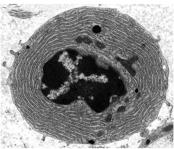
What can be concluded from the electron micrograph?
Options
A The cell secretes products that are toxic to pathogens.
B The cell synthesises a large quantity of proteins.
C The cell synthesises large quantities of antigens.
D The cell digests pathogenic bacteria.
Working
The micrograph shows a cell with a large nucleus whose heterochromatin is pushed to the periphery, giving the characteristic "clock-face" or "cartwheel" appearance, and a cytoplasm packed with parallel cisternae of rough endoplasmic reticulum (RER). This is the classic ultrastructure of a plasma cell — an activated B-lymphocyte that secretes antibodies. Antibodies are immunoglobulins, i.e. proteins, and they are synthesised and processed on the abundant RER visible throughout the cytoplasm.
- A — incorrect: plasma cells secrete antibodies, not toxins; antibodies neutralise pathogens by binding to their antigens, not by being directly toxic.
- B — correct: the extensive RER is the morphological signature of a cell producing large amounts of protein (antibodies/immunoglobulins).
- C — incorrect: plasma cells synthesise antibodies, not antigens; antigens are the foreign molecules that stimulate the immune response.
- D — incorrect: phagocytosis and digestion of bacteria is the role of phagocytes (neutrophils, macrophages), whose cytoplasm is dominated by lysosomes and vesicles, not by RER.
Answer
B
B
Background Concept
A plasma cell is a terminally differentiated B-lymphocyte whose sole function is to manufacture and secrete antibodies (immunoglobulins) at a very high rate. Because antibodies are proteins, the cell needs the cellular machinery for bulk protein synthesis, modification, and secretion:
- Rough endoplasmic reticulum (RER) — ribosome-studded membrane cisternae where proteins destined for secretion are synthesised, folded and undergo initial post-translational modification (e.g. disulphide bond formation, N-linked glycosylation).
- Golgi apparatus — receives proteins from the RER, completes glycosylation and packaging into secretory vesicles for exocytosis.
- A large euchromatic nucleus with prominent nucleolus — needed to support heavy transcription of immunoglobulin mRNA.
The hallmark ultrastructural features of a plasma cell, visible in the electron micrograph, are therefore:
- An extensive network of parallel RER cisternae filling almost the entire cytoplasm.
- A large nucleus with heterochromatin pushed to the periphery in a "clock-face" or "cartwheel" pattern.
- A well-developed Golgi apparatus (often appearing as a pale region near the nucleus).
Understanding the Question
This is a multiple-choice question that asks the candidate to interpret an electron micrograph of a blood cell and select the conclusion that the visible ultrastructure supports. The stem tells us only that the cell is a type of blood cell; the test-taker must identify which blood cell is shown and what its prominent organelle arrangement implies about its function.
The command word is essentially "conclude" — the candidate must match an organelle abundance to a function, not merely name the organelle.
Approach
- Recognise the cell from its ultrastructure: abundant RER + clock-face nucleus = plasma cell.
- Recall the function of a plasma cell: it secretes antibodies (immunoglobulins), which are proteins.
- Match the function to the option list, while actively rejecting the distractors by checking each against plasma cell biology.
Step-by-Step Reasoning
Step 1 — Identify the cell.
The micrograph shows a roughly circular cell dominated by stacked, parallel dark membranes studded with ribosomes (the RER). The nucleus is large, off-centre, and its densely staining heterochromatin is concentrated in clumps around the nuclear envelope, leaving a paler central region. There is no obvious phagosome or large lysosomal population. This combination is diagnostic of a plasma cell.
Step 2 — Link structure to function.
RER is the site of synthesis of secreted and membrane-bound proteins. The fact that RER occupies most of the cytoplasm tells us the cell is producing very large amounts of protein for export. In a plasma cell, that protein is antibody (immunoglobulin).
Step 3 — Evaluate the options.
- A — secretes products toxic to pathogens. Antibodies are not themselves directly toxic; they mark pathogens for destruction (opsonisation) or neutralise them by binding. Cells that secrete toxic products (e.g. defensins, perforin) have a different secretory apparatus and a different lineage (e.g. cytotoxic T-cells, NK cells, some epithelial cells).
- B — synthesises a large quantity of proteins. Directly supported by the visible abundance of RER, and consistent with antibody secretion. Correct.
- C — synthesises large quantities of antigens. A conceptual error: antigens are foreign molecules recognised by the immune system. Plasma cells make antibodies (which bind antigens), not antigens themselves. Even if the candidate momentarily confuses the two, the RER is not associated with antigen production — it is associated with protein secretion.
- D — digests pathogenic bacteria. That is the role of a phagocyte (neutrophil or macrophage), whose cytoplasm is filled with lysosomes, phagocytic vesicles, and often a multi-lobed nucleus — not the regular, parallel RER stacks seen here. The RER-dominant cytoplasm rules out a phagocyte.
Step 4 — Select B.
The conclusion that the micrograph supports is that the cell is a protein-secreting cell, namely a plasma cell producing antibodies.
Key Takeaways
- The ultrastructure of a cell (organelle abundance and arrangement) is a reliable clue to its function.
- An abundance of RER signals bulk synthesis of proteins for secretion.
- A plasma cell is identified by its clock-face nucleus and cytoplasm packed with RER; its function is to secrete antibodies (immunoglobulins).
- Distinguish carefully between antibodies (made by plasma cells) and antigens (foreign molecules that antibodies bind to).
- Phagocytes (which digest bacteria) are identified by their lysosomes and vesicles, not by RER.
Common Mistakes
- Confusing antibodies with antigens and selecting C — antigens are the foreign molecules that trigger the response; plasma cells produce the antibodies that target them.
- Selecting D because the cell "looks busy" — phagocytes have many lysosomes and irregular nuclei, not the regular parallel RER stacks characteristic of a plasma cell.
- Selecting A by assuming immune cells are generally "toxic to pathogens" — antibodies are not directly cytotoxic; they tag pathogens for destruction by other components of the immune system.
- Naming the organelle (RER) but not linking it to the function (protein secretion), then picking an option that mentions a function without the structural basis.
Things to Be Careful About
- Use precise CIE terminology: the cell is a plasma cell (effector B-lymphocyte), and the product is antibodies / immunoglobulins (proteins), not "antigens" or "toxins".
- Read each option as a full statement; some wrong options are wrong because of a single misused word (e.g. "antigens" instead of "antibodies").
- The presence of RER tells you about synthesis and secretion of proteins, not about phagocytosis, respiration, or any other unrelated activity — match the organelle to the function it actually performs.
Why is mitosis important in the immune response?
Options
A It allows B-lymphocytes to produce plasma cells.
B It allows B-lymphocytes to synthesise antibodies.
C It allows neutrophils to carry out phagocytosis.
D It allows T-lymphocytes to recognise foreign antigens.
Working
Mitosis is the production of genetically identical daughter cells by nuclear division, and is central to clonal expansion in the specific immune response.
- When a B-lymphocyte is activated by binding to its specific antigen (with help from a T-helper cell), it divides repeatedly by mitosis to form a clone of identical cells. These cloned cells differentiate into plasma cells, which secrete antibodies, and into memory cells.
- Option B is wrong: antibody synthesis is transcription and translation in plasma cells, not mitosis.
- Option C is wrong: phagocytosis by neutrophils is engulfment of pathogens; mitosis is not required for it.
- Option D is wrong: T-lymphocytes recognise antigens via specific T-cell receptors; recognition itself does not require mitosis.
Answer
A
A
Background Concept
The specific immune response relies on lymphocytes (B- and T-cells) that each carry a unique receptor for one specific antigen. When the body is first exposed to a pathogen, only a tiny number of lymphocytes have receptors that match its antigens. To mount an effective response, these selected lymphocytes must rapidly multiply so that there are enough effector cells to deal with the infection. This multiplication of selected lymphocytes is called clonal selection and clonal expansion, and it is achieved by mitosis — the nuclear division that produces two genetically identical daughter cells.
Once a B-lymphocyte has bound its matching antigen and received co-stimulation from a T-helper cell, it proliferates by mitosis. The resulting clone differentiates into two main cell types:
- Plasma cells — short-lived antibody-secreting factories that produce large quantities of antibody specific to the original antigen.
- Memory B-cells — long-lived cells that remain in the body and enable a faster, stronger secondary response on re-exposure to the same antigen.
Notice that mitosis produces the cells, while transcription and translation (protein synthesis) produce the antibodies inside the plasma cells. The two processes are sequential, not alternatives.
Understanding the Question
This is a multiple-choice question testing whether you can identify the role of mitosis specifically within the immune response. The command word is "Why", so you need to pick the option that correctly links mitosis to an immune function. The other options are plausible-sounding distractors that describe real immune events but are powered by different mechanisms (protein synthesis, phagocytosis, receptor binding).
Approach
For each option, ask: "Is this process driven by mitosis, or by something else?"
- Plasma cell production from activated B-lymphocytes = clonal expansion by mitosis ✓
- Antibody synthesis = transcription and translation in plasma cells ✗
- Phagocytosis = engulfment of pathogens, not cell division ✗
- Antigen recognition by T-cells = specific receptor–antigen binding ✗
Step-by-Step Reasoning
- Option A — B-lymphocytes produce plasma cells by mitosis: Correct. An activated B-lymphocyte divides repeatedly by mitosis to form a large clone; the clone's cells then differentiate into plasma cells (which secrete antibodies) and memory B-cells.
- Option B — B-lymphocytes synthesise antibodies: Incorrect. Plasma cells, not B-lymphocytes themselves, secrete antibodies. Antibody secretion is a result of transcription of the antibody gene and translation at ribosomes on the rough endoplasmic reticulum — i.e. protein synthesis, not mitosis.
- Option C — Neutrophils carry out phagocytosis: Incorrect. Phagocytosis involves the cell engulfing and digesting a pathogen using lysosomal enzymes. It does not require nuclear division; neutrophils are short-lived phagocytes that do not need to multiply to perform this function.
- Option D — T-lymphocytes recognise foreign antigens: Incorrect. Recognition is a binding event between the T-cell receptor (TCR) and an antigen presented on an MHC molecule. No cell division is needed for recognition to occur — it actually happens before the T-cell proliferates.
Key Takeaways
- Mitosis in the immune system powers clonal expansion: the rapid multiplication of the few lymphocytes whose receptors match the invading antigen.
- In B-cell responses, mitosis produces the clone from which plasma cells (antibody factories) and memory cells differentiate.
- Mitosis produces cells; transcription/translation produces the proteins (e.g. antibodies) those cells secrete. Don't confuse the two.
Common Mistakes
- Picking B because it mentions "B-lymphocytes" and "antibodies" — antibody production is protein synthesis, not mitosis; and it is plasma cells (descended from B-lymphocytes) that secrete antibodies, not the B-lymphocytes themselves.
- Picking C because phagocytosis is a well-known immune function — but phagocytosis is an engulfment process that does not depend on nuclear division.
- Picking D because T-lymphocytes do divide by mitosis after activation — but the recognition event is the receptor binding the antigen, and that happens before mitosis, not because of it.
Things to Be Careful About
- Read the option precisely: the question asks what mitosis allows, so the option must describe a process that genuinely requires cell division.
- Distinguish between the production of cells (mitosis) and the function of those cells (antibody secretion, phagocytosis, antigen recognition). Mitosis creates the workforce; the workforce then does the immune work.
What is fused with a B-lymphocyte to form a hybridoma cell in monoclonal antibody production?
Options
A antigen
B clone
C macrophage
D myeloma cell
Working
In the production of monoclonal antibodies, a B-lymphocyte (which makes a specific antibody) is fused with a myeloma cell (a cancerous plasma cell that can divide indefinitely). The resulting hybridoma cell combines the B-lymphocyte's antibody-producing ability with the myeloma cell's capacity for continuous division.
Answer
D
D
Background Concept
Monoclonal antibodies are identical antibodies produced by a single clone of B-lymphocytes, all specific to the same antigen. To produce them in useful quantities, scientists exploit a cell-fusion technique that creates a hybridoma — a hybrid cell with two desirable properties.
The two parent cells are:
- A B-lymphocyte that has been activated (e.g. by immunising a mouse with the antigen of interest). It produces the specific antibody but cannot divide indefinitely in culture.
- A myeloma cell — a cancerous plasma cell (a type of B-cell tumour). It divides continuously and indefinitely, but it does not produce the specific antibody the researcher wants.
When these two cells are fused, the hybridoma inherits antibody specificity from the B-lymphocyte and the ability to grow and divide without limit from the myeloma cell. A single hybridoma can then be cloned to give a large population of identical cells, all secreting the same monoclonal antibody.
Understanding the Question
The question asks for the name of the cell that is fused with a B-lymphocyte during the hybridoma step of monoclonal antibody production. This is a direct recall question from the Immunity topic, specifically the sub-topic covering the hybridoma method.
Approach
Recall the two-cell fusion step of the monoclonal antibody procedure: B-lymphocyte + myeloma cell → hybridoma. The cell other than the B-lymphocyte is the myeloma cell.
Step-by-Step Reasoning
- The command word is implicit (recall / identification). No working is required beyond stating the correct option.
- Eliminate the distractors:
- A. antigen — the antigen is what stimulates the B-lymphocyte to make the specific antibody; it is not a cell and is not fused with anything.
- B. clone — a clone is the resulting population of identical hybridoma cells or their secreted antibodies; it is the product, not a parent cell.
- C. macrophage — macrophages are phagocytic cells of innate immunity; they engulf pathogens and present antigens, but they are not part of the hybridoma fusion.
- D. myeloma cell — correct. The B-lymphocyte is fused with a myeloma (tumour) cell to form a hybridoma.
- The correct answer is therefore D.
Key Takeaways
- The hybridoma is made by fusing a B-lymphocyte with a myeloma cell.
- The B-lymphocyte contributes antibody specificity; the myeloma cell contributes immortality (continuous division).
- The resulting hybridoma is cloned to produce large quantities of a single (monoclonal) antibody.
Common Mistakes
- Confusing the role of the macrophage (a phagocyte that presents antigen) with the myeloma cell (a tumour cell used in the fusion).
- Selecting "antigen" — the antigen is the target, not one of the fused cells.
- Selecting "clone" — this is the downstream product, not a parent cell in the fusion step.
Things to Be Careful About
- "Myeloma" (a type of cancer of plasma cells) must not be confused with "mycelium" (fungal threads) or "myelin" (the fatty sheath around nerve axons). The spelling and the biological context matter.
Your score so far
Answer a question to start scoring
Your marks add up here as you work through the paper.