Biology 9700/22 — February/March 2024
Cambridge AS Level · AS Level Structured Questions · worked solutions for every part, with the mark scheme
Topics Cell Structure · Cell Membranes and Transport · Biological Molecules · Infectious Diseases · Enzymes · Transport in Mammals · +4 more
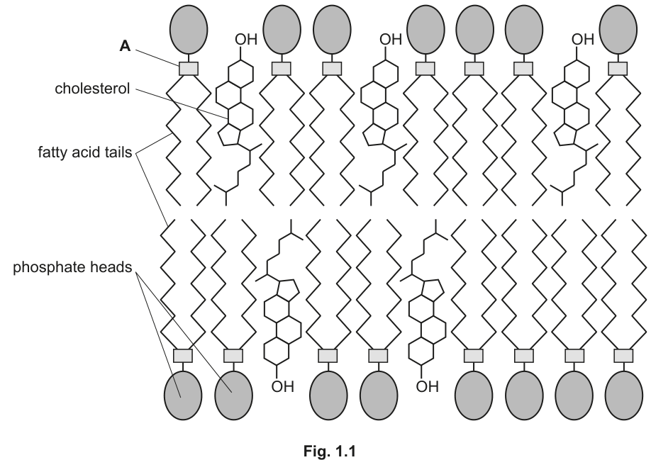
Fig. 1.1 is a diagram representing part of the phospholipid bilayer of a cell surface membrane.
Identify the part of a phospholipid molecule, labelled A in Fig. 1.1, that forms bonds with the phosphate heads and with the fatty acid tails.
Answer
Glycerol.
Glycerol
Background Concept
A phospholipid is built from one glycerol molecule ester-linked to two fatty acid tails (which form the hydrophobic, non-polar region) and to one phosphate group (which forms the hydrophilic, polar head). The glycerol is the three-carbon backbone that physically joins the two fatty acids to the phosphate head. In the bilayer, the heads face the aqueous environments on either side of the membrane while the fatty acid tails are sequestered in the hydrophobic core.
Understanding the Question
The question points to label A in Fig. 1.1, which sits between the phosphate head and the two fatty acid tails of a phospholipid. The candidate is asked to name this connecting region.
Approach
A quick structural identification: read the position of A in the diagram, recall the three components of a phospholipid, and pick the component that lies in that position.
Step-by-Step Reasoning
- A is drawn as the small square that connects the circular phosphate head above to the two zigzag fatty acid tails below.
- The only part of a phospholipid that bonds both to the phosphate head AND to two fatty acid tails is the three-carbon alcohol glycerol.
- Two of glycerol's –OH groups form ester bonds with the carboxyl groups of the two fatty acids; the third –OH bonds with the phosphate group.
Key Takeaways
- A phospholipid = glycerol + 2 fatty acids + 1 phosphate group.
- The glycerol backbone is the linker between the polar head and the non-polar tails.
Common Mistakes
- Writing "phosphate" or "fatty acid" — these are the parts that bond TO glycerol, not the part labelled A.
- Spelling "glycerol" as "glycerine" (a different compound) or "glyceral".
Things to Be Careful About
The mark scheme awards the precise term "glycerol"; the spelling matters.
Cholesterol is an important lipid component of many cell surface membranes. Fig. 1.2 shows the structure of a cholesterol molecule.
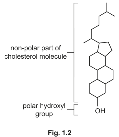
Using the information in Fig. 1.2, explain the orientation (positioning) of cholesterol molecules in the phospholipid bilayer, as shown in Fig. 1.1.
Answer
The polar –OH (hydroxyl) group of cholesterol is positioned next to the phosphate heads because it is hydrophilic / polar and can interact with them. The non-polar (hydrocarbon rings and tail) part lies among the fatty acid tails because both are hydrophobic / non-polar.
The polar –OH group aligns with the polar phosphate heads; the non-polar hydrocarbon region lies among the non-polar fatty acid tails.
Background Concept
Cholesterol is an amphipathic molecule: it has a small polar region (one –OH group) and a large non-polar region (four fused hydrocarbon rings plus a hydrocarbon tail). The fluid-mosaic bilayer is itself arranged so that polar groups face water and non-polar groups face each other in the centre. Amphipathic molecules always orient themselves so that "like meets like" — polar with polar, non-polar with non-polar.
Understanding the Question
Fig. 1.2 shows cholesterol's two parts explicitly. Fig. 1.1 shows how cholesterol sits in the bilayer. The candidate must explain WHY the molecule takes up the position shown, using the structure in Fig. 1.2.
Approach
Match the two parts of cholesterol to the two parts of the bilayer using polarity:
- polar –OH ↔ polar phosphate heads / aqueous environment
- non-polar rings/tail ↔ non-polar fatty acid tails / hydrophobic core
Step-by-Step Reasoning
- Cholesterol's –OH group is polar, so it is attracted to the polar phosphate heads and/or water. In Fig. 1.1 the –OH is drawn pointing towards the phosphate-head surface — this is the energetically favourable arrangement.
- The hydrocarbon ring system and the branched chain are non-polar (hydrophobic), so they associate with the hydrophobic fatty-acid tails in the middle of the bilayer.
- Because one end is polar and the rest is non-polar, cholesterol is held in a specific vertical orientation in the bilayer, exactly as shown in Fig. 1.1.
Key Takeaways
- Cholesterol is amphipathic; its position in the bilayer is dictated by the polarity of its two regions.
- Polar groups orient towards water; non-polar groups orient towards the hydrophobic core.
Common Mistakes
- Saying cholesterol is "trapped" or "held in place" without referring to the polar/non-polar match.
- Reversing the orientation in the answer (e.g. claiming –OH lies among the fatty acid tails).
Things to Be Careful About
The mark scheme accepts either an explicit "polar / non-polar" pairing or a "hydrophilic / hydrophobic" pairing, but BOTH ends of the molecule must be addressed for a complete explanation.
Answer
Any one from:
- Maintains / regulates the fluidity of the membrane (e.g. reduces fluidity at high temperatures and increases it at low temperatures).
- Provides (mechanical) stability to the membrane / prevents it from easily rupturing.
- Reduces the permeability of the membrane to hydrophilic / polar substances / ions.
- Reduces lateral movement of phospholipids.
One role is sufficient for the mark.
Maintains / regulates the fluidity of the membrane.
Background Concept
Cholesterol is wedged between phospholipid molecules and acts as a "membrane moderator". Its rigid steroid ring system restricts the movement of the fatty acid tails when the membrane would otherwise be too fluid (high temperature) and, by disrupting the regular packing of the tails, prevents the membrane from becoming too rigid at low temperatures. It also mechanically reinforces the bilayer.
Understanding the Question
A single-marker "state" question, so only one clear, accurate role is needed.
Approach
Pick the most commonly credited role and write it in the mark-scheme wording.
Step-by-Step Reasoning
- Most frequently rewarded answer: regulates membrane fluidity.
- Alternative correct answers: mechanical stability; reduces permeability to ions / polar molecules; restricts lateral movement of phospholipids; prevents membrane rupture.
- Any one of these, phrased concisely, scores the mark.
Key Takeaways
- Cholesterol is both a fluidity buffer and a stabiliser of the bilayer.
- A "state" command word requires only one point but it must be the precise term from the mark scheme.
Common Mistakes
- Giving a vague answer such as "it helps the membrane" — this is too imprecise to score.
- Writing "forms the bilayer" — cholesterol is a minor component; it does NOT form the bilayer.
Things to Be Careful About
"Fluidity" is the precise term; avoid the looser "keeps the membrane flexible" unless paired with a clear reference to fluidity regulation.
Answer
Sodium ions (Na⁺) are positively charged, so they are repelled by the hydrophobic / non-polar fatty acid tails in the centre of the bilayer and therefore cannot diffuse through.
Na⁺ ions are charged and are repelled by the hydrophobic fatty acid tails in the centre of the bilayer.
Background Concept
The interior of a phospholipid bilayer is hydrophobic because it is lined by the long hydrocarbon fatty acid tails. Simple diffusion across a membrane requires a substance to dissolve in this hydrophobic core. Polar molecules (water-soluble) and ions (charged) cannot dissolve in a non-polar environment and so cannot pass through by simple diffusion; they must be helped across by transport proteins.
Understanding the Question
The command word is "explain", so the candidate must give a reason, not just an observation.
Approach
State the property of the sodium ion (it is charged) and the property of the membrane interior (it is hydrophobic/non-polar), then link them with a verb of repulsion / inability to dissolve.
Step-by-Step Reasoning
- Na⁺ carries a positive charge.
- The bilayer core consists of hydrophobic (non-polar) fatty acid tails.
- Like charges and hydrophobic regions are mutually incompatible — the charged ion is repelled by / cannot dissolve in the non-polar core.
- Therefore Na⁺ cannot simply diffuse across.
Key Takeaways
- Ions and polar molecules are excluded from the hydrophobic core.
- "Repelled by the hydrophobic tails" is the mark-scheme wording — both ideas (charged AND hydrophobic tails) must be present.
Common Mistakes
- Saying only "Na⁺ is charged" with no reference to the membrane — incomplete.
- Saying "the membrane is solid" or "Na⁺ is too big" — both biologically wrong and not credited.
Things to Be Careful About
Acceptable alternatives to "hydrophobic" include "non-polar"; the mark scheme lists "hydrophobic tails / non-polar core / non-polar tails" as interchangeable.
Ions and some molecules move across cell surface membranes by facilitated diffusion and active transport.
Compare facilitated diffusion and active transport by stating one way in which they are similar and two ways in which facilitated diffusion is different from active transport.
similarity ______
difference 1 ______
difference 2 ______
Answer
Similarity
Both facilitated diffusion and active transport occur through transport (carrier / channel) proteins in the cell surface membrane. (Both can also be described as specific for the molecule/ion being transported.)
Difference 1 — direction relative to the gradient
- Facilitated diffusion: substances move down their concentration gradient (from high to low).
- Active transport: substances move against their concentration gradient (from low to high).
Difference 2 — energy requirement
- Facilitated diffusion: passive — does not require ATP / metabolic energy.
- Active transport: requires ATP / metabolic energy (from respiration).
(Equivalently: facilitated diffusion uses both channel and carrier proteins; active transport uses only carrier proteins.)
Answer
Similarity: both involve transport proteins in the membrane. Difference 1: facilitated diffusion is down the concentration gradient, active transport is against it. Difference 2: facilitated diffusion does not require ATP, active transport does.
Similarity: both use transport proteins; D1: facilitated diffusion is down the gradient (active transport is against); D2: facilitated diffusion is passive / does not use ATP (active transport requires ATP).
Background Concept
Both processes move substances across the cell surface membrane via transport proteins, but they differ in three important ways: the direction of movement relative to the concentration gradient, the source of energy used, and (in the case of channel proteins) the type of protein involved.
- Facilitated diffusion is passive: substances move down a concentration gradient through channel or carrier proteins; no ATP is needed.
- Active transport moves substances against the concentration gradient via carrier proteins and requires energy from ATP (often supplied by co-transport of ions or by direct ATP hydrolysis).
Understanding the Question
The question is a structured comparison requiring: one similarity (1 mark) and two differences (1 mark each). The candidate must phrase contrasts as point-by-point pairs, not as two separate descriptions.
Approach
- For the similarity, identify what is true of BOTH processes.
- For each difference, state the situation in facilitated diffusion AND the contrasting situation in active transport in the same sentence (or as a paired row of a table).
Step-by-Step Reasoning
Similarity — the mark-scheme-credited common feature is that both processes use transport proteins (channel and/or carrier) embedded in the membrane. Alternatives accepted by the mark scheme: both are specific to the molecule/ion; both can involve a conformational change in a carrier protein; both can transport substances into and out of the cell.
Difference 1 — direction of movement. The contrast is down the gradient (facilitated diffusion) versus against the gradient (active transport). The mark scheme phrases this as "down the concentration gradient" vs "against a concentration gradient".
Difference 2 — energy. The contrast is no ATP / passive (facilitated diffusion) versus ATP required / metabolic energy (active transport). The mark-scheme wording is "does not require ATP" vs "requires ATP".
(A third difference, also accepted: facilitated diffusion can use both channel and carrier proteins, while active transport only uses carrier proteins.)
Key Takeaways
- A good comparison states the shared feature first, then pairs the contrasts clearly.
- Phrase contrasts so facilitated diffusion is named first, then active transport — or present them as a two-column comparison.
Common Mistakes
- Saying facilitated diffusion "uses ATP" — wrong; it is passive.
- Confusing the direction of the gradient: "active transport goes down the gradient" is a common error.
- Writing similarities that are actually differences (e.g. "facilitated diffusion doesn't need ATP, active transport does" — this is a difference, not a similarity).
- Omitting the protein aspect of the similarity.
Things to Be Careful About
- One mark is for the similarity, one each for the two differences — three separate ideas, three separate sentences is safest.
- Avoid the term "facilitated diffusion is faster" — it is not a mark-scheme point and may not be credited.
Prostaglandins are small lipids produced in many tissues of the body. One role of prostaglandins is to cause inflammation at the site of an injury or infection. Inflammation is the normal first response of the immune system to injury or infection.
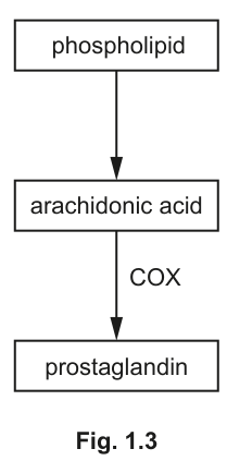
Cyclooxygenase (COX) is an enzyme that catalyses one of the steps in the reaction pathway for the formation of prostaglandins from phospholipids. The reaction pathway occurs in the smooth endoplasmic reticulum (SER) of cells. Part of the reaction pathway is shown in Fig. 1.3.
Suggest an advantage for this reaction pathway occurring in the smooth endoplasmic reticulum of a cell rather than in the cytoplasm.
Answer
Any one from:
- The SER is membrane-bound and so can supply / provide the phospholipid substrate for the pathway.
- The SER is involved in lipid synthesis / transport, so the product (prostaglandin) can be released into / transported from the SER.
- The pathway is compartmentalised (separated from other cytoplasmic reactions), providing optimum conditions / a higher concentration of enzymes / substrates for the pathway.
The SER is membrane-bound, providing phospholipid substrate and compartmentalising the pathway for optimum conditions / efficient transport of the lipid product.
Background Concept
The smooth endoplasmic reticulum (SER) is a network of membrane-bound flattened sacs (cisternae) and tubules continuous with the nuclear envelope. Unlike the rough ER, it lacks ribosomes. The SER is the main site of lipid and steroid synthesis (including phospholipids and cholesterol), and it also stores calcium ions and helps to detoxify drugs.
Because the SER is enclosed by a membrane, it separates the reactions inside from those in the surrounding cytoplasm. This compartmentalisation is a key feature of eukaryotic cells: different reactions can occur in different locations with their own optimal conditions and without interference from competing reactions.
Understanding the Question
The stem describes a pathway (phospholipid → arachidonic acid → prostaglandin) catalysed partly by COX, and states that the pathway occurs in the SER. The candidate is asked to suggest an advantage of this location, compared with the cytoplasm.
Approach
Two main families of argument are credited:
- Substrate/product supply — the SER is the site of phospholipid synthesis, so the starting material is right there, and being lipid-based the products can be transported through / stored in the SER membrane.
- Compartmentalisation — keeping the pathway in a separate compartment gives optimum conditions (pH, substrate concentration) and prevents the intermediates from interfering with other cytoplasmic reactions.
Step-by-Step Reasoning
- The first substrate of the pathway is a phospholipid, which is itself made in the SER; locating the pathway here means the substrate is readily available.
- Prostaglandins are lipids and so are soluble in membranes; they can be transported within or released from the SER easily.
- The enzymes (e.g. COX) of the pathway can be at high local concentration inside the SER, speeding up the reaction.
- Separating the pathway from the cytoplasm prevents unwanted side reactions and concentrates the substrates.
Any ONE of these points is sufficient for the single mark.
Key Takeaways
- Eukaryotic compartmentalisation allows specific reactions to occur in optimal conditions.
- The SER is a lipid-handling organelle — pathways involving lipids are commonly located there.
Common Mistakes
- Saying only "the SER has ribosomes" — the SER does NOT have ribosomes; that is the RER.
- Writing "for protection" without saying what is protected from what — too vague.
- Saying the SER "produces ATP" — that is mitochondria.
Things to Be Careful About
This is a "suggest" question — there is no single correct answer, but the answer must specifically link the structure of the SER (membrane-bound) to a feature of the prostaglandin pathway (lipid substrates/products).
Sometimes inflammation can have side-effects, such as pain. Aspirin is a drug that can be used to reduce these side-effects.
Aspirin reduces the catalytic activity of the COX enzyme by modifying the R-group of one of the amino acids.
Suggest how modifying the R-group of an amino acid in the COX enzyme can reduce the catalytic activity of the enzyme.
Answer
- The R-groups of amino acids interact via ionic bonds, hydrogen bonds and hydrophobic interactions, which contribute to the tertiary structure of the enzyme and the specific shape of the active site. Modifying an R-group disrupts some of these interactions, so the tertiary structure / shape of the active site changes.
- The active site is no longer complementary in shape to arachidonic acid (the substrate).
- The enzyme–substrate complex therefore cannot form (or forms much more slowly), so the catalytic activity is reduced.
(Equivalently: the modified R-group changes the charges / no longer provides the hydrophobic regions needed for the substrate to bind, so the activation energy is no longer lowered.)
The modified R-group disrupts the bonding that maintains the active site shape, so the active site is no longer complementary to the substrate and the enzyme–substrate complex cannot form.
Background Concept
The tertiary structure of an enzyme is held together by interactions between the R-groups of amino acids far apart in the chain: hydrogen bonds, ionic bonds, disulfide bridges and hydrophobic interactions. The precise 3-D shape of the active site depends on this tertiary structure. If the chemistry of an R-group is changed, the interactions it makes with neighbouring R-groups change, the tertiary structure distorts, and the active site loses its complementary shape. The substrate can then no longer bind (lock-and-key) or the active site can no longer mould around the substrate (induced fit). Without a productive enzyme–substrate complex, the enzyme cannot lower the activation energy and the reaction slows or stops.
Aspirin is a classic non-competitive / irreversible inhibitor of COX — it acetylates a serine residue inside the active site, blocking the channel leading to it. The question's wording is generic, however: "modifying the R-group of one of the amino acids".
Understanding the Question
The stem states that aspirin reduces the catalytic activity of COX by modifying an R-group. The candidate must explain the cascade of consequences from this single molecular change.
Approach
Step through the logic in the order in which the mark scheme rewards it:
- State which R-group interactions are affected.
- State the consequence for tertiary structure / active site shape.
- State the consequence for substrate binding (ES complex).
Step-by-Step Reasoning
- Amino-acid R-groups interact through hydrogen bonds, ionic bonds and hydrophobic interactions to maintain the 3-D shape of the protein and therefore the precise shape of the active site.
- Modifying an R-group changes its chemistry (e.g. adds a charged or bulky group). This disrupts the original interactions — for example, an ionic bond may break, or a new steric clash may appear.
- The tertiary structure of the enzyme changes, and with it the shape / conformation of the active site.
- The active site is no longer complementary in shape to arachidonic acid (the substrate for COX), so by the lock-and-key / induced-fit model the enzyme–substrate complex cannot form, or forms only at a much reduced rate.
- With no (or fewer) ES complexes, COX cannot lower the activation energy of the conversion of arachidonic acid to prostaglandin, so catalytic activity is reduced.
Key Takeaways
- Primary structure (R-group sequence) determines tertiary structure, which determines active-site shape, which determines substrate specificity.
- Changing a single R-group can therefore abolish enzyme activity.
- This is a non-competitive / irreversible form of inhibition: the substrate can still approach, but the binding site no longer fits.
Common Mistakes
- Saying "the enzyme is denatured" — denaturation usually refers to disruption by heat / pH across the whole protein; here a single R-group is changed, so "shape of the active site changes" is more accurate.
- Saying "the substrate is broken down" — aspirin is not a competitive substrate; it modifies the enzyme, not the substrate.
- Failing to link R-group change → tertiary structure → active-site shape → substrate binding; any break in this chain loses marks.
Things to Be Careful About
- The mark scheme requires any three of the listed ideas for the 3 marks; cover at least three.
- "Complementary" is the precise term to use when describing the active site and substrate relationship.
- "Enzyme–substrate complex" should be written in full (or ES complex) — do not just say "they don't bind".
Prostaglandins are examples of cell-signalling molecules.
Outline the process of cell signalling that leads to a response by the cells involved in inflammation.
Answer
- Prostaglandins are secreted / released by cells at the site of injury (or infection) and transported to target cells involved in the inflammation response.
- The prostaglandins bind to specific receptors on the target cell surface membranes.
- This binding triggers a response inside the cell, e.g. activation of a second messenger, an enzyme cascade / phosphorylation events (signal transduction), leading to the inflammation response (e.g. vasodilation, increased permeability of capillaries, recruitment of immune cells).
Prostaglandins are released by cells, travel to target cells and bind to receptors on the cell surface membrane, triggering a response inside the cell (e.g. second-messenger activation) that produces the inflammation response.
Background Concept
Cell signalling is the process by which cells communicate. A typical sequence is:
- A signalling cell secretes a signalling molecule (a ligand, e.g. a hormone, neurotransmitter or local mediator such as a prostaglandin).
- The ligand travels (by diffusion through tissue fluid, or in the blood) to a target cell.
- The ligand binds to a specific receptor on the target cell's surface membrane (or, for steroid-like lipids, inside the cell).
- Receptor binding triggers a response inside the cell — often via a second messenger (e.g. cyclic AMP) and an enzyme cascade of phosphorylation events, which amplify the signal and produce the cell's response.
Prostaglandins are local mediators (paracrine signals) — they act on neighbouring cells rather than being carried long distances in the blood.
Understanding the Question
The stem reminds the candidate that prostaglandins are cell-signalling molecules that lead to inflammation. The candidate must outline (give the main steps of) the signalling process.
The mark scheme rewards any two of: release/transport, receptor binding, and a valid example of the triggered response.
Approach
Write a tight, ordered outline covering: source and travel of the signal, binding to a receptor, and the resulting cellular response. Two well-chosen points are enough for 2 marks; covering all three makes the answer bullet-proof.
Step-by-Step Reasoning
- Step 1 — release: Prostaglandins are secreted (released) by the cells that produce them. Because they are lipids they can simply diffuse out through the cell surface membrane and travel through the tissue fluid to neighbouring target cells (paracrine signalling).
- Step 2 — receptor binding: The prostaglandin binds to a specific receptor protein on the surface membrane of the target cell involved in inflammation. The receptor is specific — only cells carrying the appropriate receptor respond.
- Step 3 — response: Receptor binding activates a signal-transduction pathway inside the cell — typically a second messenger (e.g. cAMP, Ca²⁺), an enzyme cascade and phosphorylation events. This amplifies the signal and produces the inflammation response: vasodilation, increased capillary permeability, redness, swelling, pain, recruitment of immune cells.
The mark scheme accepts any valid example at step 3 (second messenger, enzyme cascade, phosphorylation cascade or signal transduction).
Key Takeaways
- Cell signalling = ligand release → travel → receptor binding → cellular response.
- Prostaglandins are local (paracrine) lipid mediators, not classical hormones.
- A "response" in cell signalling is usually a change in cell behaviour triggered by an intracellular cascade.
Common Mistakes
- Writing "the prostaglandin enters the cell and changes the DNA" — prostaglandins bind to surface receptors, not intracellular ones (unlike steroid hormones).
- Saying "the prostaglandin is an antigen" — antigens are recognised by lymphocytes in immunity; this is a different process.
- Omitting the receptor step — the mark scheme specifically rewards "binds to receptors".
Things to Be Careful About
- The mark scheme rejects "antigens" — do not confuse cell signalling with immune recognition.
- A concrete example of the triggered response (second messenger, enzyme cascade, phosphorylation, signal transduction) is rewarded; a vague "the cell responds" is not.
Table 2.1 shows descriptions of three types of white blood cell.
Complete Table 2.1 by stating the names of these three types of white blood cell.
Table 2.1
| description | name of white blood cell |
|---|---|
| A large cell that has a bean-shaped (kidney-shaped) nucleus. It can develop into a macrophage. | |
| A cell that has a large spherical nucleus and little cytoplasm. It responds to non-self antigens. | |
| A cell that has a lobed nucleus. It is phagocytic. |
Answer
| description | name of white blood cell |
|---|---|
| A large cell that has a bean-shaped (kidney-shaped) nucleus. It can develop into a macrophage. | monocyte |
| A cell that has a large spherical nucleus and little cytoplasm. It responds to non-self antigens. | lymphocyte |
| A cell that has a lobed nucleus. It is phagocytic. | neutrophil |
monocyte; lymphocyte; neutrophil
Background Concept
Mammalian blood contains several cell types suspended in plasma. Red blood cells (erythrocytes) carry oxygen; white blood cells (leucocytes) defend the body. There are five principal types of leucocyte, but AS candidates need to recognise the three most commonly asked about: neutrophils, lymphocytes and monocytes. (Eosinophils and basophils are usually only mentioned in passing.)
Neutrophils are the most abundant white blood cell. They are small cells with a distinctive multi-lobed nucleus (typically three to five lobes joined by thin strands) and granular cytoplasm. They are highly phagocytic, engulfing and digesting bacteria at sites of infection. They are often the first responders to bacterial invasion.
Lymphocytes are the second most numerous white blood cell. They have a large, round, densely-staining nucleus that occupies most of the cell, leaving only a thin rim of cytoplasm. Lymphocytes are central to the specific immune response: they recognise non-self antigens and either (B-lymphocytes) produce antibodies or (T-lymphocytes) destroy infected cells directly. The mark scheme here insists on the umbrella term 'lymphocyte' — naming B- or T-lymphocytes specifically is ignored, not credited.
Monocytes are the largest of the white blood cells. They have a characteristic kidney- or bean-shaped nucleus (not lobed) and abundant cytoplasm. They circulate in the blood for only a few days before migrating into tissues, where they mature into macrophages — larger, long-lived phagocytes that form part of the monocyte-macrophage system.
Understanding the Question
The question is a recall task: each row of Table 2.1 gives distinctive structural AND functional clues, and the candidate must name the cell. The features are deliberately diagnostic:
- 'Bean-shaped nucleus' + 'develops into a macrophage' → monocyte.
- 'Large spherical nucleus and little cytoplasm' + 'responds to non-self antigens' → lymphocyte.
- 'Lobed nucleus' + 'phagocytic' → neutrophil.
The candidate does not need to use any calculation or analysis, just accurate terminology.
Approach
Read each row carefully, match the description to the three candidate cells, and write the name. The function usually clinches the answer when the nucleus description is ambiguous.
Step-by-Step Reasoning
- The first row describes a cell with a 'bean-shaped' nucleus that can become a macrophage. This is the definition of a monocyte — the precursor cell of the macrophage lineage.
- The second row has a 'large spherical nucleus and little cytoplasm' and 'responds to non-self antigens'. The high nucleus-to-cytoplasm ratio is the giveaway for a lymphocyte. The function confirms it: lymphocyte activation is the central event in adaptive immunity.
- The third row has a 'lobed nucleus' and is 'phagocytic'. The lobed (multi-lobed) nucleus is unique to the neutrophil among the three cells. Phagocytosis is its principal function.
Key Takeaways
- Neutrophils = multi-lobed nucleus + phagocytosis.
- Lymphocytes = round nucleus, thin cytoplasm, antigen recognition.
- Monocytes = kidney-shaped nucleus, macrophage precursor.
Common Mistakes
- Naming 'B-cell' or 'T-cell' instead of 'lymphocyte' — the mark scheme explicitly ignores these as too narrow.
- Confusing monocytes with neutrophils because both are phagocytic. The nuclear shape is the disambiguator: kidney-shaped vs lobed.
- Spelling 'neutrophil' as 'neutraphil' or 'neutraphyll'.
- Writing 'macrophage' for the first row — the question asks for the blood cell that develops into a macrophage, which is the monocyte.
Things to Be Careful About
- The mark scheme accepts only the three terms monocyte, lymphocyte and neutrophil. Subtypes (B-lymphocyte, T-lymphocyte) and incorrect plurals will be ignored.
- Each cell is identified by both a structural feature (nucleus) and a functional one (what it does). A correct name requires matching the whole description, not just one clue.
Dromedary camels are classified in the family Camelidae and live in desert habitats of North Africa and Asia. In these hot, dry environments, dromedary camels can lose up to of their body mass from dehydration, causing their blood to become more viscous (thicker).
Fig. 2.1 shows a drawing of red blood cells of a dromedary camel. Fig. 2.2 is a drawing of human red blood cells.
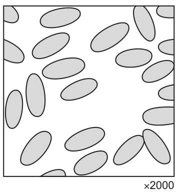
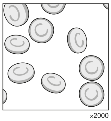
Fig. 2.1 and Fig. 2.2 show differences between the red blood cells of dromedary camels and the red blood cells of humans.
Suggest how these differences adapt dromedary camels for living in hot, dry environments.
Answer
- The elliptical (oval) shape of camel red blood cells (compared to the circular biconcave-disc shape of human red blood cells) allows easier / quicker flow / movement of the blood (compared to human red blood cells) ;
- Camel red blood cells are smaller than human red blood cells ;
- There is a larger number of camel red blood cells per unit volume ;
Together, these features allow the camel to maintain blood flow when dehydrated, since the blood becomes more viscous (thicker).
Elliptical shape and small size of camel red blood cells allow easier flow of more viscous (thicker) blood; more numerous smaller cells per unit volume aid this.
Background Concept
Red blood cells (erythrocytes) are highly specialised for oxygen transport. In most mammals they are circular, biconcave discs about across, lacking a nucleus and most organelles to maximise internal space for haemoglobin. The biconcave shape gives a large surface-area-to-volume ratio, a short diffusion distance and good flexibility for squeezing through capillaries.
Most mammals share this basic design, but there are interesting exceptions. Camelids (camels, llamas, alpacas, vicuñas, guanacos) have elliptical rather than circular red blood cells. This is unusual among mammals and is thought to be an adaptation related to dehydration and the need to tolerate high blood viscosity.
When an animal becomes dehydrated, the volume of water in the blood plasma falls. With the same number of cells in a smaller volume of plasma, the blood becomes more viscous, and the heart must work harder to pump it. The fluid dynamics of flow through narrow vessels mean that viscosity matters most in the microcirculation, where resistance is high.
Understanding the Question
The question provides two drawings (Fig. 2.1 of camel red blood cells and Fig. 2.2 of human red blood cells) at the same magnification () so that the visible differences are real, not artefacts of scale. The candidate must identify the differences from the drawings and then suggest how these differences help a camel in a hot, dry environment where it can become severely dehydrated and its blood becomes more viscous.
The command word is 'suggest' — the candidate is being asked to apply biological reasoning rather than recall a fact. Mark schemes for 'suggest' questions credit logical inferences from the given information, even if not the textbook answer.
Approach
First identify the visible differences between Fig. 2.1 and Fig. 2.2:
- Shape: camel cells are oval/elliptical, human cells are circular (biconcave disc).
- Size: at the same magnification, the camel cells appear smaller.
- Number: the camel cells appear more numerous per unit area.
Then link each difference to the selective pressure of dehydration:
- Smaller, more numerous cells in a viscous fluid flow more easily.
- Elliptical cells are more flexible and align better with flow.
The unifying selective pressure is viscous (thick) blood due to dehydration.
Step-by-Step Reasoning
-
Shape advantage: The elliptical shape of camel red blood cells (versus the biconcave disc of human red blood cells) allows the cells to deform and align with the direction of flow, particularly in narrow capillaries. In viscous blood this becomes critical because thicker fluid resists movement more strongly.
-
Size advantage: Smaller cells experience a more favourable surface-area-to-volume ratio and generate less frictional drag as they move. They also pass more easily through narrow capillaries. Together these factors reduce resistance to flow when blood is thick.
-
Number advantage: If more cells are packed into the same volume of blood, the loss of plasma water (which increases viscosity) is partially offset because the cells themselves are smaller. The relative proportion of cells to fluid remains favourable, and the smaller individual cells flow more easily than fewer large cells would.
-
The combined effect: When the camel is severely dehydrated (up to body mass loss), its blood becomes very viscous. The smaller, more numerous, elliptical cells minimise the increase in resistance to flow, so the heart can continue to circulate blood effectively without an unsustainable rise in blood pressure.
Key Takeaways
- Relating structure to function is a core skill: the unusual elliptical shape of camelid red blood cells is functionally significant.
- An unusual observation (like a different cell shape) often indicates an adaptation to a specific environmental pressure.
- The selective pressure in this question is dehydration-induced high blood viscosity.
- The mark scheme requires the link between structure and function — structure alone, or function alone, does not score full marks.
Common Mistakes
- Simply restating that 'camels store water' or 'camels can survive dehydration' — this does not address the red blood cell differences shown in the figures.
- Describing the camel red blood cells as 'biconcave' — the figures show they are NOT biconcave discs; they are elliptical/oval. The mark scheme explicitly contrasts 'lozenge/torpedo/elliptical' (camel) with 'biconcave disc' (human).
- Failing to link the cell features to blood flow / viscosity — a mark scheme point requires the link.
- Confusing red blood cells with white blood cells in the description.
- Mentioning only one structural difference when the question says 'these differences' (plural).
Things to Be Careful About
- The candidate must link structural difference to functional advantage, otherwise marks are lost.
- 'Suggest' questions allow some flexibility in wording, but the key idea of easier/quicker blood flow must appear.
- The question says 'these differences' (plural) — at least two structural differences are needed, ideally with the flow advantage stated separately as a unifying point.
- The first mark scheme point is the FUNCTIONAL advantage (easier flow); the next two are STRUCTURAL reasons (shape, size/number).
The llama is also classified in the family Camelidae. Llamas live in mountainous areas of South America, often at altitudes of or higher. As the altitude above sea level increases, the air pressure decreases.
The partial pressure of oxygen in the lungs of mammals at is .
Fig. 2.3 shows the oxygen dissociation curve of adult human haemoglobin and adult llama haemoglobin.
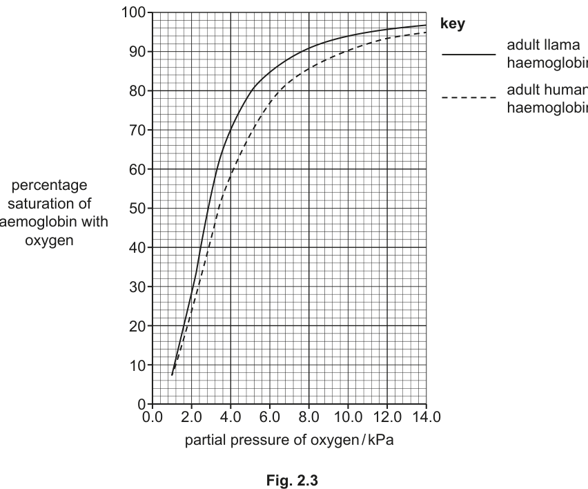
With reference to Fig. 2.3, explain how the differences between the oxygen dissociation curves for humans and llamas show that llamas are better adapted for living at high altitudes than humans.
Answer
- At the partial pressure of oxygen in the lungs at (), llama haemoglobin is more highly saturated with oxygen than human haemoglobin ;
- At , the percentage saturation is approximately for the llama and approximately for the human (a difference of ) ;
- Llama haemoglobin has a higher affinity for oxygen than human haemoglobin (the llama curve lies to the left of the human curve) ;
- Therefore more oxygen is loaded in the lungs and transported to / delivered to the tissues, where sufficient oxygen is unloaded to meet the metabolic demand despite the low atmospheric at high altitude.
Llama haemoglobin has a higher affinity for oxygen than human haemoglobin, so at the low pO2 in the lungs at 3500 m the llama haemoglobin is more highly saturated (≈86% vs ≈79-80% for human), allowing more oxygen to be delivered to the tissues.
Background Concept
The oxygen dissociation curve plots the percentage saturation of haemoglobin with oxygen against the partial pressure of oxygen (). The curve is sigmoidal because haemoglobin is a tetramer with four binding sites that show cooperativity: once one oxygen molecule binds, the others bind more easily.
Key regions of the curve:
- The flat upper plateau (above about ) corresponds to alveolar conditions in healthy lungs at sea level, where haemoglobin is ~ saturated.
- The steep middle portion (around ) corresponds to tissue conditions, where small changes in lead to large changes in saturation — ideal for oxygen unloading.
The position of the curve reflects the affinity of haemoglobin for oxygen:
- Curve to the LEFT = HIGHER affinity (binds oxygen more readily at lower ).
- Curve to the RIGHT = LOWER affinity (releases oxygen more readily at tissues).
At high altitude the atmospheric pressure falls, so the in the alveoli is lower than at sea level. This means that at a sea-level of about a mammal's haemoglobin is near saturation, but at the alveolar is only , which on the human curve corresponds to a saturation of roughly rather than at sea level. Animals native to high altitudes have evolved haemoglobin with a left-shifted curve to compensate.
Understanding the Question
The question provides a graph (Fig. 2.3) with two oxygen dissociation curves: adult llama and adult human. The llama curve is to the LEFT of the human curve, meaning llama haemoglobin has a higher affinity for oxygen. The candidate is told the partial pressure in the lungs at is .
The command word is 'explain' with the requirement to 'refer to Fig. 2.3'. This means the answer must quote specific features of the graph — curve position, saturation values, or partial pressures — and use them to support the conclusion.
Approach
- Read the two curves at the relevant ().
- Compare the saturation values.
- State the direction of the difference (which curve is to the left, which haemoglobin has higher affinity).
- Link this to oxygen loading in the lungs and delivery to tissues.
The mark scheme is 'any three from' five or six possible points — the candidate does not need all of them, but does need at least three well-supported points.
Step-by-Step Reasoning
-
Curve position: The llama curve lies to the LEFT of the human curve across the entire range. This means that at any given , llama haemoglobin is more highly saturated with oxygen than human haemoglobin.
-
Reading at 6.4 kPa: At a of (the alveolar partial pressure at ), the llama haemoglobin is approximately saturated, whereas the human haemoglobin is approximately saturated. That is a difference of about more oxygen carried by llama haemoglobin per unit volume of blood.
-
Affinity: Because the curve is to the left, llama haemoglobin has a higher affinity for oxygen — it picks up oxygen more readily at low . This is exactly what is needed at high altitude where the available oxygen is reduced.
-
Loading in lungs: The higher affinity means that even when the alveolar is reduced to , llama haemoglobin still loads a high percentage of its capacity with oxygen. A human at the same altitude would be at a much lower saturation.
-
Delivery to tissues: Despite the higher affinity, the curve is still sigmoidal, so the llama's tissues (with a tissue of about or less) still receive a substantial oxygen delivery. The llama gains at the loading end without losing too much at the unloading end.
Key Takeaways
- Left-shifted curve = higher affinity = better loading at low .
- High-altitude animals have evolved haemoglobin with a left-shifted dissociation curve.
- Read specific values from the graph to support the explanation — generic statements are not enough.
- The shape of the curve still permits unloading at tissues because the curve remains sigmoidal.
- The 'explanation' is about oxygen LOADING in the lungs; the mark scheme specifically ignores comments about higher saturation in tissues (because that is not where the advantage lies).
Common Mistakes
- Saying the llama curve is 'higher' rather than 'to the left' — the curve is shifted, not simply elevated.
- Confusing affinity with carrying capacity — they are different concepts.
- Citing values from the wrong curve (e.g. quoting human saturation when describing the llama).
- Stating that the llama haemoglobin carries 'more oxygen' without specifying per unit volume or per molecule of haemoglobin.
- Referring to tissue unloading advantages rather than lung loading — the mark scheme explicitly ignores references to higher saturation at tissue partial pressures.
- Failing to mention affinity at all — the mark scheme treats this as a separate point worth crediting.
Things to Be Careful About
- Always read the x-axis in the units shown () and the y-axis in percentage saturation.
- The mark scheme accepts either quoting specific values OR stating the relative positions of the curves — both approaches are valid.
- 'Explain' requires more than describing the graph: it requires linking the observation to the adaptive advantage.
- The numbers (, , ) are read from the graph; the mark scheme accepts values within the reading tolerance of the candidate.
Sketch a curve on Fig. 2.3 to show the effect of an increased carbon dioxide concentration on the percentage saturation of adult human haemoglobin with oxygen.
Answer
Sketch a sigmoidal curve to the RIGHT of the adult human haemoglobin curve on Fig. 2.3. The new curve should start near the origin, rise steeply in the middle and plateau near saturation at the top right; it is shifted horizontally (along the axis) so that at any given the percentage saturation is lower than for the human curve.
A sigmoidal curve drawn to the right of the human curve, showing the Bohr shift.
Background Concept
The Bohr effect (or Bohr shift) describes the rightward shift of the oxygen dissociation curve that occurs when the partial pressure of carbon dioxide rises (and/or pH falls). The molecular basis is that reacts with water inside red blood cells to form carbonic acid (catalysed by carbonic anhydrase), which dissociates to give and . The ions bind to haemoglobin and stabilise the deoxygenated (T) state, decreasing its affinity for oxygen. A small fraction of also binds directly to the N-terminal amino groups of globin chains, forming carbamino compounds, with a similar effect.
A rightward shift means that at any given , haemoglobin is LESS saturated with oxygen — so it unloads oxygen more readily to the tissues. This is biologically useful because metabolically active tissues produce , so the Bohr shift delivers more oxygen precisely where it is needed most.
The shift is small in absolute terms but physiologically very significant.
Understanding the Question
The question asks the candidate to draw a new curve on Fig. 2.3 showing the effect of increased on the percentage saturation of adult HUMAN haemoglobin. The original human curve (dashed line) is already on the graph, and the candidate must add a second sigmoidal curve shifted to the right of it.
The command word is 'sketch' — a quick, clear drawing that captures the essential feature (rightward shift) is all that is required. The mark scheme allocates 1 mark, which is given for the curve being to the right of the human curve.
Approach
- Identify the position of the existing human curve (dashed line).
- Sketch a new sigmoidal curve parallel to it, but shifted to the right.
- The new curve should still plateau at high and pass through the lower-left region near the origin.
- Do not shift the curve up or down — it is a horizontal shift only.
Step-by-Step Reasoning
- The Bohr effect shifts the dissociation curve to the right when rises (or pH falls). This is the established convention in physiology.
- A rightward shift means the curve is moved along the x-axis (the axis) so that it lies at HIGHER values than the original at any given saturation.
- The curve retains its general sigmoidal shape — start near the origin, rise steeply in the middle, and plateau at the top right.
- For the human curve specifically, the new curve should pass to the right of the dashed line at every point.
Key Takeaways
- Bohr shift = rightward shift of the dissociation curve.
- Increased / decreased pH → decreased affinity → rightward shift.
- A 'sketch' should capture the essential feature (the shift), not be a precise curve.
- The shift is horizontal (along the axis), not vertical.
Common Mistakes
- Drawing the curve to the LEFT (wrong direction).
- Drawing the curve ABOVE the human curve (a vertical shift, not a horizontal one) — the Bohr shift is a horizontal shift along the axis.
- Changing the shape of the curve (e.g. making it shallower or steeper) — only the position should change.
- Forgetting that the question specifies ADULT HUMAN haemoglobin, not llama — the new curve is added to the right of the human curve, not the llama curve.
Things to Be Careful About
- The mark is awarded solely for correct position. A curve that is sigmoidal, starts near the origin and plateaus at the top right, drawn clearly to the right of the human dashed line, scores the mark.
- Do not let the new curve cross the human curve — the entire new curve should be to the right of the human one at every shared value.
- A neat, smooth freehand curve is fine; ruler-drawn straight lines are not appropriate.
Explain the importance of the Bohr shift in metabolically active organs, such as the liver.
Answer
- Metabolically active organs (such as the liver) have a high rate of aerobic respiration, so they release more than less active tissues (the partial pressure of is higher) ;
- The increased (and the resulting fall in pH) decreases the affinity of haemoglobin for oxygen (the Bohr shift) ;
- As a result, more oxygen is released from haemoglobin into the tissue ;
- This extra oxygen supports the higher rate of aerobic respiration, which produces more ATP to meet the metabolic demand of the active organ.
Active tissues produce more CO2, which decreases haemoglobin's affinity for oxygen (Bohr shift), releasing more O2 to support the high rate of aerobic respiration and ATP production.
Background Concept
The Bohr effect (or Bohr shift) describes how the oxygen-carrying behaviour of haemoglobin responds to its chemical environment. Three factors shift the oxygen dissociation curve to the right (lower affinity, easier unloading):
- Increased (partial pressure of carbon dioxide).
- Decreased pH (increased concentration).
- Increased temperature.
All three are characteristic of METABOLICALLY ACTIVE tissues:
- Aerobic respiration produces as a waste product.
- reacts with water inside red blood cells (catalysed by carbonic anhydrase) to form carbonic acid, which dissociates to and .
- The ions bind to haemoglobin and stabilise its deoxygenated (T) state.
- A small fraction of also binds directly to haemoglobin to form carbaminohaemoglobin.
- Respiration releases energy as heat, so active tissues are slightly warmer.
The net result is that where the oxygen demand is highest, the curve is shifted right, so haemoglobin releases more oxygen — exactly the right amount, exactly where it is needed.
Metabolically active organs in this context include the liver (which carries out many energy-demanding biosyntheses and detoxification reactions), exercising skeletal muscle, the brain, the heart, and the kidneys.
The relevant chemical equation inside the red blood cell is:
The ions are the immediate cause of the decreased oxygen affinity.
Understanding the Question
The question asks the candidate to explain the importance of the Bohr shift IN METABOLICALLY ACTIVE ORGANS, using the liver as the example. The command word is 'explain' — the candidate must state the mechanism (what happens) and link it to the consequence (why it matters).
The mark scheme gives 'any three from' four possible points. The candidate needs to mention the cause (more ), the mechanism (decreased affinity), the consequence (more released), and the biological purpose (more aerobic respiration / ATP). Three of these four are sufficient for full marks.
Approach
Walk through the causal chain in the correct physiological order:
- Active organ → high respiration rate → high output.
- High → low pH (or direct effect on haemoglobin).
- Low pH / high → decreased affinity of haemoglobin for (Bohr shift).
- Decreased affinity → more released from haemoglobin to the tissue.
- More → more aerobic respiration → more ATP → meets the high metabolic demand of the organ.
The mark scheme rewards each logical step; the candidate should aim for three or four of them.
Step-by-Step Reasoning
-
Cause: A metabolically active organ such as the liver carries out many energy-demanding processes (biosynthesis, detoxification, etc.) at a high rate. This requires a high rate of aerobic respiration in its cells, which produces a correspondingly high output of . The local in the tissue is therefore higher than in less active tissues.
-
Mechanism: The increased lowers the local pH (because of the carbonic anhydrase-catalysed reaction). The lower pH (or the itself) decreases the affinity of haemoglobin for oxygen — the Bohr shift. The mark scheme specifically allows reference to 'haemoglobinic acid' (the protonated form of haemoglobin) as the mechanistic link.
-
Consequence: As a result of the lower affinity, more oxygen is released from haemoglobin to the tissue. The dissociation curve effectively shifts to the right at the tissue level.
-
Purpose: The extra oxygen supports the high rate of aerobic respiration, which produces more ATP to meet the metabolic demand of the active organ. Without the Bohr shift, the oxygen supply would be insufficient for the high demand.
Key Takeaways
- The Bohr shift is a NEGATIVE FEEDBACK mechanism: high demand → high → more released → demand met.
- The liver, heart, brain, kidneys and exercising muscle are all metabolically active and benefit from the Bohr shift.
- The mechanism involves , water, carbonic anhydrase, and haemoglobin — knowing the chemistry is not required at AS, but the link between and release is.
- The Bohr shift is a LOCAL effect: the blood in capillaries supplying an active tissue experiences the shift; blood elsewhere does not.
- The mark scheme requires the comparative 'more' to be implicit or explicit in the answer.
Common Mistakes
- Stating that the Bohr shift 'increases the affinity of haemoglobin for oxygen' — the shift DECREASES affinity.
- Reversing cause and effect: saying 'more causes more ' rather than the correct 'more causes more to be released'.
- Confusing the Bohr shift with the effect of high altitude (which is a different curve shift mechanism involving different haemoglobin).
- Failing to mention the CONSEQUENCE for respiration/ATP — the mark scheme explicitly looks for 'more aerobic respiration' or 'more ATP production'.
- Treating the Bohr shift as something that 'helps the blood carry more oxygen' — it actually helps RELEASE more oxygen to the tissues.
- Stating only that the organ 'produces ' without indicating the comparison with less active tissues.
Things to Be Careful About
- The mark scheme requires 'more' (a comparative) — simply saying the organ 'produces ' without saying 'more' is not enough; the comparison with less active tissues must be implicit or explicit.
- The cause is increased / decreased pH; the consequence is more released. Do not reverse the order.
- 'Explain' questions at AS need the WHY behind each point, not just a list of facts.
- The Bohr shift should be described as decreasing affinity, not as some other change.
Fig. 3.1 is a photomicrograph showing part of a transverse section through the root of an iris, Iris germanica. Irises are herbaceous monocotyledons. These plants have the same transport tissues as herbaceous dicotyledons, but the transport tissues are distributed differently. In monocotyledons, the central tissue in the root is parenchyma (packing tissue).
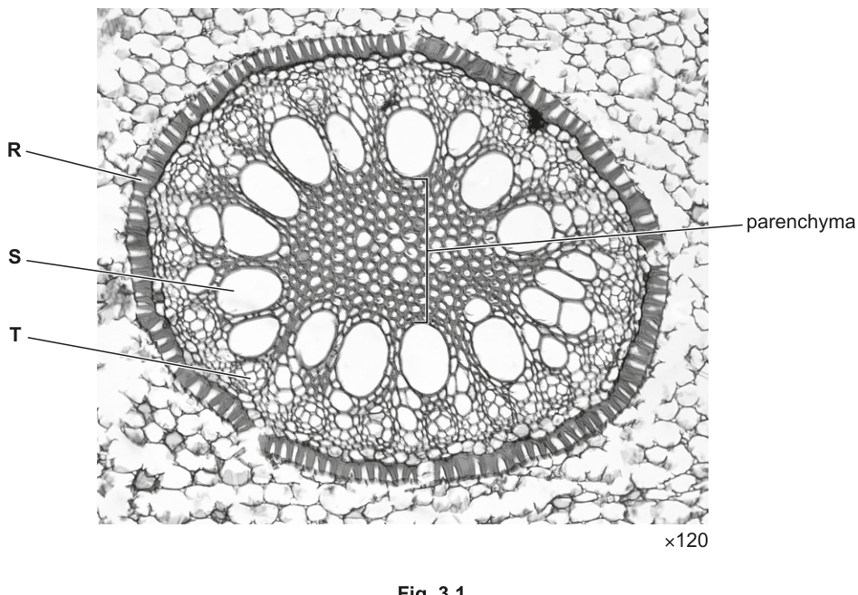
Cells R, S and T in Fig. 3.1 are found in different tissues.
Name the tissues in which the cells labelled R, S and T are found.
tissue in which cell R is found ______
tissue in which cell S is found ______
tissue in which cell T is found ______
Answer
tissue in which cell R is found: endodermis
tissue in which cell S is found: xylem
tissue in which cell T is found: phloem
R: endodermis; S: xylem; T: phloem
Background Concept
The transverse section of a young root reveals the tissues arranged from the outside in: epidermis → cortex → endodermis → pericycle → vascular tissue (xylem and phloem) → central pith (parenchyma in monocots, xylem in dicots).
- Endodermis is a single ring of tightly packed cells around the vascular cylinder. The radial and transverse walls are impregnated with suberin, forming the Casparian strip, and the cells appear as a continuous dark ring in section.
- Xylem conducts water and mineral ions from root to shoot. Xylem vessel elements are dead, hollow tubes with lignified walls; in cross-section they appear as large, open, thick-walled cells.
- Phloem translocates organic solutes (mainly sucrose). Sieve tube elements are living, lack a nucleus, and are connected end-to-end; in cross-section they appear as small cells with thin walls, often accompanied by companion cells.
In a monocot root the xylem forms a ring of separate vessels around a central parenchyma pith, with phloem strands lying between the xylem vessels.
Understanding the Question
Fig. 3.1 is a transverse section through the root of an iris (Iris germanica), a herbaceous monocotyledon, at magnification. The stem tells the candidate that the central tissue is parenchyma and that R, S and T are cells in three different tissues. They must name each tissue from its visible appearance in the labelled micrograph.
Approach
Read off where each labelled line points in Fig. 3.1 and match it to the description of that tissue:
- R points to the conspicuous dark, ring-like layer of cells just outside the vascular tissue → endodermis.
- S points to a large, open, thick-walled vessel → xylem.
- T points to a smaller cell lying between xylem vessels → phloem.
Step-by-Step Reasoning
- R: the line ends at the inner boundary of the cortex. The cells here form an unbroken ring and have thickened radial walls — characteristic of the endodermis. The endodermis separates the cortex from the stele.
- S: the line ends at a large, hollow cell with a thick wall. This is a xylem vessel element. Xylem conducts water and dissolved mineral ions upwards.
- T: the line ends at a smaller, thinner-walled cell between xylem vessels. This is a sieve tube element (phloem), which translocates organic solutes such as sucrose.
Key Takeaways
In any root TS, look for:
- the dark Casparian-strip ring = endodermis;
- large hollow vessels = xylem;
- small thin-walled cells between xylem vessels = phloem.
In monocots the xylem forms a ring of separate bundles around a parenchyma pith (unlike dicots, where the xylem forms a central star with phloem between the arms).
Common Mistakes
- Confusing the pericycle with the endodermis: the pericycle lies inside the endodermis and is usually one or two cells thick.
- Calling R "cortex" or "epidermis": the cortex lies outside the endodermis and consists of larger, loosely packed parenchyma cells.
- Confusing xylem and phloem in cross-section: xylem vessels are much larger and have thicker walls than the small sieve tube elements of the phloem.
Things to Be Careful About
The order of labels on this micrograph is unusual — R is the outer ring (endodermis), not a xylem vessel. Always read the position of the label line on the image, not the letter order on the page.
Answer
- The Casparian strip (suberin) in the endodermis blocks water moving through the apoplast pathway, forcing water to cross the plasma membrane into the symplast pathway.
- This allows the endodermis to control which substances enter the xylem / stele.
Blocks the apoplast pathway so water must cross the plasma membrane into the symplast, allowing the endodermis to control substances entering the stele.
Background Concept
Water entering a root from the soil can move through two parallel pathways:
- the apoplast — through the continuous network of porous cell walls and intercellular spaces, without crossing any membrane;
- the symplast — through the cytoplasm of cells connected by plasmodesmata, with everything enclosed by plasma membranes.
The endodermis is the boundary layer whose radial and transverse walls are impregnated with suberin, forming the waterproof Casparian strip. This blocks the apoplast pathway at the inner edge of the cortex.
Understanding the Question
Cell R is in the endodermis. Part (a)(ii) asks the candidate to outline the role of that tissue — i.e. why its structure matters for water and solute movement into the vascular cylinder.
Approach
The command word "outline" only requires a short summary of the key effects. Two marking points are available: (1) the apoplast-blocking effect and (2) the resulting symplast-only route, which allows control of what enters the xylem. The mark scheme accepts as an alternative valid point a reference to passage cells or to the Casparian strip / suberin itself.
Step-by-Step Reasoning
- Water moving in the apoplast would otherwise pass freely through cell walls and reach the xylem without crossing any membrane. The Casparian strip is waterproof, so the apoplast route is blocked at the endodermis.
- To continue inwards, water must cross the plasma membrane of an endodermal cell and enter the symplast. Crossing a selectively permeable membrane allows the cell to control which ions and molecules are admitted to the xylem.
- Optional supporting detail — passage cells (endodermal cells without a Casparian strip) allow specific solutes to pass, or the strip itself is made of suberin and is the structural feature responsible.
Key Takeaways
- Casparian strip → blocks apoplast → forces symplast → membrane crossing gives control of xylem contents.
- This is the main reason roots can selectively absorb mineral ions.
Common Mistakes
- Saying only that the endodermis "controls what enters" without explaining the apoplast/symplast mechanism — this is too vague to earn both marks.
- Describing the endodermis as "impermeable" — it is not impermeable overall, only at the Casparian strip, and passage cells exist.
- Confusing "apoplast" with "symplast" — apoplast = walls + spaces, symplast = cytoplasm + plasmodesmata.
Things to Be Careful About
The mark scheme explicitly accepts AVP including references to passage cells and to the Casparian strip / suberin — both are useful, accurate supplementary points to include if room allows.
Answer
Sucrose (also acceptable: amino acid, peptide, protein, RNA, or a named plant hormone).
Sucrose
Background Concept
Phloem translocates organic solutes — the products of photosynthesis (assimilates) and other metabolites — from sources (e.g. mature leaves, storage organs being mobilised) to sinks (e.g. roots, fruits, growing tips, storage organs). The main transported sugar in most plants is sucrose, because it is non-reducing, highly soluble and metabolically inert in transit, so it does not react with other solutes in the phloem sap.
Understanding the Question
The question asks for a single example of an organic compound moved in the phloem of an iris root. The compound must be organic (carbon-containing) and translocated (transported in the phloem), not something carried in the xylem.
Approach
Choose the most common, textbook example: sucrose. The mark scheme lists sucrose, amino acids, peptides/polypeptides/proteins, RNA, and plant hormones as acceptable answers, but explicitly rejects minerals — these are xylem-transported and would not be credited here.
Step-by-Step Reasoning
- Sucrose is the principal assimilate translocated in most plant phloem.
- It is synthesised in mesophyll cells (sources) and loaded into sieve tubes at the source end.
- It is then carried by mass flow to sinks such as roots, where it may be respired or stored as starch.
Key Takeaways
- The default answer to "what is translocated in phloem?" is sucrose.
- Other acceptable answers exist (amino acids, hormones, RNA, some proteins), but minerals are rejected — they travel in xylem.
Common Mistakes
- Writing "glucose" — glucose is rarely the major transported form in phloem because it is a reactive reducing sugar; sucrose is preferred.
- Writing a mineral ion (e.g. "nitrate", "magnesium") — explicitly rejected by the mark scheme; minerals travel in xylem.
Things to Be Careful About
Any named organic compound that is genuinely translocated in phloem earns the mark — sucrose is the safest, most widely credited answer.
The electron micrograph in Fig. 3.2 shows a section through some root cells in an onion, Allium cepa.
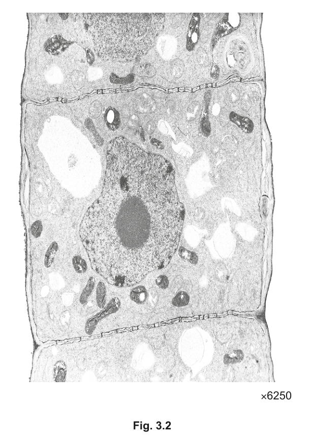
On Fig. 3.2, draw a label line and label it with the letter P to identify one plasmodesma.
Answer
Draw a label line ending on one of the narrow channels (plasmodesmata) that pass through the cell wall between two adjacent onion root cells, and label the line with the letter P.
Label line P drawn to a plasmodesma in Fig. 3.2.
Background Concept
Plasmodesmata (singular: plasmodesma) are narrow cytoplasmic channels that pass through the cell walls of adjacent plant cells, connecting their cytoplasm and allowing direct cell-to-cell transport of water, ions, small molecules and some larger molecules. They are the structural basis of the symplast pathway. In transmission electron micrographs (TEMs) they appear as thin, dark, transverse lines crossing the lighter middle-lamella region of the cell wall between two cells.
Understanding the Question
Fig. 3.2 is a TEM of onion root cells at magnification. Between adjacent cells the wall shows fine transverse dark lines — these are plasmodesmata. The candidate must add one label line, lettered P, that ends exactly on one of these channels.
Approach
Identify a clear, unambiguous plasmodesma: a thin, dark, straight line crossing the cell wall perpendicular to the wall surface, between the cytoplasm of two cells. Place the end of the label line precisely on it and letter it P. Do not label a thicker dark band (which would be a whole cell wall) or a gap (which would be an intercellular space).
Step-by-Step Reasoning
- Locate the boundaries between cells. The cell walls appear as light bands separating darker cytoplasm.
- Within those light bands, look for thin, dark, transverse lines — these are the plasmodesmata.
- Draw the label line so its tip lands on (not near, not through) one plasmodesma, and label it P.
Key Takeaways
- Plasmodesmata are the channels that create the symplast pathway in plants.
- They are visible only at EM resolution; in light micrographs they cannot be seen individually.
Common Mistakes
- Labelling the whole cell wall instead of a single plasmodesma — the line must end on a channel, not on the wall.
- Labelling the middle lamella or an intercellular space — neither is a plasmodesma.
- Putting the P too far from the structure to be clearly identified.
Things to Be Careful About
The mark scheme awards the single mark purely for "correct label to a plasmodesma", so accuracy of placement matters more than neatness. The label line should end precisely on a channel, not pass through it or point vaguely at the wall.
Table 3.1 contains information about four polysaccharides found in animals or plants.
Complete Table 3.1 by filling in the missing information.
Table 3.1
| polysaccharide | monomer | glycosidic bond(s) | function |
|---|---|---|---|
| amylopectin | -glucose | 1,4 and 1,6 | energy storage in plants |
| amylose | 1,4 | energy storage in plants | |
| cellulose | -glucose | structural role in plant cell walls | |
| glycogen | -glucose | 1,4 and 1,6 |
Answer
| polysaccharide | monomer | glycosidic bond(s) | function |
|---|---|---|---|
| amylopectin | -glucose | 1,4 and 1,6 | energy storage in plants |
| amylose | -glucose | 1,4 | energy storage in plants |
| cellulose | -glucose | 1,4 | structural role in plant cell walls |
| glycogen | -glucose | 1,4 and 1,6 | energy storage in animals |
amylose: -glucose; cellulose: 1,4; glycogen: energy storage in animals
Background Concept
Polysaccharides are polymers of monosaccharide monomers joined by glycosidic bonds:
- Starch (plant storage polysaccharide) consists of two polymers:
- amylose — long, unbranched chains of -glucose joined only by 1,4 glycosidic bonds; the chain coils into a helix.
- amylopectin — long, branched chains of -glucose with 1,4 bonds in the chains and 1,6 bonds at the branch points.
- Glycogen is the animal equivalent of starch — even more highly branched chains of -glucose with 1,4 and 1,6 bonds; used for energy storage in liver and muscle cells.
- Cellulose is the structural polysaccharide of plant cell walls: long, unbranched chains of -glucose joined by 1,4 glycosidic bonds; every other glucose is rotated 180°, allowing hydrogen bonds between chains to form strong, straight microfibrils.
Understanding the Question
Table 3.1 gives four polysaccharides with some information filled in. Three cells are missing and must be completed:
- the monomer of amylose;
- the glycosidic bond(s) of cellulose;
- the function of glycogen.
Approach
Recall the basic structure-function relationships of the four polysaccharides:
- amylose is built from -glucose;
- cellulose's β-1,4 linkages are what give it its straight, structural form;
- glycogen is the animal storage polysaccharide (equivalent to starch).
Step-by-Step Reasoning
- Amylose monomer: amylose is the unbranched component of starch, formed only of -glucose (the same monomer as the amylopectin already given in the table).
- Cellulose glycosidic bonds: cellulose chains use 1,4 glycosidic bonds between -glucose units. The alternation in orientation produces the straight, hydrogen-bonded chains.
- Glycogen function: glycogen stores glucose in animal cells (especially liver and muscle), exactly as starch does in plants.
Key Takeaways
- -glucose → storage polysaccharides (amylose, amylopectin, glycogen) → helical, soluble, easily hydrolysed.
- -glucose → structural polysaccharide (cellulose) → straight, insoluble microfibrils.
- 1,6 bonds appear only at branch points (amylopectin, glycogen); 1,4 bonds form the main chain.
Common Mistakes
- Writing "β-glucose" for amylose or glycogen — these are storage polysaccharides built from -glucose.
- Writing "1,4 and 1,6" for cellulose — cellulose has only 1,4 bonds; it is unbranched.
- Writing "energy storage in plants" for glycogen — must specify animals.
Things to Be Careful About
The mark scheme is strict on the words "" and "" and on "animals" for glycogen. Always state the bond type with the numbers in the right order (1,4 not 4,1) and use Greek letters exactly for the glucose isomer.
Table 4.1 shows a sequence of 12 nucleotides in the template strand of a short length of a DNA molecule, the corresponding primary transcript and the four amino acids coded for by the sequence. The table is incomplete.
Complete Table 4.1 to show the sequence of nucleotides in the primary transcript that would result from transcription of this short length of DNA.
Table 4.1
| position of nucleotide | 1 | 2 | 3 | 4 | 5 | 6 | 7 | 8 | 9 | 10 | 11 | 12 |
|---|---|---|---|---|---|---|---|---|---|---|---|---|
| DNA template strand | C | A | C | T | A | C | T | C | C | A | A | C |
| primary transcript | ||||||||||||
| amino acid | aa1 | aa2 | aa3 | aa4 |
Answer
| position of nucleotide | 1 | 2 | 3 | 4 | 5 | 6 | 7 | 8 | 9 | 10 | 11 | 12 |
|---|---|---|---|---|---|---|---|---|---|---|---|---|
| DNA template strand | C | A | C | T | A | C | T | C | C | A | A | C |
| primary transcript | G | U | G | A | U | G | A | G | G | U | U | G |
GUG AUG AGG UUG
Background Concept
Transcription is the first stage of protein synthesis, in which a length of DNA is copied into a single-stranded molecule of messenger RNA (the primary transcript). Only one of the two DNA strands — the template (antisense) strand — is read by RNA polymerase. The other strand is the coding (sense) strand and has the same base sequence as the mRNA (except that T in DNA corresponds to U in RNA).
The base-pairing rules for transcription are:
- A (DNA) pairs with U (mRNA)
- T (DNA) pairs with A (mRNA)
- C (DNA) pairs with G (mRNA)
- G (DNA) pairs with C (mRNA)
Crucially, RNA uses the base uracil (U) in place of thymine (T), so any A in the template DNA appears as U in the mRNA.
Understanding the Question
The question gives the 12-nucleotide sequence of a DNA template strand running from position 1 to 12. We are asked to write the corresponding 12-nucleotide primary transcript (mRNA). No further processing (5′ capping, poly-A tail, splicing) is being asked for — just the raw RNA copy.
Approach
Work position by position, writing the RNA complement of each DNA nucleotide. The two strands are antiparallel, but because the table gives the template nucleotides in the order in which they are read by RNA polymerase, the mRNA can simply be written position-by-position beneath.
Step-by-Step Reasoning
Applying the base-pairing rules to each position:
- Pos 1: C → G
- Pos 2: A → U
- Pos 3: C → G
- Pos 4: T → A
- Pos 5: A → U
- Pos 6: C → G
- Pos 7: T → A
- Pos 8: C → G
- Pos 9: C → G
- Pos 10: A → U
- Pos 11: A → U
- Pos 12: C → G
So the completed primary transcript is G–U–G–A–U–G–A–G–G–U–U–G.
Key Takeaways
- The primary transcript is a complementary RNA copy of the template DNA strand.
- Uracil (U) replaces thymine (T) in RNA, so A in the DNA template is transcribed as U in the mRNA.
- RNA polymerase reads the template 3′→5′ and synthesises the new RNA strand 5′→3′.
Common Mistakes
- Writing T instead of U in the mRNA — the molecule is RNA, so every complementary base to a DNA A must be U, not T.
- Pairing the wrong way round (e.g. writing A opposite T) — only complementary base pairing is allowed; there is no T–T or A–A pair.
- Skipping a position or miscounting columns in the table.
Things to Be Careful About
- This is transcription (DNA → RNA), not replication (DNA → DNA) and not translation (mRNA → protein). The base-pairing partners in transcription use U where DNA would have used T.
- The question is part of a sequence, so getting position 3 correct matters for parts (iii) and (iv) which refer back to it.
Table 4.2 shows all the possible template strand DNA triplets that code for the amino acids labelled aa1, aa2, aa3 and aa4 in Table 4.1.
Table 4.2
| amino acid | DNA triplets |
|---|---|
| val | CAA, CAG, CAT, CAC |
| arg | GCA, GCG, GCT, GCC, TCT, TCC |
| met | TAC |
| leu | AAT, AAC, GAA, GAG, GAT, GAC |
Complete Table 4.3 to identify the four amino acids labelled aa1, aa2, aa3 and aa4 in Table 4.1.
Table 4.3
| aa1 | aa2 | aa3 | aa4 | |
|---|---|---|---|---|
| amino acid |
Answer
| aa1 | aa2 | aa3 | aa4 | |
|---|---|---|---|---|
| amino acid | val (valine) | met (methionine) | arg (arginine) | leu (leucine) |
aa1 = val, aa2 = met, aa3 = arg, aa4 = leu
Background Concept
The genetic code is read in non-overlapping groups of three nucleotides called codons. A codon in mRNA corresponds to a triplet on the template DNA strand. The codon table is degenerate: most amino acids are specified by more than one codon, but each codon specifies only one amino acid. Table 4.2 in this question lists, for each amino acid, every possible DNA template triplet that codes for it.
The relationship between a DNA template triplet and its mRNA codon is complementary and antiparallel. In this question, Table 4.2 already gives the template strand triplets, so each codon in mRNA can be deduced from the complement of the DNA triplet shown — but the exam shortcut is to read Table 4.2 directly: any DNA template triplet listed there corresponds to that amino acid.
Understanding the Question
The 12-nucleotide template sequence must be split into four consecutive, non-overlapping triplets starting at position 1. Each triplet in turn codes for one amino acid, labelled aa1, aa2, aa3 and aa4. The amino acid labels in the original Table 4.1 sit at positions 2, 5, 8 and 11 — the middle nucleotide of each triplet — which is a useful cross-check that the reading frame begins at position 1.
Approach
- Slice the template into triplets: positions 1–3, 4–6, 7–9, 10–12.
- Look each triplet up in Table 4.2 and read off the corresponding amino acid.
Step-by-Step Reasoning
Template DNA: C A C | T A C | T C C | A A C
- Triplet 1 (positions 1–3): CAC — listed under val. → aa1 = val
- Triplet 2 (positions 4–6): TAC — listed under met. → aa2 = met
- Triplet 3 (positions 7–9): TCC — listed under arg. → aa3 = arg
- Triplet 4 (positions 10–12): AAC — listed under leu. → aa4 = leu
Cross-check via the mRNA codons (GUG, AUG, AGG, UUG): all four are standard codons and match the amino acids above.
Key Takeaways
- The genetic code is read in non-overlapping triplets from a fixed start position.
- A codon table can be used to translate a DNA template (or its mRNA) directly into an amino acid sequence.
- The code is degenerate: more than one triplet can specify the same amino acid, but the converse is not true.
Common Mistakes
- Reading the triplets with an off-by-one error (e.g. starting at position 2 instead of 1) — the amino acid labels at positions 2, 5, 8 and 11 should be the middle base of each codon.
- Confusing the DNA template with the mRNA codon when using the table; the table here lists template DNA triplets, so just match directly.
- Mixing up the rows of Table 4.2 — for example, mistaking TCC for leu (it is arg).
Things to Be Careful About
- A single mistake in the triplet grouping cascades into wrong answers for all four amino acids, costing the only mark for this sub-part (it is an "all four correct" mark).
- The amino acid label sits at the middle nucleotide of each codon, which can be used as a sanity check.
One type of gene mutation is caused by the substitution of a DNA nucleotide.
Using the information in Table 4.2, state and explain the effect on the final protein structure of a substitution of the nucleotide at position 3 in Table 4.1.
Answer
- No effect on the protein structure.
- All four triplets beginning with CA (CAA, CAG, CAT, CAC) code for valine.
- The genetic code is, redundant / degenerate.
No effect on protein structure — degeneracy of the code means all possible CA_ codons still code for valine.
Background Concept
A substitution mutation replaces one nucleotide with another at a single position. The consequence depends on whether the new triplet still codes for the same amino acid. The genetic code is degenerate (or redundant): most amino acids are specified by more than one codon, often differing only in the third (wobble) position. Substitutions at this third position are frequently silent — they change the codon but not the amino acid, leaving the protein unchanged.
Understanding the Question
The nucleotide at position 3 of the DNA template is the C that completes the first codon (CAC, coding for val). The question asks what happens if this single nucleotide is substituted — i.e. replaced by a different base — and why.
Approach
- Identify which codon the affected nucleotide belongs to: position 3 is the third base of the first triplet (CAC).
- List every possible substitution at that position.
- Look each new triplet up in Table 4.2 and check whether the amino acid changes.
Step-by-Step Reasoning
The first triplet is C A C. The first two bases (CA) are unaffected, so any substitution at position 3 only changes the third base of this codon. The four possibilities are:
- C → A: gives CAA → still val (in Table 4.2)
- C → G: gives CAG → still val
- C → T: gives CAT → still val
- C (unchanged): CAC → val
Every possible substitution produces a triplet that still codes for valine. So the first amino acid is unchanged, and because this is the only codon that position 3 lies within, the rest of the protein is also unaffected.
The reason this works is the degeneracy (redundancy) of the genetic code — specifically, the fact that valine has four codons differing only in the third base, and that this third-base "wobble" position often does not change the amino acid specified.
Key Takeaways
- Substitution mutations are not always harmful; many are silent because the code is degenerate.
- Changes at the third base of a codon are particularly likely to be silent — a feature of the genetic code sometimes called the "wobble" position.
- Whether a substitution matters depends on which amino acid, if any, the new codon specifies, and how similar the new amino acid is to the original.
Common Mistakes
- Saying the protein will change without checking Table 4.2 — the data table is the key reference, and it shows the protein does not change.
- Calling the code non-overlapping or universal instead of degenerate / redundant — these are different properties of the code.
- Confusing this with the deletion mutation in part (iv), which does change the reading frame.
Things to Be Careful About
- Position 3 is inside the first codon; the substitution is contained within that one codon and does not shift the reading frame, so only that one amino acid could possibly be affected.
- A mark is available for stating that the code is degenerate — this is the underlying biological reason a substitution can be silent.
A second type of gene mutation is caused by the deletion of a DNA nucleotide.
Using the information in Table 4.2, state and explain the effect on the final protein structure of a deletion of the nucleotide at position 3 in Table 4.1.
Answer
- The first amino acid is still val (because the new first triplet CAT also codes for valine).
- The reading frame changes from this point onwards (frameshift mutation).
- All amino acids after the mutation are altered; this may introduce a premature stop codon, giving a shorter polypeptide and an altered tertiary structure / active site.
First amino acid still val; reading frame shifted, altering all subsequent amino acids and likely producing a shorter, non-functional protein.
Background Concept
A deletion mutation removes one or more nucleotides from the DNA. If the number removed is not a multiple of three, the deletion shifts the reading frame of every codon downstream of the mutation — a so-called frameshift mutation. From the point of the deletion onwards, the ribosome reads entirely new triplets, almost always producing a completely different amino acid sequence. Frameshifts very often introduce a premature stop codon, truncating the polypeptide and almost always abolishing its function.
Understanding the Question
The question asks what happens if the single nucleotide at position 3 of the template is removed. We need to (1) re-read the new sequence in triplets from position 1, (2) check what the new codons code for, and (3) explain the consequences for the protein.
Approach
- Delete the C at position 3.
- Re-write the 11 remaining nucleotides.
- Re-read in triplets from position 1 — note the new first codon and all subsequent codons.
- Compare with Table 4.2 and explain why the protein changes.
Step-by-Step Reasoning
Original template: C A C T A C T C C A A C
Delete position 3: C A T A C T C C A A C
Re-read in triplets from position 1:
- New codon 1: CAT → val (still valine, because CAT is one of the valine codons in Table 4.2)
- New codon 2: ACT → not in Table 4.2; codes for a different amino acid (threonine)
- New codon 3: CCA → not in Table 4.2; codes for a different amino acid (proline)
- New codon 4: AC_ → incomplete; the sequence ends here
So the first amino acid is unchanged, but from codon 2 onwards every codon is read differently. In a real, longer sequence this would:
- alter the primary structure (the amino acid sequence after the mutation),
- very likely introduce a premature stop codon, producing a shorter polypeptide,
- and consequently alter the tertiary structure and the shape of the active site (if the protein is an enzyme), usually abolishing function.
The key point is that a single-nucleotide deletion is a frameshift mutation — it is not contained within a single codon the way a substitution is, and so it has effects that propagate all the way to the end of the protein.
Key Takeaways
- A deletion of a number of nucleotides that is not a multiple of three causes a frameshift.
- Frameshifts change every codon downstream of the mutation, almost always destroying protein function.
- The first amino acid may or may not be unchanged — here it happens to be unchanged because CAT still codes for val, but this is a coincidence and is not generally the case.
Common Mistakes
- Saying the protein is unchanged — this is a deletion, not a substitution, and the reading frame has shifted.
- Saying only one amino acid is affected — frameshifts affect all amino acids from the mutation point onwards.
- Forgetting to mention the possibility of a premature stop codon and a shorter polypeptide.
- Confusing deletion (this part) with substitution (part iii).
Things to Be Careful About
- The fact that the first amino acid is still val is a "lucky" consequence of the new codon CAT also being a val codon; it is not a general property of frameshifts.
- The mark scheme accepts any three of: first AA unchanged, reading frame change, all downstream AAs altered, possible premature stop / shorter polypeptide, altered tertiary structure / active site. Aim to give at least three of these for full marks.
Replication of nuclear DNA occurs just once in every mitotic cell cycle. Six named events associated with the mitotic cell cycle are listed. The events are not listed in any particular order.
Draw a circle around each event where replication of nuclear DNA occurs.
cytokinesis
interphase S phase
phase phase
mitosis
Answer
Circle interphase and S phase (both should be circled, as S phase occurs within interphase).
interphase and S phase
Background Concept
The mitotic cell cycle has two broad phases: interphase (a long growth-and-preparation period) and mitosis (the relatively short division of the nucleus), followed by cytokinesis (division of the cytoplasm). Interphase itself is divided into three sub-phases:
- G1 — cell growth and production of organelles and proteins.
- S phase — synthesis of new DNA; each chromosome is replicated to form two sister chromatids.
- G2 — further growth, production of proteins needed for mitosis, and a final check that DNA replication is complete.
DNA replication occurs only once per cell cycle, and that is during the S phase, which lies within interphase.
Understanding the Question
A list of cell-cycle events is given. The question asks which of them include DNA replication, and asks the candidate to circle each one. Because S phase is a sub-phase of interphase, both should be circled.
Approach
- Recall that DNA replication happens in S phase.
- Recognise that S phase is part of interphase, so both answers are correct.
Step-by-Step Reasoning
Going through the list:
- cytokinesis — division of the cytoplasm; no DNA replication. ✗
- interphase — contains S phase, so this includes DNA replication. ✓
- S phase — the specific stage of DNA synthesis. ✓
- G2 phase — preparation for mitosis; replication has already happened. ✗
- G1 phase — cell growth; DNA has not yet been replicated. ✗
- mitosis — nuclear division; DNA was already replicated in S phase. ✗
Both interphase and S phase should be circled.
Key Takeaways
- DNA replication occurs once per cell cycle, during S phase.
- S phase is a sub-phase of interphase, so the two answers are not contradictory.
- The other cell-cycle events (G1, G2, mitosis, cytokinesis) do not involve DNA replication.
Common Mistakes
- Circling only S phase and not interphase — the mark scheme requires both.
- Circling mitosis — DNA has already been replicated by this point; mitosis distributes the copies to daughter nuclei.
- Circling G2 or G1 — replication happens in S phase, not in the G phases.
Things to Be Careful About
- The question uses British-style "circle"; candidates should draw a clear loop around each correct word.
Answer
- DNA helicase unwinds the double helix and breaks the hydrogen bonds between the two antiparallel strands, exposing the bases.
- Both strands act as templates for the new DNA.
- Free activated DNA nucleotides pair with the exposed bases by complementary base pairing (A–T, C–G).
- DNA polymerase forms phosphodiester bonds between adjacent nucleotides; the leading strand is synthesised continuously while the lagging strand is synthesised in short Okazaki fragments that are later joined by DNA ligase.
- Replication is semi-conservative: each new DNA molecule contains one parental (conserved) strand and one newly synthesised strand.
See working — semi-conservative replication using helicase, DNA polymerase and ligase on both template strands.
Background Concept
DNA replication is the process by which a cell makes an identical copy of its nuclear DNA before dividing. Each strand of the parent double helix serves as a template for a new complementary strand, so each daughter molecule contains one old (parental) strand and one new strand. This pattern is called semi-conservative replication and was demonstrated by the classic Meselson–Stahl experiment.
Replication requires several enzymes working in a coordinated way at the replication fork (the Y-shaped point where the double helix is being opened up).
Understanding the Question
The question asks for an outline of how DNA is replicated inside the nucleus. This is a "describe" question with four marks, so four to five clearly distinct points are needed. The mark scheme warns that no more than three marks can be awarded if the candidate drifts into describing transcription (DNA → RNA) — so the answer must stay firmly on replication (DNA → DNA).
Approach
Sequence the events as they happen at the replication fork:
- Unwind the parent DNA and separate the strands.
- Use both strands as templates.
- Bring in activated nucleotides and pair them by complementary base pairing.
- Join the new nucleotides together.
- Handle the two strands' different geometries (leading vs lagging).
- State that the outcome is semi-conservative.
Step-by-Step Reasoning
- Unwinding: DNA helicase breaks the hydrogen bonds between complementary base pairs and unwinds the double helix, producing a replication fork. Topoisomerase relieves the twisting strain ahead of the fork (an additional credit-worthy point if mentioned).
- Both strands as templates: each of the two separated strands is read by the replication machinery; both carry the information needed to make a new complementary strand.
- Free activated nucleotides: the new nucleotides are added as triphosphates (dNTPs), which carry their own energy and are sometimes described as "activated". Hydrolysis of two of the three phosphates releases the energy that drives the polymerisation reaction.
- DNA polymerase: catalyses the addition of each new nucleotide to the growing strand, only in the 5′→3′ direction. It also proofreads each new base, correcting mismatches. Adjacent nucleotides are linked by phosphodiester bonds.
- Leading and lagging strands: because the two template strands are antiparallel and DNA polymerase can only synthesise 5′→3′, the strand being made towards the fork (the leading strand) is synthesised continuously, while the other strand (the lagging strand) is synthesised in short reverse-direction fragments called Okazaki fragments.
- DNA ligase: joins the Okazaki fragments together on the lagging strand by forming phosphodiester bonds between them.
- Semi-conservative outcome: each finished double helix contains one parental strand and one newly synthesised strand — i.e. half of the parent molecule is "conserved" in each daughter molecule.
Key Takeaways
- Replication is semi-conservative and occurs once per cell cycle, in S phase.
- Several enzymes cooperate: helicase (unwinding), DNA polymerase (polymerisation and proofreading), ligase (joining fragments), and topoisomerase (relieving strain).
- The two strands are replicated differently because DNA polymerase can only work 5′→3′: the leading strand continuously, the lagging strand in Okazaki fragments.
- The free nucleotides used are activated (triphosphates), which provide the energy for the polymerisation reaction.
Common Mistakes
- Drifting into transcription (RNA polymerase, mRNA, U instead of T) — the mark scheme caps the score at 3 if this happens. Stay on DNA → DNA.
- Forgetting the role of both strands as templates.
- Saying DNA polymerase "makes the new strand" without mentioning the formation of phosphodiester bonds.
- Calling replication "conservative" — replication is semi-conservative: one strand is conserved, the other is new.
- Forgetting that the lagging strand is made in fragments, or forgetting to mention ligase.
Things to Be Careful About
- The question is about replication, not transcription. The base-pairing rules for replication are the same as for transcription (A–T, C–G), but the product is DNA, not RNA, and the enzyme is DNA polymerase, not RNA polymerase.
- "Outline" implies a structured, multi-point answer; a single sentence is unlikely to score four marks.
Fig. 4.1 shows the structure of an ATP molecule.
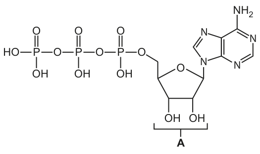
State the name of the part of the ATP molecule labelled A in Fig. 4.1.
Answer
Ribose (the mark scheme specifically does not accept "pentose").
ribose
Background Concept
An ATP (adenosine triphosphate) molecule is a nucleotide built from three components:
- A nitrogenous base — adenine (a double-ringed purine).
- A five-carbon sugar — ribose (a pentose sugar, the same sugar found in RNA).
- Three phosphate groups linked in a chain; the bonds between them are high-energy phosphoanhydride bonds whose hydrolysis releases energy that the cell can use.
Together, adenine + ribose form adenosine; adding the three phosphate groups gives adenosine triphosphate, ATP. It is worth noting that the sugar in ATP is ribose (as in RNA), not deoxyribose (as in DNA).
Understanding the Question
Fig. 4.1 shows the full structure of ATP with three phosphate groups on the left, a five-membered sugar ring in the middle, and a double-ringed nitrogenous base on the right. Label A points to the sugar ring at the centre of the molecule. We have to name this part.
Approach
- Identify the three structural regions of ATP from the figure.
- Determine which region label A is pointing to.
- Give the correct name of that region.
Step-by-Step Reasoning
In Fig. 4.1:
- The three phosphate groups sit on the left of the molecule.
- The sugar ring (five-membered, with –OH groups) is in the middle.
- The double-ringed base with the –NH2 group is on the right.
Label A is positioned beneath the middle sugar ring, so it is pointing at the sugar. The sugar in ATP is ribose. (The mark scheme explicitly does not accept "pentose" — although that is technically correct, the expected answer is the specific name.)
Key Takeaways
- ATP = adenine + ribose + 3 phosphate groups.
- The sugar in ATP (and in RNA) is ribose; the sugar in DNA is deoxyribose.
- Adenine is a purine (double-ringed base).
Common Mistakes
- Writing "pentose" or "sugar" — these are too vague; the mark scheme credits only "ribose".
- Writing "deoxyribose" — that is the sugar in DNA, not in ATP.
- Confusing label A with a phosphate group or with adenine.
Things to Be Careful About
- The mark scheme ignores "pentose" — give the specific name, ribose, to be safe.
- The 2′ and 3′ –OH groups on the sugar ring are characteristic of ribose (deoxyribose would lack the 2′ –OH), so the structure in the figure is unambiguously ribose.
The pathogen that causes cholera is a prokaryote.
Fig. 5.1 shows an electron micrograph of the pathogen that causes cholera.
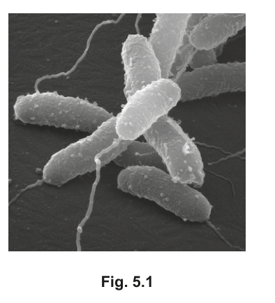
Answer
Scanning (electron microscope) / SEM.
Scanning (electron microscope) / SEM
Background Concept
Two types of electron microscope are used in biology:
- Transmission electron microscope (TEM) — a beam of electrons is passed through an ultra-thin specimen. The image is a 2D cross-section showing internal structures (e.g. ribosomes, internal membranes, chromosome).
- Scanning electron microscope (SEM) — electrons scan the surface of a specimen (which is usually coated in a thin layer of heavy metal, e.g. gold). Secondary electrons emitted from the surface are collected to build a 3D image of surface topography.
Both have much higher resolution than the light microscope because the wavelength of electrons is far shorter than that of visible light.
Understanding the Question
The question asks which type of electron microscope was used to produce Fig. 5.1. The image shows several curved, rod-shaped cells with long thin flagella clearly projecting from one end, all rendered with a strong 3D appearance — you can see the rounded contours and shadows of the cell surface.
Approach
Look at the image and decide whether it shows internal 2D structure (TEM) or external 3D surface detail (SEM). The 3D shaded appearance of whole cells on a textured surface is the giveaway for SEM.
Step-by-Step Reasoning
- The image is not a flat 2D slice; whole cells are shown in three dimensions.
- Surface features (texture of the cell wall, the curve of the flagella, the rounded ends of the rods) are clearly visible.
- This is exactly what an SEM produces — secondary electrons from the specimen's surface are used to construct a 3D topographical image.
- A TEM image of the same cells would appear as flat, electron-dense outlines with no surface shadowing.
Key Takeaways
- SEM → 3D surface images; useful for whole cells, surface structures (flagella, pili, cilia), external morphology.
- TEM → 2D internal images; useful for organelles, viruses, internal cell structure.
Common Mistakes
- Writing just "electron microscope" without specifying scanning — this does not earn the mark.
- Confusing SEM with TEM because the word "microscope" is in both — remember SEM is for surfaces, TEM is for thin sections.
Things to Be Careful About
- The clue is the 3D surface appearance. The mark scheme requires the word "scanning" (or the abbreviation SEM) — "transmission" would be wrong.
Answer
Vibrio cholerae
Vibrio cholerae
Background Concept
Cholera is an acute diarrhoeal disease of the small intestine caused by the bacterium Vibrio cholerae. The genus name Vibrio refers to the curved, comma-like shape of the cells (visible in Fig. 5.1). Pathogenic strains produce the cholera toxin (CT), which causes the secretion of large volumes of water and electrolytes into the gut lumen, leading to severe watery diarrhoea ('rice-water stools') and rapid dehydration.
Understanding the Question
The question gives an image of curved, flagellated bacteria and asks for the species name of the prokaryote that causes cholera. "Species" means the full binomial (genus species).
Approach
Recall the binomial name of the cholera pathogen. Write it in the accepted format: genus capitalised, species lower-case, both in italics.
Step-by-Step Reasoning
- Cholera is caused by the bacterium Vibrio cholerae.
- Vibrio = genus (capital V, italicised).
- cholerae = specific epithet / species (lower-case c, italicised).
- Writing only "Vibrio" would refer to the whole genus (which includes non-pathogenic marine species) and would not score the mark.
Key Takeaways
- The four major pathogens named on this syllabus are: Vibrio cholerae (cholera), Plasmodium falciparum / P. vivax / P. ovale / P. malariae (malaria), Mycobacterium tuberculosis / M. bovis (TB), and HIV (a virus, not given a binomial name).
- Always use binomial nomenclature — both genus and specific epithet — and italicise.
Common Mistakes
- Writing only "Vibrio" — genus alone does not specify the species.
- Capitalising the species: Vibrio Cholerae — the specific epithet is lower-case.
- Forgetting italics — binomials should be italicised (or underlined if handwritten).
- Spelling the species as colera or kolera — the correct spelling is cholerae.
Things to Be Careful About
- The cell in the image is described as a prokaryote — a useful sanity check that the answer should be a bacterium (binomial name) rather than a virus (which has no binomial, e.g. HIV is a name not a species).
The passage contains a description of the main features of prokaryotic cells. There is one factual error in the passage.
Prokaryotic cells are unicellular and generally between and in diameter. Prokaryotes do not have organelles surrounded by double membranes. They do have cell surface membranes, 70S ribosomes and a cellulose cell wall. The DNA of a prokaryotic cell is circular and is found free in the cytoplasm rather than enclosed in a nuclear envelope.
Identify and correct the factual error in the passage.
Answer
Prokaryotic cell walls are made of peptidoglycan (murein), not cellulose.
Prokaryotic cell walls are made of peptidoglycan (murein), not cellulose.
Background Concept
Cell walls differ chemically between domains and kingdoms:
- Plant cell walls — made of cellulose (a β-1,4-linked glucose polymer), plus pectins and hemicelluloses.
- Bacterial (prokaryotic) cell walls — made of peptidoglycan (also called murein): long polysaccharide chains of alternating NAG and NAM cross-linked by short peptide bridges.
- Fungal cell walls — made of chitin and glucans.
Peptidoglycan is the target of several antibiotics (e.g. penicillin disrupts the peptide cross-links), which is why these drugs are effective against bacteria but harmless to human cells.
The other facts in the passage are correct:
- Prokaryotes are unicellular and typically 1–5 µm in diameter (correct).
- They do not have membrane-bound organelles such as a nucleus, mitochondria or ER (correct — they have 70S ribosomes but no organelles surrounded by double membranes).
- They do have a cell surface membrane, 70S ribosomes, and a cell wall (correct — only the wall's composition is wrong).
- The DNA is circular and lies free in the cytoplasm in a region sometimes called the nucleoid, not enclosed by a nuclear envelope (correct).
Understanding the Question
The stem says the passage contains one factual error and asks you to identify and correct it. The other claims in the passage are true, so the incorrect statement must be singled out precisely.
Approach
Read each claim and check it against your knowledge of prokaryotic structure. The composition of the cell wall is the suspect claim because cellulose is the plant cell wall material.
Step-by-Step Reasoning
- Check the size: 1–5 µm diameter — true for most bacteria.
- Check the organelles claim: no double-membrane-bound organelles — true.
- Check the surface features: cell surface membrane, 70S ribosomes, cell wall — true in general.
- Check the wall composition: "cellulose cell wall" — false. Bacterial cell walls are peptidoglycan (murein); cellulose is a plant cell wall material.
- Check the DNA: circular, free in cytoplasm, no nuclear envelope — true.
- The single error is the claim that the wall is cellulose. Correct it by replacing "cellulose" with "peptidoglycan" (or "murein").
Key Takeaways
- The phrase "cellulose cell wall" is a hallmark of plant cells, not prokaryotes.
- Knowing what each cell type's wall is made of is a recurring exam point and links directly to the action of antibiotics such as penicillin.
- When asked to "identify and correct", a candidate must do both: name the wrong feature and supply the correct one. Stating only "the wall is not cellulose" without saying what it actually is would not earn the mark under this scheme.
Common Mistakes
- Writing "no cell wall" — incorrect; bacteria do have a cell wall (its composition is the issue).
- Writing "protein cell wall" — wrong material.
- Identifying the wrong statement as the error (e.g. claiming "70S ribosomes" is wrong, or saying prokaryotes do have a nucleus).
- Saying only "it is peptidoglycan" without making clear this replaces the word "cellulose" in the passage — the question asks for identification and correction, so both should be evident.
Things to Be Careful About
- "Peptidoglycan" and "murein" are both acceptable (they are synonyms).
- Do not over-correct by altering the true statements; the question only wants the one error fixed.
- The diameter range given (1–5 µm) refers to the diameter, not the length — many bacteria (including Vibrio cholerae) are 1–5 µm across but can be longer lengthwise. The mark scheme accepts the passage's wording here.
Fig. 6.1 is a simplified diagram representing a section through the human immunodeficiency virus (HIV) particle that causes HIV/AIDS. The diagram shows the virus particle about to attach to the cell surface membrane of a T-helper cell at a receptor protein called CD4. A second protein (coreceptor) called CCR5 is also necessary for the virus particle to enter and then infect the T-helper cell.
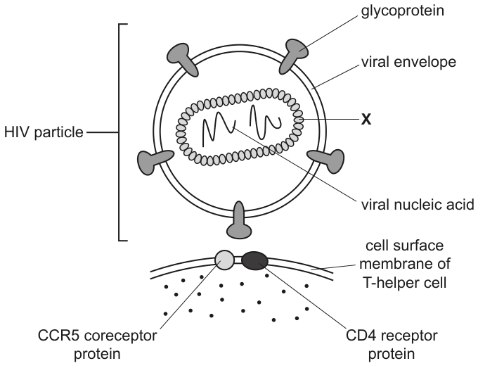
Answer
Capsid (protein coat).
capsid
Background Concept
HIV is a retrovirus. Like all viruses, it is non-cellular and consists of nucleic acid enclosed within a protein coat. The structure shown in Fig. 6.1 is the classic HIV architecture:
- an outer viral envelope (a phospholipid bilayer, originally derived from the host cell's membrane, studded with viral glycoproteins — gp120 and gp41 — that recognise host receptors);
- a capsid inside the envelope, made of repeating protein subunits called capsomeres, which assembles into a cone-shaped shell;
- within the capsid, the viral nucleic acid (in HIV, two identical single-stranded RNA molecules) together with reverse transcriptase and integrase enzymes.
The capsid's job is to protect the viral genome and to deliver it into the host cell after fusion. Without an intact capsid the RNA would be vulnerable to nucleases and the infection cycle could not proceed.
Understanding the Question
Fig. 6.1 labels several components of the HIV particle (glycoprotein, viral envelope, viral nucleic acid) but points an arrow labelled X to the protein structure immediately surrounding the viral nucleic acid. You must give the biological name of that structure.
Approach
Read the figure: X points to the inner protein layer that encloses the viral nucleic acid. From the definition above, that layer is the capsid. No further reasoning is needed; just supply the term.
Step-by-Step Reasoning
- Locate label X in Fig. 6.1. It points to the inner ring of protein subunits.
- The outer phospholipid layer is labelled viral envelope, and the squiggles inside are labelled viral nucleic acid.
- The layer sandwiched between them, surrounding the nucleic acid, is by definition the capsid.
- 'Capsomere' (a single subunit of the capsid) and 'protein coat' are accepted alternatives.
Key Takeaways
- The viral capsid is the protein shell that packages and protects the viral genome.
- HIV also possesses an outer envelope (taken from the previous host cell) studded with glycoproteins used for attachment.
Common Mistakes
- Writing 'protein' or 'glycoprotein' — these are rejected; the envelope glycoproteins are already labelled separately and protein is too vague.
- Spelling 'caspid' — rejected; spell capsid correctly.
- Calling X the envelope — the envelope is the outer phospholipid layer, not the inner protein shell.
Things to Be Careful About
When a question gives a label on a virus diagram, match the arrow to the structure it actually points to, not the structure closest to the label text. The arrow for X clearly indicates the inner coat.
Explain how the ability of the immune system to resist the damaging effects of a pathogen is affected by destruction of T-helper cells.
Answer
- Fewer cytokines are released (by T-helper cells).
- As a result, fewer plasma cells are produced so fewer antibodies are made.
- And/or macrophages are less stimulated (less antigen presentation / fewer 'angry' macrophages).
- And/or fewer T-killer cells are stimulated to divide, so fewer infected cells are destroyed.
- And/or fewer memory cells are produced in the primary response.
See answer
Background Concept
T-helper lymphocytes (CD4⁺ T-cells) are the coordinators of the adaptive immune response. They do not themselves kill infected cells or secrete large amounts of antibody; instead, they release signalling molecules called cytokines (e.g. interleukins such as IL-2) that activate and recruit other immune cells. Through cytokines, T-helper cells stimulate:
- B-lymphocytes to proliferate and differentiate into plasma cells that secrete antibodies (humoral immunity).
- Macrophages to become more aggressive phagocytes and to present antigen more efficiently.
- T-killer (cytotoxic) cells to divide and destroy infected cells.
- The formation of memory cells that underpin the secondary response.
HIV selectively infects and destroys CD4⁺ T-helper cells, so every one of these downstream responses is weakened — which is why AIDS patients succumb to opportunistic pathogens.
Understanding the Question
The question asks you to explain (3 marks) how the destruction of T-helper cells affects the immune system's ability to resist a pathogen. The command word 'explain' means each point must state what happens and why it follows from the loss of T-helper cells. The mark scheme rewards one mark for naming the lost cytokine signal and two further marks for any two of the cascade consequences.
Approach
Start with the central, defining role of the T-helper cell — cytokine release. Then pick the two downstream effects you can describe most confidently. Three marks total: one for cytokines, two for the consequences you choose.
Step-by-Step Reasoning
- Fewer cytokines released — T-helper cells are the principal cytokine source; their destruction removes the chemical signals that drive the rest of the response. (1 mark)
- Choose consequences of cytokine loss — any two of:
- B-cell arm: with less IL-2 / IL-4, B-lymphocytes divide less, so fewer plasma cells form and antibody production falls.
- Macrophage arm: macrophages are not stimulated into the 'angry' state, so phagocytosis is less aggressive and antigen presentation to other lymphocytes is reduced.
- T-killer arm: T-killer cell proliferation depends on cytokines from T-helper cells, so fewer T-killer cells differentiate and infected cells are not destroyed.
- Memory arm: the primary response generates fewer memory cells, weakening future secondary responses to the same pathogen.
- Each chosen point is one mark; only two are required.
Key Takeaways
- T-helper cells act by secreting cytokines; the consequences of their loss are therefore about signalling, not direct killing.
- Loss of T-helper function is a cascade: one lost cytokine signal affects B-cells, T-killers, macrophages and memory cells simultaneously.
- This explains the wide range of opportunistic infections seen in AIDS.
Common Mistakes
- Saying that T-helper cells 'kill the pathogen' — they do not; they coordinate others.
- Listing the consequences without linking them back to cytokine loss — the mark scheme wants the chain of reasoning.
- Naming only one downstream effect — you need two consequences to earn the two further marks.
- Confusing T-helper with T-killer cells; the question is about T-helper destruction.
Things to Be Careful About
The mark scheme accepts 'B lymphocytes' as equivalent to 'plasma cells' for the antibody point. It also allows 'AW' (alternative wording) on the macrophage point, so descriptions such as 'less antigen presentation' or 'macrophages less stimulated' are both valid. You do not need all four downstream effects; pick the two you know best.
Studies have shown that some individuals did not become infected with HIV even though they were repeatedly exposed to the virus. Later discoveries indicated that these individuals had a mutation in the gene for the CCR5 coreceptor protein.
Suggest how mutation of the gene for the CCR5 coreceptor protein provided protection against HIV infection.
Answer
The mutated CCR5 coreceptor no longer functions correctly, so HIV cannot bind / cannot trigger entry into the T-helper cell.
Virus cannot enter the T-helper cell because the CCR5 coreceptor cannot trigger endocytosis / fusion of the viral particle.
Background Concept
HIV entry into a host cell is a two-receptor event. The viral surface glycoprotein gp120 first binds the host's CD4 receptor, which causes a conformational change in gp120 that allows it to engage a co-receptor — either CCR5 (the most common route, used by so-called 'R5-tropic' strains early in infection) or CXCR4. Co-receptor engagement then allows gp41 to insert into the host membrane and trigger fusion of the viral envelope with the cell surface membrane (an endocytosis-like uptake of the capsid).
A mutation that alters the shape of CCR5 so that gp120 can no longer recognise it therefore blocks one of the two obligatory steps of viral entry. Individuals homozygous for the CCR5-Δ32 deletion — a 32-base-pair deletion that produces a non-functional, truncated receptor — are largely resistant to HIV infection.
Understanding the Question
This part gives you a scenario — repeatedly exposed individuals who remain uninfected, and a mutation in the gene encoding CCR5 — and asks you to suggest how that mutation provides protection. 'Suggest' means you are reasoning from the mechanism shown in the diagram; you do not need to cite Δ32 by name, just the logical consequence.
Approach
Ask: what does a normally functioning CCR5 do? It allows HIV to enter the T-helper cell (per Fig. 6.1 and the stem). What would a non-functional CCR5 do? It would stop that step. Write that single consequence clearly.
Step-by-Step Reasoning
- CCR5's role, given in the stem and visible in the figure, is to act as a co-receptor enabling HIV to attach to and enter the T-helper cell.
- A mutation may change the shape (tertiary structure) of the CCR5 protein so that HIV's glycoprotein can no longer bind to it.
- Without functional co-receptor binding, the virus cannot trigger fusion/endocytosis and so cannot enter the T-helper cell.
- The T-helper cells remain uninfected and continue to coordinate the immune response.
Key Takeaways
- HIV entry needs two receptor–ligand interactions: CD4 plus a chemokine co-receptor (CCR5 or CXCR4).
- A mutation that disables the co-receptor is a powerful natural resistance mechanism — this is the basis of the famous 'Berlin patient' cure and of CCR5-knockout gene-therapy trials.
- This links genetics (mutation), protein structure (receptor shape) and cell biology (viral entry mechanism).
Common Mistakes
- Saying only that the mutation 'prevents HIV from infecting the person' — too general; you must state how (entry blocked at the membrane).
- Attributing protection to the CD4 receptor — the question explicitly concerns CCR5.
- Saying the virus is 'destroyed' or 'recognised by antibodies' — neither follows from a CCR5 mutation.
Things to Be Careful About
The mark scheme phrasing is 'virus cannot enter the T-helper cell / CCR5 unable to trigger endocytosis of viral particle'. Either form is accepted, but the answer must refer to entry/endocytosis specifically.
The use of monoclonal antibodies against the CCR5 coreceptor protein (anti-CCR5) has been shown to be effective in the treatment of HIV infection.
Outline how anti-CCR5 monoclonal antibodies can be synthesised in the laboratory using the hybridoma method.
Answer
- Inject a small mammal (e.g. mouse) with the CCR5 antigen and allow an immune response to occur over several weeks.
- Extract B-lymphocytes (plasma cells) from the spleen of the immunised animal.
- Fuse the B-lymphocytes with myeloma (tumour) cells to form hybridoma cells.
- Screen/select the hybridomas to identify those producing the desired anti-CCR5 antibody.
- (AVP) Clone the selected hybridoma cells (e.g. by separating into wells) for large-scale antibody production.
See answer
Background Concept
A monoclonal antibody is an antibody preparation in which every molecule is identical, produced by a single clone of B-lymphocytes and therefore specific for one epitope on one antigen. In the body, B-cells make antibody but cannot be grown indefinitely in culture; myeloma cells can. The hybridoma method (Köhler and Milstein, 1975) gets the best of both by fusing an antibody-producing B-cell with a myeloma cell, producing a hybrid ('hybridoma') that is both immortal and antibody-secreting.
In this question the desired antibody is anti-CCR5, which blocks the CCR5 co-receptor on T-helper cells and so prevents HIV from entering — a real therapeutic strategy (e.g. leronlimab) for HIV treatment.
Understanding the Question
You must outline (3 marks) the hybridoma method. The mark scheme lists six numbered points and credits any three. 'Outline' means a concise description of each chosen stage, not a full protocol.
Approach
Use the canonical six-stage sequence as a checklist:
(1) inject antigen → (2) immune response → (3) harvest B-cells → (4) fuse with myeloma → (5) select hybridomas → (6) clone/produce.
Pick the three (or more) you can state most precisely; ensure each is a distinct stage so you don't inadvertently give overlapping content.
Step-by-Step Reasoning
- Immunisation. A small mammal (typically a mouse) is injected with the CCR5 antigen, often with an adjuvant, and boosted over several weeks so that B-lymphocytes are stimulated to produce antibodies against CCR5.
- Harvesting B-cells. B-lymphocytes (plasma cells) are extracted from the spleen (splenocytes) of the immunised animal.
- Fusion. The B-cells are fused with myeloma (tumour) cells using a fusogen such as polyethylene glycol (PEG), or by electrofusion. The resulting hybrid cells — hybridomas — combine the antibody specificity of the B-cell with the immortality of the myeloma.
- Selection/screening. The fused mixture is grown on a selective medium (e.g. HAT medium) that kills unfused myeloma cells and allows only hybridomas to survive. They are then screened (commonly by ELISA) for production of the desired anti-CCR5 antibody.
- Cloning and scale-up. Selected hybridomas are cloned (often by limiting dilution into separate wells) so that each culture is derived from a single cell, guaranteeing monoclonal antibody. The cloned hybridomas are then cultured on a large scale — in vitro in bioreactors or in vivo as ascites in mice — to harvest the antibody.
Key Takeaways
- The hybridoma method fuses an antibody-producing B-cell with an immortal myeloma cell.
- Selection (HAT medium) and screening (ELISA for the desired antibody) are the two distinct steps often confused by students.
- The product is a single antibody clone that can be manufactured in unlimited quantities.
Common Mistakes
- Forgetting that the animal must be immunised first — you cannot harvest useful B-cells without prior exposure to the antigen.
- Saying 'fuse B-cells with T-cells' — T-cells do not produce antibody.
- Confusing selection of hybridomas (HAT kills unfused cells) with screening for the desired antibody (ELISA identifies the right antibody).
- Skipping the cloning step; without cloning the antibody is not truly monoclonal.
- Omitting the source of B-cells (spleen) — the mark scheme specifically awards this point.
Things to Be Careful About
The mark scheme uses 'AW' (accept alternative wording) on the cell types and on the source animal, so 'mouse' or 'small mammal', and 'B-cells / B-lymphocytes / plasma cells / splenocytes' are all interchangeable. Note that B-cells from the spleen include plasma cells (the antibody-secretors) but the mark scheme uses the broader term 'B-lymphocytes'.