Biology 9700/12 — February/March 2024
Cambridge AS Level · Multiple Choice (AS Level) · answer key with instant marking and worked solutions
Topics Biological Molecules · Cell Structure · Cell Membranes and Transport · Nucleic Acids and Protein Synthesis · The Mitotic Cell Cycle · Transport in Plants · +5 more
Tap an option under each question to check it — your score builds as you go.
A student used a light microscope to observe a blood smear on a microscope slide.
An eyepiece graticule was used to measure the diameter of a white blood cell on the slide. The student recorded that the white blood cell was 5 eyepiece graticule units in diameter.
Which additional information does the student need to determine the diameter of the white blood cell in micrometres?
Options
A calibration of the eyepiece graticule using a stage micrometer only
B calibration of the eyepiece graticule using a stage micrometer and the magnification of the eyepiece lens
C the magnification of the eyepiece lens only
D the magnification of the eyepiece lens and the magnification of the objective lens
Working
An eyepiece graticule is a small disc bearing an arbitrary scale placed in the eyepiece. The size of each graticule division changes with the objective lens in use, so its divisions have no fixed real-world length. To convert "5 graticule units" into micrometres, the student must know how many micrometres each division represents at the magnification being used.
This is obtained by calibrating the eyepiece graticule against a stage micrometer (a slide with a known scale, e.g. divisions) at that objective. The calibration value already incorporates the effect of the objective (and eyepiece) lens, so no separate magnification values are required.
Answer
A
A
Background Concept
Light microscopes have two scales that can be used together to measure specimens:
- Eyepiece graticule — a transparent disc with an arbitrary scale (typically 100 divisions) that sits inside the eyepiece. Because it is in the eyepiece, its divisions look the same no matter which objective lens is in place, BUT the real distance each division represents depends on the objective lens in use. Its units are arbitrary "eyepiece graticule units".
- Stage micrometer — a microscope slide with a precisely known scale, usually or per division. It is used as a reference standard.
Calibration is the procedure of lining up the two scales at a given objective lens magnification and reading how many micrometres (or millimetres) correspond to one eyepiece graticule division at that magnification. The result (e.g. "1 eyepiece graticule unit = 2.5 µm at ×40") already has the magnification built in.
Understanding the Question
The student has measured a white blood cell as 5 eyepiece graticule units. The question asks what additional information is needed to turn this into an actual diameter in micrometres. The options combine calibration with or without magnification values of the eyepiece and/or objective lenses.
Approach
The aim is to convert an arbitrary graticule reading into a real length. Recognise that the calibration procedure, performed at the working magnification, already embeds that magnification into its numerical result. Therefore a separately stated lens magnification adds nothing once a calibration has been done.
Step-by-Step Reasoning
- The eyepiece graticule's divisions are not fixed in real length; they shrink (in real terms) as the objective magnification increases. A reading of "5" therefore has no meaning on its own.
- A stage micrometer carries a scale of known real length, allowing the two scales to be compared at the magnification in use.
- After calibration at, say, ×40 objective, the student obtains a value such as: .
- The white blood cell's diameter is then simply . No further magnification information is needed — the calibration has already taken it into account.
- Option B adds the eyepiece lens magnification to the calibration; this is unnecessary because the calibration itself is performed through that eyepiece and already accounts for it.
- Options C and D rely on magnification alone, ignoring that the eyepiece graticule's apparent divisions are arbitrary and must be converted using a known reference scale.
Key Takeaways
- The eyepiece graticule only gives meaningful measurements after it has been calibrated against a stage micrometer at the objective lens in use.
- A single calibration value (e.g. "1 unit = X µm at this objective") is sufficient — magnification figures do not need to be quoted separately.
- This is why the calibration must be repeated whenever the objective lens is changed.
Common Mistakes
- Choosing D — thinking the diameters depend on multiplying the real size by total magnification. With a graticule you read off the image directly; you do not then divide by magnification.
- Choosing C — believing that the magnification alone is enough. The graticule's divisions are arbitrary, so a magnification figure cannot convert them into a length without the reference of the stage micrometer.
- Choosing B — assuming the eyepiece magnification still needs to be stated. Calibration through the eyepiece already includes it.
Things to Be Careful About
- The calibration value is only valid for the objective lens used during calibration; it must be redone for any other objective.
- State the calibration result with the correct units (micrometres, µm, or mm) and to an appropriate number of significant figures.
- When recording a final size, give the figure with its unit (e.g. "≈ 12.5 µm") and remember the result is actual size, not image size.
Which statement explains why it is necessary to use an electron microscope to see the cristae of a mitochondrion?
Options
A The magnification of the electron microscope is greater than that of the light microscope.
B The membranes of the cristae are separated by a distance greater than .
C The maximum resolution of a microscope using visible light is too low.
D The wavelength of an electron beam is longer than the wavelength of visible light.
Working
Cristae are internal membrane folds of a mitochondrion. The two membranes of a crista are separated by a distance well below 200 nm. The light microscope is limited to a maximum resolution of about 200 nm because visible light has a wavelength of roughly 400–700 nm; two points closer together than this cannot be distinguished as separate. An electron beam has a much shorter wavelength, giving the electron microscope a far higher resolution, so it can resolve the cristae.
- A is wrong because the light microscope can already be magnified enough to make a mitochondrion appear large on the page; the limitation is resolution, not magnification.
- B is wrong because the cristae membranes are separated by far less than 200 nm (not more).
- D is wrong because the wavelength of an electron beam is shorter than that of visible light, not longer.
Answer
C
C
Background Concept
Resolution is the smallest distance between two points at which they can still be seen as separate objects. It is set by the wavelength of the radiation used to form the image: the shorter the wavelength, the smaller the distance that can be resolved. Magnification is the number of times larger an image is compared with the real object — it makes things look bigger but does not, on its own, reveal more detail.
A light (optical) microscope uses visible light, with wavelengths of roughly 400–700 nm. This gives a maximum useful resolution of about 200 nm — points closer than this blur into a single image no matter how much the image is magnified. A transmission electron microscope (TEM) uses a beam of electrons, whose effective wavelength is around 0.005 nm, giving a resolution on the order of 0.2–0.5 nm, sufficient to resolve sub-cellular structures such as the membranes of mitochondrial cristae.
Mitochondrial cristae are inward folds of the inner mitochondrial membrane. The two membrane layers of a crista are separated by an intermembrane-space gap of only a few nanometres, far below the ~200 nm resolution limit of the light microscope. Therefore a mitochondrion can be seen with a light microscope (it appears as a small, elongated organelle about 0.5–1 µm across), but the cristae inside it cannot be resolved.
Understanding the Question
The question asks for the statement that explains why an electron microscope is necessary to see cristae. The command word "explains" means the answer must give a reason — not just a description. Three of the four options are red herrings that test common confusions (magnification vs resolution; the actual distance involved; the comparison of wavelengths).
Approach
For each option, decide whether it correctly links the need for an electron microscope to the structure of the cristae:
- Does the option identify a property of the electron microscope that makes it suitable?
- Does it also reflect a real limitation of the light microscope when applied to cristae?
- Is the statement itself biologically/physically true?
The key contrast is resolution vs magnification.
Step-by-Step Reasoning
Option A — "The magnification of the electron microscope is greater than that of the light microscope."
This is true as a fact (electron microscopes can reach ×500 000+ versus ×1500 for a good light microscope), but it does not explain why an electron microscope is needed. Even a ×1500 light microscope makes a mitochondrion large on the slide, yet the cristae still cannot be seen — proof that magnification is not the limiting factor. Resolution is.
Option B — "The membranes of the cristae are separated by a distance greater than 200 nm."
This is factually false. The two membrane leaflets of a crista are separated by an intermembrane-space gap of only a few nanometres, not greater than 200 nm. If they were farther apart than 200 nm, the light microscope could in fact resolve them, and an electron microscope would not be needed. This option fails the test on its own internal logic.
Option C — "The maximum resolution of a microscope using visible light is too low."
This is the correct answer. The light microscope's resolution is capped at ~200 nm by the wavelength of visible light, but the gap between (and the width of) cristae membranes is much smaller. Therefore, no matter how much you magnify, the light microscope cannot resolve them. The electron microscope, with its much shorter electron wavelength, has a resolution small enough to image these structures clearly.
Option D — "The wavelength of an electron beam is longer than the wavelength of visible light."
This is the opposite of the truth. The wavelength of the electron beam in a TEM is roughly 0.001–0.01 nm, vastly shorter than the 400–700 nm of visible light. It is this shorter wavelength that gives electron microscopes their superior resolution. The option states a false comparison and therefore cannot be the explanation.
Key Takeaways
- Resolution, not magnification, is what limits what can be "seen" in detail.
- The light microscope's resolution is limited to ~200 nm by the wavelength of visible light; structures smaller than this cannot be resolved.
- Cristae membranes are separated by distances well below 200 nm, so they require an electron microscope.
- Electron microscopes achieve much higher resolution because electrons have a much shorter wavelength than visible light.
Common Mistakes
- Confusing resolution with magnification. A common error is to choose A because it "sounds technical". Remember: magnification makes an image larger; resolution makes it sharper and more detailed. Empty magnification (making a blurry image larger) reveals no new information.
- Choosing the option that sounds quantitative (B). The cristae spacing is less than 200 nm, not greater. If the membranes were spaced by more than 200 nm, the light microscope could resolve them — so this option contradicts the need for an electron microscope.
- Inverting the wavelength comparison (D). It is easy to forget the direction of the comparison: electron wavelengths are shorter than visible-light wavelengths, which is exactly why electron microscopes resolve finer detail.
Things to Be Careful About
- Always read "resolution" and "magnification" carefully — they are not interchangeable.
- When asked why a particular microscope is needed, the answer must connect the microscope's property to a limitation of the alternative (here, the light microscope's resolution limit set by visible-light wavelength).
- The values to remember: light microscope resolution ≈ 200 nm; cristae membrane spacing ≈ a few nm; electron beam wavelength ≈ 0.005 nm.
Some stains can be used to identify cell structures in living cells.
A dilute solution of one stain causes the whole cell to appear blue.
The blue colour rapidly disappears from most cell structures. Those cell structures that release energy stay blue.
Which type of cell structure is likely to stay blue?
Options
A endoplasmic reticulum
B Golgi body
C lysosome
D mitochondrion
Working
Mitochondria are the site of aerobic respiration, where energy (ATP) is released. The vital stain described (Janus Green B) is blue in its oxidised form and is kept blue in mitochondria because the electron transport chain maintains a high redox/oxygen-consuming environment, so the dye is not reduced to its colourless form. The other listed organelles do not release energy and so lose the blue colour.
Answer
D
D
Background Concept
Eukaryotic cells contain a number of membrane-bound organelles, each with a distinctive role. Of these, the mitochondrion is the site of aerobic respiration, the process by which glucose (and other respiratory substrates) is oxidised to release energy in the form of ATP. The later stages of aerobic respiration — the Krebs cycle and oxidative phosphorylation (the electron transport chain) — take place in the mitochondrial matrix and on the inner mitochondrial membrane respectively, and consume O₂ while generating ATP.
The other organelles listed have different functions:
- Endoplasmic reticulum (ER) — synthesis and transport of proteins (rough ER) and lipids (smooth ER); not a site of energy release.
- Golgi body — modification, packaging and sorting of proteins and lipids for secretion or delivery within the cell.
- Lysosome — contains hydrolytic enzymes for the breakdown of waste materials, damaged organelles and engulfed pathogens.
A vital stain is a dye that can be used on living cells without immediately killing them, allowing temporary visualisation of particular structures. The stain described in the question behaves like Janus Green B: it is blue in its oxidised form and colourless when reduced. In the cytoplasm, reducing agents reduce the dye, so it loses its colour. Inside mitochondria, however, the electron transport chain continuously oxidises substrates and consumes O₂; this oxidising environment keeps the dye in its blue form, so the mitochondria stay blue while the rest of the cell fades.
Understanding the Question
This is a multiple-choice question (Paper 1 style) that gives a behavioural description of a stain in a living cell and asks which organelle would retain the blue colour. The key clue is "Those cell structures that release energy stay blue" — the candidate must therefore identify the organelle responsible for energy (ATP) release in a eukaryotic cell.
Approach
- Read the stem carefully: the blue colour is retained specifically in cell structures that release energy.
- Recall which organelle is responsible for releasing energy (ATP) via aerobic respiration.
- Confirm that the mechanism described (oxidation keeping the dye blue) is consistent with the biochemistry of that organelle.
- Select the matching option.
Step-by-Step Reasoning
- Identify the function described in the stem. The phrase "cell structures that release energy" points directly to ATP-producing organelles. In a eukaryotic cell, ATP is produced by mitochondria (aerobic respiration) and, in plant cells, also by chloroplasts (photosynthesis). Chloroplasts are not listed, so the organelle being described is the mitochondrion.
- Match to the options. Options A (ER), B (Golgi body) and C (lysosome) are all membrane-bound organelles, but none of them carries out respiration or ATP synthesis. Option D is the mitochondrion.
- Explain the staining mechanism. Janus Green B is a redox dye: blue when oxidised, colourless when reduced. In the general cytoplasm, the dye is reduced by cellular reductants, so the colour fades. Inside mitochondria, the electron transport chain (the final stage of aerobic respiration) maintains an oxidising environment, holding the dye in its oxidised (blue) form, so the mitochondria remain visibly blue.
- Conclusion. Only the mitochondrion fits both the functional description (releases energy) and the staining behaviour (retains the blue colour), so the correct answer is D.
Key Takeaways
- The mitochondrion is the site of aerobic respiration and ATP production in eukaryotic cells.
- Janus Green B is a vital stain that selectively highlights mitochondria in living cells because the electron transport chain keeps the dye in its oxidised (blue) form.
- When a question describes a structure by its behaviour or staining reaction, link the behaviour to the underlying function (here: energy release → mitochondrion).
Common Mistakes
- Confusing the role of organelles. Some candidates associate "energy release" with the whole cell or with digestion (lysosomes), and so pick C. Lysosomes release the energy stored in chemical bonds of waste molecules only in the sense of catabolism; they do not produce ATP for the cell's general use.
- Choosing the ER or Golgi body because they are common in micrographs. These organelles are abundant but have nothing to do with ATP production, so they would lose the blue colour as the dye is reduced in the cytoplasm.
- Thinking the stain works like a simple dye. It is not colouring a chemical component of the organelle; it is being held in its oxidised (blue) state by the redox chemistry of the electron transport chain.
Things to Be Careful About
- The question specifies living cells — vital stains (such as Janus Green) are designed for this, and only mitochondria retain the blue colour because of their unique redox activity.
- The stem says "cell structures that release energy" — read this as ATP for the cell's use, which means mitochondria, not lysosomes or any digestive compartment.
- Do not be misled by the order of the options; the answer is determined by biology, not by position in the list.
When mucus is secreted from a goblet cell, these events take place.
1 addition of carbohydrate to protein
2 fusion of a vesicle with the cell surface membrane
3 extracellular release of a glycoprotein
4 separation of a vesicle from the Golgi body
What is the sequence in which these events take place?
Options
A
B
C
D
Working
Mucus is a glycoprotein. Its synthesis and secretion follow the standard secretory pathway through the Golgi body:
- Addition of carbohydrate to protein (1) — glycosylation occurs inside the Golgi body, where the protein is modified by the addition of carbohydrate groups to form the glycoprotein.
- Separation of a vesicle from the Golgi body (4) — once the glycoprotein is fully modified, a transport vesicle buds off from the trans face of the Golgi body.
- Fusion of a vesicle with the cell surface membrane (2) — the vesicle travels to and fuses with the plasma membrane (exocytosis).
- Extracellular release of a glycoprotein (3) — the contents (mucus) are released outside the cell.
The sequence is therefore 1 → 4 → 2 → 3.
Answer
A
A
Background Concept
Mucus, secreted by goblet cells in the respiratory and digestive epithelia, is a glycoprotein — a protein with carbohydrate chains covalently attached to it. Its production and release is a classic example of the secretory pathway and is used to illustrate the function of the Golgi body (also called the Golgi apparatus or Golgi complex).
The key events in glycoprotein secretion are:
- Glycosylation (addition of carbohydrate to protein) — this post-translational modification occurs in the cisternae of the Golgi body. The protein arrives from the rough endoplasmic reticulum, and enzymes within the Golgi add sugars to specific amino acid residues (e.g. serine, threonine, asparagine), building the carbohydrate side chains that make the molecule a glycoprotein.
- Vesicle budding from the Golgi — once fully modified, the glycoprotein is packaged into a secretory vesicle that pinches off from the trans face of the Golgi body.
- Vesicle trafficking — the vesicle moves through the cytoplasm toward the plasma membrane.
- Exocytosis — the vesicle membrane fuses with the cell surface membrane, opening the vesicle to the exterior and releasing the glycoprotein by exocytosis.
Understanding the Question
The question presents four events and asks the candidate to order them correctly. This is a sequencing question testing whether the student knows the order of the secretory pathway, not just the individual steps. The four events are:
- Addition of carbohydrate to protein (glycosylation)
- Fusion of a vesicle with the cell surface membrane
- Extracellular release of a glycoprotein
- Separation of a vesicle from the Golgi body
The command word is implicit but clear: arrange the events in the sequence in which they take place. Each numbered event corresponds to a single biological step, and the candidate must place all four in the correct order.
Approach
Identify each event and link it to its position in the secretory pathway. The two key facts to anchor the sequence are:
- Glycosylation (1) happens inside the Golgi, so it must occur before a vesicle separates from the Golgi (4).
- Extracellular release (3) must occur after the vesicle has fused with the cell surface membrane (2), so 2 must come immediately before 3.
This logic gives 1 → 4 → ... → 2 → 3, with no ambiguity about the remaining ordering.
Step-by-Step Reasoning
- Step 1 (event 1, glycosylation): The protein backbone of mucus is synthesised on ribosomes of the rough endoplasmic reticulum, then transported to the Golgi. Inside the Golgi cisternae, carbohydrate groups are added — this is glycosylation. So event 1 occurs first, within the Golgi body.
- Step 2 (event 4, vesicle budding): Once glycosylation is complete, the modified glycoprotein is concentrated in the trans Golgi network, and a secretory vesicle buds off (separates) from the Golgi membrane. This is event 4, and it must come after event 1.
- Step 3 (event 2, vesicle fusion): The secretory vesicle travels to the cell periphery, where its membrane fuses with the cell surface membrane (plasma membrane). This is event 2, the membrane-fusion step of exocytosis.
- Step 4 (event 3, release): Once fused, the vesicle opens to the extracellular space, and the glycoprotein (mucus) is released outside the cell. This is event 3, and it is the final step.
Eliminating the options:
- B (1 → 4 → 3 → 2): Places release (3) before membrane fusion (2) — impossible, since contents cannot appear outside the cell before the vesicle has fused with the plasma membrane.
- C (4 → 1 → 2 → 3): Places vesicle budding (4) before glycosylation (1) — impossible, since glycosylation occurs inside the Golgi and a vesicle cannot bud off carrying the glycoprotein before the carbohydrate has been added.
- D (4 → 1 → 3 → 2): Same first error as C, and again places release (3) before fusion (2).
- A (1 → 4 → 2 → 3): Correct — glycosylation inside the Golgi (1), vesicle buds off (4), vesicle fuses with plasma membrane (2), glycoprotein released outside (3).
Key Takeaways
- The secretory pathway runs: RER → Golgi (modification) → vesicle budding → vesicle trafficking → exocytosis (membrane fusion and release).
- Glycosylation always occurs inside the Golgi body, so it must precede vesicle budding from the Golgi.
- Exocytosis has two distinct sub-events: membrane fusion (2) then release of contents (3) — these cannot be swapped.
- Mucus is a glycoprotein, which makes it a useful model system for testing knowledge of the Golgi's role in post-translational modification.
Common Mistakes
- Reversing events 2 and 3 (placing release before fusion): this is biologically impossible, because the vesicle contents are inside an intact vesicle until the membrane fuses with the plasma membrane.
- Reversing events 1 and 4 (placing vesicle budding before glycosylation): this is impossible because the protein must be fully modified (glycosylated) inside the Golgi before it can be packaged into a vesicle that leaves the Golgi.
- Confusing the RER with the Golgi as the site of glycosylation: although some initial glycosylation begins in the RER, the major modification that defines a glycoprotein (such as mucus) is completed in the Golgi.
Things to Be Careful About
- The numbered events in the question refer to distinct steps, not overlapping processes. A candidate who reads the question too quickly may try to bundle (1) and (4) together, or (2) and (3) together, and lose the mark.
- The Golgi body is sometimes called the Golgi apparatus or Golgi complex — all refer to the same organelle.
- Do not confuse the cis face (receiving side, from the RER) with the trans face (shipping side, where vesicles bud off). It is the trans face that gives rise to secretory vesicles.
The diagram shows four biological features.
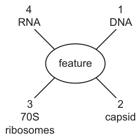
Which biological features are present in typical prokaryotes?
Options
A 1, 2 and 3
B 1, 2 and 4
C 1, 3 and 4
D 2, 3 and 4
Working
Prokaryotes (e.g. bacteria) are cells and contain:
- 1 (DNA) ✓ — a single circular chromosomal DNA, plus plasmids
- 3 (70S ribosomes) ✓ — smaller than the 80S ribosomes of eukaryotic cytoplasm
- 4 (RNA) ✓ — mRNA, tRNA and rRNA are all produced and used in prokaryotes
A capsid (2) is the protein coat of a virus. Prokaryotes are not viruses and do not possess a capsid.
Features 1, 3 and 4 are present in typical prokaryotes.
Answer
C
C
Background Concept
Prokaryotes are organisms whose cells lack a true membrane-bound nucleus and other membrane-bound organelles. Bacteria and archaea are prokaryotes. Their defining cellular features include:
- A single circular chromosome of DNA located in a region called the nucleoid (not enclosed by a membrane)
- Small plasmids (also circular DNA) carrying accessory genes
- 70S ribosomes (smaller than the 80S ribosomes found free in the cytoplasm of eukaryotes; S = Svedberg unit, a measure of how fast a particle sediments in a centrifuge and therefore an indication of size/shape)
- RNA of all the usual types: mRNA (transcribed from DNA and translated directly, since prokaryotes have no nucleus to separate transcription from translation), tRNA and rRNA
- A cell wall made of peptidoglycan (in bacteria)
Viruses are non-cellular particles. They consist of a nucleic acid core (DNA or RNA) surrounded by a protein coat called a capsid, and sometimes an outer envelope. They have no ribosomes, no cytoplasm, and no metabolism of their own — they are obligate intracellular parasites that can only replicate inside a host cell. The capsid is built from repeating protein subunits called capsomeres, and it protects the viral nucleic acid and helps the virus attach to host cells.
Understanding the Question
Fig. 5.1 lists four biological features:
- DNA
- capsid
- 70S ribosomes
- RNA
The question asks which combination of these is present in typical prokaryotes. A "typical" prokaryote here means a general bacterial cell — not an archaeon (some of which differ in detail) and certainly not a virus. The candidate must recognise which features belong to a prokaryotic cell and rule out features that are exclusive to other kinds of biological entity (in this case, the capsid, which is a viral structure).
Approach
For each of the four features, decide whether it is found inside a prokaryotic cell:
- DNA — prokaryotes have DNA, so feature 1 belongs.
- Capsid — this is exclusively a viral structure; a cellular organism does not have a capsid. Feature 2 does NOT belong.
- 70S ribosomes — these are the characteristic ribosome type of prokaryotes. Feature 3 belongs.
- RNA — prokaryotes transcribe and translate RNA as part of gene expression. Feature 4 belongs.
Features 1, 3 and 4 are correct, giving option C.
Step-by-Step Reasoning
-
Feature 1 — DNA: PRESENT. Every cellular organism uses DNA as its genetic material. Prokaryotes carry a single circular DNA molecule (the bacterial chromosome) and frequently also small circular plasmids. DNA is therefore a defining feature of prokaryotic cells.
-
Feature 2 — capsid: ABSENT. A capsid is a protein shell that encloses the nucleic acid of a virus. It is made of capsomere subunits and is part of the virion (the complete virus particle). Prokaryotes are complete cells, not viruses, and they do not make a capsid. A capsule (sometimes confused with "capsid") is an external slime/polysaccharide layer on some bacteria — but the question specifies capsid, not capsule, and even a capsule is not a feature of all typical prokaryotes.
-
Feature 3 — 70S ribosomes: PRESENT. Prokaryotic ribosomes are 70S, made of a 50S large subunit and a 30S small subunit. Eukaryotic cytoplasmic ribosomes are larger at 80S (60S + 40S). The size difference is the basis for many antibiotics (e.g. tetracyclines, streptomycin) which selectively inhibit bacterial protein synthesis without harming the host's 80S ribosomes.
-
Feature 4 — RNA: PRESENT. Prokaryotes produce all the RNA types they need:
- mRNA, transcribed from the DNA and immediately available for translation (no nuclear membrane to cross)
- tRNA, which delivers amino acids to the ribosome during translation
- rRNA, which forms the structural and catalytic core of the ribosome
So RNA is firmly a feature of prokaryotes.
-
Selection. The three features present in typical prokaryotes are 1, 3 and 4. The only option containing exactly this set is C.
Key Takeaways
- Typical prokaryotic cells contain DNA, RNA and 70S ribosomes.
- A capsid is a viral structure, never found in cellular organisms; this is one of the clearest distinctions between viruses and cells.
- The "S" in 70S/80S is a Svedberg unit, reflecting sedimentation rate — it is related to but not strictly equal to mass.
- The smaller size of prokaryotic ribosomes is exploited by many antibiotics, which is a key link between cell structure and medicine.
Common Mistakes
- Confusing "capsid" with "capsule". A capsule is an external polysaccharide layer found on some bacteria (it helps with adhesion and resisting phagocytosis), whereas a capsid is the protein coat of a virus. Only the capsid is relevant here, and it is not present in prokaryotes.
- Thinking prokaryotes "have no DNA" because they have no nucleus. This is wrong — they still have DNA, it is just not enclosed in a nuclear envelope.
- Choosing 80S ribosomes for prokaryotes. 80S ribosomes are characteristic of eukaryotic cytoplasm; prokaryotes have 70S. (Note: the ribosomes inside mitochondria and chloroplasts are also 70S, reflecting their prokaryotic evolutionary origin.)
- Selecting RNA as absent. RNA is essential for translation in every cell, including prokaryotes.
Things to Be Careful About
- The mark scheme is testing precise knowledge of what is and is not a prokaryotic feature. A vague answer that "prokaryotes are simple" is not enough; you must identify the three correct features individually.
- "Typical" prokaryote = bacterium. Do not let unusual exceptions (e.g. some archaea) distract from the standard features the syllabus requires you to know.
- Read the question carefully: it asks which features are present in prokaryotes, not which are unique to them. RNA, for example, is present in prokaryotes, eukaryotes and many viruses, but it is still a feature found in prokaryotes.
Which row shows features that occur in dicotyledonous plant cells and also in typical bacterial cells?
key
✓ = found in dicotyledonous plant cells and also in typical bacterial cells
✗ = not found in at least one of these two types of cell
Options
| 70S ribosomes | 80S ribosomes | centrioles | circular DNA | |
|---|---|---|---|---|
| A | ✓ | ✗ | ✓ | ✗ |
| B | ✓ | ✗ | ✗ | ✓ |
| C | ✗ | ✓ | ✓ | ✗ |
| D | ✗ | ✓ | ✗ | ✓ |
Working
For each feature, decide whether it is found in BOTH dicotyledonous plant cells AND typical bacterial cells:
- 70S ribosomes: ✓ — present in bacteria (their cytoplasmic ribosomes) and also in plant cells (inside chloroplasts and mitochondria).
- 80S ribosomes: ✗ — present in plant cytoplasm, but bacteria have only 70S ribosomes.
- Centrioles: ✗ — absent from plant cells (and from bacteria).
- Circular DNA: ✓ — the main bacterial chromosome is circular; plant cells also contain circular DNA in chloroplasts and mitochondria.
The pattern ✓, ✗, ✗, ✓ matches row B.
Answer
B
B
Background Concept
All cells fall into two broad categories: prokaryotic (e.g. bacteria) and eukaryotic (e.g. plant, animal and fungal cells). Prokaryotes are smaller, lack a true nucleus, and have a simpler internal organisation. Eukaryotes possess membrane-bound organelles, including a nucleus.
A common misconception is that the only ribosomes/DNA present in a eukaryotic cell are the 80S ribosomes and linear nuclear DNA. In fact, mitochondria and (in plant cells) chloroplasts are thought to have arisen from ancient prokaryotic endosymbionts and retain several prokaryotic-like features:
- 70S ribosomes (smaller, like those of modern bacteria), in addition to the 80S ribosomes of the cytoplasm.
- Circular DNA that is not associated with histones, again like bacterial DNA.
So a feature present in bacteria is not automatically absent from eukaryotes — it may still appear inside an organelle.
Understanding the Question
The question asks which row of features is found in both dicotyledonous plant cells and typical bacterial cells. The key is:
- ✓ — the feature is present in both cell types
- ✗ — the feature is missing from at least one of the two cell types
Each feature must therefore be evaluated twice: once in the context of a plant cell, and once in the context of a bacterium.
Approach
Run through each of the four features, recalling:
- What ribosomes a bacterium has (only 70S)
- What ribosomes a plant cell has (80S in cytoplasm, 70S in chloroplasts/mitochondria)
- Whether centrioles exist in plants (no) or bacteria (no)
- Where circular DNA is found (bacterial nucleoid; plant chloroplasts and mitochondria)
Then read off the matching row.
Step-by-Step Reasoning
-
70S ribosomes
- Bacteria: their cytoplasmic ribosomes are 70S → present.
- Plant cells: 80S in the cytoplasm, but 70S inside chloroplasts and mitochondria → present.
- Result: ✓ (in both)
-
80S ribosomes
- Bacteria: only have 70S ribosomes → absent.
- Plant cells: present in cytoplasm.
- Result: ✗ (not in bacteria, so not in both)
-
Centrioles
- Bacteria: have no centrioles (no microtubule-organising centre of this type).
- Plant cells: generally lack centrioles — they use other MTOCs for spindle formation.
- Result: ✗ (not in either)
-
Circular DNA
- Bacteria: their single chromosome is a circular molecule of DNA located in the nucleoid → present.
- Plant cells: chloroplasts and mitochondria each contain a small circular DNA molecule → present.
- Result: ✓ (in both)
The required pattern is ✓, ✗, ✗, ✓ — which is row B.
Key Takeaways
- Prokaryotes and eukaryotes share some features because of endosymbiotic origins of mitochondria and chloroplasts (70S ribosomes and circular DNA inside those organelles).
- 80S ribosomes and centrioles are genuinely eukaryote-specific features and are not found in bacteria.
- When asked "found in BOTH", the feature must be present in each cell type — the absence of it from even one disqualifies a ✓.
Common Mistakes
- Forgetting organelle-origin features: thinking plant cells only have 80S ribosomes and linear DNA, leading to a ✗ for 70S ribosomes and circular DNA, and so wrongly picking a row other than B.
- Treating "plant" and "animal" as interchangeable: centrioles are a feature of animal cells, not plant cells; do not assume every eukaryotic feature is present in plants.
- Misreading the key: ✗ means "not in at least one", not "in neither" — so a feature absent from bacteria alone is still ✗.
Things to Be Careful About
- Some textbooks list centrioles as present in lower plants, but for A-level purposes the safe assumption is that dicotyledonous plant cells do not contain centrioles.
- Be precise with ribosome S-values: 70S in bacteria and in chloroplasts/mitochondria of eukaryotes; 80S in the eukaryotic cytoplasm.
- Distinguish "circular DNA" (a structural feature of bacterial chromosomes and organelle genomes) from the linear, histone-bound DNA of the eukaryotic nucleus.
Which flow chart outlining the test for non-reducing sugars is correct?
Options
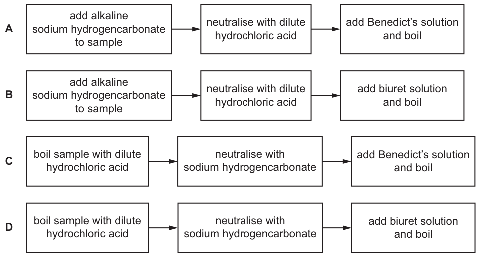
Working
The standard test for a non-reducing sugar (e.g. sucrose) requires three ordered steps:
- Hydrolyse the glycosidic bond by boiling the sample with dilute hydrochloric acid — this breaks the disaccharide into its reducing monosaccharide components.
- Neutralise the acid with sodium hydrogencarbonate (an alkali), because Benedict's reagent only works under alkaline conditions.
- Add Benedict's solution and boil — a brick-red precipitate confirms the presence of the (now reducing) sugar.
Checking the options:
- A — adds sodium hydrogencarbonate first and then "neutralises with HCl": the order is reversed and the sample is never hydrolysed.
- B — same reversed order, and biuret is used instead of Benedict's; biuret detects peptide bonds (proteins), not sugars.
- C — boil with HCl → neutralise with sodium hydrogencarbonate → add Benedict's and boil. This is the correct sequence.
- D — correct order of hydrolysis and neutralisation, but biuret is used instead of Benedict's; biuret is not a test for sugars.
Answer
C
C
Background Concept
Sugars are classified as reducing or non-reducing depending on whether they possess a free aldehyde (–CHO) or ketone group capable of donating electrons. Glucose, fructose, maltose and lactose are reducing sugars; sucrose is the classic non-reducing sugar because its two monosaccharide units are linked through both their anomeric carbons, leaving no free reducing group.
To test for a non-reducing sugar, the disaccharide must first be hydrolysed into its reducing monosaccharides, and only then can Benedict's reagent be used:
- Benedict's reagent is alkaline and contains copper(II) sulfate (Cu²⁺, blue). When heated with a reducing sugar, Cu²⁺ is reduced to Cu⁺, which precipitates as red copper(I) oxide. If the solution is not alkaline, or if no reducing group is present, the reagent stays blue.
- The biuret test is for peptide bonds in proteins — alkaline copper(II) sulfate is reduced by peptide bonds to give a violet/purple colour. It is never used to detect sugars.
Understanding the Question
The question presents four flow charts (A–D) that combine different starting reagents, different neutralising agents, and different test reagents. The student must identify the one chart that:
- Starts by hydrolysing the sample with dilute hydrochloric acid (boiled),
- Neutralises the acid with sodium hydrogencarbonate (an alkali),
- Uses Benedict's solution (not biuret) and boils to detect the now-reducing sugar.
The command word is implicit ("Which…is correct?") — only one option satisfies all three conditions.
Approach
Recall the canonical order of the non-reducing sugar test, then check each option against two criteria:
- Order of reagents: acid hydrolysis must occur before neutralisation, not after.
- Identity of test reagent: Benedict's (sugars), not biuret (proteins).
Eliminate any option that fails on either criterion.
Step-by-Step Reasoning
- Option A — begins with sodium hydrogencarbonate, then "neutralises with HCl". This is the reverse of the correct procedure; the sample is never hydrolysed and Benedict's would be added to a strongly acidic mixture (which would not work because the reaction needs alkaline conditions). Reject.
- Option B — same reversed order as A, and the final reagent is biuret. Biuret detects peptide bonds in proteins, not reducing sugars. Reject on both counts.
- Option C — boil sample with dilute HCl (hydrolyses glycosidic bond) → neutralise with sodium hydrogencarbonate → add Benedict's solution and boil. The order is correct and the test reagent is correct. Accept.
- Option D — correct hydrolysis and neutralisation order, but the final reagent is biuret. Even though the first two steps are right, biuret cannot detect a sugar. Reject.
A positive result with C would be a colour change from blue → green → yellow → orange → brick-red precipitate of copper(I) oxide, confirming the original non-reducing sugar was hydrolysed to a reducing one.
Key Takeaways
- The non-reducing sugar test has a fixed sequence: dilute HCl + heat (hydrolysis) → sodium hydrogencarbonate (neutralisation) → Benedict's + heat (detection).
- Benedict's reagent must be alkaline to work; acid must be neutralised before it is added.
- Benedict's detects reducing sugars (and, after hydrolysis, non-reducing sugars); biuret detects proteins. Mixing these up is a common error.
Common Mistakes
- Reversing the order: neutralising before hydrolysing leaves the glycosidic bond intact, so Benedict's gives a negative result regardless of what was in the sample.
- Using biuret as the final reagent — this is a test for proteins and never produces a colour change with sugars.
- Forgetting that Benedict's solution must be boiled with the sample to provide the activation energy for the reduction of Cu²⁺.
- Adding Benedict's to an acidic solution — the blue colour of Cu²⁺ is not diagnostic under acid conditions.
Things to Be Careful About
- The reagent labelled "alkaline sodium hydrogencarbonate" in distractors A and B is a red herring: it is a base, not an acid, so it cannot hydrolyse the disaccharide.
- "Neutralise with dilute hydrochloric acid" (in A and B) is impossible after the addition of sodium hydrogencarbonate without a strong acid–base reaction that would not test for sugars in any meaningful way.
- In the correct procedure, sodium hydrogencarbonate is added slowly until effervescence stops — this is the visual indicator that neutralisation is complete.
- Benedict's test is itself a screening test for reducing sugars; a non-reducing sugar test is therefore a two-stage procedure that converts a negative Benedict's result into a positive one only when a non-reducing sugar was originally present.
Which diagrams show the release of a water molecule during the formation of a glycosidic bond?
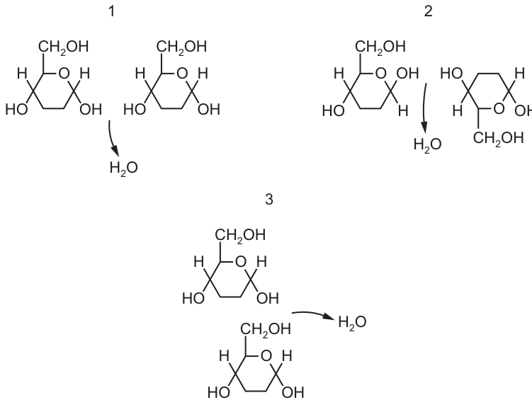
Options
A 1, 2 and 3
B 1 only
C 2 and 3 only
D 3 only
Working
A glycosidic bond is formed by a condensation reaction between two hydroxyl (–OH) groups, releasing one molecule of water. Check each diagram for two –OH groups combining to release :
- Diagram 1: C1 –OH of the first -glucose joins C4 –OH of the second -glucose, releasing → 1,4-glycosidic bond. ✓
- Diagram 2: C1 –OH of the first glucose joins C4 –OH of the inverted second glucose, releasing → 1,4-glycosidic bond. ✓
- Diagram 3: C1 –OH of the top glucose joins C6 –OH of the bottom glucose, releasing → 1,6-glycosidic bond. ✓
All three diagrams show valid glycosidic bond formation with the release of water.
Answer
A
A
Background Concept
Glucose is a monosaccharide with the molecular formula . Its carbon atoms are numbered C1 to C6, with hydroxyl (–OH) groups attached to C1, C2, C3, C4 and C6. Because each carbon carries a reactive –OH, two glucose molecules can join by a condensation reaction between any two of these hydroxyl groups. The reaction removes an –H from one hydroxyl and an –OH from the other, releasing a molecule of water () and leaving a covalent C–O–C link called a glycosidic bond.
The position of the bond matters:
- A 1,4-glycosidic bond (e.g. between C1 of one glucose and C4 of the next) is found in maltose and is the dominant linkage in starch, glycogen and cellulose.
- A 1,6-glycosidic bond (between C1 of one glucose and C6 of the next) creates a branch point in amylopectin and glycogen.
The reverse process — adding water to break a glycosidic bond — is hydrolysis.
Understanding the Question
The question shows three separate diagrams, each depicting two glucose molecules with an arrow pointing from a pair of –OH groups to a water molecule. The task is to decide which diagrams correctly represent the formation of a glycosidic bond by condensation. A common trap is to assume that only the "standard" 1,4 linkage counts, or to miss that the second glucose in diagram 2 is simply rotated through 180° — this does not change the chemistry, only the orientation of the drawing.
The command word is implicit ("Which diagrams show…") — the candidate must evaluate each one against the definition of glycosidic bond formation.
Approach
For each diagram, ask two questions:
- Are the reacting groups both –OH groups on two glucose carbons?
- Is the reaction shown as condensation (water released) and not hydrolysis (water added)?
If both answers are yes, the diagram represents glycosidic bond formation. The orientation of the molecule and the specific carbons involved (1,4 or 1,6) do not change this conclusion.
Step-by-Step Reasoning
Diagram 1: Two -glucose molecules are drawn side by side in the standard orientation. The arrow starts at the –OH on C1 of the left glucose and the –OH on C4 of the right glucose, pointing to . This is a textbook 1,4-glycosidic condensation — valid.
Diagram 2: The left glucose is in the standard orientation, but the right glucose has been flipped upside down (rotated 180°). Despite the rotation, the –OH on C1 of the left glucose still joins an –OH on C4 of the right glucose, with water released. The bond is still 1,4. The drawing is unconventional, but the chemistry is identical to diagram 1 — valid.
Diagram 3: Two glucose molecules are drawn one above the other. The –OH on C1 of the top molecule joins the –OH on C6 (the group) of the bottom molecule, releasing water. This forms a 1,6-glycosidic bond — exactly the branch-point linkage in amylopectin and glycogen — valid.
Because all three satisfy the definition of glycosidic bond formation by condensation, the answer is A: 1, 2 and 3.
Key Takeaways
- A glycosidic bond forms whenever two –OH groups (on sugar carbons) undergo condensation, releasing .
- The bond can be 1,4 or 1,6 (and 1,1 for trehalose, 1,2 for sucrose etc.) — all are genuine glycosidic bonds.
- The orientation in which the second sugar is drawn (rotated, inverted, stacked) does not change the chemistry.
- Watch the arrow: an arrow from –OH groups to water = condensation; an arrow from water to the bond = hydrolysis.
Common Mistakes
- Choosing B or C because only the 1,4 linkage was remembered as "the" glycosidic bond. Diagram 3 (1,6) is also valid.
- Rejecting diagram 2 because the second glucose "looks upside-down". The carbons still match — only the orientation is different.
- Confusing condensation with hydrolysis: if the arrow runs from into the bond, the bond is being broken, not formed.
Things to Be Careful About
- Always check that the reacting groups are actually –OH on two sugar carbons, not, for example, a hydrogen on one side.
- Remember that both 1,4 and 1,6 glycosidic bonds exist in nature and both form by the same condensation mechanism.
- The group on C6 of glucose carries the –OH involved in 1,6 linkages — easily missed if the diagram is small.
Which molecules have a structural formula that contains bonds?
1 amino acids
2 fatty acids
3 glycerol
4 protein
Options
A 1, 2 and 3
B 1, 2 and 4
C 1, 3 and 4
D 2, 3 and 4
Answer
B — 1, 2 and 4
Amino acids (1) contain a carboxyl group (–COOH), fatty acids (2) contain a carboxyl group at one end of the hydrocarbon chain, and proteins (4) contain C=O within the peptide bond (–CO–NH–) linking amino acids. Glycerol (3) has only hydroxyl (–OH) groups on its three-carbon backbone and contains no C=O bonds.
B
Background Concept
Many biologically important molecules are defined by the functional groups they carry. A carbon–oxygen double bond (C=O), sometimes called a carbonyl group, is the defining feature of two related groups:
- Carboxyl group (–COOH): C=O bonded to a hydroxyl (–OH) on the same carbon. This is the acidic group found in carboxylic acids.
- Carbonyl (C=O) within a larger structure: e.g. the C=O of an amide (peptide) bond, or the C=O of a ketone/aldehyde.
Glycerol, by contrast, is a trihydric alcohol: three carbons each bearing a hydroxyl (–OH) group but no C=O anywhere on the molecule.
Understanding the Question
The question gives four candidate molecules and asks which of them have a C=O in their structural formula. The four options A–D differ only in the presence or absence of one of the molecules (3 glycerol is the variable — does it contain C=O?), so the test hinges on knowing the functional groups of glycerol compared with those of amino acids, fatty acids and proteins.
Approach
Decide, for each molecule, whether a C=O bond is part of its structure:
- Examine the functional groups of an amino acid (the general formula: ).
- Examine the structure of a fatty acid (a long hydrocarbon chain terminating in –COOH).
- Examine the structure of glycerol ().
- Examine the peptide bond that links amino acids in a protein.
Step-by-Step Reasoning
1. Amino acids — YES, contains C=O.
Every amino acid has the general structure with at least one carboxyl group (–COOH). The carbon of this group is double-bonded to one oxygen. So C=O is present.
2. Fatty acids — YES, contains C=O.
A fatty acid is a long-chain carboxylic acid: . The terminal –COOH again contains a C=O double bond.
3. Glycerol — NO C=O.
Glycerol is propane-1,2,3-triol, . All three carbons are bonded only to –OH (and to H or C). There is no carbon–oxygen double bond anywhere in the molecule.
4. Protein — YES, contains C=O.
A protein is a polymer of amino acids joined by peptide bonds. Each peptide bond has the form –CO–NH–, and the C=O of this amide linkage is present in every residue of the polypeptide backbone. Many side chains (e.g. those of aspartate, glutamate, asparagine, glutamine) also contain additional C=O groups.
Counting: 1 ✓, 2 ✓, 3 ✗, 4 ✓ → 1, 2 and 4, which is option B.
Key Takeaways
- A C=O bond is present in any carboxyl group (–COOH) and in any amide (peptide) bond (–CO–NH–).
- Amino acids, fatty acids and proteins all contain C=O; glycerol does not.
- Recognising functional groups in molecular structures is a recurring skill — it underpins topics from lipid structure to enzyme mechanism to DNA chemistry.
Common Mistakes
- Assuming glycerol contains C=O because it is part of a triglyceride. The C=O bonds in a triglyceride come from the fatty acid carboxyl groups, not from glycerol itself. Glycerol contributes only –OH groups, which become ester linkages (C–O–C) when joined to fatty acids — and an ester C–O–C linkage has only single C–O bonds, not C=O.
- Forgetting that peptide bonds contain C=O. Some students remember peptide bonds as –CO–NH– but overlook the double-bonded oxygen, which is the very group the question is testing.
Things to Be Careful About
- When the mark scheme says "structural formula", it means a drawn-out formula showing all bonds (including double bonds) — not the molecular formula alone. So a fatty acid written as (the molecular formula) does not on its own reveal a C=O, but its structural formula clearly does.
- The carboxyl C=O and the peptide C=O are both carbonyls; the question only requires recognising that the C=O is present, not distinguishing the two contexts.
Which statement about triglycerides is correct?
Options
A In any triglyceride, all the fatty acids are saturated or all the fatty acids are unsaturated.
B Two triglycerides are joined together by an ester bond.
C Triglycerides are polar hydrophobic molecules.
D Glycerol is joined to each fatty acid by a covalent bond.
Working
A triglyceride is formed when glycerol (a trihydric alcohol) reacts with three fatty acids via three ester linkages (covalent bonds formed by condensation).
Evaluating each option:
- A is incorrect: a triglyceride can contain any combination of saturated and unsaturated fatty acids — there is no requirement that they be all one type.
- B is incorrect: triglyceride molecules are not joined to one another by ester bonds; ester bonds join fatty acids to glycerol within a single triglyceride.
- C is incorrect: triglycerides are non-polar (hydrophobic), not polar. They are insoluble in water but soluble in organic solvents.
- D is correct: each fatty acid is joined to glycerol by an ester bond, which is a covalent bond formed in a condensation reaction.
Answer
D
D
Background Concept
A triglyceride (triacylglycerol) is formed from one glycerol molecule and three fatty acid molecules. The three –OH groups on glycerol each react with the –COOH group of a fatty acid in a condensation reaction, releasing a molecule of water and forming an ester bond (–COO–). An ester bond is a type of covalent bond, in which electron pairs are shared between the bonded atoms.
Key properties of triglycerides:
- They are non-polar (hydrophobic) because the long hydrocarbon chains of the fatty acids dominate the molecule's character.
- They are insoluble in water but soluble in organic solvents such as ethanol and ether.
- They serve as an efficient energy storage molecule, yielding more than twice the energy per gram compared with carbohydrates.
Fatty acids may be saturated (only C–C single bonds in the hydrocarbon chain) or unsaturated (containing one or more C=C double bonds). A single triglyceride can contain a mixture of these — there is no rule restricting the type.
Understanding the Question
This is a multiple-choice question (Paper 1, AS Level) asking the candidate to identify the correct statement about triglycerides from four options. The command is essentially "evaluate each statement and choose the true one." No calculation is required — the skill is precise recall of triglyceride structure and properties.
Approach
Go through each option systematically and judge whether the statement is biologically accurate. Eliminate any that contain a factual error:
- Option A — claims all fatty acids in a triglyceride must be the same type. This is false; triglycerides commonly contain a mix.
- Option B — claims triglycerides are joined to each other by ester bonds. Ester bonds occur within a triglyceride (between glycerol and each fatty acid), not between triglyceride molecules.
- Option C — calls triglycerides "polar hydrophobic." The two terms are contradictory in this context: hydrophobic means water-fearing/non-polar, so a "polar hydrophobic" molecule is a misnomer.
- Option D — states glycerol is joined to each fatty acid by a covalent bond. This is true; the ester linkage is a covalent bond.
Step-by-Step Reasoning
Why A is wrong: Triglycerides in food and in body stores (e.g. adipose tissue) typically contain a mixture of saturated, monounsaturated and polyunsaturated fatty acids. There is no biological requirement for homogeneity. A vegetable oil, for example, may contain triglycerides with three different fatty acids esterified to glycerol.
Why B is wrong: The ester bond in a lipid is between a fatty acid's carboxyl group (–COOH) and glycerol's hydroxyl group (–OH). It is an intramolecular bond — it holds the three fatty acids to the glycerol backbone within ONE triglyceride. Triglyceride molecules are not bonded to one another; they aggregate via weak hydrophobic interactions, not covalent bonds.
Why C is wrong: Triglycerides consist predominantly of long hydrocarbon (C–H) chains, which are non-polar. The molecule as a whole is therefore non-polar and hydrophobic. "Polar hydrophobic" is an oxymoron — polar molecules tend to be hydrophilic. Phospholipids, by contrast, have a polar phosphate head and non-polar fatty acid tails, making them amphipathic, but triglycerides do not have this dual character.
Why D is correct: Each of the three fatty acids in a triglyceride is joined to glycerol via an ester bond, which is a covalent bond (specifically, formed by a condensation reaction between –OH and –COOH with the loss of water). All bonds in a triglyceride are covalent.
Key Takeaways
- A triglyceride = glycerol + 3 fatty acids, joined by 3 ester (covalent) bonds.
- Ester bonds are covalent, formed by condensation (releasing water).
- Triglycerides are non-polar and hydrophobic.
- Fatty acids within a triglyceride can be a mix of saturated and unsaturated types.
Common Mistakes
- Confusing the location of the ester bond: students sometimes think ester bonds join triglyceride molecules together; in fact, they join fatty acids to glycerol within a single triglyceride.
- Saying triglycerides are "polar" because they contain the C=O of the ester group — the long hydrocarbon tails dominate and the molecule overall is non-polar.
- Believing all fatty acids in a triglyceride must be the same type (saturated or unsaturated). This is not the case.
Things to Be Careful About
- "Hydrophobic" and "non-polar" are essentially synonymous in this context — do not use them as if they were independent properties.
- Distinguish between intramolecular bonds (within one molecule, e.g. ester bonds in a triglyceride) and intermolecular interactions (between molecules, e.g. hydrophobic interactions between triglyceride molecules).
- An ester bond is one specific type of covalent bond, but the question rewards the broader term "covalent bond," which is correct.
Many flowers produce a sweet solution called nectar. Bees provided with nectar use enzyme Q to change the nectar into honey.
After testing a sample of nectar for the presence of reducing sugar using standard laboratory reagents, the sample was blue. After testing a sample of honey in the same way, the sample was orange.
Which conclusion about the reaction catalysed by enzyme Q is consistent with these results?
Options
| type of reaction | substrate | product | |
|---|---|---|---|
| A | condensation | maltose | glucose |
| B | condensation | sucrose | fructose |
| C | hydrolysis | sucrose | fructose |
| D | hydrolysis | maltose | glucose |
Working
Benedict's reagent gives:
- blue = reducing sugar absent (nectar)
- orange = reducing sugar present (honey)
So enzyme Q converts a non-reducing sugar into reducing sugars.
- Condensation forms glycosidic bonds (joins monosaccharides); it cannot break a non-reducing sugar down into reducing sugars. This rules out A and B.
- Maltose is itself a reducing sugar, so if maltose were the substrate, nectar would already give an orange (positive) result. This rules out D.
- Sucrose is non-reducing (both anomeric carbons are locked in the glycosidic bond), so nectar with only sucrose tests blue. Hydrolysis of sucrose gives glucose + fructose, both of which are reducing sugars — so honey tests orange. ✓
Answer
C
C
Background Concept
Benedict's test for reducing sugars. Benedict's reagent contains Cu²⁺ ions complexed with citrate in alkaline solution. When heated with a reducing sugar, the Cu²⁺ is reduced to insoluble red/orange Cu₂O, so a positive result gives a colour change from blue (reagent colour) through green/yellow to orange and finally brick-red. A solution that stays blue after boiling contains no reducing sugar.
A sugar is "reducing" when it has a free aldehyde (CHO) group, or a free ketone group that can open to form one, capable of donating electrons and reducing Cu²⁺. A sugar is "non-reducing" when both of its anomeric (C1 in aldose / C2 in ketose) carbons are tied up in a glycosidic bond — no free end is available to reduce Cu²⁺.
- Glucose – free C1 aldehyde → reducing.
- Fructose – free C2 ketone (tautomerises) → reducing.
- Maltose – α-1,4 bond between two glucoses; one glucose's C1 is free → reducing.
- Sucrose – α-1,β-2 bond between glucose C1 and fructose C2; both anomeric carbons are in the bond → non-reducing.
Hydrolysis vs condensation.
- A condensation reaction forms a glycosidic bond by removing water (), joining monosaccharides into a disaccharide or polysaccharide. The enzyme that does this is a synthetase/ligase-type activity.
- A hydrolysis reaction breaks a glycosidic bond by adding water, splitting a disaccharide into its constituent monosaccharides. Examples: sucrase (invertase) hydrolyses sucrose; maltase hydrolyses maltose.
Understanding the Question
Two pieces of evidence are given:
- Nectar + Benedict's → blue (no reducing sugar present).
- Honey + Benedict's → orange (reducing sugar present).
Enzyme Q converts nectar into honey. We need the row in the table whose substrate, product, and reaction type are all consistent with the data.
Approach
Ask three questions in order:
- Must the substrate be reducing or non-reducing? (Nectar was blue, so substrate must be non-reducing. This eliminates maltose as substrate.)
- Must the products be reducing? (Honey was orange, so products must include at least one reducing sugar.)
- Does the named reaction match the direction of change (breakdown of a disaccharide into monosaccharides)? (That is hydrolysis, not condensation.)
Only one option survives all three tests.
Step-by-Step Reasoning
Eliminate by reaction type — A and B are wrong.
Both A and B call the reaction a condensation. A condensation joins two monosaccharides into a disaccharide and releases water; it does not split a disaccharide into monosaccharides, so it cannot be the reaction that turns nectar into honey. A and B are out.
Eliminate by substrate — D is wrong.
Option D says the substrate is maltose. But maltose is itself a reducing sugar — Benedict's on pure maltose would go orange, not blue. Since nectar tested blue, the nectar cannot contain maltose. D is out.
Confirm C.
- Substrate = sucrose. Sucrose is the canonical non-reducing disaccharide (the only common dietary sugar with no free anomeric carbon), so nectar containing only sucrose correctly tests blue.
- Reaction = hydrolysis. The glycosidic bond in sucrose is cleaved by the addition of a water molecule:
- Products = glucose + fructose. Both are reducing sugars, so the resulting honey gives the orange Benedict's result.
All three observations are satisfied → C is correct.
Key Takeaways
- A negative Benedict's result (blue) tells you the sugar is non-reducing — for the disaccharides on this syllabus, that means sucrose.
- Hydrolysis of a disaccharide always uses water to split the glycosidic bond and release monosaccharides, all of which are reducing.
- Condensation is the opposite direction (building up), and would not generate new reducing ends from a non-reducing starting material.
Common Mistakes
- Confusing hydrolysis and condensation. Many students pick B because "sucrose → fructose" looks plausible, forgetting that condensation builds rather than breaks the disaccharide. Condensation never produces monosaccharides from a disaccharide.
- Forgetting that maltose is reducing. Picking D because it names a disaccharide is the trap. Because maltose itself reduces Benedict's, the starting nectar would already have been orange, contradicting the blue result.
- Confusing glucose and fructose with sucrose. Glucose and fructose are reducing because they have a free anomeric carbon; sucrose is non-reducing because both anomeric carbons are locked in the 1,2-glycosidic bond.
Things to Be Careful About
- The Benedict's colours to remember: blue = reagent only (negative); green/yellow/orange/brick-red = progressively more reducing sugar present.
- The rule for whether a disaccharide is reducing: at least one of the two anomeric carbons must be free. Sucrose fails this rule; maltose, lactose and cellobiose pass it.
- "Enzyme Q" in the stem is fictional — do not try to identify a real enzyme; the question only requires you to match the type of reaction to the data.
- "Fructose alone" (option B) cannot be the only product of any clean disaccharide breakdown — hydrolysis of sucrose gives equimolar glucose and fructose, which is the biologically correct answer.
The diagram shows the structure of part of a peptidoglycan molecule.
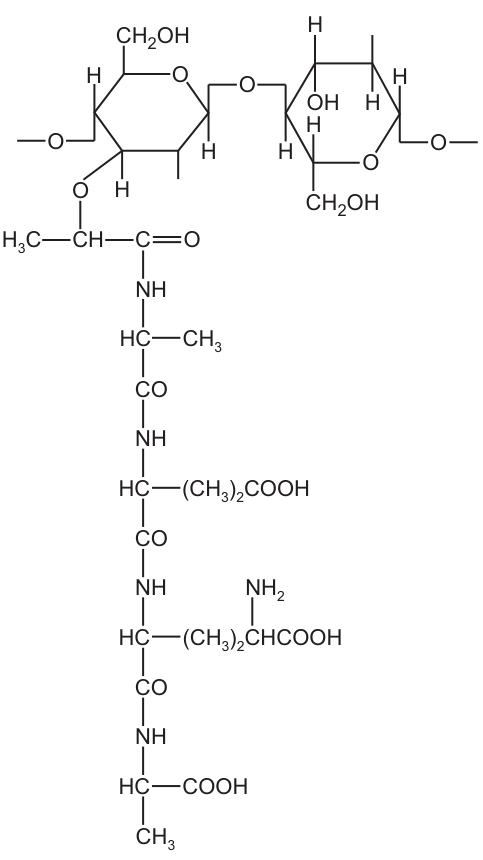
Which type of 1,4 linkage and how many peptide bonds are shown in this part of the molecule?
Options
| type of 1,4 linkage | number of peptide bonds | |
|---|---|---|
| A | -1,4 | 3 |
| B | -1,4 | 4 |
| C | -1,4 | 3 |
| D | -1,4 | 4 |
Working
The diagram shows the structure of peptidoglycan, the polymer that makes up bacterial cell walls. The two sugar rings (N-acetylglucosamine, NAG, and N-acetylmuramic acid, NAM) are connected by a β-1,4 glycosidic linkage (the oxygen bridge is in the β position, i.e. on the opposite side of the ring from the CH2OH group — analogous to cellulose, not starch).
The short peptide hanging off the NAM unit contains four amino acids (L-alanine, D-glutamic acid, L-lysine and D-alanine) joined through their α-carboxyl and α-amino groups, plus the bond linking the C-terminus of the muramic acid lactyl group to the N-terminus of L-alanine:
- lactyl –CO–NH– L-Ala
- L-Ala –CO–NH– D-Glu
- D-Glu –CO–NH– L-Lys
- L-Lys –CO–NH– D-Ala
That gives 4 peptide (amide) bonds.
Answer
D
D
Background Concept
Peptidoglycan (also called murein) is the structural polymer of bacterial cell walls. Its carbohydrate backbone is built from two alternating sugar derivatives:
- N-acetylglucosamine (NAG) – glucose with an –NHCOCH3 group on C2.
- N-acetylmuramic acid (NAM) – NAG with a lactyl (–O–CH(CH3)–COOH) group on C3, through which a short peptide is attached.
The two sugars are joined by a β-1,4 glycosidic bond (the oxygen bridge projects from C1 in the β-configuration, i.e. on the opposite face of the pyranose ring from the C6 –CH2OH). This is the same linkage as in cellulose. It is quite different from the α-1,4 linkage found in starch and glycogen, which is why lysozyme — an enzyme that cleaves β-1,4 bonds — can hydrolyse peptidoglycan but not starch.
Hanging off each NAM is a short stem peptide. The classical tetrapeptide in many Gram-positive bacteria is:
L-Ala — D-Glu — L-Lys — D-Ala
These amino acids are joined head-to-tail by peptide (amide) bonds of the form –CO–NH–, formed by condensation between the α-carboxyl group of one residue and the α-amino group of the next. A peptide of n amino acids contains n – 1 peptide bonds between amino acids, but when the chain is anchored to the muramic acid lactyl group, the bond joining the lactyl –COOH to the first amino acid (L-Ala) is also a peptide (amide) bond, so the total is n peptide bonds for an n-residue stem.
Understanding the Question
The figure shows a fragment of a peptidoglycan molecule containing:
- one disaccharide of NAG–NAM (with a dashed continuation on each side indicating that the chain extends), and
- the tetrapeptide side chain attached to the NAM residue, with all four amino acids drawn out and the –CO–NH– linkages labelled.
You are asked to identify:
- the type of glycosidic linkage joining the two sugars, and
- the number of peptide bonds visible in this fragment.
The options pair a linkage type (α-1,4 or β-1,4) with a count of peptide bonds (3 or 4). Only one combination is correct.
Approach
- For the linkage: look at the geometry around the bridging oxygen. In a Haworth projection, β-1,4 means the oxygen from C1 of the left sugar points up (same side as the –CH2OH on C5), and the O on C4 of the right sugar also comes from above — a hallmark of cellulose-like polymers, including peptidoglycan. An α-1,4 linkage (starch/glycogen) would have the C1 oxygen pointing down.
- For the peptide bonds: count every –C(=O)–NH– motif in the stem, including the one joining the lactyl group of NAM to the first amino acid (L-alanine). Four amino acids joined consecutively give three inter-residue peptide bonds, plus the anchor bond = four in total.
Step-by-Step Reasoning
- The disaccharide shows the bridging O coming off C1 in the β position (upward in the Haworth-style drawing) and re-attaching to C4 of the next sugar. This is a β-1,4 glycosidic bond, eliminating options A and B.
- Reading the stem peptide from the NAM down:
- Bond 1: lactyl –CO–NH– L-Ala (this is a peptide/amide bond; the NAM lactyl group is the "first amino acid equivalent")
- Bond 2: L-Ala –CO–NH– D-Glu
- Bond 3: D-Glu –CO–NH– L-Lys (L-Lys is the residue with the side-chain –(CH2)4–NH2 drawn)
- Bond 4: L-Lys –CO–NH– D-Ala (D-Ala is the terminal residue with the free α-COOH and the –CH3 side chain)
- That is 4 peptide bonds in total.
- The combination β-1,4 + 4 peptide bonds is option D.
Key Takeaways
- Peptidoglycan contains β-1,4 glycosidic bonds between NAG and NAM, distinguishing it from storage polysaccharides (α-1,4).
- A peptide stem of n amino acids attached to a NAM residue has n peptide/amide bonds if you include the bond linking the lactyl group to the first amino acid (or, equivalently, n – 1 if you exclude it). Always count what the question shows.
- The tetrapeptide of peptidoglycan is characteristically L-Ala — D-Glu — (meso-DAP or L-Lys) — D-Ala; the alternating L/D chirality protects the wall from host proteases.
- Lysozyme works by hydrolysing the β-1,4 bond between NAG and NAM, which is why it is antibacterial.
Common Mistakes
- Choosing C (β-1,4, 3 peptide bonds) because the four amino acids "should" give three inter-residue bonds. The mark scheme (and the chemistry) also credits the amide bond between the NAM lactyl group and L-alanine as a peptide bond, giving four.
- Choosing A by confusing the peptidoglycan linkage with the α-1,4 linkage of starch or glycogen. The two are drawn differently around C1.
- Counting only the amino–acid-to-amino–acid bonds and stopping at the first amino acid, forgetting that the chain is anchored through a peptide-like linkage to the sugar.
Things to Be Careful About
- Read every –C(=O)–N(H)– group drawn in the figure; do not assume the figure shows only the residues and omits the anchor bond.
- The β-1,4 designation refers specifically to the configuration at C1 of the donor sugar and the position on C4 of the acceptor — make sure you can recognise it in a Haworth-style sketch (the bridge projects from the same face as the C6 –CH2OH group).
- The peptide bond is a condensation product of an α-carboxyl and an α-amino group; side-chain carboxyls (e.g. the γ-carboxyl of D-glutamic acid, drawn as –(CH2)2–COOH here) are NOT involved in the main-chain peptide bonds you are counting.
A scientist investigated the progress of two enzyme-catalysed reactions in separate test-tubes, X and Y. Both reactions result in colour changes that can be detected using colorimetry.
samples were taken from each test-tube at the start of the investigation and at regular intervals for the next 5 minutes. Copper ions were added to each sample as soon as the sample was collected to inactivate the enzymes and stop the reactions from progressing further.
The absorbance of each sample was measured using a colorimeter.
The graph shows the results of this investigation.
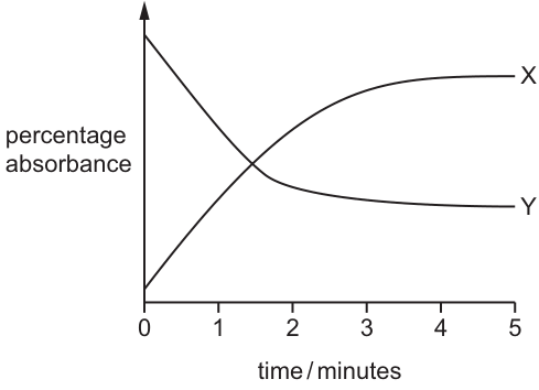
Which statement is consistent with the results shown in the graph?
Options
A The substrate in test-tube X has a higher absorbance than the product.
B The product in test-tube Y has a lower absorbance than the substrate.
C The rate of the reaction in test-tube X increased with time.
D The rate of the reaction in test-tube Y increased with time.
Working
- Test-tube X: absorbance rises from low to a high plateau, so the product absorbs more light than the substrate. A is the opposite of this and is wrong.
- Test-tube Y: absorbance falls from high to a low plateau, so the product absorbs less light than the substrate. B matches the graph.
- In both reactions the curves flatten out, showing the rate of reaction decreases with time (substrate is being used up). C and D are therefore incorrect.
Answer
B
B
Background Concept
A colorimeter measures how much light of a chosen wavelength is absorbed by a sample. In an enzyme-catalysed reaction that produces a colour change, the absorbance changes as substrate is converted to product. If the product is more intensely coloured than the substrate, absorbance rises; if it is less intensely coloured, absorbance falls. Samples are taken at intervals and the enzyme is inactivated (here with copper ions) so that the absorbance reading reflects the amount of product that had formed at that moment — the reaction is effectively "frozen".
Understanding the Question
A scientist has set up two enzyme reactions (X and Y) and followed each by taking samples at 0, 1, 2, 3, 4 and 5 minutes, stopping each sample with copper ions, and reading its absorbance. The graph plots percentage absorbance against time. We must choose the statement that is consistent with what the curves show.
Approach
Read off, for each test-tube, the direction in which absorbance is changing. A rising absorbance means product is more strongly absorbing than substrate; a falling absorbance means product is less strongly absorbing than substrate. Then consider the gradient of each curve, because the rate of reaction at any instant is given by the gradient of absorbance against time — a steeper gradient means a faster rate, and a flat curve means the reaction has effectively stopped.
Step-by-Step Reasoning
- Test-tube X: the curve starts low, rises, and then levels off. The rise tells us the product is more strongly coloured (absorbs more light) than the substrate, so option A is wrong (A says the substrate absorbs more than the product).
- Test-tube Y: the curve starts high, falls, and then levels off. The fall tells us the product is less strongly coloured than the substrate. This is exactly what option B says, so B is correct.
- The rate of reaction is given by the gradient (steepness) of the curve. For both X and Y, the curve is steepest at the start and flattens out as time goes on. This is the typical pattern for an enzyme reaction: the rate is highest when substrate concentration is highest, and falls as substrate is used up. Therefore the rate is decreasing with time, not increasing. Options C and D both claim the rate is increasing, so both are wrong.
Key Takeaways
- A colorimeter's absorbance reading is a proxy for the concentration of a coloured species; the direction of change shows whether product is more or less coloured than substrate.
- Copper ions (and similarly strong acids, alkalis or heat) are used to denature enzymes and stop a reaction at a defined time point, so each sample represents a "snapshot" of the reaction.
- The gradient of an absorbance–time graph gives the rate of the reaction. A plateau means the reaction has effectively finished (substrate exhausted or equilibrium reached).
Common Mistakes
- Assuming the higher starting line corresponds to the product: it corresponds to whatever species is more strongly coloured at the start, which is the substrate at t = 0.
- Confusing "absorbance increases" with "rate increases": absorbance tells you the cumulative amount of product so far; the rate is the slope of the curve, not the value of the curve.
- Picking an option just because the trend matches the curve in one test-tube; each statement must be evaluated against its own test-tube.
Things to Be Careful About
- A curve that plateaus does not mean the rate was zero; it means the rate has become very small because the reaction has run out of substrate or reached equilibrium.
- Copper ions denature enzymes by disrupting their tertiary structure; this is why the technique works to stop the reaction. The same idea underlies stopping an enzyme reaction with extreme pH or boiling.
A student investigated the hydrolysis of lipid in high-fat milk, using the enzyme lipase.
- of enzyme solution was added to of high-fat milk.
- The temperature was kept constant.
- The pH of the reaction mixture was recorded at time 0 minutes and every minute for 20 minutes.
Which statements correctly describe the expected results of this investigation?
1 The product forms more slowly as time proceeds because the concentration of the substrate is decreasing.
2 The pH of the reaction mixture increases rapidly in the first few minutes and then increases less rapidly.
3 The increase in the concentration of product eventually causes the lipase molecules to denature.
Options
A 1, 2 and 3
B 1 and 2 only
C 1 and 3 only
D 2 and 3 only
Working
- Statement 1: As lipase hydrolyses the triglycerides in milk, the substrate (lipid) concentration falls, so the rate of product formation slows with time. ✓ TRUE
- Statement 2: Hydrolysis of triglycerides releases fatty acids, which lower the pH (more acidic), so the pH falls, not rises. ✗ FALSE
- Statement 3: Accumulation of fatty acids lowers the pH away from the optimum, eventually denaturing the lipase. ✓ TRUE
Answer
C
C
Background Concept
Lipase catalyses the hydrolysis of triglycerides (the main lipid in milk) into fatty acids and glycerol (a monoglylyceride intermediate is also formed). Because the products include carboxylic acid groups, the reaction mixture becomes progressively more acidic as hydrolysis proceeds. The rate of an enzyme-catalysed reaction depends on substrate concentration, enzyme concentration, temperature and pH; deviation from the optimum pH (or temperature) can denature the enzyme, abolishing its activity.
Understanding the Question
The student sets up a sealed reaction of lipase + high-fat milk, holds the temperature constant, and monitors pH every minute for 20 minutes. The question asks which of three statements correctly describes the expected outcome of the experiment.
Approach
Evaluate each statement against what is known about the chemistry of lipid hydrolysis and enzyme behaviour:
- 1 → does substrate concentration fall, and does that slow the rate?
- 2 → does the pH go up or down as fatty acids are produced?
- 3 → could product accumulation denature the enzyme, and if so by what mechanism?
Step-by-Step Reasoning
Statement 1 (TRUE). The substrate (triglyceride) is being consumed. With less substrate available, fewer enzyme–substrate complexes form per unit time, so the instantaneous rate of product formation falls. The pH therefore continues to fall, but the fall per minute gets smaller. This is the standard "reaction slowing as substrate is used up" effect.
Statement 2 (FALSE). Lipid hydrolysis releases fatty acids, which dissociate to release H⁺ ions, lowering the pH. The pH therefore decreases (becomes more acidic), not increases. The rate of decrease is steepest at the start and then less steep — the direction described in the statement is wrong.
Statement 3 (TRUE). As fatty acids accumulate, the pH drifts away from lipase's optimum. Outside its optimum pH range, the tertiary structure of the enzyme is disrupted (hydrogen and ionic bonds broken), the active site loses its specific shape, and the enzyme is denatured. So an increasing concentration of acidic product can indeed denature the lipase.
Only statements 1 and 3 are correct → C.
Key Takeaways
- The products of lipid hydrolysis are fatty acids and glycerol, so pH falls during the reaction.
- A fall in substrate concentration is the main reason the rate of an enzyme-catalysed reaction decreases over time in a closed system.
- Product accumulation can change the pH away from the optimum, leading to denaturation and a reaction that effectively stops.
Common Mistakes
- Thinking hydrolysis of a lipid produces an alkaline product, leading to "pH increases".
- Confusing slowing of the reaction with denaturation by temperature (temperature was kept constant here).
- Attributing the rate decrease only to denaturation, ignoring the falling substrate concentration.
Things to Be Careful About
- Read the direction of the pH change carefully — fatty acids acidify, they do not alkalinise.
- Denaturation here is caused by pH, not by heat, because the question states the temperature was constant.
- "Product forms more slowly" is consistent with normal kinetics and does not require denaturation; statement 1 is about kinetics, statement 3 is about eventual loss of activity.
What is the correct range of measurements for the width of the cell surface membrane?
Options
A
B
C
D
Working
The cell surface membrane (plasma membrane) consists of a phospholipid bilayer plus associated proteins, and has a total thickness of approximately . This places it firmly in the range.
- A (): too small — comparable to the size of a single molecule or chemical bond, not a bilayer.
- C (): too large — closer to the size of a small organelle.
- D (): far too large — resolvable with a light microscope, whereas the membrane requires electron microscopy.
Answer
B
B
Background Concept
The cell surface membrane (plasma membrane) is described by the fluid mosaic model. It is built from a phospholipid bilayer ~ thick, studded with proteins, cholesterol, and carbohydrate chains. A single phospholipid molecule has a hydrophilic head ~ wide and two hydrophobic fatty-acid tails; two of these monolayers stacked tail-to-tail give the bilayer its characteristic thickness.
Because the membrane is only about thick — roughly — it is far below the resolving power of a light microscope (~). To visualise it, an electron microscope (resolution ) is required, and the membrane appears as a characteristic "dark–light–dark" three-layer structure (the two dense phospholipid-head regions bracketing the lighter tail region).
Understanding the Question
The question is a straight multiple-choice recall: pick the range that correctly describes how thick the plasma membrane is. All four options give numerical ranges, but in three different units (nm twice, with a hundredfold difference, and µm). The candidate must identify both the correct order of magnitude and the correct unit.
Approach
Recall the standard value: the cell surface membrane is approximately thick (sometimes quoted as ). The only option that brackets this value is B.
A quick check of the other options reveals they are wrong by orders of magnitude:
- is sub-molecular.
- is the size of small viruses / ribosomes.
- is the size of small bacteria / cellular organelles — visible with a light microscope.
Step-by-Step Reasoning
- The plasma membrane's structural unit is the phospholipid bilayer, ~ thick.
- Option B, , correctly encompasses this value.
- Each distractor represents a different biological scale:
- A is the scale of chemical bonds and small molecules.
- C is the scale of small membrane-bound structures (small vesicles, ribosomes ~).
- D is the scale of whole cells and organelles — well within light-microscope resolution.
- Therefore the answer is B.
Key Takeaways
- Plasma membrane thickness ≈ (range ).
- Membranes cannot be resolved by light microscopy; electron microscopy is required.
- Knowing the order of magnitude of common biological structures is essential for multiple-choice questions in this paper.
Common Mistakes
- Choosing A because it is the smallest non-zero range; is the size of a single molecule, not a bilayer.
- Choosing C by confusing membrane thickness with the diameter of a small organelle such as a ribosome or vesicle.
- Choosing D by forgetting that µm-scale structures are visible under the light microscope and therefore cannot be a membrane.
- Picking the right number but the wrong unit — the test always offers nm vs µm as a trap.
Things to Be Careful About
- Distinguish nm () from µm (); , roughly thicker than a real membrane.
- If a value falls into the range, check the unit carefully — both A and D share those digits but in different units.
- The accepted CIE wording is that the membrane is "about thick"; quoting is acceptable as the range.
Where in the cell surface membrane are the carbohydrate chains of glycoproteins and glycolipids mainly located?
Options
| glycoproteins | glycolipids | |
|---|---|---|
| A | inner surface | inner surface |
| B | inner surface | outer surface |
| C | outer surface | inner surface |
| D | outer surface | outer surface |
Working
In the fluid mosaic model, the carbohydrate chains attached to membrane lipids (glycolipids) and to membrane proteins (glycoproteins) all project from the outer surface of the plasma membrane. They never face the cytoplasm. This is why they are positioned to form the glycocalyx, which is involved in cell–cell recognition and signalling at the cell's external face.
Answer
D
D
Background Concept
The cell surface membrane is described by the fluid mosaic model as a phospholipid bilayer in which proteins are embedded and able to move laterally. Two of the membrane components are decorated with short carbohydrate chains:
- Glycoproteins – integral (transmembrane) proteins with a short oligosaccharide chain covalently attached to the extracellular portion of the protein.
- Glycolipids – phospholipids whose polar head group carries a short carbohydrate chain.
The carbohydrate parts of both molecules project exclusively from the outer (extracellular) surface of the bilayer. Together they form a sugar coating called the glycocalyx on the outside of the cell.
Understanding the Question
This is a one-mark MCQ testing a structural fact about membrane asymmetry. The candidate is asked to identify, for both glycoproteins and glycolipids, on which face of the bilayer the carbohydrate chains are located. The four options test the combination of locations; only one of the four combinations is correct.
Approach
Recall the simple rule: all membrane carbohydrate is on the extracellular (outer) face. Apply this uniformly to both types of molecule — glycoproteins and glycolipids — and select the option that places both on the outer surface.
Step-by-Step Reasoning
- The phospholipid bilayer has two leaflets whose polar (hydrophilic) head groups face the external environment and the cytoplasm respectively.
- Glycosylation occurs in the lumen of the ER and Golgi apparatus, and the resulting glycoproteins and glycolipids are delivered by vesicles that fuse with the plasma membrane with the same orientation. The sugar chains therefore end up facing the extracellular side when the membrane becomes the cell surface membrane.
- Because the cytoplasmic face has no carbohydrate decoration, the carbohydrate chains of both glycoproteins and both glycolipids are located on the outer surface.
- Checking the table: option D states outer surface for both, which matches.
- Options A, B and C each place at least one carbohydrate chain on the inner surface, which contradicts the established membrane asymmetry and is therefore wrong.
Key Takeaways
- Carbohydrate chains of glycoproteins and glycolipids are always on the outer surface of the plasma membrane.
- This asymmetry is established during membrane biosynthesis in the ER/Golgi and preserved when vesicles fuse with the plasma membrane.
- The outer-facing sugars form the glycocalyx, which is essential for cell recognition, cell signalling, and adhesion.
Common Mistakes
- Picking B or C because of confusion between the two molecule types — the location rule applies identically to both, so they must both be on the same (outer) surface.
- Confusing the inner/outer terminology with the cytoplasmic/extracellular faces, and accidentally selecting "inner surface" as if it were the cell's exterior.
Things to Be Careful About
- "Inner surface" and "outer surface" in this context always refer to the cytoplasmic and extracellular faces of the plasma membrane — not the inside versus outside of an organelle.
- The carbohydrate parts are short oligosaccharide chains; do not confuse them with integral membrane proteins themselves, which span the bilayer.
Sodium ions can enter cells across the cell surface membrane.
Which methods could be used by sodium ions to cross a cell surface membrane and enter a cell?
Options
A active transport only
B active transport and facilitated diffusion
C facilitated diffusion and simple diffusion
D simple diffusion only
Working
Sodium ions () are charged particles. The hydrophobic interior of the phospholipid bilayer repels charged/polar substances, so cannot cross the membrane by simple diffusion.
can cross the cell surface membrane by:
- facilitated diffusion — through channel proteins, down its electrochemical gradient (concentration of is higher outside the cell than inside);
- active transport — e.g. by a carrier protein such as the /glucose cotransporter, which uses energy (indirectly from the gradient) to move into the cell against its concentration gradient.
Simple diffusion is not possible because is a charged ion.
Answer
B
B
Background Concept
The cell surface membrane is a phospholipid bilayer. The phosphate heads are hydrophilic (water-loving) and the fatty-acid tails are hydrophobic (water-hating), so the interior of the membrane is non-polar. This has an important consequence: small, non-polar molecules such as and can dissolve in the bilayer and pass through it directly by simple diffusion, but charged ions (e.g. , , ) and large polar molecules (e.g. glucose, amino acids) cannot. They require a protein route.
There are two protein-mediated routes into a cell:
- Facilitated diffusion — ions/molecules pass through a channel or carrier protein, moving down their concentration (or electrochemical) gradient. No ATP is needed.
- Active transport — a carrier protein moves ions/molecules against their concentration gradient. This requires energy from ATP (primary active transport) or, in co-transport (secondary active transport), uses the energy stored in the electrochemical gradient of another ion (usually ) that was set up earlier by an ATP-driven pump.
Understanding the Question
The question asks which of the four transport methods (simple diffusion, facilitated diffusion, active transport) can be used by sodium ions to enter a cell. The four options pair these methods in different combinations, and we must pick the pair that is biologically correct.
Approach
- First, decide whether can perform simple diffusion. (Hint: look at the charge of the ion.)
- Then identify which protein-mediated mechanisms can move into a cell.
- Match these to the options.
Step-by-Step Reasoning
- is a charged ion. It cannot dissolve in the hydrophobic interior of the phospholipid bilayer, so simple diffusion is impossible. This eliminates options C and D.
- Facilitated diffusion is possible. In many cells, including nerve cells, the membrane contains voltage-gated or leak channel proteins. Because extracellular concentration is much higher than intracellular, opening these channels allows to flow down its electrochemical gradient into the cell. This is facilitated diffusion and does not require ATP.
- Active transport is possible. A common example is the /glucose cotransporter (SGLT) in the intestinal epithelium. Although it is technically secondary active transport (it uses the gradient set up by the / pump), it is classified as active transport because net movement is against the concentration gradient of glucose. The accompanying moves into the cell with glucose. For CIE 9700, this is credited as active transport of into the cell.
- Putting (2) and (3) together: can enter by facilitated diffusion and active transport, which matches option B.
Key Takeaways
- Charged ions cannot cross the phospholipid bilayer by simple diffusion — they need a protein.
- Ions can use both facilitated diffusion (down the gradient) and active transport (up the gradient, via pumps or co-transporters) depending on the direction relative to their gradient.
- For entering a cell, the /glucose cotransporter is the textbook example of active transport bringing into the cell.
Common Mistakes
- Choosing C or D because students forget that "ions need a protein" — they assume can diffuse like or . Small, non-polar molecules diffuse; ions do not.
- Choosing A because students only remember the / pump, which moves out of cells. Active transport of into cells still occurs via co-transporters (e.g. SGLT1).
- Confusing "active transport" with "only pump proteins that hydrolyse ATP". In CIE 9700, co-transport (e.g. with glucose or amino acids) is also called active transport because net movement is against a concentration gradient.
Things to Be Careful About
- Read the direction asked: the question is about entering the cell, not leaving it. The / pump exports , so it does not contribute to entry.
- "Simple diffusion" means diffusion directly through the phospholipid bilayer without a protein. Any answer that includes simple diffusion of an ion is biologically impossible.
- In multi-choice questions, eliminate options systematically. Eliminating C and D (which include simple diffusion) immediately narrows the answer to A or B, then decide whether facilitated diffusion also applies.
The diagram shows how an artificial partially permeable membrane was used to separate a sodium chloride solution and a sodium chloride solution in a beaker. The two sides of the beaker were labelled R and S.
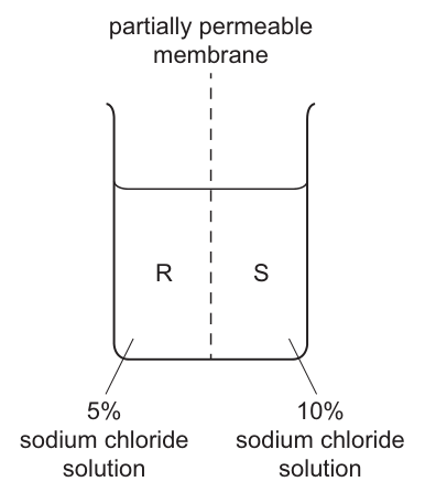
Which row correctly describes and explains what will happen in the half of the beaker labelled S?
Options
| description of S | explanation | |
|---|---|---|
| A | volume of solution increases | net movement of water from a higher water potential to a lower water potential |
| B | volume of solution increases | net movement of water from a lower water potential to a higher water potential |
| C | volume of solution decreases | net movement of water from a higher water potential to a lower water potential |
| D | volume of solution decreases | net movement of water from a lower water potential to a higher water potential |
Working
Side S contains 10% NaCl (more solute) and side R contains 5% NaCl (less solute). The more concentrated solution has the lower (more negative) water potential, so S has the lower water potential and R has the higher water potential.
Across a partially permeable membrane, water moves by osmosis down its water potential gradient — from higher water potential (R) to lower water potential (S). Therefore water enters side S and its volume increases.
Answer
A
A
Background Concept
Osmosis is the net movement of water molecules across a partially permeable membrane from a region of higher water potential to a region of lower water potential. Water potential () is measured in kPa; pure water has a water potential of 0 kPa, and adding solute lowers (makes more negative) the water potential of a solution. The greater the solute concentration, the lower the water potential. A partially permeable membrane allows water (and other small molecules) to pass but blocks the larger solute particles, so only water can move freely between the two sides.
Understanding the Question
The figure shows a beaker split by a partially permeable membrane into two halves, R and S. Side R holds a 5% NaCl solution (more dilute) and side S holds a 10% NaCl solution (more concentrated). The question asks what will happen in side S — that is, whether its volume will increase or decrease, and why. This is a multiple-choice question: we must select the row that pairs the correct direction of volume change in S with the correct biological explanation of osmosis.
The command word is implicit: we are asked to describe and explain the change. To do this we need to (1) decide which side has the higher water potential, (2) state the direction of net water movement, and (3) describe the resulting change in volume of side S.
Approach
- Compare solute concentrations on each side of the membrane.
- Translate the concentration difference into a water-potential difference: more solute → lower (more negative) .
- Apply the rule that water moves from higher to lower across a partially permeable membrane.
- Predict the effect on the volume of side S.
Step-by-Step Reasoning
- Side R has 5% NaCl; side S has 10% NaCl. S is the more concentrated solution.
- Adding solute lowers the water potential, so S has a lower water potential than R; equivalently, R has a higher water potential than S.
- By the definition of osmosis, the net movement of water is from higher water potential (R) to lower water potential (S).
- Therefore water flows into side S, so the volume of solution in S increases.
- The correct description–explanation pairing is: volume of solution increases; net movement of water from a higher water potential to a lower water potential.
Eliminating the distractors:
- B is wrong because the description (volume increases in S) is correct, but the explanation reverses the water-potential gradient — water does not move from lower to higher water potential.
- C gives the wrong description (volume decreases in S would only be true if water left S, which it does not) even though the explanation is correct.
- D is wrong on both counts: water does not leave S, and water does not move from low to high water potential.
Key Takeaways
- Solute concentration is inversely related to water potential: more dissolved solute → lower (more negative) water potential.
- Across a partially permeable membrane, the net movement of water is from higher water potential to lower water potential (down the water-potential gradient).
- A common phrasing trap: students often write "water moves from high to low concentration" — the precise wording is "high to low water potential", which is the only form that scores the mark.
Common Mistakes
- Confusing water potential with solute concentration. Water moves from the side with the higher water potential (the more dilute side) to the side with the lower water potential (the more concentrated side).
- Stating that water moves "down the concentration gradient" instead of "down the water potential gradient". The CIE mark scheme requires the exact term water potential.
- Predicting that side S (the more concentrated side) will lose water. In fact, S gains water because its water potential is lower.
- Mixing up the direction of volume change with the direction of water movement.
Things to Be Careful About
- The membrane is described as partially permeable (allows water but not solute), not fully permeable (which would let solute through as well, equalising concentrations directly rather than via osmosis).
- "Higher water potential" means the less-negative value (closer to 0 kPa); "lower water potential" means the more-negative value. Do not write "water moves from low to high water potential" — that is the opposite of osmosis.
- The volume change in S is a net result; water crosses the membrane in both directions, but more water moves from R to S than from S to R, so the net flow is into S.
Agar cubes can be used to demonstrate the effect on diffusion of changing the surface area to volume ratio.
Three different agar cubes made using a coloured indicator solution were placed into a dilute acid that diffused into the cubes. As the acid diffused into the agar cubes, the colour of the indicator solution changed.
The cubes had volumes of , and and were left in the dilute acid for 10 minutes. All other variables were kept the same.
After 10 minutes, the agar cubes were removed from the dilute acid and cut in half. The cut surfaces were observed and the results were recorded as diagrams. All diagrams were drawn to the same scale.
The results for the cube are shown.
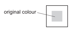
Which diagrams show the results for the and the cubes?
Options
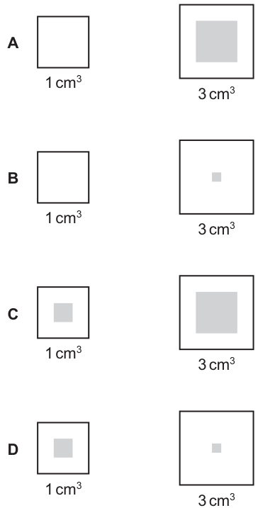
Working
The reference 2 cm³ cube (Fig. 19.1) shows a moderate central area of original colour, indicating acid has diffused part-way in from the surface.
Smaller cube (1 cm³): higher surface area to volume ratio, so acid diffuses more efficiently relative to the cube's size → less original colour remaining (a small central shaded square).
Larger cube (3 cm³): lower surface area to volume ratio, so acid diffuses less efficiently relative to the cube's size → more original colour remaining (a large central shaded square).
This pattern matches the diagrams in option C.
Answer
C
C
Background Concept
Diffusion is the net movement of particles from a region of higher concentration to a region of lower concentration. The rate at which a substance diffuses into a cell or tissue depends on:
- The concentration gradient (steeper = faster).
- The surface area available for diffusion.
- The distance the substance must travel.
The efficiency of diffusion in supplying a volume of tissue is captured by the surface area to volume ratio (SA:V). As an object grows while keeping the same shape, its volume increases faster than its surface area, so SA:V falls. A high SA:V means that diffusion can supply or remove substances fast enough to meet the needs of the whole volume; a low SA:V means the centre of the object is starved of supplies or cannot get rid of waste quickly.
In this experiment, agar cubes impregnated with a coloured indicator (e.g. pH indicator) are placed in acid. The acid diffuses in from all faces of the cube and changes the indicator's colour where it reaches. After a fixed time, the cubes are sliced open to reveal how far the acid has penetrated — a white (changed) outer ring with the original colour remaining in the centre.
Understanding the Question
The question gives the result for the 2 cm³ cube (Fig. 19.1): a square with a substantial central area still showing the original colour, surrounded by a white border where the acid has changed the indicator.
We are asked to predict what the 1 cm³ and 3 cm³ cubes would look like after the same 10 minutes in acid. All variables (time, acid concentration, temperature, indicator concentration) are constant — only the cube size changes.
Approach
The key idea is that the same diffusion time gives the same absolute penetration depth in a given material, but the relative effect on the cube depends on its size:
- The smaller the cube, the higher its SA:V, and the more completely acid reaches the centre.
- The larger the cube, the lower its SA:V, and the less completely acid reaches the centre.
So:
- 1 cm³ cube → smallest, highest SA:V → acid diffuses fastest relative to its size → least original colour remaining.
- 2 cm³ cube → reference (Fig. 19.1) → moderate original colour.
- 3 cm³ cube → largest, lowest SA:V → acid diffuses slowest relative to its size → most original colour remaining.
Step-by-Step Reasoning
- Compare the size of each cube using SA:V:
- A 1 cm cube has sides of 1 cm: SA = 6 cm², V = 1 cm³, SA:V = 6:1.
- A 2 cm³ cube has sides of ∛2 ≈ 1.26 cm: SA:V ≈ 4.76:1.
- A 3 cm³ cube has sides of ∛3 ≈ 1.44 cm: SA:V ≈ 4.16:1.
- The 1 cm³ cube has the highest SA:V, so it is the most efficiently supplied by diffusion.
- After 10 minutes in acid, the 1 cm³ cube will have the most colour change — only a small central core remains its original colour.
- The 3 cm³ cube has the lowest SA:V, so it is the least efficiently supplied by diffusion.
- After 10 minutes, the 3 cm³ cube will have the least colour change — a large central region remains its original colour.
- The reference 2 cm³ cube (Fig. 19.1) lies between these extremes, with a moderate central core of original colour.
- Matching this prediction to the options: option C shows a small central shaded square for 1 cm³ and a large central shaded square for 3 cm³ — exactly the expected pattern.
Key Takeaways
- SA:V decreases as a cube of constant shape grows larger.
- A higher SA:V means diffusion can supply the centre faster relative to its volume.
- For a fixed diffusion time, smaller cubes become fully colour-changed more easily than larger cubes.
- The agar cube experiment is a simple model of why cells must remain small: beyond a certain size, diffusion cannot meet the metabolic needs of the cell interior.
Common Mistakes
- Choosing A or B: these show the 1 cm³ cube completely white (all colour changed) and either a large or tiny centre for the 3 cm³ cube. Option A would only be correct if 10 minutes were enough for acid to fully penetrate the 1 cm³ cube, but it does not match the relative sizes shown in the reference.
- Choosing D: this swaps the expectation — showing a large original-colour region in the 1 cm³ cube. This would only be true if smaller cubes had lower SA:V, which is the opposite of reality.
- Confusing surface area with volume: a bigger cube has more surface area in absolute terms, but the SA:V ratio falls.
Things to Be Careful About
- All cubes are made of the same agar and indicator and are placed in identical acid for the same time — only the cube size varies.
- "Original colour" means the colour before acid exposure (the centre, where acid has not yet diffused).
- The diagrams are drawn to the same scale, so visual comparison of the remaining central area is valid.
- The 2 cm³ cube reference shows that 10 minutes is not long enough to fully penetrate even the medium cube, so we should expect the 3 cm³ cube to retain even more original colour and the 1 cm³ cube to retain less — but not necessarily to be completely changed.
During sperm formation in mammals, part of the structure of each chromosome is replaced with proteins called protamines. This replacement allows the DNA to be packaged much more densely than would otherwise be possible.
Which part of the chromosome is replaced by protamines?
Options
A centromeres
B chromatids
C histones
D telomeres
Working
During sperm formation (spermiogenesis), the DNA in spermatid nuclei is repackaged. The normal DNA–protein complexes use histones around which DNA is wound, but in maturing sperm these histones are progressively replaced by smaller, arginine-rich protamines. Because protamines are much smaller than histones, the DNA can be coiled far more tightly, producing the highly condensed nucleus of the mature sperm.
- A – centromeres: these are the constricted regions joining sister chromatids; they are not replaced.
- B – chromatids: these are the two copies of a replicated chromosome; they are not replaced.
- C – histones: ✓ replaced by protamines to allow denser DNA packaging.
- D – telomeres: these are the repetitive ends of chromosomes; they are not replaced.
Answer
C
C
Background Concept
In eukaryotic cells, DNA does not exist as a naked double helix. It is wrapped around small basic proteins called histones to form nucleosomes, which in turn are folded into higher-order chromatin fibres. Histones (especially H2A, H2B, H3 and H4) are rich in the positively charged amino acids lysine and arginine, which bind tightly to the negatively charged phosphate groups of DNA. This packaging compacts the DNA and helps regulate gene expression.
During spermiogenesis — the final stage of sperm production in mammals — the spermatid nucleus undergoes a dramatic remodelling. Most of the histones are replaced by smaller, even more positively charged proteins called protamines. Because protamines are roughly half the size of histones and pack DNA much more tightly (allowing the double helix to form tight toroidal coils), the sperm nucleus becomes extremely condensed. This is biologically important: a smaller, denser sperm head reduces drag, protects the DNA during transit through the male and female reproductive tracts, and silences transcription in a cell that is essentially a delivery vehicle for the paternal genome.
Understanding the Question
The stem tells us that in mammals, during sperm formation, part of each chromosome is replaced by protamines, and that this replacement allows much denser DNA packaging. We are asked which chromosome component is replaced. The four options are centromeres, chromatids, histones and telomeres — all genuine parts of a chromosome, but only one is the protein scaffolding normally associated with DNA in the nucleus.
The command word is implicit but the question is asking for an identification: which of the four structures is the one that protamines substitute for.
Approach
The key is to recognise that protamines are described as proteins that replace part of the chromosome structure, and that the outcome is denser DNA packaging. Among the four options, only histones are the protein components of chromatin that DNA is normally wound around. Centromeres, chromatids and telomeres are not protein scaffolds that DNA wraps around in the way the question describes — they are structural/functional regions of the chromosome itself. Therefore histones must be the structures that protamines substitute for.
Step-by-Step Reasoning
- Protamines are described as proteins that take the place of part of the chromosome to allow denser DNA packaging.
- In somatic cells, the proteins around which DNA is wound are histones (forming nucleosomes).
- During spermiogenesis these histones are removed and replaced by the smaller, more positively charged protamines, allowing DNA to be coiled into a much more compact structure.
- Therefore the answer is C – histones.
- The other options are not protein scaffolds that DNA is wrapped around: centromeres are the constricted attachment region for spindle fibres; chromatids are the duplicated copies of a chromosome; telomeres are the repetitive, protective ends of linear chromosomes.
Key Takeaways
- In most eukaryotic cells, DNA is packaged by winding around histone proteins into nucleosomes.
- During mammalian spermiogenesis, histones are replaced by smaller, arginine-rich protamines.
- This remodelling produces the highly condensed nucleus of the mature sperm, which protects the DNA and aids sperm motility/function.
- Centromeres, chromatids and telomeres are structural regions of the chromosome itself — not the protein packaging layer around the DNA.
Common Mistakes
- Confusing chromatids with chromatin: chromatids are the duplicated copies of a chromosome joined at a centromere; they are not the protein packaging of DNA.
- Confusing telomeres with histone-like proteins: telomeres are repetitive DNA sequences (e.g. TTAGGG in humans) at the ends of chromosomes, with associated shelterin proteins — they are not the general DNA-packaging proteins.
- Choosing centromeres because they sound structural: centromeres are regions of DNA/protein (kinetochore) used in spindle attachment, not the proteins around which the bulk of DNA is wound.
Things to Be Careful About
- The question specifies a protein replacement. Among the four options, only histones are proteins of the type that protamines (also proteins) could substitute for.
- Remember that protamine replacement is largely a feature of mammalian sperm; in many fish, for example, protamines are the dominant sperm nuclear protein and there is little or no histone-to-protamine transition in the same way.
- Although a small percentage of histones is retained in human sperm and may carry epigenetic information, the bulk of the DNA is protamine-packaged — the question is testing this general principle.
The diagram shows the mitotic cell cycle.
During which phase do chromosomes condense and become visible?
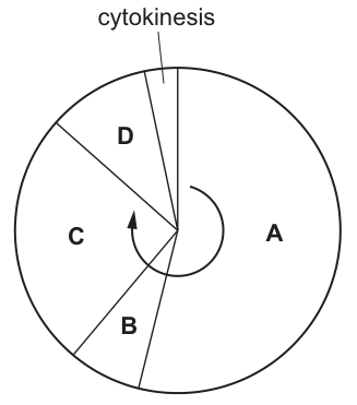
Options
A A
B B
C C
D D
Working
Chromosomes condense and become visible during prophase, the first stage of mitosis. In the diagram, the largest section A represents interphase. Following the cycle from interphase, the stages of mitosis proceed in order: D = prophase, C = metaphase, B = anaphase and telophase, before cytokinesis divides the cytoplasm.
Therefore, chromosomes condense and become visible in section D.
Answer
D
D
Background Concept
The cell cycle is the series of events that takes place in a cell leading to its division and the duplication of its DNA to produce two daughter cells. It consists of:
- Interphase (the longest phase, comprising G1, S, and G2): the cell grows, replicates its DNA, and prepares for division. During interphase, the genetic material exists as diffuse chromatin and is not visible as discrete chromosomes under a light microscope.
- Mitosis (M phase): the nucleus divides. It is divided into four stages:
- Prophase — chromatin condenses into visible chromosomes; the nuclear envelope breaks down; the centrosomes move to opposite poles and spindle fibres begin to form.
- Metaphase — chromosomes align at the cell's equator (metaphase plate), attached to spindle fibres at their centromeres.
- Anaphase — sister chromatids separate and are pulled to opposite poles by the shortening spindle fibres.
- Telophase — chromatids arrive at the poles, nuclear envelopes reform around each set, and the chromosomes begin to decondense.
- Cytokinesis — the cytoplasm divides, producing two genetically identical daughter cells.
The defining event in prophase is the condensation of chromatin into discrete, visible chromosomes. This condensation is what allows the chromosomes to be seen under the microscope and ensures they can be properly attached to spindle fibres and segregated during the later stages of mitosis.
Understanding the Question
The question asks which phase of the mitotic cell cycle chromosomes condense and become visible. The diagram shows a circular cell cycle divided into four sections (A, B, C, D) of different sizes, with cytokinesis marked as a separate wedge. The arrow in the centre indicates the direction of progression around the cycle.
The command word here is essentially "identify which" — the candidate must know that chromosome condensation is the defining event of prophase, and then correctly match prophase to the appropriate lettered section of the diagram.
Approach
- Recall that chromosome condensation and visibility is the defining event of prophase.
- Recognise that the largest section of the diagram represents interphase, because interphase occupies the majority of the cell cycle in terms of duration.
- Trace the cell cycle from interphase: prophase follows interphase as the first stage of mitosis.
- Identify which lettered section comes immediately after interphase in the direction of the arrow.
Step-by-Step Reasoning
- Section A is by far the largest wedge, indicating it is the longest phase. This is interphase (G1, S, G2), which makes up the bulk of the cell cycle.
- Following interphase, the cell enters mitosis. The first mitotic stage is prophase, where the diffuse chromatin condenses into visible chromosomes.
- In this diagram, the section that immediately follows interphase (in the direction of the cycle indicated by the arrow) is D.
- Therefore, D represents prophase — the phase during which chromosomes condense and become visible.
- The remaining sections correspond to the other mitotic stages: C = metaphase (chromosomes aligned at the equator), B = anaphase and telophase (chromatids separating and arriving at the poles, then decondensing), before cytokinesis completes cell division.
Key Takeaways
- Chromosomes condense and become visible during prophase, the first stage of mitosis.
- In a cell cycle diagram, interphase is the largest section because it is the longest in duration; the stages of mitosis are much shorter.
- The order of mitosis is: prophase → metaphase → anaphase → telophase, followed by cytokinesis.
- The size of each wedge in a cell cycle pie chart is proportional to the time the cell spends in that phase.
Common Mistakes
- Confusing prophase with telophase: In telophase, chromosomes begin to decondense and become invisible again — so this cannot be the answer.
- Confusing prophase with metaphase or anaphase: While chromosomes are visible in these stages too, the question specifically asks when they condense and become visible, which is prophase.
- Misidentifying the sections: A student who assumes the cycle simply proceeds clockwise from A and chooses B will be wrong — the layout of this particular diagram places prophase at D.
- Choosing the section labelled "cytokinesis": Cytokinesis is the division of the cytoplasm, not a stage where chromosomes condense.
Things to Be Careful About
- The term "condense" specifically refers to prophase, when chromatin first becomes visible as discrete chromosomes.
- The relative size of each section in a cell cycle diagram reflects the relative duration of that phase — interphase is largest, mitosis stages are short, and cytokinesis is brief.
- Always read the direction of the arrow in the diagram and trace from interphase to identify the first stage of mitosis (prophase).
Which statements correctly describe features of stem cells that are essential for their role in cell replacement and tissue repair?
1 After mitosis of stem cells, the daughter cells can either remain as stem cells or follow a developmental pathway that leads to the formation of specialised cells.
2 Stem cells are different to all other body cells because they retain all of the genetic information in their DNA throughout the life of the organism.
3 A small population of stem cells is retained in the body of adults throughout their life time.
4 Stem cells have more telomeres than other body cells and this allows them to undergo an unlimited number of mitotic divisions.
Options
A 1, 2, 3 and 4
B 1, 2 and 3 only
C 1 and 3 only
D 2, 3 and 4 only
Working
Evaluate each statement:
-
Statement 1 — TRUE. Stem cells can self-renew (one daughter remains a stem cell) or differentiate into specialised cells. This dual potential is the defining feature enabling cell replacement and tissue repair.
-
Statement 2 — FALSE. All nucleated body cells retain the full genome; this is not unique to stem cells. Stem cells differ in that they can express the appropriate genes to differentiate, not because they uniquely retain DNA.
-
Statement 3 — TRUE. Adult (tissue) stem cells persist in small numbers in tissues such as bone marrow, skin and intestinal epithelium throughout life, providing ongoing replacement and repair.
-
Statement 4 — FALSE. Stem cells do not have "more" telomeres than other cells; they maintain telomere length via telomerase, but even with telomerase activity their division is not strictly unlimited.
Only statements 1 and 3 are correct.
Answer
C
C
Background Concept
Stem cells are unspecialised cells with two defining properties: self-renewal (the ability to divide and produce more stem cells) and potency (the ability to differentiate into specialised cell types). These features make them essential for growth, tissue maintenance and repair.
A critical distinction is that all nucleated somatic cells in the body contain the same full genome — every cell arises by mitosis from the zygote and inherits a complete copy of the DNA. What makes stem cells different is not the presence of unique DNA, but their capacity to remain undifferentiated and to activate specific gene expression programmes that other, more committed cells have largely switched off.
Telomeres are repetitive DNA sequences at the ends of chromosomes that protect coding DNA from being lost during replication. In most somatic cells, telomeres shorten with each division (the Hayflick limit), eventually triggering cell senescence. Stem cells (and some cancer cells) express telomerase, an enzyme that replenishes telomere length, allowing many more divisions than typical somatic cells — but the divisions are still not truly unlimited in vivo.
Understanding the Question
This is a multiple-choice question (Paper 1 style) testing whether the candidate can identify which statements correctly describe properties essential to the role of stem cells in cell replacement and tissue repair. The command word is implicit "which statements correctly describe", and four statements are given with options combining them in various ways. The correct answer is C (1 and 3 only).
Approach
Go through each statement and judge it as true or false based on accurate knowledge of stem cell biology, then compare to the answer options.
Step-by-Step Reasoning
Statement 1 describes asymmetric division — a key feature of stem cells. When a stem cell undergoes mitosis, one daughter can remain a stem cell (self-renewal) while the other commits to differentiation. This is precisely what enables a continuous supply of replacement cells while preserving the stem cell pool. ✓ TRUE
Statement 2 is a common misconception. All nucleated body cells retain the full genetic information because mitosis produces genetically identical daughter cells. A red blood cell loses its nucleus and is an exception, but liver cells, skin cells, muscle cells, etc., all contain the full genome. What makes stem cells special is not retaining DNA, but the ability to use that DNA flexibly to become many different cell types. The "throughout the life of the organism" qualifier is also misleading — DNA is retained in all dividing cells, and even non-dividing cells generally keep their DNA for life. ✗ FALSE
Statement 3 is true. Adult (tissue) stem cells are found in small populations in many tissues throughout life — for example, haematopoietic stem cells in bone marrow, intestinal stem cells in the crypts of Lieberkühn, and stem cells in the basal layer of the epidermis. Their persistence is what allows ongoing replacement of short-lived cells such as blood cells and gut epithelial cells. ✓ TRUE
Statement 4 is false on two counts. First, stem cells do not have more telomeres than other cells; they have the same number of telomeres (one at each chromosome end) but maintain them better via telomerase. Second, the claim that they undergo an "unlimited" number of divisions is an overstatement — even with telomerase activity, divisions are constrained by other factors (accumulated mutations, niche signals, etc.), and embryonic stem cells still have a finite but extended capacity. ✗ FALSE
Only 1 and 3 are correct → Option C.
Key Takeaways
- Stem cells are defined by self-renewal and the ability to differentiate into specialised cells.
- All nucleated body cells share the same genome; stem cells are not unique in retaining DNA.
- Adult stem cells persist in small numbers in many tissues, supporting lifelong cell replacement.
- Stem cells maintain telomeres via telomerase, but this does not confer truly unlimited division.
Common Mistakes
- Marking statement 2 as true because it "sounds like" what stem cells do. The DNA-retention claim applies to all nucleated cells, not just stem cells.
- Marking statement 4 as true by confusing telomerase activity with having "more" telomeres, and overstating the division capacity of stem cells.
- Selecting option B (1, 2 and 3) by accepting statement 2, or option D by accepting statement 4.
Things to Be Careful About
- Distinguish between a property that is unique to stem cells and one that is shared by other cells.
- Telomeres are present in all cells; only the maintenance of telomere length via telomerase distinguishes stem (and some progenitor) cells from most somatic cells.
- "Unlimited divisions" is an overstatement — embryonic stem cells divide many more times than most somatic cells, but they are still subject to constraints.
Which statements are correct for all nucleotides?
1 The nitrogen-containing base is always attached to carbon atom 1 of the pentose.
2 The phosphate group is always attached to carbon atom 5 of the pentose.
3 A condensation reaction occurs to join the nitrogen-containing base to the pentose.
4 Nucleotides are linked together by condensation reactions between phosphate groups.
Options
A 1, 2 and 3
B 1 and 2 only
C 1, 3 and 4
D 2, 3 and 4
Working
- Statement 1: Correct — the nitrogenous base is always attached to carbon 1 (C1′) of the pentose sugar.
- Statement 2: Correct — the phosphate group is always attached to carbon 5 (C5′) of the pentose sugar.
- Statement 3: Correct — the bond between the base and the pentose is a glycosidic bond formed by a condensation reaction.
- Statement 4: Incorrect — nucleotides are joined to each other by condensation reactions between the phosphate group of one nucleotide and the 3′ hydroxyl of the pentose of the next nucleotide (forming a phosphodiester bond), not between two phosphate groups.
Answer
A
A
Background Concept
A nucleotide is the monomer of nucleic acids (DNA and RNA). Each nucleotide has three components:
- A pentose sugar — deoxyribose in DNA, ribose in RNA. The carbons of the pentose are numbered 1′ to 5′.
- A nitrogenous base — either a purine (adenine A or guanine G, double-ringed) or a pyrimidine (cytosine C, thymine T in DNA, or uracil U in RNA, single-ringed).
- A phosphate group — attached to the 5′ carbon of the sugar.
The base is always attached to C1′ and the phosphate is always attached to C5′. The bond between the base and the sugar is a glycosidic bond (formed by condensation), and the bond linking adjacent nucleotides in a polynucleotide chain is a phosphodiester bond — formed by a condensation between the 3′-OH of one sugar and the 5′-phosphate of the next nucleotide.
Understanding the Question
This is a multiple-choice question testing recall of nucleotide structure and the bonds that hold a nucleotide together and link nucleotides together. The stem stresses "for ALL nucleotides", so the answers must apply to every nucleotide — both DNA and RNA.
Approach
Check each statement against the canonical structure of a nucleotide, paying close attention to which atoms are joined by which bond.
Step-by-Step Reasoning
- Statement 1 — The nitrogenous base attaches to C1′ of the pentose in every nucleotide. ✓ Correct.
- Statement 2 — The phosphate group attaches to C5′ of the pentose in every nucleotide. ✓ Correct.
- Statement 3 — Joining the base to the sugar is a condensation reaction, releasing water and forming a glycosidic (C-N) bond. ✓ Correct.
- Statement 4 — This is the trap. Nucleotides are linked into a polynucleotide chain by condensation reactions between the 3′-OH of one sugar and the 5′-phosphate of the next nucleotide, NOT between two phosphate groups. The bond formed is a phosphodiester bond. ✗ Incorrect.
So statements 1, 2, and 3 are correct → answer A.
Key Takeaways
- In a nucleotide: base = C1′; phosphate = C5′; the third position on the sugar (C3′) is what links to the next nucleotide.
- Base–sugar bond: glycosidic (condensation).
- Sugar–phosphate–sugar backbone bond: phosphodiester (condensation) — between 3′ C of one sugar and 5′ phosphate of the next.
Common Mistakes
- Choosing D or C because of a misconception that "phosphates link to phosphates" in the sugar–phosphate backbone. The actual link is sugar (3′-OH) to phosphate (5′), giving a phosphodiester.
- Forgetting that the same numbering (C1′ for base, C5′ for phosphate) applies to BOTH deoxyribose and ribose.
Things to Be Careful About
- "Nucleotide" vs. "nucleoside": a nucleoside is base + sugar only (no phosphate). Adding a phosphate to C5′ makes it a nucleotide.
- Statement 4 is the classic distractor — the phosphodiester bond is between a sugar's 3′ hydroxyl and another nucleotide's 5′ phosphate, never directly between two phosphate groups.
How many phosphodiester bonds are present in a circular DNA molecule of 2700 base pairs?
Options
A 2699
B 2700
C 5398
D 5400
Working
A DNA base pair consists of 2 nucleotides, so:
In a circular DNA molecule there are no free 3′ or 5′ ends; each nucleotide is joined to two neighbours, so the number of phosphodiester bonds equals the number of nucleotides in that strand.
With two strands in the duplex:
Answer
D
D
Background Concept
The backbone of a DNA strand is built from alternating deoxyribose sugars and phosphate groups, linked by phosphodiester bonds. A phosphodiester bond is formed between the 3′-OH of one sugar and the 5′-phosphate of the next nucleotide, creating an unbroken chain with directionality (5′ → 3′).
A double-stranded DNA molecule has two such backbones running antiparallel to each other, with the bases meeting in the middle as complementary base pairs (A with T, G with C). One "base pair" therefore corresponds to two nucleotides — one on each strand.
The geometry of the molecule matters when counting bonds:
- In a linear DNA, each strand has two free ends (a 5′ end and a 3′ end), so two of the nucleotides are not linked on both sides. For n nucleotides, there are therefore n − 1 phosphodiester bonds per strand, or 2n − 2 in total.
- In a circular DNA (such as bacterial chromosomes, plasmids, and mitochondrial DNA), the 3′ end of the last nucleotide is bonded to the 5′ end of the first, so there are no free ends. Each nucleotide is bonded to two neighbours, and the number of phosphodiester bonds equals the number of nucleotides in that strand.
Understanding the Question
The question gives a circular DNA molecule containing 2700 base pairs and asks for the total number of phosphodiester bonds. The key feature is the word circular — without it, the answer would be 2n − 2 (i.e. 5398). Because it is circular, the simpler rule applies.
Approach
- Convert base pairs to total nucleotides (× 2).
- Recognise that "circular" removes the − 2 correction that applies to linear molecules.
- Conclude that the number of phosphodiester bonds equals the total number of nucleotides.
Step-by-Step Reasoning
- 2700 base pairs × 2 nucleotides per base pair = 5400 nucleotides in total.
- In a circular molecule, the last nucleotide's 3′ carbon is joined by a phosphodiester bond to the 5′ phosphate of the first nucleotide, closing the loop. So each strand has 2700 phosphodiester bonds (equal to the number of nucleotides per strand), and both strands combined contain 5400.
- Therefore the answer is 5400, which is option D.
Why the distractors are wrong:
- A (2699): This would be the count if you (incorrectly) treated 2700 base pairs as 2700 nucleotides in a single circular strand.
- B (2700): This treats the molecule as single-stranded and circular, ignoring the second strand.
- C (5398): This is the correct count for a linear duplex of 2700 base pairs (2 × 2700 − 2). The question explicitly states the molecule is circular, so the ends must be joined, adding 2 extra bonds.
Key Takeaways
- Each nucleotide in a strand is joined to the next by one phosphodiester bond.
- In a circular DNA, the number of phosphodiester bonds per strand equals the number of nucleotides per strand (no free ends).
- In a linear DNA, it equals n − 1 per strand, or 2n − 2 for the duplex.
- "Base pairs" refers to pairs of nucleotides, so multiply by 2 to get the total nucleotide count.
Common Mistakes
- Forgetting that a base pair = 2 nucleotides and only counting one strand.
- Subtracting 2 (treating the molecule as linear) when the question clearly says circular.
- Counting only the phosphodiester bonds in one strand and ignoring the complementary strand.
Things to Be Careful About
- Always read carefully whether the DNA is described as circular or linear — the word changes the answer by exactly 2.
- Remember that hydrogen bonds (between base pairs) are a different type of bond from phosphodiester bonds (within the sugar-phosphate backbone) and are not asked about here.
Which statements about complementary base pairing are correct?
1 Purines and pyrimidines are different sizes.
2 Complementary base pairing occurs during translation.
3 The base pairs are of different lengths.
4 Uracil forms two hydrogen bonds with adenine.
Options
A 1, 2 and 3
B 1, 2 and 4
C 1, 3 and 4
D 2, 3 and 4
Working
- Purines and pyrimidines are different sizes. — Correct. Purines (adenine, guanine) have a double-ring structure; pyrimidines (cytosine, thymine, uracil) have a single-ring structure, so they differ in size.
- Complementary base pairing occurs during translation. — Correct. tRNA anticodons pair with mRNA codons at the ribosome by complementary base pairing.
- The base pairs are of different lengths. — Incorrect. Each base pair always consists of one purine + one pyrimidine, so all base pairs are the same (uniform) width along the helix.
- Uracil forms two hydrogen bonds with adenine. — Correct. A pairs with U using two hydrogen bonds (A–T in DNA also uses two; only G–C uses three).
Statements 1, 2 and 4 are correct.
Answer
B
B
Background Concept
The nitrogenous bases in nucleic acids fall into two structural families:
- Purines — adenine (A) and guanine (G) — have a double-ring structure (a six-membered ring fused to a five-membered ring).
- Pyrimidines — cytosine (C), thymine (T, in DNA only) and uracil (U, in RNA only) — have a single-ring structure.
Because of this size difference, base pairing is always a purine with a pyrimidine (A–T, A–U, G–C). This keeps the helix a uniform width along its length, and it also allows the hydrogen-bonding pattern on each base to be geometrically complementary.
Hydrogen-bond numbers:
- A–T (DNA) and A–U (RNA): 2 hydrogen bonds
- G–C (DNA and RNA): 3 hydrogen bonds
Complementary base pairing occurs wherever a single-stranded nucleic acid template is read:
- DNA replication — new DNA strand against a DNA template.
- Transcription — mRNA strand against a DNA template strand.
- Translation — tRNA anticodon against the mRNA codon at the ribosome.
Understanding the Question
This is a multiple-choice question (Paper 1 style) that lists four statements and asks which combination is correct. Each statement tests a different aspect of base pairing:
- Statement 1: structural difference between purines and pyrimidines.
- Statement 2: where complementary base pairing happens in the central dogma.
- Statement 3: uniformity of the DNA/RNA helix width.
- Statement 4: hydrogen-bond count for an A–U pair.
The command word is implicit ("which statements … are correct") — the candidate must judge each statement independently and then pick the option that lists only the correct ones.
Approach
Judge each statement on its own merits using the rules above, then match the set of correct statements to an option (A–D).
Step-by-Step Reasoning
Statement 1 — Purines and pyrimidines are different sizes. TRUE. Purines (A, G) have two rings; pyrimidines (C, T, U) have one ring. They are different sizes, and this is the reason a purine always pairs with a pyrimidine.
Statement 2 — Complementary base pairing occurs during translation. TRUE. Translation is the assembly of a polypeptide at the ribosome using mRNA. The mRNA codon is read by a tRNA whose anticodon pairs with the codon by complementary base pairing. Without this pairing, the correct amino acid (carried by the tRNA) would not be selected.
Statement 3 — The base pairs are of different lengths. FALSE. Each base pair is one purine + one pyrimidine, and the two families have been selected by evolution to span the same distance across the helix. This is precisely why A–T and G–C base pairs are the same length — the wider purine compensates for the narrower pyrimidine.
Statement 4 — Uracil forms two hydrogen bonds with adenine. TRUE. A–U uses two hydrogen bonds (just like A–T in DNA). Only G–C uses three hydrogen bonds.
Correct statements: 1, 2 and 4 → option B.
Key Takeaways
- Purine + pyrimidine pairing produces base pairs of uniform length, not different lengths.
- Complementary base pairing is used in three processes: replication, transcription and translation (tRNA anticodon ↔ mRNA codon).
- A–U = 2 H-bonds, A–T = 2 H-bonds, G–C = 3 H-bonds.
- Uracil is found in RNA (replacing thymine during transcription); it pairs with adenine.
Common Mistakes
- Thinking complementary base pairing is unique to DNA replication or transcription and forgetting it also occurs during translation (between mRNA codon and tRNA anticodon).
- Confusing A–U with G–C and assuming A–U has three hydrogen bonds. In fact, A–U and A–T both have two; only G–C has three.
- Believing base pairs differ in length — they are uniform because a purine (wider) always pairs with a pyrimidine (narrower).
Things to Be Careful About
- Memorise the hydrogen-bond counts as a tidy set: A–T = 2, A–U = 2, G–C = 3. This set of numbers is tested repeatedly.
- "Complementary base pairing" is a description of hydrogen-bonded base–base recognition; it is not restricted to DNA–DNA.
- The exam marks "1, 2 and 4" as correct; option C (1, 3, 4) is a distractor built by swapping in the false statement 3 — watch out for this trap.
During the mitotic cell cycle, the chromosomal DNA is replicated. The specific points in DNA molecules where replication is occurring are known as replication forks.
A typical human chromosome has about 150 million base pairs of DNA. It takes about 1 hour to replicate the DNA of a typical human chromosome.
The rate of replication using a single replication fork is approximately 50 base pairs per second.
Approximately how many replication forks must occur in a typical human chromosome during DNA replication?
Options
A 835
B 41 700
C 50 000
D 3 000 000
Working
- Convert the time available into seconds:
- Calculate how many base pairs one replication fork can copy in 3600 s:
- Divide the total length of the chromosome by the amount one fork can copy:
Answer
A
A
Background Concept
A human chromosome is an extremely long DNA molecule. A single chromosome contains around 150 million base pairs of DNA, all of which must be copied accurately during the S (synthesis) phase of interphase so that each daughter cell receives a complete set of chromosomes after mitosis.
DNA replication begins at many sites along a chromosome, called origins of replication. At each origin the double helix is unwound and two replication forks move outwards in opposite directions as new DNA is synthesised. Because eukaryotic chromosomes are so long, hundreds or thousands of origins (and therefore forks) are needed so that the whole chromosome can be copied in a reasonable time (about an hour).
Understanding the Question
The question gives three pieces of information:
- Total size of a typical human chromosome: ~150 000 000 base pairs
- Time available: ~1 hour
- Rate of a single replication fork: ~50 base pairs per second
We are asked to estimate how many replication forks must work simultaneously in order to copy the whole chromosome in one hour. This is essentially a rate–time–distance calculation: total amount of work divided by amount each fork can do in the time available.
Approach
- Convert the time available (1 hour) into seconds, because the rate is given in base pairs per second.
- Multiply this by the rate of one fork to find how many base pairs one fork can copy in the available time.
- Divide the total number of base pairs in the chromosome by this amount to obtain the number of forks required.
- Match the result to the closest option.
Step-by-Step Reasoning
Step 1 — Time conversion
Step 2 — Output of one replication fork in 1 hour
A single fork adds nucleotides at ~50 bp s⁻¹, so in 3600 s it copies:
Step 3 — Number of forks required
The closest answer is 835 (option A). The small difference (833 vs 835) is just rounding of the rates given in the question.
Why the other options are wrong
- B (41 700) would be the answer if you forgot to convert hours to seconds and used minutes (60 s) instead — a classic timing error.
- C (50 000) is the number of seconds in roughly 14 hours, mixing up time and rate units.
- D (3 000 000) has no sensible interpretation in the calculation; it is far too large and would imply that each fork copies only ~50 bp in total.
Key Takeaways
- Eukaryotic chromosomes are far too long to be replicated from a single origin in a useful time.
- Hundreds to thousands of origins of replication, each producing two replication forks, fire simultaneously during S phase to copy a chromosome in about an hour.
- This question is a standard rate × time = amount calculation, disguised inside a biological context. Always check that the units of the rate and the time match before multiplying.
Common Mistakes
- Forgetting to convert 1 hour into seconds. If you treat 1 hour as 1 unit and divide 150 000 000 by 50, you get 3 000 000 — option D, the classic distractor.
- Confusing seconds with minutes, which gives 41 700 — option B.
- Multiplying instead of dividing the total bp by the per-fork output, which gives a meaningless large number.
Things to Be Careful About
- Always write the units when stating a rate: , not just 50.
- The number of forks is not the same as the number of origins: each origin produces two forks, so the number of origins is roughly half the number of forks. The question specifically asks for forks, so do not halve again.
- The data are deliberately rounded, so the answer is approximate (~835) — do not worry that 833 ≠ 835.
Which molecule has its synthesis directly controlled by DNA?
Options
A amylase
B cholesterol
C glycogen
D phospholipid
Answer
DNA codes directly for proteins via transcription and translation. Amylase is a protein (an enzyme), so its synthesis is directly controlled by DNA. Cholesterol and phospholipids are lipids, and glycogen is a polysaccharide — none of these are directly encoded by DNA; they are synthesised indirectly through enzyme-catalysed reactions.
A
A
Background Concept
The central dogma of molecular biology states that genetic information flows from DNA to mRNA (transcription) and then from mRNA to protein (translation). A gene is a sequence of DNA bases that codes, via an mRNA template, for a sequence of amino acids. Therefore, the class of molecule that is directly encoded by DNA is proteins (specifically, the order of their amino acids).
All other major biomolecules in a cell are produced through metabolic pathways catalysed by enzymes. Because those enzymes are themselves proteins, and those enzymes are encoded by genes, the synthesis of these other molecules is only indirectly controlled by DNA.
Understanding the Question
The command word is implicit — the question asks you to pick the molecule whose synthesis is directly controlled by DNA. You are given four biomolecules and must classify each by its chemical nature, then decide whether DNA's coding role reaches it directly or only via intermediate enzymes.
- Amylase — a protein (digestive enzyme; hydrolyses starch).
- Cholesterol — a lipid (sterol).
- Glycogen — a polysaccharide (storage carbohydrate in animals).
- Phospholipid — a lipid (major membrane component).
Approach
Apply the central dogma: only proteins are the direct gene product. Decide which option is a protein and which are not.
Step-by-Step Reasoning
- DNA → mRNA (transcription) → protein (translation). The direct gene product is a polypeptide/protein.
- Amylase is an enzyme, and enzymes are proteins. So amylase's amino-acid sequence is dictated directly by a gene. ✓
- Cholesterol is synthesised through a long enzyme-catalysed pathway (e.g. the mevalonate pathway). DNA only controls this indirectly, by specifying the enzymes that catalyse each step. ✗ (indirect)
- Glycogen is built from glucose by glycogen synthase and branching enzyme. Again, DNA acts only via those enzymes. ✗ (indirect)
- Phospholipids are assembled by enzymes such as acyltransferases. Like the other non-protein options, their synthesis is one metabolic step removed from a gene. ✗ (indirect)
- Therefore amylase is the only molecule here whose synthesis is directly controlled by DNA — answer A.
Key Takeaways
- The direct products of genes are proteins.
- Lipids, carbohydrates and other non-protein biomolecules are made by enzyme-catalysed pathways, so their synthesis is only indirectly under genetic control.
- This is a classic CIE distinction: directly coded = protein; everything else = indirectly coded (via enzymes).
Common Mistakes
- Picking B, C or D on the basis that "DNA controls everything the cell makes". The question is precise: it asks what is directly controlled. Lipid and carbohydrate synthesis are several enzyme-catalysed steps downstream of a gene.
- Confusing the substrate of an enzyme with its product. Glycogen is the product of glycogen synthase (a protein), but glycogen itself is not a protein.
- Forgetting that phospholipids and cholesterol are lipids, not proteins.
Things to Be Careful About
- Read the question word for word — directly controlled, not generally influenced.
- A solid understanding of the central dogma is the single most testable idea here; revising it pays off across many MCQs.
Which statement correctly describes the association between a companion cell and its sieve tube cell?
Options
A The companion cell provides all of the ATP used for energy-requiring processes in both types of cell.
B The companion cell controls the active transport of molecules through the sieve plates.
C The companion cell prevents side-to-side movement of assimilates between sieve tube cells.
D The companion cell provides a nucleus that controls cellular activities in both types of cell.
Working
Mature sieve tube elements lose their nucleus, ribosomes and most other organelles during development in order to form an unobstructed tube for translocation of assimilates. The adjacent companion cell retains a nucleus and is connected to the sieve tube element by many plasmodesmata. The companion cell's nucleus therefore directs the metabolism and cellular activities of both the companion cell and its associated sieve tube element.
- A is wrong: the companion cell does supply much of the ATP used by the sieve tube element, but it does not provide all the ATP for "both types of cell" — the companion cell also requires its own ATP.
- B is wrong: the companion cell is involved in active loading of sucrose into the sieve tube at the source, but it does not control transport through the sieve plates between adjacent sieve tube elements.
- C is wrong: the companion cell does not prevent side-to-side movement of assimilates.
- D is correct: the companion cell provides the nucleus that controls cellular activities in both cell types.
Answer
D
D
Background Concept
Phloem is the living tissue that translocates organic solutes (mainly sucrose, the main assimilate) from sources (e.g. photosynthesising leaves) to sinks (e.g. roots, fruits, growing tips). It is made up of two main cell types: sieve tube elements (also called sieve tube members) and companion cells, plus supporting phloem fibres and phloem parenchyma.
Sieve tube elements are highly specialised for transport. During their differentiation they lose their nucleus, ribosomes, vacuole, Golgi apparatus and most of their cytoplasm, leaving a hollow tube of cytoplasm bounded by a plasma membrane. The end walls between adjacent sieve tube elements are perforated by sieve plates, which allow assimilates to flow longitudinally from one element to the next. The loss of organelles means the sieve tube element cannot independently direct its own metabolism or protein synthesis.
Each sieve tube element is associated with one or more companion cells. Companion cells retain all their organelles, including a prominent nucleus, and are connected to their sieve tube element by numerous plasmodesmata. Because the sieve tube element lacks a nucleus, the companion cell's nucleus effectively controls the activities of both cells. The companion cell is also responsible for active loading of sucrose (and other assimilates) into the sieve tube element at the source — it has many mitochondria and infoldings of the plasma membrane (transfer-cell morphology) to support this energy-demanding transport via a proton-sucrose symporter driven by proton pumps.
Understanding the Question
This is a multiple-choice question that asks which of the four statements correctly describes the relationship between a companion cell and its sieve tube cell. The key fact the candidate must recall is the developmental loss of the nucleus in the sieve tube element and the resulting control exerted by the companion cell's nucleus over both cells.
Approach
Eliminate each option by recalling the specific roles and structural features of sieve tube elements and companion cells:
- Consider what the sieve tube element loses during development.
- Consider what the companion cell retains and what it does for the sieve tube element.
- Match the option that is biologically accurate.
Step-by-Step Reasoning
- Option A — The companion cell does supply much of the ATP used by the sieve tube element (because the sieve tube element has lost most of its mitochondria). However, the statement claims the companion cell provides all the ATP for "both types of cell". The companion cell itself needs ATP for its own metabolism and for the active loading of sucrose via its proton pumps. So the wording "all of the ATP…in both types of cell" is too absolute. Reject A.
- Option B — Active transport of molecules (e.g. sucrose) by the companion cell occurs across the companion cell's plasma membrane, loading the sieve tube at the source. The companion cell does not control active transport through the sieve plates. The sieve plates are passive channels for mass flow; assimilates move through them down the pressure gradient, not by active transport. Reject B.
- Option C — The companion cell has no role in preventing side-to-side (lateral) movement of assimilates between sieve tube cells. Lateral leakage is limited by structural features of the phloem (e.g. companion cells, bundle sheath), not by the companion cell blocking lateral movement. Reject C.
- Option D — This correctly identifies the key structural/functional relationship: the mature sieve tube element lacks a nucleus, and the companion cell retains a nucleus connected via plasmodesmata, so the companion cell's nucleus controls the cellular activities of both cells. Accept D.
Key Takeaways
- Mature sieve tube elements lack a nucleus, ribosomes and most organelles.
- The companion cell retains a nucleus and controls both cells' activities through plasmodesmata.
- The companion cell is metabolically very active, supplying ATP and performing the active loading of sucrose at the source.
- Movement of assimilates through sieve plates is by pressure-driven mass flow, not active transport.
Common Mistakes
- Choosing A because it is true that the companion cell supplies a lot of ATP to the sieve tube element — but the statement overgeneralises ("all…in both types of cell").
- Choosing B by confusing the loading of sucrose at the source with transport through the sieve plates — these are different processes.
- Forgetting that sieve tube elements are alive but anucleate at maturity — a key fact about phloem structure.
Things to Be Careful About
- Read absolute wording such as "all" and "both types of cell" critically — in a well-constructed MCQ these often mark a wrong answer.
- Distinguish the loading of assimilates (active transport by the companion cell at the source) from movement along the sieve tube (mass flow down a pressure gradient, no active transport through sieve plates involved).
A maize seedling was grown in soil that contained lanthanum ions labelled with a chemical that fluoresces under ultraviolet light. The diagram represents what was observed when a section of root was examined using a light microscope with ultraviolet illumination.
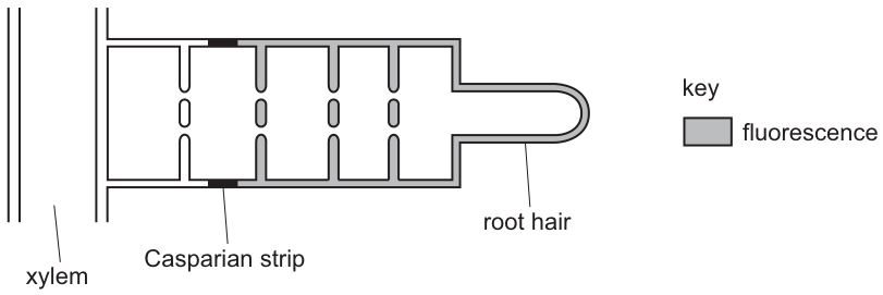
What is a correct conclusion about the transport of lanthanum ions in maize roots?
Options
A The ions are not transported through the apoplast pathway or the symplast pathway.
B The ions are transported through the apoplast pathway only.
C The ions are transported through the apoplast pathway and symplast pathway.
D The ions are transported through the symplast pathway only.
Working
The shaded (fluorescent) areas lie in the cell walls of the root hair and cortex, indicating the lanthanum ions have moved through the apoplast (cell walls and intercellular spaces). No fluorescence is seen inside any cell, so the ions have not entered the cytoplasm — the symplast pathway is not being used. The fluorescence stops abruptly at the Casparian strip of the endodermis because this suberised band is impermeable and blocks apoplast flow; the ions cannot continue because they have not crossed any plasma membrane to take the symplast route either.
Answer
B
B
Background Concept
Water and dissolved mineral ions can travel from the root surface towards the xylem by two parallel routes:
- Apoplast pathway — through the porous cell walls and the intercellular spaces between cells. This is a continuous, non-living route; substances moving here never cross a plasma membrane.
- Symplast pathway — through the cytoplasm of cells, with one cell connected to the next by plasmodesmata. To enter this pathway, a substance must cross a plasma membrane at least once.
The endodermis is the innermost layer of the cortex, and its radial and transverse walls are impregnated with suberin to form the Casparian strip. This waterproof, impermeable band seals the apoplast, so any ion or water molecule still travelling in the cell-wall continuum at this point is forced to cross the plasma membrane of an endodermal cell (i.e. to enter the symplast) if it is to reach the xylem. The Casparian strip is therefore the structure that compels solutes to be taken selectively into the symplast before they enter the vascular tissue.
Understanding the Question
A maize seedling has been grown in soil containing lanthanum ions tagged with a fluorescent marker. The question describes (and Fig. 29.1 shows) a section of root viewed under ultraviolet light. We are asked to interpret the pattern of fluorescence and decide which pathway(s) the lanthanum ions have used.
The three key observations from the diagram are:
- Fluorescence is present in the cell walls of the root hair and the cortical cells — i.e. in the apoplast.
- No fluorescence is seen inside any cell (no fluorescence in cytoplasm or vacuoles) — i.e. the symplast is empty of lanthanum.
- Fluorescence stops abruptly at the Casparian strip of the endodermis and is absent from the xylem.
The command word is implicit in the MCQ: we must select a correct conclusion, not just an observation, so we have to combine what we see with what the Casparian strip does.
Approach
Compare each option against the observations:
- A. "Not transported through either pathway" — contradicted by the clear fluorescence in the cell walls of the cortex and root hair.
- B. "Transported through the apoplast pathway only" — consistent with fluorescence in the walls and absence inside cells; also explains why the ions pile up at the Casparian strip.
- C. "Transported through both pathways" — ruled out because nothing fluoresces inside any cell; the symplast is empty.
- D. "Transported through the symplast pathway only" — ruled out for the same reason; the fluorescence is in the walls, not the cytoplasm, and the symplast route could not in any case cross the Casparian strip region to reach the xylem.
Step-by-Step Reasoning
- Locate the fluorescence: it occupies the cell walls of the root hair and the cortical cells. By definition, anything travelling exclusively in cell walls is in the apoplast. So the apoplast pathway is in use.
- Check for any fluorescence inside the cells: there is none. Lanthanum ions are therefore not crossing plasma membranes to enter the symplast. The symplast pathway is not in use.
- Consider the role of the Casparian strip: it blocks the apoplast at the endodermis. Lanthanum in the apoplast meets this impermeable barrier and simply halts. Because the ions never entered the symplast, they have no alternative route past the endodermis and do not reach the xylem.
- The pattern of fluorescence is therefore fully explained by apoplast-only transport that has been stopped by the Casparian strip. Option B captures this exactly.
Key Takeaways
- The apoplast is the network of cell walls and intercellular spaces; the symplast is the connected cytoplasm of living cells via plasmodesmata.
- To enter the symplast, a solute must cross a plasma membrane.
- The Casparian strip in the endodermis is impermeable and forces any apoplast-borne solute to either be taken into the symplast (via membrane transport) or to be stopped there.
- Ions that cannot cross plasma membranes (lanthanum is one example) are useful experimental tracers: they are confined to the apoplast and reveal exactly how far apoplastic flow can reach before being intercepted by the endodermis.
Common Mistakes
- Choosing C because the ions are clearly moving across the root — but moving across the root is not the same as moving through both pathways. The fluorescence pattern shows only one route is in use.
- Choosing D by confusing the location of the fluorescence (cell walls) with the symplast, or by assuming that any transport into the xylem must be symplastic. Here, no transport into the xylem occurs.
- Choosing A by misreading the diagram and thinking the cortex shows no fluorescence, or by forgetting that "apoplast" includes cell walls.
- Forgetting that the Casparian strip is a block on the apoplast — it does not itself indicate a pathway; it is a barrier that funnels solutes into the symplast when symplastic transport is possible.
Things to Be Careful About
- Read the diagram precisely: shaded = fluorescence, and it lies between the cells (in the walls), not within them.
- Distinguish the structural Casparian strip (a wall modification) from the symplast (cytoplasm) — they are different things; the strip blocks apoplast flow, it does not constitute a pathway.
- "Conclusion" in this style of MCQ means interpret the evidence in the light of biological principles — the question is testing whether the student understands why the pattern looks the way it does, not just whether they can name a pathway.
Which description of adhesion and cohesion is correct?
Options
A Adhesion refers to the force between the water molecules due to hydrogen bonding. Cohesion refers to the force between water molecules and the xylem vessel walls.
B Adhesion refers to the reduced friction between hydrophobic lignin walls and the water molecules. Cohesion refers to the force between water molecules and the xylem vessel walls.
C Adhesion refers to the reduced friction between hydrophobic lignin walls and the water molecules. Cohesion refers to the force between water molecules due to hydrogen bonding.
D Adhesion refers to the force between the water molecules and the xylem vessel walls. Cohesion refers to the force between water molecules due to hydrogen bonding.
Working
Adhesion = the force of attraction between water molecules and the xylem vessel wall. Cohesion = the force of attraction between water molecules, due to hydrogen bonding. Only option D gives these two definitions the correct way round.
Answer
D
D
Background Concept
Water moves up a plant in the xylem as a continuous column. The cohesion-tension theory explains this movement using two properties of water:
- Cohesion is the force of attraction between water molecules themselves. It arises from hydrogen bonding: each water molecule has a partially negative oxygen and two partially positive hydrogens, so the molecules cling together. This produces surface tension and gives water its high tensile strength, allowing the column to be pulled up without breaking.
- Adhesion is the force of attraction between water molecules and another surface, such as the hydrophilic cellulose and lignin in the walls of xylem vessels. Adhesion causes water to creep up the sides of narrow tubes (capillary action) and counteracts the downward pull of gravity on the water column.
Together, adhesion and cohesion maintain the unbroken column of water in the xylem as it is placed under tension (a pulling force) by evaporation of water from the mesophyll cell walls and stomata.
Understanding the Question
This is a multiple-choice item testing the precise definitions of the two terms adhesion and cohesion as used in the cohesion-tension theory of water transport. The distractors mix the two definitions up, and one of them introduces an incorrect idea about "reduced friction between hydrophobic lignin walls".
Approach
Match each option's definition to the correct scientific meaning:
- The correct definition of adhesion must mention water and the xylem vessel wall (water–surface attraction).
- The correct definition of cohesion must mention water–water attraction due to hydrogen bonding.
Step-by-Step Reasoning
- Option A swaps the two definitions — it claims adhesion is water–water hydrogen bonding and cohesion is water–wall. This is the wrong way round.
- Option B gives both definitions incorrectly. Lignin in xylem walls is not strongly hydrophobic at the lumen face, and adhesion is not described as "reduced friction". It also swaps the two terms.
- Option C has the wrong definition for adhesion ("reduced friction between hydrophobic lignin walls") and the right definition for cohesion. Because both terms are being asked about, the wrong definition for adhesion makes the whole option wrong.
- Option D correctly states that adhesion is the force between water and the xylem vessel walls, and cohesion is the force between water molecules due to hydrogen bonding. This matches the textbook definitions and earns the mark.
Key Takeaways
- Cohesion = water-to-water attraction (hydrogen bonding).
- Adhesion = water-to-wall attraction (e.g. to xylem vessel walls).
- These two forces together make the cohesion-tension theory work: cohesion keeps the column intact; adhesion helps the water cling to the vessel walls as it is pulled up.
Common Mistakes
- Swapping the two terms (options A and B). This is the single most common error.
- Describing adhesion as "reduced friction" or claiming the lignin walls are "hydrophobic". In the transpiration stream, the inner surface of the xylem is water-wet and adhesion to it is favourable, not reduced friction.
Things to Be Careful About
The exam will sometimes use a context such as transpiration, capillary rise, or the transpiration stream. The correct pairing of term and definition is fixed regardless of context: always adhesion = water-to-surface and cohesion = water-to-water.
Which conditions are needed to allow the mass flow of sucrose in phloem sieve tubes?
Options
| phloem sieve tube in sources | phloem sieve tube in sinks | |
|---|---|---|
| A | higher hydrostatic pressure higher water potential | lower hydrostatic pressure lower water potential |
| B | higher hydrostatic pressure lower water potential | lower hydrostatic pressure higher water potential |
| C | lower hydrostatic pressure higher water potential | higher hydrostatic pressure lower water potential |
| D | lower hydrostatic pressure lower water potential | higher hydrostatic pressure higher water potential |
Working
The mass flow (pressure-flow) hypothesis depends on a pressure gradient between source and sink:
- At the source (e.g. leaf mesophyll): sucrose is actively loaded into the sieve tube via companion cells. This lowers the water potential inside the sieve tube, so water enters from the xylem by osmosis, raising the hydrostatic pressure.
- At the sink (e.g. roots, fruits, growing tips): sucrose is actively unloaded, raising the water potential inside the sieve tube, so water leaves, lowering the hydrostatic pressure.
- The resulting pressure gradient (high → low) drives the bulk flow of sucrose solution from source to sink.
Therefore the source has higher hydrostatic pressure and lower water potential, while the sink has lower hydrostatic pressure and higher water potential.
Answer
B
B
Background Concept
Plants transport the products of photosynthesis (mainly sucrose) from "sources" (regions that make or release sugars — typically photosynthesising leaves, or storage organs during mobilisation) to "sinks" (regions that use or store sugars — roots, fruits, growing shoots, developing seeds). This long-distance transport of sucrose takes place in the phloem sieve tubes.
The accepted mechanism is the mass flow (pressure-flow) hypothesis, proposed by Ernst Münch in 1930. Bulk flow of the sucrose solution through a sieve tube is driven by a hydrostatic pressure gradient between source and sink. The key to generating this gradient is the controlled movement of sucrose (and therefore water) at each end:
- At the source, companion cells actively load sucrose into the sieve tube using a proton pump (H⁺ pumped out, then sucrose–H⁺ symport). The high internal sucrose concentration makes the sieve tube contents more negative in water potential, so water flows in osmotically from the adjacent xylem. This water entry raises the turgor (hydrostatic) pressure inside the sieve tube at the source end.
- At the sink, sucrose is actively unloaded into sink cells. The sieve tube contents become less concentrated, the water potential becomes less negative, water leaves the sieve tube back to the xylem, and the hydrostatic pressure at the sink end falls.
- The pressure difference pushes the sucrose solution along the sieve tube from source to sink.
Note that water potential (Ψ) and hydrostatic pressure move in opposite directions in this system — adding solute lowers Ψ but then water entry raises turgor pressure. Confusing the two is the most common error in this topic.
Understanding the Question
This is a multiple-choice question requiring you to match the correct combination of hydrostatic pressure and water potential in sieve tubes at the source and at the sink. The single correct option is the one consistent with the mass flow hypothesis above.
The table in the question gives four possible pairings; you must pick the one in which the source end of the sieve tube has the higher pressure and lower water potential, while the sink end has the lower pressure and higher water potential.
Approach
- Recall the mass flow hypothesis and the direction of each variable at source and sink.
- Translate the qualitative direction ("high/low") into the formal terms the question uses: hydrostatic pressure and water potential.
- Scan the four options to find the one matching both columns.
Step-by-Step Reasoning
At the source:
- Active loading of sucrose → sieve tube contents have a high solute concentration.
- High solute concentration → low (more negative) water potential inside the sieve tube.
- Low Ψ draws water in from the xylem by osmosis.
- Water entry pushes against the sieve tube wall → high hydrostatic pressure.
At the sink:
- Active unloading of sucrose → sieve tube contents have a lower solute concentration.
- Lower solute concentration → higher (less negative) water potential inside the sieve tube.
- Higher Ψ drives water out of the sieve tube back to the xylem.
- Water loss → low hydrostatic pressure.
Mass flow direction: the fluid moves from the high-pressure end (source) to the low-pressure end (sink) — exactly as described.
So the required row in the table is:
- Source: higher hydrostatic pressure, lower water potential
- Sink: lower hydrostatic pressure, higher water potential
This matches option B.
Checking the other options:
- A reverses the water potential column (higher Ψ at source is wrong — adding solute lowers Ψ).
- C reverses both columns (low pressure at source cannot drive mass flow toward the sink).
- D has higher pressure at the sink and lower water potential at the sink, the opposite of the unloading situation.
Key Takeaways
- The mass flow hypothesis is driven by a hydrostatic pressure gradient, not a water potential gradient, between source and sink.
- Water potential is low at the source and high at the sink — the opposite of the pressure direction.
- Active loading and unloading of sucrose via companion cells is what sets up and maintains the gradient.
Common Mistakes
- Writing "higher water potential at the source" because students think "lots of sugar = lots of water potential". Sucrose lowers water potential; the high water content of the sieve tube is a consequence of the low water potential, not the cause.
- Confusing the direction of flow: phloem carries sugars from source to sink, so the source end is the upstream (high-pressure) end.
- Forgetting that sieve tubes must be considered as an entire continuous tube; pressure and Ψ change along the length, not in absolute terms.
Things to Be Careful About
- "Higher" water potential means less negative (closer to zero); "lower" water potential means more negative.
- The same water that entered the sieve tube at the source must leave it at the sink — otherwise the system would not be a continuous loop with the xylem.
- Mass flow in phloem is bulk flow under pressure, not diffusion; diffusion alone is far too slow for long-distance transport.
One type of congenital heart defect is where the left and right atria are not completely separated. This is called an atrial septal defect (ASD).
ASD usually results in blood moving from the left atrium into the right atrium. This causes increased blood pressure in the right atrium and decreased blood pressure in the left atrium.
Which row describes other effects caused by ASD?
Options
| blood pressure in pulmonary artery | blood pressure in aorta | % oxygenation of blood in pulmonary artery | |
|---|---|---|---|
| A | decreased | increased | decreased |
| B | decreased | increased | increased |
| C | increased | decreased | decreased |
| D | increased | decreased | increased |
Working
In an ASD, oxygenated blood shunts from the left atrium (LA) into the right atrium (RA), where it mixes with deoxygenated venous blood returning from the body.
-
Pulmonary artery pressure: The extra blood in the RA passes into the right ventricle, which pumps a greater volume into the pulmonary artery → pulmonary artery pressure increases.
-
Aortic pressure: The LA has lost some of its blood to the shunt, so the left ventricle receives less blood and pumps less out through the aorta → aortic pressure decreases.
-
% oxygenation in pulmonary artery: The pulmonary artery normally carries deoxygenated blood. Because oxygenated blood from the LA is now mixing in via the shunt, the blood entering the pulmonary artery is partially oxygenated → % oxygenation increases.
This matches row D: increased pulmonary artery pressure, decreased aortic pressure, increased % oxygenation in the pulmonary artery.
Answer
D
D
Background Concept
The mammalian heart has four chambers: left and right atria (receiving chambers) and left and right ventricles (pumping chambers). The left side of the heart handles oxygenated blood at higher pressure (systemic circulation to the body), while the right side handles deoxygenated blood at lower pressure (pulmonary circulation to the lungs). The two sides are normally completely separated by the interatrial septum (between the atria) and the interventricular septum (between the ventricles), preventing oxygenated and deoxygenated blood from mixing.
Two key vessels emerge from the ventricles: the aorta, which carries oxygenated blood at high pressure from the left ventricle to the body, and the pulmonary artery, which carries deoxygenated blood at lower pressure from the right ventricle to the lungs. Note that the pulmonary artery is named an "artery" because it carries blood away from the heart, not because of the oxygen content of its blood.
Understanding the Question
The question describes an atrial septal defect (ASD) — a congenital hole in the wall between the two atria. It states that because the left side is normally at higher pressure, blood flows from the left atrium into the right atrium (a left-to-right shunt). This raises right atrial pressure and lowers left atrial pressure. The question then asks about the further downstream effects on three quantities: pulmonary artery pressure, aortic pressure, and the percentage oxygenation of blood in the pulmonary artery.
The command word here is essentially "which row is correct" — we have to apply our understanding of cardiac blood flow to work out the three effects.
Approach
The strategy is to follow the extra blood through the right side of the heart to see its effects on the pulmonary circuit, and to follow the lost blood on the left side to see its effect on the systemic circuit. We also need to remember what kind of blood normally travels in each vessel so that we can determine how the oxygenation changes when mixing occurs.
Step-by-Step Reasoning
1. Pressure in the pulmonary artery — INCREASES
The right atrium is now receiving its normal venous return from the body PLUS the extra blood that has shunted across from the left atrium. This larger volume flows into the right ventricle during ventricular filling. The right ventricle therefore ejects a larger stroke volume into the pulmonary artery on each beat. Pumping more blood into a closed system raises the pressure inside it, so pulmonary artery pressure increases.
2. Pressure in the aorta — DECREASES
The left atrium has lost some of its blood through the shunt. Less blood is therefore available to fill the left ventricle, so the left ventricle ejects a smaller stroke volume into the aorta. Pumping less blood into the aorta means the pressure inside it falls, so aortic pressure decreases.
3. % oxygenation of blood in the pulmonary artery — INCREASES
Under normal circumstances, the pulmonary artery carries fully deoxygenated blood from the right ventricle to the lungs. In an ASD, however, the blood reaching the right ventricle is a mixture of the body's deoxygenated venous return AND oxygenated blood that has crossed the defect from the left atrium. This mixed blood is then pumped into the pulmonary artery, which now contains a higher percentage of oxygenated blood than usual. The % oxygenation therefore increases.
These three effects — increased pulmonary artery pressure, decreased aortic pressure, and increased % oxygenation of blood in the pulmonary artery — correspond exactly to row D.
Key Takeaways
- A left-to-right shunt such as ASD transfers blood (and therefore volume) from the high-pressure systemic side to the lower-pressure pulmonary side.
- The pulmonary circuit bears the burden of the extra volume: raised pulmonary artery pressure can, over time, damage the right ventricle and the pulmonary vessels (pulmonary hypertension).
- The systemic side is under-filled, which is why aortic pressure falls and the body's tissues can receive less perfusion.
- The pulmonary artery normally carries deoxygenated blood; any condition that allows oxygenated blood to mix into the right side of the heart will raise the oxygen content of pulmonary arterial blood.
Common Mistakes
- Reversing the pressure change in the pulmonary artery. Some students assume that "less blood going to the lungs to be oxygenated" would lower pulmonary artery pressure. In fact, more blood is being delivered to the pulmonary artery because the right side is overloaded — pressure rises.
- Thinking % oxygenation of the pulmonary artery decreases. Forgetting that the pulmonary artery carries deoxygenated blood is the usual cause; any mixing with oxygenated left-atrial blood must raise the oxygen content, not lower it.
- Confusing the pulmonary artery with the pulmonary vein. The pulmonary vein carries oxygenated blood back from the lungs to the left atrium. If a student mixes these up, the oxygenation reasoning becomes inverted.
- Treating the shunt as right-to-left. Pressure gradients in the heart normally drive any septal defect left-to-right (high to low pressure). A right-to-left shunt would only occur late in the disease, once right-sided pressures have risen to match or exceed the left — and that is not the situation described in the question.
Things to Be Careful About
- The pulmonary artery is an artery by virtue of carrying blood away from the heart, not by virtue of carrying oxygenated blood. The same logic applies to the pulmonary vein, which is a vein despite carrying oxygenated blood.
- "Pressure in the pulmonary artery" should be thought of as the pressure downstream of the right ventricle — if the right ventricle is pumping more, that pressure will rise.
- A reduction in left atrial pressure does not automatically translate to a reduction in left ventricular pressure, but it does mean reduced ventricular filling and therefore reduced cardiac output into the aorta, so aortic pressure falls.
- Always follow the blood: where does the extra volume go, where does the lost volume come from, and what is the oxygen status of blood in each vessel at each step?
Which property of water, related to its role in blood and tissue fluid, is correctly described?
Options
A Water is a solvent for all biological molecules.
B Water is a solvent for most non-polar molecules.
C Water requires little energy to increase its temperature because it has a high specific heat capacity.
D Water cools down slowly because it has a high specific heat capacity.
Working
A high specific heat capacity means that a relatively large amount of thermal energy is needed to raise (or lower) the temperature of water by 1 °C. As a result, water warms up slowly and, conversely, cools down slowly. This buffers the temperature of blood and tissue fluid, helping to maintain a stable body temperature.
- A: incorrect — water is a solvent for polar/ionic molecules, not for all biological molecules (e.g. lipids are non-polar and do not dissolve in water).
- B: incorrect — water is a poor solvent for non-polar molecules, which are hydrophobic.
- C: incorrect — a high specific heat capacity means a lot of energy is required to raise the temperature, not little (the statement reverses the relationship).
- D: correct — water cools down slowly because of its high specific heat capacity.
Answer
D
D
Background Concept
Water has several biologically important properties that arise from its polar nature and the hydrogen bonds that form between water molecules. One of these is its high specific heat capacity — the amount of thermal energy required to raise the temperature of 1 g (or 1 kg) of a substance by 1 °C. For water, this is about , which is unusually high compared with most other liquids.
A high specific heat capacity means water can absorb (or release) a lot of heat energy with only a small change in its own temperature. This has two consequences that are easy to confuse:
- Water heats up slowly (a lot of heat input is needed to raise its temperature).
- Water cools down slowly (a lot of heat must be lost before its temperature falls appreciably).
In the context of blood and tissue fluid, this thermal buffering helps organisms maintain a relatively constant body temperature. Blood, which is largely water, distributes heat around the body, and tissue fluid (derived from blood plasma) carries heat to and from cells without large temperature swings.
Understanding the Question
This is a multiple-choice question asking which statement correctly describes a property of water that is relevant to its role in blood and tissue fluid. The stem frames the property in a physiological context (blood and tissue fluid), which points us towards properties that help with transport and temperature stability — most directly, the high specific heat capacity.
The command word is implicit: identify the option whose description of the property is correct. The trap is that three of the four options contain a real water property but describe it incorrectly (or describe the wrong property altogether).
Approach
Examine each option in turn:
- Test the factual content of the option (is the property itself true?).
- Test the causal/reversibility wording (does the description follow logically from the property?).
- Check relevance to the blood/tissue fluid context where the option is being used.
Step-by-Step Reasoning
Option A — "Water is a solvent for all biological molecules."
Water is a good solvent for polar and ionic substances (e.g. glucose, amino acids, ions such as Na⁺ and Cl⁻) because its partial charges interact with solutes and disrupt their interactions with one another. However, non-polar molecules such as lipids and triglycerides are hydrophobic and do not dissolve in water. So "all" is incorrect — this option is rejected.
Option B — "Water is a solvent for most non-polar molecules."
This is the opposite of the truth. Non-polar molecules are repelled by water's polar structure and tend to cluster together (the hydrophobic effect). Water is a poor solvent for non-polar molecules. Rejected.
Option C — "Water requires little energy to increase its temperature because it has a high specific heat capacity."
The property (high specific heat capacity) is correctly named, but the description reverses the meaning. A high specific heat capacity means that a large amount of energy is needed to raise the temperature, not a little. The hydrogen bonds between water molecules must be continually broken and reformed as temperature changes, which absorbs a great deal of energy. Rejected.
Option D — "Water cools down slowly because it has a high specific heat capacity."
This statement correctly identifies both the property and the consequence. Because water has a high specific heat capacity, much heat must be lost to the surroundings before its temperature falls appreciably — so it cools down slowly. This thermal stability is highly relevant to blood and tissue fluid, which together buffer the body against rapid temperature changes. Correct.
Key Takeaways
- Water's high specific heat capacity is due to extensive hydrogen bonding between its molecules.
- High specific heat capacity → water heats up slowly AND cools down slowly.
- This thermal buffering is biologically important: blood and tissue fluid help maintain a stable internal temperature.
- Water is a good solvent for polar/ionic molecules, not for non-polar ones.
- Always check the direction of cause-and-effect statements: reversing the relationship is a common distractor.
Common Mistakes
- Saying water "requires little energy to increase its temperature" because of high specific heat capacity — this reverses the definition; high specific heat capacity means a lot of energy is needed.
- Claiming water dissolves everything, including lipids and other non-polar substances.
- Confusing the specific heat capacity with the latent heat of vaporisation (which is also high for water and is related to evaporative cooling, not to heating/cooling the bulk liquid).
Things to Be Careful About
- The phrase "high specific heat capacity" is frequently misused in exam answers. Memorise the correct direction: a lot of energy in → small temperature rise; a lot of energy out → small temperature fall.
- The question is set in the context of blood and tissue fluid, but the underlying biology is a property of water itself. Don't be distracted by detailed circulatory knowledge — the answer depends on understanding one property of water.
- Watch for absolute words such as "all" and "never" in MCQ stems; they often signal an incorrect option.
Which statement about tissue fluid formation is correct?
Options
A The hydrostatic pressure of blood decreases from the arteriole end to the venule end of a capillary.
B The water potential is always higher in the blood in the capillaries than in the tissue fluid in the surrounding tissues.
C No cells move out of the blood in the capillaries into the tissue fluid.
D Most tissue fluid formation occurs at the venule end of a capillary.
Working
At the arteriole end of a capillary, the hydrostatic pressure of the blood is high (≈ 5.3 kPa), exceeding the oncotic pressure of plasma proteins, so fluid is forced out into the tissue spaces.
At the venule end, much of the hydrostatic pressure has been lost through friction against the capillary wall and the dissipation of the pressure gradient. The hydrostatic pressure here is low (≈ 2.0 kPa), lower than the plasma oncotic pressure, so most of the fluid re-enters the capillary.
Therefore, hydrostatic pressure decreases from the arteriole end to the venule end.
Answer
A
A
Background Concept
Tissue fluid is the fluid that bathes the cells of the body. It forms by ultrafiltration from the blood plasma across the thin, leaky walls of capillaries, and is then mostly reabsorbed at the downstream end. The direction and rate of fluid movement at any point along a capillary are determined by the balance of two opposing forces — the hydrostatic pressure of the blood (which pushes fluid OUT of the capillary) and the oncotic pressure created by plasma proteins such as albumin (which pulls fluid back IN by osmosis, because the capillary retains these large proteins while losing water and small solutes).
The net filtration pressure at any point is:
where is hydrostatic pressure and is oncotic pressure. Because the oncotic pressure difference is roughly constant along the capillary while the capillary hydrostatic pressure falls steadily, the net direction of fluid flow reverses along the length of the capillary: out at the arteriole end, in at the venule end.
Understanding the Question
The question is multiple choice and asks which single statement about tissue fluid formation is correct. The four options each test a different misconception or a different aspect of capillary exchange. The candidate must identify the one that is consistent with the Starling model of capillary filtration.
Approach
Recall the two key gradients along a capillary:
- Hydrostatic pressure: high at the arteriole end → low at the venule end.
- Net effect on fluid: out at the arteriole end → in at the venule end (most of the fluid that left is reabsorbed).
Then check each option against this picture.
Step-by-Step Reasoning
Option A — "The hydrostatic pressure of blood decreases from the arteriole end to the venule end of a capillary."
This is correct. Blood enters the capillary network at the arteriole end under a high hydrostatic pressure generated by the pumping of the heart and the resistance of the arterioles. As blood flows along the narrow capillary, friction against the endothelium dissipates this pressure, so by the venule end the hydrostatic pressure is markedly lower. This is precisely why ultrafiltration is greatest where the capillary begins and reabsorption occurs where the capillary ends.
Option B — "The water potential is always higher in the blood in the capillaries than in the tissue fluid."
This is incorrect. At the venule end, fluid is moving from the tissue fluid back into the blood. For osmosis to drive water in this direction, the water potential of the blood inside the capillary must be lower (more negative) than that of the surrounding tissue fluid. So water potential is not always higher in the blood — it depends on where along the capillary you are.
Option C — "No cells move out of the blood in the capillaries into the tissue fluid."
This is incorrect. Although the bulk of exchange is fluid and dissolved solutes, certain white blood cells — particularly phagocytes such as neutrophils and monocytes — can squeeze between the endothelial cells of the capillary wall by diapedesis and enter the tissues, where they engulf pathogens. Lymphocytes also recirculate between blood and lymph. So cells do move out of the blood in capillaries.
Option D — "Most tissue fluid formation occurs at the venule end of a capillary."
This is incorrect. Tissue fluid formation (ultrafiltration) is greatest at the arteriole end, where the hydrostatic pressure is highest and the net filtration pressure is strongly outward. At the venule end, the net movement is back into the capillary, not out of it.
Key Takeaways
- Hydrostatic pressure falls along a capillary; it is high at the arteriole end and low at the venule end.
- Net fluid movement is OUT at the arteriole end (tissue fluid forms) and IN at the venule end (most tissue fluid is reabsorbed).
- Plasma oncotic pressure remains relatively constant along the capillary because the large proteins are not filtered out.
- Phagocytes can leave the blood by diapedesis, so the statement "no cells move out" is wrong.
Common Mistakes
- Saying that the water potential is always higher in the blood than in the tissue fluid. Because plasma proteins cannot leave the capillary, the blood becomes more concentrated as it loses water, so by the venule end the blood actually has a lower (more negative) water potential than the tissue fluid — this is what pulls water back in.
- Confusing the arteriole and venule ends: tissue fluid forms at the arteriole end (high hydrostatic pressure) and is reabsorbed at the venule end (low hydrostatic pressure, higher effective oncotic pressure).
- Believing that cells never leave the blood in capillaries — phagocytes perform diapedesis to enter infected tissues.
Things to Be Careful About
- Use the terms hydrostatic pressure (pushes fluid out) and oncotic pressure (pulls fluid in) precisely. CIE expects these exact terms.
- Remember that the fall in hydrostatic pressure along a capillary is the reason filtration and reabsorption occur at opposite ends.
- In a comparison MCQ, do not be tempted by an answer that is "almost" correct — the wording "always" in option B is what makes it wrong, and the word "most" in option D swaps the correct end of the capillary.
Oxyhaemoglobin, carbaminohaemoglobin, haemoglobinic acid and carbonic anhydrase are found inside red blood cells.
How many of these substances will show an overall decrease in concentration as a red blood cell passes through capillaries in the lungs?
Options
A 1
B 2
C 3
D 4
Working
In the lungs, a red blood cell:
- Picks up O₂, so oxyhaemoglobin is formed (concentration increases, not decreases).
- Releases CO₂, so carbaminohaemoglobin dissociates (concentration decreases).
- Releases CO₂ from haemoglobinic acid (H₂CO₃), which is converted back to CO₂ and H₂O (concentration decreases).
- Retains carbonic anhydrase unchanged — it is the enzyme catalysing the reaction and is not consumed (concentration stays the same).
Two substances decrease in concentration.
Answer
B
B
Background Concept
Red blood cells transport O₂ from the lungs to respiring tissues and carry CO₂ back to the lungs. Inside the red blood cell, CO₂ is carried in three main forms:
- Carbaminohaemoglobin — CO₂ bound directly to the amine groups of the globin chains of haemoglobin.
- Haemoglobinic acid (carbonic acid, H₂CO₃) / hydrogencarbonate (HCO₃⁻) — about 70% of CO₂ is hydrated inside the red blood cell to form carbonic acid, which dissociates to H⁺ and HCO₃⁻. The HCO₃⁻ diffuses out into the plasma (the chloride shift).
- Dissolved CO₂ in the cytoplasm and plasma (a small proportion).
The hydration reaction is catalysed by the enzyme carbonic anhydrase:
In the lungs the direction reverses: CO₂ is released, O₂ binds to haemoglobin, and the reactions run in the opposite sense to those in respiring tissues. Crucially, carbonic anhydrase is an enzyme — it speeds up the reaction in both directions but is not used up, so its concentration inside the cell does not change as the cell passes through any capillary bed.
Understanding the Question
We are asked to count, out of the four named substances found inside red blood cells, how many will show an overall decrease in concentration during a single transit through the pulmonary (lung) capillaries. The correct answer is a number (1, 2, 3 or 4).
The four candidates are:
- Oxyhaemoglobin (HbO₂)
- Carbaminohaemoglobin (HbCO₂)
- Haemoglobinic acid (H₂CO₃)
- Carbonic anhydrase (the enzyme)
Approach
Go through each substance in turn and decide whether it goes up, down, or stays the same as the red blood cell moves from pulmonary artery → pulmonary vein (i.e. through lung capillaries). Remember: an enzyme catalyses a reaction but is not consumed, so its own concentration is unchanged.
Step-by-Step Reasoning
-
Oxyhaemoglobin. In the lungs, O₂ diffuses from alveolar air into the red blood cell and binds to deoxyhaemoglobin. The reaction Hb + O₂ → HbO₂ proceeds strongly to the right. Concentration increases — so this is not counted as a decrease.
-
Carbaminohaemoglobin. The reverse reaction occurs: HbCO₂ → Hb + CO₂, with CO₂ diffusing out into the alveoli. Concentration decreases ✓.
-
Haemoglobinic acid (H₂CO₃). As CO₂ is released and the equilibrium is pulled to the left, the chloride shift reverses: HCO₃⁻ enters the red blood cell from the plasma, combines with H⁺, and forms H₂CO₃, which is then dehydrated by carbonic anhydrase to give CO₂ + H₂O. The H₂CO₃ pool is therefore being consumed to release CO₂. Concentration decreases ✓.
-
Carbonic anhydrase. This is the enzyme that catalyses the reversible hydration/dehydration of CO₂. Enzymes are not consumed by the reactions they catalyse — they remain at essentially constant concentration. Concentration stays the same — not a decrease.
Two substances decrease in concentration: carbaminohaemoglobin and haemoglobinic acid. The answer is 2.
Key Takeaways
- In the lungs: O₂ is loaded onto haemoglobin (HbO₂ rises); CO₂ is unloaded (carbaminohaemoglobin and H₂CO₃ fall).
- Carbonic anhydrase is an enzyme. Its job is to speed up the CO₂ + H₂O ⇌ H₂CO₃ reaction; it is not used up and its concentration does not change during a single capillary transit.
- Always distinguish between substrates/products (which are consumed or produced) and the enzyme (which is neither).
Common Mistakes
- Counting carbonic anhydrase as a substance that decreases because it "is used in the reaction". It is a catalyst, not a reactant.
- Counting oxyhaemoglobin as a decrease because it "loses something". In the lungs it gains O₂, so its concentration rises.
- Forgetting that haemoglobinic acid and hydrogencarbonate (HCO₃⁻) are the same chemical system — students sometimes count them as different or miss the fall in the H₂CO₃ pool.
- Saying that all four decrease, or only one, because of muddled reasoning about the chloride shift.
Things to Be Careful About
- Read the list carefully: the question is about substances inside the red blood cell. Dissolved CO₂ and HCO₃⁻ in plasma are not in the list and should not be analysed.
- "Decrease" means a real fall in concentration during the transit, not merely a low steady-state value.
- Remember that the chloride shift is reversible: in the lungs, HCO₃⁻ moves from plasma back into the red blood cell to be converted to CO₂ — this still represents a fall in intracellular H₂CO₃ because the CO₂ is then lost to the alveoli.
- Watch the wording in similar questions: if the question had asked about tissue capillaries, the answer would be the opposite direction (O₂ decreases, CO₂ forms increase).
Which of these structures typically contain cartilage and cilia?
1 bronchi
2 bronchioles
3 trachea
Options
A 1, 2 and 3
B 1 and 2 only
C 1 and 3 only
D 3 only
Working
- The trachea contains C-shaped rings of cartilage to keep the airway open and is lined with ciliated epithelium to move mucus.
- The bronchi are branches of the trachea and likewise contain cartilage (irregular plates) and ciliated epithelium.
- The bronchioles are smaller airways that lack cartilage (they have smooth muscle in their walls) and lack cilia.
Therefore, only the trachea and bronchi (1 and 3) contain both cartilage and cilia.
Answer
C
C
Background Concept
The human gas exchange system is a branching network of airways that conduct air from the atmosphere down to the alveoli, where gas exchange occurs. As the airways branch and decrease in diameter, their wall composition changes — a key feature of the system.
- Trachea — the largest airway. Its wall is supported by C-shaped rings of hyaline cartilage that keep the lumen open during inhalation. The inner lining is a ciliated, mucus-secreting (pseudostratified ciliated columnar) epithelium.
- Bronchi — the first two branches arising from the bifurcation of the trachea (left and right primary bronchi). They retain cartilage in the form of irregular plates (rather than complete rings) and are also lined with ciliated epithelium.
- Bronchioles — smaller branches (typically < 1 mm in diameter) that lack cartilage entirely. Their walls are dominated by smooth muscle, and the lining is mainly simple cuboidal epithelium with very few or no cilia. Because there is no cartilage, bronchioles can constrict significantly (as in asthma).
Understanding the Question
The question asks which of the three listed airways typically contain both cartilage and cilia. A structure must have both features to be included; lacking either one excludes it. The correct answer must therefore list all and only the airways that satisfy both conditions.
Approach
Work through each numbered structure in turn, ticking off whether it has cartilage and whether it has cilia. The structure passes only if it has both.
Step-by-Step Reasoning
- 1 — Bronchi: ✓ cartilage (irregular plates), ✓ cilia → included.
- 2 — Bronchioles: ✗ no cartilage, ✗ no cilia → excluded.
- 3 — Trachea: ✓ cartilage (C-rings), ✓ cilia → included.
This gives 1 and 3 only, matching option C.
Key Takeaways
- Cartilage is a feature of the larger, conducting airways (trachea, bronchi) and is lost in the smaller bronchioles.
- Cilia are most abundant in the trachea and bronchi, where they beat to move mucus (and trapped particles) upward away from the lungs.
- Bronchioles are structurally distinct: no cartilage, mostly smooth muscle, sparse/no cilia, and they are the site of the greatest resistance in healthy airways.
Common Mistakes
- Choosing A (1, 2 and 3) by assuming all airways have cartilage and cilia. Bronchioles lack both.
- Choosing B (1 and 2 only) by forgetting that the trachea is also ciliated and has cartilage.
- Choosing D (3 only) by thinking bronchi have lost the ciliated lining, which is incorrect — they remain ciliated until the bronchioles.
Things to Be Careful About
- Remember that cartilage is present in bronchi as plates, not rings — the question still credits the bronchi as having cartilage.
- Cilia are present in the larger airways to drive the mucociliary escalator; their absence in bronchioles is the structural reason why infections (e.g. bronchiolitis in infants) can spread more easily to the alveoli once past the bronchi.
Which statements about the function of tissues found in the human gas exchange system are correct?
1 Collagen in the bronchi prevents them collapsing.
2 Smooth muscle in the bronchioles can contract to increase the flow of air into the alveoli.
3 Elastic fibres in the alveoli stretch and recoil during breathing.
Options
A 1 and 2
B 1 and 3
C 2 and 3
D 3 only
Working
- Statement 1: It is cartilage (C-shaped rings) in the walls of the bronchi that prevents them from collapsing during expiration, not collagen. ✗
- Statement 2: When smooth muscle in the bronchioles contracts, the bronchioles constrict, which decreases the flow of air into the alveoli (not increases). ✗
- Statement 3: Elastic fibres in the walls of the alveoli (and surrounding tissue) stretch during inspiration and recoil during expiration, helping to expel air. ✓
Only statement 3 is correct.
Answer
D
D
Background Concept
The human gas exchange system (trachea → bronchi → bronchioles → alveoli) is a branching network of tubes whose walls contain several specialised tissues, each with a distinct role. The four key tissues are:
- Cartilage – rigid (but flexible) supporting tissue that holds the airways open. It is present as incomplete C-shaped rings in the trachea and bronchi, but is absent from the bronchioles (below ~1 mm diameter), which is why bronchioles can change diameter markedly.
- Smooth muscle – present in the walls of the bronchi and bronchioles. It can contract (narrowing the lumen) or relax (widening the lumen) under autonomic and hormonal control, regulating the resistance of the airways.
- Elastic fibres – stretchy connective-tissue components in the walls of the bronchioles, alveoli and surrounding lung tissue. They allow the lungs to stretch on inspiration and snap back on expiration.
- Collagen – a fibrous structural protein found in connective tissues throughout the body (e.g. tendons, skin, bone matrix, blood-vessel walls). In the gas exchange system it is present in small amounts as a structural component of connective tissue, but it does not have the specific role of holding the bronchi open.
Understanding the Question
The question gives three statements about the function of these tissues and asks which are correct. The options then bundle them in different ways, so each statement must be judged on its own merits before choosing the answer. The key traps are:
- Confusing collagen with cartilage – both are structural, but only cartilage prevents airway collapse.
- The sign of the effect of smooth muscle contraction on airflow (it decreases, not increases, the lumen diameter).
- A correct but understated statement (3) is the only one that is biologically accurate.
Approach
Evaluate each statement independently against the known function of the named tissue, then identify which combination matches one of the four options.
Step-by-Step Reasoning
Statement 1 – "Collagen in the bronchi prevents them collapsing."
This is wrong. The tissue that prevents the bronchi (and trachea) from collapsing during expiration is cartilage, present as C-shaped (incomplete) rings in their walls. Collagen is a fibrous protein that provides tensile strength in connective tissue generally, but it is not the structure that maintains the open lumen of the bronchi. Statement 1 is incorrect.
Statement 2 – "Smooth muscle in the bronchioles can contract to increase the flow of air into the alveoli."
This is wrong. Smooth muscle is arranged circularly in the walls of the bronchioles. When it contracts, the lumen of the bronchiole narrows (bronchoconstriction), which increases resistance and decreases the flow of air into the alveoli. It is relaxation of the smooth muscle (bronchodilation) that increases airflow. Statement 2 is incorrect.
Statement 3 – "Elastic fibres in the alveoli stretch and recoil during breathing."
This is correct. The alveolar walls (and the interstitium between alveoli) contain elastic fibres. During inspiration the alveoli expand as the lungs fill with air; the elastic fibres stretch. During expiration they recoil passively, helping to reduce alveolar volume and push air out. This elastic recoil is also what drives expiration passively at rest. Statement 3 is correct.
Since only statement 3 is correct, the answer is the option that contains 3 only.
Key Takeaways
- Cartilage (C-rings), not collagen, keeps the trachea and bronchi patent.
- Smooth muscle contraction in bronchioles constricts them, reducing airflow; relaxation widens them, increasing airflow.
- Elastic fibres in alveoli and lung tissue stretch on inspiration and recoil on expiration, providing the passive elastic recoil that aids expiration.
- When tackling a "which statements are correct" MCQ, always check each statement independently and be alert to sign errors (increase vs decrease) and tissue-name confusions.
Common Mistakes
- Confusing collagen and cartilage – both are structural, but only cartilage gives the rigid-yet-flexible support that keeps the larger airways open.
- Assuming "muscle contracting" means "more flow" – muscle contracting around a tube makes the tube narrower and reduces flow; this is the basis of bronchoconstriction in asthma.
- Forgetting that elastic recoil is a real mechanism in the lungs – candidates sometimes underplay this, but it is essential to passive expiration.
Things to Be Careful About
- Read the tissue name exactly as written. "Collagen" and "cartilage" are not interchangeable, even though both contain protein.
- Read the direction of the effect carefully: contraction → constriction → ↓ airflow for circular smooth muscle around a tube.
- The bronchi and bronchioles are different structures: bronchi have cartilage, bronchioles do not — only bronchioles have a substantial smooth muscle layer able to constrict the lumen markedly.
Antibiotic-resistant strains of Mycobacterium tuberculosis are a major problem when treating TB. A new antibiotic, teixobactin, could be very effective at killing M. tuberculosis with only a small risk that the bacteria will evolve teixobactin resistance.
Penicillin and similar antibiotics bind to a single protein, but teixobactin binds to two lipids that are needed for the formation of the bacterial cell wall. Teixobactin binds to regions of the two lipids that do not vary across many different species of bacteria.
Which statements help to explain why the use of teixobactin is thought to be less likely to lead to the evolution of antibiotic resistance than the use of many other antibiotics, such as penicillin?
1 A single mutation can result in bacteria that are resistant to penicillin and similar antibiotics but at least two mutations are required to produce teixobactin-resistant bacteria.
2 Mutations can affect the structure of proteins but cannot affect the structure of lipids because only proteins are made of amino acids.
3 The lack of variation across many species of bacteria in the two lipids that bind to teixobactin suggest that the particular structure of these lipids is essential for successful bacterial cell wall formation.
Options
A 1, 2 and 3
B 1 and 2 only
C 1 and 3 only
D 2 and 3 only
Working
Statement 1 — TRUE: Penicillin binds to a single protein, so one mutation altering that protein can confer resistance. Teixobactin binds to two separate lipids, so resistance would require mutations in both — a far rarer event.
Statement 2 — FALSE: Although lipids are not themselves made of amino acids, mutations in the genes encoding the enzymes that synthesise lipids can change lipid structure. Mutations are changes to DNA and can therefore affect any product downstream, including lipids.
Statement 3 — TRUE: The two target lipids are highly conserved across many bacterial species. Strong conservation indicates that the structure is under intense selection, i.e. any change is likely to impair cell-wall formation, so resistant mutants are unlikely to survive.
Statements 1 and 3 are correct; statement 2 is not.
Answer
C
C
Background Concept
Antibiotic resistance evolves by natural selection acting on random mutations. A population of bacteria contains pre-existing genetic variation; if any individual carries a mutation that allows it to survive exposure to the antibiotic, it reproduces and its offspring inherit the resistance allele, so the resistant form becomes more common in the population over time.
The probability of resistance evolving therefore depends on two factors:
- How many mutations are needed to confer resistance. One mutation is far more likely to arise spontaneously than two simultaneous mutations, which is why multi-target drugs tend to be more durable.
- How constrained the target is. If the target molecule is essential and its structure is conserved (i.e. it cannot easily be altered without harming the bacterium), the range of mutations that confer resistance and still leave the bacterium viable is very small.
Penicillin and related antibiotics work by binding to a single bacterial enzyme involved in cell-wall synthesis (transpeptidase / penicillin-binding protein). A single amino-acid substitution in this protein can prevent binding and yet leave the enzyme functional enough for the bacterium to survive. Teixobactin, in contrast, binds to two specific lipid precursors (lipid II and lipid III) that are needed to build the bacterial cell wall.
Understanding the Question
The question is a multiple-choice item asking the candidate to judge three biological statements and select which combination correctly explains why teixobactin is expected to select for resistance less readily than penicillin. The candidate must identify which statements are biologically valid (1 and 3) and which is not (2), then choose the corresponding option (C: 1 and 3 only).
Approach
Work through each statement, decide whether it is biologically correct, then match the combination of true statements to the answer options. The reasoning rests on (a) the genetics of how many mutations are needed to confer resistance, (b) the difference between proteins and lipids as gene products, and (c) the meaning of evolutionary conservation.
Step-by-Step Reasoning
Statement 1 — At least two mutations are required to produce teixobactin-resistant bacteria. — TRUE.
- Penicillin's target is a single protein. A mutation in the gene encoding that protein can change its shape and prevent penicillin from binding. One mutation is enough.
- Teixobactin binds to two distinct lipid molecules simultaneously. For a bacterium to become resistant, both lipids would have to be altered so that teixobactin can no longer bind to them, but the cell wall can still be made.
- Two independent mutations occurring in the same bacterium at the same time is a much rarer event than one, so resistance evolves much more slowly.
Statement 2 — Mutations cannot affect the structure of lipids because only proteins are made of amino acids. — FALSE.
- This statement confuses the direct gene product (the protein/RNA encoded by a gene) with all downstream molecules in the cell. A mutation changes the base sequence of DNA, which changes the mRNA, which can change the amino acid sequence of a protein — but proteins are enzymes, and enzymes catalyse almost every other reaction in the cell, including the synthesis of lipids, carbohydrates and nucleotides.
- Therefore a mutation in a gene encoding a lipid-synthesising enzyme can change the structure of the lipid that the enzyme produces. Lipid structure is just as much under genetic control as protein structure; it is simply controlled indirectly via enzymes.
- This is why statement 2 is rejected.
Statement 3 — The lack of variation across many species in the two target lipids suggests their particular structure is essential for cell-wall formation. — TRUE.
- "Lack of variation" across many different bacterial species means these lipid sequences/structures have been conserved over a long evolutionary time. Natural selection preserves sequences that are essential — any variant that arose and reduced fitness would have been removed.
- If the structure is essential, then any mutation that altered it would either kill the bacterium or severely reduce its growth. A bacterium that is inviable or grows very poorly cannot spread antibiotic resistance through the population.
- This is exactly why the lipids are a good drug target: the bacterium is highly constrained in how it can change them.
Combining the judgements: statements 1 and 3 are correct; statement 2 is not. Only option C lists "1 and 3 only".
Key Takeaways
- Resistance evolution is faster when a single mutation can confer it on a non-essential target.
- Drugs that hit multiple targets (or highly conserved, essential targets) select for resistance more slowly.
- A gene mutation is a change to DNA; its consequences extend to all downstream products made via the proteins it encodes, including lipids, carbohydrates and other non-protein molecules.
- Evolutionary conservation of a molecule is strong evidence that its structure is functionally essential.
Common Mistakes
- Assuming mutations only affect proteins. Many students think "lipids aren't made of amino acids so they can't mutate". This is wrong: mutations in enzyme-encoding genes can change the structure of the lipid those enzymes make.
- Confusing the number of targets with the number of mutations. Teixobactin has two lipid targets, so two mutations are required — a key point that is often missed.
- Misreading "conserved" as "unchanging because unimportant". In evolutionary biology, conservation of a sequence or structure almost always indicates importance, not irrelevance.
- Choosing A because all three statements "sound scientific". Statement 2 contains a fundamental error in how genes relate to cellular molecules and must be rejected.
Things to Be Careful About
- Watch the exact wording of MCQ options: this question asks for the combination of statements that helps explain the lower risk, so any wrong statement disqualifies an option.
- The "central dogma" (DNA → RNA → protein) does not stop at proteins: proteins are the workhorses that build and modify every other class of molecule, so the effects of a mutation can reach lipids, carbohydrates and even whole organelles.
- "Lack of variation" is a population-level / comparative observation; it must be interpreted as evidence of strong purifying selection, not as a neutral trait.
Monoclonal antibodies are now being used to treat some human diseases.
What explains why monoclonal antibodies are suitable for this purpose?
1 They can divide by mitosis to produce the large numbers of antibodies required for treatment.
2 They are specific to a particular antigen.
3 They can be modified so that they do not act as antigens themselves.
Options
A 1, 2 and 3
B 1 only
C 2 and 3 only
D 2 only
Working
- Statement 1 is incorrect: antibodies are proteins secreted by hybridoma cells; the antibodies themselves do not divide by mitosis. It is the hybridoma cells (formed by fusing a B-lymphocyte with a myeloma cell) that divide to produce large numbers of identical antibodies.
- Statement 2 is correct: monoclonal antibodies are produced by a single clone of B-lymphocytes and so are specific to one antigen (one epitope).
- Statement 3 is correct: antibodies raised in mice would be recognised as foreign by the human immune system, so they are modified (e.g. humanised or chimeric antibodies) so they do not act as antigens themselves.
Answer
C
C
Background Concept
Monoclonal antibodies (mAbs) are identical antibody molecules produced by a single clone of B-lymphocytes. They are made commercially by fusing a B-lymphocyte (which makes a specific antibody but cannot divide indefinitely) with a myeloma cell (a cancer cell that divides endlessly). The resulting hybrid cell is called a hybridoma, and it combines two useful properties: it can divide continuously and it secretes one specific antibody.
For therapeutic use, monoclonal antibodies must be safe and effective inside a patient. Two biological requirements are critical:
- Specificity — the antibody must bind only to the target antigen (e.g. a protein on a cancer cell or a signalling molecule) so that healthy tissues are not damaged.
- Non-immunogenicity — if the antibody is recognised by the patient's immune system as a foreign antigen, the patient will mount an immune response against the treatment itself, neutralising it and causing harmful side effects. Mouse antibodies are therefore engineered (humanised or chimerised) to look more like human antibodies.
Understanding the Question
This is a multiple-choice question that gives three statements and asks which combination correctly explains why monoclonal antibodies are suitable for treating human disease. We must judge each statement independently and then pick the option that lists only the correct ones.
The command word is implied: "What explains…" — i.e. which statements give a true biological reason for their therapeutic suitability.
Approach
- Check each statement against established facts about monoclonal antibodies.
- Pay close attention to the subject of each sentence: in statement 1, the subject is "they" — and "they" refers back to the monoclonal antibodies, which are protein molecules, not cells.
- Statements 2 and 3 describe properties of the antibody molecule itself, which is the appropriate level for therapeutic use.
Step-by-Step Reasoning
- Statement 1 — "They can divide by mitosis…"
Antibodies are globular proteins, not cells. They cannot undergo mitosis. What divides is the hybridoma cell that secretes the antibody, and this division is in vitro in culture vessels in the laboratory — not inside the patient being treated. The statement is therefore rejected. - Statement 2 — "They are specific to a particular antigen."
Because each monoclonal antibody comes from one B-cell clone, every molecule binds the same epitope on the same antigen. This specificity is exactly what is wanted in therapy: it allows the antibody to target, for example, HER2 on certain breast cancer cells, or TNF-α in rheumatoid arthritis, without binding to unrelated molecules. Statement 2 is accepted. - Statement 3 — "They can be modified so that they do not act as antigens themselves."
Early monoclonal antibodies were made in mice. Their constant regions would be recognised as "non-self" by a human patient, triggering an immune response against the drug. Modern therapeutic antibodies are engineered — chimeric (mostly human with mouse antigen-binding regions), humanised (only the antigen-binding loops from mouse), or fully human — so they do not provoke an immune response. Statement 3 is accepted. - Conclusion: Statements 2 and 3 are correct; statement 1 is not. This combination matches option C.
Key Takeaways
- Monoclonal antibodies are specific to one antigen because they come from a single B-cell clone.
- They are humanised to prevent the patient's immune system from treating them as foreign.
- It is the hybridoma cell, not the antibody, that divides by mitosis — and this division happens in the lab, not in the patient.
Common Mistakes
- Confusing the antibody with the cell that makes it. Statement 1 looks plausible because "they produce large numbers of antibodies" is true of hybridoma cells. The slip is reading "they" as the cells rather than the antibodies. The mark scheme rejects this because antibodies are not cells.
- Assuming all three statements are true. The phrasing of option A tempts students into ticking the all-of-the-above trap without checking statement 1.
- Forgetting that mouse antibodies would be antigenic in humans. Statement 3 is a less obvious point, but it is essential to why mAbs needed to be engineered before they could be used therapeutically.
Things to Be Careful About
- Always check what the pronoun in the statement refers back to. Here "they" = monoclonal antibodies (the protein), not the hybridoma (the cell).
- Therapeutic suitability depends on both specificity (statement 2) and lack of immunogenicity (statement 3) — both must hold.
- Note that mitotic division of hybridomas is a production step in the laboratory, not a therapeutic action in the patient; the question is about why the antibodies themselves are suitable for treatment.
Measles is an infectious disease caused by a virus.
The graph shows the number of cases of measles each year in a country before and after a vaccine was introduced.
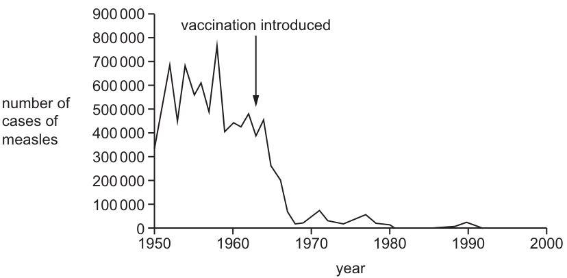
What could have caused the decrease in the number of cases of measles after vaccination was introduced?
Options
| vaccines provided artificial active immunity in people | vaccines provided artificial passive immunity in people | fewer people are able to act as hosts for the virus | ||
|---|---|---|---|---|
| A | ✓ | ✗ | ✓ | key |
| B | ✗ | ✓ | ✓ | ✓ = yes |
| C | ✗ | ✓ | ✗ | ✗ = no |
| D | ✓ | ✗ | ✗ |
Working
Vaccines contain antigens that stimulate the recipient's own immune system to produce antibodies and memory cells. This is artificial active immunity (not passive, which would require injecting ready-made antibodies).
When a large proportion of a population is vaccinated, fewer people can be infected. With fewer susceptible individuals, the virus has fewer hosts in which to replicate and spread — the basis of herd immunity. This is a correct statement for the post-vaccination decline shown in Fig. 40.1.
- Row A: artificial active immunity = ✓, fewer hosts = ✓ → both correct.
- Row B: passive immunity is wrong — vaccines do not give ready-made antibodies.
- Rows C and D each contain an incorrect statement.
Answer
A
A
Background Concept
The immune response can be classified along two axes:
- Natural vs artificial — how the antigens/antibodies are encountered.
- Natural exposure happens through ordinary infection.
- Artificial exposure happens through medical intervention (vaccine injection, antibody injection).
- Active vs passive — who makes the antibodies.
- Active immunity: the recipient's own lymphocytes produce antibodies and memory cells in response to antigens. This takes days to weeks to develop but is long-lasting.
- Passive immunity: ready-made antibodies are transferred into the recipient (e.g. from mother to fetus across the placenta, or as an antiserum injection). Protection is immediate but short-lived because no memory cells are made.
A vaccine contains antigens (attenuated, killed or subunit forms of a pathogen) that provoke the recipient's own active immune response — so vaccination is artificial active immunity.
Herd immunity describes the indirect protection of unvaccinated individuals that occurs when a sufficiently high proportion of a population is immune. With fewer susceptible people available, the pathogen cannot easily find a host, replicate and transmit, so its reproduction number () falls below 1 and incidence declines.
Understanding the Question
The graph in Fig. 40.1 shows that measles cases fluctuated at a high level (300 000–800 000 per year) between 1950 and the early 1960s, then fell sharply after the vaccine was introduced around 1963, reaching very low levels by the 1970s. The question asks which combination of statements correctly explains this decline.
The three statements to evaluate are:
- Vaccines provided artificial active immunity in people.
- Vaccines provided artificial passive immunity in people.
- Fewer people are able to act as hosts for the virus.
Approach
Decide each statement on its biological merits, then match the combination to the correct option.
Step-by-Step Reasoning
-
Statement 1 — artificial active immunity: A vaccine introduces antigens that the recipient's own immune system responds to by producing antibodies and memory B- and T-lymphocytes. This matches the definition of artificial active immunity. ✓
-
Statement 2 — artificial passive immunity: Passive immunity requires the transfer of pre-formed antibodies (e.g. an antiserum or maternal IgG). A vaccine does not contain ready-made antibodies, so this statement is false. ✗
-
Statement 3 — fewer people able to act as hosts: When a large fraction of the population is immune, the virus has fewer susceptible cells/people in which to replicate. Transmission chains break and the virus's effective reproduction number falls. This is the herd-immunity effect, and the graph supports it. ✓
Therefore the correct combination is: statement 1 ✓, statement 2 ✗, statement 3 ✓. Only option A carries this combination.
Key Takeaways
- Vaccines stimulate the recipient's own immune response → artificial active immunity.
- Passive immunity = receiving ready-made antibodies; vaccines do not do this.
- Mass vaccination reduces the number of susceptible hosts, producing herd immunity and driving down disease incidence.
Common Mistakes
- Conflating vaccines with passive immunity because "you're receiving something medical". A vaccine is antigenic material, not pre-formed antibody.
- Thinking vaccination only protects the individual. The herd-immunity (host-availability) statement is what links vaccination programmes to population-level declines such as the one shown in Fig. 40.1.
- Selecting B because "fewer hosts" is correct, overlooking that passive immunity is the wrong description for vaccination.
Things to Be Careful About
- Distinguish active vs passive (who makes the antibody) from natural vs artificial (how the antigen/antibody is delivered). A vaccine is artificial + active.
- "Fewer hosts" is a population-level concept: even vaccinated individuals can, in principle, encounter the virus, but a virus that cannot easily find an unvaccinated host cannot sustain transmission.
- On a multi-statement MCQ, check every statement in the chosen row — a single wrong tick is enough to eliminate that option.
Your score so far
Answer a question to start scoring
Your marks add up here as you work through the paper.