Biology 9700/22 — October/November 2023
Cambridge AS Level · AS Level Structured Questions · worked solutions for every part, with the mark scheme
Topics Transport in Mammals · Nucleic Acids and Protein Synthesis · The Mitotic Cell Cycle · Biological Molecules · Enzymes · Immunity · +5 more
During interphase and mitosis of the cell cycle, the chromosomes within a cell go through a number of changes. Each chromosome is composed of DNA complexed with proteins.
In interphase, individual chromosomes are too diffuse (long and thin) to be visible using a microscope. In this stage, the chromosomal material is known as chromatin.
State the term used to describe the proteins that are complexed with DNA and form part of chromatin.
Answer
Histones
Histones
Background Concept
In eukaryotic cells, nuclear DNA does not exist as a free, naked molecule. It is packaged with proteins to form a compact structure that fits inside the nucleus. The protein component of this complex is the histones — small, positively charged (basic) proteins rich in the amino acids lysine and arginine. The negatively charged phosphate groups of DNA wrap around clusters of histones called nucleosomes (eight histones forming a core, plus the DNA wound around them), and this "beads on a string" arrangement is then further coiled and folded. The whole DNA–protein complex is called chromatin; when chromatin is maximally condensed during mitosis it becomes visible as discrete chromosomes.
Understanding the Question
The stem tells us that chromosomes are made of DNA complexed with proteins, and part (a) asks specifically for the name of the proteins that associate with DNA to form chromatin. The answer is a single technical term worth one mark.
Approach
Identify the protein component of chromatin from prior knowledge of chromosome structure. CIE accepts either "histones" or "histone proteins" (or "basic proteins"); the most precise single term is histones.
Step-by-Step Reasoning
- Chromatin = DNA + protein. The protein part is the histone fraction.
- Histones are basic because their side chains carry positive charges at physiological pH, which neutralises the negatively charged phosphate–sugar backbone of DNA and allows tight association.
- Eight histones (two each of H2A, H2B, H3, H4) form a nucleosome core; DNA is wound ~1.65 times around it.
- Mark scheme: histones (also accepts "basic proteins" or "histone proteins").
Key Takeaways
- The proteins associated with DNA in chromatin are histones.
- Histones are basic proteins that allow the highly negatively charged DNA to be tightly packaged.
- Nucleosomes are the repeating structural units of chromatin.
Common Mistakes
- Writing "chromatin" itself — that is the name of the DNA–protein complex, not the protein component.
- Writing "histamine" — this is a different molecule (involved in inflammation), not a chromatin protein.
- Writing "non-histone proteins" or "acidic proteins" — the specific term asked for is histones.
Things to Be Careful About
- Spelling: "histones" not "histons" or "hystones".
- The mark scheme accepts the unqualified term "histones"; do not pad the answer.
When viewed through a microscope, a chromosome is most clearly visible during the metaphase stage of mitosis.
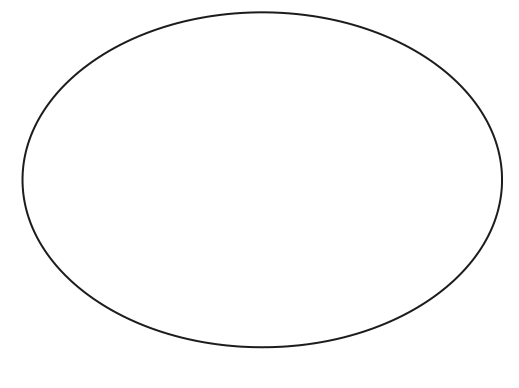
Complete Fig. 1.1 to produce a labelled diagram of the metaphase stage of mitosis in an animal cell with two chromosomes.
Answer
Key features required for full marks:
- Two chromosomes drawn along the central equator (metaphase plate), each consisting of two sister chromatids joined at a centromere.
- At least one other feature: a spindle of fibres radiating from the two poles, ideally with a pair of centrioles at each pole (animal cell).
- Spindle fibres must be drawn inside the cell membrane (no fibres extending outside).
- Three correct labels with label lines touching the structure, e.g.:
- sister chromatid(s) (or chromatid)
- centromere
- spindle / spindle fibre(s)
- centriole(s) / pole(s) / centrosome
- (spindle) equator / metaphase plate (only if a spindle label is given)
Labelled diagram: two chromosomes (each with two chromatids) at the metaphase plate, with a spindle and centrioles at the poles; labelled with chromatid, centromere, spindle (and/or centriole).
Background Concept
Metaphase is the third stage of mitosis. By the time the cell reaches metaphase:
- The nuclear envelope has broken down (end of prophase).
- The chromosomes are at their most condensed, so they are most easily seen with a light microscope.
- Each chromosome consists of two genetically identical sister chromatids held together at the centromere (the constricted region where the kinetochore protein complex assembles).
- Spindle fibres (microtubules) extend from the two poles of the cell. In animal cells, the poles are organised by a pair of centrioles sitting in a centrosome. Kinetochore microtubules attach to the centromere of each chromatid, and the chromosomes are aligned along the equator (metaphase plate) by opposing tensions from the two poles.
Key convention: in an animal cell, centrioles are present at each pole; plant cells lack centrioles and the spindle forms from microtubule-organising centres without centrioles.
Understanding the Question
The question prints a large circle representing an animal cell membrane (Fig. 1.1). The candidate is asked to complete the figure with a labelled diagram of metaphase in an animal cell containing two chromosomes. The marks are: (1) drawing the two chromosomes correctly at the equator, each with two chromatids; (2) one additional drawn feature (e.g. spindle); (3) three labels. Spindle fibres/centrioles must not extend outside the cell. The two chromosomes must not be drawn as a homologous (bivalent) pair sitting close together or apart — that is meiosis I, not mitosis.
Approach
- Draw a vertical (or horizontal) dashed/dotted line down the middle of the cell — this is the equator / metaphase plate.
- Draw two X-shaped chromosomes straddling that line, each clearly showing two chromatids meeting at a centromere. Space them apart (do not bunch them as a bivalent).
- Add a spindle of dashed lines running from the two poles to the chromosomes, and place a pair of centrioles at each pole inside the cell.
- Add label lines ending on the structure: chromatid, centromere, spindle fibre (or centriole).
Step-by-Step Reasoning
- Mark 1 — two chromosomes, each with two chromatids, along the central equator. Each chromosome is drawn as two parallel sister chromatids joined at a single centromere. They are placed at the equator of the cell. The diagram must clearly show this is mitosis (so the two chromosomes are not paired as homologues) — keep them well separated, not as a bivalent.
- Mark 2 — at least one additional drawn feature. Acceptable additional features are any of:
- a spindle drawn as fibres from the poles to the chromosomes, OR
- a centromere drawn holding the sister chromatids together, OR
- a pair of centrioles at each pole.
The spindle must stay inside the cell membrane — fibres extending past the boundary lose 2 marks.
- Mark 3 — three correct labels. Acceptable labels include: sister chromatid(s)/chromatid, centromere, spindle/spindle fibre, spindle equator/metaphase plate, centriole(s)/centrosome(s)/pole(s). Label lines must end on the structure, not float in space. "Chromosome" by itself is not credited (the mark scheme ignores "label to chromosome"). "Equator" alone is only accepted if a spindle label is also given (to clarify that it is the spindle equator).
Key Takeaways
- A chromosome at metaphase = two sister chromatids joined at the centromere.
- Chromosomes at metaphase are aligned on the metaphase plate (the spindle equator) by opposing spindle fibres.
- Animal-cell spindles are organised at each pole by a pair of centrioles within a centrosome; this is a key difference from plant cells.
- CIE drawing conventions: clear lines, label lines ending on the structure, no vague labels like "chromosome" without further qualification.
Common Mistakes
- Drawing a bivalent / homologous pair (the two chromosomes stuck together as a tetrad) — that would be meiosis I, not mitosis. Reject.
- Drawing the spindle outside the cell membrane — loses up to 2 marks.
- Labelling only "chromosome" without specifying chromatid, centromere or spindle — the mark scheme ignores this label.
- Drawing the chromosomes scattered around the cell instead of aligned at the equator.
- Forgetting that the question specifies an animal cell — centrioles should be drawn at the poles.
Things to Be Careful About
- Centrioles are pairs of short cylindrical structures at right angles to each other; they sit inside the cell at each pole.
- The cell in Fig. 1.1 is already drawn — the candidate only adds the internal structures. Do not redraw the membrane.
- Label lines must touch the structure they name; floating labels are not credited.
- Use a sharp pencil, no shading, and continuous lines (not sketching) for cell structures.
Outline the changes that occur to the structure and behaviour of a chromosome:
• from the start of the S phase to the end of interphase
• during prophase of mitosis.
Answer
From the start of S phase to the end of interphase:
- (Chromosomal) DNA replicates (DNA synthesis).
- Each new molecule of DNA complexes with histones (proteins), so sister/identical chromatids form by the end of S phase.
During prophase of mitosis:
3. The (sister) chromatids condense (become shorter and fatter) by coiling/supercoiling of DNA, and the chromosome becomes visible as two sister chromatids joined at a centromere.
4. Spindle microtubules form and attach to the centromere of each chromatid (kinetochore formation).
S phase: DNA replicates and each new DNA molecule associates with histones to form sister chromatids. Prophase: chromatids condense (shorter and fatter, by coiling) and become visible as two sister chromatids; spindle microtubules attach to the centromere.
Background Concept
The cell cycle has two main parts: interphase (G1, S, G2) and mitosis (prophase, metaphase, anaphase, telophase), followed by cytokinesis. During interphase the cell grows and the DNA is replicated so that each chromosome can be passed on in full to each daughter cell.
- S phase is the part of interphase in which DNA synthesis (replication) occurs. Each chromosome is copied to produce two identical DNA double helices. As the new DNA is synthesised, it is packaged with histones (largely from parental histones plus newly synthesised ones) to form chromatin. By the end of S phase, each chromosome consists of two genetically identical sister chromatids held together at the centromere.
- G2 is the gap after S phase, where the cell continues to grow and prepares for mitosis.
- Prophase is the first stage of mitosis. The diffuse chromatin (long and thin, invisible as discrete chromosomes) condenses by progressive coiling and supercoiling of the DNA around the histones, becoming shorter and fatter so that each chromosome is visible under the light microscope as two sister chromatids joined at the centromere. The spindle (made of microtubules) begins to form, and in animal cells the centrioles move to opposite poles. The nuclear envelope is still intact at the start of prophase and breaks down towards the end.
Understanding the Question
The command word is outline — give the main points without going into excessive detail. Two bullet areas are given: (i) from the start of S phase to the end of interphase, and (ii) during prophase. The mark scheme caps each bullet at 2 marks and the question is worth 3 marks total, so the candidate needs to make about three specific points across the two phases.
Approach
- List the key S-phase event: DNA replication and the production of sister chromatids, with histones complexing the new DNA.
- List the key prophase events: condensation of chromatids (so they become visible as two chromatids) and the formation/attachment of the spindle.
- Avoid overlap — the mark scheme forbids awarding the same event twice (e.g. it rejects stating that chromosomes condense "in prophase" if it has already been credited in S phase, and rejects "chromatin" as a credit for the DNA-replication point).
Step-by-Step Reasoning
- Mark 1 (interphase) — DNA replicates (or "DNA synthesis"). Note: the mark scheme ignores "chromatin" and rejects statements located "in G1/G2" — replication must be associated with S phase.
- Mark 2 (interphase) — Each new molecule of DNA complexes / associates with histones (proteins), and sister/identical chromatids form (qualified — must be clear they form by the end of S phase / after replication, before prophase begins). The mark scheme requires that the candidate make it explicit that the chromatids are formed by the end of interphase, not during prophase.
- Mark 3 (prophase) — Chromatids/chromosome condense (becomes shorter and fatter; coiling/supercoiling of DNA in the context of chromosome/chromatid). Do not write "chromosomes condense to form chromatin" — the direction is wrong (chromatin condenses to form chromosomes).
- Mark 4 (prophase — alternative) — (Spindle) microtubules attach to the centromere (kinetochore formation) OR the chromosome becomes attached to the spindle fibres.
Three marks are needed; a strong answer gives two interphase points and one prophase point, or one interphase point and two prophase points.
Key Takeaways
- S phase: DNA replication + assembly of new DNA with histones → formation of two sister chromatids per chromosome.
- Prophase: chromatids condense (shorter/fatter, by coiling) and become visible as two chromatids; spindle forms and attaches to centromeres via kinetochores.
- Sister chromatids are genetically identical because they are produced by semi-conservative replication of the parental DNA.
- The change from "diffuse chromatin" to "condensed chromosome" is what makes chromosomes visible under the light microscope during mitosis.
Common Mistakes
- Stating that DNA replication occurs in G1 or G2 — it occurs in S phase; the mark scheme rejects this.
- Stating that "chromatin replicates" — DNA is what replicates, not the chromatin as a whole; "chromatin" is ignored for that marking point.
- Saying "chromosomes condense to form chromatin" — backwards.
- Saying the chromatids are formed during prophase — they must be formed by the end of interphase, ready for prophase to condense them.
- Describing anaphase events (centromeres splitting, chromatids separating) — the question is about prophase, not anaphase.
Things to Be Careful About
- Use the precise terms "sister / identical chromatids", "condense" (not just "become visible"), "histones", "centromere" and "spindle / microtubules".
- Do not mix up condensation (prophase) with replication (S phase); the mark scheme explicitly rejects awarding condensation in interphase.
- "Becomes visible as two chromatids" alone is ignored (per mark scheme: I "composed of two chromatids") — pair it with the condensation point or the spindle-attachment point for credit.
- Keep the answer sequenced: S-phase first, then prophase.
The rest of this paper
5 more questions- Q2Biological Molecules · Enzymes12M
- Q3Transport in Mammals · Immunity · Cell Structure · Nucleic Acids and Protein Synthesis14M
- Q4Gas Exchange · Cell Membranes and Transport · Nucleic Acids and Protein Synthesis12M
- Q5Infectious Diseases · Transport in Mammals9M
- Q6Transport in Plants6M