Biology 9700/23 — May/June 2023
Cambridge AS Level · AS Level Structured Questions · worked solutions for every part, with the mark scheme
Topics Biological Molecules · Nucleic Acids and Protein Synthesis · Immunity · Cell Structure · Enzymes · Cell Membranes and Transport · +5 more
The cells in a tissue are often at different stages of the cell cycle.
Fig. 1.1 shows cells at different stages of the cell cycle.
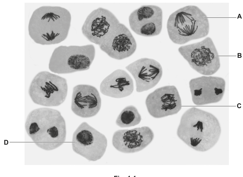
Identify the stages of mitosis occurring in the cells labelled B and C in Fig. 1.1.
B ______
C ______
Answer
B: prophase
C: metaphase (early anaphase also accepted)
B = prophase; C = metaphase
Background Concept
Mitosis is a continuous process but is divided into four named stages based on chromosome appearance and position. Recognising each stage from a micrograph depends on a small number of clear visual cues:
- Prophase — chromosomes have condensed and become visible as discrete threads within an intact nuclear area; the nucleolus is still often visible and the nuclear envelope is beginning to break down. No spindle is yet fully formed and chromosomes are scattered within the cell.
- Metaphase — chromosomes (each still composed of two sister chromatids joined at the centromere) are aligned along the equator of the spindle, the metaphase plate.
- Anaphase — sister chromatids separate at the centromere and are pulled to opposite poles, so two equal groups of daughter chromosomes are seen moving apart.
- Telophase — chromosomes reach the poles, decondense, and new nuclear envelopes re-form around each set; in plant cells a cell plate begins to form across the middle.
Understanding the Question
Fig. 1.1 is a light micrograph of plant cells in which four specific cells (A, B, C, D) are labelled. The question asks for the names of the mitotic stages shown in cells B and C, worth one mark each. You must rely only on what is visible in the micrograph, not on what you cannot see.
Approach
Look at each cell in turn and match its chromosome appearance to the diagnostic features of the stages listed above. The cell type and the surrounding cells are irrelevant to identifying the stage of a single cell.
Step-by-Step Reasoning
- Cell B shows chromosomes that are condensed and visible as a tangled mass within an area that was the nucleus, but they are not yet aligned on a plate and have not separated. The nuclear envelope is still partially present. This matches prophase. Any qualifier such as "early prophase" or "late prophase" is ignored by the mark scheme, so the safe answer is simply prophase.
- Cell C shows condensed chromosomes that appear to be gathered centrally in the cell, consistent with alignment at the metaphase plate. (If the chromosomes had already begun to separate, the cell would be at the very start of anaphase, which the mark scheme also accepts.) The simplest and most likely answer is metaphase.
Key Takeaways
- Recognising mitotic stages depends on chromosome shape and position, not on knowing which cell is "supposed" to be at which stage.
- Use a small mental checklist (condensed? aligned? separating? decondensing?) when reading any micrograph of mitosis.
- For cells where the picture is ambiguous (e.g. late metaphase versus early anaphase), the mark scheme usually accepts either, so give your best identification rather than leaving it blank.
Common Mistakes
- Confusing prophase with telophase because both can look "messy"; the key difference is that in telophase there are two separate nuclear areas re-forming, while in prophase the chromosomes are still in one area.
- Writing "metaphase/anaphase" with a slash to hedge; commit to the single best answer the micrograph supports.
- Describing chromosome appearance in the cell label rather than naming the stage.
Things to Be Careful About
- The mark scheme ignores qualifiers such as early, mid or late on prophase — do not waste time deciding between them.
- For cell C, early anaphase is an acceptable alternative; if you genuinely see chromatids starting to separate, give anaphase.
Describe the behaviour of the chromosomes in the stage of mitosis shown in cell A in Fig. 1.1.
Answer
- The sister chromatids of each chromosome separate at the centromere.
- The (daughter) chromosomes move to opposite poles of the cell.
- The chromosomes are pulled by spindle fibres that contract (shorten).
Sister chromatids separate at the centromere and are pulled by contracting spindle fibres to opposite poles of the cell.
Background Concept
Anaphase is the shortest stage of mitosis and is defined by a single event: the separation of sister chromatids and their movement to opposite ends of the cell.
Before anaphase, each chromosome consists of two sister chromatids held together at the centromere. The centromere is the region where the kinetochore forms — a protein structure to which spindle (microtubule) fibres attach. The spindle has two poles, each anchored at a centrosome, and the spindle fibres exert a pulling force on the kinetochores.
At the start of anaphase, the cohesion between sister chromatids is broken and the centromeres divide, so each chromatid becomes an independent daughter chromosome. Spindle fibres shorten (depolymerise) from both ends, pulling the daughter chromosomes apart so that one set moves towards each pole.
Understanding the Question
Cell A in Fig. 1.1 shows two clear groups of chromosomes being pulled apart to opposite ends of the cell, with the typical V-shape of chromatids being dragged centromere-first by their arms trailing behind. The question asks you to describe what is happening to the chromosomes during this stage (anaphase) for three marks.
Approach
Give the mark-scheme points in the order the events actually happen: first the chromatids separate, then they move, and the reason is the contracting spindle fibres. Use the precise words the mark scheme rewards — sister chromatids, poles, spindle fibres, contracting — rather than everyday paraphrases such as "they split and go to the ends".
Step-by-Step Reasoning
- Sister chromatids separate. Each chromosome was previously a pair of identical sister chromatids joined at the centromere. At the start of anaphase the centromeres divide, so the two chromatids of each chromosome become separate daughter chromosomes. (Mark: sister chromatids separate.)
- Chromosomes move to opposite poles. The two sets of daughter chromosomes travel to opposite ends of the cell, so the cell now contains two equal groups. (Mark: movement of daughter chromosomes to opposite poles.)
- The movement is caused by the spindle fibres. Spindle fibres attached to the kinetochores at each centromere shorten (contract), pulling the centromere first with the arms of the chromosome trailing behind. (Mark: movement by spindle fibres contracting/pulling.)
A possible extra mark-bearing point is that the centromeres lead the movement (the arms lag), which is why moving chromosomes often look like a V or a J on a micrograph.
Key Takeaways
- The hallmark of anaphase is separation of sister chromatids, not the formation of new nuclei (that is telophase).
- The motor of anaphase movement is the spindle fibres contracting — the cell does not push the chromosomes apart; the spindle pulls them.
- Each point in the answer must contain both the observation and, where appropriate, the mechanism (e.g. separated AND pulled by contracting spindle fibres).
Common Mistakes
- Saying the chromosomes "split" without specifying that it is the centromere that divides and the sister chromatids that separate. The mark scheme explicitly rejects answers that treat a chromosome as already being two chromatids and only the "chromosome" moving.
- Saying the chromatids move to the same pole (this would give an unequal division and is rejected).
- Saying the chromosomes are "moved by the spindle" without saying how — the mark scheme requires the spindle to be contracting/pulling.
- Describing events of telophase (chromosomes decondense, nuclear envelope reforms) when the cell shown is clearly anaphase. The mark scheme allows partial credit for telophase answers, but a wrong-stage answer can never earn full marks.
Things to Be Careful About
- The image shows chromatids being pulled to opposite poles — commit to anaphase; do not hedge by also describing telophase.
- Do not say the cell "pulls" the chromosomes; the spindle fibres pull them.
- Spelling centromere correctly (not centrosome; the centrosome is the organelle at the pole, not the joining region of the chromosome).
Cell D in Fig. 1.1 is in interphase.
Describe the role of DNA ligase in interphase.
Answer
- DNA ligase is active during the S phase (synthesis phase) of interphase, when DNA replication occurs.
- It joins Okazaki fragments on the lagging strand.
- It does this by catalysing the formation of phosphodiester bonds between adjacent nucleotides, completing the sugar–phosphate backbone of the new DNA strand.
During S phase of interphase, DNA ligase joins Okazaki fragments on the lagging strand by catalysing phosphodiester bond formation, completing the sugar–phosphate backbone.
Background Concept
Interphase is the part of the cell cycle between mitoses. It has three sub-phases:
- G1 — cell growth and normal metabolism.
- S phase — DNA is replicated semi-conservatively: each double helix is opened up and a new complementary strand is built alongside each old template.
- G2 — further growth and preparation for mitosis; organelles are duplicated.
During S phase the two new DNA strands are made in different ways because DNA polymerase can only add nucleotides to a free 3′-OH end and can only travel in one direction (5′ → 3′):
- The leading strand is synthesised continuously towards the replication fork.
- The lagging strand is synthesised away from the fork, in short pieces called Okazaki fragments, which are later joined together.
DNA ligase is the enzyme that seals those Okazaki fragments into one continuous strand. It does this by catalysing the formation of a phosphodiester bond between the 3′-OH of one fragment and the 5′-phosphate of the next, completing the sugar–phosphate backbone.
Understanding the Question
The question has already told you that cell D is in interphase and asks you to describe the role of DNA ligase in interphase — i.e. specifically during the S phase, when DNA replication occurs. Three marks are available for distinct points.
Approach
The strongest answer moves from context (where in the cell cycle) to mechanism (what the enzyme does and where on the DNA it acts) to molecular detail (the type of bond it forms). The mark scheme credits any three of these ideas.
Step-by-Step Reasoning
- Locate the action in the cell cycle. DNA ligase acts during the S phase of interphase, when DNA replication is taking place. (The question already gives the interphase context, so naming the S phase is the precise version.)
- Name what is being joined. DNA ligase joins Okazaki fragments — the short, newly synthesised pieces of DNA on the lagging strand. (Without the qualifier "on the lagging strand" the point is incomplete.)
- Name the bond formed. It catalyses the formation of phosphodiester bonds between adjacent nucleotides, which joins the 3′ end of one fragment to the 5′ end of the next.
- State the outcome. This produces a continuous sugar–phosphate (polynucleotide) backbone, so the lagging strand becomes a single, unbroken DNA strand.
Any three of these four points earn three marks.
Key Takeaways
- DNA ligase is specifically a "DNA-joining" enzyme, not a DNA-building enzyme; it does not add new nucleotides, it seals nicks between nucleotides already in place.
- It acts on the lagging strand only; the leading strand is made continuously and does not need ligation.
- Its specific chemistry is phosphodiester bond formation in the sugar–phosphate backbone — using the term "phosphodiester bond" rather than "bond" is worth a mark in itself.
Common Mistakes
- Saying DNA ligase "joins the two strands of DNA together" — that is not its function; it joins fragments within a single new strand.
- Putting it on the leading strand (no Okazaki fragments are made there).
- Confusing DNA ligase with DNA polymerase (the latter adds new nucleotides; the former joins already-added fragments).
- Saying it forms "hydrogen bonds" — hydrogen bonds hold the two complementary strands together via base pairing, not adjacent nucleotides in the same strand.
- Saying only that it "completes the DNA" without specifying the bond or the substrate — too vague for a mark.
Things to Be Careful About
- The question says "in interphase" — you must anchor your answer to the S phase of interphase, not to mitosis.
- "Phosphodiester bond" must be spelled (and understood) correctly; the bond links the 3′ carbon of one sugar to the 5′ carbon of the next via a phosphate group.
- "Backbone" refers to the sugar–phosphate strand, not the double helix as a whole.
Telomerase is an enzyme that is active during interphase in some cells.
Telomerase helps to maintain telomeres present on chromosomes.
State the location of telomeres on a chromosome.
Answer
Telomeres are located at both ends of a chromosome.
At both ends of the chromosome.
Background Concept
A chromosome is a single, very long DNA molecule packaged with histone proteins. Each chromosome has two ends — the regions where the DNA cannot continue any further. These specialised end regions are called telomeres.
Telomeres consist of short, repetitive, non-coding DNA sequences (in humans, the repeat TTAGGG) plus associated proteins. They perform two important jobs:
- They prevent the natural ends of linear DNA from being recognised by the cell as DNA damage.
- They provide a buffer of non-coding DNA that can be lost during replication without losing genes (see part (b)(ii)).
Understanding the Question
This is a one-mark "state the location" question. It tests whether you know the position of telomeres on a chromosome, which is the prerequisite for understanding why telomerase matters in part (b)(ii).
Approach
State the location in a single short sentence using the precise wording the mark scheme accepts: at both ends of the chromosome. Avoid vague answers like "at the tip" or "on the outside".
Step-by-Step Reasoning
A chromosome is linear and has exactly two ends. Telomeres cap both of those ends, so the correct answer is "at both ends of the chromosome" (or "at the ends of a chromosome").
Key Takeaways
- Telomeres are a structural feature of chromosome ends, not a protein or a gene.
- The fact that they are at both ends (not one) is part of the definition — a chromosome that lost a telomere at one end would be unstable.
Common Mistakes
- Saying telomeres are "on the chromosome" without specifying the ends.
- Confusing telomeres with centromeres (the centromere is in the middle of the chromosome, where sister chromatids are joined; telomeres are at the two ends).
Things to Be Careful About
- The mark scheme explicitly accepts "at the ends of a chromosome" as well as "at both ends" — either wording is fine, but you must include the idea of ends.
Some people with a long lifespan have cells showing a higher than normal activity of telomerase.
Suggest why a long lifespan could result from a higher telomerase activity.
Answer
- Higher telomerase activity keeps the telomeres longer / maintained for a longer time, so chromosomes do not lose important coding genes from their ends.
- This allows the cells to undergo more cell cycles (more rounds of DNA replication and mitosis) and so the person can continue to replace damaged or worn-out cells, supporting a longer lifespan.
Telomeres are maintained for longer, so more cell cycles can occur and fewer coding genes are lost, allowing damaged cells to be replaced for longer.
Background Concept
DNA replication has an end-replication problem. Because DNA polymerase synthesises only in the 5′ → 3′ direction and needs an RNA primer to start, the very last few nucleotides at the 5′ end of each new strand cannot be replaced once the primer is removed. As a result, every time a normal somatic cell divides, its telomeres shorten by a small amount.
Once telomeres become too short, the chromosome ends look like broken DNA, which triggers cell-cycle arrest or apoptosis (programmed cell death). This is thought to be one of the reasons tissues lose function as we age.
Telomerase is a ribonucleoprotein enzyme that adds telomeric repeat sequences back onto chromosome ends, counteracting this shortening. It is active in germ cells, stem cells, and most cancer cells, but in most adult somatic cells it is switched off, so telomeres progressively shorten with age.
Understanding the Question
The question gives you a real-world observation: people with a long lifespan sometimes have cells with higher-than-normal telomerase activity. It then asks you to suggest why this might lead to a longer life. This is a "suggest" question — there is no single right answer, but the mark scheme lists the specific biological links it will credit. You need to choose the two most relevant points.
Approach
Build a short causal chain: higher telomerase activity → telomeres maintained for longer → chromosomes retain their coding genes → cells can keep dividing for longer → tissues can keep replacing old/damaged cells → lifespan is extended.
Pick the two or three steps from this chain that carry the most marks. The mark scheme credits any of these, so a focused two-point answer is enough.
Step-by-Step Reasoning
- Telomeres are maintained / not shortened as much. With more telomerase, the telomeric repeats are replaced as fast as they are lost, so chromosome ends stay longer.
- More cell cycles are possible. Telomeres of usable length delay the trigger that would otherwise stop the cell cycle, so the cell can keep replicating its DNA and dividing by mitosis many more times. (The mark scheme explicitly rejects the idea that telomerase speeds up the cell cycle — the point is that more cycles are possible, not faster ones.)
- Fewer coding genes are lost from the chromosome ends. Because the telomeric buffer is preserved, the erosion does not reach into the coding regions of the chromosome, so genetic information is retained.
- Tissues can keep replacing damaged, worn-out or old cells. With more available cell divisions, stem cells and their descendants can renew tissues for longer, delaying the functional decline that usually accompanies ageing.
Any two of the above points earn the two marks.
Key Takeaways
- Telomerase activity and telomere length are linked to replicative capacity, not to the speed of cell division.
- The "end-replication problem" is the fundamental reason telomeres shorten at all — it is a direct consequence of how DNA polymerase works.
- The connection between telomeres, cell replacement and ageing is a recurring theme in modern biology; questions often use it to test whether you can apply a molecular idea to a whole-organism outcome.
Common Mistakes
- Saying telomerase "speeds up mitosis" or "makes cells divide faster" — the mark scheme rejects this; telomerase does not change the rate of division, it changes the number of divisions possible.
- Saying only that "telomeres are protected" without linking this to anything downstream (cell cycles, gene loss, tissue renewal).
- Writing about cancer rather than ageing — high telomerase is a feature of cancer cells, but the question is explicitly about why a long lifespan could result.
- Vague claims about "cells stay younger" without naming a mechanism.
Things to Be Careful About
- Keep the chain logical: telomerase → telomere length → cell-cycle capacity → tissue renewal → lifespan.
- The mark scheme allows several different pairs of points; you do not need to make every link, but you must give at least two distinct creditable ideas.
The rest of this paper
5 more questions- Q2Infectious Diseases · Immunity · Gas Exchange11M
- Q3Cell Structure · Transport in Mammals · Enzymes10M
- Q4Biological Molecules · Nucleic Acids and Protein Synthesis · Cell Membranes and Transport · Immunity10M
- Q5Transport in Plants · Enzymes · Biological Molecules10M
- Q6Biological Molecules · Cell Structure · Cell Membranes and Transport8M