Biology 9700/21 — May/June 2023
Cambridge AS Level · AS Level Structured Questions · worked solutions for every part, with the mark scheme
Topics Cell Membranes and Transport · Immunity · Transport in Plants · Cell Structure · Biological Molecules · Nucleic Acids and Protein Synthesis · +2 more
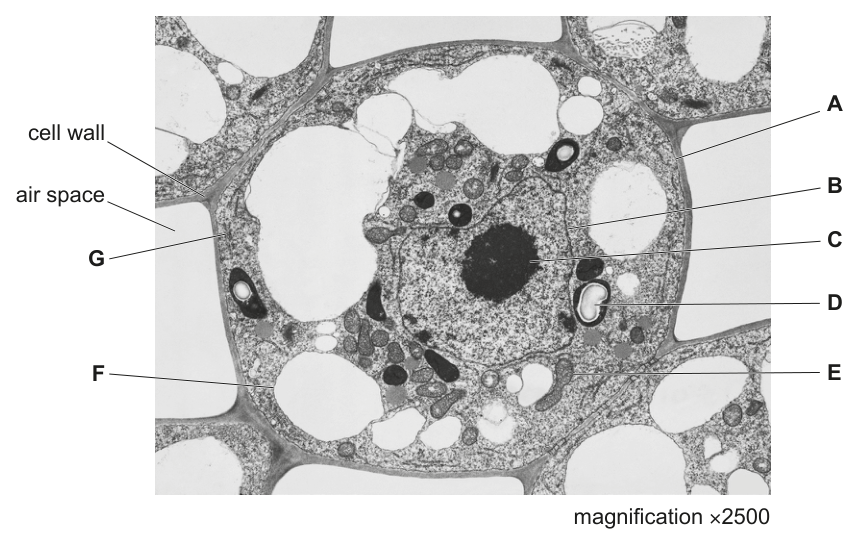
Fig. 1.1 is a transmission electron micrograph of a cell from the stem of sago pondweed, Stuckenia pectinata.
State the evidence from Fig. 1.1 that shows that the cell is from the stem of S. pectinata and not from the mesophyll of a leaf.
Answer
Presence of (many) small vacuoles / no large (central) vacuole
OR the nucleus is centrally located / not at the edge of the cell.
many small vacuoles (no large central vacuole) / centrally located nucleus
Background Concept
Plant cells vary in structure according to their function and location. Mesophyll cells of a leaf are specialised for photosynthesis and have very characteristic features: many chloroplasts distributed around the cell periphery, one large central vacuole that pushes the cytoplasm and nucleus to the edge of the cell, and a thin cell wall. Stem cells (especially in non-photosynthetic tissues) lack this photosynthetic specialisation, so they typically have few or no chloroplasts, may contain several smaller vacuoles rather than a single large one, and the nucleus remains more centrally placed.
Understanding the Question
The candidate is shown a TEM of a cell from the stem of the aquatic plant Stuckenia pectinata. The task is to give one piece of evidence visible in the image that rules out a leaf mesophyll origin. The command word is state, so a single clear observation is required.
Approach
Examine Fig. 1.1 and compare what is visible with the expected organisation of a mesophyll cell. Look for: chloroplast density, vacuole arrangement (one big vs many small) and the position of the nucleus.
Step-by-Step Reasoning
The micrograph shows:
- A handful of chloroplasts (label G) — far fewer than the many chloroplasts a mesophyll cell would pack against its wall.
- Several small vacuolar spaces (D points to one) rather than a single large central vacuole that fills most of the cell.
- A centrally placed nucleus (B) and nucleolus (C), not pushed to the cell periphery by a large vacuole.
Any one of these observations is sufficient and is enough to show the cell is not a mesophyll cell.
Key Takeaways
- Cell structure reflects function: mesophyll cells are photosynthetic and have many chloroplasts and one large central vacuole.
- Stem cells (especially in non-green tissues) have a different ultrastructure: fewer chloroplasts, often several smaller vacuoles and a more central nucleus.
- A single well-chosen observation is enough to answer a "state" question.
Common Mistakes
- Stating a feature without comparing it explicitly to a mesophyll cell (e.g. "the cell has a nucleus").
- Claiming "no chloroplasts" if at least one is clearly visible in the image.
- Confusing the tonoplast with the cell surface membrane when describing vacuole boundaries.
Things to Be Careful About
- Read the question as "state evidence… NOT from the mesophyll", so a comparison must be implied even if only one feature is named.
- Look carefully at vacuole size and number — the cell shown has several small vacuoles, not one dominant central one.
- Nucleus position is a giveaway: a peripheral nucleus implies a large central vacuole (mesophyll); a central nucleus implies small/multiple vacuoles (not mesophyll).
Complete each row in Table 1.1 to identify a cell structure shown in Fig. 1.1 that carries out the function listed.
Table 1.1
| function | name of cell structure | letter on Fig. 1.1 |
|---|---|---|
| gas exchange | ||
| production of subunits of ribosomes | ||
| active transport of ions | ||
| aerobic respiration |
Answer
| function | name of cell structure | letter on Fig. 1.1 |
|---|---|---|
| gas exchange | cell surface membrane | A |
| production of subunits of ribosomes | nucleolus | C |
| active transport of ions | cell surface membrane | A |
| aerobic respiration | mitochondrion | E |
gas exchange — cell surface membrane (A); subunits of ribosomes — nucleolus (C); active transport — cell surface membrane (A); aerobic respiration — mitochondrion (E)
Background Concept
Each membrane-bound organelle carries out a specific role that depends on its structure:
- The cell surface (plasma) membrane is a thin phospholipid bilayer across which O₂ and CO₂ diffuse freely; it also houses carrier proteins for active transport.
- The mitochondrial inner membrane has a large surface area and is also thin enough to permit gas diffusion, so it qualifies for gas exchange in the same way.
- The nucleolus, a dense region inside the nucleus, is the site where rRNA combines with proteins to assemble the large and small subunits of ribosomes (it does not build whole ribosomes).
- Several membranes — cell surface membrane, tonoplast, mitochondrial inner membrane and nuclear envelope — all carry transport proteins able to perform active transport of ions using ATP.
- The mitochondrion is the site of aerobic respiration: the link reaction and Krebs cycle in the matrix, and the electron transport chain / oxidative phosphorylation on the inner membrane.
Understanding the Question
The candidate is given a four-row table listing cellular functions and must (1) name the structure responsible, and (2) give the letter that labels that structure on Fig. 1.1. One mark is awarded per correct row.
Approach
For each function, recall the organelle that performs it, then match the organelle to the letter assigned in the micrograph. In Fig. 1.1: A = cell surface membrane, B = nucleus, C = nucleolus, D = vacuole, E = mitochondrion, F = other membrane, G = chloroplast.
Step-by-Step Reasoning
- Gas exchange — small, lipid-soluble gases (O₂, CO₂) diffuse across thin membranes. The cell surface membrane (A) is the principal site; the mitochondrial membrane (E) is also accepted.
- Subunits of ribosomes — assembled in the nucleolus (C).
- Active transport of ions — requires ATP and specific carrier proteins. The cell surface membrane (A) is the most frequently cited site, but the tonoplast (F), mitochondrial membrane (E) and nuclear envelope (B) are all acceptable.
- Aerobic respiration — the mitochondrion (E) is the only organelle that carries out the Krebs cycle and oxidative phosphorylation.
Key Takeaways
- The cell surface membrane is central to gas exchange and to active transport of ions.
- The nucleolus builds ribosomal subunits, not whole ribosomes.
- Mitochondria are the unique site of aerobic respiration in eukaryotes.
- Several different membranes can perform active transport.
Common Mistakes
- Writing "cell membrane" rather than the precise "cell surface membrane".
- Saying the nucleus (rather than the nucleolus) makes ribosomes.
- Listing a whole organelle such as "mitochondrion" for active transport without specifying the membrane involved.
- Confusing the cell wall with the cell surface membrane (the cell wall is fully permeable and not a site of selective transport).
Things to Be Careful About
- The mark scheme requires "cell surface membrane" (not "cell membrane") for full credit.
- The nucleolus produces ribosomal subunits, not whole ribosomes — these are joined together in the cytoplasm.
- For active transport, any membrane with the appropriate carrier protein is acceptable; the most common answer is the cell surface membrane (A).
Plant vacuoles develop when vesicles fuse together. The vacuoles increase in size as more vesicles fuse.
Fig. 1.2 shows the movement of vesicles within a plant cell during the development of a vacuole.
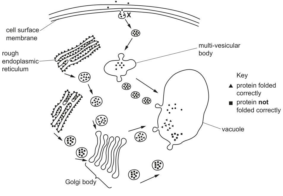
Name the process that is occurring at X.
Answer
Endocytosis (or pinocytosis).
endocytosis (or pinocytosis)
Background Concept
Cells move material across the plasma membrane in bulk by endocytosis and exocytosis:
- Endocytosis — the plasma membrane invaginates around extracellular material and pinches off to form a vesicle inside the cell.
- Pinocytosis — a form of endocytosis in which small droplets of extracellular fluid (and any dissolved solutes) are taken up in small vesicles; sometimes called "cell drinking".
- Phagocytosis — endocytosis of large solid particles such as bacteria or cell debris; these end up in large phagosomes and is performed by specialised phagocytes. Phagocytosis is not the same as pinocytosis, even though both are forms of endocytosis.
Understanding the Question
In Fig. 1.2, label X points to a vesicle interacting with the cell surface membrane. The command word "name" requires a single term.
Approach
Look at the size of the vesicle at X and the direction of movement: a small vesicle being internalised from the cell surface membrane indicates endocytosis / pinocytosis. A large vesicle engulfing a whole cell or large particle would indicate phagocytosis.
Step-by-Step Reasoning
The vesicle at X is small, comparable with the other transport vesicles in the diagram. Material is being taken up into the cell (the arrow points inward). This is the textbook description of endocytosis; because the vesicle is small and carries fluid, the more specific term pinocytosis is also accepted. Phagocytosis is explicitly rejected by the mark scheme because the vesicles shown are too small to be phagosomes and the cell is not a phagocyte.
Key Takeaways
- Endocytosis is the bulk uptake of material by vesicle formation at the cell surface membrane.
- Pinocytosis = endocytosis of fluid in small vesicles; phagocytosis = endocytosis of large particles.
- The size of the vesicle and the cell type determine the correct name.
Common Mistakes
- Writing "phagocytosis" — rejected by the mark scheme because the vesicles are small, not large phagosomes, and the cell shown is not a phagocyte.
- Writing "active transport" or "diffusion" — these are not bulk transport and do not form vesicles.
- Writing "osmosis" — that refers specifically to water movement, not to vesicle formation.
Things to Be Careful About
- "Endocytosis" is the broadest correct answer; "pinocytosis" is also accepted.
- "Phagocytosis" must be avoided here — it is reserved for the uptake of large particles by phagocytes.
Some of the vesicles formed by the Golgi body pass to the vacuole. These vesicles contain proteins that have been folded correctly and some that have not folded into their correct shapes. The proteins that have not folded correctly pass to the vacuole where they are broken down.
Explain how proteins that have not folded correctly are broken down in the vacuole.
Answer
- Proteases / peptidases (hydrolytic enzymes) catalyse the hydrolysis of the misfolded proteins;
- peptide bonds are broken (by the addition of water);
- this releases shorter peptides and amino acids.
hydrolytic enzymes (proteases) hydrolyse peptide bonds to release amino acids / shorter peptides
Background Concept
Proteins are polymers of amino acids joined by peptide bonds (a type of covalent bond). Hydrolysis is a chemical reaction in which a bond is broken by the addition of a water molecule: the –OH from water goes to one fragment, the –H to the other. Hydrolytic enzymes speed up hydrolysis. Enzymes that act on proteins are called proteases (or peptidases / proteolytic enzymes); they are one class of the acidic hydrolases stored in lysosomes (animal cells) and vacuoles (plant cells). Plant vacuoles are therefore often described as having a lysosome-like role: they can digest unwanted or damaged macromolecules, including proteins that have not folded correctly.
Understanding the Question
The stem tells the candidate that misfolded proteins reach the plant vacuole and are broken down there. The task is to explain how this breakdown happens — i.e. the chemistry and the enzymes involved. Three marks are available.
Approach
Construct a logical sequence: identify the type of reaction, name the enzymes, name the bond broken, and state the products.
Step-by-Step Reasoning
- The vacuole contains hydrolytic enzymes, including proteases (peptidases).
- These enzymes catalyse the hydrolysis of the misfolded proteins — water is added across the bond.
- The bonds broken are the peptide bonds that link amino acids in the polypeptide chain.
- The products are shorter peptides and individual amino acids that the cell can recycle.
The mark scheme accepts any three of: hydrolysis / use of water; proteases / peptidases; peptide bonds broken; products are peptides / amino acids. The term "hydrolytic enzymes" alone is sufficient for the first point if hydrolysis is implied.
Key Takeaways
- Protein breakdown in the vacuole is by hydrolysis, catalysed by proteases.
- The bonds cleaved are peptide bonds (NOT glycosidic, ester or phosphodiester bonds).
- Products are peptides and amino acids, which the cell can reuse.
- Vacuoles carry out lysosome-like digestive functions in plant cells.
Common Mistakes
- Writing that "bonds" (without specifying peptide bonds) are broken — the mark scheme insists on peptide bonds for full credit.
- Naming an incorrect bond (e.g. glycosidic, ester or phosphodiester) — these are explicitly rejected.
- Omitting any mention of hydrolysis or water.
- Saying "the protein is broken into amino acids only" — shorter peptides are also produced.
Things to Be Careful About
- The mark scheme requires the type of reaction (hydrolysis) AND/OR the name of the enzymes (proteases) AND the bond (peptide) AND the products (peptides / amino acids). Three of these four ideas are needed for three marks.
- "Hydrolytic enzymes" is an acceptable shorthand for proteases in the vacuole.
- Do not confuse protein breakdown with denaturation — denaturation is loss of 3-D shape, not peptide bond cleavage.
Small vacuoles in S. pectinata may have roles similar to lysosomes in animal cells.
Describe the role of lysosomes in animal cells in defence against pathogens.
Answer
- Lysosomes fuse with phagosomes (the vesicles containing engulfed pathogens) to form phagolysosomes;
- hydrolytic enzymes (e.g. proteases and lysozymes) inside the lysosome digest / break down the pathogen into harmless / soluble products.
lysosomes fuse with phagosomes to form phagolysosomes; hydrolytic enzymes digest the pathogen
Background Concept
Lysosomes are spherical, membrane-bound organelles that contain hydrolytic (digestive) enzymes active at acidic pH. The enzyme set includes proteases, lipases, nucleases, carbohydrases and lysozymes. They are the cell's recycling and defence centres.
Phagocytes (e.g. neutrophils and macrophages) are white blood cells that engulf invading bacteria and other pathogens. The pathogen is surrounded by a piece of the cell surface membrane that pinches off inside the cell to form a phagosome.
When a phagosome meets a lysosome, the two membranes fuse and the lysosomal enzymes are released into the now combined vesicle, called a phagolysosome. The pathogen is hydrolysed into smaller, harmless, soluble molecules that the phagocyte can either exocytose or reuse.
Understanding the Question
The question asks the candidate to describe the role of lysosomes in animal cells specifically in defence against pathogens. Two marks are available. The command word "describe" requires specific named structures and processes, not a vague statement.
Approach
Hit the two key points the mark scheme rewards:
- The formation of a phagolysosome by fusion of lysosome and phagosome (1 mark).
- Digestion / breakdown of the pathogen by lysosomal enzymes, with a supporting detail (a named enzyme, named substrate or named product) for the second mark.
Step-by-Step Reasoning
- A phagocyte (such as a neutrophil) engulfs a pathogen into a phagosome.
- The phagosome fuses with a lysosome, producing a phagolysosome.
- The hydrolytic enzymes of the lysosome (e.g. proteases that digest proteins, lysozymes that break down peptidoglycan in bacterial cell walls) hydrolyse the pathogen's macromolecules.
- The products (amino acids, sugars, nucleotides, fatty acids) are harmless, soluble and may be reused by the cell or exported.
Any one of the following earns the second mark: two named enzymes, two named substrates, or a statement that the products are harmless / useful / soluble.
Key Takeaways
- Lysosomes work together with phagosomes to form phagolysosomes.
- The hydrolytic enzymes in the phagolysosome digest the engulfed pathogen.
- The result is harmless, soluble products that the cell can excrete or recycle.
- This is one half of the non-specific immune response (alongside the inflammatory response).
Common Mistakes
- Writing that lysosomes "engulf" pathogens — they don't; phagocytes engulf pathogens, and lysosomes only fuse afterwards.
- Confusing lysosomes with phagosomes or with the phagocyte itself.
- Mentioning only one enzyme without giving a specific second example or named substrate.
- Describing the role of lysosomes in autophagy (organelle turnover) — the mark scheme explicitly ignores this; the question is about defence against pathogens.
Things to Be Careful About
- The fusion step must be stated: lysosome + phagosome → phagolysosome.
- A named enzyme, a named substrate, or a description of the products is needed for the second mark.
- Stay on the topic of defence against pathogens; do not drift into organelle recycling.
The rest of this paper
5 more questions- Q2Biological Molecules9M
- Q3Cell Membranes and Transport · Transport in Plants · Immunity10M
- Q4Nucleic Acids and Protein Synthesis · The Mitotic Cell Cycle13M
- Q5Transport in Mammals11M
- Q6Transport in Plants6M