Biology 9700/12 — May/June 2023
Cambridge AS Level · Multiple Choice (AS Level) · answer key with instant marking and worked solutions
Topics Biological Molecules · Cell Structure · Transport in Plants · Nucleic Acids and Protein Synthesis · Cell Membranes and Transport · Gas Exchange · +5 more
Tap an option under each question to check it — your score builds as you go.
A graticule and a micrometer scale can be used to measure the size of biological structures that are viewed with a microscope.
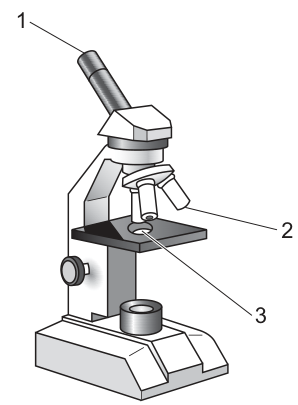
Which row shows the correct locations for the placement of a graticule and a micrometer scale on the microscope shown?
Options
| graticule | micrometer scale | |
|---|---|---|
| A | 1 | 2 |
| B | 1 | 3 |
| C | 2 | 3 |
| D | 3 | 1 |
Working
The eyepiece graticule is a small glass disc with an engraved scale that fits inside the eyepiece lens, so it must be placed at position 1 (eyepiece). The stage micrometer is a specially engraved glass slide used as a reference scale; it is placed on the microscope stage at position 3. Position 2 (the objective lens) is not the correct location for either piece of equipment.
Answer
B
B
Background Concept
Two pieces of equipment are used to measure the size of an object under a light microscope:
- Eyepiece graticule: a small circular glass disc bearing a fine scale (e.g. 100 divisions across a 1 cm line). It is inserted into the eyepiece lens, where it appears superimposed on the specimen image. Because the apparent size of its divisions changes with objective magnification, the graticule must be calibrated before each objective is used.
- Stage micrometer: a glass slide bearing a scale of known length (typically 2 mm in 0.01 mm divisions). It is placed on the microscope stage like a normal slide, viewed through the same objective as the specimen, and used to calibrate the graticule.
A calibrated graticule then lets the observer read the size of a specimen directly from the graticule scale and convert it to a real measurement (in mm, µm or nm) by multiplying by the calibration factor for the objective in use.
Understanding the Question
The diagram shows a light microscope with three numbered parts:
- 1 — the eyepiece (ocular) lens at the top of the body tube
- 2 — the objective lens, on the nosepiece, pointing down at the stage
- 3 — the stage, the flat platform that holds the slide
The question asks where the graticule and the stage micrometer should each be placed, and which of the four answer combinations is correct.
Approach
Match each piece of equipment to the part of the microscope it belongs in:
- The graticule is built into / inserted into the eyepiece → position 1.
- The stage micrometer is a slide placed on the stage → position 3.
Therefore the answer is the row graticule = 1, micrometer = 3.
Step-by-Step Reasoning
- The graticule must be viewed at the same plane as the specimen, so it sits inside the eyepiece (position 1). It cannot sit in the objective or on the stage because it would not be seen as a superimposed scale on the image.
- The stage micrometer is itself a microscope slide, so it must be clipped onto the stage (position 3) in place of a specimen slide during calibration.
- The objective lens (position 2) is a fixed optical component with no slot or space for either item.
- Combining these, graticule = 1 and micrometer = 3, which is option B.
Key Takeaways
- Graticule → eyepiece; stage micrometer → stage.
- The stage micrometer is a reference slide of known length, used only to calibrate the graticule.
- Calibration must be repeated for every objective, because the apparent length of graticule divisions changes with magnification.
- The conversion used is: actual size = (number of graticule divisions × calibration factor for that objective).
Common Mistakes
- Confusing the two and placing the graticule on the stage: the stage micrometer goes on the stage, not the graticule.
- Placing the stage micrometer in the eyepiece: the eyepiece graticule goes in the eyepiece, not the micrometer.
- Thinking the objective (position 2) is involved: the objective is a sealed optical element; neither item is inserted there.
Things to Be Careful About
- Remember the direction of the question: "graticule" then "micrometer scale"; the columns of the table are in that order, so the correct row must put 1 in the graticule column and 3 in the micrometer column.
- Final answers in µm (or even nm for very small structures such as ribosomes) — always convert mm to µm () or nm () when calculating actual sizes.
The rest of this paper
39 more questions- Q2Cell Structure1M
- Q3Cell Structure · Nucleic Acids and Protein Synthesis1M
- Q4Cell Structure1M
- Q5Cell Structure1M
- Q6Cell Structure1M
- Q7Biological Molecules1M
- Q8Biological Molecules1M
- Q9Biological Molecules1M
- Q10Biological Molecules1M
- Q11Biological Molecules1M
- Q12Biological Molecules1M
- Q13Enzymes1M
- Q14Enzymes1M
- Q15Cell Membranes and Transport1M
- Q16Cell Membranes and Transport1M
- Q17Cell Membranes and Transport1M
- Q18Cell Membranes and Transport1M
- Q19Cell Membranes and Transport1M
- Q20The Mitotic Cell Cycle1M
- Q21The Mitotic Cell Cycle1M
- Q22Nucleic Acids and Protein Synthesis1M
- Q23Nucleic Acids and Protein Synthesis1M
- Q24Nucleic Acids and Protein Synthesis1M
- Q25Nucleic Acids and Protein Synthesis1M
- Q26Transport in Plants1M
- Q27Transport in Plants1M
- Q28Transport in Plants1M
- Q29Transport in Plants1M
- Q30Transport in Plants1M
- Q31Transport in Plants1M
- Q32Biological Molecules1M
- Q33Transport in Mammals1M
- Q34Transport in Mammals1M
- Q35Gas Exchange1M
- Q36Gas Exchange1M
- Q37Gas Exchange1M
- Q38Infectious Diseases1M
- Q39Infectious Diseases1M
- Q40Immunity1M