Biology 9700/11 — May/June 2023
Cambridge AS Level · Multiple Choice (AS Level) · answer key with instant marking and worked solutions
Topics Cell Structure · Transport in Mammals · Transport in Plants · Nucleic Acids and Protein Synthesis · Biological Molecules · Cell Membranes and Transport · +5 more
Tap an option under each question to check it — your score builds as you go.
The electron micrograph shows onion root cells prepared using a freeze-fracture technique. The cells were quickly frozen and then physically broken apart. Freeze fracture breaks apart cells along weak areas, such as membranes and the surfaces of organelles.
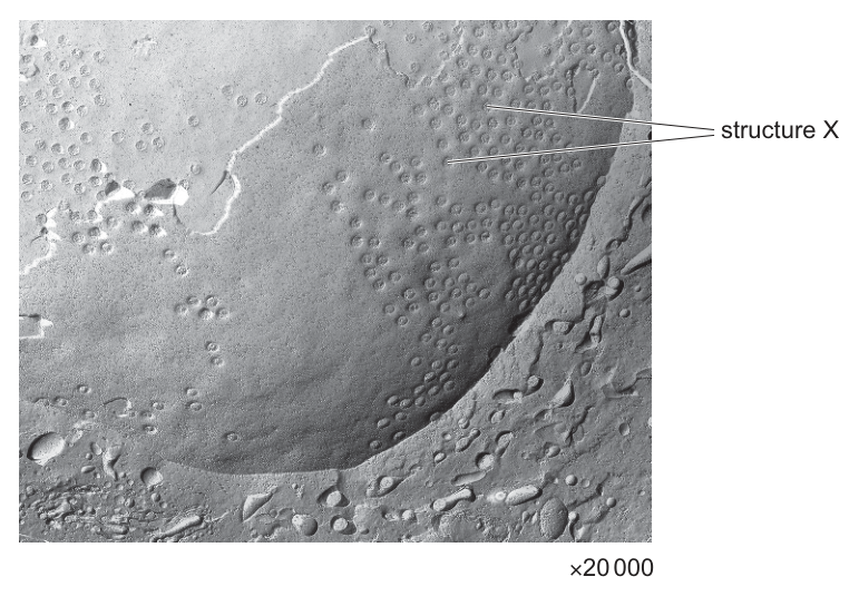
Which statement best explains the appearance of the electron micrograph?
Options
A The cells were broken apart at the endoplasmic reticulum; structure X is a ribosome.
B The cells were broken apart at the nuclear envelope; structure X is a nuclear pore.
C The cells were broken apart at the nuclear envelope; structure X is a ribosome.
D The cells were broken apart at the tonoplast; structure X is a plasmodesma.
Working
Freeze-fracture splits the cell along weak membranes. The micrograph shows a large, curved surface studded with many regularly spaced circular depressions — this is the characteristic appearance of the nuclear envelope, which is perforated by nuclear pores.
- A is wrong: rough ER would show ribosomes (small dense particles), not circular pores, and ribosomes do not appear as ring-shaped depressions.
- C is wrong: although the surface is the nuclear envelope, structure X is a nuclear pore, not a ribosome (ribosomes are not embedded in the nuclear envelope as ring structures).
- D is wrong: plasmodesmata pass through cell walls between adjacent plant cells, not through the tonoplast (vacuolar membrane), and would not appear as numerous regular pores on a single curved surface.
Answer
B
B
Background Concept
Freeze-fracture is a preparation technique used in electron microscopy. Cells are rapidly frozen and then physically cleaved; the fracture plane travels along the weakest internal interfaces, which are the hydrophobic interiors of membranes. When a membrane is split open, the two inner leaflets are exposed, revealing any structures embedded in or associated with that membrane.
The nuclear envelope is a double membrane perforated by nuclear pores — large protein complexes (~125 nm in diameter) that regulate the passage of molecules between the nucleus and cytoplasm. When the nuclear envelope is fractured open, the pores appear as circular depressions or rings distributed across the exposed inner surface.
By contrast:
- Rough endoplasmic reticulum (RER) carries ribosomes, which are small, dense, granular particles attached to the cytoplasmic face — not ring-shaped pores.
- The tonoplast is the single membrane surrounding the central vacuole; it is a smooth membrane without the regular pore pattern shown here.
- Plasmodesmata are cytoplasmic channels that pass through the cell wall between adjacent plant cells; they are not features of the tonoplast.
Understanding the Question
The stem explains that freeze-fracture reveals the surfaces of membranes and organelles. The image shows a very large, curved membrane surface covered in many regular circular structures, with one labelled "structure X". The candidate must identify:
- Which membrane/organelle has been exposed by the fracture (i.e. what surface we are looking at).
- What structure X is — the ring-shaped feature on that surface.
The command word is implicit ("which statement best explains") — the task is selection of the correct biological interpretation.
Approach
Two diagnostic features guide the answer:
- Size and curvature of the exposed surface: very large and dome-like — consistent with the nuclear envelope of a eukaryotic cell, which is the largest internal membrane surface in many cells.
- Nature of structure X: regularly spaced circular rings — the signature of nuclear pores, not ribosomes (which are granular, not ring-shaped) and not plasmodesmata (which span cell walls and would not be this numerous or uniformly distributed on a membrane surface).
Matching each option against these features eliminates A, C and D, leaving B.
Step-by-Step Reasoning
- The exposed surface is large, curved, and uniformly covered in circular features. In an onion root cell (a eukaryotic plant cell), the largest organelle surface with regular circular openings is the nuclear envelope, which has hundreds of nuclear pores.
- Structure X is one of these circular openings — therefore a nuclear pore, a channel composed of nucleoporins that controls transport between the nucleus and cytoplasm.
- Option A suggests ER + ribosome. Ribosomes on the RER appear as small dense granules (≈20–30 nm), not as ring-shaped pores, so A is incorrect.
- Option C correctly identifies the surface as the nuclear envelope but misidentifies the structure as a ribosome. Ribosomes are not embedded as rings in the nuclear envelope; the ring-shaped features are nuclear pores, so C is incorrect.
- Option D suggests the tonoplast and plasmodesma. Plasmodesmata are channels through cell walls, not through the tonoplast, and they would not produce this regular pore pattern on a single curved membrane. D is incorrect.
- Option B — nuclear envelope exposed by fracture, structure X is a nuclear pore — fits both the surface and the structure shown.
Key Takeaways
- Freeze-fracture reveals the inner surfaces of biological membranes; the fracture plane follows membrane interiors.
- The nuclear envelope has a distinctive appearance after fracture: a large curved surface dotted with nuclear pores.
- Ribosomes appear as dense granules on rough ER, never as ring-shaped pores.
- Plasmodesmata are cell-wall channels and are unrelated to tonoplast structure.
Common Mistakes
- Confusing nuclear pores with ribosomes because both are small circular features on micrographs — remember pores are rings/holes in the nuclear envelope, while ribosomes are solid dense particles.
- Assuming the curved surface must be a vacuole (large plant cell organelle) — vacuoles have a smooth tonoplast without regular pore arrays.
- Choosing any option that mentions plasmodesmata, which pass through cell walls, not membranes.
Things to Be Careful About
- Read the option's both parts together — the surface identification and the structure identification must both be correct for the option to be right (this is why C is wrong despite correctly naming the nuclear envelope).
- "Pore" in biology does not just mean a small hole — a nuclear pore is a specific large protein complex; recognising its characteristic ring appearance after freeze-fracture is the key diagnostic.
The rest of this paper
39 more questions- Q2Cell Structure1M
- Q3Cell Structure1M
- Q4Cell Structure · Transport in Plants1M
- Q5Cell Structure1M
- Q6Cell Structure1M
- Q7Nucleic Acids and Protein Synthesis1M
- Q8Biological Molecules1M
- Q9Biological Molecules1M
- Q10Biological Molecules1M
- Q11Biological Molecules1M
- Q12Enzymes1M
- Q13Enzymes1M
- Q14Cell Membranes and Transport1M
- Q15Cell Membranes and Transport1M
- Q16Cell Membranes and Transport1M
- Q17The Mitotic Cell Cycle1M
- Q18The Mitotic Cell Cycle1M
- Q19Nucleic Acids and Protein Synthesis1M
- Q20Nucleic Acids and Protein Synthesis1M
- Q21Nucleic Acids and Protein Synthesis1M
- Q22Nucleic Acids and Protein Synthesis1M
- Q23Transport in Plants1M
- Q24Biological Molecules1M
- Q25Transport in Plants · Cell Membranes and Transport1M
- Q26Transport in Plants1M
- Q27Transport in Plants1M
- Q28Transport in Mammals1M
- Q29Transport in Mammals1M
- Q30Transport in Mammals1M
- Q31Transport in Mammals1M
- Q32Transport in Mammals1M
- Q33Transport in Mammals1M
- Q34Gas Exchange1M
- Q35Gas Exchange1M
- Q36Infectious Diseases1M
- Q37Infectious Diseases1M
- Q38Infectious Diseases1M
- Q39Immunity1M
- Q40Immunity1M