Biology 9700/23 — October/November 2022
Cambridge AS Level · AS Level Structured Questions · worked solutions for every part, with the mark scheme
Topics Nucleic Acids and Protein Synthesis · Cell Membranes and Transport · Transport in Mammals · Biological Molecules · Immunity · Cell Structure · +3 more
During translation, a polypeptide is synthesised when amino acids are added to a growing chain of amino acids.
Fig. 1.1 shows part of a growing chain of amino acids and the amino acid cysteine.
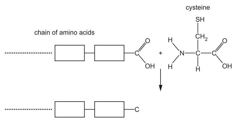
Complete Fig. 1.1 by showing the formation of the bond between cysteine and the growing chain of amino acids in the process of translation.
Answer
The completed diagram shows:
Three marking points satisfied:
- C-N peptide bond between the carboxyl C of the chain and the amino N of cysteine.
- C=O remains on the carboxyl C and an N-H remains on the amino N.
- HO is shown as a product of the condensation reaction.
See diagram — C-N peptide bond formed between chain carboxyl C and cysteine amino N; C=O and N-H of the peptide group drawn; H₂O released.
Background Concept
During translation, amino acids are joined together by peptide bonds. A peptide bond is the covalent link formed when the carboxyl group (-COOH) of one amino acid reacts with the amino group (-NH₂) of the next, releasing one molecule of water. This is a condensation reaction. The resulting linkage -C(=O)-N(H)- is called a peptide group, and the whole polymer of amino acids is a polypeptide.
Understanding the Question
Fig. 1.1 shows the growing polypeptide chain ending in a free carboxyl group (-COOH), together with a free cysteine molecule (-NH₂, central C-H, -CH₂-SH side chain and -COOH). The candidate must complete the lower half of the figure by drawing the new peptide bond, the remaining atoms on the C and N of the peptide group, the rest of cysteine, and the water molecule released.
Approach
The peptide bond is a condensation:
The -OH is removed from the carboxyl C, and one H is removed from the amino N; together they form H₂O. The C then carries a C=O, and the N carries a single N-H.
Step-by-Step Reasoning
- C-N bond (1st marking point): Connect the terminal carboxyl C of the chain to the amino N of cysteine with a single bond — this is the new peptide bond.
- =O on C and -H on N (2nd marking point): After losing -OH, the carboxyl C retains a C=O double bond. After losing one H, the amino N retains a single N-H. Together these form the planar peptide group -C(=O)-N(H)-.
- Water molecule (3rd marking point): Show H₂O as a product, with O from the carboxyl -OH and the two H atoms (one from the amino group, one from the carboxyl -OH).
- Rest of cysteine: The remainder of cysteine (-CH(CH₂SH)-COOH) is bonded to the amino N through its central C-H.
Key Takeaways
- Peptide bonds form by condensation between -COOH and -NH₂.
- Each peptide bond releases one H₂O.
- The peptide group is -C(=O)-NH-, planar due to partial double-bond character.
- Cysteine is a sulphur-containing amino acid with a -CH₂-SH side chain; here, the SH is not directly involved in the peptide bond.
Common Mistakes
- Connecting the wrong atoms (e.g. the -CH₂-SH side chain to the chain instead of the amino N).
- Leaving -OH on the carboxyl C, forgetting that the OH has been lost.
- Drawing the water but not indicating it as a product.
- Reversing which H is removed from the amino group — there are two on -NH₂; only one leaves.
Things to Be Careful About
- The new bond is C-N (single), not C=N.
- The peptide group must show C=O on the carbon and N-H on the nitrogen.
- The water is a product, drawn separately from the joined molecule.
State the name of the covalent bond that forms when cysteine is added to the growing chain of amino acids.
Answer
Peptide (bond). (Amide is also accepted.)
Peptide (bond)
Background Concept
The covalent bond formed between two amino acids during translation is the peptide bond. It links the α-carboxyl carbon of one amino acid to the α-amino nitrogen of the next, with loss of water. In organic chemistry the same linkage is called an amide bond, and exam mark schemes generally accept either name. The term "polypeptide bond" is not accepted.
Understanding the Question
Part (ii) follows directly on from (i), where the candidate has just drawn the bond linking cysteine to the chain. The question simply asks for the name of this covalent bond.
Approach
Recall the standard name used in the syllabus for the linkage between amino acids in a polypeptide chain.
Step-by-Step Reasoning
- The C-N bond formed in (i) is a peptide bond.
- "Amide" is an accepted alternative.
- Avoid the rejected wording "polypeptide bond".
Key Takeaways
- Peptide bond = amide bond between two amino acids.
- The bond is covalent and forms by condensation.
Common Mistakes
- Writing "polypeptide bond" — not the correct term and specifically rejected by the mark scheme.
- Writing "hydrogen bond" or "ionic bond" — different types of interaction, not the covalent link between adjacent amino acids.
Things to Be Careful About
- "Peptide bond" or "amide bond" are both acceptable; "polypeptide bond" is not.
State the organelle where the reaction shown in Fig. 1.1 takes place.
Answer
Ribosome.
Ribosome
Background Concept
Translation is the assembly of a polypeptide from mRNA codons, using tRNAs that deliver amino acids. This process takes place on ribosomes — small ribonucleoprotein particles found free in the cytoplasm or bound to the rough endoplasmic reticulum (RER).
Understanding the Question
Part (iii) asks for the organelle at which the peptide bond formation shown in Fig. 1.1 actually occurs in the cell.
Approach
Recall the cellular location of translation.
Step-by-Step Reasoning
- Translation occurs on ribosomes.
- The mark scheme also accepts "rough endoplasmic reticulum" (full term only; "rough ER" / "RER" alone is ignored).
- The ribosome types (70S, 80S) are ignored; only the organelle name "ribosome" is credited.
Key Takeaways
- Translation = ribosome.
- Free ribosomes in the cytoplasm make cytosolic proteins; ribosomes on the RER make secreted/membrane proteins.
Common Mistakes
- Writing "RER" or "rough ER" without spelling it out — not accepted.
- Writing "cytoplasm" alone — translation does not occur in the cytoplasm as such but on ribosomes, which sit in the cytoplasm.
- Writing the ribosome type (70S/80S) instead of the organelle name.
Things to Be Careful About
- Quote the full term accepted by the mark scheme ("ribosome" or "rough endoplasmic reticulum").
Fig. 1.2 is a ribbon diagram showing the three-dimensional structure of a protein from the bacterium Streptococcus.
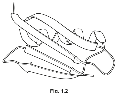
Describe the secondary structure of the protein shown in Fig. 1.2.
Answer
Any two from:
- -helix / alpha-helix
- -pleated sheet(s)
- region of no fixed shape / random coil
α-helices and β-pleated sheets (with regions of no fixed shape / random coil)
Background Concept
The secondary structure of a protein is the regular local folding of the polypeptide backbone, stabilised by hydrogen bonding between peptide groups. The two main types are:
- -helix: a right-handed coil with hydrogen bonds between the N-H of one peptide group and the C=O of the peptide group four residues earlier.
- -pleated sheet: extended strands lying side-by-side, with hydrogen bonds between N-H and C=O of adjacent strands.
Some regions of a protein have no regular pattern; these are described as random coil or regions of no fixed shape.
In a ribbon diagram, -helices appear as coiled/spiral ribbons and -pleated sheets appear as flat arrows.
Understanding the Question
Fig. 1.2 is a ribbon diagram of a Streptococcus protein. The candidate must describe which secondary structures are visible.
Approach
Look at the ribbon diagram and recognise the coiled ribbons (-helices) and the flat arrows (-pleated sheets). Note that some regions are not regular.
Step-by-Step Reasoning
- The flat arrows in Fig. 1.2 represent -pleated sheets.
- The coiled ribbons represent -helices.
- Any region that is neither a helix nor a sheet is a region of no fixed shape / random coil.
- The candidate should name at least two features (any two from the three) for the 2 marks.
Key Takeaways
- Ribbon diagrams use specific shapes to denote secondary structure: coiled ribbons for -helices, flat arrows for -strands/sheets.
- "Random coil" or "region of no fixed shape" is an acceptable third category.
Common Mistakes
- Calling an -helix an "-sheet" or a "-helix" — easy to mix up the Greek letters.
- Writing just "helix" or "sheet" without the alpha/beta qualifier (mark scheme specifically rejects "A" or "a" for alpha and "B" or "b" for beta).
- Missing the random-coil regions entirely.
Things to Be Careful About
- Use the Greek letters or spell out "alpha"/"beta" in full; "A" or "B" alone is rejected.
- "-pleated sheet" must include "pleated" — "-sheet" is acceptable shorthand.
Explain why the protein shown in Fig. 1.2 has tertiary structure, but not quaternary structure.
Answer
- The protein consists of a single polypeptide chain / one chain of amino acids, so it has tertiary structure (folding of one polypeptide).
- Quaternary structure requires two or more polypeptides, which it does not have.
Single polypeptide chain; quaternary structure requires two or more polypeptides.
Background Concept
Protein structure is described at four levels:
- Primary: linear sequence of amino acids linked by peptide bonds.
- Secondary: regular local folding (-helix, -pleated sheet) stabilised by hydrogen bonds between peptide groups.
- Tertiary: overall 3D folding of a single polypeptide, stabilised by interactions between R-groups.
- Quaternary: assembly of two or more polypeptide chains into a functional unit, stabilised by interactions between R-groups on different chains.
The key distinction between tertiary and quaternary structure is the number of polypeptide chains involved.
Understanding the Question
Fig. 1.2 shows a single folded ribbon — i.e., one polypeptide chain. The candidate must explain why this protein has tertiary structure but no quaternary structure.
Approach
Recall the definitions: tertiary = one polypeptide folded; quaternary = two or more polypeptides assembled.
Step-by-Step Reasoning
- Fig. 1.2 shows one continuous ribbon, so the protein is a single polypeptide chain.
- A single polypeptide, when folded, has tertiary structure.
- Quaternary structure requires two or more separate polypeptide chains interacting, which this protein does not have.
- Therefore the protein has tertiary structure only.
Key Takeaways
- Tertiary = folding of one polypeptide.
- Quaternary = two or more polypeptides interacting.
- A protein made of a single chain cannot have quaternary structure.
Common Mistakes
- Saying the protein "does not have enough amino acids" — the issue is not size but chain number.
- Saying the protein "is not folded enough" — folding of a single chain is tertiary, regardless of how compact it is.
- Confusing quaternary with tertiary.
Things to Be Careful About
- The answer hinges on polypeptide count, not on size, complexity, or absence of prosthetic groups.
An analysis of the amino acid composition of the protein in Fig. 1.2 showed that it does not contain any cysteine residues.
Explain how the three-dimensional structure of the protein shown in Fig. 1.2 is held in place.
Answer
Secondary structure (held in place by hydrogen bonds in the backbone):
- In an -helix, each N-H forms a hydrogen bond with a C=O three or four amino acids further along the same polypeptide.
- In a -pleated sheet, each N-H forms a hydrogen bond with a C=O on an adjacent region of the polypeptide.
Tertiary structure (held in place by interactions between R-groups):
- Hydrogen bonds between polar R-groups (e.g. those with C=O and HN groups).
- Ionic bonds between charged R-groups (e.g. -NH and -COO groups).
- Hydrophobic interactions between non-polar R-groups.
(No disulfide bonds because the protein contains no cysteine.)
Hydrogen bonds within the backbone hold secondary structure (α-helix and β-pleated sheet); hydrogen bonds, ionic bonds, and hydrophobic interactions between R-groups hold tertiary structure. No disulfide bonds (no cysteine).
Background Concept
A protein's 3D shape is held in place by a combination of weak and strong interactions:
- In secondary structure, regular hydrogen bonding between peptide groups (N-H and C=O) of the backbone produces -helices and -pleated sheets.
- In tertiary structure, the overall fold is stabilised by interactions between R-groups: hydrogen bonds between polar R-groups, ionic (salt-bridge) interactions between positively and negatively charged R-groups, hydrophobic interactions between non-polar R-groups, and disulfide bonds (when cysteine residues are present and oxidised).
Because the Streptococcus protein in question contains no cysteine residues, no disulfide bonds are possible. The candidate must explain the structure using the other interactions only.
Understanding the Question
Part (iii) provides an extra piece of information: the protein has no cysteine. The candidate must explain how the 3D structure is nevertheless held in place.
Approach
Cover both secondary structure (backbone hydrogen bonds) and tertiary structure (R-group interactions). Avoid disulfide bonds entirely.
Step-by-Step Reasoning
- Secondary structure: in an -helix, hydrogen bonds form between the N-H of one peptide group and the C=O of the peptide group four residues earlier in the same chain. In a -pleated sheet, hydrogen bonds form between N-H and C=O groups on adjacent strands of the polypeptide.
- Tertiary structure — hydrogen bonds: polar R-groups (those carrying C=O or N-H groups, e.g. serine, threonine, asparagine) form hydrogen bonds with one another.
- Tertiary structure — ionic bonds: charged R-groups (e.g. -NH on lysine, -COO on aspartate/glutamate) attract each other to form salt bridges.
- Tertiary structure — hydrophobic interactions: non-polar R-groups (e.g. the hydrocarbon side chains of leucine, isoleucine, valine, phenylalanine) cluster together in the interior of the protein, away from the aqueous cytosol.
- Disulfide bonds: not present because there are no cysteine residues.
Key Takeaways
- Secondary structure depends on hydrogen bonds between peptide groups of the backbone.
- Tertiary structure depends on interactions between R-groups: hydrogen bonds, ionic bonds, hydrophobic interactions and (when cysteine is present) disulfide bonds.
- A protein lacking cysteine cannot form disulfide bonds, but the other interactions still suffice to hold the fold in place.
Common Mistakes
- Mentioning disulfide bonds — explicitly wrong because the protein has no cysteine.
- Confusing backbone hydrogen bonds (secondary) with R-group hydrogen bonds (tertiary).
- Saying "weak bonds" without naming them.
- Only discussing one level (e.g. just secondary or just tertiary) and ignoring the other.
Things to Be Careful About
- Be specific: name the groups involved (NH, C=O, NH, COO) and where the bonds form (backbone vs R-groups).
- The question explicitly excludes disulfide bonds via the cysteine information — do not contradict the question.
The rest of this paper
5 more questions- Q2Immunity · Cell Structure · Infectious Diseases14M
- Q3Transport in Plants7M
- Q4The Mitotic Cell Cycle · Nucleic Acids and Protein Synthesis11M
- Q5Cell Membranes and Transport · Transport in Mammals · Nucleic Acids and Protein Synthesis12M
- Q6Cell Membranes and Transport · Transport in Mammals4M