Biology 9700/22 — October/November 2022
Cambridge AS Level · AS Level Structured Questions · worked solutions for every part, with the mark scheme
Topics Biological Molecules · Cell Structure · Nucleic Acids and Protein Synthesis · Transport in Mammals · The Mitotic Cell Cycle · Immunity · +5 more
The mammalian circulatory system is adapted for the long-distance transport of the respiratory gases, oxygen and carbon dioxide.
The system is described as a closed double circulation.
Name:
• the two different circulations of the double circulatory system of mammals
• the main vein returning deoxygenated blood to the heart.
Answer
- Pulmonary (circulation)
- Systemic (circulation)
- Vena cava (inferior or superior vena cava)
Pulmonary and systemic circulations; vena cava.
Background Concept
Mammals have a closed double circulation: blood passes through the heart twice for every complete circuit of the body. This is more efficient than the single circulation of fish because oxygenated and deoxygenated blood are kept separate, allowing higher metabolic rates to be sustained.
The two circuits are:
- The pulmonary circulation – carries deoxygenated blood from the right ventricle to the lungs (via the pulmonary artery) and returns oxygenated blood to the left atrium (via the pulmonary veins).
- The systemic circulation – carries oxygenated blood from the left ventricle around the body (via the aorta) and returns deoxygenated blood to the right atrium.
The vena cava (actually two vessels: the superior and inferior venae cavae) is the largest vein and empties deoxygenated blood directly into the right atrium of the heart.
Understanding the Question
The question asks for three one-word/short-phrase answers worth 3 marks: the two circulations of the mammalian double system, and the main vein bringing deoxygenated blood back to the heart.
Approach
This is a recall question. Simply state the two names of the circuits and the name of the vein. Avoid inventing terms; the mark scheme rejects "systematic" in place of "systemic".
Step-by-Step Reasoning
- Circulation 1: blood goes from the heart to the lungs and back → pulmonary.
- Circulation 2: blood goes from the heart to the rest of the body and back → systemic.
- Main vein to the heart: the two large veins joining the right atrium are the superior and inferior vena cava; either name (or just "vena cava") is accepted.
Key Takeaways
- Double circulation = pulmonary + systemic.
- Vena cava is the main vessel returning deoxygenated blood to the right atrium.
- Spelling matters: "systemic", not "systematic".
Common Mistakes
- Writing "systematic" – this is a different word and is rejected.
- Calling the pulmonary artery a vein because it carries deoxygenated blood. (Arteries carry blood away from the heart; veins carry blood towards the heart – irrespective of oxygen content.)
Things to Be Careful About
- Either "vena cava" or "superior/inferior vena cava" is acceptable.
- "Venae cavae" (plural) is also accepted in CIE mark schemes.
In a closed circulation, blood is kept within vessels at all times.
Name the type of blood vessel that connects capillaries and veins.
Answer
Venule
Venule
Background Concept
Blood flows from the heart through progressively smaller vessels: arteries → arterioles → capillaries → venules → veins → back to the heart. As blood leaves a capillary bed it first drains into a small vessel called a venule, which then merges with other venules to form a vein.
A venule is a tiny (typically 8–100 µm diameter) thin-walled vessel that collects blood from several capillaries. Its wall is essentially a continuation of the capillary endothelium, with a thin outer layer of connective tissue. Because it lacks a substantial smooth-muscle layer, it cannot constrict strongly and is the principal site of fluid and white-blood-cell exchange in the microcirculation.
Understanding the Question
The question requires the name of the vessel that links the capillary network to a vein. It is worth 1 mark.
Approach
Recall the standard order of vessels in the circulatory system and pick the one that sits between capillaries and veins.
Step-by-Step Reasoning
- Trace the flow: artery → arteriole → capillary → venule → vein.
- The vessel between capillaries and veins is the venule.
Key Takeaways
- Capillaries drain into venules, which join to form veins.
- Venules are very small; their walls are mostly endothelium.
Common Mistakes
- Writing "vein" (too large; capillaries do not join veins directly).
- Writing "venial" or "venus" – spelling errors that lose the mark.
Things to Be Careful About
- Singular or plural ("venule / venules") is accepted by CIE; either is correct here.
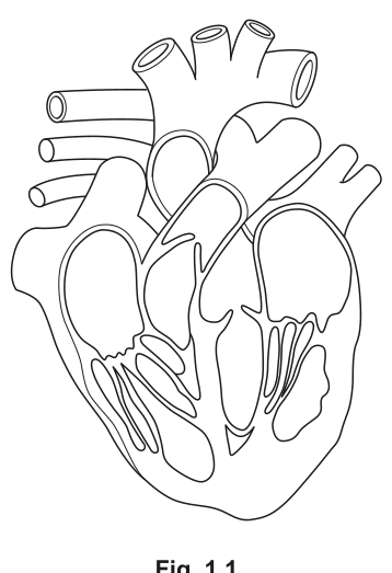
Fig. 1.1 is a diagram of a section through the heart.
On Fig. 1.1:
• add a label line and the letter L to show the artery that takes blood from the heart to the lungs
• add a label line and the letter R to show the valve that closes when the right ventricle is in systole.
Answer
- L – label line drawn to the pulmonary artery (the vessel leaving the right ventricle and branching into left and right pulmonary arteries).
- R – label line drawn to the right atrioventricular valve (tricuspid valve), the valve between the right atrium and right ventricle that closes when the right ventricle contracts (systole).
L = pulmonary artery; R = right atrioventricular (tricuspid) valve.
Background Concept
The mammalian heart has four chambers and four valves:
- Two atrioventricular (AV) valves – the right (tricuspid) and left (bicuspid/mitral) valves between each atrium and ventricle. These close during ventricular systole to prevent backflow into the atria.
- Two semilunar valves – the pulmonary (right) and aortic (left) valves at the outlets of the ventricles. These open during ventricular systole and close during diastole.
So when the right ventricle contracts (right systole):
- The right AV (tricuspid) valve closes ← this is the valve the question wants.
- The pulmonary semilunar valve opens, allowing blood to be ejected into the pulmonary artery.
The pulmonary artery is the only artery in the body that normally carries deoxygenated blood – it carries it from the right ventricle to the lungs.
Understanding the Question
Two label lines and letters must be added to the printed diagram of the heart in Fig. 1.1.
- Letter L → the artery carrying blood from the heart to the lungs.
- Letter R → the valve that closes when the right ventricle is contracting.
Note that closing during systole is the defining property of the atrioventricular valve, not the semilunar valve. The pulmonary semilunar valve is open during right ventricular systole, so it would be wrong to label it R.
Approach
- Identify the pulmonary artery on the diagram – it leaves the right ventricle (on the viewer's left in anatomical view) and goes upward, branching toward each lung.
- Identify the right AV (tricuspid) valve – it is the AV valve on the same side of the heart as the right ventricle, guarding the entrance from the right atrium into the right ventricle.
- Draw label lines from each structure to the margin of the figure and write L and R at the end of each line.
Step-by-Step Reasoning
- The pulmonary artery is recognisable because it is the vessel emerging from the conus arteriosus of the right ventricle and immediately splitting into a left and right branch (the bifurcation is usually drawn clearly on heart diagrams).
- The tricuspid / right AV valve sits between the right atrium and right ventricle. In an anterior view of the heart it appears on the viewer's right (because the heart's right side is on the patient's right).
- Do not label the pulmonary veins (these carry oxygenated blood from the lungs to the heart, not to the lungs).
- Do not label the pulmonary semilunar valve (it opens during right systole, not closes).
- Do not label the chordae tendineae; the mark scheme specifically rejects these as the target of the R label.
Key Takeaways
- Ventricular systole = ventricles contract = AV valves close, semilunar valves open.
- Right ventricle → pulmonary artery (only artery carrying deoxygenated blood).
- Right AV valve = tricuspid valve.
Common Mistakes
- Labelling the aorta instead of the pulmonary artery – they both leave the heart, but only the pulmonary artery goes to the lungs.
- Labelling the pulmonary semilunar valve as the valve that closes during right systole – this is the wrong valve because it OPENS during systole.
- Labelling the left AV (bicuspid) valve – this is on the left ventricle, not the right.
Things to Be Careful About
- The label line should touch (or clearly end at) the structure being labelled. A line that points to nearby muscle or to the chordae tendineae will be rejected.
- The letters L and R must be written at the end of each label line.
The entry of carbon dioxide into red blood cells results in the production of hydrogencarbonate ions. This involves the enzyme carbonic anhydrase.
Complete the passage summarising the production of hydrogencarbonate ions by:
• writing the correct biological term in the spaces provided
• writing the molecular formula for two of the terms in the spaces in brackets.
Carbonic anhydrase has an overall spherical shape and is known as a ______ protein. The enzyme acts within the cell so can be described as an ______ enzyme. When blood passes into the capillary network through actively respiring tissues, carbon dioxide () diffuses into red blood cells and carbonic anhydrase catalyses a reaction where ______ (______) is combined with to form ______ (), which rapidly forms ______ ions (______) and hydrogencarbonate ions ().
Answer
Carbonic anhydrase has an overall spherical shape and is known as a globular protein. The enzyme acts within the cell so can be described as an intracellular enzyme. When blood passes into the capillary network through actively respiring tissues, carbon dioxide () diffuses into red blood cells and carbonic anhydrase catalyses a reaction where water () is combined with to form carbonic acid (), which rapidly forms hydrogen ions () and hydrogencarbonate ions ().
globular; intracellular; water (H₂O); carbonic acid; hydrogen (H⁺).
Background Concept
Around 70% of carbon dioxide is transported in the plasma as hydrogencarbonate ions (), but the ions are made inside the red blood cell. The series of reactions is catalysed by the enzyme carbonic anhydrase, which has a roughly spherical/globular tertiary structure and is found within the cytosol of erythrocytes (so it is intracellular).
The reaction is:
Step 1: + water → carbonic acid (slow unless catalysed).
Step 2: carbonic acid → hydrogen ion + hydrogencarbonate ion (essentially instantaneous).
The hydrogencarbonate ions diffuse out of the red cell into the plasma in exchange for chloride ions – this is the chloride shift, which maintains electrochemical balance. The hydrogen ions are buffered by haemoglobin, preventing the blood from becoming too acidic.
Protein classification:
- Globular proteins are roughly spherical, water-soluble, and usually have metabolic roles (enzymes, antibodies, some hormones, haemoglobin).
- Fibrous proteins are long, insoluble, and structural (collagen, keratin).
Enzyme classification by location:
- Intracellular enzymes work inside the cell (e.g. carbonic anhydrase, enzymes of glycolysis, Krebs cycle enzymes).
- Extracellular enzymes are secreted and work outside the cell (e.g. digestive enzymes such as amylase, trypsin).
Understanding the Question
The question has five blanks in a passage, each worth 1 mark. For two of the blanks the mark scheme requires both the name of the species and its molecular formula. Specifically the mark scheme requires formulae for the first reactant (water) and for the hydrogen ion, but the carbonic acid formula is also given in the passage so it does not need to be supplied.
Approach
- Recall the chemical equation for the carbonic anhydrase reaction.
- Identify each blank with the appropriate biological term and, where the bracket is provided, the corresponding formula.
- Use the precise CIE wording: "globular" and "intracellular" – not "intercellular", not "spherical" as a substitute for "globular".
Step-by-Step Reasoning
- Type of protein – "has an overall spherical shape" → globular protein (compare fibrous proteins such as collagen, which are long and rope-like).
- Enzyme location – "acts within the cell" → intracellular (mark scheme rejects "intercellular").
- Species combined with CO₂ – water, formula .
- Intermediate product – carbonic acid, formula (formula already in the passage, so only the name is needed here).
- Cation produced alongside hydrogencarbonate – hydrogen ion, formula .
Key Takeaways
- Carbonic anhydrase reaction: .
- The enzyme is intracellular and globular.
- Most is transported as in the plasma after the chloride shift.
Common Mistakes
- Writing "intercellular" – the mark scheme explicitly rejects this; the correct term is intracellular ("within a cell").
- Writing "fibrous" instead of "globular" – fibrous proteins are not enzymes.
- Confusing the two formulae: is water; is carbonic acid; is hydrogencarbonate; is the hydrogen ion. Slips such as writing or are common and lose marks.
- Writing "hydrogen ion" without a formula or "H" without the charge – the question asks for the molecular formula, so the superscript plus sign is part of the mark.
Things to Be Careful About
- CIE mark schemes require the name of the species and the correct molecular formula for the two bracketed blanks; giving only one half loses the mark.
- The question does not ask for the chloride shift or for buffering of by haemoglobin, so do not include these – they would be irrelevant padding.
The rest of this paper
5 more questions- Q2Cell Structure · Nucleic Acids and Protein Synthesis · The Mitotic Cell Cycle · Immunity15M
- Q3Infectious Diseases · Nucleic Acids and Protein Synthesis11M
- Q4Gas Exchange8M
- Q5Biological Molecules · Cell Membranes and Transport · Cell Structure · Enzymes10M
- Q6Transport in Plants5M