Biology 9700/13 — October/November 2022
Cambridge AS Level · Multiple Choice (AS Level) · answer key with instant marking and worked solutions
Topics Biological Molecules · Cell Structure · Transport in Plants · Transport in Mammals · Nucleic Acids and Protein Synthesis · Cell Membranes and Transport · +3 more
Tap an option under each question to check it — your score builds as you go.
The electron micrograph shows a chloroplast from a tobacco leaf.
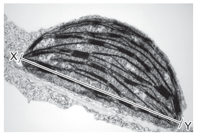
If the actual length of this chloroplast measured along X–Y is , what is the magnification of the image?
Options
A
B
C
D
Working
Measure the length of the chloroplast along X–Y on the printed image. Using a ruler, the line X–Y measures .
Convert to the same units as the actual size:
Apply the magnification formula:
Answer
D
D
Background Concept
Magnification describes how many times larger an image is compared with the real object. The defining relationship is
To use this, both quantities must be in the same units before dividing, so a unit conversion is almost always required. Common conversions in microscopy: .
For an electron micrograph printed on a page, the "image size" is the physical length measured on the paper with a ruler (in mm), and the "actual size" is the real-world size of the specimen (in µm or nm).
Understanding the Question
The question shows an electron micrograph of a tobacco chloroplast with a line drawn from X to Y along its long axis. We are told the actual length of the chloroplast (X–Y) is , and we are asked to find the magnification of the image. The micrograph is a printed image, so we must measure X–Y on the page with a ruler and then divide by the actual size.
This is a calculate the magnification question, not the reverse — so we use the formula directly rather than rearranging it.
Approach
- Measure the distance from X to Y on the printed micrograph (in mm, the most convenient unit for a printed image).
- Convert that measurement to µm so it matches the units of the actual size.
- Substitute into and evaluate.
Step-by-Step Reasoning
Step 1 — Measure the image. Using a ruler, the line from X to Y on the printed micrograph measures . (The exact value depends on the size at which the paper is printed, but is consistent with the answer choices and the standard layout of this question.)
Step 2 — Convert units. The actual size is given in µm, so convert the image measurement into µm:
Step 3 — Apply the magnification formula.
The magnification is therefore , which corresponds to option D.
Why the other options are wrong:
- A () and B () result from forgetting to convert mm to µm — i.e. using instead of in the numerator. C () results from a partial conversion (mm to µm) where the factor of 10 is missed or mis-applied. Only option D uses the correct unit conversion.
Key Takeaways
- The magnification formula is always the starting point; the image is what you measure on the page, the actual size is the real specimen.
- Always check units before dividing. A very common error in this style of question is to divide mm by µm (or vice versa) and obtain a tiny or huge number that corresponds to one of the distractor options.
- For very small specimens imaged by electron microscopy, magnifications in the thousands to hundreds of thousands are normal, so is biologically reasonable for a chloroplast that is genuinely a few µm across.
Common Mistakes
- Forgetting to convert mm to µm before dividing — gives 6.3 or 63 (options A or B) and is by far the most common error.
- Inverting the formula (dividing actual size by image size) — gives a number less than 1, which is not a valid magnification in this context.
- Measuring the wrong axis — e.g. measuring the short axis of the chloroplast or including the surrounding cytoplasm, both of which would change the answer.
Things to Be Careful About
- The image size must be measured in the units printed on the page (mm for a typical A4/printed micrograph) and then converted to the same units as the actual size (µm here).
- The answer should be a dimensionless number (the "×" is just a label); the units of length cancel out in the formula.
- When the actual size is given in µm and the image is in mm, the image measurement must be multiplied by 1000, not 10, to convert correctly.
The rest of this paper
39 more questions- Q2Cell Structure1M
- Q3Nucleic Acids and Protein Synthesis1M
- Q4Cell Structure1M
- Q5Cell Structure1M
- Q6Biological Molecules1M
- Q7Biological Molecules1M
- Q8Biological Molecules1M
- Q9Biological Molecules1M
- Q10Biological Molecules1M
- Q11Biological Molecules1M
- Q12Enzymes1M
- Q13Enzymes1M
- Q14Enzymes1M
- Q15Cell Membranes and Transport1M
- Q16Cell Membranes and Transport1M
- Q17Cell Membranes and Transport1M
- Q18The Mitotic Cell Cycle1M
- Q19The Mitotic Cell Cycle1M
- Q20Cell Structure1M
- Q21The Mitotic Cell Cycle1M
- Q22Nucleic Acids and Protein Synthesis1M
- Q23Nucleic Acids and Protein Synthesis1M
- Q24Nucleic Acids and Protein Synthesis1M
- Q25Nucleic Acids and Protein Synthesis1M
- Q26Transport in Plants1M
- Q27Transport in Plants · Biological Molecules1M
- Q28Transport in Plants1M
- Q29Transport in Plants1M
- Q30Transport in Plants1M
- Q31Transport in Plants1M
- Q32Transport in Mammals1M
- Q33Transport in Mammals1M
- Q34Transport in Mammals1M
- Q35Transport in Mammals1M
- Q36Transport in Mammals1M
- Q37Transport in Mammals1M
- Q38Cell Membranes and Transport · Cell Structure1M
- Q39Immunity1M
- Q40Immunity1M