Biology 9700/12 — May/June 2022
Cambridge AS Level · Multiple Choice (AS Level) · answer key with instant marking and worked solutions
Topics Biological Molecules · Cell Structure · Transport in Plants · Transport in Mammals · The Mitotic Cell Cycle · Gas Exchange · +5 more
Tap an option under each question to check it — your score builds as you go.
The photomicrograph shows a bronchiole and alveoli.
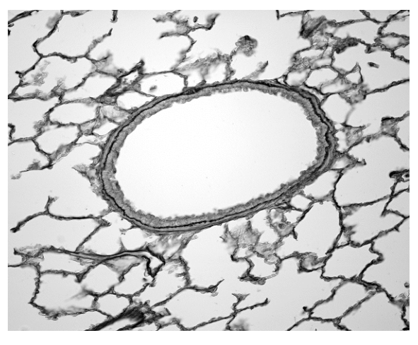
The magnification of the image is .
What is the maximum diameter of the bronchiole lumen?
Options
A
B
C
D
Working
Measure the maximum diameter of the bronchiole lumen on the image (the widest dimension across the open space inside the bronchiole wall). The lumen measures approximately across on the printed image.
Use the magnification formula rearranged to find actual size:
Convert the image measurement to the same units as the answer choices (micrometres): .
Answer
C —
C
Background Concept
When a specimen is viewed through a microscope, what you see on the printed page or screen is a magnified version of the real structure. The relationship between what you measure on the image, the actual size of the object, and the magnification is given by:
Rearranged to find the actual (real) size of the specimen:
Units must be consistent. Because light micrographs of cells and tissues typically produce results in the micrometre () range, image measurements taken in millimetres must be converted: .
In the lung, a bronchiole is a small airway (< 1 mm diameter) that branches from a bronchus and leads to the alveoli. It is lined by a ciliated epithelium and has smooth muscle in its wall, but (unlike larger bronchi) no cartilage. The lumen is the hollow interior of the tube through which air flows.
Understanding the Question
You are given a light micrograph (Fig. 1.1) of lung tissue at a stated magnification of . A large oval structure in the centre is the bronchiole, surrounded by a network of thin-walled air sacs (alveoli). The task is to determine the actual maximum diameter of the bronchiole lumen — i.e. the widest internal distance across the open space inside the bronchiole wall.
The command word is "what is…", which simply requires a numerical answer chosen from the four options. The biological skill is the magnification calculation, not tissue identification.
Approach
- Identify the lumen on the micrograph — it is the clear (white) space inside the dark ring of the bronchiole wall. The maximum diameter is the longest straight line that fits across this opening.
- Measure this maximum diameter on the printed image using a ruler, in millimetres.
- Convert to micrometres.
- Divide by the magnification () to obtain the actual size.
- Select the option closest to the calculated value.
Step-by-Step Reasoning
Step 1 — Locate the lumen. On Fig. 1.1, the bronchiole lumen is the bright, open region in the centre of the image, bounded by a dark, somewhat granular wall (epithelium plus smooth muscle). It is roughly oval.
Step 2 — Measure the maximum diameter. Placing a ruler across the widest part of the lumen gives a reading of approximately on the printed page.
Step 3 — Convert units. The answer options are in micrometres, so convert first:
Step 4 — Apply the magnification formula.
Step 5 — Choose the closest option. rounds to , which matches option C.
Quick sanity check on the distractors:
- A () would correspond to a lumen only across on the image — far too small for the prominent central structure shown.
- B () corresponds to about on the image, smaller than the lumen clearly is.
- D () would require an image measurement of about , which exceeds the lumen's apparent width.
Key Takeaways
- The magnification formula is the workhorse of all microscopy calculations in this syllabus.
- Always convert units consistently (mm ↔ µm ↔ nm) before dividing, and check that the magnitude of your answer is biologically plausible.
- When asked for the "maximum" diameter, take the longest straight line across the structure, not a perpendicular or average value.
- A real bronchiole lumen is typically in the range of – (–), so an answer of ~ is biologically reasonable.
Common Mistakes
- Dividing instead of multiplying by magnification (or vice versa) when rearranging the formula.
- Forgetting to convert mm to µm — leaving the answer as and then not recognising the required unit in the options.
- Measuring the outside diameter of the bronchiole (including the wall) rather than the lumen — the question specifically asks for the lumen, so the dark wall is excluded.
- Measuring the wrong axis of an oval lumen — "maximum diameter" means the longest, not the average, dimension.
- Quoting too many significant figures — the image measurement is approximate, so a value such as is unjustified; is the sensible precision.
Things to Be Careful About
- The magnification given () refers to the printed image as you see it, not to an additional screen or book magnification. Measure the lumen directly on the figure as printed.
- Distinguish carefully between the bronchiole (the central tube with a definite wall) and the surrounding alveoli (the thin-walled sacs that look like a honeycomb). The lumen belongs to the bronchiole.
- When estimating from a printed image, allow a small tolerance in the measurement; the question is designed so the correct option sits clearly above or below the others once the calculation is done.
The rest of this paper
39 more questions- Q2Cell Structure1M
- Q3Cell Structure1M
- Q4Cell Structure1M
- Q5Cell Structure1M
- Q6Biological Molecules1M
- Q7Biological Molecules1M
- Q8Biological Molecules1M
- Q9Biological Molecules1M
- Q10Biological Molecules1M
- Q11Biological Molecules1M
- Q12Enzymes1M
- Q13Biological Molecules1M
- Q14Cell Membranes and Transport1M
- Q15Cell Membranes and Transport1M
- Q16Cell Membranes and Transport1M
- Q17The Mitotic Cell Cycle1M
- Q18The Mitotic Cell Cycle1M
- Q19The Mitotic Cell Cycle1M
- Q20The Mitotic Cell Cycle1M
- Q21Nucleic Acids and Protein Synthesis1M
- Q22Nucleic Acids and Protein Synthesis · Infectious Diseases1M
- Q23Nucleic Acids and Protein Synthesis1M
- Q24Transport in Plants1M
- Q25Transport in Plants1M
- Q26Transport in Plants1M
- Q27Transport in Plants1M
- Q28Transport in Plants1M
- Q29Transport in Plants1M
- Q30Transport in Mammals1M
- Q31Transport in Mammals1M
- Q32Transport in Mammals1M
- Q33Transport in Mammals1M
- Q34Transport in Mammals1M
- Q35Gas Exchange1M
- Q36Cell Structure · Gas Exchange1M
- Q37Infectious Diseases1M
- Q38Immunity1M
- Q39Transport in Mammals1M
- Q40Immunity1M