Biology 9700/11 — May/June 2022
Cambridge AS Level · Multiple Choice (AS Level) · answer key with instant marking and worked solutions
Topics Biological Molecules · Cell Structure · Transport in Mammals · Cell Membranes and Transport · The Mitotic Cell Cycle · Nucleic Acids and Protein Synthesis · +5 more
Tap an option under each question to check it — your score builds as you go.
A student used a stage micrometer scale to calibrate an eyepiece graticule.
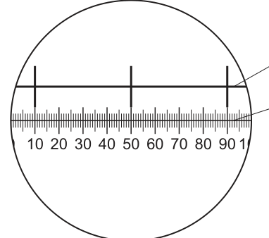
The diagram shows the view of both the stage micrometer scale and the eyepiece graticule seen by the student. The divisions on the stage micrometer scale are apart.
The student removed the stage micrometer scale and viewed a slide with blood cells on it. The same lenses were used so that the magnification remained unchanged.
The student measured the diameter of one of the white blood cells on the slide using the eyepiece graticule and recorded that it was 8 eyepiece units.
What is the correct diameter of this white blood cell in micrometers?
Options
A 0.2
B 0.8
C 20
D 800
Working
From Fig. 1.1:
- The 3 large tick marks on the stage micrometer align with positions 10, 50, and 90 on the eyepiece graticule.
- 2 intervals of the stage micrometer (each ) therefore span 80 eyepiece units (from 10 to 90).
- Real distance = .
- So 1 eyepiece unit = .
For the white blood cell:
- 8 eyepiece units = .
Answer
C
C
Background Concept
The eyepiece graticule is a small glass disc with an engraved scale that sits inside the eyepiece of a light microscope. It allows the user to take measurements of specimens on a slide, but the scale is arbitrary: the apparent size of each division depends on the magnification being used. To turn "eyepiece units" into real units (mm or µm), the graticule must first be calibrated using a stage micrometer — a slide with a precisely known scale engraved on it. In this question the stage micrometer divisions are () apart.
When the stage micrometer and the eyepiece graticule are viewed together at the same magnification, the alignment of the two scales tells us how many eyepiece units correspond to a known real distance. This ratio is only valid at the magnification used for calibration; changing the objective or eyepiece lens requires re-calibration.
Understanding the Question
The student has:
- Calibrated the eyepiece graticule by viewing both the stage micrometer and the eyepiece graticule simultaneously at one magnification (Fig. 1.1).
- Removed the stage micrometer and put a slide of blood cells on the stage, keeping the same magnification.
- Used the (now-calibrated) eyepiece graticule to measure a white blood cell as 8 eyepiece units across.
- We are asked for the actual diameter of the cell in micrometres (µm).
The diagram Fig. 1.1 is the key to determining the value of one eyepiece unit.
Approach
- From Fig. 1.1, identify the alignment points where the stage micrometer and eyepiece graticule line up.
- Calculate the real distance (using the stage micrometer calibration of 0.1 mm per division) that corresponds to a known number of eyepiece units.
- Divide to find the real distance per eyepiece unit, converting to µm.
- Multiply by 8 to obtain the actual diameter of the white blood cell.
Step-by-Step Reasoning
From Fig. 1.1:
- The 3 large tick marks of the stage micrometer line up with positions 10, 50, and 90 on the eyepiece graticule.
- So 2 stage micrometer intervals (each ) span 80 eyepiece units.
- Real distance = .
- eyepiece units, so 1 eyepiece unit = .
For the white blood cell:
- The cell measured 8 eyepiece units across.
- Actual diameter = .
Sanity check: leukocytes (white blood cells) are typically around 10–20 µm in diameter, so 20 µm is biologically reasonable. Red blood cells, by contrast, are about 7 µm across and would not be 20 µm.
Key Takeaways
- The eyepiece graticule must be calibrated against a stage micrometer before it can be used to measure specimens.
- The calibration only applies at the magnification used; changing the objective or eyepiece requires re-calibration.
- . Be careful with unit conversions.
- The actual size of a cell is always far smaller than its appearance in eyepiece units.
Common Mistakes
- Forgetting to convert mm to µm (e.g. writing instead of ).
- Confusing the two scales — the stage micrometer is the known reference, the eyepiece graticule is the one being calibrated.
- Misreading the alignment in the diagram (e.g. using 4 stage micrometer divisions instead of 2, or 40 eyepiece units instead of 80).
- Taking instead of .
Things to Be Careful About
- The question asks for the answer in µm (micrometres), not mm — convert at the end.
- Each stage micrometer division is .
- The same lenses are used for both calibration and measurement, so the calibration factor still applies.
- White blood cells (leukocytes) are around 10–20 µm in diameter, so 20 µm is biologically sensible — use this as a check against distractors like (far too large) or / (smaller than most bacteria).
The rest of this paper
39 more questions- Q2Cell Structure1M
- Q3Cell Structure1M
- Q4Cell Structure1M
- Q5Cell Structure1M
- Q6Biological Molecules1M
- Q7Biological Molecules1M
- Q8Biological Molecules1M
- Q9Biological Molecules1M
- Q10Biological Molecules1M
- Q11Biological Molecules1M
- Q12Biological Molecules1M
- Q13Enzymes1M
- Q14Cell Membranes and Transport1M
- Q15Cell Membranes and Transport1M
- Q16Cell Membranes and Transport1M
- Q17Cell Membranes and Transport1M
- Q18The Mitotic Cell Cycle1M
- Q19The Mitotic Cell Cycle1M
- Q20The Mitotic Cell Cycle1M
- Q21The Mitotic Cell Cycle1M
- Q22Nucleic Acids and Protein Synthesis1M
- Q23Nucleic Acids and Protein Synthesis1M
- Q24Nucleic Acids and Protein Synthesis1M
- Q25Nucleic Acids and Protein Synthesis1M
- Q26Transport in Plants1M
- Q27Transport in Plants1M
- Q28Transport in Plants1M
- Q29Transport in Plants1M
- Q30Transport in Mammals1M
- Q31Transport in Mammals1M
- Q32Transport in Mammals1M
- Q33Transport in Mammals · Gas Exchange1M
- Q34Transport in Mammals1M
- Q35Gas Exchange1M
- Q36Gas Exchange1M
- Q37Infectious Diseases1M
- Q38Immunity1M
- Q39Immunity1M
- Q40Immunity1M