Biology 9700/23 — October/November 2021
Cambridge AS Level · AS Level Structured Questions · worked solutions for every part, with the mark scheme
Topics Biological Molecules · Nucleic Acids and Protein Synthesis · Enzymes · Transport in Mammals · Cell Membranes and Transport · Transport in Plants · +5 more
Fig. 1.1 is a diagram showing a stage in protein synthesis.
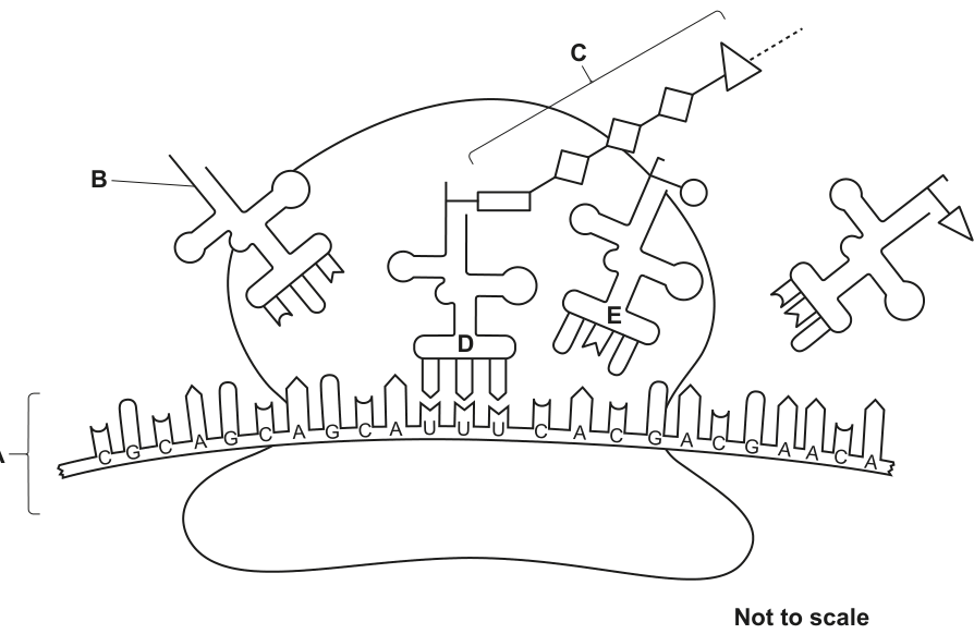
Name the stage of protein synthesis that is shown in Fig. 1.1.
Answer
Translation
Translation
Background Concept
Protein synthesis has two main stages. Transcription is the first stage: in the nucleus, the DNA double helix unwinds and one strand (the template strand) is used to synthesise a complementary single-stranded messenger RNA (mRNA) molecule. Translation is the second stage: the mRNA leaves the nucleus and binds to a ribosome in the cytoplasm. Transfer RNA (tRNA) molecules, each carrying a specific amino acid, decode the mRNA codons one at a time. The ribosome joins the amino acids together with peptide bonds, building up a polypeptide chain.
The diagram in Fig. 1.1 shows all the visual hallmarks of translation: a ribosome, an mRNA strand passing through it, tRNAs delivering amino acids, and a growing polypeptide chain.
Understanding the Question
Fig. 1.1 is described as showing a stage of protein synthesis. You are asked to name that stage. The structures visible in the diagram (ribosome, mRNA, tRNAs, polypeptide) are unique to the second stage of protein synthesis.
Approach
Recognise the structures in the diagram and match them to the correct stage. A ribosome with mRNA and tRNAs, building a polypeptide, is the textbook picture of translation.
Step-by-Step Reasoning
- The large structure in the centre of Fig. 1.1 is a ribosome — the site of translation.
- A single-stranded molecule (A) with a written sequence of bases passes through the ribosome — this is the mRNA being read.
- Clover-leaf shaped molecules (B) deliver amino acids and have an anticodon that base-pairs with mRNA codons — these are tRNAs.
- A chain of amino acids (C) is attached to one of the tRNAs and is growing as new amino acids are added — this is the (poly)peptide product.
- The combination of ribosome + mRNA + tRNAs + growing polypeptide is the definition of translation.
Key Takeaways
- Transcription = DNA → mRNA (in the nucleus)
- Translation = mRNA → polypeptide (at the ribosome)
- A diagram of translation always shows a ribosome, mRNA, tRNAs and a polypeptide chain
Common Mistakes
- Writing "protein synthesis" instead of "translation" — the question asks for the stage, not the overall process
- Confusing transcription with translation — transcription does not involve ribosomes or tRNAs
Things to Be Careful About
The mark scheme requires the specific word "translation". Simply describing the diagram or naming a structure (e.g. "ribosome") does not score the mark.
Identify A, B and C in Fig. 1.1.
A ______
B ______
C ______
Answer
A – messenger RNA / mRNA
B – transfer RNA / tRNA
C – (poly)peptide (chain)
A: messenger RNA / mRNA; B: transfer RNA / tRNA; C: (poly)peptide (chain)
Background Concept
Three types of RNA are involved in translation:
- mRNA (messenger RNA) — a long single-stranded RNA molecule that carries the genetic code from DNA to the ribosome. Its sequence of bases is read in groups of three (codons), each codon specifying an amino acid (or a stop signal).
- tRNA (transfer RNA) — a small RNA molecule with a characteristic clover-leaf shape. It has an anticodon (a triplet of bases) at one end that base-pairs with a complementary codon on the mRNA, and carries the corresponding amino acid on its 3' end.
- rRNA (ribosomal RNA) — combines with proteins to form ribosomes.
The polypeptide is the chain of amino acids linked by peptide bonds. It is the immediate product of translation. A functional protein is the folded, three-dimensional form of one or more polypeptide chains — a polypeptide on its own is not yet a protein.
Understanding the Question
Fig. 1.1 labels three structures: A, B and C. You must name each one. The diagram provides visual cues (shape, position, what each molecule is attached to) that identify the molecules unambiguously.
Approach
Use shape and position to identify each labelled structure. mRNA is the long strand threading through the ribosome; tRNA is the clover-leaf; the polypeptide is the chain of amino acids.
Step-by-Step Reasoning
- A is the long strand of bases passing horizontally through the ribosome. The bases are written on it (C, G, C, A, G, C, …). A long RNA molecule carrying a sequence of codons is mRNA.
- B is the clover-leaf shaped molecule with an anticodon at one end (base-pairing with the mRNA) and an amino acid attached at the other. This is tRNA.
- C is the chain of linked shapes (each shape representing an amino acid) attached to the central tRNA. A linear chain of amino acids joined by peptide bonds is a (poly)peptide chain.
Key Takeaways
- mRNA = long single strand threading through the ribosome, displaying codons
- tRNA = clover-leaf shape, with an anticodon and a carried amino acid
- Polypeptide = chain of amino acids linked by peptide bonds (the immediate product of translation)
Common Mistakes
- Calling the polypeptide "protein" — a polypeptide is the chain, while a protein is the folded functional form. The mark scheme rejects "protein", "(primary structure of) protein" and "amino acid chain" for label C.
- Calling tRNA simply "RNA" — the answer must specify "transfer RNA" or "tRNA".
Things to Be Careful About
The mark scheme requires the precise terms: "messenger RNA / mRNA" for A, "transfer RNA / tRNA" for B, and "(poly)peptide (chain)" for C. Imprecise synonyms are not credited.
State the base sequences at D and E.
D ______
E ______
Answer
D – AAA
E – GUG
D: AAA; E: GUG
Background Concept
A codon is a sequence of three bases on the mRNA. There are 64 possible codons (4³ combinations of A, U, G, C), and they specify 20 different amino acids (with some redundancy) plus three stop signals. The ribosome reads codons sequentially in the 5' to 3' direction along the mRNA. Each tRNA carries an anticodon (a triplet of bases) that base-pairs with the complementary codon on the mRNA, and brings the amino acid specified by that codon.
Understanding the Question
In Fig. 1.1, two tRNAs (labelled D and E) sit inside the ribosome, each base-paired with a codon on the mRNA. You are asked to read off the three-base sequence (the codon) at the position of each tRNA.
Approach
Locate the tRNAs D and E in the diagram, then read the three bases of the mRNA directly beneath each tRNA. Read the bases in the 5' to 3' direction (the order in which the ribosome reads them).
Step-by-Step Reasoning
- The tRNA at D is bound to a codon on the mRNA. The three bases of this codon, read 5' to 3', are AAA. So D = AAA.
- The tRNA at E is bound to the adjacent codon. The three bases of this codon, read 5' to 3', are GUG. So E = GUG.
- D and E are two adjacent codons on the same mRNA, being read in sequence by the ribosome. The two tRNAs occupy adjacent sites on the ribosome (D in the P site, E in the A site), which is why their codons lie side by side on the mRNA.
Key Takeaways
- Codons are read 5' to 3' on the mRNA
- A codon = three consecutive bases
- Each tRNA's anticodon base-pairs with one codon on the mRNA
Common Mistakes
- Reading the anticodon on the tRNA instead of the codon on the mRNA — the question asks for the base sequence at positions D and E (which are on the mRNA).
- Reading the bases in the wrong direction (3' to 5' instead of 5' to 3').
- Mixing up the order of the three bases within a triplet.
Things to Be Careful About
The question asks for the base sequence at D and E — this is the mRNA codon, not the anticodon. Codons are always written 5' to 3'. The mark scheme will also accept the names of the bases (e.g. "adenine, adenine, adenine" for AAA), but letters are cleaner and faster on an exam paper.
Mutagenesis is a process that leads to a change in the amino acid sequences of proteins. Scientists carry out mutagenesis to investigate the importance of particular amino acids in protein structure and function.
Outline how changing one amino acid in the -globin polypeptide of haemoglobin may change the structure and function of a molecule of haemoglobin.
Answer
- The new amino acid has a different R group / side chain (e.g. glutamic acid replaced by valine).
- The new R group may form different bonds with other R groups (or prevent bonds that previously existed).
- This changes the secondary / tertiary structure of the -globin polypeptide (and may alter the quaternary structure of haemoglobin).
- The ability of haemoglobin to bind oxygen is reduced (or otherwise altered).
Different R group → different bonds between R groups → change in 2°/3°/4° structure → reduced or altered ability to bind oxygen.
Background Concept
Proteins have four levels of structure, each building on the one before:
- Primary (1°) structure — the linear sequence of amino acids linked by peptide bonds. This is determined by the gene.
- Secondary (2°) structure — local folding patterns such as α-helices and β-pleated sheets, stabilised by hydrogen bonds between the peptide backbone.
- Tertiary (3°) structure — the overall three-dimensional shape of a single polypeptide chain, stabilised by interactions between the R groups (side chains) of the amino acids: hydrogen bonds, ionic bonds, disulfide bridges and hydrophobic interactions.
- Quaternary (4°) structure — the way multiple polypeptide subunits fit together in a multi-subunit protein.
Every amino acid has the same backbone (–NH–CHR–CO–) but a unique R group. R groups differ in size, charge, polarity and ability to form bonds, and it is these differences that determine how a polypeptide folds. The primary structure therefore determines the higher levels of structure.
Haemoglobin is a globular protein with four polypeptide subunits (2 α-globin and 2 β-globin), each carrying a haem group with an Fe²⁺ ion that reversibly binds O₂. The 3D folding of each subunit, and the way the four subunits pack together, are essential for cooperative O₂ binding.
A classic example is the mutation that causes sickle cell anaemia: a single base substitution in the HBB gene changes codon 6 of β-globin from GAG to GUG, replacing glutamic acid (polar, negatively charged) with valine (non-polar, hydrophobic). One amino acid change — yet the consequences for structure and function are severe.
Understanding the Question
You are told that mutagenesis changes the amino acid sequence of a protein and that scientists use it to investigate which amino acids matter for structure and function. The question asks you to outline how changing one amino acid in the β-globin polypeptide may change the structure and function of a haemoglobin molecule. "Outline" means you should give a clear chain of linked points — not a disconnected list, and not a single sentence.
Approach
Trace the chain of consequences: amino acid change → R-group change → different bonding between R groups → change in folding (2°/3°/4° structure) → change in function (O₂ binding). The mark scheme caps structure points at 3, so to score the full 4 marks you need at least one function point about oxygen binding.
Step-by-Step Reasoning
- The new amino acid has a different R group / side chain. The sickle-cell example makes this concrete: glutamic acid (charged, polar) is replaced by valine (non-polar, hydrophobic). Any substitution puts a different R group in that position, with different chemistry.
- The new R group may form different bonds with other R groups, or may prevent bonds from forming. Different chemistry = different bonding. A hydrophobic valine tends to cluster with other hydrophobic residues; the charged glutamic acid it replaced would have formed ionic bonds and hydrogen bonds with polar/charged partners. The bonding pattern around the substituted position changes.
- This changes the secondary, tertiary and/or quaternary structure of the protein. The polypeptide folds differently; the 3D shape of β-globin is altered; the way β-globin packs against the α-globin subunits in the haemoglobin tetramer (4° structure) may also change. The overall globular shape is different.
- The change in structure alters the function of haemoglobin. The shape of the O₂-binding site depends on the 3D folding of the subunits, so a different shape means a different (usually lower) ability to bind O₂ (and sometimes CO₂). The percentage saturation of haemoglobin with O₂ at a given partial pressure is altered, and the volume of O₂ carried per unit of blood falls.
The mark scheme credits up to 3 structure points (any combination of points 1–5 from its list) plus 1 function point (point 7). At least one of your four points must address function, or you cannot reach 4.
Key Takeaways
- A protein's primary structure determines its higher levels of structure (and therefore its function).
- A single amino acid change can have a large effect if the new R group has very different chemistry from the original (e.g. polar → non-polar).
- The chain of consequence is: DNA mutation → different mRNA codon → different amino acid → different R group → different bonds → different 3D shape → altered function.
- The sickle-cell mutation (Glu → Val at position 6 of β-globin) is the canonical example.
Common Mistakes
- Talking about DNA instead of the protein — the question is about the amino acid in the protein, not the underlying DNA. (The DNA is also different, but that is not what the question is asking about.)
- Saying haemoglobin has an "active site" — the mark scheme explicitly rejects this. Haemoglobin has binding sites for O₂ on its haem groups; these are not called active sites (active sites are for enzymes).
- Talking about quaternary structure for a single polypeptide — the mark scheme rejects "quaternary" if applied to a single β-globin chain. Quaternary structure refers to the arrangement of multiple subunits.
- Vague answers — "the protein is changed" or "function is affected" score nothing. You must say what changes and why.
- Ignoring function — the mark scheme requires at least one function point (about O₂/CO₂ binding); four structure-only points score only 3.
Things to Be Careful About
- Specify the R group (not just "the amino acid") when describing what changes.
- Specify the level of structure (2°, 3°, 4°) that is affected.
- Specify what aspect of function changes (binding of O₂/CO₂, affinity, percentage saturation).
- The mark scheme accepts up to 3 structure points plus the 1 function point — make sure your 4 marks include a function point.
- Refs to DNA and red blood cells are ignored by the mark scheme — keep your answer about the protein itself.
The rest of this paper
5 more questions- Q2Enzymes8M
- Q3Transport in Mammals11M
- Q4Biological Molecules · Cell Membranes and Transport · Transport in Plants10M
- Q5Infectious Diseases · Immunity11M
- Q6Nucleic Acids and Protein Synthesis · Biological Molecules · Cell Structure · Gas Exchange · The Mitotic Cell Cycle11M