Biology 9700/22 — October/November 2021
Cambridge AS Level · AS Level Structured Questions · worked solutions for every part, with the mark scheme
Topics Biological Molecules · Cell Membranes and Transport · The Mitotic Cell Cycle · Transport in Mammals · Infectious Diseases · Nucleic Acids and Protein Synthesis · +5 more
There are two types of cell, prokaryotic and eukaryotic. Bacterial cells are prokaryotic and plant cells are eukaryotic.
There are differences in the structural features that are common to bacterial cells and plant cells. For example, the cell surface membrane in a plant cell contains cholesterol, but in a bacterial cell the membrane contains molecules known as hopanoids. Cholesterol and hopanoids have the same function.
Some of the main structural features common to both types of cell are shown in Table 1.1.
Complete Table 1.1 by giving one difference between a bacterial cell and a plant cell for each structural feature listed.
The difference between the cell surface membranes of the two types of cell has been completed for you.
Table 1.1
| feature common to bacterial and plant cells | bacterial cell | plant cell |
|---|---|---|
| cell surface membrane | contains hopanoids | contains cholesterol |
| ribosome | ||
| DNA | ||
| cell wall |
Answer
| feature | bacterial cell | plant cell |
|---|---|---|
| ribosome | 70S (smaller) | 80S (larger) |
| DNA | circular (and naked, free in cytoplasm) | linear, with histones, enclosed by a nuclear envelope |
| cell wall | (mainly) peptidoglycan / murein | (mainly) cellulose |
One mark per correctly completed row; only one mark is available for the DNA row, so only one valid pair of contrasting descriptions is required.
Ribosome: 70S (bacterial) vs 80S (plant); DNA: circular/naked/free in cytoplasm (bacterial) vs linear/with histones/in nucleus (plant); Cell wall: peptidoglycan (bacterial) vs cellulose (plant).
Background Concept
Prokaryotic and eukaryotic cells share several features (cell surface membrane, ribosomes, DNA, cell wall) but each is built on a fundamentally different plan. Bacteria are prokaryotes: their DNA is not contained within a nucleus, their ribosomes are smaller, and their cell walls are made of peptidoglycan. Plant cells are eukaryotes: their DNA is linear and packaged with histone proteins inside a nucleus, their cytoplasmic ribosomes are larger, and their cell walls are made of cellulose. The completed cell surface membrane row in the question hints at the wider theme: even where both cell types use the same feature, the molecular details differ.
Understanding the Question
The stem of the question tells us that hopanoids in the bacterial cell surface membrane play the same role as cholesterol in the plant cell surface membrane. The candidate is then asked to complete Table 1.1 with one difference per feature, for the remaining three rows: ribosomes, DNA and cell wall. Each feature is worth one mark, EXCEPT the DNA row, which is worth one mark in total and only needs ONE valid pair of contrasting statements (e.g. 'circular vs linear' would score, even if the other two DNA differences are not given).
Approach
Read across each row and pick the single, clearest contrast that examiners will accept. Use the textbook numbers (70S vs 80S) and the textbook molecules (peptidoglycan vs cellulose) for unambiguous marks. For DNA, choose whichever pair you can write most confidently – the mark scheme allows circular vs linear, naked vs histones, or free in cytoplasm vs enclosed by nuclear envelope, but only one is needed for the single available mark.
Step-by-Step Reasoning
- Ribosomes: Bacterial (prokaryotic) ribosomes are described as 70S and are smaller; plant (eukaryotic) cytoplasmic ribosomes are 80S and larger. The 'S' stands for Svedberg units, a measure of how fast particles sediment in a centrifuge, which depends on size and shape.
- DNA (one mark only, choose one valid pair):
- Shape: bacterial DNA is a single circular molecule; plant nuclear DNA is linear (on multiple chromosomes).
- Proteins: bacterial DNA is 'naked' – it has no associated histones; plant DNA is complexed with histone proteins.
- Location: bacterial DNA lies free in the cytoplasm (in a region called the nucleoid); plant DNA is enclosed by a nuclear envelope inside a nucleus.
Only ONE of these contrasts is needed to earn the single mark for the row.
- Cell wall: Bacterial cell walls are mainly composed of peptidoglycan (also called murein); plant cell walls are mainly composed of cellulose.
Key Takeaways
- 70S ribosomes (bacteria) vs 80S ribosomes (plants) is a classic prokaryote/eukaryote distinction.
- Bacterial DNA is circular, naked and cytoplasmic; plant DNA is linear, histone-bound and nuclear.
- Peptidoglycan vs cellulose is the defining cell wall contrast – the molecule targets of antibiotics such as penicillin.
Common Mistakes
- Writing 'no nucleus' for the plant instead of giving the bacterial/plant contrast on the DNA row.
- Stating that bacteria have 'no ribosomes' – they do, they are just smaller.
- Calling the plant wall 'peptidoglycan' or the bacterial wall 'cellulose'.
- Spelling the bacterial wall molecule as 'peptidoglican' – correct spelling is peptidoglycan (or murein).
Things to Be Careful About
The DNA row is restricted to one mark, so writing all three contrasts does not earn three marks – only one. Use precise terminology: '70S' and '80S' are stronger than 'small' and 'large', although 'smaller/larger' is also credited.
One role of the cell surface membrane of bacterial cells and plant cells is the transport of substances into and out of cells.
Explain how membrane carrier proteins and membrane channel proteins are involved in the transport of substances into and out of cells.
Answer
Any three from:
- Carrier and channel proteins transport ions and hydrophilic / polar molecules that cannot pass directly through the phospholipid bilayer.
- Carrier proteins are used in both active transport and facilitated diffusion; channel proteins are used in facilitated diffusion.
- Carrier proteins have specific binding sites and undergo a conformational change to move the substance across the membrane (using ATP in active transport, and moving substances against the concentration gradient during active transport, or down the concentration gradient during facilitated diffusion).
- Channel proteins form hydrophilic / water-filled pores; some are selective (by size and charge) and allow substances to move down their concentration gradient (e.g. aquaporins for water).
Three marking points covering (i) role in transporting ions/polar molecules that cannot cross the bilayer, (ii) the difference between carrier (active transport + facilitated diffusion) and channel (facilitated diffusion only) proteins, and (iii) one piece of structural detail for each protein type.
Background Concept
The fluid mosaic model describes the cell surface membrane as a phospholipid bilayer in which proteins are embedded. The phospholipid bilayer is hydrophobic in its interior, so small non-polar molecules (e.g. O₂, CO₂) cross it freely by simple diffusion, but ions and polar molecules cannot. Two kinds of integral membrane protein solve this problem:
- Channel proteins form a fixed hydrophilic pore through which ions or polar molecules can pass down their concentration gradient (facilitated diffusion). Some are selective (e.g. Na⁺ or K⁺ channels), and some are gated, opening only in response to a stimulus.
- Carrier proteins bind a specific molecule/ion, change shape, and release it on the other side. They mediate facilitated diffusion (down the gradient) and, when coupled to ATP hydrolysis, active transport (against the gradient).
Understanding the Question
The question sets the context ('one role of the cell surface membrane … is the transport of substances') and asks the candidate to explain how the two protein types carry out transport. The command word is 'explain', so each mark requires a statement, not a single word. Three marks are available.
Approach
Pick the three strongest, most distinct points the mark scheme lists. A clean strategy is: (1) state what the proteins collectively do – transport ions and polar molecules that cannot cross the bilayer; (2) state which protein type does what (carrier = active transport + facilitated diffusion; channel = facilitated diffusion); (3) give one piece of structural/mechanistic detail for either protein type. This structure guarantees coverage of the high-scoring points.
Step-by-Step Reasoning
- Point 1 – what they transport: The phospholipid bilayer has a hydrophobic core, so charged ions and hydrophilic/polar molecules cannot diffuse across it directly. Carrier and channel proteins provide an alternative route. (This is also the 'why we need them' explanation.)
- Point 2 – type of transport: Both types of protein allow facilitated diffusion (down the concentration gradient). Only carrier proteins can additionally perform active transport (against the concentration gradient) because they can be linked to ATP hydrolysis.
- Point 3 – carrier protein detail: Each carrier has a specific binding site for its substrate; binding causes a conformational change that shifts the substrate across the membrane. In active transport, this is coupled to ATP hydrolysis.
- Point 4 (alternative) – channel protein detail: Channel proteins form a hydrophilic (water-filled) pore; many are selective, allowing only certain ions (by size and charge) through, and the substance moves down its concentration gradient. Aquaporins are specialised channel proteins that allow larger quantities of water to cross.
Any three of these earn the three marks.
Key Takeaways
- Channel proteins mediate facilitated diffusion only; carrier proteins mediate both facilitated diffusion and active transport.
- The key structural difference: channels form a fixed pore; carriers change shape around the bound substrate.
- Specificity comes from binding sites (carrier) and pore dimensions/charge (channel).
Common Mistakes
- Saying 'channel proteins are used in active transport' – they are not.
- Writing 'diffusion' without specifying 'facilitated' for protein-mediated transport.
- Confusing 'water-filled' with 'water-pumping' – aquaporins let water through, they do not pump it actively.
- Listing 'ATP' as a function of channel proteins – it is a function of (some) carrier proteins only.
Things to Be Careful About
The mark scheme accepts 'moves substances against the concentration gradient' as a description of active transport and 'moves substances down the concentration gradient' as a description of facilitated diffusion. The 'down the gradient' statement is credited only ONCE in the whole answer (even if given for both carrier and channel points), so spend your three marks on three different ideas.
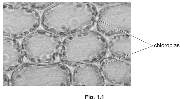
Fig. 1.1 is a photomicrograph showing chloroplasts in plant leaf cells.
Explain why the chloroplasts are seen only around the periphery (edge) of each plant cell.
Answer
The chloroplasts are pushed to the periphery (edge) of the cell by the large central vacuole, which is turgid / full of cell sap and occupies the centre of the cell.
The chloroplasts are displaced to the edge by the large, turgid central vacuole.
Background Concept
A mature plant cell typically has one large permanent vacuole bounded by a selectively permeable membrane called the tonoplast. The vacuole is filled with cell sap (a watery solution of salts, sugars and pigments) and, when fully hydrated, it is turgid and pushes the cytoplasm and all the organelles it contains – including the chloroplasts – outwards against the cell wall. This is why a leaf mesophyll cell viewed under the light microscope shows chloroplasts arranged in a ring around the edge, with an apparently empty region in the middle.
Understanding the Question
The question gives the candidate Fig. 1.1, a photomicrograph of leaf cells with chloroplasts clearly lining the inner edge of each cell wall, and asks them to explain this arrangement. The mark scheme explicitly ignores references to 'light absorption' (a tempting but not-credit-worthy point), so the answer must address the physical reason – the vacuole.
Approach
Look at the figure: large clear central region, chloroplasts around the rim. Connect this to the structure of a mature plant cell: the clear region IS the vacuole. The answer is therefore one short sentence about the vacuole pushing the chloroplasts outwards.
Step-by-Step Reasoning
- A mature plant cell has a large, permanent, central vacuole filled with cell sap.
- When the cell is turgid, the vacuole takes up most of the cell volume, occupying the centre.
- The cytoplasm (and the chloroplasts suspended in it) is squeezed into a thin layer between the vacuole and the cell wall – the periphery.
That single chain of reasoning is the mark. The mark scheme accepts any one of three equivalent statements:- chloroplasts at periphery because pushed by the vacuole;
- vacuole is turgid / full of cell sap;
- the (large permanent) vacuole is in the centre of the cell.
Key Takeaways
- A plant cell's apparent empty centre is the vacuole, not a hole.
- The vacuole physically displaces the cytoplasm and its organelles to the cell periphery.
- Chloroplast arrangement maximises light capture (peripheral cytoplasm is closest to the cell wall, where light enters first), but that functional argument is not credited here.
Common Mistakes
- Saying 'chloroplasts move to the edge to absorb more light' – the mark scheme rejects this.
- Saying the cell is 'empty in the middle' – it isn't; that is the vacuole.
- Confusing the vacuole with the nucleus – the nucleus is a much smaller structure embedded in the cytoplasm at the periphery.
Things to Be Careful About
Keep the answer to one clear point – the question is only worth one mark, and over-elaborating wastes time and risks introducing an uncredited (and rejected) idea such as light absorption.
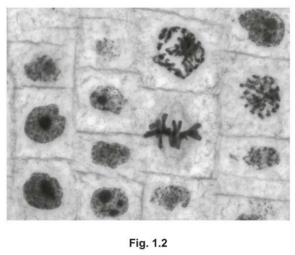
Fig. 1.2 shows plant cells in a root tip where cell division by mitosis is taking place.
Identify two cells in Fig. 1.2 that are in different stages of mitosis.
Draw a label line to each cell and add the name of the stage of mitosis that is shown by the cell.
Answer
Label any one cell whose chromosomes are aligned along the equator (middle) of the cell as metaphase, and label any one cell whose chromosomes appear as a loose mass of condensed chromatin that has not yet aligned as prophase.
Metaphase – label a cell with chromosomes lined up along the equator; Prophase – label a cell with condensed chromosomes that have not yet aligned.
Background Concept
Mitosis is conventionally divided into four stages defined by chromosome behaviour:
- Prophase: chromosomes condense and become visible as discrete threads; the nuclear envelope breaks down; no alignment has occurred yet.
- Metaphase: chromosomes line up along the equator (metaphase plate), each attached to spindle fibres from opposite poles.
- Anaphase: sister chromatids separate at the centromere and are pulled to opposite poles.
- Telophase: chromatids reach the poles, decondense, and a new nuclear envelope forms around each set; cytokinesis then divides the cytoplasm.
The root tip of a plant is a classic site of active mitosis, so a section through it shows many cells at different stages, often side by side.
Understanding the Question
The candidate is given Fig. 1.2, a photomicrograph of a plant root tip in which several cells show distinctive chromosome configurations. The task is to identify two cells that are in different stages of mitosis, draw a label line from each, and write the name of the stage. Two marks are available, one for each correctly named cell.
Approach
Scan the figure for the two stages that are easiest to recognise unambiguously:
- a cell with chromosomes lined up in a single plane across the middle of the cell (metaphase);
- a cell where the chromosomes are condensed into a dark, tangled mass but have not yet aligned (prophase).
Draw a straight label line from each chosen cell to a clear area of the page and write the stage name. Other stages (anaphase, telophase) are also acceptable, but metaphase and prophase are the most clearly visible in the supplied image.
Step-by-Step Reasoning
- In Fig. 1.2, the central cell whose chromosomes form a neat row across the middle is in metaphase.
- The cells (e.g. lower left or upper left) in which the chromosomes are condensed into a dark, scattered, irregular mass with no alignment are in prophase.
- A cell whose chromatids are visibly separating into two groups pulled towards opposite poles would be anaphase; a cell with two reforming nuclei at opposite ends would be telophase – the mark scheme credits any two correctly named stages, but the clearest pairs visible in the figure are prophase + metaphase.
Key Takeaways
- Stage identification is based on chromosome arrangement, not on cell size or shape.
- Prophase = condensed but unaligned; metaphase = aligned at the equator; anaphase = separating to poles; telophase = two reforming nuclei.
- A root tip is the standard specimen for studying mitosis because the meristematic cells there are dividing rapidly.
Common Mistakes
- Confusing prophase with telophase – in prophase there is one mass of condensing chromatin; in telophase there are TWO separate groups at opposite ends.
- Calling metaphase 'anaphase' because the chromosomes look like a line – alignment is the key, not separation.
- Labelling a cell that is in interphase (no visible chromosomes, only a nucleus) as a stage of mitosis – it is not.
Things to Be Careful About
The mark scheme requires the label to be attached to a cell that genuinely shows that stage; the line must end ON the cell, not on a neighbouring one. Do not label more than the requested two cells – extra incorrect labels can lose the marks.
The rest of this paper
5 more questions- Q2Transport in Mammals · Biological Molecules · Cell Membranes and Transport11M
- Q3Infectious Diseases · Biological Molecules · Nucleic Acids and Protein Synthesis11M
- Q4Transport in Plants9M
- Q5Nucleic Acids and Protein Synthesis · Infectious Diseases · Gas Exchange · Transport in Mammals · The Mitotic Cell Cycle · Immunity16M
- Q6Enzymes · Biological Molecules4M