Biology 9700/21 — October/November 2021
Cambridge AS Level · AS Level Structured Questions · worked solutions for every part, with the mark scheme
Topics Biological Molecules · Transport in Mammals · Transport in Plants · Cell Structure · Cell Membranes and Transport · Infectious Diseases · +3 more
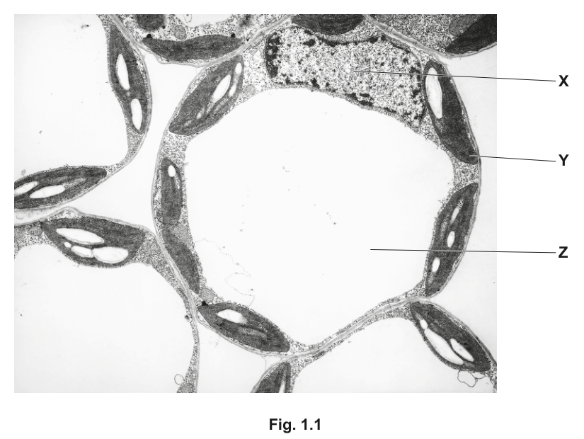
Fig. 1.1 is a transmission electron micrograph of cells from the leaf of a plant.
Name the cell structures X, Y, and Z.
X ______
Y ______
Z ______
Answer
X nucleus
Y chloroplast
Z vacuole
X = nucleus; Y = chloroplast; Z = vacuole.
Background Concept
A mature plant leaf mesophyll cell is a eukaryotic cell bounded by a cellulose cell wall, with a large central vacuole, peripheral chloroplasts and a nucleus. Under the transmission electron microscope these three structures have very characteristic appearances:
- The nucleus is a large, roughly spherical, often heterochromatin-darkened body enclosed by a double nuclear envelope with pores. It usually sits in the cytoplasm pressed against the cell wall by the vacuole.
- The chloroplast is a lens-shaped organelle about 5–10 µm long, with a double envelope and internal stacks of thylakoid membranes (grana) connected by stromal lamellae. Chloroplasts are pushed to the cell periphery by the vacuole.
- The vacuole is a single, large, fluid-filled compartment bounded by a membrane (the tonoplast). In a mature plant cell it occupies most of the cell volume and appears as a pale, empty-looking region in TEM preparations because its contents are washed out during fixation.
Understanding the Question
The question asks the candidate to read off three labels on a printed TEM of plant leaf cells. X points to a dense rounded body in the cytoplasm, Y points to an oval, internally-striped organelle at the cell edge, and Z points to the large pale central region of each cell.
Approach
Match each label to the structure that best fits both its position in the cell and its appearance in the micrograph: dense rounded body in the cytoplasm = nucleus; oval striped organelle at the periphery = chloroplast; large pale central region = vacuole.
Step-by-Step Reasoning
- X is shown as a dark, rounded structure sitting in the thin layer of cytoplasm between the vacuole and the cell wall. Its size, shape and central position are diagnostic of the nucleus.
- Y is one of the elongated, dark organelles pressed against the inner surface of the cell wall. The dark transverse bands are the grana (stacks of thylakoids), so Y must be a chloroplast.
- Z is the very large, electron-lucent region that fills most of the cell. This is the vacuole, which in a mature mesophyll cell occupies 80–90% of the cell volume.
Key Takeaways
On a TEM of a plant cell: a dense rounded body in the cytoplasm = nucleus; a striped oval organelle at the cell edge = chloroplast; the large pale central region = vacuole.
Common Mistakes
- Calling the chloroplast a "mitochondrion" — mitochondria are smaller, lack grana, and are not pushed to the cell periphery.
- Calling the vacuole a "hole" or "empty space" — it is a membrane-bound organelle (tonoplast).
- Confusing the nucleus with a nucleolus or starch grain — the nucleus is bounded by a nuclear envelope and contains chromatin, not a single dense inclusion.
Things to Be Careful About
Use the exact accepted terms: nucleus, chloroplast, vacuole. Spelling (especially "vacuole", not "vacuole/vacule") and the singular form are credited. Each correct name earns one mark.
State two ways in which the structure of an animal cell differs from plant cells such as those shown in Fig. 1.1.
Answer
- Animal cells do not have a (large permanent) vacuole (whereas the plant cells in Fig. 1.1 have a large central vacuole).
- Animal cells do not have chloroplasts (whereas the plant cells in Fig. 1.1 contain chloroplasts).
(Any two of: no cell wall; no plasmodesmata; nucleus more central; centrioles present; glycogen rather than starch stored; no amyloplasts.)
Animal cells have no chloroplasts and no (large permanent) vacuole.
Background Concept
Plant and animal cells are both eukaryotic and share the same basic machinery — nucleus, mitochondria, ER, ribosomes, Golgi, plasma membrane. They differ in a small set of features that reflect their different lifestyles: plants are autotrophic and sessile, so they make their own food (chloroplasts) and need structural support and water balance (cell wall, large central vacuole); animals are heterotrophic and motile, so they store carbohydrate as glycogen and use centrioles during cell division.
Diagnostic plant features (visible in Fig. 1.1):
- Cell wall of cellulose outside the plasma membrane.
- Chloroplasts at the cell periphery, with internal grana.
- Large central vacuole occupying most of the cell.
- Plasmodesmata linking adjacent cells through the walls.
- Starch grains inside chloroplasts/amyloplasts.
Diagnostic animal features (not in Fig. 1.1):
- Centrioles in the centrosome.
- Glycogen granules instead of starch.
- More central nucleus (because there is no large vacuole pushing it sideways).
- No cell wall, no chloroplasts, no large permanent vacuole.
Understanding the Question
The command word is "state", so brief, factual points are needed. The question asks for two differences, and the mark scheme offers a generous list of acceptable answers. The points must be structural differences, not functional ones.
Approach
Look at what Fig. 1.1 shows in the plant cells (cell wall, chloroplasts, large central vacuole, starch, peripheral nucleus) and for each decide whether an animal cell has the same or lacks it. Pick the two differences that the candidate can express most clearly.
Step-by-Step Reasoning
The clearest, most commonly credited pair:
- Chloroplasts: the plant cells in Fig. 1.1 contain chloroplasts (the striped organelles at the periphery). Animal cells never contain chloroplasts because they do not photosynthesise.
- Vacuole: the plant cells have a single, large, central vacuole that fills most of the cell. Animal cells have only small, temporary vesicles that would not be called a vacuole in this sense.
Other valid differences (any one of these could replace either of the two above):
- No cellulose cell wall in animal cells.
- Animal-cell nucleus lies more centrally; plant-cell nucleus is pushed to the periphery by the vacuole.
- Animal cells store carbohydrate as glycogen, not starch.
- Animal cells contain centrioles (plant cells generally do not).
- Plasmodesmata are absent in animal cells.
Key Takeaways
The signature differences between animal and plant cells: cell wall, chloroplast, large central vacuole, plasmodesmata, starch are plant-only; centrioles and glycogen granules are animal-only. The position of the nucleus also differs as a consequence of the large vacuole.
Common Mistakes
- Stating features that both cell types have ("animal cells have a nucleus") — this is not a difference.
- Functional statements instead of structural ones ("animals move") — credit requires a structural difference.
- Vague answers such as "different shapes" or "animals don't have green colour" — too imprecise to score.
- Writing "plant cells have more organelles" — the question asks what animal cells lack, not quantity.
Things to Be Careful About
The mark scheme phrases each answer as a positive statement about what plant cells have that animal cells do not (or vice versa). State the plant feature first, then make clear the animal cell lacks it, e.g. "(plant cells have) chloroplasts; animal cells do not." Any two of the listed differences are accepted.
Cell structure Y in Fig. 1.1 contains a large starch granule (grain).
Name the chemical reagent used to test for starch and state the colour change that will be seen if starch is present.
reagent ______
colour change ______
Answer
reagent: iodine solution (in potassium iodide)
colour change: from orange/brown to blue-black
Iodine solution; (orange/brown) to blue-black.
Background Concept
Starch is a mixture of two -glucose polymers, amylose (mostly linear/helical) and amylopectin (branched). The helical coils of amylose form a hydrophobic channel into which polyiodide ions ( / ) from an iodine solution slot. The charge-transfer complex between the trapped polyiodide and the amylose helix absorbs visible light strongly, producing the characteristic deep blue-black colour. Iodine solution alone (iodine dissolved in aqueous potassium iodide, giving ) is yellow-brown, so a colour change is only seen if starch is present.
Understanding the Question
The question has two parts worth one mark each: name the reagent, and state the colour change. "State" means a brief factual answer with no extra reasoning required. The mark scheme credits the starting colour as well as the final colour.
Approach
Recall the standard test for starch:
- Reagent: iodine in potassium iodide solution, normally referred to as "iodine solution".
- Positive result: orange/brown blue-black.
- Negative result: stays orange/brown.
Step-by-Step Reasoning
- Reagent (1 mark): iodine solution (or "iodine in potassium iodide solution"). "Iodine" on its own is the element and is not credited.
- Colour change (1 mark): state both the starting colour and the positive colour, e.g. "(from) orange/brown to blue-black". Saying only "goes black" misses the starting colour, although many mark schemes still award the mark because the question asks for the colour change observed; the safest answer includes both colours.
Key Takeaways
Starch test = iodine solution; positive result = blue-black. The colour is due to a polyiodide–amylose inclusion complex, not a chemical reaction that alters the iodine.
Common Mistakes
- Confusing with the Benedict's test for reducing sugars (reagent: Benedict's; positive: brick-red precipitate on heating).
- Confusing with the biuret test for proteins (reagent: biuret/NaOH + CuSO; positive: purple/lilac).
- Writing "iodine" rather than "iodine solution".
- Quoting only the final colour, not the change.
Things to Be Careful About
The reagent is correctly written as "iodine solution" or "iodine in potassium iodide"; "iodine" alone is rejected. The colour change is orange/brown blue-black (or black).
Starch granules contain amylose and amylopectin.
Describe the similarities and differences between the structure of amylose and the structure of amylopectin.
Answer
Similarities:
- both are polymers / polysaccharides;
- both are composed of -glucose monomers;
- both contain 1,4-glycosidic bonds / linkages between the glucose monomers in their chains.
Differences:
- amylose is unbranched (a single chain), whereas amylopectin is branched;
- amylopectin has 1,6-glycosidic bonds at the branch points (amylose has none);
- amylose has a helical structure, whereas amylopectin does not;
- amylose is a smaller molecule (fewer glucose residues) than amylopectin.
Both are α-glucose polysaccharides with 1,4-glycosidic bonds; amylose is unbranched and helical, amylopectin is branched with 1,6-glycosidic bonds at branch points.
Background Concept
Starch is the main storage polysaccharide of plants and is a mixture of two polymers of -glucose:
- Amylose (~20–30% of starch) is essentially a single, unbranched chain of -glucose units linked by 1,4-glycosidic bonds. Because every glucose is in the configuration, the chain curls into a left-handed helix. This helix is what traps the polyiodide ions in the iodine test, giving the blue-black colour. A typical amylose molecule contains a few hundred to a few thousand glucose units.
- Amylopectin (~70–80% of starch) has the same 1,4-linked -glucose backbone but is heavily branched: roughly every 24–30 glucose units along the chain, a short side chain is attached via a 1,6-glycosidic bond. The many short branches make amylopectin more compact than amylose and give it a much larger overall molecular size (tens of thousands of glucose units).
The 1,4- and 1,6-glycosidic bonds are formed by condensation reactions, releasing water. The numbering refers to the carbon atoms of the glucose ring that the oxygen bridge connects (C1 of one glucose to C4 of the next in the main chain, C1 of the branch-point glucose to C6 of a glucose in the main chain).
Understanding the Question
The command word is "describe", which here means "set out the relevant features" — both what the two molecules have in common and how they differ. The mark scheme allows up to 3 marks for similarities and 3 for differences, with 4 marks available in total. A clear, well-organised answer should give at least two similarities and at least two differences.
Approach
- State the shared features (composition, type of bond, polymer class).
- State the contrasting features (branching, additional bond type, shape, size).
Step-by-Step Reasoning
Similarities (3 marks available):
- Both are polymers / polysaccharides of glucose.
- The monomer in both is -glucose (not -glucose — that distinction is what allows the chain to coil).
- Both are joined by glycosidic bonds, and in particular the main chain in both is held together by 1,4-glycosidic bonds (C1 of one glucose to C4 of the next).
Differences (3 marks available, 1 needed):
- Branching: amylose is unbranched; amylopectin is branched.
- Bond type at branch points: amylopectin contains 1,6-glycosidic bonds (at the branch points); amylose does not contain 1,6 bonds.
- Shape: amylose is helical; amylopectin is not (it is more compact/globular).
- Size: amylose is a smaller molecule; amylopectin is much larger.
Key Takeaways
- Both amylose and amylopectin are polymers of -glucose with 1,4-glycosidic bonds.
- The defining difference is branching (amylose unbranched, amylopectin branched) and the resulting 1,6-glycosidic bonds at branch points in amylopectin.
- The configuration and the lack of branches allow amylose to coil into a helix; the many short branches of amylopectin prevent a regular helix.
Common Mistakes
- Stating "amylose is branched, amylopectin is unbranched" — the opposite of the truth.
- Saying the monomer is "-glucose" or just "glucose" — only -glucose is correct.
- Calling the bonds "peptide bonds" or "hydrogen bonds" — the linkages are glycosidic.
- Failing to specify the bond numbers (1,4 and 1,6) — the type of bond is the precise marking point.
- Forgetting to describe both similarities and differences.
Things to Be Careful About
The mark scheme distinguishes "glycosidic" from "glucosidic" (the latter is accepted as alternative wording in the syllabus) and specifically requires the 1,4 / 1,6 numbering. State explicitly — the same helical/coiling properties that allow starch to store compactly and to give the iodine test depend on the configuration.
The rest of this paper
5 more questions- Q2Transport in Mammals · Cell Membranes and Transport · Transport in Plants12M
- Q3Infectious Diseases · Biological Molecules · Immunity13M
- Q4The Mitotic Cell Cycle · Nucleic Acids and Protein Synthesis10M
- Q5Transport in Plants9M
- Q6Transport in Mammals5M