Biology 9700/23 — October/November 2019
Cambridge AS Level · AS Level Structured Questions · worked solutions for every part, with the mark scheme
Topics Transport in Mammals · Nucleic Acids and Protein Synthesis · Cell Structure · Biological Molecules · The Mitotic Cell Cycle · Transport in Plants · +3 more
Fig. 1.1 is a diagram of a molecule of haemoglobin.
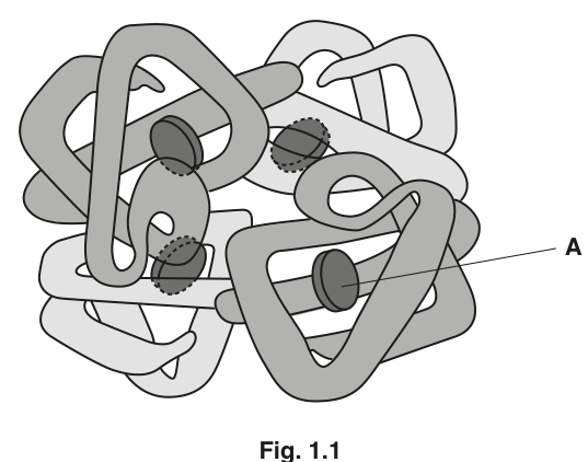
Name the structure labelled A on Fig. 1.1.
Answer
Haem (group).
Haem (group).
Background Concept
Haemoglobin is a conjugated globular protein. Each of its four polypeptide chains (two α and two β) carries a non-protein component — a flat, disc-shaped haem group. A non-protein component that is tightly and permanently associated with a protein and is required for the protein's biological activity is called a prosthetic group. The haem group is built around a porphyrin ring with a central iron ion (Fe²⁺).
Understanding the Question
The figure shows the quaternary structure of haemoglobin, with the four folded chains and a labelled disc inside one chain. The command word is "name", so a single term is required.
Approach
Recognise that the labelled disc inside a subunit is the haem prosthetic group, and recall the term accepted by the mark scheme.
Step-by-Step Reasoning
The label A points to the flat disc-shaped structure inside one of the four subunits. This is the haem group — the porphyrin ring with a central Fe²⁺ ion. The mark scheme accepts "haem", "heme", or "prosthetic group", but does NOT accept "iron", "iron ion", "Fe" or "porphyrin ring" on its own, because those describe only part of the structure or only the metal at its centre.
Key Takeaways
- Haem = the prosthetic group of haemoglobin (and of myoglobin and cytochromes).
- It is a porphyrin ring with a central Fe²⁺ ion; the iron is the part that actually binds O₂.
- "Prosthetic group" is a wider term and is an acceptable alternative.
Common Mistakes
- Writing "iron" or "Fe²⁺" — this is the central ion of the haem group, not the group itself; the mark scheme explicitly ignores it.
- Writing "porphyrin ring" — this is the organic ring, again only part of the structure; ignored by the mark scheme.
- Writing "heme" (American spelling) — accepted, but stick with "haem" in CIE papers.
Things to Be Careful About
The CIE mark scheme is strict: write "haem" (or "prosthetic group") and nothing more, otherwise the mark is lost.
State the function of structure A.
Answer
To bind oxygen in the lungs and release oxygen in respiring tissues (where pO2 is low).
To bind oxygen in the lungs and release oxygen in respiring tissues.
Background Concept
The haem group is the functional heart of haemoglobin. The Fe²⁺ ion at its centre binds O₂ reversibly. In the lungs, where the partial pressure of oxygen (pO2) is high, each of the four haem groups combines with an O₂ molecule to form oxyhaemoglobin. In actively respiring tissues, where pO2 is low, the reverse reaction occurs and O₂ is released for the cells to use. The four haem groups give haemoglobin its cooperative binding — the binding of one O₂ makes the next easier, and release of one makes the next easier to release.
Understanding the Question
The command word is "state the function", so a concise statement is required. The mark scheme accepts any of three equivalent phrasings; the cleanest candidate response covers binding in the lungs and release in the tissues.
Approach
Recall the role of the haem group: reversible combination with oxygen, driven by differences in pO2 between the lungs and respiring tissues.
Step-by-Step Reasoning
The haem group is the oxygen-binding site of haemoglobin. A good answer makes both directions of transport explicit: O₂ is taken up where pO2 is high (the lungs) and released where pO2 is low (respiring tissues). Either direction alone is enough to score a mark; both together fully justifies the function.
Key Takeaways
- The haem group reversibly binds O₂.
- Uptake in the lungs and release in respiring tissues are two sides of the same reversible reaction.
- The four haems in one haemoglobin molecule allow four O₂ molecules to be carried.
Common Mistakes
- Writing "forms bonds" — this is too vague and is rejected by the mark scheme.
- Writing "carries oxygen" without saying where it is picked up or released.
- Confusing the haem group with the globin chains — it is the iron/porphyrin complex that actually binds O₂.
Things to Be Careful About
Avoid saying that haemoglobin itself binds oxygen when the question asks specifically about the haem group; the polypeptide chains do not bind O₂ directly — they hold the haem groups in the right orientation.
Haemoglobin is described as a globular protein. Explain why this protein is described as globular.
Answer
Any two from:
- It has a (roughly) spherical / ball-like shape.
- It is (water-)soluble because hydrophilic R-groups are on the outside of the molecule and hydrophobic R-groups are on the inside.
- It has a dynamic role in metabolism / it functions in a metabolic process.
Globular because it is spherical and water-soluble, with hydrophilic R-groups on the outside and hydrophobic R-groups on the inside.
Background Concept
Proteins are classified structurally as either fibrous or globular. Fibrous proteins (e.g. collagen, keratin) are long, parallel polypeptide chains that form rope-like or sheet-like structures; they are usually insoluble. Globular proteins (e.g. haemoglobin, enzymes, antibodies) fold into compact, roughly spherical shapes in which hydrophobic R-groups are buried in the interior and hydrophilic R-groups project into the surrounding aqueous cytosol or plasma, making the molecule water-soluble. The soluble, compact shape suits a dynamic role: the protein must move, bind, and release small molecules quickly.
Understanding the Question
The command word is "explain why", so points must justify the description, not just restate it. The mark scheme offers up to four creditworthy points, of which any two are required.
Approach
For each marking point, identify a property of globular proteins that is true of haemoglobin. The strongest pair of points is shape AND solubility/R-group arrangement, because together they explain the very term "globular" and the functional consequence of solubility.
Step-by-Step Reasoning
- Shape — globular proteins are spherical or ball-like. The mark scheme rejects "circular" / "round" / "3D"; "spherical" or "ball-like" are required.
- Solubility — globular proteins are water-soluble because they form hydrogen bonds with surrounding water. "Soluble" without further qualification is acceptable.
- R-group arrangement — this is the structural reason behind the solubility: hydrophilic R-groups on the outside interact with water; hydrophobic R-groups on the inside are away from it. Note that the mark scheme rejects any reference to "tails" (an amino acid does not have a tail).
- Function — globular proteins typically have dynamic, metabolic, or physiological roles (e.g. transport, catalysis, signalling), unlike fibrous proteins which are mainly structural.
Key Takeaways
- Globular = compact, spherical, water-soluble, dynamic in function.
- The inside/outside segregation of R-groups (hydrophobic in, hydrophilic out) is what makes the protein soluble.
- Haemoglobin is a classic globular transport protein.
Common Mistakes
- Writing "circular" or "round" — these are rejected; "spherical" or "ball-like" is required.
- Saying the R-group "tails" are hydrophobic — the mark scheme rejects any use of "tail" for an R-group.
- Confusing globular with fibrous — a fibrous protein (e.g. collagen) is long, parallel, and insoluble.
Things to Be Careful About
Two marks are available, and the safest two points are (a) the spherical/ball-like shape and (b) water solubility with hydrophilic R-groups on the outside and hydrophobic R-groups on the inside. Adding the dynamic/metabolic function point is optional but useful when marks are tight.
The gene codes for the -globin polypeptide.
State why a polypeptide, such as -globin, is described as a polymer.
Answer
A polypeptide is a polymer because it is a long-chain macromolecule made of many amino acid monomers joined together by peptide bonds.
A polypeptide is a polymer: many amino acid monomers joined by peptide bonds form a long-chain macromolecule.
Background Concept
A polymer is a large molecule (macromolecule) made of many repeating small units called monomers, joined together by covalent bonds. In proteins the monomers are amino acids, and the bond between them is the peptide bond (formed by a condensation reaction between the carboxyl group of one amino acid and the amino group of the next). A long chain of amino acids is a polypeptide; a protein is one or more polypeptide chains folded into a specific 3D shape.
Understanding the Question
The question explicitly links β-globin to the gene that codes for it, but the command word "state why" targets the definition of "polymer", so the answer must say what monomers the polypeptide is made from and what holds them together.
Approach
Two marking points are available: name the monomer (amino acid) AND give the linker (peptide bond) OR describe the size ("macromolecule" / "long-chain") and the repetition ("many (sub-)units").
Step-by-Step Reasoning
- Monomer — many amino acids. The mark scheme demands the phrase "amino acids" — "subunits" alone is not credited.
- Linker — peptide bonds join amino acids to one another. "Dipeptide bond" is rejected by the mark scheme, and "peptide bond between two amino acids" is also rejected on its own (it describes a dipeptide, not a polymer). The correct phrasing is "peptide bonds" (plural) between amino acids.
- The mark scheme offers an alternative route: call the polypeptide a "macromolecule" or "long-chain (molecule)" or describe it as "repeated/many (sub-)units/monomers". Any of these, combined with the amino-acid point, is sufficient.
Key Takeaways
- Polymer = many monomers joined by covalent bonds.
- Polypeptide = many amino acids joined by peptide bonds.
- "Macromolecule" and "long-chain" are alternative ways to express the same idea.
Common Mistakes
- Writing "dipeptide bond" — the mark scheme rejects this; the correct term is "peptide bond".
- Writing "subunits" without ever specifying amino acids — this is too vague and is not credited.
- Confusing the peptide bond with the glycosidic or phosphodiester bond — they look similar but join different monomers.
Things to Be Careful About
The cleanest, fullest answer is: a polypeptide is a polymer because it is a long-chain macromolecule made of many amino acid monomers joined by peptide bonds. This covers both marking points at once.
A single base change in the DNA of the gene results in a change to the amino acid sequence of -globin. In the sequence, a single glutamic acid is replaced by valine.
Outline the effects of this change in the amino acid sequence of -globin on the structure and function of a haemoglobin molecule.
Answer
Any three from:
- The R-group of glutamic acid is polar / hydrophilic, while the R-group of valine is non-polar / hydrophobic.
- This changes the tertiary structure of the β-globin chain (its folding becomes less globular).
- This changes the quaternary structure of haemoglobin, so the haemoglobin molecules become 'sticky' and associate to form fibres.
- The haemoglobin is less (water-)soluble.
- The haemoglobin is less efficient at binding / transporting oxygen (lower affinity for O₂).
Polar glutamic acid → non-polar valine alters tertiary and quaternary structure; haemoglobin becomes 'sticky', forms fibres, becomes less soluble and less efficient at binding oxygen.
Background Concept
This is the molecular basis of sickle-cell anaemia. The HBB gene codes for the β-globin polypeptide. A single base substitution (GAG → GTG) changes one codon: glutamic acid (glu) is replaced by valine (val) at position 6 of the β chain. Glutamic acid has a polar, hydrophilic R-group; valine has a non-polar, hydrophobic R-group. R-group character determines the interactions (hydrogen bonds, ionic bonds, hydrophobic interactions) that hold a protein in its 3D shape. Changing one R-group can therefore ripple through every level of structure above the primary.
Understanding the Question
The question gives a specific substitution (glu → val) and asks for its effects on haemoglobin structure AND function. Three marks are available; the candidate must show the chain of consequences from primary structure (the substitution itself) to tertiary and quaternary structure (the new R-group changes the folding), then to function (fibre formation, lower solubility, lower O₂ affinity).
Approach
Walk up the hierarchy of protein structure: primary → tertiary → quaternary, and at each level say what changes and why. Then state the functional consequences for haemoglobin as a whole.
Step-by-Step Reasoning
- R-group character — the mark scheme demands BOTH halves of the contrast: glu is polar/hydrophilic AND val is non-polar/hydrophobic. This is the root cause of everything that follows.
- Tertiary structure — the new R-group cannot form the same interactions as the original; the β-globin chain folds differently. The mark scheme allows "less globular" as an alternative way to express this.
- Quaternary structure — the change in β-globin alters how the four chains fit together; the haemoglobin molecules now expose hydrophobic patches that allow them to stick to one another.
- Fibre formation — the "sticky" haemoglobin molecules polymerise into long fibres inside red blood cells, distorting them into the rigid sickle shape.
- Lower solubility — hydrophobic interactions drive the aggregation; the molecules become less soluble in the cytoplasm.
- Lower O₂ affinity / capacity — the altered quaternary structure is less efficient at binding and releasing O₂ (in some descriptions the Bohr effect is shifted too).
- AVP — the mark scheme leaves room for a valid extra point, e.g. reduced red-cell flexibility leading to capillary blockage.
Key Takeaways
- A single amino-acid substitution is a primary-structure change, but its consequences propagate to higher structure levels because R-group character governs folding.
- The sickle-cell mutation is the textbook example of how a tiny genetic change can have dramatic phenotypic effects.
- A complete answer must connect structure (R-group, tertiary, quaternary, fibres) to function (lower solubility, lower O₂ binding).
Common Mistakes
- Stating only that "the shape changes" without naming the R-group polarity contrast — the mark scheme demands both halves.
- Saying "haemoglobin becomes insoluble" — the mark scheme ignores "insoluble" and wants "less soluble".
- Saying "haemoglobin cannot bind oxygen" — the mark scheme ignores this; the correct phrasing is "less efficient at binding/transporting oxygen" or "lower affinity for oxygen".
- Confusing tertiary with quaternary structure — the β-globin chain itself has a tertiary structure; how the four chains fit together is the quaternary structure of haemoglobin.
Things to Be Careful About
The most common reason candidates lose marks is stopping at "the shape changes". The marker needs to see the R-group contrast explicitly, and ideally to see both the tertiary and quaternary effects and at least one functional consequence. Three well-chosen marks: (R-group polarity contrast) + (change in tertiary structure) + (fibre formation / lower O₂ affinity) cover the biology cleanly.
Haemoglobin interacts with carbon dioxide and carbon monoxide.
Outline the role of haemoglobin in the transport of carbon dioxide.
Answer
Any three from:
- CO2 combines with haemoglobin to form carbaminohaemoglobin.
- CO2 reacts with the terminal amine groups (–NH2) of the polypeptide chains.
- Each polypeptide chain can carry one molecule of CO2, so one haemoglobin molecule can carry four CO2 molecules.
- CO2 remains bound to haemoglobin until it reaches a region of low pCO2 (or high pO2), e.g. the lungs / alveoli, where it is released.
- Hydrogen ions (from the dissociation of carbonic acid in red blood cells) bind to haemoglobin, forming haemoglobinic acid (HHb); this helps buffer the blood.
Haemoglobin carries CO2 as carbaminohaemoglobin (CO2 binds to terminal –NH2 groups); each chain carries one CO2, so four per Hb; the binding is reversed in the lungs; H+ ions are also buffered by haemoglobin.
Background Concept
Carbon dioxide is transported from respiring tissues to the lungs in three ways:
- Dissolved in the plasma (~5%).
- As hydrogencarbonate ions (HCO3⁻) in the plasma (~85%) — formed inside red blood cells when CO2 reacts with water (catalysed by carbonic anhydrase) to give H2CO3, which then dissociates into H+ and HCO3⁻. HCO3⁻ leaves the cell in exchange for Cl⁻ (the chloride shift).
- Bound to haemoglobin as carbaminohaemoglobin (~10%) — the question is about this fraction.
Carbaminohaemoglobin forms when CO2 reacts with the terminal –NH2 groups of the polypeptide chains. The reaction is reversible: in the lungs, where pO2 is high and pCO2 is low, the CO2 dissociates and is exhaled. The binding of CO2 to haemoglobin is favoured when haemoglobin is deoxygenated (the Haldane effect); release is favoured when haemoglobin is oxygenated (the Bohr effect).
The H+ ions generated in the red cell are mopped up by haemoglobin, which acts as a buffer: they bind to histidine residues and to the imidazole groups of the globin chains, forming haemoglobinic acid (HHb). This prevents the blood pH from falling too low, and the same effect is reversed in the lungs (oxygenation of haemoglobin releases H+, which combines with HCO3⁻ to reform H2CO3 and then CO2 and water).
Understanding the Question
The stem says haemoglobin interacts with both CO2 and CO. The question asks specifically about the role of haemoglobin in CO2 transport. "Outline" is the command word, so the answer must give a connected series of points rather than a single fact.
Approach
Build a logical sequence: how the CO2 binds to haemoglobin (amine groups, forming carbaminohaemoglobin), how many can be carried, where it stays bound, and what happens at the lungs. The H+/buffer point is an additional creditworthy idea.
Step-by-Step Reasoning
- Binding site — CO2 combines with the terminal amine groups (–NH2) of the globin chains. The mark scheme accepts –NH or –NH2.
- Product — the product is carbaminohaemoglobin. Note: "carboxyhaemoglobin" and "carbonylhaemoglobin" are explicitly rejected by the mark scheme; these are wrong names (carboxyhaemoglobin is in fact the complex with CO, the toxic gas — not CO2).
- Capacity — each of the four polypeptide chains can carry one CO2, so one haemoglobin molecule can carry four CO2 molecules in addition to four O2.
- Reversibility / location of release — the CO2 remains bound until the haemoglobin reaches a region of low pCO2 (or high pO2), i.e. the lungs, where it dissociates and is exhaled.
- Buffering — H+ ions (from the dissociation of carbonic acid inside the red cell) bind to haemoglobin, forming haemoglobinic acid (HHb). This is a separate but linked role of haemoglobin in CO2 transport; it is not CO2 carriage itself, but it is essential to the overall process.
- AVP — the mark scheme also credits valid points such as the transport of hydrogencarbonate ions in the plasma as a consequence of the chloride shift, or the Bohr/Haldane effect.
Key Takeaways
- About 10% of CO2 in the blood is carried as carbaminohaemoglobin.
- CO2 binds to the –NH2 groups of the globin chains (not to the haem group, which carries O2).
- Deoxygenated haemoglobin binds CO2 better than oxygenated haemoglobin (Haldane effect); this is the link between O2 release and CO2 uptake in respiring tissues, and the reverse in the lungs.
- Haemoglobin also buffers H+ ions produced from the dissociation of carbonic acid.
Common Mistakes
- Writing "carboxyhaemoglobin" — the mark scheme rejects this; the correct term is "carbaminohaemoglobin". "Carboxyhaemoglobin" actually refers to the CO (carbon monoxide) complex, which is the wrong gas.
- Saying the CO2 binds to the haem group — it binds to the protein, not the haem.
- Saying haemoglobin "carries CO2 in the plasma" — the carrier of CO2 in the plasma is hydrogencarbonate (HCO3⁻); haemoglobin-bound CO2 travels inside red blood cells.
- Stating the H+ point without naming the product (HHb) or with no reference to the source of the H+ (carbonic acid dissociation).
Things to Be Careful About
The cleanest three-point answer is: (CO2 binds to terminal –NH2 groups) + (forms carbaminohaemoglobin) + (released in lungs at low pCO2). Adding the H+ buffering point as a fourth idea makes the answer feel more rounded and would also be accepted.
The rest of this paper
5 more questions- Q2The Mitotic Cell Cycle6M
- Q3Transport in Plants · Nucleic Acids and Protein Synthesis8M
- Q4Transport in Mammals14M
- Q5Cell Structure · Enzymes · Immunity14M
- Q6Cell Structure · Cell Membranes and Transport6M