Biology 9700/12 — February/March 2019
Cambridge AS Level · Multiple Choice (AS Level) · answer key with instant marking and worked solutions
Topics Biological Molecules · Cell Structure · Transport in Plants · Transport in Mammals · Cell Membranes and Transport · The Mitotic Cell Cycle · +5 more
Tap an option under each question to check it — your score builds as you go.
The diagram below was drawn from an electron micrograph of an animal cell.
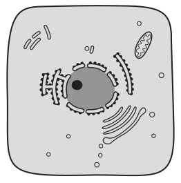
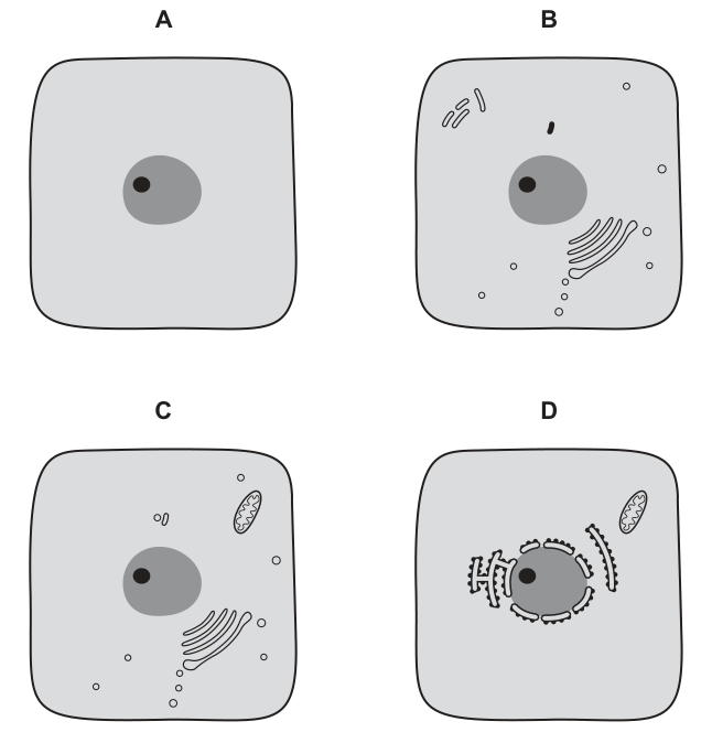
Which diagram would represent the same cell seen under a simple light microscope, using daylight as the only light source?
Options
Working
A simple light microscope using daylight has a resolution limit of about , so only the largest cellular structures can be distinguished: the cell (surface) membrane, the nucleus, and the nucleolus. Organelles such as mitochondria, rough endoplasmic reticulum, ribosomes and the Golgi apparatus are all below this resolution limit and are only resolvable with an electron microscope.
Looking at the options:
- A shows only the cell membrane, nucleus and nucleolus — matches what is resolvable with a light microscope.
- B, C and D each show additional organelles (mitochondria, Golgi apparatus, rough ER) that a light microscope cannot resolve.
Answer
A
A
Background Concept
A microscope's usefulness is governed by two distinct properties: magnification (how many times larger the image is than the object) and resolution (the minimum distance between two points at which they can still be seen as separate). Increasing magnification without improving resolution simply produces a larger but blurrier image — empty magnification.
- A simple light microscope using daylight has a resolution of roughly (0.2 µm). This is sufficient to distinguish the cell (surface) membrane, the nucleus and (with staining) the nucleolus, but not small cytoplasmic organelles.
- An electron microscope uses a beam of electrons (very short wavelength) and can resolve structures down to about , so it reveals the full ultrastructure: mitochondria, rough endoplasmic reticulum with ribosomes, Golgi apparatus, vesicles, and so on.
A useful size reference: mitochondria are typically – wide, ribosomes are , and ER cisternae are similarly sub-resolution for standard light microscopy.
Understanding the Question
The electron micrograph (Fig. 1.1) shows a typical animal cell packed with organelles: nucleus with nucleolus, mitochondria, rough ER studded with ribosomes, Golgi apparatus, and various vesicles. The question asks which of the four options (A–D) is the same cell as it would appear under a simple light microscope with daylight illumination — i.e. with no staining tricks, no phase contrast, no oil immersion, just a basic school-style light microscope.
The command word is implicit ("which diagram would represent…") — this is a multiple-choice selection, so the answer is the single option that shows only what a light microscope can resolve.
Approach
- Identify the resolution limit of a simple light microscope.
- List the structures in the EM image and decide which are above and which are below that limit.
- Pick the option that contains only the resolvable structures and excludes all sub-light-microscope organelles.
Step-by-Step Reasoning
- The cell (surface) membrane is well above the resolution limit → visible.
- The nucleus (a few µm across) is well above the resolution limit → visible.
- The nucleolus (–) is large enough to be seen, often as a darker spot inside the nucleus → visible.
- Mitochondria (–) sit around the resolution limit. With careful staining (e.g. Janus Green) they can be seen, but in a plain, unstained, daylight, simple light microscope they are not reliably resolvable → not visible in this scenario.
- Ribosomes (), rough ER cisternae, Golgi cisternae and small vesicles are all well below the light-microscope resolution limit → not visible.
Now check the four options:
- A — cell membrane + nucleus + nucleolus only. ✔ Matches a light-microscope view.
- B — adds a small structure (mitochondrion) and Golgi-like stacks. ✗ These are not resolvable here.
- C — adds a mitochondrion and a Golgi apparatus. ✗ Not resolvable.
- D — adds mitochondria and rough ER with ribosomes. ✗ Not resolvable.
Therefore A is the only option that represents what the cell would actually look like under a simple light microscope using daylight.
Key Takeaways
- Resolution, not magnification, determines what is visible in a microscope.
- Light microscopy reveals only the largest organelles (cell membrane, nucleus, nucleolus) in unstained, daylight conditions.
- Mitochondria, ER, Golgi, ribosomes and vesicles require an electron microscope (or specific stains / fluorescence) to be seen.
Common Mistakes
- Choosing an option simply because it "looks like" the EM image (B, C or D contain familiar organelles) — confusing detail with what the lower-resolution instrument can actually show.
- Assuming mitochondria are always visible by light microscopy. In practice they require a special stain (e.g. Janus Green); in a plain, daylight, simple light microscope they are not resolvable.
- Confusing magnification with resolution: high magnification alone does not let a light microscope reveal ultrastructure.
Things to Be Careful About
- The question specifies a simple light microscope with daylight — no staining, no phase contrast, no fluorescence, no oil immersion. This is the lowest-power, lowest-resolution realistic microscopy setup, so the visible structures are reduced to the absolute minimum.
- A nucleolus can be seen as a dense body inside the nucleus in some light-microscope preparations, so it is acceptable in option A and not a reason to reject it.
- The rough ER in option D is decorated with ribosomes, both of which are sub-light-microscope resolution — strong evidence that D is an EM-style image, not a light-microscope one.
The rest of this paper
39 more questions- Q2Cell Structure1M
- Q3Cell Structure1M
- Q4Cell Structure1M
- Q5Cell Structure1M
- Q6Cell Structure1M
- Q7Biological Molecules1M
- Q8Biological Molecules1M
- Q9Biological Molecules1M
- Q10Biological Molecules1M
- Q11Biological Molecules1M
- Q12Biological Molecules1M
- Q13Cell Membranes and Transport1M
- Q14Biological Molecules1M
- Q15Enzymes1M
- Q16Enzymes1M
- Q17Cell Membranes and Transport1M
- Q18Cell Membranes and Transport1M
- Q19The Mitotic Cell Cycle1M
- Q20The Mitotic Cell Cycle1M
- Q21Nucleic Acids and Protein Synthesis1M
- Q22Nucleic Acids and Protein Synthesis1M
- Q23Transport in Plants1M
- Q24Transport in Plants1M
- Q25Transport in Plants1M
- Q26Transport in Plants1M
- Q27Transport in Plants1M
- Q28Transport in Mammals1M
- Q29Transport in Mammals1M
- Q30Transport in Mammals1M
- Q31Transport in Mammals1M
- Q32Transport in Mammals1M
- Q33Gas Exchange1M
- Q34Gas Exchange1M
- Q35Infectious Diseases1M
- Q36Infectious Diseases1M
- Q37Infectious Diseases1M
- Q38The Mitotic Cell Cycle · Immunity1M
- Q39Immunity1M
- Q40Immunity1M