Biology 9700/23 — May/June 2018
Cambridge AS Level · AS Level Structured Questions · worked solutions for every part, with the mark scheme
Topics Cell Structure · Biological Molecules · Transport in Mammals · Nucleic Acids and Protein Synthesis · Infectious Diseases · The Mitotic Cell Cycle · +5 more
Fig. 1.1 is a photomicrograph of root tip meristem. Different stages of the cell cycle are visible.
Some cells are in the same stage of the cell cycle and some are in the same stage of mitosis.
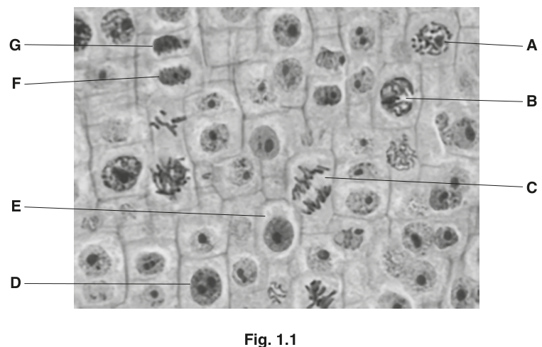
Cell D and cell E are in the same stage of the cell cycle.
State the difference between the nucleus of cell D and cell E.
Answer
Cell D has two nucleoli, while cell E has one nucleolus.
Cell D has two nucleoli; cell E has one nucleolus.
Background Concept
During interphase the cell is not dividing but is preparing for mitosis. The DNA is dispersed as chromatin inside a nucleus bounded by a nuclear envelope, and within that nucleus one or more nucleoli are visible. A nucleolus is the dense, rounded body inside the nucleus where ribosomal RNA is transcribed and ribosomal subunits are assembled. The number of nucleoli visible in an interphase nucleus can vary between species, between tissues and even between cells of the same tissue at different points in interphase, because nucleoli fuse and separate as the cell moves through G1, S and G2. Despite this variability, the number of nucleoli is one of the easiest features to use when comparing two apparently similar interphase nuclei in a single field of view.
Understanding the Question
Cells D and E in Fig. 1.1 both look like 'resting' interphase cells — each has a clear, rounded nucleus with no condensed chromosomes. The question asks for a single observable difference between their nuclei. Looking carefully at the photomicrograph, the nucleus of cell D contains two small dark dots (nucleoli), while the nucleus of cell E contains only one dark dot. This is the difference the mark scheme requires.
Approach
The strategy is to compare the two nuclei feature by feature — size, shape, staining intensity and especially internal structures. The key internal structure to look for is the nucleolus (a dense, rounded, darkly stained body inside the nucleus). Counting nucleoli in each cell gives the answer.
Step-by-Step Reasoning
- Identify the cells in interphase. Cells D and E both have intact nuclear envelopes, no visible chromosomes and a granular chromatin appearance — characteristic of interphase.
- Look inside each nucleus for nucleoli, which appear as small, dense, darkly stained round bodies.
- In cell D, two such bodies are visible; in cell E, only one is visible.
- State the difference precisely: cell D has two nucleoli, cell E has one nucleolus.
Key Takeaways
- Interphase nuclei can be distinguished by the number, size and position of their nucleoli.
- A single observable difference (here, nucleolus number) is enough to score the mark — no need to over-describe.
- Always check inside the nucleus, not just its outline, when comparing interphase cells.
Common Mistakes
- Stating that one nucleus is 'bigger' or 'darker' — this is too vague and is not credited.
- Confusing the nucleolus with the whole nucleus, or with chromatin patches.
- Describing differences in the cytoplasm outside the nucleus — the question specifies the nucleus.
Things to Be Careful About
- Use the exact term 'nucleolus' (plural 'nucleoli'); do not write 'dot', 'speck' or 'body' as a substitute.
- Make the comparison explicit — 'D has two, E has one' — rather than just describing one cell.
Name the stage of mitosis occurring in each of cells A, B and C.
cell A ______
cell B ______
cell C ______
Answer
- Cell A: prophase
- Cell B: prophase
- Cell C: anaphase
A = prophase; B = prophase; C = anaphase
Background Concept
Mitosis is conventionally divided into four stages — prophase, metaphase, anaphase and telophase — whose order reflects the progressive movement of duplicated chromosomes from a diffuse state (interphase) into two separated, identical sets at opposite poles of the cell. The diagnostic features are:
- Prophase — chromosomes condense and become visible as distinct threads; the nuclear envelope begins to break down and a spindle forms. In early prophase the chromosomes are long and thin; by late prophase they are shorter, thicker and clearly separate.
- Metaphase — chromosomes align on the equator (metaphase plate), held there by spindle fibres attached to their centromeres.
- Anaphase — sister chromatids separate at the centromere and are pulled to opposite poles, often appearing as two V-shapes pointing toward the poles as the centromeres lead.
- Telophase — chromosomes arrive at the poles, decondense, and new nuclear envelopes form.
Understanding the Question
The candidate is shown a root-tip meristem micrograph with seven labelled cells (A–G). The question asks for the mitotic stage of three of them — A, B and C. Cells F and G are post-cytokinesis daughter cells, D and E are interphase, and only A, B and C are in mitosis.
Approach
For each labelled cell, look at the chromosomes and decide which stage the chromosome appearance and position match. The decision tree is essentially: are chromosomes condensed? (Yes → mitosis.) Are they scattered? (prophase.) Are they on the equator? (metaphase.) Are they moving apart? (anaphase.) Are they at the poles with reforming nuclei? (telophase.)
Step-by-Step Reasoning
- Cell A — chromosomes are condensed and visible inside the nucleus, but not yet aligned. The chromosomes are relatively long and thin and still within the area of the original nucleus. This is prophase (early/mid). The mark scheme credits prophase here.
- Cell B — chromosomes are condensed, thicker and more clearly separated than in A, but still no equatorial plate and no separation of chromatids. This is still prophase (mid/late). The mark scheme credits prophase here too.
- Cell C — chromosomes are no longer at a single position; sister chromatids can be seen as two groups being pulled apart, with the characteristic V-shape of moving chromatids pointing toward each pole. This is anaphase.
Key Takeaways
- The defining feature of prophase is condensed but unaligned chromosomes inside the nucleus.
- The defining feature of anaphase is separating chromatids moving toward opposite poles.
- Both 'early' and 'late' views of prophase can occur in a single meristem field — knowing the broad stage is what matters.
Common Mistakes
- Calling cell B 'metaphase' because the chromosomes look orderly — they must be on the equator for metaphase.
- Calling cell C 'telophase' because chromatids are at the poles — but in C they are clearly moving, which is anaphase.
- Writing 'prophase' for cell C — chromosomes in C are visibly in two groups moving apart, not condensing together.
Things to Be Careful About
- The mark scheme accepts either 'prophase' or a more precise 'early/mid prophase' / 'mid/late prophase' for A and B; it rejects 'late prophase' for A and 'early prophase' for B.
- Always quote the stage, not a description ('chromosomes are separating' is not a stage name).
Cells F and G are newly formed cells. Cytokinesis has occurred with the formation of a cell plate.
Describe the events that have occurred in the stage of mitosis immediately before cytokinesis.
Answer
- The (daughter) chromosomes have arrived at opposite poles of the cell.
- The nuclear envelope reassembles around each group of chromosomes.
- The chromosomes decondense / become long and thin / uncoil (back into chromatin).
- Nucleoli reappear.
- The spindle disassembles.
See working
Background Concept
Telophase is the fourth and final stage of mitosis. It begins as the separated sister chromatids (now properly called daughter chromosomes) reach the two poles of the cell, and it ends when two new nuclei have formed and the cell is ready to divide its cytoplasm in cytokinesis. Telophase essentially reverses prophase: the events of nuclear envelope breakdown, chromosome condensation and spindle assembly are undone. Because plant cells then form a cell plate across the middle of the cell (as seen in cells F and G of Fig. 1.1), cytokinesis in plants follows telophase by laying down a new wall between the two daughter nuclei.
Understanding the Question
The question asks for the events of the stage of mitosis immediately before cytokinesis — that is telophase. It is a 'describe' question, so the candidate needs to give a small set of named, ordered events; the mark scheme allows any two from a long list for the two available marks.
Approach
Recall the events of telophase from memory, and present them in the order in which they occur: chromosome position → nuclear envelope reformation → chromosome decondensation → nucleolus reappearance → spindle disassembly. Pick the ones most easily remembered and most clearly creditable.
Step-by-Step Reasoning
- By telophase the daughter chromosomes have reached the poles — they are no longer moving; they sit as two distinct groups, one at each end of the old spindle.
- A new nuclear envelope assembles around each group of chromosomes (in telophase, fragments of the old envelope and components of the endoplasmic reticulum coalesce around the chromosomes).
- Inside each reforming nucleus, the chromosomes begin to decondense — they uncoil and become long and thin, returning to the diffuse chromatin state characteristic of interphase.
- As the chromatin becomes diffuse, nucleoli reappear within the new nuclei (they had disappeared during prophase).
- The mitotic spindle breaks down — microtubules depolymerise once they are no longer needed.
- In plant cells, vesicles from the Golgi apparatus then fuse across the middle of the cell to form the cell plate seen in cells F and G of the micrograph; in animal cells a cleavage furrow forms instead.
Key Takeaways
- Telophase reverses prophase: nuclear envelope reforms, chromosomes decondense, nucleoli return, spindle disappears.
- Cytokinesis is a separate process from telophase — telophase finishes when two nuclei exist; cytokinesis then divides the cytoplasm.
- In plants, the cell plate (precursor of the new middle lamella and cell wall) is the visible sign of cytokinesis.
Common Mistakes
- Saying 'chromosomes disappear' — they do not disappear, they decondense back into chromatin.
- Saying 'nuclear envelope breaks down' — that is prophase, not telophase.
- Confusing cytokinesis with telophase and describing the cell plate as a telophase event — cytokinesis is what follows telophase, not part of it.
- Omitting the position of the chromosomes ('at the poles'); a description of telophase without this is incomplete.
Things to Be Careful About
- The mark scheme ignores the stage name ('I name of stage') but credits the events, so the candidate should focus on describing what happens rather than naming telophase.
- Two marks are available; the mark scheme allows any two of the listed points, but a confident candidate usually gives three or four for safety.
The rest of this paper
5 more questions- Q2Cell Structure · Biological Molecules · Cell Membranes and Transport · Transport in Mammals12M
- Q3Transport in Plants · Nucleic Acids and Protein Synthesis · Immunity15M
- Q4Gas Exchange · Transport in Mammals · Infectious Diseases · Nucleic Acids and Protein Synthesis14M
- Q5Enzymes · Biological Molecules7M
- Q6Infectious Diseases · Cell Structure6M