Biology 9700/21 — May/June 2018
Cambridge AS Level · AS Level Structured Questions · worked solutions for every part, with the mark scheme
Topics Cell Structure · The Mitotic Cell Cycle · Nucleic Acids and Protein Synthesis · Enzymes · Biological Molecules · Transport in Mammals · +3 more
Fig. 1.1 is a transmission electron micrograph of a cell from the root of thale cress, Arabidopsis thaliana.
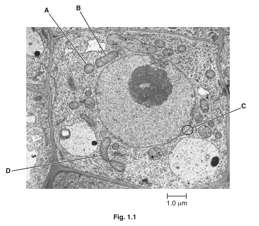
The structures labelled A and B on Fig. 1.1 are sections of two mitochondria.
Suggest why A and B are different shapes.
Answer
A and B are sections cut through the mitochondria in different planes / at different angles / orientations, so A is a cross section (appearing circular) and B is a longitudinal section (appearing elongated).
A is a cross section and B is a longitudinal section because the mitochondria have been cut in different planes.
Background Concept
A transmission electron micrograph (TEM) is a two-dimensional image of an extremely thin slice (around 70 nm thick) cut through a cell. Anything in that slice appears flat, but the three-dimensional organelle it came from may have been cut at any angle. The same is true of a loaf of bread: an end-cap slice and a slice through the side of the same loaf look very different. In cell biology, a cross section (transverse) of a mitochondrion shows a roughly circular profile, while a longitudinal section (cut along the long axis) shows an elongated oval. Mitochondria are not rigid spheres — they are dynamic, flexible organelles that change shape, fuse and divide, so a population always contains a mix of profiles even before considering the plane of section.
Understanding the Question
The question refers to two labelled mitochondria, A and B, in Fig. 1.1. A is round, B is elongated. The question asks the candidate to suggest why they look different, given that both are described as mitochondria. The command word 'suggest' means any biologically reasonable idea is acceptable as long as it fits the evidence.
Approach
The most defensible single-mark answer is the plane-of-section idea, because the shapes A (circular) and B (elongated) are the classic signatures of a cross section and a longitudinal section of the same elongated organelle. Alternative creditable points (flexibility, age, variety of shapes) are also acceptable; any one of them scores the single mark.
Step-by-Step Reasoning
- Mitochondria are typically elongated, sausage-shaped organelles. A three-dimensional mitochondrion is roughly cylindrical.
- If you slice such a cylinder perpendicular to its long axis you see a circle — that is what A looks like.
- If you slice the same cylinder parallel to its long axis you see an elongated oval — that is what B looks like.
- Therefore A and B are most likely cut in different planes (A = cross section, B = longitudinal section).
- Other acceptable ideas the mark scheme credits: mitochondria are flexible/change shape, come in a variety of sizes and shapes, or are of different ages/stages of development.
Key Takeaways
- The plane of section determines the 2-D shape of a 3-D organelle in a TEM — this is a recurring theme in cell biology micrographs.
- Mitochondria are dynamic, changing shape, dividing and fusing; they are not fixed spheres or rods.
Common Mistakes
- Stating that A and B are 'different types of mitochondria' — there is only one type, the difference is in the cut.
- Saying that one is old and the other new without further qualification, when a more direct explanation (plane of section) is available.
Things to Be Careful About
- The mark scheme demands a comparative answer; simply describing one mitochondrion and not contrasting it with the other is not enough.
The structure labelled D on Fig. 1.1 is a mitochondrion about to divide.
Explain the importance of the division of mitochondria for the cell shown in Fig. 1.1 and for cells in the root tips of thale cress.
Answer
- Mitochondria provide (most of the) ATP / energy for the cell, so dividing allows the cell to increase the number of mitochondria to meet energy demand.
- Root tip cells are dividing rapidly by mitosis; new cells need a full complement of mitochondria, so mitochondrial division ensures that mitochondria are shared between daughter cells and that numbers are maintained in each new cell. It also replaces old / worn-out / damaged mitochondria.
Mitochondrial division increases the number of mitochondria to provide ATP for growth and division, and ensures daughter cells receive enough mitochondria when the parent cell divides.
Background Concept
Mitochondria are the site of aerobic respiration and produce the bulk of a cell's ATP. A cell's ATP demand rises sharply when it is growing (synthesising new cytoplasm, organelles and membranes) and when it is dividing (mitosis itself is energetically expensive, and the daughter cells need to be self-sufficient). Because mitochondria cannot be made de novo in the cytoplasm — they arise only from pre-existing mitochondria — the cell increases its mitochondrial number by mitochondrial division (a process related to binary fission in bacteria, reflecting the endosymbiotic origin of mitochondria).
The root tip of a plant is a meristem: a region of rapid mitosis that produces new cells to extend the root. Every cell in this region is, or has recently been, dividing.
Understanding the Question
The question is about the cell shown in Fig. 1.1 (a root cell of Arabidopsis thaliana) and the importance of mitochondrial division for this cell and for root tip cells generally. Two marks are available, so two distinct ideas are needed.
Approach
Think about (a) why any cell needs more mitochondria (energy + replacement of old ones) and (b) why this is particularly important in root tips, where cells are dividing rapidly and each daughter cell must inherit mitochondria.
Step-by-Step Reasoning
- Mitochondria produce most of a cell's ATP via aerobic respiration; the more ATP-demanding the cell, the more mitochondria it needs.
- A growing cell and a dividing cell both have a high ATP demand, so producing more mitochondria by division meets that demand.
- In a root tip, mitosis is rapid and continuous; when a parent cell divides, its mitochondria are partitioned between the two daughter cells.
- Without mitochondrial division before cell division, daughter cells would receive fewer and fewer mitochondria each generation; division therefore keeps the number per cell constant.
- Mitochondrial division also replaces old, worn-out or damaged mitochondria with new, functional ones — equivalent to quality control.
Key Takeaways
- Mitochondrial division is essential for energy supply, for cell growth, and for ensuring that daughter cells inherit sufficient mitochondria.
- Root meristems are a context where mitochondrial demand is particularly high because of the rate of mitosis.
Common Mistakes
- Writing vague answers like 'cell needs energy' without linking it to mitochondria, or 'more mitochondria for energy' without explaining why more is needed (the mark scheme ignores unqualified 'energy production').
- Listing uses of ATP in the cell — these are explicitly ignored by the mark scheme.
- Forgetting the root-tip context: this is a meristem where cell division is rapid, and that is why mitochondrial numbers must be replenished.
Things to Be Careful About
- The mark scheme requires the reason the cell needs more mitochondria, not just the bare statement that it does.
- The phrase 'energy production' is rejected; you must state ATP, or that mitochondria provide energy/ATP.
Within a cell, substances move between the nucleus and the cytoplasm. The area labelled C in Fig. 1.1 shows an area where this communication occurs.
Make a large, labelled drawing of area C to show where this communication occurs.
Answer
A large, clear drawing of area C with:
- the nuclear envelope shown as two parallel membranes (double lines) drawn as continuous lines that close at each end, with a clear gap (the nuclear pore) between the two membranes;
- a label line ending on the nuclear pore labelled 'nuclear pore';
- neat, continuous lines, no shading, label line with no arrowhead, drawn larger than the original micrograph detail.
Drawing: nuclear envelope as two parallel membranes with closed ends and a labelled nuclear pore (gap).
Background Concept
The nuclear envelope surrounds the nucleus and is made of two membranes (an inner and an outer membrane) separated by a narrow perinuclear space. The two membranes are continuous with each other at their edges, so the envelope encloses a closed compartment. At intervals, the two membranes are pulled apart by protein complexes to form nuclear pores — channels about 100 nm across that allow the regulated passage of molecules (mRNA, ribosomal subunits, proteins with nuclear localisation signals) between the nucleus and the cytoplasm. The question says area C is "where this communication occurs", which means C is showing a nuclear pore.
Understanding the Question
Part (b) is a drawing question. The candidate is asked to make a large, labelled drawing of area C from Fig. 1.1, the small region of the nuclear envelope containing the nuclear pore. The marks (2) are for two specific features the mark scheme identifies.
Approach
Apply the standard CIE biological-drawing conventions:
- Use a sharp pencil and clear, continuous lines (no sketching or shading).
- Draw larger than the original micrograph detail.
- Show structures as they actually appear: the two membranes of the nuclear envelope with a gap (the pore) between them.
- Add a label line ending on the structure, with no arrowhead, written in pencil.
- Only label what is asked for and what you can see.
Step-by-Step Reasoning
- Identify what is at area C in the micrograph: a piece of nuclear envelope that shows a small gap (a nuclear pore) in the otherwise continuous double membrane.
- Decide what to draw: a section of the nuclear envelope in the same orientation as the micrograph, but enlarged. The two membranes should be drawn as two parallel lines that come together and close at each end of your drawing.
- In the middle of the drawing, leave a small gap in both membranes — this is the nuclear pore.
- Add a label line from the gap, ending exactly on the pore, with the words 'nuclear pore'.
- The two marks are: (1) drawing the nuclear envelope as two membranes with closed ends and a gap; (2) adding the label 'nuclear pore' to the gap.
Key Takeaways
- The nuclear envelope is a double membrane; nuclear pores are gaps in it where the two membranes are pulled apart by a protein complex.
- Drawing conventions matter as much as biological accuracy in CIE marking.
Common Mistakes
- Drawing the envelope as a single line — it must be two lines representing the two membranes.
- Leaving the ends of the envelope open, or showing the two membranes not joining up.
- Using an arrowhead on the label line, or letting the label line cross other structures.
- Adding shading or colouring, which the CIE convention forbids.
- Drawing individual pores as holes through the cytoplasm rather than through the envelope.
Things to Be Careful About
- The label 'nuclear pore' must be written horizontally if possible, and the label line must end on the pore, not in the cytoplasm or the nucleoplasm.
- Do not draw individual molecules (e.g. mRNA) passing through the pore — the question only asks for a drawing that shows where the communication occurs, not the molecules themselves.
- The drawing should be larger than the corresponding area in the micrograph, not the same size.
Outline the functions of the nucleus in non-dividing cells, such as the cell in Fig. 1.1.
Answer
- The nucleus contains chromosomes / chromatin / DNA / genes / genetic material — the coded information for the cell.
- The sequence of bases in DNA carries the information needed to synthesise polypeptides / proteins (transcription of genes produces mRNA).
- The nucleus contains the nucleolus, which is the site of manufacture of ribosomal sub-units / ribosomes.
- The nuclear envelope protects the DNA from degradation by, e.g., cytoplasmic enzymes.
(Plus any one AVP, e.g. post-transcriptional modification of mRNA, or DNA repair.)
The nucleus houses the DNA, controls transcription, contains the nucleolus (ribosome subunit assembly) and protects the DNA from degradation.
Background Concept
The nucleus is the largest organelle in a eukaryotic cell and the store of the cell's genome — the complete set of DNA molecules that code for every protein the cell can make. In a non-dividing (interphase) cell, the DNA is dispersed as chromatin (DNA wrapped around histone proteins) and is not condensed into visible chromosomes.
Within the nucleus:
- The nucleolus is a dense, non-membrane-bound region where ribosomal RNA (rRNA) is transcribed and combined with proteins to form the large and small ribosomal sub-units, which are then exported through nuclear pores to the cytoplasm.
- The nuclear envelope (double membrane with nuclear pores) separates the contents of the nucleus from the cytoplasm. This compartmentalisation allows mRNA to be transcribed and processed (capping, poly-A tail, splicing) safely inside the nucleus before being exported.
The general flow of information is: DNA → (transcription) → mRNA → (translation in cytoplasm) → protein. The nucleus is the site of the first two of these steps and the assembly point for the ribosomes that carry out the third.
Understanding the Question
The question asks the candidate to outline the functions of the nucleus in a non-dividing cell — the cell in Fig. 1.1. The command word 'outline' means to give the main points, briefly, in a logical order. Four marks means four distinct creditable points are needed; the mark scheme provides eight candidate ideas and a mark can be earned for any four of them.
Approach
The strongest answers group the points into clear themes:
(a) the nucleus as the store of genetic information;
(b) the nucleus as the site of transcription;
(c) the nucleolus and ribosome manufacture;
(d) protection of DNA by the nuclear envelope.
Step-by-Step Reasoning
- Contains genetic material — the nucleus contains the chromosomes/chromatin, i.e. the DNA, which carries the genes/inherited information. (Mark 1)
- Codes for proteins — the sequences of bases in DNA determine the sequences of amino acids in polypeptides/proteins. (Mark 2)
- Transcription — within the nucleus, genes are transcribed into mRNA. (Mark 3)
- Nucleolus — the nucleolus, inside the nucleus, is the site of manufacture of the sub-units of ribosomes. (Mark 4)
- (Mark 5, AVP) Protection — the nuclear envelope physically separates the DNA from cytoplasmic enzymes that would otherwise degrade it, and the nuclear pores regulate what enters and leaves.
- (Mark 6, AVP) Other valid points include: post-transcriptional modification of mRNA (capping, splicing, polyadenylation); DNA repair; the nucleus responding to signals that control gene expression.
For four marks, the four most commonly credited points are: contains DNA/genes, codes for protein synthesis, contains the nucleolus / manufactures ribosomal sub-units, and protects the DNA from degradation.
Key Takeaways
- The nucleus is not just a 'control centre' (which the mark scheme ignores) — it has specific, named functions: storing DNA, transcribing genes, assembling ribosomes and protecting the genome.
- The nucleolus is a feature inside the nucleus, not a separate organelle.
- Precise vocabulary (chromatin, transcription, ribosomal sub-units) scores where vague wording (e.g. 'controls the cell') does not.
Common Mistakes
- Writing 'the nucleus controls the cell's activities' — explicitly ignored by the mark scheme because it is too vague.
- 'mRNA travels through nuclear pores' — also ignored because it is implicit in the structure; the marks want what the nucleus does, not the transport step.
- Saying the nucleus 'makes ribosomes' without mentioning the nucleolus; or saying 'the nucleus makes proteins', which is wrong — proteins are made by ribosomes in the cytoplasm.
- Confusing transcription (in the nucleus) with translation (in the cytoplasm).
Things to Be Careful About
- Use the precise term: 'manufacture of ribosomal sub-units' or 'assembles ribosomal sub-units' is the wording the mark scheme credits. 'Makes ribosomes' is accepted as an alternative; 'makes proteins' is not.
- The question specifies a non-dividing cell, so do not write about mitosis (chromosome behaviour, spindle, etc.) — those functions belong to the nucleus only during cell division.
The rest of this paper
5 more questions- Q2—10M
- Q3Enzymes10M
- Q4Biological Molecules · Transport in Mammals10M
- Q5Immunity10M
- Q6Transport in Plants · Cell Membranes and Transport11M