Biology 9700/22 — February/March 2018
Cambridge AS Level · AS Level Structured Questions · worked solutions for every part, with the mark scheme
Topics Cell Structure · Cell Membranes and Transport · Immunity · Infectious Diseases · Gas Exchange · Biological Molecules · +5 more
Fig. 1.1 is an electron micrograph of part of a eukaryotic cell.
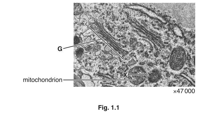
State how it is possible to deduce that Fig. 1.1 is a transmission electron micrograph and not a scanning electron micrograph.
Both the Golgi body and the rough endoplasmic reticulum are part of the internal network of membranes in cells.
Outline structural features shown in Fig. 1.1 that identify G as the Golgi body and not the rough endoplasmic reticulum.
Calculate the actual diameter, X–Y, of the mitochondrion labelled in Fig. 1.1.
Write down the formula that you will use to make your calculation. Give your answer to the nearest whole nanometre (nm).
actual diameter = ______
The inner and outer membranes of the mitochondrion have a fluid mosaic structure similar to other cell membranes. They are both approximately 6 to 7 nanometres (nm) thick.
Outline the fluid mosaic model of membrane structure.
There is space below for a diagram.
The inner and outer membranes of the mitochondrion differ in the detail of their membrane components. The inner membrane is also much less permeable than the outer membrane.
Suggest one way in which the structure of the inner membrane may differ from that of the outer membrane to produce a less permeable inner membrane.
The rest of this paper
5 more questions- Q2Immunity · Infectious Diseases · Gas Exchange17M
- Q3Biological Molecules · Enzymes11M
- Q4Transport in Plants5M
- Q5Transport in Mammals6M
- Q6Nucleic Acids and Protein Synthesis · The Mitotic Cell Cycle12M