Biology 9700/23 — October/November 2017
Cambridge AS Level · AS Level Structured Questions · worked solutions for every part, with the mark scheme
Topics Cell Membranes and Transport · Cell Structure · Transport in Mammals · Gas Exchange · Biological Molecules · Immunity · +4 more
Answer all questions.
Fig. 1.1 is a transmission electron micrograph of a part of an animal cell.
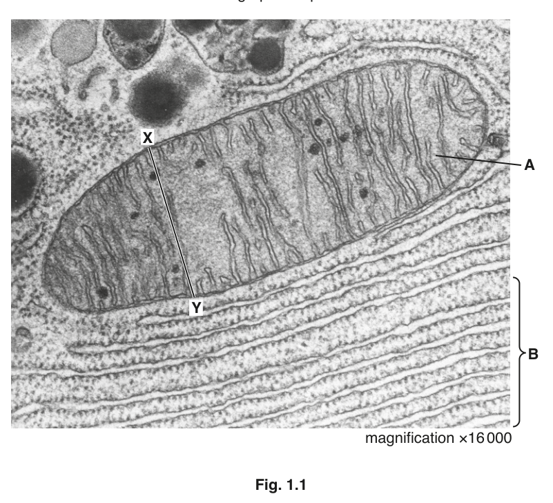
Fig. 1.1
Calculate the actual width of the organelle labelled A, as shown by line X–Y.
State the formula that you will use and show your working.
Give your answer in µm and to one decimal place.
actual width = ______
Working
Rearranged:
Length of line X–Y on the micrograph =
Answer
actual width = 2.5 µm
2.5 µm
Background Concept
When a biological specimen is viewed using a microscope, the image we see is larger than the real object by a factor called the magnification. The relationship between the size of the image, the size of the real object and the magnification is:
Rearranging:
In the light microscope, sizes are usually quoted in micrometres (µm), with . Electron micrographs are usually given with a stated magnification printed on the figure (here, ×16 000), and the candidate must measure the image and convert the measurement to a real biological size.
Understanding the Question
The question gives a transmission electron micrograph of a mitochondrion (organelle A) at ×16 000 magnification. A line X–Y is drawn across the width of the mitochondrion. The candidate is asked to:
- State the formula linking magnification, image size and actual size.
- Use the measured length of X–Y to calculate the actual width of the mitochondrion.
- Give the answer in µm, to one decimal place.
The command words are calculate and state — the working must be shown in full and the formula clearly given.
Approach
- Measure the X–Y line on the printed micrograph with a ruler. The accepted measurement is 40 mm (alternatives 38/39 or 40/41 mm are allowed and give 2.4 or 2.6 µm respectively).
- Convert the millimetre measurement to micrometres so it is compatible with the answer unit.
- Apply the magnification formula and divide to obtain the actual size.
- Round to one decimal place in µm.
Step-by-Step Reasoning
- Measure the length of line X–Y. On a standard print of this paper it is approximately 40 mm.
- Convert units: . This step is essential because the answer is required in µm and the magnification is a dimensionless number, so the numerator must be in µm.
- State the formula as required by the question:
- Substitute:
- Calculate:
- Format: the answer is given to one decimal place in µm, exactly as requested.
Key Takeaways
- The magnification formula can be remembered as a triangle (M over I, A under I), or written out: .
- Always convert measurements to the same units as the answer before dividing.
- The accepted answer range covers slight measurement variation (38–41 mm gives 2.4–2.6 µm).
Common Mistakes
- Dividing the wrong way round: image ÷ magnification is correct; magnification ÷ image is not.
- Forgetting to convert mm to µm, so quoting an answer of 0.0025 (mm) or 0.025 (cm).
- Giving too many decimal places (e.g. 2.50) — the question asks for one decimal place.
- Using the wrong formula form, e.g. , which would give 640 000 µm.
Things to Be Careful About
- The mark scheme explicitly caps the marks if the formula is wrong but the calculation is right, or if the answer is correct but the units are not converted. Always show the formula and unit conversion clearly.
- The measurement tolerance means small reading errors are accepted; do not worry about a 1 mm difference.
- Keep one decimal place in the final answer.
Name the organelle A and state its role in cells.
name = ______
role = ______
Answer
Name: mitochondrion (mitochondria)
Role: site of aerobic respiration / produces (provides) ATP
mitochondrion; produces ATP / site of aerobic respiration
Background Concept
The mitochondrion is a double-membrane-bound organelle found in nearly all eukaryotic cells. The outer membrane is smooth; the inner membrane is folded into cristae, which project into the matrix. The folds give a large surface area for the electron transport chain and ATP synthase, allowing the organelle to carry out aerobic respiration — the oxidation of respiratory substrates (e.g. glucose, fatty acids) using oxygen to generate ATP.
Understanding the Question
The micrograph shows an organelle with a clearly visible double membrane and many parallel inner folds (cristae). The candidate is asked to name this organelle and to state its role in cells.
Approach
- Identify organelle A from the two key ultrastructural features visible in the TEM: the double membrane and the cristae.
- Recall the main function: the production of ATP through aerobic respiration.
Step-by-Step Reasoning
- Identification: the dark, oval organelle crossed by the X–Y line shows a smooth outer boundary and many parallel inward folds. This is the classic appearance of a mitochondrion.
- Function: the cristae house the electron transport chain and ATP synthase; the matrix contains the enzymes of the Krebs cycle. Together they carry out oxidative phosphorylation to generate ATP, the universal energy currency.
- Wording the answer: "produces ATP" or "site of aerobic respiration" are both accepted. The mark scheme also accepts "releases/provides energy" and any valid AVP such as the urea cycle or β-oxidation of fatty acids.
Key Takeaways
- Mitochondria are identified by their double membrane and cristae in electron micrographs.
- Their principal function is ATP synthesis by aerobic respiration.
Common Mistakes
- Calling it a chloroplast (it is an animal cell, and there are no thylakoids).
- Vague function statements such as "makes energy" — the mark scheme requires ATP or aerobic respiration specifically.
- Confusing the matrix with cytoplasm.
Things to Be Careful About
- The question is in an animal cell, so a chloroplast is impossible.
- "Provides energy" alone is too vague; "provides ATP" or "ATP for energy-requiring processes" is precise.
Name the cell structure labelled B and state one reason for your answer.
name = ______
reason = ______
Answer
Name: rough endoplasmic reticulum (RER)
Reason: ribosomes are attached to the membrane (visible as the small dark dots studding the parallel membranes).
rough endoplasmic reticulum; ribosomes are attached to the membrane
Background Concept
The endoplasmic reticulum (ER) is a system of flattened membrane-bound sacs (cisternae) continuous with the nuclear envelope. There are two forms:
- Rough ER (RER): studded with ribosomes on the cytoplasmic face; it is the site of synthesis of proteins destined for secretion, insertion into membranes, or delivery to lysosomes.
- Smooth ER (SER): no ribosomes; site of lipid/steroid synthesis and detoxification.
The two are sometimes continuous with each other, but in a micrograph they are distinguished by the presence or absence of ribosomes.
Understanding the Question
Structure B is the bracketed region below the mitochondrion, showing parallel membrane cisternae. The candidate is asked to name this organelle and give one reason for the identification.
Approach
- Look for the characteristic parallel cisternae (suggesting ER rather than Golgi) and any small dense granules on their cytoplasmic surface.
- The dense granules are ribosomes, so the organelle is rough ER.
- Give one observable reason (ribosomes attached) — this is what the mark scheme credits.
Step-by-Step Reasoning
- Shape: B consists of a stack of parallel flattened sacs — characteristic of endoplasmic reticulum.
- Surface texture: the membrane surface carries many small dark dots, which are ribosomes; the presence of ribosomes makes it rough ER specifically.
- Reason: "ribosomes are attached to the membrane" is the single most direct and mark-scheme-aligned justification. The mark scheme notes that this reason is also accepted if the candidate simply identifies the organelle as ER/RER, but it is much safer to give the explicit ribosome reason.
Key Takeaways
- Rough vs smooth ER is distinguished by the presence/absence of ribosomes.
- In a TEM, ribosomes appear as small dense granules, ~20 nm in diameter, on the cytoplasmic face of the membrane.
- RER is the site of synthesis of membrane and secretory proteins.
Common Mistakes
- Calling it Golgi apparatus (Golgi is also stacked cisternae but is not continuous with the nuclear envelope and typically has more dilated, curved cisternae with associated vesicles).
- Calling it smooth ER (no ribosomes would be visible).
- Identifying it as ribosomes alone — that is a structure, not the organelle as a whole.
Things to Be Careful About
- Give one reason, but make it a precise observable one. The mark scheme specifically requires "ribosomes are attached" or equivalent wording.
- Avoid vague statements such as "it looks rough" — say why it looks rough.
The rest of this paper
5 more questions- Q2Transport in Mammals10M
- Q3Gas Exchange · Biological Molecules · Immunity12M
- Q4Infectious Diseases12M
- Q5Transport in Plants · Cell Membranes and Transport9M
- Q6Cell Membranes and Transport · The Mitotic Cell Cycle · Nucleic Acids and Protein Synthesis10M