Biology 9700/22 — February/March 2017
Cambridge AS Level · AS Level Structured Questions · worked solutions for every part, with the mark scheme
Topics Cell Membranes and Transport · Biological Molecules · Transport in Plants · The Mitotic Cell Cycle · Infectious Diseases · Cell Structure · +5 more
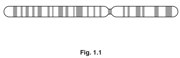
A diagram of a chromosome from a dividing cell is shown in Fig. 1.1.
A dividing cell is at risk of losing genetic material each time DNA replication occurs.
On Fig. 1.1, add a label line and the letter G to show the location on the chromosome of an area that helps to prevent the loss of genes.
Answer
On Fig. 1.1, draw a label line from the letter G to one end of the chromosome.
Label line and letter G added to one end of the chromosome (telomere region)
Background Concept
Every chromosome in a eukaryotic cell ends in a special protective region called a telomere. Telomeres are short, repetitive, non-coding DNA sequences (in humans the repeat is TTAGGG) packaged with specific proteins that form a "cap" on the chromosome end. They do not code for proteins, so the cell can afford to lose them gradually.
This matters because DNA replication on a linear chromosome has an unavoidable problem: at each replication fork, the new strand must be built from an RNA primer, and the very last primer at the 5' end of the new strand cannot be replaced by DNA. The result is that a small number of nucleotides are lost from each end of every chromosome with every cell division — the so-called end-replication problem. Without telomeres, essential coding genes near the chromosome ends would be eroded away. Telomeres therefore act as disposable buffers that absorb this shortening, sacrificing themselves to preserve the coding DNA further in.
Understanding the Question
The question gives you Fig. 1.1, a single duplicated chromosome drawn as two sister chromatids joined at a centromere. It asks you to add a label line and the letter G to the region that helps prevent the loss of genes. You are not asked to name the region — only to mark its position on the diagram.
Approach
Identify the two rounded ends of the chromosome (the telomeres). Draw a clean label line from one of those ends to a clear space outside the chromosome, then write the letter G at the end of the line. Either end is accepted by the mark scheme.
Step-by-Step Reasoning
- The chromosome in Fig. 1.1 has three obvious regions: a centromere (the constriction in the middle) and two ends.
- The "area that helps to prevent the loss of genes" is the telomere, located at each end of the chromosome.
- Draw a label line from one of the two ends (not the centromere, not the body of a chromatid) outwards into clear space on the page.
- Write the letter G at the end of the label line.
- The mark scheme awards the mark for the combination of label line and letter G touching one of the two chromosome ends.
Key Takeaways
- Telomeres are repetitive, non-coding DNA–protein caps at the ends of eukaryotic chromosomes.
- They protect coding regions from being lost during the end-replication problem of DNA replication.
- On a chromosome diagram, telomeres are the two rounded ends of the structure — never the centromere in the middle.
Common Mistakes
- Labelling the centromere instead of an end — the centromere is the constriction in the middle, not a telomere.
- Drawing the label line to the middle of a chromatid rather than to the very end of the chromosome.
- Adding the line but forgetting to write the letter G, or vice versa — both are needed.
- Putting the letter G on the chromosome itself rather than at the end of a label line.
Things to Be Careful About
- The mark requires both a label line AND the letter G — not just one.
- Either end is accepted; do not waste time deciding between left and right.
- Do not label the centromere or the chromatid body — only the chromosome ends qualify.
The chromosome shown in Fig. 1.1 consists of one long DNA molecule associated with histone proteins.
Name one stage of mitosis in which a chromosome would have the same general structure as the chromosome shown in Fig. 1.1.
..............................................................................................................................................
Answer
Anaphase / telophase.
Anaphase or telophase
Background Concept
Mitosis is a continuous process divided into four named stages, defined by what the chromosomes are doing:
- Prophase — chromosomes condense and become visible as two sister chromatids joined at the centromere.
- Metaphase — chromosomes line up on the metaphase plate (cell equator); each still appears as two chromatids joined at the centromere.
- Anaphase — centromeres split; sister chromatids separate and are pulled to opposite poles by spindle fibres.
- Telophase — chromosomes arrive at the poles, decondense, and a new nuclear envelope forms around each set.
A chromosome with the "two-chromatid-joined-at-centromere" structure shown in Fig. 1.1 is the duplicated form. In strict biological terms this is the appearance most characteristic of prophase and metaphase, when the chromosome is fully condensed but the centromere has not yet split. By anaphase the chromatids are no longer joined; by telophase the chromosome is unwinding.
Understanding the Question
The question gives you Fig. 1.1 (a duplicated chromosome) and asks for one stage of mitosis in which a chromosome would have the same general structure. The published mark scheme for this question credits anaphase or telophase as the accepted answer, so write one of these to earn the mark.
Approach
Read the question as a recall task: state one mitotic stage. The mark scheme accepts anaphase or telophase — write either on the answer line.
Step-by-Step Reasoning
- The chromosome in Fig. 1.1 has two chromatids joined at a centromere.
- The credited answers in the mark scheme for this question are anaphase and telophase.
- Write one of these stage names on the dotted line.
Key Takeaways
- A chromosome with two chromatids joined at a centromere is the duplicated form of a chromosome.
- In strict biology, this structure is most clearly seen in prophase and metaphase.
- The mark scheme for this specific paper accepts anaphase or telophase.
Common Mistakes
- Writing interphase — this is not a stage of mitosis (interphase precedes mitosis and is not part of it).
- Writing prophase or metaphase — these are biologically the more obvious choices, but they are NOT the credited answers on this paper's mark scheme; writing them risks losing the mark.
- Misspelling the stage name (e.g. "ana phase", "telophaze").
Things to Be Careful About
- The mark scheme is the authority for what earns the mark. To be safe, write the answer the mark scheme credits — here, anaphase or telophase.
- The question asks for one stage; do not list several.
Name the stage in the mitotic cell cycle during which the cytoplasm and the cell divide to produce two genetically identical daughter cells.
..............................................................................................................................................
Answer
Cytokinesis.
Cytokinesis
Background Concept
The mitotic cell cycle has two major parts:
- Interphase (G1, S, G2) — the cell grows, replicates its DNA (in S phase), and prepares for division.
- Mitotic (M) phase — the cell divides. M phase itself is split into:
- Mitosis (prophase, metaphase, anaphase, telophase) — division of the nucleus, distributing one copy of each chromosome to each pole.
- Cytokinesis — division of the cytoplasm and the cell itself, producing two separate daughter cells.
Mitosis and cytokinesis are sequential but distinct events. Mitosis finishes when two genetically identical sets of chromosomes are sitting at opposite poles inside one cell. Cytokinesis is the step that physically splits that cell into two daughter cells, each with its own nucleus and a complete set of chromosomes.
Understanding the Question
The question asks for the name of the stage in the mitotic cell cycle during which the cytoplasm and the cell divide to produce two genetically identical daughter cells. The answer is the stage that comes after mitosis.
Approach
Recall the cell-cycle term that specifically refers to the splitting of the cytoplasm and the cell itself.
Step-by-Step Reasoning
- The mitotic cell cycle comprises interphase, mitosis, and cytokinesis.
- Mitosis divides the nucleus; cytokinesis divides the cytoplasm and the cell.
- Write cytokinesis on the dotted line.
Key Takeaways
- Cytokinesis is the physical division of the cytoplasm that produces two separate daughter cells.
- It is distinct from mitosis, which divides the nucleus.
- In animal cells, cytokinesis is achieved by a cleavage furrow; in plant cells, by a cell plate forming across the middle of the cell.
Common Mistakes
- Writing telophase — telophase is the final stage of mitosis (nuclear division), not of cytoplasmic division.
- Writing mitosis or M phase — too vague; the question asks for the specific stage.
- Writing interphase — this is the growth/DNA-replication phase, the opposite of division.
Things to Be Careful About
- The command word is Name, so a single term is required.
- Spelling: "cytokinesis" (not "cytokenisis" or "cytokynesis").
The control of the cell cycle can be affected by extracellular chemical messengers that bind to proteins and glycoproteins in the cell surface membrane. The overall mechanism is known as cell signalling.
State the term used to describe the proteins and glycoproteins that function in this way.
..............................................................................................................................................
Answer
Receptor(s).
Receptor(s)
Background Concept
Cell signalling is the process by which cells communicate with each other using chemical messengers. A typical sequence is:
- A signalling cell releases a chemical messenger (a ligand) such as a hormone, growth factor, or neurotransmitter.
- The messenger travels (through blood, across a synapse, or through tissue fluid) to the target cell.
- The messenger binds to a specific protein or glycoprotein in the target cell's cell surface membrane.
- That membrane protein is called a receptor.
- Binding of the messenger changes the receptor's shape, triggering a response inside the cell (e.g. activation of an enzyme, change in gene expression, change in ion permeability).
The cell surface membrane is described as a fluid mosaic, and its proteins and glycoproteins perform many roles: some are transporters, some are enzymes, and some are receptors. The ones that bind extracellular chemical messengers are receptors.
Understanding the Question
The question describes extracellular chemical messengers binding to proteins and glycoproteins in the cell surface membrane, and asks for the term for those membrane proteins.
Approach
Recall the single word that names the membrane proteins/glycoproteins that bind extracellular chemical messengers.
Step-by-Step Reasoning
- The chemical messenger is the ligand.
- The membrane protein it binds to is the receptor.
- Write receptor(s) on the dotted line.
Key Takeaways
- A receptor is a membrane protein (or glycoprotein) that binds a specific extracellular chemical messenger (ligand).
- Receptor binding is the first step in converting an external signal into an internal cellular response.
- Different cell types have different receptors, allowing them to respond to different signals.
Common Mistakes
- Writing ligand — that is the chemical messenger, not the membrane protein.
- Writing enzyme, channel or transporter — these are other types of membrane protein, not the ones that bind signalling molecules.
- Writing antigen — that is a marker recognised by the immune system, not a signalling receptor.
Things to Be Careful About
- The mark scheme accepts receptor or receptors; either singular or plural is fine.
- The question explicitly says "proteins and glycoproteins" — both are receptors, so a single term covers both.
- Do not write a description (e.g. "a protein that binds hormones") — the mark scheme explicitly ignores descriptions and credits only the term.
The rest of this paper
5 more questions- Q2Infectious Diseases · Cell Structure · Biological Molecules · Cell Membranes and Transport · Immunity20M
- Q3Enzymes · Nucleic Acids and Protein Synthesis11M
- Q4Transport in Mammals · Biological Molecules · Transport in Plants10M
- Q5Gas Exchange8M
- Q6Cell Membranes and Transport · Transport in Plants · Biological Molecules7M