Biology 9700/23 — May/June 2015
Cambridge AS Level · AS Level Structured Questions · worked solutions for every part, with the mark scheme
Topics Biological Molecules · Cell Membranes and Transport · Cell Structure · The Mitotic Cell Cycle · Nucleic Acids and Protein Synthesis · Infectious Diseases · +5 more
The cell surface membrane has a fluid mosaic structure.
Describe what is meant by the term fluid mosaic.
Answer
- The phospholipid (and protein) molecules are able to move about / diffuse within the membrane.
- Protein molecules are scattered through the membrane and different types of protein are present.
Phospholipids (and proteins) can move within the membrane; proteins are scattered and exist in more than one type.
Background Concept
The cell surface membrane is described by the fluid mosaic model (Singer and Nicolson, 1972). "Fluid" refers to the fact that the molecules that make up the membrane are not rigidly fixed in place — they can move relative to one another, much like a liquid. "Mosaic" refers to the patchwork appearance created by the variety of different molecules (phospholipids, proteins, cholesterol, glycolipids and glycoproteins) embedded in the bilayer. Phospholipids move mainly by lateral diffusion within their own leaflet, and many membrane proteins also drift laterally, although some are anchored to the cytoskeleton.
Understanding the Question
The command word is "describe", so the candidate must give a brief, accurate account of what the two parts of the term "fluid mosaic" actually mean. Two marks are available, so two clear, distinct points are required.
Approach
Break the term into its two halves: the fluid part (movement of molecules) and the mosaic part (the scattered, varied arrangement of proteins). One mark is earned for each.
Step-by-Step Reasoning
- Point 1 (fluid): State that the phospholipid and protein molecules can move about / diffuse within the membrane. This is the "fluid" aspect. The movement is largely lateral (sideways), although flip-flop between leaflets is rare.
- Point 2 (mosaic): State that the protein molecules are scattered through the membrane, with different types of protein present (e.g. extrinsic, intrinsic, transmembrane). This is the "mosaic" aspect.
Key Takeaways
- "Fluid" = the membrane is not rigid; its components can move.
- "Mosaic" = it contains a variety of different molecules arranged in a patchwork.
Common Mistakes
- Writing only about phospholipids and forgetting that proteins also move.
- Describing the bilayer structure instead of the fluid / mosaic arrangement — this answers a different question.
- Confusing the term with the Davson–Danielli sandwich model.
Things to Be Careful About
- Both components of the term must be addressed: the fluid nature (movement) AND the mosaic nature (varied scattered proteins).
- The mark scheme requires that the point about proteins covers their scattered/varied nature; simply stating "there are proteins" is too vague.
In 1934, the biologists Davson and Danielli published their suggestion for the structure of the cell surface membrane, as shown in Fig. 1.1.
They suggested that the membrane was a phospholipid bilayer with a layer of hydrophilic protein on both surfaces.
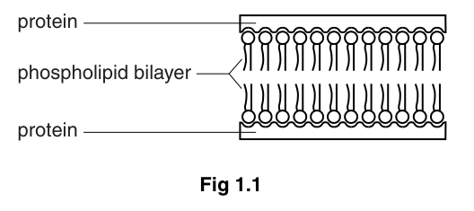
State one way in which the Davson-Danielli structure is similar to the fluid mosaic structure and one way in which it differs from the fluid mosaic model.
similarity
difference
Answer
Similarity: Both models have a phospholipid bilayer (and both contain protein).
Difference: In the Davson–Danielli model, protein is found only as continuous layers on the outer surfaces of the bilayer. In the fluid mosaic model, proteins are found in different locations (e.g. embedded within, or spanning, the bilayer), different types of protein are present, and cholesterol molecules are also part of the structure.
Similarity: both have a phospholipid bilayer (and both contain protein). Difference: Davson–Danielli has protein only on the outer surfaces as continuous layers, whereas the fluid mosaic model has proteins in different positions (including within the bilayer) and also includes cholesterol.
Background Concept
Two historical models describe the cell surface membrane:
- Davson–Danielli (1935): A phospholipid bilayer sandwiched between two continuous layers of protein. The hydrophilic protein was thought to coat the hydrophilic phospholipid heads on each side.
- Fluid mosaic (1972): A phospholipid bilayer in which proteins are scattered through the bilayer in various positions — some on the surface, some partially embedded, some fully spanning the bilayer. Cholesterol molecules are also interspersed between the phospholipid tails, and some carbohydrates are attached to lipids and proteins.
Understanding the Question
The candidate must identify one similarity between the two models (max 1 mark) and one key way in which they differ (max 1 mark), for a total of 2 marks. The question stem describes Davson–Danielli and Fig. 1.1 shows it diagrammatically, so the candidate has both the text and the figure to work from.
Approach
Look first at the Davson–Danielli model on the figure: a phospholipid bilayer with protein layers on each side. Then look at the fluid mosaic model and identify features that overlap (similarity) and features that are not present in the older model (difference).
Step-by-Step Reasoning
- Similarity: Both models contain a phospholipid bilayer and both contain protein. The mark scheme accepts either of these as the similarity point.
- Difference: The Davson–Danielli model places protein only on the two outer surfaces as continuous sheets. The fluid mosaic model places proteins in many different positions (extrinsic on the surface, intrinsic partially embedded, transmembrane spanning the bilayer), has different types of protein, and includes cholesterol molecules. Any one of these constitutes a valid difference.
Key Takeaways
- The main shared feature of the two models is the phospholipid bilayer.
- The crucial advance of the fluid mosaic model is the recognition that proteins are dispersed throughout the bilayer in many positions, and that cholesterol is a key component.
Common Mistakes
- Saying the difference is that "the fluid mosaic has more protein" — this is not specific; the mark scheme wants the position/distribution of the protein.
- Suggesting the Davson–Danielli model has no protein (it does — on the surfaces).
- Failing to mention the position of the protein in either model.
Things to Be Careful About
- The mark scheme asks for the difference to be from the perspective of one model or the other, but accepts "ora" (or reverse argument), so either phrasing is acceptable as long as the position/distribution of protein is made clear.
- The Davson–Danielli model is sometimes called the "sandwich" model; this name is not required but reflects the continuous protein layers.
One way in which substances can cross cell membranes is by active transport.
Describe the mechanism of active transport.
Answer
- Active transport requires energy (in the form of ATP, released from respiration).
- It uses a carrier (transport) protein in the membrane.
- The substance binds to a specific binding site on the carrier protein, which then undergoes a conformational change.
- This moves the substance across the membrane against its concentration gradient (from a region of low concentration to a region of high concentration).
Active transport requires energy (ATP), uses a carrier protein with a specific binding site, and the carrier undergoes a conformational change to move the substance against its concentration gradient.
Background Concept
Active transport is the movement of substances across a cell membrane against their concentration gradient (or electrochemical gradient), using energy derived from the hydrolysis of ATP. Because the cell is moving substances from where they are less concentrated to where they are more concentrated, this is "uphill" movement that cannot occur spontaneously by diffusion. The energy and specificity come from specialised membrane proteins called carrier (transport) proteins, which can change their three-dimensional shape to shuttle the bound substance across the bilayer.
Understanding the Question
The command word is "describe" and the mark allocation is 3. The candidate must give a short, ordered account of the steps involved in active transport. Five separate marking points are listed in the scheme, but only three are required.
Approach
Recall the key features of active transport and select three from the mark scheme list:
- energy / ATP requirement
- carrier (transport) protein
- conformational change of the carrier
- movement against the concentration gradient
- specific binding site
Step-by-Step Reasoning
- Point 1 — energy: State that the process requires energy supplied as ATP (released by respiration in the cell). The mark scheme explicitly rejects the phrase "ATP energy" — write "energy" or "ATP", not both fused together.
- Point 2 — carrier protein: State that a carrier (or transport) protein in the membrane carries the substance across. "Pump" is an acceptable alternative wording.
- Point 3 — conformational change: State that the carrier protein changes shape to move the substance across the membrane.
- Point 4 — against the gradient: State that the substance is moved against its concentration gradient (from low to high concentration). This is what distinguishes active transport from diffusion and facilitated diffusion.
- Point 5 — specific binding site: State that the substance binds to a specific site on the carrier protein (the carrier is specific for the substance it transports).
Any three of the above earn full marks.
Key Takeaways
- Active transport moves substances against their concentration gradient using ATP.
- The work is done by a carrier protein that changes shape.
- Carrier proteins are specific — each one binds only certain molecules or ions.
Common Mistakes
- Confusing active transport with diffusion or facilitated diffusion (which are passive, do not use ATP, and only move substances down the gradient).
- Saying the cell "uses energy" without specifying ATP — the mark scheme accepts "energy" but specifying ATP is more precise.
- Writing "ATP energy" as a single phrase — the mark scheme rejects this.
- Omitting the role of the carrier protein or its conformational change.
Things to Be Careful About
- The mark scheme uses "R ATP energy" — the candidate must write "energy" OR "ATP", not "ATP energy".
- The mark scheme uses "A pump" — using the word "pump" instead of "carrier protein" is acceptable.
- Specific binding site is sometimes called the "active site" of the carrier — both are accepted.
High temperature can damage cell membranes. One factor contributing to this damage is the denaturation of membrane proteins.
Describe how proteins become denatured at high temperature and explain how this could lead to damaging cell membranes.
Answer
How proteins denature at high temperature (max 2):
- The increased kinetic energy of the molecules at high temperature causes vibration within the polypeptide chain.
- This vibration breaks the weak bonds (hydrogen, ionic and hydrophobic interactions) that hold the protein in its specific tertiary structure.
- As a result, the protein loses its specific 3D shape, including the shape of any active / binding site.
How this damages the cell membrane (max 2):
4. The membrane proteins lose their function — for example, carrier proteins can no longer transport polar molecules / ions across the membrane, and receptor proteins can no longer bind signalling molecules.
5. The membrane becomes leaky and loses its partially permeable nature, so the cell can no longer control which substances enter or leave.
6. The interaction between the proteins and the phospholipid bilayer is disrupted, further destabilising the membrane structure.
High temperature breaks the weak bonds maintaining the tertiary structure of membrane proteins, causing them to lose their specific 3D shape. This means the proteins lose their function (e.g. transport, receptors, cell signalling) and the membrane becomes leaky, losing its partially permeable nature.
Background Concept
Protein structure: The function of a protein depends on its precise three-dimensional shape. The tertiary structure is the overall 3D folding of a single polypeptide chain, maintained by weak interactions — hydrogen bonds, ionic bonds, hydrophobic interactions (and, in some proteins, disulfide bridges). The quaternary structure is the arrangement of two or more polypeptide subunits. The shape of any active site or binding site is determined by the tertiary (and sometimes quaternary) structure.
Denaturation: When a protein is exposed to extremes of temperature or pH, the weak bonds holding its tertiary structure are disrupted. The polypeptide chain unfolds or refolds incorrectly, the active/binding site loses its specific shape, and the protein can no longer perform its function. Denaturation is usually irreversible for globular proteins.
Membrane proteins: Many membrane proteins (carriers, channels, receptors, enzymes, cell-adhesion molecules) must maintain a specific 3D shape to work. Their proper position in the phospholipid bilayer also depends on interactions between the hydrophobic regions of the protein and the hydrophobic fatty-acid tails of the phospholipids.
Understanding the Question
The question has two halves worth 3 marks in total. First, "describe how proteins become denatured at high temperature" (max 2 marks). Second, "explain how this could lead to damaging cell membranes" (max 2 marks, but combined total is capped at 3). The candidate must produce a coherent causal chain: heat → bond breakage → loss of shape → loss of function → membrane damage.
Approach
For the first half, focus on the molecular events: kinetic energy, vibration, bond breakage, loss of tertiary (or quaternary) structure, loss of specific shape. For the second half, link the loss of protein function to specific membrane-level consequences: failed transport, failed signalling, lost cell adhesion, leakiness, disrupted protein–bilayer interaction.
Step-by-Step Reasoning
Denaturation (max 2):
- At high temperature, molecules have greater kinetic energy and vibrate more vigorously. This vibration disrupts the weak interactions (hydrogen bonds, ionic bonds, hydrophobic interactions) that maintain the tertiary (and quaternary) structure of the protein.
- The protein therefore loses its specific 3D shape, and any active site or binding site within it loses its precise geometry. The protein is now denatured.
Membrane damage (max 2, but total capped at 3):
- Loss of protein function: The denatured membrane proteins can no longer perform their roles. For example:
- Carrier and channel proteins can no longer transport specific polar molecules or ions.
- Receptor proteins can no longer bind hormones or other signalling molecules, so cell signalling fails.
- Cell-surface glycoproteins involved in cell–cell adhesion no longer function, so cells may detach.
- Membrane-bound enzymes lose their catalytic activity.
- Leakiness: The membrane loses its partially permeable nature. Substances that should be retained leak out, and substances that should be excluded leak in, so the cell can no longer control its internal environment.
- Disrupted protein–bilayer interaction: Many membrane proteins are held in place by hydrophobic interactions between their non-polar amino acid side chains and the fatty-acid tails of the phospholipids. When the protein's tertiary structure is lost, these interactions are weakened, and the protein may move out of position or detach, further destabilising the membrane.
Key Takeaways
- Protein function depends on tertiary structure, which is held by weak bonds.
- Heat denatures proteins by breaking those weak bonds, leading to loss of specific shape and loss of function.
- For a membrane, denatured proteins mean failed transport, failed signalling, lost adhesion, leakiness, and disrupted bilayer interactions.
Common Mistakes
- Describing denaturation without explaining the mechanism (e.g. just writing "the protein denatures" without mentioning bonds and shape).
- Stating that "the protein is destroyed" or "killed" — proteins are not alive; they denature (lose shape) and may coagulate but are not "killed".
- Confusing the active site of an enzyme with the binding site of a carrier protein — both rely on tertiary structure, but they have different functions.
- Describing the effect of heat on phospholipids (which also melt and make the membrane more fluid / leaky) instead of on the proteins — this is a real effect but does not address this question, which is specifically about protein denaturation.
- Forgetting to link the loss of shape to loss of function; the question demands that the consequence for the membrane be explained.
Things to Be Careful About
- The mark scheme accepts "loss of shape of the active site in correct context" as a shorthand for the more detailed point about tertiary structure.
- The mark scheme rejects points that are not specifically about membrane damage — generic statements about "the cell dying" are too vague.
- "Disruption of the interaction between the protein and the phospholipid bilayer" is a specific marking point and should be mentioned if possible.
- The total mark is 3, so the candidate should select 2 strong denaturation points and 1–2 strong damage points rather than padding the answer.
The rest of this paper
5 more questions- Q2The Mitotic Cell Cycle · Nucleic Acids and Protein Synthesis7M
- Q3Cell Structure · Infectious Diseases · Immunity11M
- Q4Transport in Mammals · Biological Molecules · Enzymes11M
- Q5Cell Structure · Transport in Plants · Biological Molecules · Cell Membranes and Transport14M
- Q6(outdated) Ecology7M