Biology 9700/21 — October/November 2014
Cambridge AS Level · AS Level Structured Questions · worked solutions for every part, with the mark scheme
Topics Gas Exchange · Cell Structure · Cell Membranes and Transport · Infectious Diseases · Immunity · Biological Molecules · +5 more
Fig. 1.1 is an electron micrograph of cells from the lining of the small intestine.
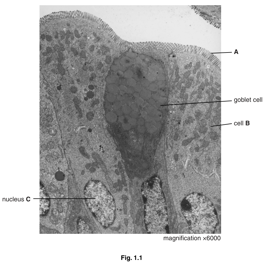
Identify the structures labelled A and state their role for the cell.
Answer
- A = microvilli
- (Microvilli) increase the surface area for absorption across the membrane.
Microvilli; they increase the surface area for absorption.
Background Concept
The epithelial lining of the small intestine is specialised for absorbing the products of digestion. Each epithelial cell projects tiny finger-like extensions of its apical plasma membrane into the lumen. These extensions are called microvilli (singular: microvillus). A whole field of microvilli at the surface of an epithelial cell is visible under the electron microscope as a dense, regular fringe — the brush border. Microvilli should not be confused with villi, which are much larger finger-like folds of the entire intestinal wall containing many cells, blood capillaries and a lacteal.
The function of microvilli follows directly from their shape: by folding the membrane repeatedly, they greatly increase the membrane surface area packed into a small volume of lumen. More membrane means more carrier proteins and channel proteins per unit length of gut, so absorption is faster.
Understanding the Question
Part (a) has two marks. The question asks you to (i) name the structure labelled A at the apical surface of the cell and (ii) state its role. You should give the correct technical term and a clear functional point that is worth a mark in its own right.
Approach
Look at the dark fringe along the top of every cell in the micrograph. These are microvilli seen in longitudinal section; the question is about their identity and what they are for. Think surface-area-to-volume: more membrane = more absorption.
Step-by-Step Reasoning
- The structures at A project from the apical (lumen-facing) surface of each epithelial cell. They are short, regular, densely packed, and far smaller than a cell — consistent with microvilli, not villi.
- Each microvillus is an extension of the plasma membrane, supported inside by a core of actin filaments. Because each cell has hundreds of microvilli, the total apical membrane area is enormously increased.
- This expanded membrane houses many transport proteins (e.g. SGLT1 for glucose, PepT1 for amino acids/peptide uptake), so the rate of absorption is much higher than it would be across a flat membrane.
The mark scheme specifically rejects the term "villi" — villi are visible to the naked eye, while the structures here require an electron microscope to resolve.
Key Takeaways
- Microvilli = sub-cellular projections of the apical plasma membrane, visible only on EM.
- They increase surface area for absorption of digested nutrients.
- They are not the same as villi (which are folds of the gut wall visible without a microscope).
Common Mistakes
- Writing "villi" instead of "microvilli" — wrong scale, and the mark scheme rejects this.
- Saying microvilli "increase surface area for digestion" — digestion happens in the lumen by extracellular enzymes; microvilli are for absorption.
- Saying "for protection" — that is the role of goblet cells/mucus, not microvilli.
Things to Be Careful About
The mark scheme accepts "excretion/secretion" as alternatives to absorption for the second mark, but the safest, most precise answer in the context of the small intestine is absorption.
There are many mitochondria in cell B.
Suggest why cell B contains a large number of mitochondria.
Answer
- (Mitochondria) synthesise / release ATP;
- ATP is required for the active uptake / transport of (digested) nutrients (e.g. glucose, amino acids) across the membrane.
Mitochondria provide ATP for active transport/uptake of nutrients.
Background Concept
Mitochondria are double-membrane organelles whose inner membrane is folded into cristae. They are the site of aerobic respiration in eukaryotes, where glucose (and other substrates) is oxidised to release the energy needed to phosphorylate ADP to ATP. The number of mitochondria in a cell is correlated with its energy demand — cells that do a lot of work contain many.
Active transport moves substances across a membrane against their concentration gradient, using specific carrier proteins and energy from ATP hydrolysis. In the small intestine, the final uptake of glucose (via SGLT1), amino acids and many ions uses secondary active transport coupled to the Na⁺ gradient that is itself maintained by the Na⁺/K⁺ pump — a process ultimately dependent on ATP.
Understanding the Question
Cell B is a columnar absorptive epithelial cell adjacent to the goblet cell. The question states that cell B contains many mitochondria and asks you to suggest why. Two marks are available — one for the immediate role of the mitochondria (making ATP) and one for what that ATP is used for (active transport).
Approach
The trick is not to give one vague point ("for energy") — energy is not a thing the cell uses; ATP is. State that mitochondria produce ATP, and then name the energy-requiring process. The mark scheme rejects "provide energy" alone because "energy" is not a markable substance; it specifically requires the word ATP.
Step-by-Step Reasoning
- Mitochondria carry out aerobic respiration. Glucose + O₂ → CO₂ + H₂O + ATP. The ATP is exported into the cytoplasm.
- The cell's main energy-requiring job at the apical surface is the active uptake of digested nutrients against their concentration gradients.
- Because active transport is happening continuously as food is absorbed, ATP is consumed continuously, and so the cell maintains a high density of mitochondria close to the apical membrane.
The mark scheme also accepts "pinocytosis" or "secretion" as alternatives to active uptake, because both are ATP-dependent processes carried out by these cells.
Key Takeaways
- Mitochondria = site of aerobic respiration → ATP production.
- Active transport (e.g. of glucose, amino acids) needs ATP.
- Cell B has many mitochondria because it does a lot of active transport.
Common Mistakes
- Writing "to provide energy" — the mark scheme rejects this; you must say ATP.
- Writing "for respiration" — respiration happens IN the mitochondria; that is not the reason the cell has many of them.
- Naming the wrong process — e.g. "for diffusion" or "for passive transport". These do not need ATP and so do not explain the abundance of mitochondria.
Things to Be Careful About
The mark scheme uses R (reject) on "energy" because the term is too vague in a biology exam. Always name the molecule (ATP) and the process (active transport / uptake).
Calculate the actual length of the nucleus C.
Show your working and express your answer to the nearest 0.1 micrometre.
answer = ______
Working
Answer
4.8 µm
4.8 µm
Background Concept
The fundamental equation that connects what you see on a micrograph with the real object is:
Rearranged to find the actual (real) size:
On the CIE A-level Biology course you must always:
- convert the image size to the same units as the answer (here µm) before dividing;
- show your working — the formula and the substitution;
- quote the answer to a sensible number of significant figures and with the correct unit.
Understanding the Question
The micrograph is printed at ×6000. Nucleus C is a long oval structure in the lower left of the image. You must measure its length on the page, divide by the magnification, and give the answer to the nearest 0.1 µm. Two marks: one for the correct method (formula and substitution) and one for the final value.
Approach
- Use a ruler to measure the long axis of nucleus C in millimetres on the printed image.
- Convert mm → µm by multiplying by 1000.
- Divide by the magnification (6000).
- Round to 0.1 µm.
The mark scheme accepts image lengths of 29, 30 mm (giving 4.83 ≈ 4.8 and 5.0 µm). Slight differences in where the candidate starts and ends the measurement produce answers of 4.7, 4.8, 5.0 or 5.2 µm — all accepted. If the only error is a wrong conversion but the formula is correct, the method mark is still awarded.
Step-by-Step Reasoning
Measured image length of nucleus C ≈ 29 mm.
- Convert mm to µm:
- Apply the formula:
- Round to the nearest 0.1 µm:
(If the candidate measured 30 mm instead, the answer is exactly 5.0 µm.)
Marking notes:
- Both marks for the correct final value (within the accepted range).
- If the final answer is wrong but the formula image length ÷ 6000 is shown correctly, one mark is awarded for the method.
Key Takeaways
- Always convert to compatible units before dividing.
- 1 mm = 1000 µm; 1 µm = 1000 nm.
Common Mistakes
- Dividing before converting units (e.g. 29 / 6000 mm = wrong unit on the answer).
- Forgetting to convert mm → µm, so leaving the answer in mm or in some other awkward unit.
- Quoting too many significant figures (e.g. 4.833 µm) when the question specifies 0.1 µm — round correctly.
- Multiplying instead of dividing by the magnification. (Easy to do under exam pressure.)
Things to Be Careful About
The mark scheme allows a fairly wide tolerance because the candidate is judging where the nucleus starts and ends by eye. As long as you show the formula and the conversion, you can still score the method mark even if your final number is outside the accepted range.
There are many goblet cells within the epithelium lining the trachea and the bronchi in the gas exchange system.
Describe the role of goblet cells in the gas exchange system.
Answer
- Goblet cells secrete / release mucus;
- pathogens / bacteria / viruses / dust stick to (become trapped in) the mucus;
- this prevents pathogens reaching the cells lining the trachea / bronchi / alveoli (or prevents them entering the circulatory system), reducing the chance of infection.
Goblet cells secrete mucus that traps pathogens, preventing them from reaching the cells lining the airways/alveoli and reducing infection.
Background Concept
The gas exchange system is constantly exposed to the external air and therefore to airborne particles and microorganisms. The conducting airways (trachea, bronchi, bronchioles) are lined with a pseudostratified ciliated epithelium that contains three main cell types:
- Ciliated cells — bear motile cilia on their apical surface that beat in a coordinated wave to move the mucus layer upwards (the mucociliary escalator).
- Goblet cells — specialised secretory cells whose apical cytoplasm is packed with mucinogen granules (visible as dark vesicles in the micrograph, Fig. 1.1). On stimulation they release mucins, which hydrate to form sticky mucus.
- Brush / basal cells — replace damaged cells.
Goblet cells produce mucus — a sticky, glycoprotein-rich layer that sits on top of the cilia. Anything inhaled that is larger than a few micrometres becomes trapped in this layer.
Understanding the Question
The question has already moved context away from the small intestine and into the gas exchange system. Goblet cells in the airway perform a similar secretory role to those in the gut, but the function they support is different: instead of lubricating the epithelium for absorption, the mucus they produce is part of a defence system. Three marks are available — the mark scheme caps at three and lists four creditable ideas, so you need three of them.
Approach
Lay out the chain in order:
- What the goblet cell secretes → mucus.
- What happens to pathogens in the mucus → they stick / become trapped.
- The protective outcome → pathogens cannot reach the cells lining the trachea/bronchi/alveoli (and therefore cannot reach the blood); the chance of infection is reduced.
Step-by-Step Reasoning
- Secretion: Goblet cells release mucin glycoproteins that absorb water to form mucus.
- Trapping: The mucus is sticky; airborne pathogens (bacteria, viruses, fungal spores), dust and pollutant particles adhere to it.
- Barrier effect: Because pathogens are held within the mucus layer (which sits on top of the epithelium), they do not contact the underlying epithelial cells of the trachea or bronchi, and they do not reach the alveoli where gas exchange occurs. The cilia then beat to move the mucus — and the trapped pathogens — up and out of the airways (this is the mucociliary escalator). The pathogens are ultimately swallowed or coughed out.
- Result: The epithelium and the gas-exchange surface are kept clean; the chance of infection is lowered.
The mark scheme allows any three of the points above; maximum 3.
Key Takeaways
- Goblet cells secrete mucus.
- Mucus traps pathogens, dust and particles.
- This prevents infection of the airway epithelium and the alveoli.
- Combined with cilia, this is the mucociliary escalator (a defence system, not a gas-exchange one).
Common Mistakes
- Saying goblet cells "filter the air" or "clean the air" — too vague; the mark scheme wants the chain of secretion → trapping → prevention of infection.
- Writing "goblet cells protect against infection" without naming the mucus or describing how the mucus acts.
- Confusing goblet cells with ciliated cells — goblet cells secrete; ciliated cells move the secretion.
- Saying the mucus reaches the alveoli — the mucus layer thins out and largely disappears in the bronchioles; the gas-exchange surface itself is not covered in thick mucus.
Things to Be Careful About
The mark scheme accepts any wording for "pathogens" (bacteria, viruses, microorganisms, dust) and explicitly accepts "trapped by mucus". Do not write "kill pathogens" — mucus does not kill them; it traps them, and they are later removed mechanically.
State two ways in which the cells lining the alveoli in the lungs differ from cell B shown in Fig. 1.1.
-
______
-
______
Answer
- Alveolar cells are thin / flat / squamous (cell B is columnar / tall).
- Alveolar cells have (far) fewer mitochondria (and have no microvilli).
Alveolar cells are thinner/flatter (squamous, not columnar) and have fewer mitochondria (and no microvilli).
Background Concept
Cell shape and organelle content are dictated by the function a cell performs.
- Cell B (small-intestine epithelial cell) is a tall, columnar cell. Its apical surface is covered in microvilli and its cytoplasm is full of mitochondria because its job is to absorb nutrients by active transport — a process that needs lots of membrane area (microvilli) and lots of ATP (mitochondria).
- Alveolar cells (pneumocytes, type I and II) form the gas-exchange surface. Their function is diffusion of O₂ and CO₂ between alveolar air and pulmonary capillary blood. For diffusion to be fast, the barrier must be as thin as possible. Type I pneumocytes are therefore extremely flat (squamous), typically only ~0.1–0.2 µm thick.
Because alveolar cells exchange gases by simple diffusion, they do not need an expanded membrane area (no microvilli) and do not need much ATP (no active transport). They therefore have very few mitochondria.
Understanding the Question
The question gives you cell B from Fig. 1.1 (columnar, full of microvilli, packed with mitochondria). It asks for two ways in which the cells lining the alveoli differ. The mark scheme allows any two from:
- thinner/flatter/squamous (and not columnar);
- (far) fewer mitochondria;
- no microvilli.
Approach
Look at cell B and think about what features it has because it is an absorbing cell. Then decide which of those features would be unnecessary or unsuitable for a cell whose function is gas exchange by diffusion. The two cleanest contrasts are shape (columnar vs squamous) and organelle content (lots of mitochondria vs few; many microvilli vs none).
Step-by-Step Reasoning
- Shape: Cell B is columnar — taller than it is wide. Alveolar cells are squamous — flat, plate-like. Difference 1: alveolar cells are thinner/flatter (squamous, not columnar).
- Microvilli: Cell B has dense microvilli because absorption needs surface area. Alveolar cells have a smooth apical membrane; adding microvilli would only increase diffusion distance and slow gas exchange. Difference 2 (alternative): alveolar cells have no microvilli.
- Mitochondria: Cell B has many mitochondria because active transport needs ATP. Alveolar cells exchange gases by passive diffusion, which needs no ATP. Difference 2 (alternative): alveolar cells have (far) fewer mitochondria.
Any two of these three differences scores full marks.
Key Takeaways
- Function determines structure: absorbing cells are columnar with microvilli and many mitochondria; gas-exchange cells are squamous with smooth membranes and few mitochondria.
- The thinness of alveolar cells is what makes diffusion fast enough to oxygenate the blood.
Common Mistakes
- Writing only vague descriptions such as "alveolar cells are different" — you must name the specific feature.
- Saying alveolar cells "have more mitochondria" — the opposite is true; gas exchange uses no ATP.
- Saying alveolar cells "have microvilli" — microvilli are on absorptive epithelia, not on the gas-exchange surface.
- Naming features that are not visible or implied in Fig. 1.1 (e.g. "no nucleus") — the question asks about differences, but the marking points are limited to the three listed above.
Things to Be Careful About
The mark scheme specifies A (accept) "thin / flat / squamous" and explicitly accepts "not columnar". It does not accept "round" or "round cells" — round is not a meaningful contrast to columnar. Stick to the squamous vocabulary.
The rest of this paper
5 more questions- Q2Infectious Diseases · Immunity12M
- Q3Biological Molecules · Nucleic Acids and Protein Synthesis · Transport in Mammals · Gas Exchange14M
- Q4The Mitotic Cell Cycle10M
- Q5(outdated) Ecology7M
- Q6Transport in Plants6M